Note: Descriptions are shown in the official language in which they were submitted.
DEMANDES OU BREVETS VOLUMINEUX
LA PRESENTE PARTIE DE CETTE DEMANDE OU CE BREVETS
COMPREND PLUS D'UN TOME.
CECI EST LE TOME 1 DE 2
NOTE: Pour les tomes additionels, veillez contacter le Bureau Canadien des
Brevets.
JUMBO APPLICATIONS / PATENTS
THIS SECTION OF THE APPLICATION / PATENT CONTAINS MORE
THAN ONE VOLUME.
THIS IS VOLUME 1 OF 2
NOTE: For additional volumes please contact the Canadian Patent Office.
CA 02565175 2006-11-01
r
7-
Cancer Diagnosis And Treatment Using Anti-ROB01 Antibody
CROSS-REFERENCES
The present invention relates to methods for the
diagnosis and treatment of cancer and a method for monitoring
progression of hepatitis, as well as to a cell growth
inhibitor and an anticancer agent.
BACKGROUND
In genetic screening studies of drosophila, ROB01 has
been identified as a molecule regulating the midline crossing
of axons, and has been reported to work as a receptor for the
Slit protein in subsequent studies (Kidd et al., Cell, 92,
205-215, 1998, Wang et al., Cell, 96, 771-784, 1999, Kidd et
al., Cell, 96, 785-794, 1999, Brose et al., 96, 795-806, 1999).
In addition, regarding the relationship between ROB01 and
cancer, the chromosome region 3p12 where the human ROB01 gene
is present is highly defective in lung cancer, and expression
is suppressed by methylation of the ROB01 promoter region in
breast cancer and kidney cancer, suggesting that ROB01 gen can
be a tumor suppressor gene (Dallol et al., Oncogene, 21, 3020-
3028, 2000). Indeed, hyperplasia of tracheal epithelial cells
was observed in mice by deleting the first immunoglobulin
region present at the N-terminus of ROB01, which is similar to
the minimal defect in the ROB01 gene identified in lung cancer
patients (Xian J et al., PNAS, 98, 15062-15066, 2001).
1
CA 02565175 2006-11-01
Conversely, it has been reported that ROB01 was expressed in
new blood vessels of cancers, and that increased expression of
Slit2, a ligand for ROB01, on the cancer cells induces cancer
neovascularization to direct cancer growth (Wang et al.,
Cancer Cell, 4, 19-29, 2003).
Meanwhile, the expression of the Slit2 gene, which is a
ligand for ROB01, is also suppressed in a number of cancer
types by methylation or the like, and overexpression of Slit2
or addition of Slit2 induces a growth inhibition and apoptosis
in lung cancer, breast cancer and large intestine cancer cells.
These observation suggests that Slit2, a ligand for ROB01, is
also thought to be a tumor suppressor gene (Dallol et al.,
Cancer Research, 62, 5874-5880, 2002, Dallol et al., Cancer
Research, 63, 1054-1058, 2003 ). However, it is not clear in
this report through which receptor Slit2 exerts its effect in
inhibiting the cell growth, thus the relationship between
ROB01 and cancer has not been absolutely clear.
Cancer is the most common cause of death in Japan. Among
them, primary hepatocellular cancer is one type of cancer with
poor prognosis, representing the third leading case of death
(13%) in male and the fourth (9.0%) in female in 2001 (excerpt
from "Population Dynamics Statistics", Statistics and
Information Department, Minster's Secretariat, Ministry of
Health, Labour and Welfare). The number of chronic patients
caused by viral infection is on the rise year after year and
many of them lead to hepatic cirrhosis and then to
2
CA 02565175 2006-11-01
hepatocellular cancers. Extremely strong demands exist for a
diagnostic procedure at an early stage in the progression from
hepatic cirrhosis to hepatocellular cancer and for a treatment
of hepatocellular cancer. It is believed that without a
groundbreaking solution, the number of deaths will follow an
increasing trend in the 10 to 15 years to come.
Current hepatocellular cancer diagnostic procedure
comprises a comprehensive evaluation based on biochemical data
such as the serum value of GOP/GTP, alkaline phosphatase,
albumin and the like, or a tumor marker AFP (a-fetoprotein),
and diagnostic imaging. Then, if necessary, a small amount of
tissue fragment is taken by needle biopsy for pathological
judgment to confirm the diagnosis. Currently, tumor markers
are mainly used for the diagnosis of hepatocellular cancer.
The positive rate of alpha fetoprotein (AFP), which is the
most common marker, is 60 to 70 percent in hepatocellular
cancer patients, although it is sometimes also positive in
chronic liver disease patients or pregnant female. Another
hepatic cancer tumor marker PIVKA-II is positive in less than
50 percent of the patinet, and the specificity for
hepatocellular cancer is thought to be higher than AFP. Mainly
these two examinations are currently in practice. In either
case, false positive or double negative cases exist, thus a
tumor marker with high specificity is needed.
Histological examination of the sample collected by
needle biopsy is an important test for confirmed diagnosis of
3
CA 02565175 2006-11-01
liver diseases. In particular, as the quantity of specimen may
be limited, a more definite diagnosis technique is required.
It is desired in the art to develop an antibody against an
antigen specifically expressed in hepatic cancer to allow for
not only pathological characteristics but also identification
of hepatocellular cancer from a non-cancer tissue at an early
stage.
In the current situation of diagnosis and monitoring of
liver disease, progression from inflammation to fibrosis and
malignant transformation are diagnosed by examination with
multiple markers and examination by biopsy. In many hepatic
cancer patients, the progression occurs from viral infection
to hepatitis, chronic hepatitis, hepatic cirrhosis and then
hepatic cancer. Consequently, a simple method for diagnosis
and monitoring of liver diseases will be useful, not only in
terms of healthcare economy, but also in mitigating the burden
on the patients and in obtaining accurate medical guidelines.
Regarding treatment of hepatocellular cancer, many
medical facilities are centered mainly on three types of
therapy: surgical removal, transcatheter arterial embolization
therapy, and percutaneous ethanol injection therapy. Either
method has advantages and disadvantages, and even when
transcatheter arterial embolization therapy is selected, which
has a relatively broad application range and survival
advantages, the rate of complete cure is currently thought to
be on the order of 10%. Thus there is a great demand for a
4
CA 02565175 2006-11-01
novel therapy.
Targeted therapy by monoclonal antibody against cancer
specific tumor antigen provides a better outcome in breast
cancer and in lymphoma and the like through an action
mechanism different from conventional chemotherapy, although
no clinical application has done for hepatocellular cancers
yet. The action mechanisms of these antibody drugs include
antibody dependent cytotoxicity (ADCC) via effector cells and
complement-dependent cytotoxicity (CDC) via the complement,
agonistic action by the function of the antibody itself, and
the neutralization capability of the antibody. Recently
molecular therapies have been applied in clinical sites. An
antibody drug therapy which applies these molecular therapies
and targets to a neoplasm-specifically expressed molecule
found on hepatic cancer cells is expected to be developed in
the future.
The following are documents related to the present
invention: W099/20764; W098/48051; W001/46697; W003/29488;
W001/00828; W001/57207; W001/92581; W002/04514; W002/14500;
W002/29103.
SUMMARY OF INVENTION
An object of the present invention is to provide a novel
method for diagnosing and treating cancer, as well as a novel
cell growth inhibitor and anticancer agent, and to provide a
method for diagnosing and monitoring liver disease.
CA 02565175 2006-11-01
The present inventors discovered that ROB01 was highly
expressed in cancer cells, such as hepatocellular cancer, lung
cancer, breast cancer, uterine cancer, gastric cancer, brain
tumor, large intestine cancer and the like. In addition, they
measured the complement-dependent cytotoxicity (CDC) of the
anti-ROB01 antibody, and found that the anti-ROB01 antibody
had a CDC activity against ROB01 expressing cells. They also
found that the concentration of ROB01 in blood increased with
the progression of liver diseases. From the above observations,
the present inventors discovered the effectiveness of the
anti-ROB01 antibody in the diagnosis, prevention and treatment
of cancers overexpresping ROB01, such as hepatocellular cancer,
and achieved a method for diagnosing and monitoring liver
diseases.
The present invention provides a method for diagnosing
cancer comprising detecting ROB01 protein. In the method of
the present invention, preferably the extracellular region of
the ROB01 protein is detected. The method of the present
invention is carried out preferably using an antibody that
recognizes ROB01 protein. Preferably, in the method of the
present invention, ROB01 protein in the blood, serum or plasma,
or ROB01 protein isolated from a cell is detected.
In another aspect, the present invention provides a
method for diagnosing cancer comprising the steps of:
(a) collecting a sample from a subject; and
(b) detecting ROB01 protein contained in the collected
6
CA 02565175 2006-11-01
sample.
In another aspect, the present invention provides a kit
for diagnosing cancer, comprising an antibody that binds to
ROB01 protein. Preferably, the cancer is hepatocellular cancer.
Also, in the kit of the present invention, the antibody
preferably binds to the extracellular region of the ROB01
protein.
In another aspect, the present invention provides a
pharmaceutical composition comprising an antibody that binds
to ROB01 as an active ingredient. The present invention also
provides a cell growth inhibitor comprising an antibody that
binds to ROB01 as an active ingredient. The present invention
also provides an anticancer agent comprising an antibody that
binds to ROB01 as an active ingredient. Preferably, the
antibody that binds to ROB01 has cytotoxicity. The cancer is
preferably a hepatocellular cancer.
In another aspect, the present invention provides a
method for treating a disease caused by abnormal cell growth,
comprising administrating to a patient in need of such
treatment a pharmaceutical composition comprising an antibody
that binds to ROB01 as an active ingredient. The present
invention also provides a method for treating cancer,
comprising administrating to a patient in need of such
treatment a pharmaceutical composition comprising an antibody
that binds to ROB01 as an active ingredient. Preferably, the
cancer is hepatocellular cancer.
7
CA 02565175 2006-11-01
In another aspect, the present invention provides a
method for inducing cell damages in a ROB01 expressing cell by
bringing a ROB01 expressing cell into contact with an antibody
that binds to ROB01. The present invention also provides a
method for inhibiting the growth of a ROB01 expressing cell by
bringing a ROB01 expressing cell into contact with an antibody
that binds to ROB01. Preferably, the antibody that binds to
ROB01 has cytotoxicity. Preferably, the ROB01 expressing cell
is a cancer cell.
In still another aspect, the present invention provides
an antibody that binds to ROB01 and has cytotoxicity against a
ROB01 expressing cell.
In another aspect, the present invention provides a kit
for monitoring the progression of hepatitis comprising an
anti-ROB01 antibody. Preferably, the anti-ROB01 antibody
specifically recognizes ROB01. Preferably, the kit of the
present invention predicts the progression from hepatitis or
hepatic cirrhosis to hepatic cancer. In a preferred embodiment,
the kit of the present invention contains a first anti-ROB01
antibody immobilized on a support and a second anti-ROB01
antibody labeled with a labeling substance.
In still another aspect, the present invention provides a
method for monitoring the progression of hepatitis by
measuring ROB01 in a test sample. Preferably, the ROB01 in the
test sample is measured using an anti-ROB01 antibody.
Preferably, the anti-ROB01 antibody specifically recognizes
8
CA 02565175 2016-02-25
51481-6
ROB01. Preferably, the test sample is blood, serum or plasma. Preferably, the
monitoring method of the present invention predicts the progression from
hepatitis or
hepatic cirrhosis to hepatic cancer. In a preferred embodiment, the monitoring
method of the present invention is carried out using a first anti-ROB01
antibody
immobilized on a support and a second anti-ROB01 antibody labeled with a
labeling
substance.
The present invention as claimed relates to:
- an anti-hepatic cancer agent comprising an antibody that binds to
ROB01 as an active ingredient;
- use of an antibody that binds to ROB01 for treating hepatic cancer in
a patient in need of such treatment;
- a method for inducing cell damage in a ROB01-expressing hepatic
cell comprising bringing a ROB01 expressing hepatic cell into contact with an
antibody that binds to ROB01;
- a method for inhibiting the growth of a ROB01-expressing hepatic cell
comprising bringing a ROB01-expressing hepatic cell into contact with an
antibody
that binds to ROB01;
- use of an antibody that binds to ROB01 for inducing cell damage in a
ROB01-expressing hepatic cell;
- use of an antibody that binds to ROB01 for inhibiting the growth of a
ROB01-expressing hepatic cell; and
- use of an antibody that binds to ROB01 for monitoring progression
from hepatitis or hepatic cirrhosis to hepatic cancer.
9
CA 02565175 2016-02-25
51481-6
DESCRIPTION OF DRAWINGS
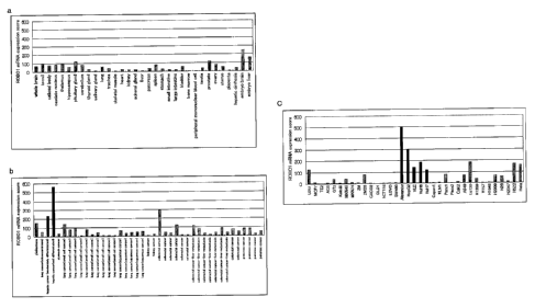
Fig. 1 shows the results of ROB01 gene expression analysis using
GeneChip U133. Fig. la: ROB01 gene expression analysis in normal
tissues/non-cancer sites; Fig. lb: ROB01 gene expression analysis in clinical
samples; Fig. lc: ROB01 gene expression analysis in cancer cell lines;
Fig. 2 shows the results of ROB01 gene expression analysis using
GeneChip U95;
Fig. 3 shows the results of Western analysis of transient expression of
full length ROB01 gene in COS7 cell and HEK293 cell lysates, and the results
of
Western analysis in the supernatant of the culture thereof;
Fig. 4 shows the results of Western analysis of the hepatic cancer cell
line lysate using anti-ROB01 monoclonal antibody A7241A;
Fig. 5 shows the results of immunohistological staining analysis of
hepatocellular cancer paraffin preparations using anti-ROB01 monoclonal
antibody
A7241A;
9a
CA 02565175 2006-11-01
Fig. 6 shows the results of Western analysis of soluble
ROB01 in patient serum using anti-ROB01 monoclonal antibody
A7241A;
Fig. 7 shows the results of Western analysis of lysate of
HEK293 cell forced to express the full length ROB01 gene using
sROB01-immunized rabbit serum;
Fig. 8 shows the results of FACS analysis of lysate of
HEK293 cell forced to express the full length ROB01 gene using
sROB01-immunized rabbit serum;
Fig. 9 shows the results of FACS analysis of lysate of
HepG2 cell using sROB01-immunized rabbit serum;
Fig. 10 shows a standard curve for ROB01 measurement by
enzyme immunoassay using rabbit anti-sROB01 antibody;
Fig. 11 shows the correlation between ROB01 concentration
and the severity of liver diseases;
Fig. 12 shows the variation in ROB01 concentration of
liver disease patients;
Fig. 13 shows the results of measurement of CDC activity
of anti-ROB01 antiserum against ROB01-expressing HEK293 cell;
Fig. 14 shows the results of measurement of CDC activity
of anti-ROB01 monoclonal antibody against ROB01-expressing
HEK293 cell;
Fig. 15 shows the results of measurement of CDC activity
of anti-ROB01 monoclonal antibody against ROB01-expressing
Alexander cell; and
Fig. 16 shows the results of measurement of ADCC activity
CA 02565175 2006-11-01
of anti-ROB01 monoclonal antibody against ROB01-expressing
HEK293 cell.
DETAILED DESCRIPTION OF INVENTION
The method of the present invention is characterized by
the detection of ROB01 protein. ROB01 (Roundabout 1) is an
axon guidance receptor protein, and its amino acid sequence
and the gene sequence coding therefor are disclosed in GenBank
ID NM_002941 (SEQ ID NOs: 9 and 10) for variant 1, and GenBank
ID NM_133631 (SEQ ID NOs: 11 and 12) for variant 2. In the
present invention, ROB01 protein is meant to include both the
full length protein and fragments thereof. A fragment is a
polypeptide containing any region of the ROB01 protein, and
may not have the function of the natural ROB01 protein.
Examples of fragments include, but are not limited to, a
fragment containing an extracellular region of the ROB01
protein. The extracellular region of the ROB01 protein
corresponds to positions 1-859 in the amino acid sequence of
SEQ ID NO: 11. In addition, the membrane spanning region
corresponds to positions 860-880 in the amino acid sequence of
SEQ ID NO: 11 (Sundaresan, et al., Molecular and Cellular
Neuroscience 11, 29-35, 1998).
In the present invention, the expression of ROB01 was
found to be enhanced at both the genetic level and the protein
level with extremely high frequencies in hepatocellular cancer.
In addition, the analysis of clinical samples and cancer cells
11
CA 02565175 2006-11-01
line of other cancer species suggested that the enhanced
expression was shown not only in hepatocellular cancer, but
also in lung cancer, breast cancer, uterine cancer, gastric
cancer, brain tumor, large intestine cancer and the like. It
was also shown that immunohistological diagnosis can be
carried out using a monoclonal antibody specific to ROB01. In
addition, ROB01 was found to be shedded in vivo, and a soluble
ROB01 (sROB01) was present in the blood of cancer patients,
indicating that sROB01 is useful as a serodiagnosis marker of
cancer.
Detection of ROB01
ROB01 protein detected in the present invention is
preferably human ROB01 protein, but any ROB01 may be used in
the invention, including, but is not limited to, canine ROB01,
feline ROB01, mouse ROB01 and hamster ROB01.
In the present invention, detection may be quantitative
or non-quantitative. Examples of non-quantitative detection
include measurement as to merely whether ROB01 protein is
present, measurement as to whether a given quantity or more
ROB01 protein is present, measurement comparing the amount of
ROB01 protein with other sample (for instance, control sample).
Examples of quantitative detection include measurement of
ROB01 protein concentration, measurement of ROB01 protein
quantity, and the like.
Test samples are not particularly limited as long as they
12
CA 02565175 2006-11-01
may contain ROB01 protein, and are preferably those collected
from the bodies of living organisms such as mammals, more
preferably those collected from humans. Specific examples of
test samples include, for instance, blood, interstitial tissue
fluid, plasma, extravascular fluid, cerebrospinal fluid,
synovial fluid, pleural fluid, serum, lymph, saliva, urine and
the like, preferably blood, serum, or plasma. Preferably, the
test samples used in the present invention also include those
derived from the original test samples, such as the culture
solution of cells collected from the body of a living organism.
The cancer to be diagnosed is not particularly limited
and may be any cancer, including hepatic cancer, pancreatic
cancer, lung cancer, large intestine cancer, breast cancer,
kidney cancer, brain tumor, uterine cancer, lung cancer,
gastric cancer, prostate gland cancer, leukemia, lymphoma and
the like. Hepatic cancer is preferred, and hepatocellular
cancer is more preferred.
In the present invention, when ROB01 protein is detected
in a test sample, and the amount of ROB01 protein detected is
determined to be higher than a negative control or a healthy
subject, the subject is determined as having cancer or as
having high potentiality to develop cancer.
In addition, progression of a liver disease can be
monitored by measuring the concentration of ROB01 protein in a
patient having the liver disease.
A preferred embodiment of the diagnosis method of the
13
CA 02565175 2006-11-01
present invention is detection of ROB01 protein released from
cells and present in blood. Particularly preferably, a
fragment containing the extracellular region of the ROB01
protein is detected.
Method for detecting ROB01 protein contained in a test
sample is not limited, but preferably include, detection by an
immunological method using an anti-ROB01 antibody. Examples of
immunological methods include, for instance, radioimmunoassay,
enzyme immunoassay, fluorescence immunoassay, luminescence
immunoassay, immunoprecipitation method, immunonephelometry,
Western blot, immunostaining, immunodiffusion method and the
like, preferably enzyme immunoassay, and particularly
preferably enzyme-linked immunosorbent assay (ELISA) (for
instance, sandwich ELISA). The immunological methods such as
ELISA can be carried out by those skilled in the art according
to well known methods.
For instance, general detection of ROB01 protein in a
test sample using an anti-ROB01 antibody may be carried out by
immobilizing an anti-ROB01 antibody on a support, adding a
test sample, incubating the sample to bind ROB01 protein to
the anti-ROB01 antibody, washing, and detecting the ROB01
protein bound to the support via the anti-ROB01 antibody.
Examples of supports used for immobilizing anti-ROB01
antibody in the present invention may include, for instance,
insoluble polysaccharides such as agarose and cellulose,
synthetic resins such as silicon resin, polystyrene resin,
14
CA 02565175 2006-11-01
polyacrylamide resin, nylon resin and polycarbonate resin, and
insoluble supports such as glass. The support may be used in
the form of beads or plates. In case of beads, a column may be
filled with the beads. In case of plates, multi-well plate
(96-well multi-well plate or the like), biosensor chip and the
like can be used. For binding the anti-ROB01 antibody to the
support, conventional binding methods may be used, such as
chemical bond or physical adsorption. Commercially available
supports may be used for these purposes.
Binding of anti-ROB01 antibody to ROB01 protein is
conventionally carried out in a buffer solution, such as
phosphate buffer solution, Tris buffer solution, citric acid
buffer solution, borate buffer solution, carbonate buffer
solution, and the like. In addition, regarding incubation
conditions, for instance, incubation for 1 hour to 24 hours at
4 C to room temperature is carried out under conventionally
used conditions. The procedure may optionally contain a
washing step using a buffer solution containing a surfactant
such as Tween 20 or the like, as long as it does not prevent
binding of the anti-ROB01 antibody to ROB01 protein.
In the ROB01 protein detection method of the present
invention, a control sample may be prepared in addition to the
test sample to be tested for ROB01 protein. Examples of
control samples include a negative control sample that does
not contain ROB01 protein, and a positive control sample that
contains ROB01 protein. In this case, the ROB01 protein can be
CA 02565175 2006-11-01
detected in the test sample by comparison with results
obtained from the negative control sample without ROB01
protein, and results obtained from the positive control sample
with ROB01 protein. In addition, a series of control samples
with increment in concentration is prepared and a standard
curve is established from the detection result for each
control sample. ROB01 protein contained in a test sample may
be quantitatively determined from the numerical value for the
test sample based on the standard curve.
In a preferred embodiment, the ROB01 protein bound to the
support via the anti-ROB01 antibody is detected using an anti-
ROB01 antibody labeled with a labeling substance. For instance,
a test sample is brought into contact with an anti-ROB01
antibody immobilized on a support, and after washing, ROB01 is
detected using a labeled antibody that specifically recognizes
the ROB01 protein.
The labeling of an anti-ROB01 antibody can be carried out
by generally known methods. Labeling substances well known to
those skilled in the art may be used, for example, fluorescent
dyes, enzymes, coenzymes, chemiluminescent substances.
Examples of the labeling substance include radioisotopes (32P,
C, 1251, 3H, 131 and the like), fluorescein, rhodamine, dansyl
chloride, umbelliferone, luciferase, peroxidase, alkaline
phosphatase, B-galactosidase, B-glucosidase, horseradish
peroxidase, glucoamylase, lysozyme, saccharide oxidase,
microperoxidase, biotin and the like. Preferably, when using
16
CA 02565175 2006-11-01
biotin as a labeling substance, a biotinylated antibody is
added and then avidin conjugated with an enzyme such as
alkaline phosphatase is added. Well known methods can be used
for preparing a conjugation of the labeling substance and the
anti-ROB01 antibody, such as, the glutaraldehyde method, the
maleimide method, the pyridyl disulphide method and the
periodic acid method.
In a specific example, a solution containing anti-ROB01
antibody is added to a support, such as a plate having the
anti-ROB01 antibody immobilized onto the support. After
washing the plate, it is blocked with, for instance, BSA,
gelatine, albumin or the like, to prevent non-specific binding
of proteins. The plate is washed again, and a test sample is
added to the plate. After incubation, the plate is washed, and
a labeled anti-ROB01 antibody is added. After an adequate
incubation, the plate is washed and the labeled anti-ROB01
antibody remaining on the plate is detected. The detection can
be carried out by methods well known to those skilled in the
art. For instance, in the case of labeling by a radioactive
substance, it may be detected by liquid scintillation or the
RIA method. In the case of labeling by an enzyme, a substrate
is added and the enzymatic modification of the substrate, for
instance color development, can be detected using a photometer.
Examples of substrates include 2,2-azinobis(3-
ethylbenzothiazolin-6-sulfonic acid) diammonium salt (ABTS),
1,2-phenylene diamine(ortho-phenylene diamine), 3,3',5,5'-
17
CA 02565175 2006-11-01
tetramethylbenzin (TMB) and the like. In the case of a
fluorescent substance, it may be detected with a
spectrofluorimeter.
In a particularly preferred embodiment of the present
invention, ROB01 protein is detected using a biotin-labeled
anti-ROB01 antibody and avidin.
In a specific example, a solution containing anti-ROB01
antibody is added to a support to immoblize the anti-ROB01
antibody to the support, such as a plate. After washing the
plate, it is blocked with, for instance, BSA or the like, to
prevent non-specific binding of proteins. The plate is washed
again, and a test sample is added to the plate. After
incubation, the plate is washed, and a biotinylated anti-ROB01
antibody is added. After an adequate incubation, the plate is
washed, and avidin conjugated with an enzyme such as alkaline
phosphatase or peroxidase is added. After incubation, the
plate is washed, a substrate corresponding to the enzyme
conjugated to avidin is added, and the ROB01 protein is
detected with the enzymatic modification of the substrate as
the indicator.
In another preferred embodiment of the present invention,
ROB01 protein may be detected using one or more species of a
primary antibody that specifically recognizes the ROB01
protein, and one or more species of a secondary antibody that
specifically recognizes the primary antibody.
For instance, a test sample is brought into contact with
18
CA 02565175 2006-11-01
one or more species of an anti-ROB01 antibody immobilized on a
support, incubated and washed. Then the ROS01 protein bound is
detected with a primary anti-ROB01 antibody and one or more
species of a secondary antibody that specifically recognizes
the primary antibody. In this case, the secondary antibody is
preferably labeled with a labeling substance.
In another embodiment of the present invention, ROB01
protein is detected using agglutination reaction. In this
method, ROB01 can be detected using a carrier sensitized with
an anti-ROB01 antibody. Any carriers may be used as carriers
to be sensitized with the antibody, as long as they are
insoluble, do not provoke non-specific reactions, and are
stable. For instance, carriers include latex particles,
bentonite, collodion, kaolin, immobilized sheep red blood cell
and the like, preferably latex particles. For instance,
polystyrene latex particles, styrene-butadiene copolymer latex
particles, polyvinyl toluene latex particles may be used as
latex particles. Polystyrene latex particles is preferred. The
sensitized particles are mixed with a sample and stirred for a
predetermined length of time. Since the higher the
concentration of anti-ROB01 antibody contained in the sample,
the larger the degree of agglutination of the particles become,
ROB01 can be detected by direct observation of the
agglutination. Also the turbidity due to agglutination may be
measured with a spectrophotometer.
In another embodiment of the present invention, ROB01
19
CA 02565175 2006-11-01
protein may be detected using a biosensor that employs, for
instance, the surface plasmon resonance phenomenon. A
biosensor that utilizes surface plasmon resonance phenomenon
is able to detect protein-protein interaction in real time as
a surface plasmon resonance signal, with a small amount of
protein without labeling. For instance, binding of ROB01
protein to anti-ROB01 antibody can be detected using a
biosensor such as the BIAcore (manufactured by Amersham
Biosciences). In a specific example, a test sample is brought
into contact with a sensor chip where an anti-ROB01 antibody
has been immobilized, and ROB01 protein binding to anti-ROB01
antibody can be detected as a variation in the resonance
signal.
The detection method of the present invention may be
automated using a variety of automatic examination apparatus,
allowing examination for a number of samples to be carried out
at once.
It is also an object of the present invention to provide
a diagnosis drug or kit for detecting ROB01 protein in a test
sample for the diagnosis of cancer. Such a diagnosis drug or
kit contains at least an anti-ROB01 antibody. If the diagnosis
drug or kit is based on an EIA method, such as the ELISA
method, a carrier for immobilizing the antibody may be
included, and the antibody may be pre-bound to the carrier. If
the diagnosis drug or kit is based on an agglutination method
using a carrier such as latex, a carrier with adsorbed
CA 02565175 2006-11-01
antibody may be included. In addition, the kit may suitably
contain a blocking solution, a reaction solution, a reaction
stop solution, a reagent for processing a sample, and the like.
Preparation of anti-ROB01 antibody
The anti-ROB01 antibody used in the present invention
specifically binds to ROB01 protein, regardless of the origin,
type (monoclonal, polyclonal) and shape thereof. Well known
antibodies such as mouse antibodies, rat antibodies, human
antibodies, chimeric antibodies, and humanized antibodies may
be used in the invention. The antibody may be a polyclonal
antibody, but a monoclonal antibody is preferred.
The anti-ROB01 antibody used in the present invention can
be obtained as a polyclonal or monoclonal antibody using well
known means. In particular, monoclonal antibodies that are
derived from a mammal are preferred as the anti-ROB01 antibody
used in the present invention. Monoclonal antibodies derived
from a mammal include those produced by hybridoma, and those
produced by a host that has been transformed with an
expression vector containing the antibody gene by a genetic
engineering method.
A monoclonal antibody-producing hybridoma can be prepared
basically using well known techniques, in the following way.
An animal is immunized with ROB01 as a sensitizing antigen
according to a conventional immunization method. Immunocytes
from the animal is fused with a well known parental cell by a
21
CA 02565175 2006-11-01
conventional cell fusion method, and screening for an
antibody-producing monoclonal cell by a conventional screening
method.
Specifically, a monoclonal antibody may be prepared in
the following way. First, ROB01 is expressed and used as a
sensitizing antigen to generate antibodies. The gene/amino
acid sequence of ROB01 is disclosed in the GenBank Accession
Number BF059159 (NM_133631). The gene coding for ROB01 is
inserted into a well known expression vector system and
transformed a suitable host cell. The human ROB01 protein of
interest is purified from the host cell or the culture
supernatant by a well known method. Alternatively, natural
ROB01 may also be purified and used.
Next, the purified ROB01 protein is used as a sensitizing
antigen. Alternatively, a partial peptide from ROB01 can also
be used as a sensitizing antigen. In this case, the partial
peptide may be obtained by chemical synthesis based on the
amino acid sequence of the human ROB01, or by expression of a
portion of the ROB01 gene inserted into an expression vector,
or by degradation of the natural ROB01 with a protease. The
region and size of ROB01 to be used as the partial peptide is
not limited.
The type of mammals to be immunized with the sensitizing
antigen are not particularly limited but is preferably
selected based on the compatibility with the parental cell
used for cell fusion. In general, rodents, for instance, mouse,
22
CA 02565175 2006-11-01
rat and hamster, or rabbit, monkey and the like are used.
An animal is immunized with the sensitizing antigen
according to a well known method. In general, a mammal is
immunize by injecting the sensitizing antigen
intraperitoneally or subcutaneously into the mammal.
Specifically, a sensitizing antigen is suitably diluted and
suspended in PBS (Phosphate-Buffered Saline), physiological
saline or the like, and mixed with a suitable amount of
conventional adjuvant, for instance Freund complete adjuvant
as desired. A mammal is administered with the emulsion several
times every 4 to 21 days. A suitable carrier may also be used
with the sensitizing antigen during immunization. If a partial
peptide with a particularly small molecular is used as the
sensitizing antigen, it is desirable to conjugate the peptide
with a carrier protein such as albumin and keyhole limpet
hemocyanin before immunization.
After a mammal is immunized as described above and the
increase in the desired antibody level in the serum is
observed, the immunocytes are taken out from the mammal and
are subjected to cell fusion. Preferred immunocytes include,
in particular, the spleen cells.
A mammalian myeloma cell may also be used as a parent
cell for cell fusion with the immunocyte. Preferably, known
variety cell lines are used as the myeloma cell such as P3
(P3x63Ag8.653) (J. Immunol. (1979) 123, 1548-1550),
P3x63Ag8U.1 (Current Topics in Microbiology and Immunology
23
CA 02565175 2006-11-01
(1978) 81, 1-7), NS-1 (Kohler, G. and Milstein, C., Eur. J.
Immunol. (1976) 6, 511-519), MPC-11 (Margulies, D. H. et al.,
Cell (1976) 8, 405-415), SP2/0 (Shulman, M. et al., Nature
(1978) 276, 269-270), FO (de St. Groth, S. F. et al., J.
Immunol. Methods (1980) 35, 1-21), S194 (Trowbridge, I. S., J.
Exp. Med. (1978) 148, 313-323), and R210 (Galfre, G. et al.,
Nature (1979) 277, 131-133).
The cell fusion of the immunocyte and the myeloma cell
may be effected principally according to a known method such
as a method of Kohler and Milstein et al. (Kohler, G. and
Milstein, C., Methods Enzymol. (1981) 73, 3-46).
More specifically, the cell fusion is carried out in a
conventional nutritional medium in the presence of, for
example, a cell fusion-promoting agent. The cell fusion-
promoting agent include, for example, polyethyleneglycol (PEG),
Sendai virus (HVJ) or the like. An auxiliary agent such as
dimethylsulfoxide can also be used to increase the fusion
efficiency as needed.
The ratio of the number of the immunocyte to the myeloma
cell to be used may be appropriately determined. For example,
the number of the immunocyte is preferred to be set at 1 to 10
times that of the myeloma cell. The culture medium to be used
in the above-mentioned cell fusion includes culture media
suitable for the growth of the above-mentioned myeloma cell
line, for example, RPMI 1640 culture medium and MEM culture
medium, and a standard culture medium which is used for this
24
CA 02565175 2006-11-01
type of cell culture. A serum supplement such as fetal calf
serum (FCS) may be used in combination.
In cell fusion, predetermined number of the immunocytes
and myeloma cells are thoroughly mixed in the culture medium,
a PEG solution previously heated to about 37oC (for example,
an average molecular weight of about 1000 to 6000) is added at
a concentration of 30 to 60% (w/v) and mixed to form a desired
fusion cell (hybridoma). Then, the process of sequential
addition of an appropriate culture medium, centrifugation and
removal of a supernatant is repeated to remove the cell fusion
agent and those which are undesirable for the growth of the
hybridoma.
The resulting hybridoma is then selected by culturing it
in a standard selection culture medium such as HAT culture
medium (a culture medium containing hypoxanthine, aminopterin,
and thymidine). The cultivation in the above-mentioned HAT
culture medium is continued for sufficient time (usually from
several days to several weeks) so that cells other than the
desired hybridoma (non-fused cells) will die. Then, a
hybridoma that produces a desired antibody is screened and
monocloned by a standard limiting dilution method.
Note that the antibody that recognizes ROB01 can also be
prepared using the method described in International
Publication W003/104453.
A desired antibody may be screened and monocloned by a
known screening method based on an antigen-antibody reaction.
CA 02565175 2006-11-01
'
For example, an antigen is bound to a support such as beads
made of polystyrene or the like or a commercially available
96-well microtiter plate, then a culture supernatant of
hybridoma is added. After the support is washed, an enzyme-
labeled secondary antibody or the like is added to determine
whether or not a desired antibody reacting with the
sensitizing antigen is contained in the culture supernatant.
The hybridoma that produces a desired antibody can be cloned
by a limiting dilution method or the like. The antigen used
for immunization may be used in the screening procedure.
In addition to the method where an animal other than
human is immunized with an antigen to obtain a hybridoma, it
is also possible to sensitize a human lymphocyte in vitro with
ROB01, and the resulting sensitized lymphocyte is fused with a
human myeloma cell having the ability to divide permanently,
whereby a desired human antibody having the activity of
binding to ROB01 can be obtained (see JP-B-1-59878).
Alternatively, ROB01 is administered to a transgenic animal
having the repertoire of all the genes for human antibody to
obtain a cell producing the anti-ROB01 antibody. The cell is
immortalized and a human antibody against ROB01 may be
obtained from the immortalized cell (see International Patent
Application Nos. WO 94/25585, WO 93/12227, WO 92/03918 and WO
94/02602).
The thus prepared hybridoma that produces a monoclonal
antibody can be subcultured in a standard culture medium, or
26
CA 02565175 2006-11-01
can be stored for a long period of time in liquid nitrogen.
In order to obtain a monoclonal antibody from the
hybridoma, the hybridoma is cultured according to a standard
method and an antibody is obtained as the culture supernatant.
Alternatively, the hybridoma is administered to and grown in a
mammal compatible with the hybridoma and an antibody is
obtained from the ascites of the mammal. The former method is
suitable for obtaining high-purity antibodies, whereas the
latter is suitable for mass production of antibodies.
According to the present invention, a recombinant
monoclonal antibody produced by genetic engineering techniques
can also be used as a monoclonal antibody. The antibody gene
is cloned from the hybridoma, incorporated into an appropriate
vector and introduced into the host cell to produce a
recombinant-type monoclonal antibody (see, for example,
Vandamme, A. M. et al., Eur. J. Biochem. (1990) 192, 767-775,
1990). Specifically, mRNA encoding the variable (V) region of
the anti-ROB01 antibody is isolated from the hybridoma
producing the anti-ROB01 antibody. The isolation of mRNA is
carried out by a known method such as guanidine
ultracentrifugation (Chirgwin, J. M. et al. Biochemistry
(1979) 18, 5294-5299) or the AGPC method (Chomczynski, P. et
al., Anal. Biochem. (1987) 162, 156-159) to prepare total RNA,
and then a desired mRNA is prepared by using an mRNA
Purification Kit (manufactured by Pharmacia). Alternatively,
mRNA can be directly prepared by using a OuickPrep mRNA
27
CA 02565175 2006-11-01
Purification Kit (manufactured by Pharmacia).
cDNA coding for the V region of the antibody is
synthesized from the resulting mRNA by using a reverse
transcriptase. The synthesis of the cDNA is carried out by
using, for example, AMV Reverse Transcriptase First-strand
cDNA Synthesis Kit (manufactured by Seikagaku Kogyo).
Alternatively, cDNA may be synthesized and amplified by the
5'-RACE method (Frohman, M. A. et al., Proc. Natl. Acad. Sci.
USA (1988) 85, 8998-9002, Belyavsky, A. et al., Nucleic Acids
Res. (1989) 17, 2919-2932) using a 5'-Ampli FINDER RACE Kit
(manufactured by Clontech), PCR and the like.
The desired DNA fragment is purified from the resulting
PCR product and ligated with a vector DNA. Then a recombinant
vector is constructed therefrom and introduced into E. coli or
the like, and a colony is selected, whereby a desired
recombinant vector is prepared. The nucleotide sequence of the
desired DNA is checked by a known method such as the dideoxy
nucleotide chain termination method. Once the desired DNA
encoding the V region of the anti-ROB01 antibody is obtained,
and the DNA is incorporated into an expression vector
containing DNA encoding the constant region (C region) of a
desired antibody.
In order to produce the anti-ROB01 antibody to be used in
the present invention, the antibody gene is incorporated into
an expression vector so as to be expressed under the control
of the expression regulatory region, for example, an enhancer
28
CA 02565175 2006-11-01
or a promoter. Subsequently, a host cell is transformed with
the expression vector, and the antibody is expressed in the
cell.
The antibody gene may be expressed in the cell by
separately introducing DNAs encoding the heavy chain (H chain)
and the light chain (L chain) of the antibody into expression
vectors and co-transforming a host cell with the vectors; or
by introducing DNAs encoding the H chain and the L chain into
a single expression vector and transforming a host cell with
the vector (see WO 94/11523).
When an antibody gene is isolated and introduced into a
suitable host to produce an antibody, a combination of
suitable host and expression vector can be used. Eucaryotic
cells to be used as a host include animal cells, plant cells
and fungal cells. Known animal cells include (1) mammalian
cells, for instance, CHO, COS, myeloma, BHK (baby hamster
kidney), HeLa and Vero, (2) amphibian cells, for instance,
Xenopus laevis oocyte, or (3) insect cells, for instance, sf9,
sf21, Tn5 and the like. Known plant cells include the
Nicotiana genus, for instance, those derived from Nicotiana
tabacum, which is grown in callus culture. Known fungal cells
include yeast, for instance, the Saccharomyces genus such as
Saccharomyces serevisiae, filamentous fungus, for instance,
the Aspergillus genus such as Aspergillus niger, and the like.
When using a prokaryotic cell, a production system using
bacterial cell are available. Known bacterial cells include
29
CA 02565175 2006-11-01
Escherichia coli (E. coli) and Bacillus subtills. The target
antibody gene is introduced into these cells by transformation,
and the antibody may be obtained by culturing the transformed
cells in vitro.
In addition to the above host cells, a transgenic animal
can be used for the production of a recombinant antibody. For
example, an antibody gene is inserted into the middle of a
gene encoding a protein produced specifically into milk (such
as goat f3-casein) to prepare a fusion gene. A DNA fragment
containing the fusion gene comprising the antibody gene is
injected into a goat's embryo, which is then introduced into a
female goat. A desired antibody can be obtained from milk
produced by a transgenic goat which is born from the goat that
had received the embryo or offspring thereof. To increase the
amount of milk containing the desired antibody produced by the
transgenic goat, an appropriate hormone may be administered to
the transgenic goat (Ebert, K.M. et al., Bio/Technology (1994)
12, 699-702).
In the present invention, an artificially modified
recombinant antibody, for instance, a chimeric antibody, a
humanized antibody, can be used with the aim of decreasing
heterologous antigenicity against human. These modified
antibodies can be prepared using a known method. A chimeric
antibody is an antibody comprising the variable regions on the
heavy chain and the light chain of an antibody from a mammal
other than human, such as mouse, and the constant regions on
CA 02565175 2006-11-01
the heavy chain and light chain from a human antibody. It is
obtained by ligating the DNA coding for the variable region of
the mouse antibody and the DNA coding for the constant region
of the human antibody, and incorporating into an expression
vector, and introducing a host for antibody production.
C regions from the human antibody is used as the C region
in the chimeric antibody or the humanized antibody. For
example, Cyl, Cy2, Cy3 or Cy4 can be used for the H chain, and
CK or Ck can be used for the L chain. The C region of the human
antibody may be modified in order to improve the stability of
the antibody itself or the production process.
A chimeric antibody is composed of the variable region of
an antibody derived from a non-human mammal and the constant
region derived from a human antibody. On the other hand, a
humanized antibody is composed of the complementarity
determining region of an antibody derived a non-human mammal,
and the framework region and the constant region derived from
a human antibody. Since the antigenicity of the humanized
antibody is expected to be reduced in human body, the
humanized antibody is useful as an active ingredient of a
therapeutic agent of the present invention.
A humanized antibody, also referred to as a "reshaped
humane antibody", is obtained by grafting the complementarity
determining region (CDR) of an antibody from a non-human
mammal, such as a mouse, into the complementarity determining
region of a human antibody. Specifically, a DNA sequence
31
CA 02565175 2006-11-01
designed to ligate a mouse antibody CDR to the framework
region (FR) of a human antibody is synthesized by PCR using as
primers several oligonucleotides constructed to have
overlapping portions at the ends of both CDR and FR.
The obtained DNA is ligated with the DNA coding for the
constant region of the human antibody, then incorporated into
an expression vector, which is introduced into and expressed
by a host to obtain the antibody (see European Patent EP
239400 and International Publication WO 96/02576).
The framework region of the human antibody to be ligated
via the CDR is selected such that the complementarity
determining region will form a favorable antigen-binding site.
As necessary, amino acids in the framework region of an
antibody variable region may be substituted, so that the
complementarity determining region of a reshaped human
antibody forms an appropriate antigen-binding site (Sato, K.
at al., Cancer Res. (1993) 53, 851-856).
In addition, a method for obtaining a human antibody is
also known. For instance, a human lymphocyte is sensitized
with a desired antigen or a cell expressing the desired
antigen in vitro, and sensitized lymphocyte is fused with a
human myeloma cell, for instance U266, to obtain the desired
human antibody capable of binding to the antigen (refer to
Japanese Patent Publication No. H1-59878). In addition, a
transgenic animal having the entirety of the repertoire of
human antibody genes can be immunized with the desired antigen
32
CA 02565175 2006-11-01
to obtain the desired human antibody (refer to International
Publication WO 93/12227, WO 92/03918, WO 94/02602, WO 94/25585,
WO 96/34096 and WO 96/33735). In addition, a technique where a
human antibody is selected by panning from a human antibody
library is also known. For instance, the variable region of
the human antibody is expressed as a single chain antibody
(scFv) on the surface of a phage by the phage display method,
and a phage binding to the antigen is selected. The gene of
the selected phage is analyzed to determin the sequence of the
DNA coding for the variable region of the human antibody
binding to the antigen. Once the DNA sequence of the scFv
binding to the antigen is detemined, a suitable expression
vector containing the sequence can be prepared to produe the
human antibody. These methods are well known, and described in
International Publication WO 92/01047, WO 92/20791, WO
93/06213, WO 93/11236, WO 93/19172, WO 95/01438 and WO
95/15388.
The antibody to be used in the present invention is not
limited to the whole antibody molecule and may be a fragment
of the antibody or a modified fragment thereof as long as it
binds to ROB01, including a divalent antibody and a monovalent
antibody. Examples of the fragment of the antibody include Fab,
F(ab')2, Fv, Fab/c having one Fab and a full Fc, and a single
chain Fv (scFv) where the Fv of the H chain and the L chain
are linked via an appropriate linker.
Specifically, an antibody is treated with an enzyme such
33
CA 02565175 2006-11-01
as papain or pepsin to provide a fragment of the antibody.
Alternatively, a gene encoding such an antibody fragment is
constructed and introduced into an expression vector, and the
antibody fragment is expressed in a suitable host cell (see,
for example, Co, M. S. et al., J. Immunol. (1994) 152, 2968-
2976, Better, M. & Horwitz, A. H. Methods in Enzymology (1989)
178, 476-496, Academic Press, Inc., Plueckthun, A. & Skerra, A.
Methods in Enzymology (1989) 178, 476-496, Academic Press,
Inc., Lamoyi, E., Methods in Enzymology (1989) 121, 652-663,
Rousseaux, J. et al., Methods in Enzymology (1989) 121, 663-
669, Bird, R. E. et al., TIBTECH (1991) 9, 132-137).
The scFv can be obtained by linking the H chain V region
and the L chain V region of an antibody. In the scFv, the H
chain V region and the L chain V region are preferably linked
via a linker, preferably a peptide linker (Huston, J. S. et
al., Proc. Natl. Acad. Sci. U.S.A. (1988) 85, 5879-5883). The
H chain V region and the L chain V region in scFv may be
derived from any antibody described as an antibody in this
specification. For example, any single chain peptide having 2
to 25 amino acid residues may be used as the peptide linker
for ligating the V regions. DNA encoding scFv can be obtained
by amplifying a fragment by PCR using as a template a DNA
portion encoding all or a desired amino acid sequence of the
sequences of DNA encoding the H chain or the H chain V region
of the above-mentioned antibody and DNA encoding the L chain
or the L chain V region of the above-mentioned antibody with a
34
CA 02565175 2006-11-01
primer pair that defines the both ends thereof. Then the
fragment is amplified with a combination of DNA encoding a
peptide linker portion and a primer pair which defines both
ends to be ligated to the H chain and the L chain. Once DNA
encoding scEv is prepared, an expression vector containing the
DNA and a host cell transformed with the expression vector can
be obtained according to a standard method. The scEv can be
obtained from such a host according to a standard method.
These antibody fragments can be produced in a host by
obtaining the gene thereof in the same manner as described
above and by allowing it to be expressed.
A modified antibody conjugated with any of a variety of
molecules such as polyethylene glycol (PEG) can also be used
in the invention. It is also possible to conjugate the
antibody with a cytotoxic agent, such as a radioisotope, a
chemotherapeutic agent and a cell-derived cytotoxin. Such a
modified antibody can be obtained by chemically modifying the
antibody obtained as above. Methods of modifying an antibody
have already been established in the art. The term "antibody"
in the present invention also encompasses such a modified
antibody.
Further, the antibody to be used in the present invention
may be a bispecific antibody. The bispecific antibody may have
antigen-binding sites that recognize different epitopes on the
ROB01 molecule. Alternatively, one of which may recognize
ROB01, and the other may recognize a cytotoxic agent, such as
CA 02565175 2006-11-01
a radioactive substance, a chemotherapeutic agent or a cell-
derived toxin. In this case, the cytotoxic agent can directly
act on a cell expressing ROB01 to specifically damage the
tumor cells to inhibit the proliferation of the tumor cells.
The bispecific antibody can also be produced by ligating an HL
pair of two types of antibodies, or by fusing hybridomas
producing different monoclonal antibodies to provide a fusion
cell producing the bispecific antibody. Furthermore, the
bispecific antibody can also be produced by genetic
engineering techniques.
Antibodies can be expressed from the antibody gene
constructed as described above by a known method. In the case
of a mammalian cell, the gene can be expressed by operably
linking a conventional useful promoter, an antibody gene to be
expressed and a poly A signal at the 3'-downstream of the gene.
A promoter/enhancer includes, for example, a human
cytomegalovirus immediate early promoter/enhancer.
Further, examples of the promoter/enhancer used for
expressing antibodies to be used in the present invention
include, for example, viral promoter/enhancers such as
retrovirus, polyoma virus, adenovirus and simian virus 40
(SV40), mammalian promoter/enhancers such as human elongation
factor la (HEF1a).
Antibodies can be readily expressed by the method of
Mulligan et al. (Nature (1979) 277, 108) when SV40
promoter/enhancer is used, and by the method of Mizushima et
36
CA 02565175 2006-11-01
al. (Nucleic Acids Res. (1990) 18, 5322) when HEFla
promoter/enhancer is used.
In the case of E. coli, the gene can be expressed by
operably linking a conventional useful promoter, a signal
sequence for antibody secretion and an antibody gene to be
expressed. A promoter includes, for example, lacZ promoter and
araB promoter. The gene can be expressed by the method of Ward
et al. (Nature (1989) 341, 544-546; FASEB J. (1992) 6, 2422-
2427) when the lacZ promoter is used, and by the method of
Better et al. (Science (1988) 240, 1041-1043) when the araB
promoter is used.
A signal sequence for antibody secretion may be used for
producing the antibody in the periplasm of E. coli, such as
pelB signal sequence (Lei, S. P. et al., J. Bacteriol. (1987)
169, 4379). After isolating the antibody produced in the
periplasm, the antibody is appropriately refolded for use.
A replication origin may be derived from SV40, polyoma
virus, adenovirus, bovine papilloma virus (BPV). To amplify
the gene copy number in a host cell system, the expression
vector may contain as a selection marker the aminoglycoside
transferase (APH) gene, the thymidine kinase (TK) gene, the E.
coli xanthine guaninephosphoribosyl transferase (Ecogpt) gene,
the dihydrofolate reductase (dhfr) gene or the like.
Any expression system, for example, a eukaryotic cell or
a prokaryotic cell can be used for producing the antibody to
be used in the present invention. Examples of the eukaryotic
37
CA 02565175 2006-11-01
cell include established animals cells such as mammalian cells,
insect cells, filamentous fungus cells, and yeast cells and
the like. Examples of the prokaryotic cell include bacteria
cells such as E. coli cells.
The antibody to be used in the present invention is
preferably expressed in a mammalian cell such as a CHO, COS,
myeloma, BHK, Vero, or Hela cell.
Subsequently, the transformed host cell is cultured in
vitro or in vivo to produce a desired antibody. The host cell
may be cultured according to a known method. For example, DMEM,
MEM, RPMI1640 and IMDM can be used as a culture medium, and a
serum supplement such as fetal calf serum (FCS) may be used in
combination.
The thus expressed and produced antibody can be purified
using known methods conventionally applied in protein
purification. For example, the antibody can be isolated and
purified by appropriately selecting and combining affinity
columns such as Protein A column, chromatography columns,
besides the above-mentioned , filters, ultra filtration,
salting-out, dialysis and the like (Antibodies A Laboratory
Manual, Ed Harlow, David Lane, Cold Spring Harbor Laboratory,
1988).
The antigen binding activity of the antibody may be
measured by a known method (Antibodies A Laboratory Manual. Ed
Harlow, David Lane, Cold Spring Harbor Laboratory, 1988) by,
for instance, ELISA (enzyme linked immunosorbent assay), EIA
38
CA 02565175 2006-11-01
,
(enzyme immuno assay), RIA (radioimmuno assay) or
immunofluorescence.
Pharmaceutical composition
In another aspect, the invention features a
pharmaceutical composition comprising an antibody that binds
to ROB01 as an active ingredient. In addition, the present
invention features a cell growth inhibitor, in particular an
anticancer agent, comprising an antibody that binds to ROB01
as an active ingredient.
In the present invention, the term "comprising an
antibody that binds to ROB01 as an active ingredient" means
comprising an anti-ROB01 antibody as a major active component,
and is not meant to restrict the content ratio of the anti-
ROB01 antibody.
The antibody contained in the cell growth inhibitor of
the present invention is not particularly limited, as long as
it binds to ROB01. Preferably, it is an antibody that binds
specifically to ROB01, and more preferably, it is an antibody
that has cytotoxicity. In addition, the antibody used in the
present invention may be an antibody with a modified glycosyl
chain. It is known that cytotoxicity of an antibody can be
increased by modifying its glycosyl chain. For instance,
antibodies with modified glycosylation (W099/54342 and the
like), antibodies that are deficient in fucose added to the
glycosyl chain (W000/61739, W002/31140 and the like)),
39
CA 02565175 2006-11-01
antibodies having a glycosyl chain with a bisecting GloNAc
(W002/79255 and the like) are known as those having modified
glycosyl chain.
In the present invention, for instance, the cytotoxity
includes antibody-dependent cell-mediated cytotoxicity (ADCC)
activity, complement-dependent cytotoxicity (CDC) activity,
and the like. In the present invention, CDC activity means a
cytotoxicity caused by the complement system. ADCC activity
means that when a specific antibody attaches to the cell
surface antigen of the target cell, an Fcy receptor carrier
cell (immune cell, etc.) binding to the Fc portion thereof via
the Fcy receptor damages the target cell.
An anti-ROB01 antibody can be tested for its ADCC
activity or CDC activity by well known methods (for instance,
Current protocols in Immunology, Chapter7. Immunologic studies
in humans, Editor, John E, Coligan et al., John Wiley & Sons,
Inc., (1993) and the like).
Specifically, first, effector cells, complement solution,
and target cells are prepared.
(1) Preparation of effector cells
Spleen is extirpated from a CBA/N mouse or the like, and
spleen cells are separated in RPMI1640 culture medium
(manufactured by GIBCO). After washing in the same culture
medium containing 10% fetal bovine serum (FBS, manufactured by
HyClone), the cells are adjusted at a concentration of 5x10⁶/ml,
to prepare effector cells.
CA 02565175 2006-11-01
(2) Preparation of complement solution
Baby Rabbit Complement (manufactured by CEDARLANE) is
diluted 10-fold in a culture medium (manufactured by GIBCO)
containing 10% FBS, to prepare a complement solution.
(3) Preparation of target cell
Cells expressing ROB01 (cells transformed with a gene
coding for ROB01, hepatic cancer cells, lung cancer cells,
breast cancer cells, uterine cancer cells, gastric cancer
cells, large intestine cancer cells, and the like) are
radioactively labeled by incubating with 0.2mCi of sodium
chromate-5ICr (manufactured by Amersham Pharmacia Biotech) in a
DMEM culture medium containing 10% FBS for one hour at 37 C.
After radioactive labeling, cells are washed three times with
RPMI1640 culture medium containing 10% FBS, and adjusted at a
concentration of 2x10⁵/m1 to prepare the target cells.
Next, the ADCC activity or the CDC activity is measured.
In the case of ADCC activity measurement, 501.11 each of target
cell and anti-ROB01 antibody are added to a 96-well U bottom
plate (manufactured by Beckton Dickinson), and incubated on
ice for 15 minutes. Thereafter, 100p1 of effector cell are
added and incubated in a carbon dioxide incubator for 4 hours.
The final concentration of antibody is 0 or 10pg/ml. After the
culture, 100p1 of supernatant is collected, and the
radioactivity is measured with a gamma counter (COBRAII AUTO-
GAMMA, MODEL D5005, manufactured by Packard Instrument
Company). The cytotoxicity (%) can be determined according to
41
CA 02565175 2006-11-01
the equation:
(A-C)/(B-C)x100
wherein A represents the radioactivity (cpm) in a sample, B
represents the radioactivity (cpm) in a sample where 1% NP-40
(manufactured by Nakarai) has been added, and C represents the
radioactivity (cpm) of a sample containing the target cells
only.
Meanwhile, in the case of CDC activity measurement, 50111
each of target cell and anti-ROB01 antibody are added to a 96-
well flat-bottomed plate (manufactured by Becton Dickinson),
and incubated on ice for 15 minutes. Thereafter, 100111 of
complement solution is added, and incubated in a carbon
dioxide incubator for 4 hours. The final concentration of
antibody is 0 or 3pg/ml. After the culture, 100111 of
supernatant is collected, and the radioactivity is measured
with a gamma counter. The cytotoxicity can be determined in
the same way as in the ADCC activity.
Cells of which proliferation is inhibited by the anti-
ROB01 antibody are not particularly limited as long as they
express ROB01, and are preferably cancer cells, and more
preferably hepatic cancer cells, lung cancer cells, breast
cancer cells, uterine cancer cells, gastric cancer cells,
brain tumor cells and large intestine cancer cells. The anti-
ROB01 antibody can be used for the purpose of treating and
preventing diseases attributable to cell proliferation, for
instance, hepatocellular cancer, lung cancer, breast cancer,
42
CA 02565175 2006-11-01
uterine cancer, gastric cancer, brain tumor, large intestine
cancer and the like.
The cell growth inhibitor and anticancer agent of the
present invention may be administered orally or parenterally.
Preferably it is administered parenterally in the form of, for
example, injectable formulation, nasal administration
formulation, pulmonary administration formulation,
percutaneous administration formulation. An injectable
formulation may be administered systemically or locally, for
instance, by intravenous injection, intramuscular injection,
intraperitoneal injection, subcutaneous injection, and the
like. The administration route may be suitably selected
according to the age of the patient and the symptoms. The dose
may be selected within the range of 0.0001mg to 1000mg per kg
body weight per administration. Alternatively, the dose may be
selected for instance within the range of 0.001 to
100000mg/body per patient. However, the therapeutic agent of
the present invention is not limited to these doses.
The cell growth inhibitor and anticancer agent of the
present invention can be formulated according to conventional
methods (for instance, Remington's Pharmaceutical Science,
latest edition, Mark Publishing Company, Easton, U.S.A), and
may also contain pharmaceutically acceptable carriers and
additives, for example, but not limited to, surfactant,
diluent, colorant, perfume, preservative, stabilizer, buffer,
suspending agent, isotonization agent, bonded, disintegrant,
43
CA 02565175 2012-06-04
5'1481-6
lubricant, fluidity promoting agent, flavoring agent and the like. Specific
examples
include light anhydrous silicic acid, lactose, crystalline cellulose,
nnannitol, starch,
carmellose calcium, carmellose sodium, hydroxypropyl cellulose, hydroxypropyl
methyl cellulose, polyvinylacetal diethylaminoacetate, polyvinylpyrrolidone,
gelatine,
medium chain fatty acid triglyceride, polyoxyethylene hardened castor oil 60,
white
sugar, carboxymethyl cellulose, corn starch, inorganic salt, and the like.
In addition, the present invention provides a method for inducing
damages in a ROB01 expressing cell and a method for inhibiting cell growth by
contacting a ROB01 expressing cell with an antibody that binds to ROB01. The
antibody that binds to ROB01 to be contained in the cell growth inhibitor of
the
present invention is as described above. The cell that is bound by the anti-
ROB01
antibody is not particularly limited as long as the cell is expressing ROB01,
preferably a cancer cell, more preferably a hepatic cancer cell, a lung cancer
cell, a
breast cancer cell, a uterine cancer cell, a gastric cancer cell, a brain
tumor cell and a
large intestine cancer cell.
44
CA 02565175 2012-06-04
= 51481-6
Examples
The present invention will be described in further detail with the
following examples; however these examples are not to limit the scope of the
present
invention.
Example 1
mRNA expression analysis of ROB01 in various types of cancer 1-1. ROB01 gene
expression analysis using Gene chip
To search for genes for which the expression is enhanced in cancer
cells, various RNAs as well as total RNAs shown in Table 1 were prepared from
various extracted tissues by conventional method using ISOGEN (manufactured by
Nippon Gene) and analyzed.
Table 1. Tissues and cell lines used for the ROB01 gene expression analysis
0
Fh
Tissue On Lett* Tissue Origin
Number of Tumor Cell line Medium Serum (%)
rt
Cases
O whole brain Clontech 64020-1 .
101041 doblastoma clinical *single 3 brain tumor U251
DMEM 10
r+ tonsil Clontech 6574-1 1030830 lung
cancer(adenocarcinorna) clinical sample 12 breast cancer MCF7
RPM!' 640 10
Pil G") callosal body ,
Clontech 6577-1 , 1010486 hepatic cancer (moderately differentiated) _
clinical sample , 3 esophageal cancer TE2 RP/4111640 10
F-' (D caudate nucleus Clontech 6575-1
120289 hepatic cancor(weil differentiated) clinical sample 3
stomach cancer AGS RPk411640 10
0
thalamus , Glontech 6582-1 1070147
stomach cancer clinical sample 3 G13 DMEIA 10
Z hippocampus _ Clontech 6578-1 õ
1050638 , lung cancor(small cell cancer) clinical sample 1
Katolll RP MI1640:DMEM-,1:1 10
PituitarY gland Clontech 6584-1 2010981 lung
cancer(small cell cancer) , clinical sample 1 MK N45 RPMI1640
10 _
(/) = M cerebellum Clontech 64035-1 ,
1010033 lung canter(small cell cancer) clinical sample 1 _
MK N74 RPM11640 10 ,
liZi thyroid gland _ Strati:mane 735040
510225 lung canceriernall cell cancer) clinical sample 1
2141 DMEM 10 _,
II salivary Fiend , Clontoch 64026-1
1011322 lung cancer!omall cell cancer) clinical sample _ 1
214103 OME M 10
Cl) (D lung , clinical sample _ 14887
lung cancer(small cell cancer) clinical sample 1 colorectal cancer
CACO2 , DMEM 20 _
O 1) trachea . Clontech 6401)1-I ,
1010201 lung cancer(small cell cancer) clinical sample 1 DLD1
RPk111640 10 _
skeletal muscle Ambion 7982 091P0101C lung
cancer(small cell cancer) clinical sample 1 , hC7116 McCoy5A 10
_
t-..1. I-''-.
(D 0 heart Ambion7966 110P438 lung
cancer(small cell cancer) clinical sample 1 _ LOVO HamFI 2:0 ME
M.1:1 10 _
a 0 kidney Ambion 7976 071F'048 king
cancer(small call cancer) , clinical sample 1 5W480 RPM11640 10
_
r+ adrenal gland Clentech64096-1
2020671 lung cancor(squamous cancer) clinical sample 1 hepatic
cancer Alexander DMEM 10 ,
l-i= 0,1 liver clinical sample (Surgery) 144
lung cancor(raquamous cancer) clinical sample 1 HepG2 DMEM 10
,
O 0 pancreas Ambion 7954
091P0104A lung cancer(spumous cancer) , clinical sample_ 1
HLE DMEM _ 10
(0 Pil spleen Ambion 7970 061P18A lung
cancer(sautimous cancer) clinical sample 1 HuH6 DMEM 10 -
1.-il stomach -., clinical samPle (Surgerv) MN15
lung cancer(equamous cancer) clinical sample 1 HuH7
DMEM 10 0
r+ 14 small intestine _ Ambion 7984
091P02131A _ kidney cancer , clinical sample 1 pancreas cancer
Capenl DMEM 20
O (1) large intestine _
Ambion 7986 071P109 kidney cancer clinical sample 1 KLM1
RPMIl 640 _ 111 _ 0
I" bladder Ambion 7900, 81P0101A
colorectal cancer clinical sample 1 ,... Panel RPMIl 640
,.. 10 10
O (17
bone marrow Clontech64106-1 , 1110932 colorectal
cancer , clinical sample 1 P aca2 PPM11640 . 10 ,..õ in
4
(D al Zai
peripheral mononuclear 01
C7) 0 blood cell clinical sample -
colorectal cancer clinical sample 1 kidney cancer Caki2
RPM11640 10 I--µ
(D P
.,3
,
testis Clontech64027-1 6120257
colorectal cancer clinical sample 1 lung cancer , A549 ,
DMEM 10 , 01
0. prostate Ambion 7988 0131P0103A
colorectal cancer clinical sample , 1 Lu130 RPM11640 10
..
H- 0 ovary Arnbion 7974 051P42A
colorectal cancer , clinical sample1 141350 RPMIl 640 10 0
PO Di uterus Stratagene 735042 1100640
colorectal cancer-liver metastasis clinical sample 1 , 14157 RPM
640 10 _ 0
II Placenta Ambion 7950 061P33B ,
colorectal cancer-liver metastasis clinical sample , 1 1-11648
HamF12:DMEMa1l _ 10 in
1
,
II hepatic cirrhosis , clinical sample ,
. colorectal cancer-liver metastasis ,clinical sample 1 1-
12009 HamF12CMEW1:1 10 l-r
I--. I-I' embryo brain Clontoch64094-1 ,
2020902 colorectal cancer-liver metastasis clinical sample 1
1-123 RPMI1640 10
I
Co (D embryo liver CHEMICON356
210691178 colorectal cancer-liver metastasis clinical sample 1
142347 RPMI1640 _ 10 , 0
Lk/ far coloractal cancer-fluor
metastasis clinical sample 1 14522 RP M11640 _ 10
1--`
__ 0 colorectal cancer-liver
metastasis clinical sample 1 corvix cancer Hob 0 (dEM 10
= 0
colorectal cancer clinical sample 1 ,
l..0 r+ colorectal cancer
clinical sample 1 ,
O
pancreas cancer clinical sample 1
O 0
pancreas cancer clinical sample 1 ,
1-T1 (f) pancreas cancer
clinical sample 1
O I-'.
pancreas cancer clinical sample 1
C) 0
rr (4
0
l'i I--.
(D 0
fi
cr co
1.<
(D
P.)
0
CA 02565175 2006-11-01
Affymetrix) according to the Expression Analysis Technique
Manual (manufactured by Affymetrix). Genes overexpressed in
cancer cells was searched based on the mean value of the
expression score set at 100 for total gene, it was apparent
that expression was notably enhanced for ROB01 mRNA (Probe ID:
213194_at HG-U133A), by 26-fold in moderately-differentiated
hepatocellular cancer (236.4), and by 62-fold in poorly-
differentiated hepatocellular cancer (563), compared to
healthy liver (9.1). In addition, the expression of ROB01 mRNA
was also enhanced in large intestine cancer, with a two-fold
enhancement or more in five out of eight cases in primary
large intestine cancer, compared to healthy large intestine
(21.4). In addition, enhancement was also marked in three out
of seven cases in metastasizing hepatic cancer of large
intestine cancer, compared to healthy liver and large
intestine. One case where ROB01 was enhanced two-fold or more
compared to healthy lung was also present in pulmonary
parvicellular cancer (Fig. la,b).
In the analyses in cancer cell lines, expression of ROB01
showed a score of 100 in U251, which is a cell line derived
from human brain tumor, Alexander, HLE, HuH6, HuH7 and HepG2,
which are derived from a human hepatic cancer cell line,
Lu1320 and H522, which are derived from a human lung cancer
cell line, and Hela, which is derived from human uterine
cervical cancer, and the like, suggesting that expression of
ROB01 is stimulated not only in hepatocellular cancer, large
47
CA 02565175 2006-11-01
intestine cancer and lung cancer shown above, but also in a
wide range of cancers such as brain tumor and uterine cervical
cancer (Fig. 1c).
In addition, analyses were carried out in the same way as
described above in well-differentiated, moderately-
differentiated (attributed to HBV or HCV viral infection) and
poorly-differentiated hepatocellular cancer for hepatocellular
cancers, as well as for hepatitis site, hepatic cirrhosis site,
which are non-cancer sites, and healthy liver, using GeneChip7m
HG-U95B Target (manufactured by Affymetryx). Total RNA was
prepared from extracted tissues each from three cases, and 51g
each of total RNA from three cases were mixed and subjected to
the GeneChip analysis. The values are shown with the mean
value of the total chip score normalized to 100.
As a result, the ROB01 gene (Probe ID: 55461_at_HG-U95B)
was expressed in an extremely low amount in healthy liver and
non-cancer sites, while a remarkable increase in expression
was observed in from well-differentiated hepatocellular cancer
to poorly-differentiated hepatocellular cancer. It clearly
shows that the expression was enhanced in hepatocellular
cancer (Fig. 2).
Example 2
1-2. ROB01 mRNA expression analysis by quantitative PCR
Quantitative PCR was carried out using RNA prepared from
healthy liver, hepatitis site, and hepatic cirrhosis site, as
48
CA 02565175 2006-11-01
well as from tissue extirpated from hepatic cancer and from
non-cancer site from the same tissue. PCR reaction was carried
out with the iCycleriQ real time PCR analysis system
(manufactured by BIO-RAD) using as the template DNA a single-
stranded cDNA synthesized using the reverse transcriptase
Superscript II (manufactured by GIBCO BRL) from the total RNA
prepared from each tissue, to quantify the amount of
expression of mRNA expression. The primer for ROB01 was
designed according to GenBank ID (NM_133631). Each 25.pL PCR
reaction solution was prepared to contain 500mM KC1, 100 mM
Tris-HC1 (pH8.3), 20mM MgC12, 0.1% gelatine, 1.25 mM dNTPs
(dATP, dCTP, dGTP, dTTP) each, 1pL of each single-stranded
cDNA, 5 pmol each of ROB01 sense primer (SEQ ID NO: 1) and
ROB01 antisense primer (SEQ ID NO: 2), 0.75pL of SYBR Green I
(1000-fold diluted solution, manufactured by Takara Shuzo),
0.25pL of recombinant Tag polymerase Mix (FG Pluthero, Rapid
purification of high-activity Taq DNA polymerase, Nucl. Acids.
Res. 1993 21: 4850-4851.). The reaction comprised of a primary
denaturation for 3 minutes at 94 C, and 40 cycles of 15
seconds at 94 C, 15 seconds at 63 C, and 30 seconds at 72 C.
The amount expressed in each authentic preparation was
calculated using the software associated with the iCycler iQ
real time analysis system. In addition, the amount of human 13-
actin gene expressed in individual RNA was also analyzed in
the same way as described above using a sense primer (SEQ ID
NO: 3) and an antisense primer (SEQ ID NO: 4) specific to
49
CA 02565175 2006-11-01
human 8-actin. The analysis result of ROB01 was corrected with
the analysis result of human 8-actin (ROB01/6-actin x 100) and
served as the amount of ROB01 mRNA expressed.
As a result, similarly to the result from the GeneChip
analysis, expression of ROB01 mRNA was almost not observed in
healthy liver and hepatitis site, as well as hepatic cirrhosis
site; in contrast, the expression of ROB01 mRNA was observed
to be enhanced in a number of hepatocellular cancer sites. In
particular, in comparisons of cancer site and non-cancer site
within the same tissue, the expression was enhanced two-fold
or more in 8 cases out of the 9 cases analyzed (Table 2).
CA 02565175 2006-11-01
Table 2. Rate of enhancement of ROB01 gene expression by
quantitative PCR analysis
Quantitative PCR analysis (versus beta-actin
(0/))
Patient No. Stage Non-cancer Cancer Site Expression
site increase rate
#1 moderately- 16 712 44,5
differentiated
#12 moderately- 62 488 7.9
differentiated
#15 moderately- 19 40 2.1
differentiated
#21 moderately- 19 64 3.4
differentiated
#30 moderately- 46 118 2.6
differentiated
#22 poorly- 16 304 19.0
differentiated
#104 poorly- N/A 826
differentiated
#108 poorly- 15 32 2.1
differentiated
#111 poorly- 161 43 0.3
differentiated
#115 poorly- 154 685 4.4
differentiated
Pair with a difference of 8 cases
two-fold or more:
Example 3
Preparation of anti-ROB01 antibody
In order to test the possiblity of detecting cancer using
an anti-ROB01 antibody, an anti-ROB01 antibody was generated.
3-1. Preparation of antigen
3-1-1. Isolation of ROB01 cDNA
In order to carry out expression of ROB01, a ROB01 cDNA
was first isolated as follows. A single stranded cDNA was
prepared from Hep3B cell following the method described above,
51
CA 02565175 2006-11-01
and used as a template in amplification by the PCR method
using primer RBV2F-TA (SEQ ID NO: 5) and RBR-TA (SEQ ID NO: 6).
Primer RBV2F-TA was designed to hybridize with the 5'-end of
the ROB01 gene (GenBank: NM_133631), and RBR-TA was designed
to hybridize to the 3'-end. The PCR method was carried out by
preparing the reaction solution according to protocols of the
LA-PCR kit (TAKARA manufactured by), and comprised of a
primary denaturation for two minutes at 95 C, and 30 cycles
of 15 seconds at 94 C, 15 seconds at 63 C, and 5 minutes at
72 C, and then the last elongation reaction under conditions
comprising 10 minutes at 72 C. As a result, a band near
approximately 5 kbp corresponding to the predicted ROB01
sequence was successfully detected. The specifically amplified
fragment obtained by the PCR method was inserted into
pcDNA3.1/V5-His TOPO (manufactured by Invitrogen) by the TA
cloning method. The base sequence was examined by an
established method to confirm that the isolated cDNA
corresponded to ROB01.
3-1-2. Preparation of recombinant baculovirus expressing the
N-terminal site of ROB01
A region containing from the N-terminus to the first
immuno globulin region (Igl) of ROB01 was expressed as a
fusion protein with the membrane protein gp64 of the
baculovirus with the ROB01 cDNA isolated above as a template,
the gene coding for the region containing from the N-terminus
52
CA 02565175 2006-11-01
to the first immuno globulin region (Igl) of ROB01 was
amplified by the PCR method using the RB_BVF primer (SEQ ID
NO: 7) and the RB_BVR primer (SE0 ID NO: 8), and inserted into
the pGEM-Te vector (manufactured by Promega). After verifying
the base sequence by an established method, a gene fragment
was digested with the restriction endonuclease KpnI and
inserted into the pBucSurf vector (manufactured by Novagen) to
construct a transfer vector ROBO1N/pBS. Then, 4pg of
ROBO1N/pBS was cut using the restriction endonuclease Bp1I
(manufactured by Fermentas) and linearized and introduced
together with Bac-N-Blue DNA into Sf9 cell according to the
instructions of Invitrogen to prepare a recombinant
baculovirus expressing a fusion protein of ROB01-Igl and gp64.
The recombinant virus prepared as above was added to
infect Sf9 cells (2 x 10⁶ cells/mL) at the MOI of 5, which were
then cultured at 27 C for 3 days. Budding baculoviruses (BV)
expressing the fusion protein of ROB01-1g1 and gp64 were
recovered from the culture supernatant after 3 days culture.
The culture solution was centrifuged at 800 x g for 15 minutes
to remove cells and cell debris, then the recovered culture
supernatant was centrifuged at 45,000 x g for 30 minutes. The
precipitate was resuspended in PBS, and cell components were
removed by further centrifuging at 800 x g. The supernatant
was centrifuged again at 45,000 x g, and the obtained
precipitate was suspended with PBS to serve as the BV fraction
to be used as antigen for immunization.
53
CA 02565175 2006-11-01
3-2. Preparation of anti-ROB01 monoclonal antibody
An anti-ROB01 monoclonal antibody was generated using as
antigen the ROB01-Igl expressing BV prepared by the above
method. ROB01-Igl expressing BV corresponding to a protein
amount of 1 mg suspended in PBS were mixed with 200 ng of
pertussis toxin, and subcutaneously injected into a gp64
transgenic mouse (W003/104453) as an initial immunization. In
a subsequent immunization, only ROB01-Igl expressing BV
corresponding to a protein amount of 500pg was injected
subcutaneously. As a final immunization, 250 pg of ROB01-Igl
expressing BV was administered intravascularly. After 3 days,
spleen cells were isolated from the mouse, and fused with
mouse P3U1 cells by a conventional method to establish a
hybridoma cell. A hybridoma cell producing anti-ROB01 antibody
was selected by ELISA, in which the antigen ROB01-Igl
expressing BV used for immunization was immobilized on a solid
phase. For the ELISA method, ROB01-Igl expressing BV was left
for one day and night in a 96-well flat-bottomed plate
(manufactured by Falcon) at 4 C at a final concentration of
10pg/ml, then blocked with TBS buffer solution containing 40%
Block Ace reagent (manufactured by Dainippon Pharmaceutical
Co., Ltd). A hybridoma culture supernatant was added, and the
reaction was let to take place at room temperature for one
hour. Next, HRP labeled antimouse IgG antibody (manufactured
by Jackson) was added at room temperature for 1 hour, washed 4
54
CA 02565175 2006-11-01
times, then 3,3',5,5'-tetramethylbenzin (TMB) reagent
(manufactured by Sigma) was added at room temperature for one
hour. The reaction was stopped with 0.5N sulfuric acid, and
the optical density at 492nm was measured with the microplate
reader Multickan JX (manufactured by Labsystems).
As a result, hybridoma cells A7241A and A7225A were
successfully established which produce a monoclonal antibody
that binds to ROB01. Each monoclonal antibody was prepared
from the culture supernatant of the hybridoma cells by
ammonium sulfate precipitation.
Example 4
Detection of ROB01 protein molecule using anti-ROB01 antibody
In order to test the reactivity of the anti-ROB01
antibody prepared by the above description, ROB01 was detected
using cell lysates from cell lines overexpressing ROB01 and
from various cancer cell lines. First, reactivity of the anti-
ROB01 antibody A7241A was verified by Western analysis using
HEK293 cells forced to express ROB01. The full length ROB01
gene expression vector (ROB01/pcDNA3.1) containing cDNA coding
for ROB01 inserted into pcDNA3.1/V5-His TOPO (manufactured by
Invitrogen) was used as an animal cell expression vector. Next,
lpg of ROB01/psDNA3.1 or pcDNA3.1 (Mock) as a negative control
was introduced into 5 x 10⁴ COS7 cells or 2 x 10⁵ HEK293 cells
using the FuGene6 reagent (manufactured by Roche Diagnostics),
to express ROB01 transiently. Cells were recovered three days
CA 02565175 2006-11-01
after introduction of the expression vector, and the cultured
cells were solubilized in the RIPA buffer solution (150 mM
sodium chloride, 1% NP-40, 0.5% deoxycholic acid, 0.1% SDS, 50
mM tris-hydroxymethyl-aminomethane hydroxy aminomethane
hydrochloride (pH 8.0)) to prepare a cell lysate.
An amount corresponding to 3pg of protein of each lysate
was subjected to SDS-polyacrylamide gel. The proteins were
separated by SDS-PAGE and transferred to Hybond-P
(manufactured by Amersham Bioscience). Then, the protein was
detected with ECL plus (manufactured by Amersham Bioscience)
using an anti-His antibody (manufactured by Sigma) or the
A7241A antibody (lpg/mL) as primary antibody, and using HRP
labeled antimouse IgG antibody (manufactured by Jackson) as
secondary antibody. A band of approximately 260kD molecular
weight reacting specifically with the anti-His antibody was
detected. This band was considered as the full length ROB01. A
band with the same molecular weight was also observed in the
cell lysate using the anti-ROB01 antibody A7241A. From the
above results, the anti-ROB01 antibody A7241A was shown to be
capable of detecting a full length ROB01 protein specifically.
A few bands with small molecular weights were also detected
for A7241A, but not detected with the anti-His antibody,
suggesting that they are degradation products lacking the C-
terminus. Indeed, a band of approximately 120kD molecular
weight was detected in the culture supernatant (Fig. 3).
Although the estimated molecular weight of full length ROB01
56
CA 02565175 2006-11-01
is 190kD, it was detected at a higher position (approximately
260kD) than 250kD of the prestained molecular weight markers
(manufactured by Bio-Rad), probablly due to modification of
glycosyl chain. In addition, a band thought to be ROB01 was
also detected in the Mock test using HEK293 cell and pcDNA3.1.
ROB01 is thought to be expressed in HKE293 cells, which were
derived from human embryo kidney, since ROB01 is expressed
during the fetal stage.
Next, Western analysis was carried out for cell lysates
from various cancer cell lines. As a result, a band of
approximately 260kD molecular weight thought to be the full
length ROB01 was detected successfully only in the cell lines
with a high mRNA expression score, which is consistent with
the GeneChip U133 analysis results (Fig. 4). In addition, a
plurality of bands with small molecular weights were detected
in the same way, suggesting that, depending on the cell line,
the number of links in a glycosyl chain is different, or ROB01
with different molecular weights are present due to
degradation or splicing.
It was examined whether soluble ROB01 fragment can be
detected in the culture supernatant of cancer cell lines
expressing ROB01. In the culture supernatant of a hepatic
cancer cell line highly expressing ROB01, a band with the same
molecular weight as the culture supernatant of the
overexpressing cells was also detected by the anti-ROB01
antibody (Fig. 3).
57
CA 02565175 2006-11-01
From the above result, it was clear that the ROB01
monoclonal antibody A7241A can specifically detect ROB01, and
that the degree of mRNA expression by the GeneChip analysis
was consistent with the degree of ROB01 protein expression. In
addition, the examination using anti-ROB01 antibody clearly
showed that soluble ROB01 fragment was present in the culture
supernatant of ROB01 expressing cell, strongly suggesting that
the presence or the absence of cancer cell can be determined
by detecting soluble ROB01.
Example 5
Immunohistological staining of hepatocellular cancer tissue
Immunohistological staining analysis of clinical sample
of hepatocellular cancer was carried out using anti-ROB01
monoclonal antibody.
A section from a fixed paraffin embedded preparation of a
hepatocellular cancer extirpated tissue sliced to 4pm was
mounted on a slide glass, and left at 37 C for about 16 hours
to dry extensively. The slide glass was deparaffinized by
soaking three times in 100% xylene for 5 minutes each, then
hydrophylized by soaking three times in 100% alcohol for 5
minutes and in 70% ethanol for 5 minutes. Then, after washing
three times in 50 mM TBS buffer solution (50 mM Tris, pH 7.4,
150 mM NaCl) for 5 minutes, the antigen was activated by
reacting in a citrate buffer (10mM, pH 7.0) at 120 C for 10
minutes. Following the activation of the antigen, the slide
58
CA 02565175 2006-11-01
glass was washed with a TBS buffer solution three times for 5
minutes each. Then, A7241A antibody diluted to 10pg/mL was
reacted at room temperature for one hour. Next, the endogenous
peroxidase was inactivated with 0.3% hydrogen peroxide for 15
minutes at room temperature. After washing three more times
with a TBS buffer solution, ENVISION+ kit/HRP (manufactured by
DAKO) was added as the secondary antibody for one hour. After
washing three times with TBS buffer solution for 5 minutes
each, DAB (3,3'-diaminobenzidine tetrahydrochloride) was added
for color development. Hematoxylin was used for contrast
staining of the nucleus.
As a result, as shown in Fig. 5, hepatocellular cancer
tissue was stained specifically by the anti-ROB01 antibody,
indicating that the expression of ROB01 protein was
specifically stimulated in hepatocellular cancer. In addition,
the anti-ROB01 antibody was shown to be useful for the
immunohistochemical diagnosis of cancers highly expressing
ROB01, such as hepatocellular cancer.
Example 6
Detection of soluble ROB01 protein (sROB01) in the serum of
hepatocellular cancer patient
The results of Example 4 showed that a fragment of ROB01
protein was released and existed as soluble ROB01. From this
fact, a soluble ROB01 protein (sROB01) is considered to be
present in the serum of a patient with cancer expressing ROB01
59
CA 02565175 2006-11-01
at a elevated level, such as a hepatocellular cancer, and is
useful as a diagnosis marker. The presence of soluble ROB01 in
each serum from 24 hepatocellular cancer patients, 6 hepatic
cirrhosis patients and 6 hepatitis patients was examined by a
Western analysis using the A7241A antibody. The SDS-PAGE and
the Western analysis were carried out as described above with
5111 of each patient serum. The culture supernatant of the
hepatic cancer cell line Alexander (ALX) was used as a
positive control. As a result, as shown in Fig. 6, sROB01 was
detected in the serum of a hepatocellular cancer patient in 23
out of 24 cases, while it was not detected in the sera of
hepatic cirrhosis and hepatitis patients. From these results,
sROB01 was shown to be present also in the serum of a patient
with ROB01-expressing cancer, and the detection of sROB01 was
shown to be extremely efficient as a serodiagnosis marker of
ROB01-expressing cancer, such as hepatocellular cancer.
Example 7
Preparation of soluble ROB01 (sROB01-His)
A soluble ROB01 with a His-tag added to the C-terminus of
the extracellular region of ROB01 (sROB01-His) was prepared as
described below.
The gene coding for the extracellular region was
amplified by the PCR method with ROB01 cDNA as a template and
using the primer RBV2F-TA (SEQ ID NO: 13) and the primer
RB_SH_TA (SEQ ID NO: 14). The PCR product was directly
CA 02565175 2006-11-01
inserted into the pBlueBack4.5-TOPO vector and the sequence
was analyzed to generate a transfer vector sROB01/pBB having
the correct base sequence. A recombinant baculovirus was
prepared using 41g of sROB01/pBB by a similar method to
Example 3.
Next, sROB01-His was prepared as described below. 2 x 10⁶
/mL of Sf9 cells were infected with the sROB01-His expressing
recombinant baculovirus at an MOI of 5, cultured for 3 days at
27 C, and the culture supernatant was recovered. sROB01-His
contained in the culture supernatant was purified using Ni-NTA
superflow (QIAGEN) according to the enclosed protocol. The
purification product was concentrated using Centircon-10
(manufactured by Amicon), and the buffer was exchanged to PBS
to prepare sROB01-His.
Example 8
Evaluation of serum from sROB01-immunized rabbit
8-1. Detection of ROB01 protein molecule by Western analysis
The purified sROB01 antigen (100pg/0.5mL/animal)
suspended in PBS was mixed with 0.5mL of Freund complete
adjuvant (manufactured by DIFCO) to form an emulsion, and
administered by subcutaneous injection to New Zealand White
rabbits (10 weeks old females, manufactured by Clea Japan) as
an initial immunization. Subsequently, with an interval of 2
weeks, a total of four immunizations were carried out by
subcutaneous injections of an emulsion containing 100pg/0.5mL
61
CA 02565175 2006-11-01
of purified sROB01 antigen suspended in PBS mixed with 0.5mL
of Freund complete adjuvant. Blood was collected prior to
immunization and after the third and the fourth immunization,
and the increase in the levels of the antibody against 5ROB01
was determined by the ELISA method. sROB01 was immobilied on a
solid phase onto an ELISA polystyrene plate, and a series of
dilution of rabbit antiserum was added to the plate. The plate
was reacted with horseradish peroxidase labeled antirabbit IgG
antibody (manufactured by Cappel) and colored with 3,3',5,5'-
tetramethylbenzidine reagent (TMB reagent; manufactured by
Cytech) to assay the antibody titer. After the increase in the
antibody level was observed, whole blood was collected to
obtain anti-ROB01 rabbit antiserum.
The full length ROB01 molecule was detected by Western
analysis using the serum from 5ROB01 immunized rabbit. Cell
lysate was prepared from HEK293 cell overexpressing a full
length ROB01 using RIPA buffer solution as described above,
and an amount corresponding to 10pg of protein was subjected
to SDS-polyacrylamide gel. The proteins were separated and
transferred to Hybond-P (manufactured by Amersham Bioscience).
Then, the proteins were detected with ECL plus (manufactured
by Amersham Bioscience) using a 100-fold diluted serum from
sROB01 immunized rabbit as primary antibody, and using HRP
labeled antirabbit IgG antibody (manufactured by Amersham
Bioscience) as secondary antibody. A band of approximately
260kD molecular weight thought to be the full length ROB01
62
CA 02565175 2006-11-01
molecule was detected, which was similarly to the positive
control A7241A antibody (Fig. 7). From the above results, the
serum from an sROB01 immunized rabbit was shown to be capable
of detecting the full length ROB01 molecule by the Western
analysis.
8-2. Detection of ROB01 protein by FACS analysis
In order to evaluate whether serum from sROB01 immunized
rabbit could detect ROB01 on the cell surface, FACS analysis
was carried out using HEK293 cells forced to express ROB01.
HEK293 cells forced to express ROB01 or negative control
HEK293 cells were suspended in a FACS solution (PBS containing
1% albumin and 0.1% NaN2). Serum from sROB01 immunized rabbit
was added to the cell suspension and let to react at 4 C for
60 minutes. After washing twice with a FACS solution, FITC
labeled antirabbit F(ab)2 antibody (manufactured by DAKO) was
added and let to react at 4 C for 30 minutes. Then, the cells
were washed twice with a FACS solution and analyzed by FACS
with FACScalibur (manufactured by Beckton Dickinson) following
the user manual. The anti-V5-tag antibody (manufactured by
Invitrogen) was used as the positive control for the
experiment, and the FITC labeled antimouse IgG antibody
(manufactured by Jackson) was used as the secondary antibody.
Since V5-tag was conjugated to the C terminus of the
intracellular region of ROB01, a FACS solution containing 0.1%
saponin was used when the primary antibody was used, so that
63
CA 02565175 2006-11-01
the antibody can detect the intracellular V5-tag.
The result is shown in Fig. 8. Similarly to the peak
shift of the positive control anti-V5 antibody, a specific
peak shift was also detected in the blood of sROB01-immunized
rabbit only in HEK293 cell forced to express ROB01.
In addition, FACS analysis was carried out according to
the same method as described above in hepatic cancer cell line
HepG2 cells, which inherently express ROB01, as shown in Fig.
9. A specific peak shift was observed in the serum of sROBOI
immunized rabbit, showing that ROB01 molecule having the
original structure on the cell surface could also be detected.
Example 9
Purification of rabbit anti-human ROB01 polyclonal antibody
Anti-human ROB01 rabbit polyclonal antibody prepared from
serum of sROB01 immunized rabbit was purified by affinity
chromatography with solid-phased ROB01. A sROB01-His affinity
column was prepared using CNBr-Activated Sepharose 4B
(Amersham Pharmacia Biotech #17-0430-02) according to the
enclosed text, and immobilizing 0.7 mg of purified sROB01-His
antigen per lmL of gel, according to the enclosed
documentation. Then, anti-ROB01 rabbit polyclonal antibody was
purified according to conventional methods from the rabbit
serum obtained in Example 8.
Example 10
64
CA 02565175 2006-11-01
Establishment of ROB01 measurement system and Detection of
ROB01 in blood by ELISA
An ELISA detection system was constructed using the anti-
ROB01 rabbit polyclonal antibody obtained in Example 9. Anti-
ROB01 rabbit polyclonal antibody was diluted in PBS at a
concentration of 5pg/mL, and was dispensed into a 96-well
immuno-plate, 50pL per well each. After the plate was let to
stand overnight at 2 to 8 C, the plate was washed three times
with PBS containing 0.05% Tween 20, and coated with 150pL
Immunoassay Stabilizer (ABI #10-601-001) per well for one hour.
After the solution was discarded, the plate was dried at 37 C
for two hours to obtain a solid-phased anti-ROB01 antibody
plate. For the biotinylated anti-ROB01 antibody used for
detection, anti-ROB01 rabbit polyclonal antibody and Sulfo-
NHS-LC-Biotin (Pierce #21335) were prepared at 0.12mg/mL and
46pg/mL, respectively, using a 50mM carbonate buffer solution
at pH 8.5, and let to stand at room temperature for two hours.
Unreacted biotin reagent was removed using PD-10 (Pharmacia
#17-0851-01), and biotinylated anti-ROB01 antibody was diluted
to lpg/mL with PBS containing 30% calf serum. Biotinylated
anti-ROB01 antibody was detected using streptavidin labeled
peroxidase (Vector #SA-5004) diluted at 3pg/mL in tris-
hydroxymethyl-aminomethane buffered physiological saline
containing 30% calf serum. TMB reagent (Cytech #TM490041) was
used as the substrate for peroxidase and TMB stop agent
(Cytech #TSB999) was used as stop reagent for the substrate
CA 02565175 2006-11-01
reaction.
ROB01 concentration was measured in the sera from 72
cases of healthy subjects, 79 cases of hepatic cancer patients,
67 cases of hepatic cirrhosis/chronic hepatitis patients and
22 cases of other cancer patients. Purified sROB01-His was
used as a standard for measuring ROB01 concentrations. Serum
or sROB01-His was diluted to 1/90 fold with tris-
hydroxymethyl-aminomethane buffer solution containing 20%
rabbit serum and 1% BSA, respectively, and 100pL each was
dispensed into each well of the antibody solid phased plate.
After incubating at room temperature for two hours, 25pL of
biotinylated anti-ROB01 antibody was dispensed into each well.
After incubating at room temperature for two hours, the
reaction solution was removed from the well, 100pL each of
streptavidin labeled peroxidase reagent was dispensed. After
incubating at room temperature for 30 minutes, the plate was
washed 5 times with PBS containing 0.05 % Tween 20. 100pL each
of TMB reagent was added to each well, and incubated at room
temperature for 30 minutes. 100pL of TMB stop reagent was
added, and the absorption at a wavelength of 450nm was
measured with an EIA plate reader (Corona Electric Co., #MTP-
120) with a reference wavelength of 630nm.
As a result, a concentration-dependent increase of
absorption was observed with sROB01-His. The concentration in
each serum was determined by regression to the standard
absorption curve (Fig. 10). The results are shown in Tables 3,
66
CA 02565175 2006-11-01
4, 5 and 6.
Table 3. Concentration of ROB01 in blood of healthy subjects
_
ID MOI ID ME01 ID R01301
NO. (ng/ml) NO. WAD NO. (ng/ml)
1 1210 44 ' 31 240 36 61 1270 29
2-1211 44 32 241 45 62 1271 27
3 1212 37 33 242 19 63 1272 33
4 1212 29 34 243 35 64 1273 28
,5 1214 56 35 241 39 ' 65 1274 29
= 6-1215
34?' 36 2-45 36 66 1215 36
7-1216 ' 37 246 36 67 1276 47
8 1217 88 38 247 39 68 1277 41
9 1218 33 39 248 42 69 1278 33
1219 38 40 249 27 70 1279 38
11 1220 42 41 250 24 71 1280 33
12 1221 41 42 251 34 72 1281 56
13 1222 33 48 252 29 Mean 36
14 1223 39 44 253 30 SD 8
1224 30 45 254 33 Mean+3SD 58
16 1225 38 46 255 23 Mean+6SD 81
1? 1226 30 47 256 15'
18-1227 44 48 257 28
19 1228 36 49 258 39
1229 37 50 259 41
21 1230 43 SI 260 40
22 1231 44 52 261 4
23 1232 38 53 262 35
24 1223 40 54 263 34
.
1234 22 55 264' 36'
26 1235 26 56 265 34
27 1236 35 57 266 22'
28 1237 40 -58 267 21
29 1238 41 59 268 35
1239 42 60 269 31
67
CA 02565175 2006-11-01
'
Table 4. Concentration of ROB01 in blood of hepatic cancer
patients
_
ID APP P1YKA AFP-L3 80801 ID APP P1YKA AFP-L3 R01301
NO. (ng/m1) (U/ml) (%) (WC)NO. (mg/oil) (Wm') (%) (nem!)
. ,
_ 1 428 2647 1292 75 41 346 7 10 93
2 442 1976 31.8 163 , 42 221 7 16 0
51
3 271 1670 130 79.4 152 43 33k 6.9_ 12 0
50,
4 538 168 226, 44 584 6.2 19 0 51
229 146 30.6 196 45 95 6 42 6.7 91
6, 381 125, 17 11.6 170 46 82 5.7 15 0 51
7 159 101, 13 143 , 47, 340 5.6 16 0
42
8 168 82.7 2.4 112, 48 264 5 13 83
9, 545 73.317.9 176 , 49 4 5 26 0 52
585, 60.7 56 49.4 58, 50 130 4.8 15 0 58
11 42 56.3 10, 12.6 83, 51, 23 4 14 45
12 512 48.5 41 47.1 70, 52 256 4 25 - 44
_ 13 360 47.9 10 3.9 83 53 341_ 3 73
14 356 39 1565 76 54 483 3 17 , 41
520, 36.9_ 10 7.2 81 55 466 3 15 43
16 66 34 89 139 56 228 2 15 36
17, 337 32.5 1,7 79 57 36 1, 14, 47
18 510 30.6 1.5 92 58 426 56
19 322 30 1824 83 59 251. 53
225 29 19, 80 60 141 53
21 231 28 160 61 230 31
22 314 28 - 2.1 , 103 62 366 27
23, 350 26 18 98 63 452 115
24 402 25 2117 , 80 64 328,. III
289 22 10 7.8, 107 65 429, 47 111
26, 549 21 1193 75 66 507, 108
27 155 21 26 5.8k,. 80 67 239 II 77
28, 527 19 14 47 68 41 70
29 122 19_ 167 60 69, 203 14 67,
151 17 559 72 70 50. 65
31 5 16.4 8.8 00 71 457 64
_
32, 481 14.2 315 2 129 72 266 63
33_ 330_ 14 91 147 73 320 21 - 60
24 359 13 14 37 74 279 50
196_ 12 10, 2 152 75 255, , 49
36 425 11.9 892 0.5 157 76 534. 47
37 28 10.7, 31 2.4 92, 77 395 44
38 271 10.1, 13.8 63 78 487 - - - 80
39 108 9 33 57 79 75-1 - - 55,
453 8 12 0 105 posi- n 27 16 6 36
tive % 46 30 18 46
Note: Blanks in the table indicate "unmeasured" .
68
CA 02565175 2006-11-01
Table 5. Concentration of ROB01 in blood of hepatic cirrhosis
and chronic hepatitis patients
ID ding- AFP PIVKA AFP-L3 ROBOI 113 d)ag- AFP PIYHA AFP-L3 110001
NO. nosis (ng/m1) (UM) (8) (ng/ml) NO. nosis (ng/ml) (U/m1)
(8) (ng/m1)
1 791 LC 59.8 9.8 112 36 570 LC 3
50
2 515 LC 41- -- 129 37 561 LC 2 93- 40
3 740 LC 36 96 2.1 95 38 1030 LC 2 14 28
4 71 LC 33 125 39, 671 LC 1 87
187 LC 24.4 12 5.5 107 40 1198 LC 26 al
6 443 LC 22 - 106 41 479 LC - - 59
7 20 LC 20 94 42 172 LC 1 21 - 50
,
8 777 LC 20 92 43 49 LC I - - 47
9 675 LC 17 95 44 409 LC I - ,- 45
601 LC 16 10 - 87 45, 374 LC - 12 - 105
11 445 LC 13- - 88 46 222 LC -- -
102
12 378 LC 13 22 - 87 47 1143 LC 11 91
13 940 LC 9 75 48, 565 LC 87
14 628 LC 8 57 49 870 LC 15 83
260 LC 6.3 10 0 61 50 380 LC 80
16 245 LC 6 A - 82 51 193 LC 76
17 67 LC 6 18 79 52 169 LC 66
18 775 LC 6 61 53 921 LC 10 62
19 932 LC 6 35 58 54 922 LC 14 50
137 LC 6 60 55 691 LC 48
21 574 LC 6 - - 50 56 981 LC 30
22 220 LC 5.7 0 87 57 916 LC 66
23 533 LC 5 105 58 1035 LC 10 53
24 1165 LC 5 57 59 192 CH 17 43
59 LC 5, 11 59 GO 458 . CIL 15 43
26 62 LC 4 11 110 61 379 CH 6 14 32
27 1149 LC 4 10 101 62 772 CH 4 14 28
28 80 LC 4 - 77 63 421 CH 3 32
29 375 LC 4 - - 48 64 568 CH - - - 39,
878 LC 4 46, 65 1090 CH 13 33
31 1128 LC 4 47 66 302 CH, 17 59
32 69 LC 3, 10 76 67 285 CH 45
33 138 LC 3 74 n 8 0 0 IS
34 513 LC 3 - 66 5 15 0 0 22
105 LC 3 12 54
Note: Blanks in the table indicate "unmeasured" .
69
CA 02565175 2006-11-01
=
Table 6. Concentration of ROB01 in blood of cancer patients
other than hepatic cancer
ROB01
ID number diagnosis
(ng/m1)
1 43 leukemia 27
2 58 leukemia 61
3 84 leukemia 56
4 182 leukemia 64
204 leukemia 52
6 159 lung cancer 128
7 161 lung cancer 57
8 76 brain tumor 15
9 29 gallbladder cancer 41
175 cholangiocarcinoma 53
11 168 colorectal cancer 50
12 68 pancreas cancer 54
13 71 esophageal carcinoma 10
14 106 myeloma 31
49 stomach cancer 1
16 50 stomach cancer 80
17 69 stomach cancer 43
18 70 stomach cancer 67
19 105 stomach cancer 18
113 stomach cancer 39
21 147 stomach cancer 128
submandibular gland
22 48 30
cancer
positive n 2
9.1
From the fact that the mean value and standard deviation
were 36ng/mL and 8ng/mL in the measurements of ROB01
CA 02565175 2006-11-01
concentration in 72 cases of healthy subject sera, 81 ng/mL,
that is mean value + (6 x standard deviation), was taken as
the cutoff value. As a result, all the cases of healthy
subject serum samples were below this cutoff value; in
contrast, 46% (36 cases out of 79 cases) of hepatic cancer
patient samples, 22% (15 cases out of 67 cases) of hepatic
cirrhosis/chronic hepatitis patient samples and 9% (2 cases
out of 22 cases) of other cancer patient samples showed not
less than the cutoff value, indicating ROB01 positive. The
positive rate of AFP, which is generally used as a diagnostic
marker for hepatic cancer, was 46% (27 cases out of 59 cases)
and 15% (8 cases out of 52 cases) in hepatic cancer patient
samples and hepatic cirrhosis/chronic hepatitis patient
samples, respectively, thus the sensitivity and specificity in
hepatic cancer diagnosis were approximately equivalent between
AFP and ROB01. Meanwhile, 10 out of 32 cases of AFP negative
hepatic cancer patient samples showed ROB01 positive, and
PIVKA was also negative in 5 cases of these 10 cases. It
suggests that hepatic cancer can be diagnosed with ROB01 even
in the cases that could not be diagnosed as hepatic cancer by
existing diagnostic methods. In fact, the positive rate was
46% by AFP alone, while it was 63% by a combination with ROB01.
The above results showed that ROB01 measurement system
has an approximately equivalent capability to the existing AFP
measurement system in the diagnosis of hepatic cancer, and it
is possible to establish a cancer diagnostic method with high
71
CA 02565175 2006-11-01
sensitivity and specificity by combining ROB01 with existing
cancer markers.
The values shown in Tables 3, 4, 5 and 6 are plotted by
disease and shown in Fig. 11. The mean ROB01 concentration in
sera of healthy subjects (NHS), chronic hepatitis (CH),
hepatic cirrhosis (LC) and hepatic cancer (HCC), were 34.8,
39.3, 74.0 and 84.4 ng/mL, respectively. This result clearly
shows that the ROB01 concentration in the sera from each group
of healthy subject, chronic hepatitis, hepatic cirrhosis and
hepatic cancer patients increased proportionally to the
progression of liver disease.
Next, in order to determine whether the ROB01
concentration varies also in time-course measurements for each
individual, ROB01 concentration in blood samples collected
before onset of hepatic cancer was measured. ROB01
concentration in blood was observed to be increased
proportionally to progression in 2 out of 3 cases tested (Fig.
12). No increase proportional to progression was observed in
one case, probablly because the maximum value had been reached,
which is higher than the healthy subject (30 to 40 ng/mL).
These results show that not only the diagnosis of hepatic
cancer but also the severity of liver disease in the patient
can be managed by measuring or monitoring the time-course of
the ROB01 concentration in serum from the patients with liver
disease.
72
CA 02565175 2006-11-01
Example 11
Measurement of complement-dependent cytotoxicity (CDC
activity)
Creation of human albumin veronal buffer (HAVB)
A solution was prepared by dissolving 12.75 g of NaC1
(Special Grade, Wako Pure Chemical Industries, Ltd.), 0.5625 g
of Na-barbital (Special Grade, Wako Pure Chemical Industries,
Ltd.), and 0.8625 g of barbital (Special Grade, Wako Pure
Chemical Industries, Ltd.) in Milli Q water and filled to 200
mL, and autoclaveed (121 C, 20 minutes). A volume of 100 mL of
autoclaved hot Milli Q water was added and found to be pH 7.43
(recommended pH: 7.5), which served as 5X veronal buffer. An
amount of 0.2205 g of CaC12=21-12C0 (Special Grade, Junsei
Chemical Co., Ltd.) was dissolved in 50 mL Milli Q water to
obtain 0.03 mol/L, which served as a CaC12 solution. An amount
of 1.0165 g of MgC12'6H20 (Special Grade, Junsei Chemical Co.,
Ltd.) was dissolved in 50 mL Milli Q water to obtain 0.1 mol/L,
which served as a MgC12 solution. A volume of 100 mL of 5X
veronal buffer, 4 mL of human serum albumin (25% Buminate
(registered trademark), 250 mg/mL human serum albumin
concentration, Baxter), 2.5 mL of CaC12 solution, 2.5 mL of
MgC12 solution, 0.1 g of KC1 (Special Grade, Junsei Chemical
Co., Ltd.), and 0.5 g of glucose (D(+)-glucose, glucose
anhydrous, Special Grade, Wako Pure Chemical Industries, Ltd.)
were dissolved in Milli Q water, and brought to 500 mL. This
served as HAVE. After sterilization by filtration, the
73
CA 02565175 2006-11-01
solution was stored at a set temperature of 5 C.
Preparation of target cell
HEK293 cell forced to express ROB01 was cultured in a
DMEM culture medium (SIGMA) supplemented with 10% FBS (Thermo
Trace) and 0.5 mg/mL Geneticin (GIBCO), released from the dish
using cell detachment buffer solution (GIBCO), dispensed at
lx104 cells/well in each well of a 96-well U bottomed plate
(BECTON DICKINSON), and cultured overnight. To the culture
5.55MBq of chromium-51 was added and incubated in a 5% carbon
dioxide incubator at 37 C for one hour. The cells were washed
twice with HAVB, and 5011L of HAVB was added to prepare a
target cell.
Preparation of baby rabbit complement
For a complement solution, baby rabbit complement (BABY
RABBIT COMPLEMENT, CEDARLANE), which was prepared immediately
before use, was dissolved in 1 mL per vial of injectable
distilled water (Fuso Pharmaceutical Industries, Ltd.) at the
time of examination.
Chromium release test (CDC activity)
Anti-ROB01 antiserum (anti-ROB01 rabbit polyclonal
antibody) was diluted with HAVB to obtain 1/50 and 1/500 fold
antibody solutions. A volume of 50 pL each of antibody
solution was added to target cells, and let to stand on ice
74
CA 02565175 2006-11-01
for 15 minutes. Next, 100pg/mL each of complement solution was
added to each well (with final antibody dilution ratios of
1/200 and 1/2000), and was let to stand in a 5% carbon dioxide
incubator at 37 C for 90 minutes. After centrifugation of the
plate, 10011L each of supernatant was collected from each well,
and the radioactivity was measured with a gamma counter. The
specific chromium release rate was determined according to the
following equation:
specific chromium release rate (%) = (A-C)/(B-C)x100
where A represents the radioactivity (cpm) in a well, B
represents the mean value of radioactivity (cpm) in a well
where 100 pL of 2% NP-40 aqueous solution (Nonidet P-40,
Nacalai Tesque Co, Ltd.) and 50 gL of HAVB were added to the
target cells, and C represents the mean value of radioactivity
(cpm) in a well where 150 pL of HAVB was added to the target
cells. The test was carried out in triplicate, and mean value
and standard deviation were calculated for the CDC activity
(%) =
The results are shown in Fig. 13. It was apparent that
the anti-ROB01 antiserum, that is the anti-ROB01 polyclonal
antibody, showed dose-dependent CDC activity against ROB01
expressing HEK293 cells. No CDC activity was detected with
rabbit preserum or without addition of antibody.
Example 12
Preparation of monoclonal antibody that binds to extracellular
CA 02565175 2006-11-01
region of ROB01
12-1. Preparation of recombinant baculovirus expressing the N-
terminal region of ROB01
The fibronectin III region (FnIII) present in the
extracellular region of ROB01 was expressed as a fusion
protein with the baculovial membrane protein gp64. A gene
coding for the third fibronectin region of ROB01 was amplified
by the PCR method with the ROB01 cDNA as a template, using the
gp4F primer (SEQ ID NO: 15) and the gp4R primer (SEQ ID NO:
16), and inserted into the pGEM-Te vector (manufactured by
Promega). After the nucleotide sequence was confirmed by an
established method, a gene fragment cleaved by restriction
endonuclease KpnI was inserted into the pBucSurf vector
(manufactured by Novagen), to construct the transfer vector
ROBO1gp4/pBS. Then, 4pg of ROBOlgp4/pBS was cut and linearized
with the restriction endonuclease Bp1I (manufactured by
Fermentas), and introduced together with a Bac-N-Blue DNA into
Sf9 cell according to the instructions from Invitrogen, to
prepare a recombinant baculovirus that expresses a fusion
protein of FnIII from ROB01 and gp64.
SEQ ID NO: 15: GGTACCCGCACCCAGTGCCCCACCCCAAGG
SEQ ID NO: 16: GGTACCGCATCTGAAATCTGCTGAGCGAGG
The recombinant virus prepared as described above was
added to infect Sf9 cells (2 x 10⁶ cells/mL) at an MOI of 5,
which were cultured at 27 C for 3 days. Budding baculoviruses
(BV) expressing the fusion protein of ROB01-FnIII and gp64
76
CA 02565175 2006-11-01
were recovered from the culture supernatant after 3 days
culture. The culture solution was centrifuged at 800 x g for
15 minutes to remove cells and cell debris, and the culture
supernatant was centrifuged at 45,000 x g for 30 minutes. The
precipitate was suspended in PBS and further centrifuged at
800 x g to remove cell constituents. The supernatant was
centrifused again at 45,000 x g and the precipitate was
suspended in PBS to serve as BV fraction for use in
immunization.
12-2. Preparation of anti-ROB01 monoclonal antibody
The ROB01-FnIII expressing BV prepared by the above
method was used as an antigen to generate an anti-ROB01
monoclonal antibody. ROB01-FnIII expressing BV corresponding
to a protein amount of lmg was suspended in PBS and mixed with
200 ng of pertussis, and then subcutaneously injected into a
gp64 transgenic mouse (W003/104453) for an initial
immunization. In a subsequent immunization, only ROB01-FnIII
expressing BV corresponding to a protein amount of 500pg was
injected subcutaneously. As a final immunization, 250 pg of
ROB01-FnIII expressing BV was administered intravascularly.
After 3 days, spleen cells were isolated from the mouse and
fused with mouse NS-1 cells according to conventional methods
to establish a hybridoma cell. Hybridoma cell producing anti-
ROB01 antibody was selected by ELISA using the solid-phased
ROB01-FnIII expressing By, the antigen used for immunization.
77
CA 02565175 2006-11-01
In the ELISA method, ROB01-FnIII expressing BV was left for
one day and night in a 96-well flat-bottomed plate
(manufactured by Falcon) at 4 C at final concentration of
10pg/ml, and blocked with a TBS buffer solution containing 40%
Block Ace reagent (manufactured by Dainippon Pharmaceutical
Co., Ltd). A hybridoma culture supernatant was added, and the
reaction was let to take place at room temperature for one
hour. Next, HRP labeled antimouse IgG antibody (manufactured
by Jackson) was added at room temperature for one hour, washed
4 times, and 3,3',5,5'-tetramethylbenzin (TMB) reagent
(manufactured by Sigma) was added at room temperature for one
hour. The reaction was stopped with 0.5N sulfuric acid, and
the optical density at 492nm was measured with the microplate
reader Multickan JX (manufactured by Labsystems).
A hybridoma cell B2318C producing a monoclonal antibody
that binds to ROB01 was established successfully. The
monoclonal antibody was prepared from the culture supernatant
of the hybridoma cells by the ammonium sulfate precipitation
method.
Example 13
Measurement of complement-dependent cytotoxicity (CDC
activity)
Preparation of human albumin'veronalthuffer (HAVB),
target cells, and baby rabbit complement were carried out
similarly to Example 11.
78
CA 02565175 2006-11-01
B2318C antibody (anti-ROB01 monoclonal antibody) was
diluted with HAVB, 50 pL each was added to target cells, and
let to stand on ice for 15 minutes. Next, 100pg/mL each of
complement solution was added to each well (prepared with
final concentrations of antibody of lpg/mL and 10pg/mL), and
incubated in a 5% carbon dioxide incubator at 37 C for 90
minutes. After centrifugation of the plate, 100pL each of
supernatant was collected from each well, and the
radioactivity was measured with a gamma counter. The specific
chromium release rate was determined according to the
following equation:
specific chromium release rate (%) = (A-C)/(B-C)x100
where A represents the radioactivity (cpm) in a well, B
represents the mean value of radioactivity (cpm) in a well
where 100 pL of 2% NP-40 aqueous solution (Nonidet P-40,
Nacalai Tesque Co, Ltd.) and 50 pL of HAVB were added to the
target cells, and C represents the mean value of radioactivity
(cpm) in a well where 150 pL of HAVB was added to the target
cells. The test was carried out in triplicate, and mean value
and standard deviation were calculated for the CDC activity
(%).
The results are shown in Fig. 14. The anti-ROB01
monoclonal antibody B2318C demonstrated dose-dependent CDC
activity against ROB01 expressing HEK293 cells.
The anti-ROB01 monoclonal antibody B2318C demonstrated
dose-dependent CDC activity on Alexander (PLC/PRF/5) cells,
79
CA 02565175 2006-11-01
also a liver cancer cell line, which is similar to the effect
against ROB01 expressing HEK293 cell (Fig. 15).
Example 14
Measurement of ADCC activity using mouse bone marrow-derived
effector cells
14-1. Preparation of mouse bone marrow-derived effector cell
solution
Bone marrow cells were collected from the femoral bone of
a SCID mouse (10 weeks old male, Clea Japan), and suspended at
5x10⁵ cells/mL in a 10% FBS/RPMI 1640 culture medium. Mouse GM-
CSF (PeproTech) and human IL-2 (PeproTech) were added at
lOng/mL and 5Ong/mL, respectively, and the cells were cultured
in a 5% carbon dioxide incubator at 37 C for 5 days. After the
culture, the cells were peeled with a scraper, washed once
with culture medium, suspended at 5x10⁶/cells/mL in 10%
FBS/RPMI 1640 culture medium to prepare a mouse bone marrow-
derived effector cell solution.
14-2. Preparation of target cell
HEK293 cells overexpressing ROB01 were maintained in a
DMEM culture medium (manufactured by Sigma) containing 10% FBS
(manufactured by ThermoTrace) and 500 ng/mL Geneticine
(Invitrogen), and removed from the dish using the Cell
Dissociation Buffer (Invitrogen). The cells were dispensed at
lx104 cells/well in each well of a 96-well U bottomed plate
CA 02565175 2006-11-01
(Falcon), and cultured overnight. After the culture, 5.55MBq
of chromium-51 was added and incubated in a 5% carbon dioxide
incubator at 37 C for four hours. The cells were washed three
times with culture medium, and 50pL of 10% FBS/RPMI1640
culture medium was added to prepare a target cell.
14-3. Chromium release test (ADCC activity)
A volume of 50 pL of B2318C antibody (anti-ROB01
monoclonal antibody) solution was added to target cells, and
let to stand on ice for 15 minutes. Then, 100pL of mouse bone
marrow-derived effector cell solution (5x10⁵ cells/well) was
added, and incubated in a 5% carbon dioxide incubator at 37 C
for 4 hours (prepared at final antibody concentrations of
lpg/mL and 10pg/mL). The plate was centrifuged and the
radioactivity in 100pL of culture supernatant was measured
with a gamma counter. The specific chromium release rate was
determined according to the following equation:
specific chromium release rate (%)=(A-C)x100/(B-C)
where A represents the mean value of radioactivity (cpm) in a
well, B represents the mean value of radioactivity (cpm) in a
well where 100 pL of 2% NP-40 aqueous solution (Nonidet P-40,
Code No.252-23, Nacalai Tesque Co, Ltd.) and 50 pL of 10%
PBS/RPMI culture medium were added to the target cells, and C
represents the mean value of radioactivity (cpm) in a well
where 150 pL of 10% FBS/RPMI culture medium was added to the
target cells. The test was carried out in triplicate, and mean
81
CA 02565175 2006-11-01
value and standard deviation were calculated for the ADCC
activity (%).
The results are shown in Fig. 16. The anti-ROB01
monoclonal antibody B2318C demonstrated dose-dependent ADCC
activity against R0301 expressing HEK293 cells.
These results demonstrated that anti-ROB01 monoclonal
antibody would be effective in the treatment of ROB01-
expressing cancer.
The present invention is useful in the diagnosis and
treatment of cancers, as well as in monitoring of progression
of hepatitis.
82
DEMANDES OU BREVETS VOLUMINEUX
LA PRESENTE PARTIE DE CETTE DEMANDE OU CE BREVETS
COMPREND PLUS D'UN TOME.
CECI EST LE TOME 1 DE 2
NOTE: Pour les tomes additionels, veillez contacter le Bureau Canadien des
Brevets.
JUMBO APPLICATIONS / PATENTS
THIS SECTION OF THE APPLICATION / PATENT CONTAINS MORE
THAN ONE VOLUME.
THIS IS VOLUME 1 OF 2
NOTE: For additional volumes please contact the Canadian Patent Office.