Note: Descriptions are shown in the official language in which they were submitted.
CA 02565382 2006-11-21
22819-621
USE OF A VISCOELASTIC COMPOSITION FOR TREATING INCREASED
INTRAOCULAR PRESSURE
=
TECHNICAL FIELD OF THE INVENTION
The present invention relates to the field of
medicine. More specifically, the invention is concerned
with the field of ophthalmology, The invention provides a
notel mi-hod of t7-eating increased intraocular pressure
(Top) in the eye of a human or an animal and a
medicament, such as a medical device, that is useful in
the method.
B;CKGROUND OF THE INVENTION
Glaucoma is caused by a number of different eye
diseases that, in most cases, produce increased pressure
within the eye. This elevated pressure is caused by a
backup of fluid in the eye, and will, over time, cause
damage to the optic nerve,
Glaucoma may be treated by medicaments daily in
order to down-regulate aqueous humour production or
increase outflow of aqueous humour. Alternatively, the
glaucoma may be treated by surgery in order to allow for
drainage of the aqueous humour and thereby lower the 'OP.
Laser surgery (laser trabeculoplasty) is currently
the major surgical technique employed. This non-invasive
procedure takes between 10 and 20 minutes, is painless,
and can be performed in either a doctor's office or an
outpatient facility. The intense heat of the laser causes
some areas of the eye's drain to shrink, resulting in
adjacent areas stretching open and permitting the fluid
to drain more easily. Complications are few, which is why
this procedure has become increasingly popular.
The major invasive surgical technique is a glaucoma
filtration procedure called trabeculectomy. In this
procedure, the surgeon makes an opening by retrieving a
small section of the trabecular meshwork, the eye's
CA 02565382 2006-11-02
WO 2005/105037 PCT/SE2005/000663
2
drain. By penetration of the sclera, the anterior chamber
is reached and aqueous fluid can be released to a
subconjunctival space. This procedure is usually done
under local anaesthesia. In some patients, surgery is
about 80-90% effective in lowering pressure. Although
trabeculectomy is a relatively safe surgical procedure,
about 30-50% of patients develop cataracts within five
years of surgery. Approximately 10-15% of patients
require additional surgery.
Newer surgical techniques, such as viscocanalostomy
and deep sclerectomy, avoid penetration of the trabecular
meshwork (DH Johnson and M Johnson, Glaucoma surgery and
aqueous outflow: how does non-penetrating glaucoma
surgery work?, Arch Ophthalmol (2002) 120(1):67-70). In
viscocanalostomy, highly viscous hyaluronic acid
compositions are used to prevent healing and
postoperative scarring of the channel that is formed
within the tissue. This procedure reduces complications
seen with trabeculectomy. Viscocanalostomy involves
creation of a large scleral flap (after conjunctival
opening) of about one third of the scleral thickness:
performing a second scleral excision inside the first
flap up to a thin scleral layer covering the choroid;
preparation of this flap into the roof of Schlemm
(unroofing) and into the cornea, thus creating a
"Descemet's window"; expanding Schlemm's canal with
hyaluronic acid; and suturing of the first scleral flap.
The many steps make the procedure difficult and time-
consuming.
In a minority of patients, various types of drainage
implants, made of inter alia metal, plastics, silicon or
collagen, are inserted. These may help avoiding
inflammation and scar formation that prevent successful
drainage of the aqueous fluid.
Optionally, healing of the created channel and scar
formation may be prevented by addition of chemicals, such
as Mitomycin C and 5-fluorouracil (5-FU).
CA 02565382 2006-11-02
WO 2005/105037 PCT/SE2005/000663
3
US patent application publication 2002/0072673 Al
and US patents 5,360,399 and 6,375,642 31 are concerned
with viscocanalostomy.
US patent 6,558,342 31 discloses an intraocular
tube, which upon implantation may be used to inject fluid
or viscoelastic material into to the anterior chamber or
under the conjunctiva.
US patent 6,142,969 discloses implantation of a
fluid shunting device into the anterior chamber. During
the procedure, a channel is created, which optionally is
filled temporarily with a viscoelastic substance to
prevent backflow of aqueous humour before the device is
inserted.
US patent 5,360,425 discloses insertion of a needle
to the subconjunctival space and infusion of a fluid,
such as sodium hyaluronate. Thereafter, a fistula is
created by ablation of the sclera employing laser pulses
from an optic fibre.
US patent 4,955,883 discloses that a fistula can be
perpetuated in the sclera using a combination of
goniopuncture and cauterisation. During this procedure,
the anterior chamber can be filled with a viscoelastic
material.
US patent 4,716,154 discloses that a gel of cross-
linked hyaluronic acid can be used as a substitute for
vitreous humour. US patent 5,092,837 discloses that a
viscoelastic substance can be instilled in the anterior
chamber to prevent collapse during insertion of a
permanent implant. US patent 5,811,453 discloses that
injection of viscoelastic materials in the anterior
chamber ameliorates inflammatory conditions resulting
from glaucoma filtration surgery.
' EP 1 129 683 Al discloses injectable compositions of
hyaluronic acid gel which are useful as artificial
vitreous bodies. US patent 5,827,937 discloses a
viscoelastic gel comprising cross-linked hyaluronic acid
that is useful in eye surgery. WO 98/26777 discloses a
CA 02565382 2006-11-02
WO 2005/105037 PCT/SE2005/000663
4
composition that is injected into the anterior chamber
during eye surgery.
US patent 6,383,219 discloses an implant made of
cross-linked hyaluronic acid, which is useful for deep
sclerectomy for draining aqueous humour during surgical
treatment of glaucoma.
US patent application publication 2003/0211166 Al
discloses compositions of microspheres formed of cross-
linked hyaluronic acid. The compositions are allegedly
designed to be injected into Schlemm's canal.
US patent 6,495,608 and WO 92/00745 discloses
injection of a viscoelastic composition into the anterior
or posterior chamber, which composition is removed at the
end of surgery.
C Raitta at a/, Acta Opthalmologica 66:544-551
(1988), discloses subconjunctival injection of cross-
linked hyaluronic acid in rabbits without change of IOP.
W02004/026347 discloses surgical creation of a
channel between the anterior chamber and ocular veins in
the sclera.
Known invasive treatments have some drawbacks in
that they are complicated and time-consuming. Moreover,
invasive treatment of glaucoma is not very effective,
since the created channels tend to heal and form scars.
SUMMARY OF THE INVENTION
It is an object of the present invention to provide
a novel method of treating increased intraocular pressure
in the eye of a human or an animal in need thereof.
It is also an object of the present invention to
provide a method of treating increased intraocular
pressure in the eye of a human or an animal in need
thereof, which is rapid and cost-effective.
It is one object of the present invention to provide
an improved method of penetrating sclerostomy, which
avoids drawbacks and/or complications with known methods.
CA 02565382 2006-11-02
WO 2005/105037 PCT/SE2005/000663
It is also an object of the present invention to
provide an improved method of penetrating sclerostomy
that provides a durable lowering of increased intraocular
pressure.
5 It is another object of the present invention to
provide a method of treating increased intraocular
pressure in the eye of a human or an animal in need
thereof by administration of a suitable medium.
It is yet another object of the present invention to
provide use of a medium for the manufacture of a
medicament, such as a medical device, for treatment of
increased intraocular pressure.
For these and other objects that will be evident
from the following disclosure, the present invention
provides according to one aspect a method of treating
increased intraocular pressure in the eye of a human or
an animal in need thereof, comprising the step of:
(i) injecting a viscoelastic medium into at least one
sclerally penetrating fistula in said eye such that said
fistula is filled with said medium.
According to another aspect, the invention provides
a method of treating increased intraocular pressure in
the eye of a human or an animal in need thereof,
comprising the steps of:
(i) creating at least one sclerally penetrating fistula
in said eye; and
(ii) injecting a viscoelastic medium into said at least
one fistula such that said fistula is filled with said
medium.
In a preferred embodiment of this method, said creating
of at least one fistula of step (i) is immediately
followed by said injecting of said medium of step (ii).
In preferred methods according to the invention, the
fistula is created by penetration of the sclera following
surgical displacement of the conjunctiva. In other
preferred methods according to the invention, the fistula
CA 02565382 2006-11-02
WO 2005/105037 PCT/SE2005/000663
6
is created by penetration of both the sclera and the
conjunctiva. In all methods, the resulting scleral
fistula is filled with the viscoelastic medium according
to the invention.
Thus, the invention resides in the finding that
treatment of increased intraocular pressure can
advantageously be conducted by administration of a
viscoelastic medium into one or more penetrating
fistulae, i.e. full-thickness fistulae. The fistulae
according to the invention extend through the sclera,
i.e. distal to the sclerocorneal limbus, and optionally
through the conjunctiva, and a viscoelastic medium is
injected into the fistulae. This rapid procedure leaves
the viscoelastic medium in the sclerally penetrating
fistulae, which prevents healing of the fistulae and
accompanying scar formation.
According to one embodiment of the invention, said
at least one fistula extends between a position distal to
the sclerocorneal limbus and the anterior chamber of said
eye. According to another embodiment of the invention,
said at least one fistula extends between a position
distal to the sclerocorneal limbus and the posterior
chamber of said eye. According to yet another embodiment
of the present invention, said at least one fistula
extends between a position distal to the sclerocorneal
limbus and the vitreous body of said eye.
In a specific embodiment of the present invention,
said methods according to the invention are for treatment
of glaucoma in the eye of a human or an animal.
According to yet another aspect of the present
invention, there is provided a novel use of a
viscoelastic medium for the manufacture of a medicament,
such as a medical device, for treatment of increased
intraocular pressure in the eye of a human or an animal
by administration of said medicament into at least one
sclerally penetrating fistula of said eye such that said
fistula is filled with said medicament.
CA 02565382 2006-11-21
22819-621
7
According to still another aspect of the present
invention, there is provided a composition for treating
increased intraocular pressure in an eye of a human or an
animal, the composition comprising a stabilised
polysaccharide or a derivative thereof and a biocompatible
carrier, the composition being adapted for administration
into at least one sclerally penetrating fistula of the eye
such that the fistula is filled.
According to yet another aspect of the present
invention, there is provided a commercial package comprising
a viscoelastic medium, together with a written matter
describing instructions for the use thereof for treating
increased ocular pressure in an eye of a human or animal by
administration of the viscoelastic medium into at least one
sclerally penetrating fistula of the eye such that the
fistula is filled with the viscoelastic medium.
CA 02565382 2006-11-21
22819-621
7a
According to an embodiment of the present invention,
said viscoelastic medium is selected from the group
consisting of media comprising stabilised polysaccharides
and derivatives thereof. In particular embodiments, said
vicoelastic medium is selected from media comprising
stabilised glycosaminoglycans and derivatives thereof. In
other particular embodiments, said viscoelastic medium is
selected from the group consisting of media comprising
stabilised hyaluronic acid, stabilised chondroitin
sulphate, stabilised heparin, and derivatives thereof. In
a specific embodiment, said viscoelastic medium is
selected from the group consisting of media comprising
cross-linked hyaluronic acid and derivatives thereof.
In preferred embodiments of the invention, said
viscoelastic medium is present as gel particles.
Preferably, said medicament is for treatment of
glaucoma in the eye of a human or an animal.
BRIEF DESCRIPTION OF THE DRAWINGS
Fig 1 depicts cross-sectional views ot. an eye bulb
and creation of fistulae according to the invention.
Fig 2 is a photograph displaying creation of a
fistula according to the invention.
Fig 3A is a photograph of a histological tissue
section encompassing a fistula according to the
invention, stained with biotin-labelled hyaluroniclacid
binding protein (HABP) using avidin-biotin-peroxidase and
DAB.
Fig 3S is a photograph of a histological tissue
section encompassing a fistula according to the
invention, stained with haematoxylin and eosin.
DETAILED DESCRIPTION OF THE INVENTION
The present invention concerns an improved method
for treatment of increased intraocular pressure, which is
typically associated with glaucoma. In essence, the
method involves administration of a viscoelastic medium
CA 02565382 2006-11-02
WO 2005/105037
PCT/SE2005/000663
8
into a sclerally penetrating fistula. According to
another aspect, the invention resides in an innovative
combination of penetrating sclerostomy and administration
of a viscoelastic medium into the resulting fistula.
In its most general form, the invention provides a
method of treating increased intraocular pressure in the
eye of a human or an animal in need thereof, comprising
the step of:
(i) injecting a viscoelastic medium into at least one
sclerally penetrating fistula in said eye such that said
fistula is filled with said medium.
According to another aspect, the invention provides
a method of treating increased intraocular pressure in
the eye of a human or an animal in need thereof,
comprising the steps of:
(i) creating at least one sclerally penetrating fistula
in said eye; and
(ii) 'injecting a viscoelastic medium into the at least
one fistula such that said fistula is filled with said
medium.
In preferred methods according to the invention, the
fistula is created by penetration of both the conjunctiva
and the sclera. In other preferred methods according to
the invention, the fistula is created by penetration of
the sclera. In the latter methods, the conjunctiva has
been made not to cover the sclera at the penetration
site. For practical purposes, this means that the
conjunctiva has been temporarily displaced by a suitable
surgical procedure. Following the formation of a scleral
fistula filled with the viscoelastic medium according to
the invention, the conjunctiva is surgically restored or
is allowed to heal spontaneously.
As used herein, the term "treating" involves any
kind of preventive, alleviating or curative treatment.
As used herein, the term "intraocular pressure", or
"IOP", refers to the pressure inside the eye. The
intraocular pressure is routinely measured by
CA 02565382 2006-11-02
WO 2005/105037 PCT/SE2005/000663
9
ophthalmologists using the assumption that the pressure
required to flatten a given area of the cornea. Normal
eye pressures range from about 10 to 21 mm Hg.
Accordingly, the term "increased intraocular pressure"
refers to intraocular pressures exceeding the normal
range of 10-21 mm Hg.
In an embodiment of the invention, the method is for
treating glaucoma or elevated intraocular pressure
associated with glaucoma. The invention is useful for
treatment of all types of glaucoma where invasive
treatment is an option, including open-angle glaucoma,
angle-closure glaucoma, secondary glaucoma, etc.
By the term "creating" is meant any type of invasive
activity resulting in the creation of a fistula,
including use of traditional instruments. Instruments
that are useful according to the present invention
include needles, cannulas, knives, scalpels, etc. The
fistula is created from the exterior of the eye, i.e.
from the outside of the conjunctiva or sclera, to the
interior of the eye, i.e. to the anterior or posterior
chambers or the vitreous body.
By term "sclerally penetrating fistula", as used
herein, is meant a non-natural, created passage, i.e. a
channel or tract, formed directly in the scleral tissue.
Thus, the fistula does not involve any artificial tube or
the like. By using a tissue channel rather than an
artificial tube, irritation and clogging of the channel
can be decreased or avoided. The fistula extends
throughout the sclera to the interior of the eye. Thus,
the exterior opening of the sclerally penetrating fistula
according to the invention is arranged subconjunctivally,
distal to the sclerocorneal limbus and proximal to the
choroid and the retina, typically 4-7 mm from the limbus.
The interior opening of the fistula is arranged in the
anterior or posterior chamber of the eye. Alternatively,
the interior opening is arranged in the vitreous body of
the eye.
CA 02565382 2006-11-02
WO 2005/105037 PCT/SE2005/000663
Alternatively, the conjunctiva may be surgically
displaced, a sclerally penetrating fistula is created and
filled with a medium according to the invention, and the
conjunctiva is restored to its original place. This
5 creates a subconjunctival drainage from the interior of
the eye, e.g. the anterior chamber, maintains the medium
according to the invention in the fistula, and maintains
a suitable IOP.
The terms "proximal" and "anterior" have their
10 standard meaning in the field of ophthalmology, i.e.
referring to objects closer to the front of eye (i.e. the
cornea). In contrast, the terms "distal" and "posterior"
refer to objects closer to the back of eye (i.e. the area
surrounding the optic nerve).
According to the invention, the ophthalmologist may
create one or more sclerally penetrating fistula(e). If
more than one fistula is created, the fistulae may be
created during the same procedure or at different
occasions. Moreover, the fistulae may end in the same
chamber or may end in different chambers. Optionally, the
fistula(e) may end in the vitreous body. The number and
arrangement of the fistulae are decided by the
ophthalmologist depending on several considerations,
including the width of the fistula and the desired
intraocular pressure lowering effect.
In certain methods according to the invention, the
created fistula does not penetrate any visible ocular
veins in the sclera. In order to decrease irritation
and/or pain, it is advantageous to avoid, as far as
possible, direct contact with blood during the procedure.
Penetration of the ciliary body shall also be avoided.
In certain embodiments of the method, penetration of
the trabecular meshwork is avoided. In certain other
embodiments, the trabecular meshwork may be penetrated.
It shall be noted that this penetration involves the
making of a full-thickness, scleral fistula of limited
width, typically the size of a cannula, e.g. 32-18 gauge.
CA 02565382 2006-11-02
WO 2005/105037 PCT/SE2005/000663
11
Without administration of the viscoelastic medium
according to the invention, the fistula would quickly
heal, whereas administration of the viscoelastic medium
into the fistula prevents healing and makes the fistula
permanent.
In contrast, trabeculectomy involves cutting out a
segment of the sclera and removing it permanently, and an
artificial lake is created between the sclera and the
conjunctiva. The scleral space that is created in
trabeculectomy is too large to heal spontaneously.
In a first embodiment, a fistula according to the
invention is created with a suitable instrument, such as
a needle, through the conjunctiva and the sclera into the
anterior chamber. In a second embodiment, a fistula
according to the invention is created with a suitable
instrument, such as a needle, through the conjunctiva and
the sclera into the posterior chamber. In a third
embodiment, a first fistula according to the invention is
created with a suitable instrument, such as a needle,
through the conjunctiva and the sclera into the anterior
chamber, and a second fistula according to the invention
is created with a suitable instrument, such as a needle
through the conjunctiva and the sclera into the posterior
chamber.
In alternative embodiments, the conjunctiva is
surgically displaced (temporarily), and a fistula
according to the invention is created with a suitable
instrument through the sclera. Following administration
of a medium according to the invention into the fistula,
the conjunctiva is restored to its original place.
By the term "viscoelastic medium", as used herein,
is meant a medium that exhibits a combination of viscous
and elastic properties. As is well known by the skilled
man, the viscoelastic properties can be determined with a
rheometer. In oscillating mode, the elastic modulus (G')
and the viscous modulus (G") can be determined at a
CA 02565382 2006-11-02
WO 2005/105037 PCT/SE2005/000663
12
frequency of 1 Hz. For a viscoelastic medium according to
the invention, the following relationship is satisfied:
0.05 ______ G'preferably G
(C7+C) (CT1+C)
Specifically, the viscoelastic medium according to
the invention is injectable through a 32-18 gauge needle
by application of a pressure of 1-50 N. In particular,
the medium, or a medicament, such as a medical device,
comprising the medium, is injectable into a sclerally
penetrating fistula according to the invention such that
said fistula is filled with said medium or medicament.
Viscoelastic media according to the invention
include gels, solutions, suspensions, slurries and
mixtures. The medium includes a physiological salt
solution and optionally other active substances, such as
cytotoxic substances, anti-inflammatory substances, etc.
Suitable viscoelastic media also include media containing
stabilised dextran and derivatives thereof, such as
dextranomer. The media containing dextranomer may be in
the form of particles.
Viscoelastic media according to the invention
include, without being limited thereto, media containing
stabilised polysaccharides and derivatives thereof. In
such media, the polysaccharide, or at least one of the
polysaccharides, provides the viscoelastic properties of
the medium. Suitable viscoelastic media contain
stabilised derivatives of starch. Suitable viscoelastic
media can also contain stabilised glycosaminoglycans and
derivatives thereof, such as stabilised hyaluronic acid,
chondroitin sulphate and heparin, and derivatives
thereof. The viscoelastic medium may also be a
combination of two or more suitable viscoelastic media.
By the term "stabilised", as used herein, is meant
any form of chemical stabilisation that, under
physiological conditions, renders the polysaccharide more
stable to degradation than the parent compound.
CA 02565382 2006-11-02
WO 2005/105037 PCT/SE2005/000663
13
Stabilised polysaccharides and derivatives thereof
include e.g. cross-linked and partially cross-linked
polysaccharides and derivatives thereof.
By the term "derivative", as used herein, is meant
any suitable form of derivative of a polysaccharide,
including cross-linked and substituted polysaccharides,
such as a sulphated polysaccharide.
Viscoelastic media according to the invention are
biocompatible, sterile and readily injectable through
standard needles used in medicine, such as 32-18 gauge
needles. Optionally, the polysaccharide of the
viscoelastic medium is of non-animal origin.
Advantageously, the polysaccharides of the viscoelastic
media according to the invention are stable, but not
permanent, under physiological conditions. According to
an embodiment of the invention, at least 50%, preferably
at least 70%, more preferably at least 90%, of the
polysaccharides providing viscoelasticity to the medium
remains for at least two weeks in vivo, more preferably
between two weeks and two years. The polysaccharide(s)
providing viscoelasticity to the medium according to the
invention is preferably degraded after five years or more
in vivo. The term "degraded" implies that less than 20%,
preferably less than 10%, of the polysaccharide remains
in the body. Thus, the viscoelastic medium will not
remain permanently in the tissue. It will eventually be
degraded following the formation of a permanent scleral
fistula.
The polysaccharide of the viscoelastic medium
according to the invention is preferably more resistant
to degradation in vivo than natural hyaluronic acid. The
prolonged presence of the stable polysaccharide providing
viscoelasticity prevents healing of the created channels
and thereby improves the outcome of the treatment.
A preferable viscoelastic medium according to the
invention contains cross-linked hyaluronic acid and
CA 02565382 2006-11-02
WO 2005/105037 PCT/SE2005/000663
14
derivatives thereof. One type of suitable cross-linked
hyaluronic acid is obtainable by cross-linking of
hyaluronic acid, optionally non-animal, using the method
of US patent 5,827,937.
In brief, said method involves forming an aqueous
solution or suspension of a water soluble, cross-linkable
polysaccharide; initiating a cross-linking of the
polysaccharide in the presence of a polyfunctional cross-
linking agent; sterically hindering the cross-linking
reaction from terminating before gelation occurs, whereby
an activated polysaccharide is obtained; and
reintroducing sterically unhindered conditions for the
activated polysaccharide so as to continue the cross-
linking thereof up to a viscoelastic gel.
The cross-linking agent to be used in connection
with this particular method is any previously known
cross-linking agent useful in connection with
polysaccharides, consideration being taken to ensure that
the biocompatibility prerequisites are fulfilled.
Preferably, however, the cross-linking agent is selected
from the group consisting of aldehydes, epoxides,
polyaziridyl compounds, glycidyl ethers and
divinylsulfones. Of these, glycidyl ethers represent an
especially preferred group, of which 1,4-butanediol
diglycidyl ether can be referred to as a preferred
example.
In this particular method, the initial cross-linking
reaction in the presence of a polyfunctional cross-
linking agent can be performed at varying pH values,
primarily depending on whether ether or ester reactions
should be promoted.
In a preferred embodiment of the invention, the
viscoelastic medium is present as gel particles or gel-
like particles of any shape. A major volume, or more than
50% (v/v), of the particles have a size of at least 10
pm, preferably in the range of 10 pm - 5 mm, such as in
CA 02565382 2006-11-21
22819-621
the range of 10 um - 0.9 mm, more preferably in the range
of 0.15-0.95 mm in the presence of a physiological salt
solution. In preferred embodiments, more than 70% (v/v),
preferably more than 90% (v/v), of the particles are
5 within the given size limits under physiological
conditions.
It follows that in certain embodiments of the
invention, the created fistula is filled with a large
number of small gel particles. In certain other
10 embodiments, the fistula is filled with only a few gel
particles. It is even a possibility that the entire
fistula is filled with one large particle of any suitable
shape.
It goes without saying that the size of the gel
15 particles according to the invention is dependent upon
e.g. the ionic strength and pH of the solvent, solution
or' carrier that is included in and/or surrounding the gel
particles. Throughout this specification, given particle
sizes assume physiological conditions, particularly
isotonic conditions. It shall be noted that, while it is
preferred that the gel particles contain and are
dispersed in a physiological salt solution, it is
contemplated that the gel particles according to the
invention can temporarily be brought to different sizes
by subjecting the gel particles to a solution of another
tonicity. Particles that are within the scope of this
invention exhibit a particle size within the given ranges
under physiological conditions, e.g. when administrated
sclerally in the body or when subjected to a
physiological, or isotonic, salt solution, i.e. a
solution with the same tonicity as the relevant
biological fluids, e.g. isoosmotic with serum.
In certain embodiments of the invention, essentially
all fluid may be incorporated in gel particles, which
means that the viscoelastic medium will consist of the
gel particles essentially without any free fluid.
ak 02565382 2006-11-21
22819-621
16
As used herein, a physiological, or isotonic,
solution is a solution having an osmolarlty in the range
of 200-400 mOam/1, preferably 250-350 mOsm/1, more
preferably approximately 300 mOsm/1. For practical
purposes, this osmolarity is easily achieved by
preparation of a 0.9% (0.154 M) NaCl solution.
When the viscoelastic medium is present as particles
of cross-linked hyaluronic acid, a major volume, or more
10. than 50% (v/v), preferably more than 70% (v/v), more
=
= preferably more than 90% (v/v) of the particles have a
size smaller than 5 mm, preferably Smaller than 0.9 mm,
preferably in the range of 10 pm - 0.9 nun, such as 0,15-
0.95 mm.
A suitable way of obtaining a desired particle size
involves producing a gel made of cross-linked hyaluronic
acid at a desired concentration and subjecting the gel to
physical disruption, such as mincing, mashing or allowing
the gel to pass through a filter with suitable particle
size. The resulting gel particles are dispersed in a
physiological salt solution, resulting in a gel
dispersion or slurry with particles of desired size.
Particle size may be determined in any suitable way,
such as by, laser diffraction, microscopy, filtration,
etc, and is decided by the longest distance between two
ends of the particle. The specific shape of the gel
particles is not critical. For spherical particles, the
diameter equals the size for this purpose. The size range
may be regulated by mechanical disruption, such as
mincing, mashing, filtration, etc, of a gel of a suitable
Concentration of the desired viscoelastio medium.
Another aspect of the invention is the hardness of
the gel. The gel hardness can readily be regulated by
adjustment e.g. of the concentration and type of cross-
linking agent, if any. Thus, harder gels can be achieved
by a higher degree of cross-linking in the gel. Other
CA 02565382 2006-11-02
WO 2005/105037
PCT/SE2005/000663
17
factors influencing the hardness of the gel are e.g. pH
and temperature. Harder gels and particles made thereof
are generally less viscoelastic and have a longer half-
life in vivo than softer gels. For use in the present
invention, it is critical that the gel retains enough
viscoelastic properties so that it is still injectable.
When the injectable medium is a hyaluronic acid
medium, the hyaluronic acid concentration is 5 mg/ml or
higher. It is preferred that the hyaluronic acid
concentration is in the range of 5-100 mg/ml, more
preferred 10-50 mg/ml, such as approximately 20 mg/ml.
According to the invention, the viscoelastic medium
is injected in the previously created fistula(e). The
viscoelastic medium may be injected immediately following
the creation of the fistula or at a later occasion.
Optionally, viscoelastic medium in the fistula may be
replaced, refilled or replenished by a subsequent
injection of the same or another viscoelastic medium.
The fistula is filled with the medium according to
the invention. By the term "filled" is meant that the
medium is administrated throughout at least the scleral
part of the fistula. Optionally, both the scleral and the
conjunctival part, if any, of the fistula are filled with
the medium.
The injected volume is determined by the number and
size of the fistulae. In a typical fistula, created with
a 27 gauge needle, a volume in the range of 1-10 pl is
typically injected. For other needle sizes, the volume is
adapted to the size of the fistula, such as in the range
of 0.1-50 pi, typically 0.1-10 pl. Without being limited
thereto, a fistula according to the invention typically
have a diameter in the range of 0.1-2.0 mm, such as 0.2-
1.0 mm, and a length in the range of 2-15 mm, such as 3-
10 mm.
The injected viscoelastic medium is not withdrawn
from the fistula by the ophthalmologist; rather, the
CA 02565382 2006-11-02
WO 2005/105037 PCT/SE2005/000663
18
medium is left in the fistula, where it prevents healing
and/or scar formation and allows for durable intraocular
pressure lowering. Thus, a preferred method according to
the invention involves the additional step of leaving
said medium in the created fistula.
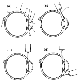
With reference to the drawings, fig la shows a
sectional view of an eye (1), where the anterior and
posterior chambers (2,3) of the eye (1) are separated by
the iris (4) and the pupil (5). Evident in the figure are
also the cornea (6), the sclera (7), the retina (8) and
the optic nerve (9). Fig lb-d shows various possibilities
for the sclerally penetrating fistula, which is created
in certain methods of treatment according the invention.
The fistula is created with a needle (10), which is
connected to a syringe (11) containing the viscoelastic
medium according to the invention.
In an advantageous embodiment of the invention, the
fistula is created using a standard needle (10), and the
viscoelastic medium is continuously injected into the
fistula from a syringe (11) coupled to the needle (10)
while the needle (10) is withdrawn, resulting in a
penetrating fistula filled with the viscoelastic medium.
The method involves insertion of a needle (10) into
the sclera (7) of the eye (1), 4-7 mm behind the limbus
(the junction between the sclera (7) and cornea(6)). As
shown in fig lb, the needle (10) is made to penetrate the
sclera (7) and reach the anterior chamber angle (12).
During withdrawal of the needle (10), the viscoelastic
medium is continuously expelled sclerally throughout the
length of the fistula. Thereby, improved drainage of
aqueous humour is achieved easily and rapidly. Also, the
created drainage is prevented from healing by the
expelled viscoelastic substance. Thereby, a long-lasting
or permanent fistula is created, which allows for
sufficient drainage of aqueous humour.
CA 02565382 2006-11-02
WO 2005/105037 PCT/SE2005/000663
19
In an alternative embodiment, shown schematically in
fig 1c, the needle (10) is made to penetrate the sclera
(7) and reach the posterior chamber (3). During
withdrawal of the needle (10), the viscoelastic medium is
continuously expelled sclerally in the thus created
fistula.
In another embodiment, shown schematically in fig
ld, the needle (10) is made to penetrate the sclera (7)
and reach the vitreous body (13). During withdrawal of
the needle (10), the viscoelastic medium is continuously
expelled sclerally in the resulting fistula. In fig ld,
two alternative ways of creating the fistula are shown.
It shall be noted that while traditional surgical
treatments involves creation and closure of a scleral
flap, surgical removal of tissue and creation of
channels, the present method involves direct penetration
of the sclera using e.g. a needle or a cannula. This
procedure simplifies the creation of a drainage channel.
Without being limited thereto, the present invention
will in the following be further illustrated by way of
examples.
EXAMPLES
Example 1 Preparation of non-animal stabilised hyaluronic
acid
As previously exemplified in e.g. US patent
5,827,937, 10 g of hyaluronic acid prepared by
fermentation of Streptococcus was dispersed in 100 ml of
1% NaOH, pH>9. Cross-linking agent in the form of 1,4-
butanediol diglycidyl ether was added to a concentration
of 0.2%. The resulting composition was incubated at 40 C
for 4 h.
The incubated composition was diluted with an acidic
water solution to reach neutral pH under mixing, yielding
CA 02565382 2006-11-02
WO 2005/105037 PCT/SE2005/000663
a final hyaluronic acid concentration of 20 mg/ml, and
again incubated for 12 h at 70 C. The viscoelastic slurry
that resulted from this second incubation was then cooled
to room temperature and mashed to its final particle
5 size, approximately 0.8 mm.
Example 2 Pre-clinical study of non-animal stabilised
hyaluronic acid in a rabbit eye
' The objective of the study is to show that
10 injections of a viscoelastic composition, such as non-
animal stabilised hyaluronic acid, in the eye will
provide a functional drainage model for glaucoma
treatment.
18 rabbits divided in three groups were used in the
15 study. The rabbits were anesthetized according to
standard procedures. The composition, 20 mg/ml of the
non-animal stabilised hyaluronic acid obtainable by the
method of example 1 (commercially available from Q-Med
AB, Uppsala, Sweden), was injected in one eye and the
20 opposite eye was the untreated control. As shown in fig
lb and fig 2, an approximately 5 mm long fistula was
created with a needle in the sclera by penetrating the
conjunctiva and moving the needle through the sclera to
the angle of the anterior chamber. The composition was
injected into the sclera of the eye during withdrawal of
the needle, thereby filling the fistula with the
composition. The needles used were 27, 23 and 18 gauge
needles.
The goal was to create a drainage from the anterior
chamber to the subconjunctival tissue. The amount of
composition used, size and type of needle and injection
site was recorded. The injection site was checked
visually before and after injection. The animals were
observed daily according to standard procedures.
At weeks 8 and 16, nine of the animals were examined
and euthanized, and histological samples were taken from
the injection sites. Photographs of the histological
CA 02565382 2006-11-02
WO 2005/105037
PCT/SE2005/000663
21
samples are shown in Fig 3. The fistulae, shown in cross-
sections in fig 3, were maintained after 8 as well as 16
weeks and still contained the stabilised hyaluronic acid,
as seen in Fig 3A (staining using hyaluronic acid binding
protein). Cells had not penetrated into the fistulae.
There was no evidence of adverse tissue reactions, nor
formation of tissue within the fistulae.
In the samples collected after 16 weeks of exposure
to the composition, histochemical staining (Fig 3B,
staining with haematoxylin and eosin) indicated that the
walls of the fistulae were covered with endothelial
cells, i.e. an early sign that the fistulae may be
permanent.
Example 3 Pre-clinical study of stabilised hyaluronic
acid in a rabbit eye
Rabbits are anesthetized according to standard
procedures. The compositions used are slurries containing
stabilized hyaluronic acid gel particles with a
hyaluronic acid concentration of 10, 30, and 50 mg/ml,
respectively. The compositions are injected in one eye
and the opposite eye is the untreated control. In each
slurry, a major volume of the particles are approximately
0.1, 0.4 and 0.8 mm, respectively.
1-3 fistulae per eye is(are) created with a needle
in the sclera by penetrating the conjunctiva and moving
the needle through the sclera to the anterior (fig lb) or
posterior chamber (fig lc). The composition is injected
into the sclera of the eye during withdrawal of the
needle.
The amount of composition used, size and type of
needle and injection site is recorded. The injection site
is checked visually before and after injection.
The animals are observed daily according to standard
procedures. At week 8 and 16, animals are examined and
euthanized, and histological samples are taken from the
injection sites.
CA 02565382 2006-11-02
WO 2005/105037 PCT/SE2005/000663
22
Example 4 Administration of stabilized hyaluronic acid
in a rabbit eye
The general procedure of example 2 was followed
using three different cannula sizes: 18G, 23G and 27G was
used. The diameter of the channel formed was
approximately equal for all cannulas. The size of the
channel seems to be more dependent on the amount of
material injected than the diameter of the cannula.
Persistent channels were found in 2/3 eyes with 18G, 2/3
eyes with 27G and 3/3 eyes with 23G cannula.
Example 5 Fluid flow through gel particles of stabilized
hyaluronic acid.
The extrusion force for an aqueous composition
containing 20 mg/ml of a non-animal stabilised hyaluronic
acid obtainable by the method of example 1 (commercially
available from Q-Med AB, Uppsala, Sweden), in the form of
gel particles (average diameter 400 pm) was determined to
21 N through a 30 gauge needle, and 4 N through a 23
gauge needle.
In the first set of experiments, the possibility of
flow through the gel particles was studied by applying a
flow of saline through the gel particles by means of a
pump. A glass column (diameter 5 mm) was filled with gel
particles to a height of 30 mm (approximately 1 ml gel
particles). The flow of saline through the gel particles
was controlled with a pump. Saline was able to flow
through this column with a flow rate of 125 pl/min.
In the second set of experiments, a glass column
(diameter 10 mm) was filled with 1 ml of the composition,
and an aqueous solution of 0.9% NaCl was applied at a
pressure of 29 mm Hg, a pressure corresponding to the IOP
with untreated glaucoma. This pressure resulted in a flow
of 160 pl/h (2.7 pl/min) through the composition. For
comparison, the aqueous humour flow in a healthy eye is
in the range of 1.8-4.3 pl/min, typically 2.75 pl/min
CA 02565382 2006-11-02
WO 2005/105037 PCT/SE2005/000663
23
(Brubaker RF, "Flow of aqueous humor in humans [The
Friedenwald Lecture]", Investigative Ophthalmology &
Visual Science 32:3145-3166 (1991)).
The experiments performed demonstrate that saline
can flow through gel particles after application of a
pressure of 29 mm Hg. The flow rate is of the same
magnitude as the aqueous humour flow in normal human
eyes. Without being bound to any particular theory, it is
contemplated that the saline will flow between the gel
particles in the same way as solvent flows through
chromatographic gel beads, such as Sephadex, in size
exclusion chromatography.