Note: Descriptions are shown in the official language in which they were submitted.
CA 02565732 2006-11-03
WO 2005/116632 PCT/US2005/015754
ANALYTICAL SYSTEMS, DEVICES, AND CARTRIDGES THEREFOR
FIELD
[0001] In general, this invention is in the field of multiple analyte
detection
and quantification, and more specifically, multiple analyte detection and/or
quantification
using more than one measurement technique, a single sample, and a single
device.
BACKGROUND
[0002] Currently, it is common practice to detect or quantify distinct
analytes
using distinct detection or quantification techniques. For example, enzyme
assays,
immunoassays, chemical colorimetric assays, fluorescence labeling and
measurement,
chemiluminescent labeling and measurement, and electrochemiluminescent
labeling and
measurement, are a few exemplary well-known analytical techniques that may be
used to
detect the presence of various analytes. Many of these techniques are
perfoimed on a test
strip or cartridge.
[0003] The test strips typically have specific zones or sites for testing
located
at various positions about the strip. Some of these strips contain an array of
test sites for
the multiple testing of a single analyte, or for the simultaneous testing of
multiple analytes.
Depending on the specific detection or quantification technique used, the test
strips may or
may not be used in combination with a separate measurement device. For
example, where
quantitative optical detection is required, an additional measurement device
is also required
to read the results of the test strip or cartridge. This is unlike the case
with qualitative
visual assays, for example, like those used in most over-the-counter pregnancy
tests, where
an observable color change on the test strip itself indicates the results of
the test. Perhaps
the best known example of a test strip used in combination with a separate
device is a
glucose test strip used in combination with a glucose meter.
[0004] However, independent of whether additional measurement devices are
employed with the test strips, different detection and quantification
techniques are not
typically combined together. This is partly because each technique has a
unique sensitivity,
robustness, and tolerance. In addition, each technique typically has unique
physical and
chemical requirements. Further, it is often the case that the physical
location of the test site
read zones must be fixed or predeteimined in order to enable a corresponding
measurement
1
CA 02565732 2006-11-03
WO 2005/116632 PCT/US2005/015754
device to read the test results. This is because the optical components within
the
measurement device are at a fixed location and the read zone must, therefore,
be in a fixed
location corresponding with the optical components so that a reading may be
obtained (e.g.,
typical in most optically read glucose test strips).
[0005] In addition, the test sample dilution factor and detection system
required to obtain the optimal testing conditions for one analyte are often
incompatible with
the dilution factor and detection system required for a second analyte. Thus,
in order to test
for both analytes, the user must either take multiple samples from the patient
for use with
different test strips, or draw one large sample for division into multiple
samples so that the
multiple samples may be used as different samples for different test strips.
Requiring that
multiple samples, or one large sample, be withdrawn is not only inconvenient
for the
patient, but can be painful as well, for example, when the sample is blood and
it is
withdrawn via venipuncture or multiple finger lances.
[0006] Therefore, running multiple tests on a single cartridge when multiple
detection or quantification techniques are required or are desirable has
heretofore been
limited. Indeed, when the use of different techniques is required or
desirable, the user
most often employs multiple instruments, sometimes from multiple vendors, in
order to
obtain the test results. In the case where the user is a physician or
laboratory technician,
these devices can clutter and reduce the availability of highly valued bench
space.
[0007] In addition, commercially available analytical devices are limited in
that they either measure a single analyte or, if they can measure multiple
analytes, require a
large sample size. For example, the DCA 2000 system (Bayer Corporation,
Diagnostics
Division, Tarrytown, NY) can measure hemoglobin Al c ("HbAlc") using a very
small
sample (1 tiL) of blood, but can only detect a single analyte on a single
cartridge using a
small volume. It is a one analyte per cal tiidge test. When the DCA 2000 is
configured to
detect more than one analyte on a single cartridge, the sample volume required
is much
larger. For example, a test to detect microalbumin and creatinine requires a
40-4, urine
sample. Similarly, the Piccolo Point of Care Chemistry and Electrolyte System
(Abaxis,
Inc., Union City, CA) can run a panel of up to about 12 tests, but it requires
100 [iL of a
blood, plasma or serum sample.
2
CA 02565732 2014-01-30
76135-141
[0008] Generally, commercially available analytical devices are also limited
in
that they are not capable of performing software updates (e.g., assay
improvements or menu
expansions) in a manner transparent to the user. Further, although some
devices designed for
point-of-care medical use perform automatic Quality Control ("QC") checks,
many ask the
user to run control samples manually to assure accurate performance. The user
is also asked
to upload software or data for new assays, etc., manually. These operations
require the user to
have a more intimate knowledge of QC testing requirements and instrument
maintenance than
many potential users are willing or are able to acquire. In addition, devices
without automatic
update capabilities inevitably wind up obsolete as new tests, algorithms, and
procedures are
developed.
[0009] Accordingly, it would be desirable to have systems, devices, and
cartridges capable of performing multiple tests on a single sample, using more
than one
detection or quantification technique. In addition, it would be desirable to
provide cartridges
and devices capable of performing these features using a small sample volume.
It would also
be desirable to have a device that provides automatic QC checks, updates, and
data storage.
[0010]
SUMMARY
[0010a] According to an aspect of the present invention, there is provided a
cartridge comprising at least two test sites having at least two test site
read zones for the
detection or quantification of at least two different analytes and configured
to use at least two
different techniques for the detection or quantification of the at least two
different analytes,
wherein the first of the at least two test sites comprises more layers than
the second of the at
least two test sites, and the at least two test sites are connected to a
sample distribution layer.
[0010b] According to another aspect of the present invention, there is
provided
a cartridge comprising: a bottom layer, wherein, at least a portion of the
bottom layer is non-
porous; a sample distribution layer; and at least two test sites having at
least two test site read
zones; wherein the test sites are adjacent to or embedded within the sample
distribution layer
3
CA 02565732 2014-01-30
,
76135-141
and configured to detect at least two analytes using two different techniques,
and wherein the
first of the at least two test sites comprises more layers than the second of
the at least two test
sites, and the at least two test sites are connected to a sample distribution
layer.
[0010c] According to another aspect of the present invention, there is
provided
a system for detecting or quantifying at least two different analytes
comprising: a device,
wherein the device comprises a port configured to accept at least a portion of
a cartridge, the
cartridge having at least two test site read zones and the portion having at
least one test site
read zone, a light source, and an array detector; memory; and a processing
module configured
to receive signals from the array detector and to perform an image analysis of
the cartridge to
identify the location of at least one of the test site read zones, wherein the
system enables the
detection or quantification of the at least two analytes using at least two
different detection or
quantification techniques, and the at least two test sites are connected to a
sample distribution
layer.
[0010d] According to another aspect of the present invention, there is
provided
a kit for detecting or quantifying at least two different analytes comprising:
the system
described above; and a cartridge having test sites, the test sites further
having test site read
zones.
[00100 According to another aspect of the present invention, there is provided
a device for detecting or quantifying at least two different analytes
comprising: a port
configured to accept at least a portion of a cartridge, the carrtridge having
at least two test site
read zones and the portion thereof having at least one test site read zone; a
light source; an
array detector; memory; and a processing module configured to receive signals
from the
detector and to perform an image analysis of the cartridge to identify the
location of at least
one of the test site read zones, wherein the device enables the detection or
quantification of
least two analytes using at least two different detection or quantification
techniques, and the at
least two test sites are connected to a sample distribution layer.
3a
CA 02565732 2014-01-30
76135-141
1001011 According to another aspect of the present invention, there is
provided
a computer readable medium containing executable code for performing an image
analysis of
a cartridge comprising at least two test site read zones for the detection or
quantification of at
least two different analytes and configured to use at least two different
techniques for the
detection or quantification of the at least two different analytes, wherein
the image analysis
identifies the location of at least one of the test site read zones.
[0010g] According to another aspect of the present invention, there is
provided
a method for detecting or quantifying at least two different analytes on a
cartridge using at
least two different detection or quantification techniques comprising the
steps of: acquiring
calibration information for a cartridge having at least two test site read
zones; acquiring an
image of the cartridge using an array detector; performing an image analysis
of the cartridge
to identify the location of at least one of the test site read zones; and
cycling through specific
detection or quantification techniques corresponding to the detection or
quantification
techniques required by the test site read zones, wherein at least two
different techniques are
used.
[0011] Described herein are systems, devices, cartridges, and kits for
detecting
and/or quantifying at least two different analytes using at least two
different techniques, in a
single sample. Methods for detecting two different analytes using at least two
different
techniques are also described. In general, the cartridges described here
comprise at least two
test sites for the detection or quantification of at least two different
analytes and are
configured to use at least two different techniques for the detection or
quantification of the at
least two different analytes. The precise location of at least one test site
read zone is not
dependent on a corresponding measurement device.
[0012] The cartridges may comprise a bottom layer, wherein at least a portion
of the bottom layer is non-porous, a sample distribution layer, and at least
two test sites
3b
CA 02565732 2006-11-03
WO 2005/116632 PCT/US2005/015754
having at least two test site read zones. The test sites are typically
embedded in, or are
adjacent to, the sample distribution layer and are configured to detect at
least two analytes
using two different techniques. As noted above, the location of at least one
test site read
zone is not dependent on a corresponding measurement device.
[0013] In some variations, the sample distribution layer comprises a porous
material; in other variations, the sample distribution layer comprises an open
channel
capillary layer. The cartridge can also include a red blood cell separating
layer, alone, or in
combination with a retaining layer. The retaining layer is configured to
adhere together the
bottom layer, the sample distribution layer, the test sites, and any
additional optional layers.
[0014] In some variations, the cartridge comprises at least three test site
read
zones. In other variations, the cartridge comprises at least, four, five, or
six test site read
zones. At least one test site may be configured to detect or quantify an
analyte that is
treatment, disease, disorder, or ailment specific. Similarly, at least one
test site may be
configured to detect or quantify an analyte that is a substance of abuse, a
medicament ora
by-product thereof, an environmental toxin or contaminant, or a biological or
chemical
warfare agent. The test sites may be of the same height, or may be of
different heights.
Similarly, some test sites may be of the same height while other test sites on
the same
cartridge may be of a different height. As should be evident, a mixture of
heights on a
single test cartridge is possible.
[0015] The cartridge can further comprise a unique identifier tag, such as a
bar code, a mechanical pattern, a microchip, or a printed pattern. The
cartridge may also be
packaged in a sealed, but openable moisture resistant package. In some
variations, the
cartridge is configured to accept a sample volume of about 20 1.1L or less,
and in some
variations the sample is a bodily fluid, such as whole blood, plasma, serum,
sweat, saliva,
tears, interstitial fluid, spinal fluid, ocular fluid, pus, milk, semen,
amniotic fluid, vaginal
secretions, mucous secretions, and urine.
[0016] Systems for detecting or quantifying at least two different analytes
are
also provided. In general, the systems comprise a device, memory, and a
processing
module. The device comprises a port configured to accept at least a portion of
a cartridge,
the portion having at least two test site read zones, a light source, and an
array detector.
The device may also have electrical contacts for communication with
electrochemical tests
4
CA 02565732 2006-11-03
WO 2005/116632 PCT/US2005/015754
on the cartridge. The processing module is configured to receive signals from
the array
detector and to perform an image analysis of the cartridge to identify the
location of the test
site read zones and the optimal portions of the image for accurate and precise
determination. The system enables the detection or quantification of the at
least two
analytes using at least two different detection or quantification techniques.
These detection
or quantification techniques can be independently selected from the group
consisting of
enzyme assays, specific binding assays, immunoassays, nucleic acid
hybridization assays,
fluorescence labeling, chemiluminescent labeling, electrochemiluminescent
labeling,
fluorescence measurement, chemiluminescent measurement,
electrochemiluminescent
measurement, reflectance measurement, transmittance measurement, absorbance
measurement, turbidity measurement, electrochemistry, and combinations thereof
The
preferred location of these detection techniques is not fixed in that their
locations may be
independently selected to be optimal for the functioning of each cartridge
test combination.
[0017] The processing module may also be configured to determine an error
condition, for example conditions such as an expired cartridge, an inadequate
sample
volume, an impossible analyte value, a reagent malfunction, a mechanical
malfunction, an
electronic malfunction, and mixtures thereof Similarly, the processing module
may be
automatically upgradeable. In addition, the system may be configured to read a
unique
identifier tag on the cartridge, and the system may be self-calibrating. The
system may also
comprise a server connection line, non-volatile memory, a computer, or
mixtures thereof
Systems, devices, and methods for automatically obtaining software upgrades,
new test
software algorithms, specific lot calibration information, specific lot
expiration
information, and related software and data are also provided.
[0018] Kits for detecting or quantifying at least two different analytes are
also
described here. In general, the kits comprise cartridges, with or without
optional
instructions. In some variations, the kits comprise the system described just
above, and a
cartridge. The cartridges of the kits may be configured so that at least a
portion of the
cartridge is configured to protrude from the port of the device. This
protruding portion may
comprise a red blood cell separator, a unique identifier tag, or mixtures
thereof The
cartridge may also be disposable.
[0019] Devices for detecting or quantifying at least two different analytes
are
also provided here, and typically comprise a port configured to accept at
least a portion of a
CA 02565732 2006-11-03
WO 2005/116632 PCT/US2005/015754
cartridge, the portion having at least one test site read zone, a light
source, an array
detector, memory, and a processing module. The processing module is configured
to
receive signals from the array detector and to perfoinr an image analysis of
the cartridge to
identify the location of the test site read zones. The device enables the
detection or
quantification of the at least two analytes using at least two different
detection or
quantification techniques.
[0020] The light source may comprise at least one light emitting diode
("LED"), an incandescent lamp or other radiant energy source emitting a broad
range of
wavelengths, with or without a filter wheel, or combinations thereof. The
array detector
typically comprises charge coupled device ("CCD") or complementary metal-oxide
semiconductor ("CMOS") technology. The processing module may be configured to
determine an error condition, such as those mentioned above. The device may
also be
configured to read a unique identifier tag on the cal ttidge. The device
may also comprise
polarization optics, a back-up power source, non-volatile memory, and
combinations
thereof. In some variations, the device occupies no more than about 1 cubic
foot of
volume.
[0021] A computer readable medium containing code for performing an
image analysis of a cartridge is also described here. Generally speaking, the
cartridge has
at least two test site read zones, for the detection or quantification of at
least two different
analytes and is configured to use at least two different techniques for the
detection or
quantification of the at least two different analytes. The image analysis
identifies the
location of at least one test site read zone. In some variations the computer
readable
medium is firmware, in other variations, the computer readable medium is
software.
[0022] Also described here are methods for detecting the presence or absence
of, or for quantifying, at least two different analytes on a single cartridge
using at least two
different detection or quantification techniques. In general, the methods
typically comprise
the steps of acquiring calibration information for a cartridge having at least
two test site
read zones, acquiring an image of the cartridge using an array detector,
performing an
image analysis of the cartridge to identify the location of at least one test
site read zone, and
cycling through specific detection or quantification techniques corresponding
to the
detection or quantification techniques required by the test sites, wherein at
least two
different techniques are used.
6
CA 02565732 2006-11-03
WO 2005/116632 PCT/US2005/015754
BRIEF DESCRIPTION OF THE DRAWINGS
[0023] FIGS. 1A and 1B provide illustrative schematics of suitable systems
and devices as described herein.
[0024] FIGS. 2A and 2B are exploded views depicting illustrative cartridge
configurations.
[0025] FIGS. 2C and 2D illustrate masks or reticles used with negative and
positive photoresist techniques respectively.
[0026] FIGS. 3A-3G depict exemplary sample distribution layer
configurations.
[0027] FIG. 4A illustrates a sample distribution layer having a portion
configured to protrude from the port of a corresponding device, where the
portion
comprises a sample collection port.
[0028] FIG. 4B illustrates a sample distribution layer having a portion
configured to protrude from the port of a corresponding device, where the
portion
comprises a sample collection port having a red blood cell separator
homogenously mixed
therethroughout.
[0029] FIG. 4C illustrates a sample distribution layer having a portion
configured to protrude from the port of a corresponding device, where the
portion
comprises a sample collection port, where the entrance to the sample
distribution layer has
a red blood cell separator barrier.
[0030] FIG. 4D illustrates a sample distribution layer having a portion
configured to protrude from the port of a corresponding device, where the
portion has
multiple sample collection ports.
[0031] FIG. 4E depicts a sample distribution layer having a portion
configured to protrude from the port of a corresponding device, where the
portion has both
a sample collection port, and a unique identifier tag.
7
CA 02565732 2006-11-03
WO 2005/116632 PCT/US2005/015754
[0032] FIG. 4F depicts a sample distribution layer having electrochemistry
capabilities and a portion configured to protrude from the port of a
corresponding device,
where the portion has a unique identifier tag.
[0033] FIG. 5A illustrates a sample distribution layer where one test site
receives whole blood for testing, while the others receive plasma.
[0034] FIG. 5B shows a sample distribution layer having a portion configured
to protrude from the port of a corresponding device, and a layered test site.
[0035] FIGS. 6A and 6B depict cross-sectional views of illustrative cartridge
configurations.
[0036] FIG. 6C provides a top view of FIGS. 6A and 6B.
[0037] FIG. 7A depicts an illustrative cross-sectional view of a configuration
suitable for use with the cartridges herein described.
[0038] FIG. 7B is a top view of FIG. 7A.
[0039] FIG. 8A depicts an illustrative cross-sectional view of a configuration
suitable for use with the cal hidges herein described.
[0040] FIG. 8B is a top view of FIG. 8A.
[0041] FIG. 9A depicts an illustrative cross-sectional view of a configuration
suitable for use with the cartridges herein described.
[0042] FIG. 9B is a top view of FIG. 9A.
[0043] FIG. 10 depicts an illustrative cross-sectional view of a configuration
suitable for use with the cartridges herein described when optical detection
is required.
[0044] FIGS. 11A and 11B depict illustrative cross-sectional views of
configurations suitable for use with the cartridges described here, when
optical detection is
required.
8
CA 02565732 2006-11-03
WO 2005/116632 PCT/US2005/015754
DETAILED DESCRIPTION
[0045] In general, the cartridges, systems, and devices described herein are
capable of detecting or quantifying at least two different analytes using at
least two
different techniques, and are capable of using these different techniques to
test a single
sample. Thus, tests requiring different detection techniques due to different
sensitivity
requirements or chemistries, for example, can be combined in the same test
cartridge and
can be run using a single sample. Having the capability to measure multiple
analytes using
different techniques may provide greater flexibility in the types of tests
that can be am, and
greater flexibility in the number and location of individual test sites on the
cartridge.
[0046] It should be understood that when the phrase detecting or quantifying
is used throughout the specification, it is meant to include detection (e.g.,
detecting the
presence or absence of an analyte) or quantification (e.g., quantifying the
amount of analyte
present in a given sample), alone, or in combination. Detection and
quantification are not
mutually exclusive for the purposes described herein. Examples of detection
and
quantification techniques suitable for use with the devices and cartridges
described herein
include enzyme assays, specific binding assays, immunoassays, fluorescence
labeling and
measurement, chemiluminescent labeling and measurement,
electrochemiluminescent
labeling and measurement, reflectance measurement, transmittance measurement,
absorbance measurement, turbidity measurement, electrochemistry, and
combinations
thereof. As should be apparent, also included within this description is the
use of two
different types of the same technique (e.g., two different types of
electrochemistry
techniques) therefore, making the two techniques "different." For example,
competitive
and sandwich immunoassays are different techniques, as are heterogeneous and
homogenous immunoassays. Similarly, an immunoassay employing reflectance
measurement is a different technique from the same type of immunoassay
employing
fluorescence measurement. In this manner, two or more different concentration
ranges of
an analyte may be performed and fall within the scope of this invention if a
different
technique is employed for measurement of each concentration range.
I. General Uses
[0047] The systems, devices, and cartridges described herein may be used for
any number of purposes. For example, they may be used for comprehensive
diagnostic
testing for use at a physician's office, clinic, pharmacy, hospital bedside,
emergency room,
9
CA 02565732 2006-11-03
WO 2005/116632 PCT/US2005/015754
mobile medical facility, military facility, or the like. That is, a cartridge
may be configured
to run multiple tests to aid in the diagnosis of a particular disease,
disorder, or ailment. For
example, someone suffering from a sore throat may be tested for strep throat,
mononucleosis, pharyngitis, tonsillitis, and the like, using a single
cartridge and a single
sample. Similarly, someone suspected of suffering from a sexually transmitted
disease may
be tested for chlamydia, genital herpes, AIDS, gonorrhea, syphilis, and the
like, using a
single cartridge and a single sample. This is so even though different
analytes may need to
be detected using different technologies in order to confirm the presence or
absence of a
particular disease.
[0048] The cartridges and devices described herein may also be configured to
run multiple tests in order to ascertain levels of particular analytes of
interest. This may be
useful, for example, in order to detect ineffectively low, as well as
potentially hazardous
high, blood analyte concentrations. This type of configuration may also be
useful to detect
the presence of a particular disease (e.g., diabetes, hypothyroidism, etc),
monitoring a
disease, stratification of a disease, and/or assessing risk for a given
disease or condition.
For example, typically more than one analyte (or elevated concentrations of
various
analytes) are associated with a given disease, and the detection of these
analytes (or the
detection of their elevated concentrations) can help determine from which
disease a person
may be suffering.
[0049] This type of configuration may also be used to monitor patient
compliance with various treatment regimes. For example, blood may be taken as
a sample,
and the concentration of various medications in the blood may be quantified.
Monitoring
patient compliance may be particularly useful in the case of psychotic
patients, where it
may be difficult to otherwise determine compliance (e.g., by simply asking the
patient).
Thus, by way of example, a psychiatrist may obtain critical information about
a mood
stabilizer concentration in the bloodstream of a patient, as well as the
safety of that blood
level as it may affect the health of various organs. That is, potentially
adverse side-effects
involving injury to the liver, kidneys, or other internal organs for which
there are
corresponding and specific detectable substances in the bloodstream, may be
monitored in
this way. For example, in the case of treating bipolar disorder, valproic acid
may be
administered. A test may be configured to monitor the valproic acid
concentration (to
make sure the treatment is effective), while at the same time configured to
monitor various
CA 02565732 2006-11-03
WO 2005/116632 PCT/US2005/015754
enzymes of interest to ensure that liver damage does not occur. A typical
combination of
tests on a single cartridge for this type of analysis, for example, might
include test sites for
valproic acid and liver enzymes such as alanine aminotrasferase ("ALT,"
"SGPT"),
aspartate aminotransferase ("AST," "SGOT"), and lactate dehydrogenase ("LDH").
[0050] The cartridges may also be configured to run tests for various
substances of abuse. These substances may include street drugs such as heroin,
cocaine,
crystal meth, ecstasy, lysergic acid diethylamide ("LSD"), and the like, which
may be
particularly useful for the police force. Similarly, these tests may also be
useful for
physicians, by helping them rapidly detect a particular drug overdose when a
patient arilives
at the hospital unconscious, for example. The substances of abuse may also
include various
steroids, which may be particularly useful for testing athletes prior to
competition.
[0051] In addition to medical applications, the systems, devices, cartridges,
kits, and methods described here may also find utility in areas such as
environmental and
food testing. For example, the cartridges may be configured to detect various
environmental toxins or contaminants (e.g., mercury, lead, heavy metals, etc.)
in order to
determine compliance with certain environmentally set standards. Similarly,
the cartridges
may be configured to detect or quantify environmental toxins and contaminants
in a patient
sample. Foods may also be tested for various contaminants using the
cartridges, systems,
and devices described here. As will be discussed in more detail below, in
instances where
food is used as a sample, it is likely that the food will need to be
homogenized in a suitable
medium to provide a fluid faun.
[0052] The systems, devices, and cartridges may also be configured to detect
or quantify various biological and chemical warfare agents. This may be useful
during
times of war, for example, for use at various military facilities.
[0053] Below is a list of exemplary analytes suitable for detection using the
systems, devices, cartridges, kits and methods described herein, as well as
their clinical
utilities, and biological or therapeutic concentration ranges (taken from
Norbert W. Tietz,
"Textbook of Clinical Chemistry." W.B. Saunders Company, Philadelphia, PA,
1986). It
should be noted, that when reference is had to the detection of at least two
different
analytes, it is meant to include the case wherein the at least two different
analytes are
structurally and chemically the same, however, having different concentration
ranges. As
11
CA 02565732 2006-11-03
WO 2005/116632 PCT/US2005/015754
should be evident, any type of analyte may be tested using the systems,
devices and
cartridges herein described. Accordingly as used herein, when reference is had
to the term
"analyte," it should be understood that such term is meant to include any
chemical entity,
such as a protein, DNA (single stranded or fragments thereof), small molecule,
or the like,
which may be quantitatively or qualitatively detected. The following table is
meant to be
illustrative only, and in no fashion limiting.
Table 1: Exemplary analytes, their clinical utilities, and their biological or
therapeutic concentration ranges.
Analyte Utility Typical Concentration Range
(Serum or Plasma)
Alanine Aminotransferase (ALT, SGPT) Liver 5-28 U/L
Albumin (plasma) Liver 3.4-5.2 g/dL
Albumin (urine) Kidney <80 mg/day
Antibiotic for Severe Infection
Amakacin 1-40 p.g/mL (1.7-68 pmol/L)
(Hospital)
Therap: 125-250 ng/mL (433-903
. Amitriptyline Depression nmol/L)
Toxic: >500 ng/mL (>1805 nmol/L)
Amylase Pancreas 20-160 U/L
Aspartate Aminotransferase (AST,
Liver 8-75 U/L
SOOT)
Bilirubin Liver <2-<16 mg/dL
(34.2-274 ttmol/L)
2-22 pg/mL (up to ¨200 pg/mL with
Brain Natriuretic Peptide (BNP) . Congestive Heart Disease
heart disease or renal failure)
Calcitonin (hCT) Bone formation 30-670 pg/mL
Cancer chemotherapeutic agents Cancer Various
Therap: 8-12 p.g/mL (34-51 umol/L)
Carbamazepine Epilepsy, Bipolar Disorder
Toxic: >15 ug/mL (>63 mon))
Cardiac Troponin I (cTnI) Acute Myocardial Infarction ng/mL
Cholesterol (HDL) Diabetes & Heart Disease 5-85 mg/dL (0.13-
2.2 mmol/L)
Cholesterol (LDL) Diabetes & Heart Disease 10-235 mg/dL
(0.26-6.09 mmol/L)
45-310 or more mg/dL (1.17-8.03 or
Cholesterol (total) Diabetes & Heart Disease
more mmol/L)
Chorionic Gonadotropin (hCG) Pregnancy <3-140,000
mIU/mL
Cortisol Endocrinology 5-23 p.g/dL (83-
635 nmol/L)
Infection, Heart Disease &
C-Reactive Protein (CRP) 1-825 p.g/dL
Atherosclerosis
Creatine Kidney; Muscle 0.17-0.93 mg/dL
(13-71 p.mol/L)
Creatine Kinase (activity) Acute Myocardial Infarction (AMI) 10-200
U/L
ng/mL; 39-185 ng/mL peak during
Creatine Kinase Isoenzyme MB (CKMB) Acute Myocardial Infarction (AMI)
AMI
Creatinine (blood) Kidney 0.2-1.2 mg/dL
(18-106 mon)
12
CA 02565732 2006-11-03
WO 2005/116632 PCT/US2005/015754
Analyte Utility Typical Concentration Range
(Serum or Plasma)
Kidney; Normalization of Analyte
Creatinine (urine) 8-26 (mg/d)/kg (71-230 pmol-d-i.kg-1)
Concentration
Therap: 0.8-2.0 ng/mL (1.0-2.6
Digoxin Cardiac Arrythmias nmol/L)
Toxic: >2.5 ng/mL (>3.2 nmol/L)
Estradiol Endocrinology 0-500 pg/mL (0-1835
pmol/L)
Estriol (Free & Total) Endocrinology 1.0-350 g/L (12.1-
1215 nmol/L)
Estrogens, Total Endocrinology <30-31,000 pg/mL
(ng/L)
Plasma: .-16.5 (0-165
g/L)
al-Fetoprotein (AFP) Fetal Development
Amniotic Fluid: 0.02-5.0 mg/dL (0.2-
50 mg/L)
Follicle Stimulating Hormone (hFSH) Fertility 1-250
mIU/mL (IU/L))
Antibiotic for Severe Infection
Gentamycin 1-10 pg/mL (2.1-20.9 mon)
(Hospital)
Glucagon Endocrinology 11-117 pg/mL (ng/L)
Glucose Diabetes 20-400 mg/dL (1.11-
22.2 mmol/L)
Detection and Monitoring of Acute
Haptoglobin 26-267 mg/dL (260-2670 mg/L)
Phase Reactions and Hemolytic States
HbAlc Diabetes 2-20%
WB: 9-22.5 g/dL (1.4-3.49 mmol/L)
Hemoglobin Anemia Plasma: 1-4 mg/dL
(0.16-0.62
pmol/L)
Normal: 5-15 mon
Homocysteine Heart Disease Risk
Abnormal: 16-100+ mon
Antibiotic for Severe Infection
Kanamycin 1-40 ps/mL (2-82 mol/L)
(Hospital)
Lactate Dehydrogenase (LDH; lactate
Liver 55-1500 U/L (30 C)
pyruvate)
Therap: 0.6-1.2 mEq/L (mmol/L)
Lithium Bipolar Disorder
Toxic: >2 pg/mL mEq/L (mmol/L)
Luteinizing Hormone (hLH) Fertility 3-200 mIU/mL (IU/L)
Myoglobin MI 21-66 pg/L (5-1000
pg/L assay range)
Therap: 50-150 ng/mL (190-570
Nortriptyline Depression nmol/L)
Toxic: >500 ng/mL (>1900 nmol/L)
Paraquat Toxic Chemical 0.1-64 p.g/mL (0.39-
249 mon)
N-term: 230-630 pg/mL (ng/L)
C-term: 430-1860 pg/mL (ng/L)
Parathyroid Hormone (hPTH) Calcium Metabolism & Bone
Immuno Nuclear Mid Molecule: 0.29-
0.85 ng/mL (29-85 pmol/L)
Therap: 15-40 g/mL (65-170
Phenobarbital Epilepsy p.mol/L)
Toxic: >35 g/mL (>151 pmol/L)
13
CA 02565732 2006-11-03
WO 2005/116632 PCT/US2005/015754
Analyte Utility Typical Concentration Range
(Serum or Plasma)
Therap: 10-20 p.gimL (40-79 p.mol/L)
Phenytoin (diphenylhydantoin) Epilepsy
Toxic: >20 pg/mL (>79 mon)
<3.0 ng/mL (p.g/L)
Phosphatase, Acid Prostate
0.11-0.60 U/L
Phosphatase, Alkaline (ALK-P) Bone and Liver Diseases/Cancer 20-165 U/L
Potassium Electrolyte Status 3-12 mEq/L (mmol/L)
Progesterone Fertility 0.11-30 ng/mL (0.35-
95.4 nmol/L)
Normal: 0-4 ng/mL (0-0.12 nmol/L)
Prostate Cancer & Prostate Cancer: 50+ ng/mL
(+1.52 nmol/L
Prostate Specific Antigen (PSA)
Hyperplasia Ultrasensitive
(Recurrence): 1.01
ng/mL (0.3 pmol/L)
Protein, Total Nutritional Status; Disease Diagnosis 3.6-8.0
g/dL (36-80 g/L)
Renin Blood Pressure 0.1-13.2 (ng/h)/mL
Sodium Electrolyte Status 116-166 mEq/L
(mmol/L)
Somatotropin (hGH) Endocrinology <1-50 ng/mL ( g/L)
Free: 0.03-10.2 ng/dL (1.05-354
Testosterone Endocrinology pmol/L)
Total: 5-707 ng/dL (0.17-24.6 nmol/L)
Therap: 6-20 ug/mL (44-111 gmol/L)
Theophylline Asthma
Toxic: >20 ug/mL (>110 mon)
Thyroid Microsomal Antibodies Thyroid ND or <1:10 dilution
(IFA)
0.01-50 p.IU/mL (mIU/L)
Thyroid Stimulating Hormone (hTSH) Thyroid
Normal: 0.4-6.0 IU/rnL (mIU/L)
Free: 0.8-2.4 ng/dL (10.3-31.0
Thyroxine (T4) Thyroid pmol/L)
Total: 4.5-12 pg/dL (58-154 nmol/L)
Transferrin Iron Metabolism 130-400 mg/dL (1.3-
4.0 g/L)
Triglycerides Diabetes & Heart Disease 10-288 mg/dL (0.11-
3.25 mmol/L)
Free: 120-660 pg/dL (1.85-10.16
pmol/L)
Triiodothyronine (T3) Thyroid
Total: 30-275 ng/dL (0.46-4.26
nmol/L)
Urea Nitrogen Kidney 3-40 mg/dL (1.1-14.3
mmol urea/L)
Uric Acid Gout 2.0-8.2 mg/dL (0.12-
0.48 mmol/L)
Therap: 50-100 g/mL (347-693
Valproic Acid Epilepsy, Bipolar Disorder mon)
Toxic: >100 p.g/mL (>693 mon)
Antibiotic for Severe Infection
Vancomycin (Hospital) Toxic: 80-100 ug/mL
(mg/L)
Vitamins & Nutrients Nutritional Status Varies
Warfarin (coumadin) Anticoagulant Therapy 1-10 p.g/mL (3-32
p.mol/L)
14
CA 02565732 2006-11-03
WO 2005/116632 PCT/US2005/015754
Systems
[0054] The systems described herein enable the detection or quantification of
at least two different analytes using at least two different techniques. In
general, the
systems comprise a device, memory, and a processing module. The device
comprises a
port configured to accept at least a portion of a cartridge, a light source,
and an array
detector. The portion of the cartridge that enters the device has at least one
of the two or
more test site read zones for the detection or quantification of at least one
of the two or
more different analytes. However, as will be described in more detail below,
any number
of analytes may be detected using the systems, devices, and cartridges
described herein, as
is practicable or desirable.
[0055] Making reference now to the drawings, where like numerals indicate
like elements throughout the views, FIG. 1A provides an illustrative example
of how the
system may be configured. Shown there is an external view of device (100),
having a port
(102), which is configured to accept at least a portion of a cartridge (104).
The port may
also include automatic insertion and ejection capabilities. Shown are various
control knobs
and switches (106), which may be useful to turn the device on, and control
several of its
features. A line to a power supply is provided (108) as well as a cable (110)
to enable
attachment to a processing module (PM) such as a computer or a personal
computer ("PC")
board (112) if desirable. Indeed, the device may be configured to include a
PM, such as a
PC board, a display (D) to display information (e.g., test results,
maintenance updates,
upgrade alerts, etc.), and a printer (not shown) to print out the test
results, etc.
[0056] While the power supply line (108) is shown, it should be understood
that the device may also be battery operated. In addition, the device may also
have a back
up power supply, for example, a battery (not shown), to help power the device
in the case
of a power outage. For this reason, it may also be desirable that the device
has some non-
volatile memory as well.
[0057] The device may also comprise a sliding, or otherwise openable (e.g.,
hinged) sample door (not shown). In this way, the sample door may be opened to
enable
access to the cartridge, once the cartridge is inserted into the device. The
sample door, for
example, may optimally be placed at a position corresponding to the location
of the
cartridge so that a sample may be placed on the cartridge with relative ease
after the
cartridge has been inserted into the device. Alternatively, the device may
comprise a
CA 02565732 2006-11-03
WO 2005/116632 PCT/US2005/015754
second access port that allows application of the sample after the cartridge
is inserted into
the device.
[0058] It should be understood that while device (100) is depicted here as
having a rectangular form, the device may have any suitable or desirable
geometry. The
device may also be of any desirable size. In some variations, it may be
desirable that the
device has a size of about 2 cubic feet or less, or about 1 cubic foot or
less, which would
help minimize bench top clutter. Similarly, while the control knobs and
switches (106) are
depicted in FIG. 1A as located on the side of device (100), it should be
understood that
these knobs and switches may be located at any desirable or convenient place
on the
device. For example, they may be on the front of the device, on the back of
the device, or
combinations thereof. Some or all of the functions of these control knobs and
switches
may optionally be performed with a touch screen.
[0059] FIG. 1B provides an illustrative schematic of the inside of a device.
As shown there, device (114) has cartridge (116) therein. Illustrative light
sources are
depicted by one or more LEDs (118) and an incandescent lamp (120). A filter
wheel (122)
may also be used. Multiple light sources may be housed within the device as
shown in
FIG. 1B, or only one light source may be used. The light source should be
configured to
direct light to the cartridge (116). The cartridge output is typically
directed toward an array
detector (124). The light sources may be configured to illuminate light onto
the cartridge
from below the cartridge, from the top of the cartridge, from the side of the
cartridge, or
combinations thereof. Accordingly, the array detector will typically be
located at a
convenient output location, depending on the direction of the light source
input. The array
detector (124) can comprise CCD technology, CMOS technology, or the like, as
well
known in the art.
[0060] Also shown in FIG. 1B is an optional cable (126) so that a PM (e.g., a
PC board) may be connected to the device. A display (D) may be hooked up to
the PM in
order to monitor the results, and to display other information. Similarly, a
wireless
connection (W) or other internet connection capability (e.g., modem, cable,
etc.) may be
hooked up to the PM. Line (128) enables the device to be used with an
additional
electrochemistry measurement subsystem (EC) that may optionally be internal to
the
device. The EC may comprise conductors adapted for electrical connection with
an
electrochemical analyzer. As described above, an optional printer (not shown)
may also be
16
CA 02565732 2006-11-03
WO 2005/116632 PCT/US2005/015754
used with the device. Any or all of the foregoing elements (e.g., device, EC,
PM, W, D,
printer) may be optionally contained within a single housing.
[0061] As will be discussed in more detail below, the system may also be
configured to read a unique identifier tag on the cartridge. In this way, the
system may be
able to identify the type, number, and approximate location of the test site
read zones, as
well as able to determine calibration, algorithm, and lot information
therefor. Accordingly,
the device may further comprise a scanning window to image the tag (similar to
those used
at grocery stores), a scanning or swiping slot (similar to those used with
credit cards), or a
non-contact electronic method of obtaining infoimation from a microchip
embedded in, or
attached to, the cartridge. Similarly, the cartridge having a unique
identifier tag thereon
may be configured to be fully inserted into the device so that the tag can be
read.
[0062] However, the system need not obtain the exact read zone information
from the unique identifier tag. Indeed, the system comprises a processing
module
configured to receive signals from the array detector and to perform an image
analysis, or
scan of the cartridge in order to identify the type and location of the read
zones. The
processing module (PM) is shown in FIG. lA as being external to the device,
but it may
also be within the device itself, as shown in FIG. 1B.
[0063] The processing module may be code or logic, implemented in
hardware logic (e.g., an integrated circuit chip, Programmable Gate Array
(PGA),
Application Specific Integrated Circuit (ASIC), etc.) or in a computer
readable medium
such as, for example, magnetic storage medium (e.g. hard disk drives, floppy
disks, tape),
optical storage (e.g., CD-ROMs, optical disks, etc.), volatile and non-
volatile memory
devices (e.g., EEPROMs, ROMs, PROMs, RAMs, DRAMs, SRAMs, firmware,
programmable logic, etc.). Code in the computer readable medium is accessed
and
executed by a processor.
[0064] Accordingly, also provided herein is a computer readable medium
containing code for performing an image analysis of a cartridge comprising at
least two test
site read zones for the detection or quantification of at least two different
analytes, and
configured to use at least two different techniques for the detection or
quantification of the
at least two different analytes. The image analysis identifies the location of
the at least two
17
CA 02565732 2006-11-03
WO 2005/116632 PCT/US2005/015754
test site read zones. In some variations, the computer readable medium is
firmware, and in
other variations, it is software.
[0065] The image analysis may be performed in a manner described by
Neeley in "Reflectance Digital Matrix Photometry." Clin. Chem. Vol. 29, No. 6,
1038-
1041 (1983); "An Instrument for Digital Matrix Photometry." Clin. Chem. Vol.
27, No. 10
1665-1668 (1981); and "A Reflectance Photometer with a Square Photodiode Array
Detector for Use on Multilayer Dry-Film Slides." Clin. Chem. Vol. 34, No. 11,
2367-2370
(1988), using algorithms developed for machine vision systems, or by similar
algorithms
for image algebra. In general, the processing module receives signals from the
array
detector that provide a general image of the cartridge. The areas to be
measured are then
subdivided, grid-like, into small subunits. Image analysis then proceeds by
the
identification of clusters of pixels that have similar intensity values, which
are located
within the outer edges of a larger cluster of pixels, and which are located
approximately at
the predetermined locations for the group of tests performed by the cartridge
being used.
[0066] Therefore, in operation, the type and location of the particular test
sites on the cartridge will first be identified. The system will typically
then check to see if
the test procedure, algorithm, and calibration values are stored in the system
memory, in
order to enable the tests to be run. As with the processing module (PM), the
system
memory (M) may be external to the device, as depicted in FIG. 1A, or it may be
within the
device itself, as depicted in FIG. 1B. The system memory may also be part of
the
processing module. If the system has previously encountered cartridges from
the same
manufacturing lot (and therefore having the same calibration parameter values,
test
procedures, and algorithms), the system will have this information stored in
its memory.
Therefore, the system may notify or signal to the operator that testing may
proceed. The
notification may occur, for example, as a word prompt on a display (e.g., LCD
screen, etc.),
by an audible signal, mixtures of prompts and signals, and the like.
[0067] If the algorithms and test procedures for the cartridge are present in
the system memory, but the specific lot information (calibration parameters,
expiration
dating, etc.) is not, the system may automatically download these values from
a remote
source, such as a host server computer, via a server connection (e.g., a
direct dial-up line,
such as a land line, cell phone line, etc. or an internet connection, such as
a cable or other
wired line, wireless and satellite lines and the like). However, if the
testing procedures,
18
CA 02565732 2006-11-03
WO 2005/116632 PCT/US2005/015754
algorithms, and calibration parameters are not stored in the system memory (as
might
happen if the test has only recently been added to the menu of available
tests), the system
may download both the appropriate testing procedures and algorithms to run and
interpret
the test, as well as the specific lot infounation. If the cartridge employs
electrochemical
methods for detection, alone or in combination with optical methods, the
active electrical
contacts and their specific functions may be obtained and used in a similar
manner. The
processing module of the system may also contain executable code that enables
the system
to automatically transmit test results and sample ID information to a host
server's
confidential database for retrieval by authorized professionals. Any of the
foregoing
functions may also optionally be under the control of the user.
[0068] After the algorithms, test procedures, and lot information have been
obtained, the system then utilizes the light source and the array detector to
acquire an initial
"dry" image (or series of images) of the cartridge, as described above. That
is, images of
the cailiidge surface are obtained under different conditions of illumination,
for example,
using selected wavelengths of radiation from one or more light sources, which
are
optionally combined with one or more filters in the detection light path. The
system then
stores the images in its memory for later use. The operator may now put a
sample in the
cartridge, as signaled by the system, as described above.
[0069] When the presence of a sample is detected (optically or by methods of
employing an electrical change such as conductivity or capacitance), the
processing module
will direct the system to perfoun a variety of measurements specific for the
test or
combination of tests on the cartridge. This might result in three or more
measurement
modes being repeatedly activated in sequence. For example, a 605 nm LED might
be
turned on for reflectance measurements at two different read zones, and then a
500 nm
LED might be turned on (and a filter inserted in the detection light path to
block essentially
all the light output from that LED) in order to perfolln fluorescence
measurements at three
different read zones. Images of the entire cartridge surface acquired under
these two
different conditions of illumination would be stored and compared to
subsequent images
taken under the same conditions.
[0070] When the change in pixel intensity in the general surface locations for
the tests reaches a predetermined level of insignificance, or when the rate of
change reaches
a steady state, image acquisition is stopped and calculations are performed to
determine the
19
CA 02565732 2006-11-03
WO 2005/116632 PCT/US2005/015754
reflectance, fluorescence, etc., or the rate of change thereof, for each test
region as
appropriate for that test. These numerical values are then converted, using
stored
algorithms and calibration parameter values, into analyte concentrations that
are reported
via the display (D) on the device or on the PC, or by some other suitable
method. These
values may also be printed, communicated to a PC for storage in a patient
database, or
both. The operation of the system would be similar to the foregoing for tests
requiring
chemiluminescent or electrochemical detection, however, with these techniques,
no light
source would be needed. Electrochemical detection would not require any use of
the
optical capabilities of the device.
[0071] The processing module of the devices and systems described here may
also be configured to detect particular error conditions. These error
conditions may be, for
example, the detection of an expired cal tlidge, an inadequate sample
volume, an impossible
analyte value, a reagent malfunction, a mechanical malfunction, and mixtures
thereof.
Should an error condition be detected, an appropriate signal can be displayed
by the device.
The display may further indicate whether device repair is required. This type
of
notification may help to facilitate the expeditious replacement of faulty
parts. The
processing module may also be configured to transmit the error condition to a
host server
via a server connection line. In this way, if repair is required, the owner of
the host server
may be able to intervene and help repair the system or device in a timely
manner. In
addition, the processing module can be configured to inactivate the system or
device so that
erroneous test results are not obtained or reported should the owner try to
operate the
system or device while it is malfunctioning.
[0072] In some variations, the processing module is automatically
upgradeable. In these variations, the device may have a server line
connection, enabling
the connection to a remote source such as a host server. Here, the upgrading
can occur
automatically during the normal course of device operation without the need
for
involvement by the device operator. The server line connection may also
provide for
automatic communication with the host server on an as-needed basis. For
example,
automatic messages such as periodic maintenance reminders or notification of
existing
hardware or software upgrades may be sent to the system. In some variations,
the system is
also self-calibrating. That is, the system may perform routine calibrations
using ratioing
techniques, internal standards, and controls, and other techniques known in
the art.
CA 02565732 2006-11-03
WO 2005/116632 PCT/US2005/015754
[0073] As should be evident from the system descriptions above, individual
devices are also provided. In general, the devices comprise a port configured
to accept at
least a portion of a cartridge, the cartridge having at least two test site
read zones and the
portion having at least one test site read zone, a light source, an array
detector, memory,
and a processing module. The processing module is configured to receive
signals from the
array detector to perform an image analysis of the cartridge to identify the
location of the
read zones.
[0074] As described above, the light source may comprise one or more solid
state devices (LEDs, laser diodes, or the like) or may comprise an
incandescent lamp or
other radiant energy source emitting a broad range of wavelengths (e.g., about
300 nm to
about 1000 tun for a tungsten light source). A filter wheel may be optionally
employed.
The device may also comprise polarization technology to enable the perfomiance
of
fluorescence polarization immunoassays. Similarly, the device may also be
configured to
detect temperature changes, and provide for temperature control.
III. Cartridges
[0075] In general, the cartridges comprise at least two test sites for the
detection or quantification of at least two different analytes, and are
configured to use at
least two different techniques for the detection or quantification of the at
least two different
analytes. It should be understood that when reference is had to the phrase
"test site," it is
meant to describe an area, or areas, of a cartridge that are occupied by the
reagents and
zones necessary to perfoint a given test, as described herein. Obviously, some
test sites
will not require the use of any reagents. Similarly, the tem]. "read zone" or
"test site read
zone" when referenced herein, is meant to describe the area, or areas, of the
test site where
the results of the test are obtained. Since the location of at least one of
the read zones on
the cartridge is identified by the system or device during testing, the exact
location of at
least one of the test sites and read zones need not be fixed. That is, the
location of at least
one of the test sites and read zones is not dependent upon a corresponding
measurement
device.
[0076] In general, the cat ___ hidges comprise a bottom layer, a sample
distribution layer, and at least two different test site read zones. The
bottom layer is
typically non-porous (e.g., a plastic, glass, or the like) and may be
transparent, when optical
transmission measurement of analytes is desirable. The sample distribution
layer allows
21
CA 02565732 2006-11-03
WO 2005/116632 PCT/US2005/015754
the sample to flow to the various test sites. The test sample may be any
suitable fluid. For
example, the test sample may be a bodily fluid, such as whole blood, plasma,
serum, sweat,
saliva, tears, interstitial fluid, spinal fluid, ocular fluid, pus, milk,
semen, amniotic fluid,
vaginal secretions, mucous secretions, and urine. Similarly, the test sample
may be water
(suspected of being contaminated), or may be a food product. In cases where
the sample is
a food product, the food product will typically need to be ground up, or
homogenized and
mixed in an appropriate medium. Further manipulations may be required (such as
extraction or purification) to prepare a sample suitable for testing. The test
sites may be
embedded in, or be adjacent to, the sample distribution layer and are
configured to detect at
least two analytes using two different techniques.
[0077] The cartridges may be configured to accept a small sample volume,
for example, a 20 i_LL or 10 .1, sample of blood. This provides the advantage
of allowing
multiple tests to be perfohned using a small sample volume. However, a sample
may also
be diluted to provide for a larger sample. For example a 10 iuLL volume of
blood may be
diluted ten fold to provide a sample volume of 100 fiL. Therefore, multiple
tests may be
perfohned using a small sample of blood that has been extracted from a
patient, and then
subsequently diluted. In this way, patient pain may be minimized. Accordingly,
the
cartridges may be configured to accept any suitable sample volume.
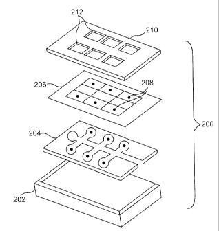
[0078] An exploded view of one variation of a suitable cartridge is depicted
in FIG. 2A. Shown there is cartridge (200) comprising a bottom layer (202), a
sample
distribution layer (204), a test site layer (206) having test sites (208), and
a retaining layer
(210). The retaining layer is shown as having transparent or open windows
(212) for the
optical detection of analytes from the corresponding test sites below. In this
variation,
bottom layer (202) and retaining layer (210) are typically constructed of a
non-porous
material, for example, plastic, glass, or the like, and sample distribution
layer (204) is an
open channeled capillary layer, punched out of plastic, for example. In this
variation, the
sample distribution layer (204) is sandwiched between the bottom layer (202)
and the test
site layer (206).
[0079] An exploded view of another suitable cartridge variation is depicted in
FIG. 2B. Shown there is cal hidge (214) comprising bottom layer (216),
sample
distribution layer (218), and test site (220). In this variation, sample
distribution layer
22
CA 02565732 2006-11-03
WO 2005/116632 PCT/US2005/015754
(218) may be constructed of a porous material or membrane having a hydrophobic
surrounding, to limit or prevent fluid flow thereto. For example, the
hydrophobic
surrounding may be a wax, or the like, shown here by diagonal striping. Bottom
layer
(216) is typically made from a non-porous material.
[0080] An optional retaining layer (not shown) may also be employed to
retain or hold the layers together. Such a layer may overlap entirely with the
sample
distribution layer, or may only overlap the sample distribution layer at its
edges or corners.
The retaining layer may also be a mesh, a nylon, or the like. In addition, the
retaining layer
may be occlusive or sealing in nature, in order to prevent evaporation
therethrough. Of
course, separate sealing layers, or portions thereof are also acceptable. As
noted above,
however, the retaining layer is optional, and the layers can be held together
by any suitable
fastening method. For example, the layers may be held together using
mechanical
clamping, snap-fitting, heat shrinking, gluing (using any suitable adhesive),
and the like.
[0081] While not shown in FIGS. 2A or 2B, the cartridge may also comprise
a red blood cell separator layer, in order to remove the red blood cells
before they reach the
test sites. In this way, red blood cells that may interfere with certain
optical measurements
are removed. This layer may be placed immediately below the sample
distribution layer,
for example, and may be contemporaneous therewith, or only cover a portion
thereof.
[0082] As shown in FIG. 2B, test site (220) is adjacent to the sample
distribution layer and, as will be described in more detail below, is
configured to detect a
given analyte. Test site (220) is shown here as having two layers, but as will
be evident
from the test site description below, any number of layers as practicable or
desirable may
be used. In this way, the test sites may be of varying heights. That is, one
test site may
have only one layer, while another test site at a different location on the
cartridge may have
two or more layers. In addition, the test sites may be of varying widths and
lengths.
[0083] Generally speaking, the sample distribution layer may be made using
any number of techniques. For example, the sample distribution layer may be
made using
processes such as lasering, embossing, Lithographic Galvanoformung Abformung
("LIGA"), electroplating, electroforming, photolithography, reactive ion
etching, ion beam
milling, compression molding, casting, reaction injection molding, injection
molding,
micromachining, and the like.
23
CA 02565732 2006-11-03
WO 2005/116632 PCT/US2005/015754
[0084] In certain variations, it may be desirable to make the sample
distribution layer using photolithography techniques. For example, polymers
can be
incorporated into a lateral flow or filtration membrane, using negative or
positive
photoresist-type materials. The photoresist materials could be impregnated
into the
membrane by screen-printing, spraying, dipping, reverse roller coating,
gravure coating, or
the like. The membrane would then be exposed to UV light, using a
photolithography
mask or reticle, so that certain areas are protected from exposure. The
membrane would
then be developed using an appropriate solvent to wash away material that had
either not
been polymerized (e.g., in the case of negative photoresist) or that have been
converted to a
soluble folin (e.g., in the case of positive photoresist). Membrane
development can be
done in any number of ways. For example, the membrane can be developed using
filtration
on a flat bed, or by dipping the membrane into a suitable solvent.
[0085] An exemplary configuration of a mask or reticle used with negative
photoresist is shown in FIG. 2C. Here, the polymers in the resist become cross-
linked in
the areas that are exposed (E) to UV light. These cross-linked polymers are
insoluble in the
solvent selected to dissolve the resist from the unexposed (UE) regions of the
membrane
during development. An exemplary configuration of a mask or reticle used with
positive
photoresist is shown in FIG. 2D. Here, the region exposed (E) to UV light
converts to a
soluble form (e.g., a carboxylic acid), which may be dissolved away using a
suitable
solvent (e.g., a weak water-based alkali solvent). The unexposed (LIE) region
remains
insoluble.
[0086] Sample distribution layers made using photoresist techniques may
offer several advantages. For example, the membrane would not have to be cut
or stamped
out to form a pattern, thus eliminating the need for difficult and precise
manufacturing
procedures. Instead, manufacturing would be simple, and the process could be
easily
scaled using different sized and shaped photolithography masks or reticles.
Similarly,
crosstalk between different test sites would be eliminated.
[0087] As noted above, the cartridges may comprise any number of test sites
and test site read zones and have any number of configurations. For example,
the cartridge
may have two or more, three or more, four or more, five or more, six or more,
eight or
more, or ten or more test sites and corresponding read zones, and the like.
Indeed, any
24
CA 02565732 2006-11-03
WO 2005/116632 PCT/US2005/015754
number of test sites may be used as practicable or desirable. Some of these
test sites may
be used for redundancy or for control testing purposes.
[0088] Shown in FIGS. 3A-3G are illustrative configurations of sample
distribution layers suitable for use with the cartridges herein described. The
sample
distribution layer may provide for multiple test site locations (as
illustrated by the black
dots) in an orderly fashion as depicted in FIG. 3A. The sample distribution
layer may also
provide for multiple test sites throughout the cartridge in order to optimize
the space
available for the test sites as demonstrated in FIG. 3B. The sample
distribution layer may
be configured such that all the test sites are on one side of the cartridge as
in FIG. 3C.
[0089] The sample distribution layer may also be amorphous in order to
provide for a random distribution of the test sites, as depicted in FIG. 3D.
Another
variation of the sample distribution layer, configured to provide a star type
of configuration
is shown in FIG. 3E. It should be pointed out that the sample distribution
layer may have
more than one sample entrance port, as shown in FIG. 3F. In this way, two
different
samples may be tested simultaneously if desirable (for example, two samples of
blood, a
sample of urine and blood, and the like). It should be understood, that while
two different
sample entrance ports are depicted in FIG. 3F, any number of ports (e.g., 3,
4, 5, or more)
may be used. FIG. 3G depicts one variation where the test sites and read zones
are radially
distributed around a sample entrance port (SEP). In this way, equal sample
distribution to
the test sites may be facilitated. Again, because the location of the read
zones is identified
by the system or device prior to testing, the sites may be located anywhere
throughout the
cartridge.
[0090] The cartridges may also be designed such that a portion of the
cartridge is configured to protrude from the port of a corresponding device.
This may, for
example, help with the insertion and removal of the cartridge in the device,
in the case that
the device does not have an automatic insertion and ejection feature. Top
views of
illustrative depictions of such cartridges are shown in FIGS. 4A-4F. In FIG.
4A, the
protruding portion (400) has a sample collection port (402) thereon. In this
way, the
cartridge may first be inserted into the device, and then the sample placed in
the sample
collection port (402), which protrudes from the device port.
CA 02565732 2006-11-03
WO 2005/116632 PCT/US2005/015754
[0091] A similar configuration is shown in FIG. 4B. Shown there is
protruding portion (404) and sample collection port (406). Within sample
collection port
(406) is a red blood cell separator (408). Red blood cell separators are well
known in the
art, and can comprise for example, certain plant proteins (e.g., lectins,
soybean
hemagglutinins, etc.), certain anti-red blood cell antibodies (e.g., a-RBC),
or certain
polymeric materials, as described below. Shown in FIG. 4C is a protruding
portion (410)
having a sample collection port (412) thereon. A red blood cell separator
barrier (414)
lines the entrance to the sample distribution layer in order to separate out
the red blood cells
prior to testing.
[0092] FIG. 4D shows another configuration of the cartridge having a
protruding portion (416). As demonstrated by FIG. 4D, the protruding portion
may have
any type of configuration or geometry. For example, it may be narrower than
the
remaining cartridge, or may be wider than the remaining cartridge as shown in
FIG. 4D. In
addition, the protruding portion may have multiple sample collection ports
(418) thereon.
As described above, these collection ports may further comprise a red blood
cell separator.
[0093] FIG. 4E provides an illustration of a cartridge having a protruding
portion (420). In this variation, the protruding portion has the shape of an
elongated oval,
but as described above the protruding portions may have any desirable
geometry. The
protruding portion of FIG. 4E has a sample collection port (422) and a unique
identifier tag
(424). The unique identifier tag (424) is shown as a bar code, but any unique
pattern may
be used. The pattern may be produced for example, by mechanical methods, or by
printing.
Similarly, the unique identifier tag may be a microchip or the like. As
described above, the
unique identifier tag can enable the system or device to determine the
location, number, and
types of test sites and read zones on the cartridge prior to testing, or can
directly or
indirectly provide calibration, algorithm, and test procedure information. As
noted above,
in the case where the unique identifier tag is outside the port, the device
may comprise a
scanning window to image the tag (similar to those used at grocery stores), a
scanning or
swiping slot (similar to those used for credit cards), and the like. In this
way, the tag can be
read by the device prior to its insertion. However, the cartridge may also be
fully inserted
into the device so that the unique identifier tag may be read, and then
ejected so that the
protruding portion is again outside the device port.
26
CA 02565732 2013-05-14
67044-78
[00941 Also shown in FIG. 4F are connectors (430) to enable electrochemical
analysis. For example, connectors (430) may pluginto a corresponding socket
within the
device. Similarly, the connectors may instead be POGO pins, for attachment to
a
corresponding socket. It should be understood that while FIGS. 4A-4F depict
various
configurations in which the cartridge has a portion configured to protrude
from a
corresponding device, the cartridges need not have such a protruding portion,
such as those
cartridges described above. ,
[00951 FIG. 5A shows a configuration that uses red blood cell separators in
the sample distribution layer itself. In this way, only those tests that
require red blood cell
removal will have a red blood cell separator. One way to accomplish this is
shown in FIG.
5A. Shown there is sample distribution layer (500) configured for the
detection of five
Replytes. Red blood cell separator barriers (502) may be placed immediately
prior to the
test site openings (504). Thus, as whole blood (508) flows through the sample
distribution
layer and encounters the red blood cell separator barriers (502), red blood
cells are removed
from the sample leaving only plasma (506). Plasma (506) continues through test
site
opening (504) for testing. Similarly, where no red blood cell separator bather
(502) is
present, whole blood continues through the test site opening (504) for
testing, as depicted
by (510). While the red blood cell separators (502) are depicted in FIG. 5A as
being of the
=
same general nature, it should be understood that each red blood cell
separator may be
different if desirable. That is, one red blood cell separator may use plant
proteins, while
another may use anti-red blood cell antibodies.
= [0096] The red blood cell separators may also be incorporated into a
polymer
bead. The bead could swell, for example, when contacted by a sample of whole
blood.
However, the pores in the swelled polymer bead could be configured to be small
enough to
exclude red blood cells, allowing only plasma to pass through. Examples of
suitable
polymers for forming such beads are acidic or basic hydrogels, which are
triggered to swell
by a change in pH, and ionic hydrogels, which are triggered to swell by a
change in ionic
= strength. These are known in the field of controlled drug delivery.
Hydrogels made with
polyvinyl alcohol are described in, e.g., U.S. Patent No. 6,608,117.
oth'er Suitable hydmgel materials include
hydrolyzed polyacrylonitrile, polyacrylamide, starches, gelatins, and the
like.
27
CA 02565732 2006-11-03
WO 2005/116632 PCT/US2005/015754
[0097] FIG. 5B shows how the test site may be located on top of a sample
distribution layer. Shown there is cartridge (512) comprising a sample
distribution layer
(514), and a portion configured to protrude from a corresponding measurement
device
(516). In this example, the portion (516) has a sample port (518) thereon.
Also shown is
test site (520), here shown as three layers. As explained above, any number of
layers may
be used for the test sites.
[0098] As noted above, any number of detection or quantification techniques
may be used with the cartridges and devices described herein. That is, one
technique may
be used at one test site, while another technique may be used at several
others. Suitable
techniques include enzyme assays, immunoassays, fluorescence labeling and
measurement,
chemiluminescent labeling and measurement, electrochemiluminescent labeling
and
measurement, reflectance measurement, transmittance measurement, absorbance
measurement, turbidity measurement, electrochemistry, and the like. FIGS. 6A
and 6B
provide illustrative depictions of sample test site configurations.
[0099] Shown in FIG. 6A is a cross-section of a cartridge (600) suitable for
use with the devices described herein. The cartridge (600) comprises a bottom
layer (602),
a sample distribution layer (604), and a test site layer (606). The test site
layer (606)
depicted in FIG. 6A has two non-porous portions (608) to prohibit fluid from
flowing
therein. The test site layer also has a conjugate zone (610). As used herein,
the term
conjugate zone is meant to describe an area of the test site occupied by a
diffusely
immobilized conjugate, a conjugate being any label coupled to a specific
binding member
of a binding pair. Exemplary binding members include, without limitation,
analytes,
analyte analogs, antibodies, nucleic acids or fragments thereof, lectins, and
the like.
Exemplary labels include, without limitation, fluorescent molecules or
microparticles,
colored molecules or microparticles, enzymes, coenzymes, and the like. Shown
in
conjugate zone (610) are diffusely immobilized conjugates, which will bind to
an analyte of
interest or to a non-diffusely immobilized specific binding member.
[0100] In the variation depicted in FIG. 6A, for example, the specific binding
member may be an antibody, and the test site may be configured to run a
homogenous
immunoassay (i.e., an immunoassay that does not require the separation of free
conjugate
from bound conjugate prior to measurement). Also typically included in the
conjugate
zone are other reagents, substrates, enzymes, and indicators, as needed to run
a given
28
CA 02565732 2006-11-03
WO 2005/116632 PCT/US2005/015754
reaction. Thus, in this design, the conjugate zone and indicator zone (i.e.,
the area of the
test site occupied by signal-developing reagents such as enzymes and enzyme
substrates)
are the same. These zones will be described in more detail below when
reference is had to
various test site configurations.
[0101] Another variation is depicted in FIG. 6B, this time having two test
site
layers. Shown there is a cross-section of a cartridge (612). The cartridge
comprises bottom
layer (614), sample distribution layer (616), and test site layers (618) and
(624). Test site
layer (618) has non-porous portions (620) and a conjugate zone (622). Test
site layer (624)
has non-porous portions (626) and an indicator zone (628). As with the test
site depicted in
FIG. 6A, the test site in FIG. 6B may be configured to run a homogenous
immunoassay.
Here however, the conjugate zone and indicator zones are separated. In this
way, the
indicator may be separate from the specific binding member, which may be
advantageous
for example, to reduce the generation of background color. FIG. 6C provides a
top view of
the test sites of FIGS. 6A and 6B. As shown there, the analyte will be
detected
immediately above the conjugate zone location (630). The sample distribution
layer within
the cartridge is depicted with dashed lines as (632).
[0102] FIG. 7A provides a cross-section of another suitable cartridge
configuration, here having a test site with separate conjugate and capture
zones. Thus, for
example, the test site of FIG. 7A may be configured to perform a heterogeneous
immunoassay (i.e., an immunoassay requiring the separation of free conjugate
from bound
conjugate prior to measurement). Cartridge (700) comprises bottom layer (702),
sample
distribution layer (704), and test site layers (706) and (712). Test site
layer (706) has non-
porous portions (708), and a conjugate zone (710). The conjugate zone (710),
similar to
that described above, may contain conjugates and/or other reagents (here shown
by dots).
Immediately above the conjugate zone (710) is a capture zone (716) within test
site layer
(712) where the conjugate is captured via binding or reaction. As used herein,
the phrase
"capture zone" is meant to describe an area of a test site wherein a conjugate
is bound by a
non-diffusely immobilized specific binding member. Additional fluid,
containing non-
bound reagents, continues to flow through test site layer (712) until it
reaches non-porous
barriers (714).
[0103] A top view of FIG. 7A is provided in 7B. As can be seen, the
conjugate is captured for detection at location (718) corresponding to capture
zone (716).
29
CA 02565732 2006-11-03
WO 2005/116632 PCT/US2005/015754
Non-captured reagents and additional reagent material are washed out to
location (720),
which corresponds to locations before non-porous regions (714). Also shown in
FIG. 7B is
sample distribution layer (722), here shown as dashed lines because it is
located two layers
below the top most test site layer.
[0104] FIG. 8A provides another cross-sectional depiction of a suitable
cartridge configuration. Similar to the configuration of FIG. 7A, the test
site may be
configured to perform a heterogeneous immunoassay (e.g., sandwich,
competitive,
subtractive, etc.), except that in this configuration, the conjugate zone
(818) and the capture
zone (822) are in the same test site layer (812). Here, cartridge (800)
comprises bottom
layer (802), sample distribution layer (804), and test site layers (806) and
(812). Test site
layer (806) has non-porous portions (808) and a porous portion (810) for fluid
to flow
therethrough. In this way, the sample can flow from sample distribution layer
(804)
through porous portion (810) and to test site layer (812) to porous portion
(816). The
sample then flows through conjugate zone (818) where the analyte binds or
reacts with the
reagents contained therein. The reacted analyte (for example, a complex
between the
analyte and its labeled specific binding member, or labeled conjugate) then
continues to
flow through porous portion (820), and to capture zone (822) where the free
conjugate or
the analyte: :conjugate complex is captured via reaction or binding.
Additional un-reacted
analyte and unbound reagent continue to flow through porous portion (824), but
stop at a
place (826) before non-porous portion (814). A top view of FIG. 8A is provided
in FIG.
8B. The read zone for detection is shown at location (828), which corresponds
to capture
zone (822). The sample distribution layer is shown as (830).
[0105] Another configuration of a suitable cartridge is depicted in FIG. 9A.
Similar to the cartridge depicted in FIG. 8A, the test site of the cartridge
of FIG. 9A may be
configured to run a heterogeneous immunoassay. Here the conjugate zone (914)
is not in
the same test site layer (916) as the capture zone (922). Instead, the
conjugate zone (914) is
in test site layer (910). The conjugate zone (914) is surrounded by non-porous
regions
(912). In this configuration, the sample flows through sample distribution
layer (904), as
indicated by the arrows, through the conjugate zone, and then through to test
site layer
(916). The free conjugate or analyte::conjugate complex is then captured at
capture zone
(922). Unreacted analyte and unbound reagent continue to flow through porous
portion
(924) until they stop at a location (926) before non-porous region (918). A
top view of
CA 02565732 2006-11-03
WO 2005/116632 PCT/US2005/015754
FIG. 9A is provided in FIG. 9B. The read zone for detection is located at
position (928),
which corresponds to capture zone (922). The sample distribution layer is
shown as (930).
101061 The capture zone may be produced by depositing reagent
microparticles with the desired component (e.g., an antibody or antigen) onto
a membrane
or other porous material. The reagent microparticles may be adsorbed or
chemically
coupled to the surface of the membrane or porous material. In addition, the
size and
chemical properties of the microparticles can be arranged so that they are
unable to migrate
(e.g., the diameter of the microparticles may be configured to be larger than
the average
pore size of the capture zone material). The capture zone may also be produced
by direct
binding of the desired component (antibody, antigen, antigen-analog, etc.) to
the membrane
or porous material by procedures familiar to those skilled in the art.
[0107] As noted above, the systems, devices, and cartridges described here
are configured to allow testing of more than one analyte, using more than one
measurement
technique. That is, a cartridge may have 5 test sites for example, one of
which is
configured to employ fluorescence measurement, three of which are configured
to employ
reflectance measurements, and one of which is configured to employ
chemiluminescence
measurement. While test sites capable of perfolining electrochemical detection
have not
been shown in detail throughout the figures, it should be understood that
cartridges having
test sites capable of perfoiming electrochemical detection are within the
scope of the
invention (e.g., cartridges with various electrochemical sensors or cartridges
with test sites
that have electrodes in contact with electrochemical reagents). Test site
configurations are
also provided that allow for optical transmission measurements.
[0108] Depicted in FIG. 10, is a cross-section of a suitable cartridge
configuration illustrating a test site allowing for optical measurements. As
shown,
cartridge (1000) comprises nonporous bottom layer (1002), nonporous
transparent
membrane layer (1008), sample distribution layer (1010), and nonporous
transparent top
layer (1014). The bottom layer (1002), and sample distribution layer (1010)
have several
non-transparent regions depicted as (1004), and (1012) respectively. In this
instance, non-
transparent regions (1012) are also non-porous in order to prohibit the sample
flow
therethrough. In this way, light (L) may be illuminated through transparent
portions (1006)
of bottom layer. Sample flows from the left into sample distribution layer
(1010).
31
CA 02565732 2006-11-03
WO 2005/116632 PCT/US2005/015754
[0109] In this configuration, light (L) passes through sample distribution
layer (1010) where it will shine through the sample and analytes contained
therein.
However, because the sample is prevented from passing through non-porous
region (1012),
the transparent region (1013) will contain no sample, and hence no analyte,
and may thus
be used as a control. The light (L) that shines through test region (1011) can
detect an
analyte at its corresponding wavelength, and will pass the infoiniation on to
detector (D).
Similarly, the light (L) shining through the control region (1013) will serve
as the control
light path (CLP).
[0110] FIGS. 11A and 11B show variations of cartridge configurations
employing various transparent regions. As shown in FIG. 11A, cartridge (1100)
comprises
nonporous bottom layer (1102), sample distribution layer (1108), and nonporous
top
transparent layer (1110). In this variation, light (L) from a suitable light
source is
illuminated through the top transparent layer (1110), through the sample
distribution layer
(1108), and then reflected off a portion (1101) of the bottom layer (1102).
The reflected
light then reflects off opposite portion (1103) of bottom layer (1102) and
continues to travel
back up through sample distribution layer (1108) and top transparent layer
(1110) to be
detected by detector (D). The portion of the bottom layer for reflecting the
light may be
made in any suitable manner. For example, the bottom layer may be molded to
include the
various angles, or additional portions may be adhered to the bottom layer in
order to create
the angles as necessary. The angles (1104) and (1106) may be adjusted as
desired to
increase, decrease, or change the pattern of the reflected light.
[0111] The configuration of FIG. 11B is similar to the configuration of FIG.
11A, except that the portion configured to reflect light is in the sample
distribution layer
(1116) as opposed to bottom layer (1114). Accordingly, since the portion
configured to
reflect the light is not in the bottom layer (1114), the bottom layer need not
be transparent.
Having the portion configured to reflect the light be located in the sample
distribution layer
(1116) may help increase detection sensitivity. As in the case with the
configuration of
FIG. 11A, the angles (1118) and (1120) in FIG. 11B, may be adjusted in any
suitable
fashion to adjust the length and pattern of the light reflected through the
portion.
IV. Conjugate or Indicator Zone Configurations
[0112] As described above, there are a number of suitable conjugate or
indicator zone configurations. The configuration selected is typically
dependent on the
32
CA 02565732 2006-11-03
WO 2005/116632 PCT/US2005/015754
chemical nature of the test to be performed. Generally speaking, these
configurations can
be designed by giving consideration to the concentration range of the analyte
to be
measured, and the molecular weight and structure of the analyte.
[0113] For example, some analytes may be present in high concentrations and
will be detectable as a result of a reaction with enzymes or color-fonning
reagents. These
analytes can be measured directly, or upon complex fonnation or reaction with
a reagent in
an indicator zone. Exemplary analytes falling within this category include
lithium, sodium,
hemoglobin, bilirubin, and the like.
[0114] The analyte may also be non-enzymatically, or enzymatically redox
reactive. These types of redox reactions may occur with or without the
consequent
production of a common redox intermediate such as nicotinamide adenine
dinucleotide in
an oxidized or reduced foun ("NAD" or "NADH"), nicotinamide adenine
dinucleotide
phosphate ("NADP"), flavine adenine dinucleotide in an oxidized or reduced
fonn ("FAD"
and "FADH2"), and hydrogen peroxide. Similarly, these reactions may occur with
or
without the gain or loss of electrons from an electrochemical sensor. When
redox
intermediates are produced, they may optionally oxidize or reduce a
chromogenic substrate.
Analytes suitable for detection in this fashion include, without limitation,
total cholesterol,
HDL-cholesterol, glucose, 0-hydroxybutyrate, hemoglobin, and the like.
[0115] Some analytes can be cleaved by a hydrolytic enzyme in order to
produce a substance that either has properties allowing it to be detected
directly by physical
methods (e.g., colorimetry, reflectometry, fluorescence, electrochemistry,
etc.) or that has
redox reactivity similar to the cases described above (e.g., cholesterol
esters, triglycerides,
etc.). For analytes that are reactive by the above criteria, but are present
in such low
concentrations that direct redox reaction or detection will not be measurable,
immunochemical or other specific binding assays may be appropriate. Similarly,
for
analytes that are not reactive by the above criteria, immunochemical or other
specific
binding assays may be appropriate.
[0116] For low molecular weight analytes having one, or a few, epitope(s)
that bind an antibody, a homogeneous or heterogeneous competitive or
competitive
inhibition immunoassay may be appropriate. Exemplary analytes falling within
this
33
CA 02565732 2006-11-03
WO 2005/116632 PCT/US2005/015754
category include valproic acid, carbamazepine, cortisol, thyroxine ("T4"),
triiodothyronine
("T3"), digoxin, phenytoin, phenobarbitol, theophylline, and the like.
[0117] For high molecular weight compounds with more than one epitope, a
heterogeneous or homogeneous immunoassay employing one or two antibodies
(sandwich,
competitive or competitive inhibition) may be appropriate, depending on the
analyte
concentration. Analytes falling within this category include, without
limitation,
hemoglobin Al c ("HbAl c"), chorionic gonadotropin ("hCG"), thyroid
stimulating
hormone ("TSH"), high sensitivity TSH, brain natriuretic peptide ("BNP"),
cardiac
troponin I ("cTnI"), creatine kinase isoenzyme MB ("CKMB"), cytokines,
microalbumin,
myoglobin, and the like. If the analyte is present at extremely low
concentrations, a
sandwich immunoassay employing fluorescent microparticles or an enzyme label
might be
desirable. Chemiluminescent detection may also be employed to help improve
sensitivity.
[0118] If the analyte is an enzyme or other macromolecule with catalytic
activity, there may be more than one desirable test site configuration
possible. For
example, an activity assay may be perfolined employing one or more substrates,
which are
converted to one or more products that are either directly or indirectly
detectable.
Exemplary analytes within this category include, aspartate aminotransferase
("SGOT"),
alanine aminotransferase ("ALT"), alkaline phosphatase ("ALK-P"), amylase,
creatine
kinase ("CK"), and the like. Similarly, a mass assay (sandwich, competitive,
or
competitive inhibition immunoassay), as outlined above for high molecular
weight
compounds may be used, for example, when the analyte is CKMB, or the like.
V. Test Site Configurations
[0119] The following are examples of various test site
configurations that
may be used with the cartridges herein described. It should be understood that
these
examples are not comprehensive or exhaustive of the many variations of test
site
configurations suitable for use with the described cartridges. These examples
are non-
limiting and for illustrative purposes only.
A. Apoenzyme Reactivation Immunoassay System ("ARIS") Assay
[0120] This type of homogeneous immunoassay is particularly amenable to
small molecule analytes such as valproic acid, carbamazepine, or thyroxine,
but it may also
be used to detect larger analytes such as immunoglobulins. A conjugate is
constructed in
34
CA 02565732 2006-11-03
WO 2005/116632 PCT/US2005/015754
which the analyte is coupled covalently to Ravin adenine dinucleotide ("FAD").
This
conjugate competes with the unlabeled analyte in the sample for binding to a
specific
antibody. FAD-conjugated analyte that is not bound to antibody, due to
competition with
free analyte from the sample, is free to bind to apo-glucose oxidase,
activating it. The
resulting glucose oxidase activity is directly proportional to the amount of
analyte in the
sample.
[0121] For example, making reference now to FIG. 6A, conjugate zone (610)
could contain apo-glucose oxidase, a preformed complex of FAD-analyte::anti-
analyte
antibody, glucose, horseradish peroxidase ("HRP"), and a chromogenic,
fluorogenic, or
chemiluminescent HRP substrate. When the sample competes off the FAD-analyte
from
the FAD-analyte::anti-analyte antibody complex, the FAD-analyte is free to
bind to apo-
glucose oxidase, thus activating it. In Figure 6B, conjugate zone (622) could
contain apo-
glucose oxidase, a preformed complex of FAD-analyte::anti-analyte antibody,
and HRP,
while indicator zone (628) could contain glucose and a chromogenic,
fluorogenic, or
chemiluminescent HRP substrate.
B. Enzyme Multiplied Immunoassay Technique ("EMIT") Assay
[0122] This type of homogeneous immunoassay is particularly amenable to
small molecule analytes, such as phenytoin, valproic acid, and thyroxine, but
it may also be
used to detect larger analytes such as immunoglobulins. A conjugate is
constructed in
which the analyte is covalently coupled to an enzyme. This conjugate competes
with the
unlabeled analyte in the sample for binding to a specific antibody. In one
example,
analyte-conjugated enzyme bound to antibody exhibits reduced activity. Analyte
present in
the sample will compete for antibody binding, releasing the analyte-conjugated
enzyme.
Therefore, the higher the concentration of analyte in the sample, the higher
the observed
enzyme activity will be. With appropriate temperature monitoring and
correction by the
system or device, a reaction rate could be determined, as opposed to simply an
endpoint.
(Indeed, in some instances, temperature monitoring will be very desirable, for
example,
where the activity of an enzyme is being measured directly.)
[0123] For example, making reference now to FIG. 6A, conjugate zone (610)
could contain a preformed complex of analyte-enzyme: :anti-analyte antibody,
along with
enzyme substrates. When the sample competes off the analyte-enzyme from the
analyte-
CA 02565732 2006-11-03
WO 2005/116632 PCT/US2005/015754
enzyme: :anti-analyte antibody complex, the analyte-enzyme is free to act on
its
chromogenic, fluorogenic, or chemiluminescent substrate. Thus, the rate of
color
formation is directly proportional to the concentration of analyte in the
sample. Similarly,
referring now to FIG. 6B, conjugate zone (622) could contain the prefollned
complex of
analyte-enzyme::anti-analyte antibody, while indicator zone (628) could
contain the
chromogenic, fluorogenic, or chemiluminescent enzyme substrate.
[0124] Typical enzymes used for EMIT assays include lysozyme, glucose-6-
phosphate dehydrogenase, malate dehydrogenase and13-galactosidase.
C. Competitive Binding Assay
[0125] In one version of this type of assay, the analyte in the sample
competes with a labeled analyte for binding to a specific binding partner. The
amount of
label associated with the binding partner at the end of the assay is inversely
proportional to
the concentration of analyte in the sample. For example, in FIG. 7A, conjugate
zone (710)
might contain a preformed complex of labeled analyte::anti-analyte antibody.
When the
sample contacts this complex, it competes with the bound label for the
antibody binding
sites, displacing the label from the complex. Capture zone (716) might contain
a non-
diffusely immobilized antibody against the first antibody. The first antibody
is captured at
this site, along with its remaining bound label. The signal measured at this
site is indirectly
proportional to the concentration of analyte in the sample. In the case of
valproate,
valproate in the sample might compete with a labeled valproate analog for
binding to a goat
anti-valproate antibody. This antibody flow t toward the capture zone, where
it is captured
by a donkey anti-goat antibody. Unbound analyte and labeled analyte flow
radially away
from the capture zone, and the signal in the capture zone is indirectly
proportional to the
concentration of valproate in the sample.
[01261 In another version of this assay type, the specific binding partner
carries the label. In this case, the labeled binding partner that is not bound
to analyte from
the sample is free to bind to an analyte or an analyte analog that is, for
example,
immobilized in a capture zone. For example, in FIG. 8A, the conjugate zone
(818) may
contain one or more labeled antibodies or other binding partners, specific for
the analyte to
be tested. The labeled antibody or other binding partner will mix with the
analyte in the
sample and then flow to the capture zone (822), where that labeled antibody or
binding
36
CA 02565732 2006-11-03
WO 2005/116632 PCT/US2005/015754
partner not bound to the analyte can bind to analyte that has been immobilized
there. For
example, in the case of thyroxine, the labeled antibody may be an anti-
thyroxine antibody.
The labeled antibody that does not react with thyroxine in the sample will
flow to the
capture zone and react with a thyroxine antigen immobilized there.
D. Sandwich Binding Assay
[0127] This type of heterogeneous assay is particularly amenable to large
molecule analytes with at least two specific binding sites, such as human
chorionic
gonadotroptin ("hCG") and thyroid stimulating hoimone ("TSH"). To construct
this assay,
one of two specific binding partners is conjugated to a label. In the first
step of the
reaction, the analyte in the sample mixes with, and is bound to, the labeled
binding partner.
The reaction mixture then flows to the capture zone, where the labeled binding
partner that
has bound analyte is captured using a second binding partner for the same
analyte. The
signal, read at the capture zone, is directly proportional to the
concentration of analyte in
the sample.
[0128] For example, making reference to FIG. 8A, conjugate zone (818)
could contain a diffusely immobilized labeled antibody specific for the
analyte in question.
When the sample contacts this antibody, the analyte binds to it. The mixture
then flows on
to capture zone (822), where a second antibody specific for the analyte is
nondiffusely
immobilized. This antibody captures the analyte::labeled antibody complex. In
the case of
hCG in blood, the labeled antibody in the conjugate zone may be specific for
one epitope
on hCG. The antibody immobilized in the capture zone would then be specific
for a second
epitope.
E. Sample Treatment to Develop Color
[0129] Here, a test sample can be optionally diluted and then added to the
cartridge and treated with a reagent present in an indicator zone, in the
diluent, in a capture
zone, or all three, to produce a color reaction with a component of the sample
that is
detected at a read zone. In the case of hemoglobin in blood, the sample
diluent can contain
a detergent to lyse the red blood cells, and an oxidizing agent such as
potassium
ferricyanide to oxidize the hemoglobin to methemoglobin. The red-brown color
read at the
capture zone is directly proportional to the amount of hemoglobin in the
sample.
37
CA 02565732 2006-11-03
WO 2005/116632 PCT/US2005/015754
F. Enzyme Assay
[0130] Here, enzyme activity is measured by adding a sample to a cartridge
containing enzyme substrates that are diffusely immobilized in an indicator
zone. The
mixture flows to the indicator zone where color development (or any other
detectable
change) occurs. At this point, the signal is detected. The test site
configuration depicted in
FIG. 5 is amenable to this type of assay. For example, when testing for
alanine
aminotransferase ("ALT") in blood, substrates for the transaminase reaction (a-
ketoglutarate and alanine, along with an appropriate buffer containing
pyridoxal phosphate
cofactor), pyruvate oxidase and sodium phosphate, and horseradish peroxidase
and its
chromogenic substrate, TMB or the like, are diffusely immobilized in the
indicator zone.
When the dried materials are reconstituted with a sample containing
transaminase activity,
color (reflectance change caused by generation of oxidized TMB chromophore) is
generated at a rate proportional to the concentration of ALT.
G. Alkaline Phosphatase Assay
[0131] For alkaline phosphatase, a fluorogenic substrate such as 4-
methylumbelliferone 7-phosphate (MUP) can be hydrolyzed to a fluorescent
compound
(methylumbelliferone) by the action of alkaline phosphatase. A solution of MUP
in an
appropriate buffer can be deposited onto the membrane in the indicator zone
and dried.
When the sample rehydrates the mixture, the MUP is hydrolyzed at a rate
directly
proportional to the concentration of alkaline phosphatase in the blood sample,
yielding a
proportionate rate of increase in fluorescence. This rate of increase in
fluorescence is
converted to enzyme units by the processing module according to algorithms and
calibration factors stored in the memory.
H. Total Cholesterol Redox Chemical Assay
[0132] When total cholesterol is the reactive analyte, the cartridge could be
configured to allow for the following chemical reactions:
(cholesterol esterase) Cholesterol Esters + H20 --> Cholesterol + Fatty
Acids
(cholesterol oxidase) Cholesterol + 02 Cholestene-3-one + H202
(peroxidase) H202 + Indicator --> H20 + Oxidized Indicator
(color or fluorescence)
38
CA 02565732 2006-11-03
WO 2005/116632 PCT/US2005/015754
I. Method of Making a Dry Reagent Test Site for Glucose
[0133] An aqueous solution of indicator (TMB, N,N,N,N-tetramethyl-
benzidine ¨ or a fluorogenic substrate such as Amplex Red from Molecular
Probes, Inc.,
that is oxidized to resorufin), glucose oxidase, horseradish peroxidase, and
25mM MES
buffer, pH 6, can be dispensed onto the indicator zone. The drop size will
typically depend
on the thickness of the support used, and may vary from about 0.1 AL to about
5 L. After
the droplet has been absorbed by the membrane support at the indicator zone,
the cartridge
can then be transferred to a drying oven (40 C) maintained at a low relative
humidity
(under 10%) with constant air circulation. Once the fluid has evaporated, the
cartridge may
be removed and transferred into a foil-laminate pouch containing a packet with
a small
amount of desiccant (e.g., about 0.5 g to about 1.0 g of molecular sieve
material or silica
gel). The pouch may then be sealed with a heat sealer and stored until the
test is needed.
[0134] Again, it should be understood that in all the exemplary test site
configurations described just above, the detectable change at the read zone
(change in
reflectance, fluorescence, color, transmittance, absorbance, etc.) need not be
at a precise
location on the cartridge. The array detector in combination with the
processing module
and its imaging analysis capability, enable the system to identify the
location of the test site
read zones, as well as enable the system to detenuine the optimal portion of
the image from
which to extract quantitative infaunation.
VI. Kits
[0135] Kits for detecting or quantifying at least two different analytes are
also
provided. For example, the kits may comprise a system and a cartridge. The
system of the
kit may be any of the systems described above, for example, one comprising a
device,
memory, and a processing module. The device typically comprises a port
configured to
accept at least a portion of a cartridge, a light source, and an array
detector.
[0136] In some variations, at least a portion of the cartridge is configured
to
protrude from the port of the device. In this way, the protruding portion may
comprise a
red blood cell separator, a unique identifier tag, or mixtures of both, as
described above.
The cartridge may also be disposable, for example, configured for a single
use. The
cartridge of the kits may also be packaged in a sealed, but openable, moisture
resistant
packaging.
39
CA 02565732 2006-11-03
WO 2005/116632 PCT/US2005/015754
[0137] Similarly, the kits may comprise the cartridge itself, or packets of
various cartridges. In this way, different cartridges can be shipped together,
where each
cartridge has a different, or similar, diagnostic or analytical capability.
The kits may also
comprise instructions for using the described cartridges, devices, or systems.
VII. Methods
[0138] As should be evident from the description herein throughout,
methods for detecting or quantifying at least two different analytes using the
cartridges and
devices herein are also provided. In general, the methods comprise the steps
of acquiring
calibration information for a cartridge having at least two test sites
thereon, acquiring an
image of the cartridge using an array device, performing an image analysis of
the cartridge
to identify the location of at least one of the read zones, and cycling
through specific
detection or quantification techniques required by the tests. At least two
different detection
techniques are used. In addition, methods of reviewing one or more test
results, wherein
the test results are produced by the methods herein described, are also
provided. Methods
of diagnosing or aiding diagnosis of a disease or condition using the
techniques described
herein are also provided.
[0139] As has been described, the systems, devices, cal __ hidges, kits,
computer
readable media, and methods described herein provide for the detection or
quantification of
at least two different analytes in a single sample using at least two
different techniques. It
should be understood, however, that the systems, devices, cartridges, kits,
computer
readable media, and methods described, are not limited to the precise examples
herein set
forth. Accordingly, modifications of the above-described systems, devices,
cartridges, kits,
computer readable media and methods, which are apparent to those of skill in
the art, are
intended to be within the scope of the appended claims.