Note: Descriptions are shown in the official language in which they were submitted.
CA 02566066 2009-02-02
SKIN LESION EXCISER AND SKIN-CLOSURE DEVICE THEREFOR
BACKGROUND OF THE INVENTION
1. Field of Invention
[0002] The present invention relates to the excision of skin tags, moles,
lesions
and other types of discrete patches or points on the skin (herein collectively
referred to as
lesions) from a human or animal.
2. Description of the Related Art
[0003] In 1996, the Center for Disease Control estimated that approximately 2
million skin lesions were excised (from humans) per year in the United States.
This
estimate was based on voluntary reporting by several centers and is most
likely an
underestimate of the actual number of skin lesions excised. In that same year,
it was
estimated that approximately 8 million skin lesions were excised (again, from
humans)
per year in industrialized nations worldwide.
[0004] The current medical practice model for treatment of skin cancer
involves preliminary screening of skin lesions. This requires surgical
excision of
CA 02566066 2006-11-08
WO 2005/112790 PCT/US2005/016699
-2-
the skin lesion typically done in the office of a plastic surgeon. Alternative
methods by which dermatologists can biopsy lesions in screening for cancer.
include shaving small segments for microscopic analysis, or-punch biopsy. A
punch biopsy involves coring out a small sample of the skin lesion and then
leaving the skin defect open with a covering bandage. Because it is such a
small
sample, no skin closure is used.
[0005] When an individual identifies a mole or skin lesion that he or she
wishes excised, either for cosmetic purposes or screening for skin cancer, the
first
approach is often a visit to the family practice physician or internist. At
that time,
evaluation of the lesion isperformed and if necessary, referral to the
dermatologist
or plastic surgeon is given.
[0006] Plastic surgeons or other physicians performing surgical excision
typically prepare and drape the area, inject the area locally with an
anesthetic such
as lidocaine, and then perform a surgical excision using a scalpel. The skin
is re-
approximated and closed using suture material, which is sewn and then tied.
[0007] These methods of skin lesion excision can be awkward, time
consuming and inconvenient. Often patients fail to follow up with screening
for
skin lesions because of the inconvenience and fear of surgical procedures even
though minor. A device and/or method of simply and effectively excising skin
lesions while the underlying skin is simultaneously re-approximated and closed
is
highly desirable. Patients would then be more likely to follow through with
the
procedures and derive greater satisfaction overall. This would also lead to
earlier
detection of skin cancer when it is more easily treated.
SUMMARY OF THE INVENTION
[0008] In accordance with the present invention, devices and methods are
provided by which skin lesions are excised safely and effectively with
substantially
simultaneous closure of the skin. The excision and closure of the excision
site
through use of the present invention could change the paradigm for screening
and
treatment of skin cancer in the industrialized world.
[0009] The inventive devices are quick and easy to manipulate, and the
method requires only a minimum of local anesthesia or analgesia for patient
CA 02566066 2009-02-02
-3-
comfort. The inventive methods could be performed in the office of the
internist or family
practice physician where the patient initially presents and often by a
physician extender,
such as a nurse practitioner, under the supervision and guidance of the
physician.
[0010] Through use of the present invention, it would be unnecessary for
patients
to make a secondary appointment with another physician for examination and
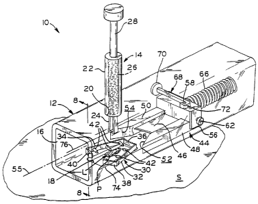
potential
excision of the lesion. The usual 30-minute procedure could be reduced to 2 or
3 minutes
using the present invention. Moreover, the excised lesion may be easily
retrieved from
the inventive device and submitted for pathologic examination.
[0011] The present invention also provides a device for excising tissue and
closing
a wound that results from excision of the tissue. The device comprises
structure defining
an aperture into which tissue to be excised is exposed, a cutting member for
excising the
tissue, a closure member for closing the wound, and an actuatable drive member
associated with the structure. The drive member is actuatable to move the
cutting member
relative to the aperture for excising the tissue that is exposed in the
aperture and for closing
the wound with the closure member. The drive member includes first and second
portions.
The first portion of the drive member supports the cutting member and the
second portion
of the drive member moves the closure member from an open condition to a
closed
condition in which the closure member closes the wound. The first portion of
the drive
member includes collapsible legs that engage a side surface of a recess in the
structure so
that, when the drive member is actuated, the second portion of the drive
member begins to
move the closure member toward the closed condition prior to the collapsible
legs
collapsing and prior to the first portion of the drive member moving the
cutting member to
excise the tissue that is pulled through the aperture.
[0012] The present invention also provides a method for excising tissue and
closing a wound that results from excision of the tissue. The method comprises
the steps
of. exposing the tissue to be excised into an aperture formed in a structure;
moving a
portion of a drive member over a first distance to move a closure member into
engagement
with the tissue that is exposed in the aperture; moving the drive member over
a second
distance beyond the first distance so as to move a cutting member relative to
the aperture
for excising the tissue that is exposed in the aperture; and moving the drive
member over a
third distance beyond the second distance for closing the wound with the
closure member.
CA 02566066 2006-11-08
WO 2005/112790 PCT/US2005/016699
-4-
[0013] The present invention still further provides a method for excising
tissue and closing a wound that results from excision of the tissue. The
method
comprises the steps of exposing the tissue to be excised into an aperture
formed in
a structure; moving a portion of a drive member over a first distance to move
a
closure member into a closed condition for clamping the tissue that is exposed
in
the aperture; moving the drive member over a second distance beyond the first
distance so as to move a cutting member relative to the aperture for excising
the
tissue that is exposed in the aperture.
[0014] The present invention also provides an exciser for excising tissue.
The exciser comprises structure defining an aperture into which tissue to be
excised is exposed, a cutting member for excising the tissue, and an
actuatable
drive member associated with the structure. The drive member is actuatable to
move the cutting member relative to the aperture for excising the tissue that
is
exposed in the aperture. The exciser also comprises an actuator handle that is
associated with the drive member. Movement of the actuator handle relative to
the
structure actuates the drive member.
[0015] The present invention also provides a method of excising tissue.
The method comprises the steps of: exposing tissue into an aperture defined in
a
structure; supporting a cutting member on an actuatable drive member that is
movable relative to the structure; associating an actuator handle to the drive
member; and moving the actuator handle to actuate the drive member to move.the
cutting member relative to the aperture for excising the tissue that is
exposed in the
aperture.
[0016] The present invention also provides a closure member for clamping
tissue adjacent a wound. The closure member comprises a planar body portion
including first and second retaining portions and a deformable portion that
connects the first and second retaining portions. The closure member has an
open
condition in which the deformable portion spaces the first and second
retaining
portions apart from one another so that the tissue to be clamped may be placed
between the first and second retaining portions. The closure member also has a
closed condition in which the deformable portion is elastically deformed to
bring
the first and second retaining portions together to clamp the tissue between
the first
CA 02566066 2006-11-08
WO 2005/112790 PCT/US2005/016699
-5-
and second retaining portions. The body portion of the closure member is
planar in
both the open and closed conditions. The closure member further includes a
first
set of tines that extends outwardly of the first retaining portion and a
second set of
tines that extends outwardly of the second retaining portion. The first and
second
sets of tines engage the tissue to secure the body portion to the tissue when
the
closure member is in the closed condition.
BRIEF DESCRIPTION OF THE DRAWINGS
[0017] The above-mentioned and other features and advantages of this
invention, and the manner of attaining them, will become more apparent and the
invention itself will be better understood by reference to the following
description
of embodiments of the invention taken in conjunction with the accompanying
drawings, wherein:
[0018] Figure 1 is an oblique view of a first embodiment of the inventive
device positioned against the skin of the patient and in a first state, prior
to lesion
excision, with the forceps retracted;
[0019] Figure 2 shows the device of Figure 1 in a second, sequential state,
prior to lesion excision, with the forceps extended and capturing the lesion
to be
excised;
[0020] Figure 3 shows the device of Figure 1 in a third, sequential state,
prior to lesion excision, with the forceps shown in a lesion-pulling position
and the
safety pin removed;
[0021] Figure 4 shows the device of Figure 1 in a fourth, sequential state,
during lesion excision, with the staple partially closed through the skin
surrounding
the lesion;
[0022] Figure 5 shows the device of Figure 1 in a fifth, , sequential state,
during lesion excision, with the staple more fully closed;
[0023] Figure 6 shows the device of Figure 1 in a sixth, sequential state,
after lesion excision, with the staple fully closed, the forceps being
withdrawn
from the device and removing the lesion from the skin;
CA 02566066 2006-11-08
WO 2005/112790 PCT/US2005/016699
-6-
[0024] Figure 7 shows the device of Figure 1 in a seventh, sequential state,
after lesion excision, the device housing being removed from the skin, the
forceps
holding the excised lesion fully removed from the device housing;
[0025] Figure 8 is an enlarged fragmentary sectional view of the exciser of
Figure 1 along line 8-8, showing the staple retention feature of the housing
and the
position of a lesion to be excised from the skin;
[0026] Figure 9 is an oblique view of a second embodiment of the
inventive device located on the skin of the patient, assembled and in a first,
open
position;
[0027] . Figure 10 is a view of the component parts of the device of Figure 9
in adisassembled state;
[0028] Figure 11 is an oblique view of the first applicator block of the
device of Figure 9, with the male staple half inserted therein;
[0029] Figure 12 is an oblique view of the second. applicator block of the
device of Figure 9, with the female staple half inserted therein;
[0030] Figure 13 is an oblique view of the blade assembly of the device of
Figure 9;
[0031] Figure 14 is an oblique view of the blade assembly of Figure 13
fitted to the second applicator block of Figure 12;
[0032] Figure 15 is a view of the male and female staple halves of Figures
11 and 12, respectively, shown interfitted;
[0033] Figure 16 shows the device of Figure 9 in a first state, prior to
lesion
excision;
[0034] Figure 17 shows the device of Figure 9 in a second, sequential state,
prior to lesion excision and during interfitting of the staple halves;
[0035] Figure 18 shows the device of Figure 9 in a third, sequential state,
prior to lesion excision but after closure of the staple;
[0036] Figure 19 shows the device of Figure 9 in a forth, sequential state,
during lesion excision;
[0037] Figure 20 shows the device of Figure 9 in a fifth, sequential state,
upon lesion excision;
CA 02566066 2006-11-08
WO 2005/112790 PCT/US2005/016699
-7-
[0038] Figure 21 shows the device of Figure 9 in a sixth, sequential state,
after upon completion of the excision and during partial release of the closed
staple
from the device;
[0039] Figure 22 is an oblique view of a third embodiment of the inventive
device;
[0040] Figure 23 is a sectional view of the device of Figure 22 in a fully
opened state;
[0041] Figure 24 is a sectional view of the device of Figure 22 in a first
state, prior to lesion excision, the integral tweezers or forceps of the
device closed
on the lesion to be excised;
[0042] Figure 25 shows the device of Figure 22 in a second, sequential
state, prior to lesion excision and during closure of the staple halves
through the
skin surrounding the lesion;
[0043] Figure 26 shows the device of Figure 22 in a third, sequential state,
subsequent to closure of the staple and during lesion excision;
[0044] Figure 27 shows the device of Figure 22 in a fourth, sequential
state, subsequent to lesion excision and during removal of the excised lesion
from
the skin;
[0045] Figure 28 is a side view of a fourth embodiment of the inventive
device in a fully opened state, with separate, known tweezers or forceps also
shown;
[0046] Figure 29 shows the separate tweezers pulling the lesion away from
the skin and the device of Figure 28 in a second, sequential state, prior to
lesion
excision and during closure of the staple halves through the skin surrounding
the
lesion;
[0047] Figure 30 shows the device of Figure 28 in a third, sequential state,
after closure of the staple and during lesion excision;
[0048] Figure 31 shows the device of Figure 28 in a fourth, sequential
state, subsequent to lesion excision and during removal of the excised lesion
from
the skin with the tweezers;
CA 02566066 2006-11-08
WO 2005/112790 PCT/US2005/016699
-8-
[0049] Figure 32 is a disassembled view of a first embodiment of a
two-piece staple for use with the inventive device of Figure 22 or 28, the
staple
pieces shown attached thereto;
[0050] Figure 33 is a disassembled view of a second embodiment of a two-
piece staple for use with the inventive device of Figure 22 or 28, the staple
pieces
shown attached thereto.
[0051] Figures 34a-34d schematically illustrate a sectional view a device
constructed in accordance with a fifth embodiment of the present invention;
[0052] Figure 35 is an oblique view of a first exemplary device constructed
in accordance with the fifth embodiment of the present invention;
[0053] Figure 36 is an oblique, exploded view of the device of Figure 35;
[0054] Figure 37 is a sectional view of the device of Figure 35;
[0055] Figure 38 is a plan view of a drive member of the device of
Figure 35 in a first position relative to a base plate portion of the device;
[0056] Figure 39 is a plan view of the drive member in a second position
relative to the base plate portion;
[0057] Figure 40 is a plan view of the drive member in a third position
relative to the base plate portion;
[0058] Figure 41 is a plan view of the drive member in a fourth position
relative to the base plate portion;
[0059] Figure 42 is an oblique view of a second exemplary device
constructed in accordance with the fifth embodiment of the present invention;
[0060] Figure 43 is an oblique, exploded view of the device of Figure 42;
[0061] Figure 44 is a sectional view of the device of Figure 42;
[0062] Figure 45 is a plan view of a drive member of the device of
Figure 42 in a first position relative to a base plate portion of the device;
[0063] Figure 46 is a plan view of the drive member in a second position
relative to the base plate portion;
[0064] Figure 47 is a plan view of the drive member in a third position
relative to the base plate portion;
[0065] Figure 48 is a plan view of the drive member in a fourth position
relative to the base plate portion;
CA 02566066 2006-11-08
WO 2005/112790 PCT/US2005/016699
-9-
[0066] Figure 49 is a first exemplary embodiment of a closure member for
use with the devices of Figures 35 and 42;
[0067] Figure 50 is a second exemplary embodiment of a closure member;
[0068] Figure 51 is a third exemplary embodiment of a closure member;
[0069] Figure 52 is a.fourth exemplary embodiment of a closure member;
[0070] Figure 53 is a fifth exemplary embodiment of a closure member;
[00711 Figure 54 is a sixth exemplary embodiment of a closure member;
[0072] Figure 55 is a seventh exemplary embodiment of a closure member;
[0073] Figure 56 is an eighth exemplary embodiment of a closure member;
[0074] Figure 57 is a ninth exemplary embodiment of a closure member;
[0075] Figure 58 is a tenth exemplary embodiment of a closure member;
[0076] Figure 59 is an eleventh exemplary embodiment of a closure
member;
[0077] Figure 60 is a twelfth exemplary embodiment of a closure member;
[0078] Figure 61 is a thirteenth exemplary embodiment of a closure
member;
[0079] Figure 62 is a fourteenth exemplary embodiment of a closure
member; and
[0080] Figure 63 is a fifteenth exemplary embodiment of a closure
member.
[0081] Corresponding reference characters indicate corresponding parts
throughout the several views. The exemplifications set out herein illustrate
various
embodiments of the invention and such exemplifications are not to be construed
as
limiting the scope of the invention in any manner.
DETAILED DESCRIPTION OF THE INVENTION
[0082] Figure 1 shows exciser 10, a first embodiment of the present
invention which includes base assembly 12 and separable forceps assembly 14.
It
is envisioned that exciser 10 may be a single use device, all or part of which
may
be discarded after a lesion has been excised therewith.
[0083] Base assembly 12 includes transparent, elongate plastic housing or
frame 16 which, as shown, has the shape of a parallelepiped. It is envisioned,
CA 02566066 2006-11-08
WO 2005/112790 PCT/US2005/016699
-10-
however, that housing 16 may be of any suitable shape. The lower side of
housing 16, that side which, in use, lies against skin S of the patient, is
provided
with rectangular first aperture 18 which frames lesion L to be excised. At a
location directly opposite first aperture 18, the upper side of housing 16 is
provided
with circular second aperture 20 into which the end of cylindrical body 22 of
forceps assembly 14 is inserted.
[0084] Forceps assembly 14 further includes forceps or tweezers 24 having
a pair of elongate, separable, somewhat flexible arms which are retractable
into
and extendable from the interior of cylindrical forceps body 22, and spring 26
which acts to urge tweezers 24 into the interior body 22. Forceps assembly 14
is
also provided with plunger 28 which, when depressed with the thumb, urges
tweezers 24 out of body 22 against the action of spring 26, the extended
tweezers
urged into an open position in which its arms are spread. Release of plunger
28
allows spring 26 to force tweezers 24 upwardly and into body 22, closing the
tweezers. Those of ordinary skill in the art will recognize that forceps
assembly 14
may include a mechanism similar to slender, elongate tools commonly used by
mechanics for grasping small parts such as screws and nuts, for example, which
have been dropped into hard to reach places. Such grasping tools typically
employ
spring-biased tweezers which are opened by depression of a plunger, as
described
above. Alternatively, forceps assembly 14 may include a mechanism (not shown)
by which tweezers 24 are similarly extended from a body and opened, or
retracted
into the body and closed, by turning a screw threaded into the body, the tip
of the
screw attached to the tweezers inside the body. As a further, unshown
alternative,
second aperture 20 may be enlarged, or housing 16 otherwise adequately
fashioned
to allow the lesion to be manually captured with an ordinary pair of tweezers
or
forceps.
[0085] Disposed inside housing 16, adjacent to first aperture 18, is a skin-
closure device which may be made of a surgical stainless steel or a suitable
plastic
material: Unitary staple 30, in its opened condition, is somewhat V-shaped,
having
a pair of distant, splayed straight legs, 32 and 34, each having an end
integrally
connected to central portion 36 which extends between one end of the legs. The
free end of legs 32 and 34 are respectively provided with barbs 38 and 40
which,
CA 02566066 2006-11-08
WO 2005/112790 PCT/US2005/016699
-11-
when the legs are proximate and the staple is closed, interlock and hold the
staple
in its closed condition. Staple 30 may be lightly adhered to the inside
surface of
housing 16 to help maintain its position prior to being closed.
[0086] The interfacing, or inward sides of legs 32 and 34 are provided with
a plurality of pointed pins 42 which extend therefrom and which, when the
staple is
closed, are alternating relative to the legs from which they extend. When the
staple
is closed, and pins 42 extend through the skin below the excision site, the
pointed
free end of each pin 42 abuts or is at least proximal the inward side of the
opposite
leg. It is to be understood that staple 30, and/or any of the other skin-
closure
devices or staples described further herein below, are exemplary embodiments
which may be adapted for use with the inventive excisers. It is envisioned
that
other types of skin-closure devices which serve to close or maintain closed
the skin
at the lesion excision site may also be in accordance with the present
invention,
and such devices or the use thereof fall within the scope of the present
invention.
[0087] Housing 16 is provided with inverted U-shaped clip 43 (Figure 8)
which is integrally molded or otherwise attached thereto at the edge of
rectangular
first aperture 18 nearest blade assembly 44. Clip 43 surrounds three sides of
staple
central portion 36 to prevent its movement longitudinally of housing 16 when
engaged by the blade assembly, as disclosed further below. Notably, the
opening
of clip 43 is located over first aperture 18 such that, upon removal of base
assembly 12 from the skin of the patient after excision of the lesion, closed
staple 30 may exit the housing with clearance between its central portion 36
and
the adjacent edge of first aperture 18. Note that excisers and skin-closure
devices
of different sizes may be provided to accommodate the excision various sized
lesions and closure of skin at the excision site.
[0088] Also disposed within housing 16 is blade assembly 44 which
includes surgical steel blade 46 fixed between wedges or hammers 48 and 50.
Hammers 48 and 50 are staple-engaging portions of blade assembly 44 and are
provided with surfaces 52 and 54 which are curved or flat and are oblique to
the
longitudinal axis 55 of housing 16. As will be described further hereinbelow,
during actuation of exciser 10, hammers 48 and 50 and blade 46 move
coincidentally such that surfaces 52 and 54 slidably engage legs 32 and 34 of
CA 02566066 2006-11-08
WO 2005/112790 PCT/US2005/016699
-12-
staple and move legs 32 and 34 together, thereby closing the staple and the
skin
simultaneously with the excision of the lesion from the skin by blade 46.
Notably,
the sharp edge of blade 46 is located adjacent to surfaces 52 and 54, and
slicing of
the lesion from the skin occurs as opposite portions of legs 32 and 34 along
axis 55
are squeezed together by surfaces 52 and 54 to their closed distance from each
other. Notably, too, above-described clip 43 is located well beneath blade 46
so
that the clip will not interfere with the blade's movement.
[0089] Blade assembly 44 further includes block 56 to which hammers 48
and 50 and blade 46 are attached. Block 56 is provided with post 58 which
extends
vertically and hole 60 (Figures 3-7) which extends laterally. Base assembly 12
is
also provided with removable elongate safety pin 62 which, prior to actuation
of
exciser 10, extends into hole 60 and through hole 64 in housing 16.
[0090] Compression spring 66 is provided inside housing 16, and has one
end fixed relative to the housing; the other end abuts block 56. Spring 66
thus
urges blade assembly 44 from its cocked position along axis 55 toward staple
30.
With safety pin 62 installed, blade assembly 44 is retained in its cocked
position
against the force of compression spring 66 and may not be inadvertently
actuated
or triggered. With safety pin 62 installed, blade assembly 44 thus may not be
slidably moved within housing 16 along axis 55. Base assembly 12 also provided
with plunger 68 which extends through the lateral wall of housing 16 and has
head 70, the depression of which triggers blade assembly 44 once safety pin 62
has
been removed.
[0091] Plunger 68 is provided with J-shaped latching end or hook 72
which, in the blade assembly cocked position, partially surrounds post 58, the
free
end of hook 72 extending laterally in a direction perpendicular to axis 55 and
abutting the post. Depression of plunger head 70 moves plunger 68 laterally
such
that post 58 is no longer captured within hook 72 and, with safety pin 62
removed,
spring 66 will then immediately force blade assembly 44 to move along axis 55
toward the lesion and staple 30.
[0092] The operation of exciser 10 will now be described with sequential
reference to Figures 1-7. The body 16 of base assembly 12 is placed against
skin S
of the patient such that lesion L to be excised is framed by aperture 18, and
safety
CA 02566066 2006-11-08
WO 2005/112790 PCT/US2005/016699
-13-
pin 62 is removed. Referring to Figure 2, plunger 28 of forceps assembly 14 is
depressed against spring 26 and tweezers 24 are extended into the interior of
housing 16 and expand. The free ends of tweezers 24, which may be serrated for
enhanced gripping ability, acquire or grab the lesion and, with reference to
Figure 3, plunger 28 is released. Under the influence of spring 26, tweezers
24 are
at least partially retracted into cylindrical body 22 and pull the lesion
upwardly
through aperture 18. Parallel lines 74 and 76 are etched or printed onto the
transparent lateral sides of body 16, and blade 46 lies and moves in a plane
containing lines 74 and 76; these lines thus establish the location on the
skin at
which the lesion will be excised by the blade.
[0093] Because body 16 is transparent, the doctor or nurse practitioner can
establish the desired elevated position of the lesion by first sighting lines
74 and 76
laterally through the body such that they are viewed as being superposed, and
adjusting the lesion with forceps assembly 14, if and as necessary, such that
perimeter P of lesion L, which may be irregularly shaped, is pulled to a
position
above the superposed lines, as best shown in Figure 8. So positioned, the
lesion
will, after actuation of the blade assembly, be placed in proximity to blade
46
which cuts the skin located outside lesion perimeter P. In adjusting forceps
assembly 14, its body 22 may be moved relative to base assembly housing 16, or
its plunger 28 may be pulled further upward, drawing tweezers 24 further into
body 22. Alternatively, as mentioned above, the lesion may be captured
manually
using an ordinary pair of tweezers or forceps and appropriately positioned
prior to
triggering blade assembly 44. As a further alternative, the lesion may be
captured
with a skin hook (not shown) and appropriately positioned prior to triggering
the
blade.
[0094] Once the lesion is in its desired position within housing 16, blade
assembly 44 is triggered by depression of plunger head 70. In immediate
response
to the free end of plunger hook 72 sliding clear of block post 58, blade
assembly 44
quickly moves along axis 55. Blade 46 passes below the free ends of tweezers
24
and through the skin outside of lesion perimeter P, slicing the lesion from
the skin
while staple 30 simultaneously closes the skin at a location below the
excision site.
During closure of staple 30, as surfaces 52 and 54 of hammers 48 and 50
slidably
CA 02566066 2006-11-08
WO 2005/112790 PCT/US2005/016699
-14-
engage and close legs 32 and 34, pins 42 pierce and protrude through the skin
of
the patent, and hold the staple in place and prevent it from being pulled from
the
re-approximated skin after closure. During the simultaneous excision and
closure,
the shorn edges of the skin on opposite sides of the excision are captured
between
staple legs 32, 34, and are upwardly diverted, resulting in a desirable,
elliptically-
shaped closure. Further, the dermis of these shorn skin edges, rather than
merely
the epidermis, is brought into abutting contact, thereby allowing the stronger
parts
of the skin to mend together and speeding the excision site healing time.
[0095] Referring to Figures 5 and 6, the flat interfacing and parallel
surfaces of hammers 48 and 50 are spaced such that central portion 36 of
staple 30
fits closely therebetween and when barbed ends 38 and 40 of the staple become
interlocked, the staple will assume a rectangular shape which is smaller than
the
periphery of rectangular first aperture 18. After blade assembly 44 has
traveled its
entire distance along axis 55, the lesion will be fully excised from the skin
and
staple 30 is completely closed. Base assembly 12 may then be removed from the
patient's skin, closed staple 30 passing through first aperture 18. Forceps
assembly 14, still gripping the excised lesion, may then be withdrawn from
hole 20
of housing 16. In Figure 7, forceps assembly 14 is shown having been
completely
and separably withdrawn from base assembly 12 with the excised lesion captured
between the ends of tweezers 24. The excised lesion may then be discarded or
sent
to a laboratory for biopsy or other analysis as appropriate.
[0096] It is envisioned that after approximately four days the excision
wound will have sufficiently healed that staple 30 may be removed. Staple 30
may
be removed by cutting it, perhaps at its central portion 36, and peeling its
legs 32, 34 away from the skin and withdrawing pins 42 therefrom.
[0097] Referring now to Figures 9-21, there is shown exciser 100, a second
embodiment of the present invention which was prototyped and successfully used
in animal experiments.
[0098] Exciser 100 comprises first applicator block 102 and second
applicator block 104. Disposed between the applicator blocks is blade
assembly 106. Guide rods 108 are fixed within bores 110 provided in first
applicator block 102 and slidably extend through bores 112 in second
applicator
CA 02566066 2006-11-08
WO 2005/112790 PCT/US2005/016699
-15-
block 104. First and second applicator blocks 102 and 104 maybe made of a
polymeric material such as nylon, for example.
[0099] Blade assembly 106 comprises block portion 114 and blade 116.
Block portion 114 is made of a material similar to that of applicator
blocks 102, 104, and blade 116 is surgical steel or suitable plastic material,
like
blade 46 of first embodiment exciser 10. Blade 116 is attached to block
portion 114 through means of fastener 120 or by any other suitable means.
Guide
rods 108 slidably extend through bores 118 provided in blade assembly block
portion 114.
[00100] The basic components of exciser 100 and its associated skin-closure
device are shown in Figure 10. Two-part staple 122 comprises interfitting male
half 124 and female half 126. Male staple half 124 comprises a pair of
parallel rod
portions 128, and female staple half 126 comprises a pair of similarly spaced
parallel tube portions 130. Rod portions 128 each include extending portion
132
and pointed engaging portion 134. Tube portions 130 each.include extending
portion 136 and engaging portion 138. As further described hereinbelow, each
solid engaging portion 134 of the male staple half slidably and interferingly
engages its mating hollow engaging portion 138 of female staple half 124
during
closure of the staple. When staple halves 124 and 126 are separated or less
than
fully seated, staple 122 is in its open condition, and when staple halves 124
and 126 are fully engaged, staple 122 is in its closed condition. The
interference fit
between engaging portions 134 and 138 ensure that staple 122 remains in its
closed
condition after excision of the lesion.
[00101] Extending between and fixed to rod portions 128 of male staple
half 124 is elongate leg 140, and extending between and fixed to tube portions
130
of female staple half 126 is elongate leg 142. When fitted into exciser 100,
or
when staple 122 is closed, legs 140 and 142 are parallel and extend in
directions
perpendicular to the longitudinal directions of rod and tube portions 128,
130.
Each of legs 140 and 142 is provided with a plurality of sharpened pins 144,
which
correspond to pins 42 of first embodiment exciser 10 shown in Figures 1
through 8.
Pins 144 extend in the longitudinal directions of engaging portions 134 and
136
and, when the staple 122 is closed, the pins of the male and female staple
halves
CA 02566066 2006-11-08
WO 2005/112790 PCT/US2005/016699
-16-
are misaligned such that they alternate along the legs, and the pointed tips
of the
pins of one staple half are in close proximity to the leg of the opposite
staple half.
Notably, when staple 122 is closed as shown in Figure 15, engaging portions
134
of male staple half 124 extend beyond the engaging portion 138 of female
staple
half 126 and into the female staple half's tubular extending portions 136. The
distance between parallel legs 140 and 142 when staple 122 is closed may be
limited by the length of female staple half engaging portion 138 relative to
its
leg 142, i.e., the ends of engaging portions 136 abut leg 140, thereby
minimizing
the distance between the staple legs.
[00102] Referring again to Figure 9, it can be seen that prior to excision of
lesion L from skin S, extending portions 132, 136 of respective male and
female
staple halves 124, 126 are received into holes 146, 148 in first and second
applicator blocks 102, 104, respectively. That is, holes 146 receive extending
portions 132 of male staple half 124, and the male staple half is slid into
first
applicator block 102 until the interfacing surfaces of the first applicator
block and
leg 140 abut. Similarly, extending portions 136 of female staple half 126 are
slidably received in holes 148 provided in second applicator block 104, with
the
interfacing surfaces of the second applicator block and leg 142 abutting.
[00103] Figures 9 and 16 show exciser 100 loaded with a staple 122 and in
its open condition, in which legs 140 and 142 are distant. So configured,
exciser 100 is placed onto skin S of the patient. Perimeter P of lesion L to
be
excised is framed between legs 140 and 142 of the staple and also between the
parallel engaging portions 134 of the male staple half 124. Is it again noted
that
excisers and staples of different sizes may be provided to accommodate the
excision various sized lesions and closure of the excision site. During
operation of
exciser 100, first applicator block 102 is held stationary relative to the
patient's
skin and second applicator block 104 and blade assembly 106 are moved relative
to
first applicator block 102 along guide rods 108.
[00104] Lesion L to be excised with exciser 100 may be pulled away from
skin S through a means of ordinary tweezers or forceps (not shown).
Alternatively,
the lesion may be captured and pulled away from the skin with a skin hook (not
shown). Lesion L is pulled through exciser 100, between the staple legs and
the
CA 02566066 2006-11-08
WO 2005/112790 PCT/US2005/016699
-17-
engaging portions of the male staple half, to an extent which places its
perimeter P
on the side of the plane defined by blade 116 opposite that on which staple
122 is
located. This ensures that the entire lesion, and not just a portion thereof,
will be
excised by blade 116 and the staple will close the skin beneath the excision
site by
pinching together, between proximate legs 140, 142, only skin located outside
of
perimeter P. As described above, the sharpened pins of the staple pierce the
skin
and hold the staple in place on the patient during healing. The excision site
is
closed by staple 122 into an elliptical shape, and the dermis of the skin,
rather than
merely the epidermis is brought into and held in abutting contact by the
closed
staple to promote faster healing.
[00105] Referring to Figures 16-20, the sequence of movements of
exciser 100 and its staple halves are shown sequentially. Prior to the cutting
of the
skin by blade 116, it can be seen (Figures 16-18), that planar blade 116
overlies flat
surface 150 of second applicator block 104 and thus cannot begin cutting
engagement with the patient's skin until blade assembly 106 is moved relative
to
second applicator block 104 along guide rods 108.
[00106] Figure 17 shows the second applicator block 104 and blade
assembly 106 having been moved together along guide rods 108 toward first
applicator block 102 such that engaging portions 134 and 138 of male and
female
staple halves 124 and 126 have entered into partial engagement. Thus, it can
be
seen that closure of staple 122 has begun prior to any cutting by blade 116.
[00107] Figure 18 shows that further movement of second applicator
block 104 and blade assembly 106 together along guide rods 108 toward first
applicator block 102 has completely closed staple 122, applicator blocks 102
and 104 being in their closest proximity to each other. Notably, unlike first
embodiment exciser 10, in which excision of the lesion and closure of the
excision
site are done substantially simultaneously, exciser 100 completely closes
staple 122 prior to any cutting by blade 116. Lesion L, which had previously
been
pulled outwardly away from the rest of the patient's skin by ordinary tweezers
or
forceps, is held in place such that its perimeter P is above the plane defined
by flat
blade 116 by the staple. Pins 144, which pierce the skin, support the lesion
above
CA 02566066 2006-11-08
WO 2005/112790 PCT/US2005/016699
-18-
the plane defined by flat blade 116; but the lesion may still be grasped by
the
tweezers or forceps for easy handling after excision.
[00108] Referring to Figure 19, it can be seen that movement of blade
assembly 106 relative to second applicator block 104 along guide rods 108 and
toward first applicator block 102 forces blade 116 over the closed staple and
through the patient's skin, preferably outside of the perimeter of the lesion.
Here it
can be seen that as blade 116 is moved, it is received in recess 152 formed in
first
applicator block 102.
[00109] Referring to Figure 20, exciser 100 is shown in a position in which
the lesion has been completely severed and perhaps removed from the excision
site
by the tweezers or forceps. In this position, the interfacing surfaces of
first
applicator block 102 and blade assembly block portion 114 abut, and further
movement of blade assembly 106 along guide rods 108 away from second
applicator block 104 is prevented.
[00110] Finally, with reference to Figure 21, blade assembly 106 is
reversely slid along guide rods 108 back to its initial position relative to
second
applicator block 104, and second applicator block 104 and blade assembly 106
are
held together. First applicator block 102 is moved away from second applicator
block 104 and blade assembly 106, withdrawing guide rods 108 therefrom.
Extending portions 132 of staple 122 are withdrawn from holes 146 in first
applicator block 102. The position of staple 122 of course remains stationary
relative to skin S. Extending portions 136 of staple 122 are then withdrawn
from
holes 148 in second applicator block 104 and the exciser completely removed
from
the patient. The extending portions of staple 122 may then be trimmed to
reduce
the size of the staple. As noted above, it is anticipated that staple 122
would
remain in place for approximately four days while the excision site heals,
after
which the staple halves may be separated by pulling them apart, overcoming the
interference fit between the engaging portions 134 and 136. Alternatively, the
staple may be cut in any convenient manner such that it may be removed in
pieces
from the patient.
[00111] Referring now to Figures 22-27 there is shown exciser 200, a third
embodiment of the present invention which is formed of elongate first and
second
CA 02566066 2006-11-08
WO 2005/112790 PCT/US2005/016699
-19-
halves 202 and 204, each respectively having a handle portion 206, 208 and a
jaw
portion 210, 212. First and second halves 202 and 204 are pivotally joined
together through rivets 214 to form a basic structure similar to an ordinary
pair of
pliers or clippers. Formed in first and second halves 202 and 204 is central
recess 216, in which is disposed barrel 218. Barrel 218 has the general form
of a
parallelepiped having closed sides and open ends. Opposite sides of barrel 218
are
provided with holes through which rivets 214 extend, thereby securing barrel
218
to the rest of exciser 200. Extending through the open ends of barrel 218 are
integral tweezers or forceps 220 comprising first and second flexible arms 222
and 224. Arms 222 and 224 are fixed together at attached end 226 of
tweezers 220. Fixed to attached end 226 are short rods 228 which are separated
from and attached to each other through neck 230. Rods 228 extend in
directions
parallel to the longitudinal axes of rivets 214.
[00112] Neck 230 extends through slot 232 centrally provided in elongate
spring steel strip 234, the opposite ends 236 of which are pivotally attached
to first
and second exciser halves 202 and 204. Spring steel strip is plastically
deformed at
its center, and retains and controls longitudinal movement of integral
tweezers or
forceps 220 through the engagement of rods 228 with the portions of strip 234
on
opposite sides of slot 232.
[00113] First and second arms 222 and 224 of tweezers 220 are provided
with plastically deformed portions 238 which, when tweezers 220 are
longitudinally moved in the direction of arrow 252, causes the opposed free
ends 240 of first and second arms 222 and 224 to move towards each other and
close. As discussed further hereinbelow, the closing action of free ends 240
of
integral tweezers or forceps 220 capture the lesion to be excised, and
longitudinal
movement of tweezers 220 in the direction of arrow 252 pulls the lesion to be
excised away from the skin.
[00114] Jaw portions 210 and 212 are each provided with opposed blades or
cutting edges 242 which, when the jaws are closed, move towards each other
and,
when the jaws are fully closed, abut each other. Thus, skin located outside
perimeter P of lesion L to be excised is pinched between blades 242 and cut
from
the remainder of the skin thereby. Blades 242, jaw portions 210, 212,
CA 02566066 2006-11-08
WO 2005/112790 PCT/US2005/016699
-20-
halves 202, 204 or indeed entire exciser 200 may be made of surgical stainless
steel.
[00115] Near the free ends of jaw portions 210 and 212 are located opposed,
staple-engaging portions having flat surfaces 244 to which are adhered first
and
second separate staple halves 246 and 248 which comprise staple 250, another
embodiment of a skin-closure device in accordance with the present invention.
When staple halves 246 and 248 are separated or at least not fully engaged,
staple 250 has an open condition. First and second staple halves 246 and 248
are,
and thus staple 250 is, closed through manipulation of exciser 220 which
interlocks
the staple halves to each other. With the staple halves in this fully engaged
state,
the staple has a closed condition.
[00116] The operation of exciser 200 is now discussed with reference to
Figures 24-27. In a first state shown in Figure 24, free ends 240 of the
integral
tweezers or forceps capture lesion L to be excised from skin S, and the lesion
is
pinched therebetween as handle portions 206 and 208 are closed towards each
other slightly.
[00117] In a second, sequential state shown in Figure 25, further movement
of handle portions 206 and 208 towards each other causes spring steel strip
234 to
flex and its center to move in the direction of arrow 252, which forces
tweezers 220 in that direction. Movement tweezers 220 upward in the direction
of
arrow 252 brings deformed portions 238 of first and second arm 222 and 224
into
sliding engagement with the opening of barrel 218 and forces free ends 240 of
the
first and second arms 222 and 224 closer together, pinching lesion L as it is
pulled
away from skin S. After tweezer free ends 240, and lesion L therebetween, have
moved to a position within the jaws formed by portions 210 and 212 such that
lesion perimeter P is past blades 242, staple halves 246 and 248 enter
engagement
with the skin outside of perimeter P and with each other in the manner
disclosed
further hereinbelow.
[00118] In a third sequential state shown in Figure 26, handle portions 206
and 208 have been brought further together, and tweezers have moved further in
the direction of arrow 252. In this state, staple 250 is fully closed, and
blades 242
are brought into abutting engagement with each other, severing lesion L from
CA 02566066 2006-11-08
WO 2005/112790 PCT/US2005/016699
-21-
skin S below lesion perimeter P. Although staple 250 may achieve its fully
closed
condition prior to actual engagement of blades 242 with skin S, the closing of
the
staple and the excision of lesion L may alternatively occur substantially
simultaneously.
[00119] In a fourth sequential state shown in Figure 27, exciser 200, with
excised lesion L still captured between tweezer free ends 240, is removed from
the
patient, staple 250 having closed-skin S below the excision site such that the
dermis located on opposite sides of the excision site are in abutting contact
and an
elliptically-shaped closure wound is formed as described above. The adhesive,
which holds staple halves 246 and 248 to their respective flat surfaces 244 of
the
staple-engaging portions at the free ends of the exciser jaws, breaks free
upon
slight release of handle portions 206, 208 which are urged away from each
other by
spring steel strip 232, and exciser 200 can then be freely removed, leaving
staple 250 behind. As handle portions 206, 208 are more fully released,
tweezers 220 move in a direction opposite to arrow 252, allowing free ends 240
to
separate, freeing excised lesion L.
[00120] Referring now to Figures 28-31 there is shown exciser 300, a fourth
embodiment of a device according to the present invention, in a series of
sequential
states of operation. Exciser 300, like exciser 200 has a basic structure
similar to
that of an ordinary pair of pliers or clippers, and a common skin-closure
device
may be used with these exciser embodiments.
[00121] Exciser 300 has a pair of elongate first and second halves 302
and 304, each respectively having handle portion 306, 308 and jaw portion 310,
312, halves 302 and 304 being pivotally joined together by pin 314. Rather
than
being provided with integral tweezers or forceps, as exciser 200 is, exciser
300 is
used with separate, known tweezers or forceps 320 as shown. Tweezers 320 are
used to capture and pull lesion L away from the skin S of the patient prior to
moving handle portions 306 and 308 towards each other to close the skin
closure
device or staple, and excise lesion L. Alternatively, the lesion may be
captured and
pulled with a skin hook (not shown). Except for these differences, the
structure
and operation of exciser 300 are substantially identical to those of exciser
200.
CA 02566066 2006-11-08
WO 2005/112790 PCT/US2005/016699
-22-
[00122] Exemplary tweezers 320 have first and second arms 322 and 324
joined at attached end 326. With the ends of jaw portions 310, 312 placed
against
skin S and lesion L placed loosely therebetween, tweezer free ends 340, which
may
be serrated, grasp lesion L which is then pulled away from skin S of the
patient and
into the jaws of exciser 300. Once the captured lesion has been pulled into
jaw
portions 310 and 312 to an extent that lesion perimeter P is above blades 342,
handle portions 306 and 308 are squeezed further together, and staple halves
246
and 248 which comprise staple 250 are brought into engagement with the skin
outside the outer perimeter of the lesion L and with each other, as shown in
Figure 29.
[00123] In Figure 30, staple 250 is fully closed on skin S and blades 342
sever lesion L from skin S at a location outside lesion perimeter P, as
described
above. As noted above, although staple 250 may achieve its fully closed
condition
prior to actual engagement of blades 342 with skin S, the closing of the
staple and
the excision of lesion L may alternatively occur substantially simultaneously.
The
lesion held by tweezers 320 is then removed from the excision site. In Figure
31,
the jaws of exciser 300 are separated, causing the adhesive, which held staple
halves 246, 248 to flat surfaces 344 of the staple-engaging portions of the
jaws, to
break free. The resulting elliptically-shaped excision wound, in which the
dermis
located on opposite sides of the excision is held in abutting contact by
staple 250,
is substantially identical to that resulting from use of exciser 200.
[00124] Referring now to Figures 32 and 33, there are respectively shown
staples 250a and 250b, first and second embodiments of staple 250 which can be
used with either of above-described excisers 200 and 300. Identical elements
of
staples 250a and 250b are identified with a common reference numeral, whereas
corresponding elements of staples 250a and 250b are identified
alphanumerically
with a common numeric portion an alphabetic character (a or b) which
correlates
with a particular embodiment staple 250a or 250b. Each embodiment of staple
250
comprises staple halves 246 and 248 which, in the figure, are respectively
shown
adhered to flat surfaces 244,344 of jaw portions 210, 310 and 212, 312 of
excisers 200, 300. Those skilled in the art will recognize that this
association
between staple halves and jaw flat surfaces maybe reversed. Staple
CA 02566066 2006-11-08
WO 2005/112790 PCT/US2005/016699
-23-
halves 246, 248 may be made of surgical stainless steel or a suitable plastic
material.
[00125] Each staple half 246 is provided with elongate flat central
portion 360 extending between legs 362 and 364. A suitable releasable
adhesive 366, which is later broken free during removal of the exciser from
the
patient as described above, is provided between the outer planar surface of
flat
central portion 360 and the abutting surface 244, 344 of jaw portion 210,310.
[00126] Similarly, each staple half 248 is provided with elongate flat central
portion 370 extending between legs 372 and 374, staple half 248 being
releasably
adhered to its mating jaw surface 244, 344 by adhesive 366.
[00127] Pointed pins 368 extend from the inner planar sides of flat central
portions 360, 370, and when staple 250 is closed, the terminal ends of pins
368 of
one staple half abut the interfacing inner surface of the other staple half.
Further,
with staple 250 closed, the pins alternate along the staple length on the
basis of
which staple half they extend. from. Moreover, each staple half 246, 248 is
substantially symmetrical about the center of its central portion 360,370,
thereby
allowing the staple halves to each be oriented on flat surfaces 244,344 in
either of
two orientations 180 degrees apart; i.e., the locations of legs 362 and 364 of
staple
half 246, or the locations of legs 372 and 374 of staple half 248 may be
switched
relative to the exciser.
[00128] Referring to Figure 32, the ends of legs 362a and 364a are provided
with barbs 376 which, when staple 250a is closed, are interconnected with
barbs 378 provided at the ends of legs 372a and 374a, the interconnecting
barbs
holding staple 250a in its closed condition. The interconnection of barbs 376
and 378 occurs as they slide past each other, resiliently deflecting at least
one leg
of each interconnecting pair, and become hooked to each other.
[00129] Referring to Figure 33, the legs 362b and 364b are substantially
tubular and telescopically engage legs 372b and 374b, which are interference
fitted
therein during closure of staple 250b to maintain its closed condition. The
engaging surfaces of legs 362b, 364b and 372b, 374b may be smooth, their
sliding
interference fit being substantially as disclosed above with respect to rod
CA 02566066 2006-11-08
WO 2005/112790 PCT/US2005/016699
-24-
portions 128 and tube portions 130 of staple 122 of second embodiment
exciser 100 (see Figure 15).
[00130] Staple halves 246b, 248b which are made of plastic may
alternatively have its legs 372b, 374b provided with ribs 380, as shown in
Figure 33, which are compressed as they are forced into smooth-walled hollow
legs 362b, 364b, the compression of ribs 380 providing a secure interference
fit
between the interconnected legs. As shown in Figure 33, the interior surfaces
of
hollow legs 362b and 364b may be also provided with recesses 382 into which
ribs 380 are received as legs 372b, 374b are forced therein, the interfitting
engagement of ribs 380 and recesses 382 holding staple 250b in its closed
condition.
[00131] Figures 34a-34d schematically illustrate a sectional view a
device 410 constructed in accordance with a fifth embodiment of the present
invention. The device 410 may be used for excising tissue and closing a wound
that results from excision of the tissue. Figures 34a-34d schematically
illustrate
the device 410 excising a lesion 412 from skin 414.
[00132] The device 410 includes a housing 418. An aperture 420 extends
vertically through the housing 418. The device 410 also includes a cutting
member 422 and a clamping member 424. At least one actuator 426 is movable
relative to the housing 418 for moving the cutting member 422 and for closing
the
closure member 424. An optional second actuator that cooperates with the
actuator 426 for closing the closure member 424 is shown by dashed lines at
428 in
Figures 34a-34d.
[00133] To remove the lesion 412 from the skin 414, the housing 418 is
positioned relative to the skin 414 so that the lesion 412 is located directly
below
the aperture 420. As is shown in Figure 34a, the lesion 412 is pulled through
the
aperture 420 of the housing 418 using a skin hook 432 or other suitable device
for
grabbing the lesion 412. When the lesion 412 is pulled through the aperture
420,
the skin 414 adjacent the lesion 412 is tensioned.
[00134] Next, as is shown in Figure 34b, the actuator 426 of the device 410
is moved relative to the housing 418. Movement of the actuator 426 causes the
closure member 424 to begin to close. As the closure member 424 begins to
close,
CA 02566066 2006-11-08
WO 2005/112790 PCT/US2005/016699
-25-
tines (not shown) of the closure member 424 pierce the tensioned skin 414 and
the
closure member begins to pinch the skin adjacent the lesion 412.
[00135] As shown in Figure 34c, further movement of the actuator 426
relative to the housing 418 causes the cutting member 422 to begin excising
the
lesion 412 from the skin 414. When the lesion 412 is completely removed from
the skin 414, as shown in Figure 34d, further movement of the actuator 426
closes
the closure member 424 to close a wound that results from excision of the
lesion 412. Alternatively, the closure member 424 may be closed prior to the
cutting member 422 completely removing the lesion 412 from the skin 414. The
following description with reference to Figures 35-48 will describe two
devices
constructed in accordance with the fifth embodiment of the present invention.
[00136] Figure 35 is an oblique view of a first exemplary device 510
constructed in accordance with the fifth embodiment of the present invention.
Figure 36 is an exploded oblique view of the device 510 and Figure 37 is a
sectional view of the device 510. The device 510 includes a housing 512 having
a
base plate portion 514 and a top plate portion 516.
[00137] The base plate portion 514 of the housing 512 includes lower and
upper surfaces 520 and 522, respectively. As best shown in Figure 37, a
cavity 536 extends upwardly into the lower surface 520 of the base plate
portion 514. The cavity 536 is generally elliptical and terminates at an upper
surface 538 (Figure 37). An elliptical aperture 540 extends through the base
plate
portion 514 and into the cavity 536.
[00138] As best shown in Figure 36, a first recess 544 extends downwardly
into the upper surface 522 of the base plate portion 514. As will be described
in
detail below, the first recess 544 receives a driver 640 of the device 510.
The first
recess 544 includes a generally planar bottom surface 546.
[00139] The first recess 544 includes a narrow section having laterally
opposite side surfaces 558 and 560. Corners 562 and 564, which preferably have
equal radii, form transitions between the laterally opposite side surfaces 558
and 560 and a wider section of the first recess 544. As will be discussed
below, the
location of the corners 562 and 564 determines a timing for cutting the lesion
and
clamping the skin adjacent the lesion.
CA 02566066 2006-11-08
WO 2005/112790 PCT/US2005/016699
-26-
[00140] As shown in Figure 36, another section of the first recess 544 is
located between laterally opposite side surfaces 582 and 584. A protruding
portion 586 that includes the elliptical aperture 540 extends upwardly into
the first
recess 544 in a location between the side surfaces 582 and 584. Two elongated
slots 588 and 590 connect the first recess 544 to the cavity 536 on
longitudinally
opposite ends of the protruding portion 586.
[00141] Two pivot pin holes 592 and 594 and a second recess 600 extend
into the bottom surface 546 of the first recess 544. The pivot pin holes 592
and 594 are located in the slot 590 and a curved end surface 596 of the first
recess 544. The second recess 600 extends longitudinally through a center of
the
first recess 544. Two axially extending ribs 602 extend into the second recess
600
near the curved end surface 596 of the first recess 544. The ribs 602 form
three
elongated slots in the second recess 600.
[00142] The base plate portion 514 also includes four fastener holes 604.
The four fastener holes 604 extend between the lower and upper surfaces 520
and 522 of the base plate portion 514.
[00143] The top plate portion 516 of the housing 512 also a lower
surface 610 (Figure 37) and an upper surfaces 612 (Figure 36). An elliptical
aperture 621 extends through the top plate portion 516 of the housing 512. The
elliptical aperture 621 extends between the lower and upper surfaces 610 and
612
and has a location corresponding the elliptical aperture 540 of the base plate
portion 514. The upper surface 612 of the top plate portion 516 is chamfered
adjacent the elliptical aperture 621.
[00144] Four fastener holes 622 and two pivot pin holes 624 and 626 also
extend through top plate portion 516 between the lower and upper surfaces 610
and 612. Each of the four fastener holes 622 is associated with and has a
location
corresponding to a fastener hole 604 of the base plate portion 514 of the
housing 512. Each of the fastener holes 622 is located in a circular boss 628
that
extends upwardly above the upper surface 612 of the top plate portion 516.
Each
of the two pivot pin holes 624 and 626 is associated with and has a location
corresponding to a pivot pin hole 592 and 594 of the base plate portion 514 of
the
housing 512.
CA 02566066 2006-11-08
WO 2005/112790 PCT/US2005/016699
-27-
[00145] Two longitudinally elongated slots 632 and 634 extend through the
top plate portion 516. The slot 634 has a length and width of approximately
four
times the length and width of the slot 632.
[00146] The device 510 also includes a driver 640. Figure 36 illustrates an
oblique view of the driver 640 and Figures 38-41 illustrate a plan view of the
driver located in the first recess 544 of the base plate portion 514 of the
housing 512. For clarity in viewing the driver 640 in Figures 38-41, the
protruding
portion 586 of the base plate portion 514 that includes the elliptical
aperture 540 is
not shown.
[00147] The driver 640 is injection molded from a plastic material that is
flexible enough to allow for a living hinge to be formed on the driver.
Exemplary
plastic materials include nylon, polycarbonate, polyester, or any other
suitable
polymer. The driver 640 includes a yoke portion 642 and a driving portion 644.
The yoke portion 642 of the driver 640 includes longitudinally spaced first
and
second end portions 650 and 652, respectively, and laterally spaced first and
second side portions 654 and 656, respectively, that collectively surround a
central
opening 658.
[00148] As shown in Figure 38, the first and second side portions 654
and 656 of the yoke portion 642 of the driver 640 connect the first and second
end
portions 650 and 652. The second end portion 652 includes first and second
pivotal drive portions 680 and 682 that are connected to the first and second
side
portions 654 and 656, respectively, with living hinges. As shown in Figure 38,
a
pivot pin hole 692 extends through the first pivotal drive portion 680 and a
pivot
pin hole 708 extends through the second pivotal drive portion 682.
[00149] The yoke portion 642 of the driver 640 also includes proximal and
distal engaging members 714 and 716. The proximal engaging- member 714
extends into the central opening 658 from the first end portion 650. The
proximal
engaging member 714 includes a recessed end portion 734 (Figure 37). The
distal
engaging member 716 is interposed between the first and second pivotal drive
portions 680 and 682. The distal engaging member 716 also includes a recessed
end portion 770 (Figure 37).
CA 02566066 2006-11-08
WO 2005/112790 PCT/US2005/016699
-28-
[00150] The driving portion 644 of the driver 640 is located longitudinally
opposite the first end portion 650 of the yoke portion 642 from the central
opening 658. The driving portion 644 includes a blade support portion 778, and
first and second collapsible legs 782 and 784, respectively.
[00151] The blade support portion 778 includes circular boss 790
(Figure 38) that extends upwardly from the blade support portion 778. A
rectangular protrusion (not shown) extends downwardly from the blade support
portion 778 for being received in the second recess 600 of the base plate
portion 514.
[00152] The first and second collapsible legs 782 and 784 are located on
laterally opposite sides of the blade support portion 778. The first and
second
collapsible legs 782 and 784 extend between the blade support portion 778 and
the
first end portion 650 of the yoke portion 642. The first collapsible leg 782
includes
first and second leg portions 796 and 798, respectively. Living hinges connect
the
first and second leg portions 796 and 798 to one another and to the blade
support
portion 778 and the first end portion 650 of the yoke portion 642. The second
collapsible leg 784 includes first and second leg portions 810 and 812,
respectively. Living hinges connect the first and second leg portions 810 and
812
to one another and to the blade support portion 778 and the first end portion
650 of
the yoke portion 642.
[00153] With reference again to Figure 36, the device 510 also includes a
cutting member or blade 830. The blade 830 includes an aperture 836 that is
sized
for receiving the circular boss 790 that extends upwardly from the blade
support
portion 778 of the driving portion 644 of the driver 640. The blade 830 also
includes a sharpened edge 838. The blade 830 is formed from surgical steel. In
one embodiment of the invention, the blade 830 is formed from 440 stainless
steel.
[00154] The device 510 also includes an actuator handle 844. The actuator
handle 844 is molded from plastic. An aperture 846 extends through the
actuator
handle 844 for receiving the circular boss 790 that extends upwardly from the
blade support portion 778 of the driving portion 644 of the driver 640 and a
fastener 848 that fixes the actuator handle 844 to the boss 790.
CA 02566066 2006-11-08
WO 2005/112790 PCT/US2005/016699
-29-
[00155] To assemble the device 510, the driver 640 is placed in the first
recess 544 of the base plate portion 514 of the housing 512 so that the
central
opening 658 of the yoke portion 642 of the driver receives the protruding
portion 586 of the base plate portion. When the driver 640 is located in the
base
plate portion 514 of the housing 512, the proximal and distal engaging
members 714 and 716 extends through the slots 588 and 590 in the base plate
portion 514 and slightly into the cavity 536 (Figure 37) in the lower surface
520 of
the base plate portion 514.
[00156] The blade 830 is then placed on the driving portion 644 of the
driver 640 so that the circular boss 790 extends through the aperture 836 in
the
blade. The blade 830 is also received in the first recess 544 of the base
plate
portion 514. The side surfaces 558 and 560 of the first portion 550 of the
first
recess 544 prevent the blade 830 from rotating about the circular boss 790 and
relative to the base plate portion 514.
[00157] The top plate portion 516 of the housing 512 is placed over the base
plate portion 514 of the housing. The slot 634 of the top plate portion 516
receives
the circular boss 790 of the blade support portion 778 of the driving portion
644 of
the driver 640. The slot 632 of the top plate portion 516 receives a portion
of the
distal engaging member 716. When the top plate portion 516 is properly
positioned relative to the base plate portion 514, the pivot pin holes 624 and
626,
the fastener holes 622, and the elliptical aperture 621 of the top plate
portion 516
are in locations corresponding to the pivot pin holes 592 and 594, fastener
holes 604, and the elliptical aperture 540 of the base plate portion 514.
Also, the
pivot pin holes 692 and 708 of the first and second drive portions 680 and 682
of
the second end portion 652 of the yoke portion 642 of the driver 640 are
aligned
with the pivot pin holes 592 and 594 of the base plate portion 514 and the
pivot pin
holes 624 and 626 of the top plate portion 516.
[00158] Four fasteners 854 (Figure 35) are then inserted through the fastener
holes 622 of the top plate portion 516 and the fastener holes 604 of the base
plate
portion 514 to hold the top plate portion relative to the base plate portion.
A first
pivot pin 856 is inserted through the pivot pin hole 624 of the top plate
portion 516, the pivot pin hole 692 in the first pivotal drive portion 680 of
second
CA 02566066 2006-11-08
WO 2005/112790 PCT/US2005/016699
-30-
end portion 652 of the yoke portion 642, and the pivot pin hole 592 in the
base
plate portion 514 of the housing 512. A second pivot pin 858 is inserted
through
the pivot pin hole 626 of the top plate portion 516, the pivot pin hole 708 in
the
second pivotal drive portion 682 of second end portion 652 of the yoke
portion 642, and the pivot pin hole 594 in the base plate portion 514 of the
housing 512.
[00159] The aperture 846 in the actuator handle 844 is aligned with the
circular boss 790 of the blade support portion 778 of the driving portion 644
of the
driver 640. The actuator handle 844 is pressed onto the circular boss 790 and
a
fastener 848 is inserted into the aperture 846 to lock the actuator handle to
the
boss. When the actuator handle 844 is placed on the circular boss 790, the
circular
boss 790 is located at a first end of the slot 634.
[00160] After assembly of the device 510 is complete, the device 510 is
turned over so that a clip 870 may be attached to the device. Exemplary clips
are
illustrated in Figures 49-63 and will be discussed later in this application.
The
clip 870 of Figure 37 includes opposite first and second retaining portions
872
and 874, respectively, that when pressed together clamp onto tissue.
[00161] To attach the clip 870 to the device 510, the clip 870 is placed in
the
cavity 536 that extends into the lower surface 520 of the base plate portion
514.
The first retaining portion 872 of the clip 870 is placed on the recessed end
portion 734 of the proximal engaging member 714 and the second retaining
portion 874 of the clip 870 is placed on the recessed end portion 770 of the
distal
engaging member 716.
[00162] To use the device 510 for removing a lesion or other portion of
tissue, the lower surface 520 of the base plate portion 514 of the housing 512
of the
assembled device 510, to which the clip 870 has been attached, is placed
against
the patient's tissue so that the elliptical apertures 540 and 621 overlie the
portion of
tissue to be removed. A skin hook, or another device for grabbing the
patient's
tissue, is inserted through the elliptical apertures 540 and 621 of the
housing 512
and hooks the portion of tissue. The portion of tissue is pulled through the
clip 870
and the elliptical apertures 540 and 621 of the housing 512 so that the tissue
is
placed under tension.
CA 02566066 2006-11-08
WO 2005/112790 PCT/US2005/016699
-31-
[00163] Pushing the actuator handle 844 through the slot 634 toward the
elliptical aperture 621 actuates the device 510. When the actuator handle 844
begins to move toward the elliptical aperture 621, the first and second
collapsible
legs 782 and 784 are pressed against the side surfaces 558 and 560 of the
first
recess 544 in the base plate portion 514, as is shown in Figure 38. As a
result, the
first and second collapsible legs 782 and 784 are prevented from collapsing
and the
movement of the actuator handle 844 causes the yoke portion 642 of the driver
640
begin to close the clip 870 so that tines of the clip penetrate the tissue and
the clip
begins to pinch the tissue. The first and second drive portions 680 and 682 of
the
yoke portion 642 of the driver 640 rotate to move the distal engaging member
716
by an amount equal to the movement of the proximal engaging member 714 to
begin to close the clip 870. Figure 39 illustrates the device 510 at a
position in
which the clip 870 begins to close.
[00164] Adjusting the location of the corners 562 and 564 enables the timing
for cutting the tissue with the blade 830 and clamping the tissue with the
clip 870
to be controlled. The longitudinal lengths of side surfaces 558 and 560,
illustrates
in Figure 38 as distance X, controls the timing of when the first and second
collapsible legs 782 and 784 of the driving portion 644 begin to collapse.
Thus, by
adjusting the distance X, the device 510 timing for cutting the tissue with
the
blade 830 and clamping the tissue with the clip 870 may be adjusted. For
example,
when the distance X is increased, the yoke portion 642 of the driver 640 will
close
the clip 870 prior to the driving portion 644 moving the blade 830 to cut the
tissue.
[001 651 When the first and second collapsible legs 782 and 784 of the
driving portion 644 of the driver 640 move out of contact with the side
surfaces 558 and 560 and over corners 562 and 564, respectively, the first and
second collapsible legs 782 and 784 begin to collapse, as is shown in Figure
39.
When the first and second collapsible legs 782 and 784 begin to collapse, the
blade
support portion 778 begins to move toward the first end portion 650. During
the
movement of the blade support portion 778 toward the first end portion 650,
the
blade 830 cuts through the tissue that extends through the elliptical aperture
621 of
the top plate portion 516 to excise the tissue from the tissue that extends
through
the elliptical aperture 540 of the base plate portion 514.
CA 02566066 2006-11-08
WO 2005/112790 PCT/US2005/016699
-32-
[00166] The tissue that extends into elliptical aperture 540 remains pinched
between the first and second retaining portions 872 and 874 of the clip 870.
After
the blade support portion 778 of the driving portion 644 of the driver 640
contacts
the first end portion 650 of the yoke portion 642 of the driver, as is shown
in
Figure 40, further movement of the actuator handle 844 through the slot 634
toward the elliptical aperture 621 results in movement of the proximal
engaging
member 714 toward the center of the elliptical aperture 540 and equal movement
of the distal engaging member 716 toward the center of the elliptical aperture
540.
The proximal and distal engaging members 714 and 716 continue to move together
as the actuator handle 844 is moved through the slot 634 toward the elliptical
aperture 621. When the proximal and distal engaging members 714 and 716 move
toward each other, the first and second retaining portions 872 and 874 of the
clip 870 are pressed together into the position shown in Figure 41. When the
actuator handle 844 reaches the end of the slot 634, the clip 870 fully
occludes the
wound that results from the excision of the tissue.
[00167] After the clip 870 fully occludes the wound, the actuator handle 844
is moved in the slot 634 in a direction away from the elliptical aperture 621.
Movement of the actuator handle 844 away from the elliptical aperture 621
causes
the proximal and distal engaging member 714 and 716 to move apart from one
another and to release the clip 870. As an alternative to manually moving the
actuator handle 844 to remove the clip 870, the device 510 may be modified to
include a biasing member or spring (not shown) that biases the first end
portion 650 of the yoke portion 642 of the driver 640 relative to the base
plate
portion 514 to remove the clip 870 from the proximal and distal engaging
member 714 and 716. For example, the biasing member may be an integral part of
the driver 640.
[00168] Preferably, all of the parts of the device 510, with the exception of
the blade 830 and the clip 870, are molded from plastic material. In a
preferred
embodiment, the device 510 is a single use device that is discarded after its
use,
while the clip 870 remains attached to the skin to close the wound.
[00169] Figure 42 is an oblique view of a second exemplary device
constructed in accordance with the fifth embodiment of the present invention.
CA 02566066 2006-11-08
WO 2005/112790 PCT/US2005/016699
-33-
Figure 43 is an exploded oblique view of the device 1010 and Figure 44 is a
sectional view of the device 1010.
[00170] The device 1010 includes a housing 1012 having a base plate
portion 1014 and a top plate portion 1016. The base plate portion 1014 of the
housing 1012 includes a lower surface 1020 (Figure 44) and an upper
surfaces 1022 (Figure 43). A cavity 1034 (Figure 44) extends upwardly into the
lower surface 1020 of the base plate portion 1014. The cavity 1034 terminates
at
an upper surface 1036. An elliptical aperture 1038 extends through the base
plate
portion 1014 through the upper surface 1036 of the cavity 1034.
[00171] With reference to Figure 43, a first recess 1044 extends downwardly
into the upper surface 1022 of the base plate portion 1014. The first recess
1044
includes a generally planar bottom surface 1046. A protruding portion 1056
having the elliptical aperture 1038 extends upwardly from the bottom. surface
1046
of the first recess 1044. As shown in Figure 44, elongated slots 1066 and 1074
are
located on opposite sides of the protruding portion 1056 and connect the first
recess 1044 to the cavity 1034.
[00172] The first recess 1044 includes a narrowed section having laterally
opposite side surfaces 1058 and 1060. As shown in Figure 45, corners 1062
and 1064, which preferably have equal radii, form transitions between the
laterally
opposite side surfaces 1058 and 1060 and a wider section of the first recess
1044.
As will be discussed below, the location of the corners 1062 and 1064
determines a
timing for cutting the lesion and clamping the skin adjacent the lesion.
[00173] The first recess 1044 also includes a generally elliptical section
that
includes arcuate side surfaces 1068 and 1070. The arcuate sides surfaces 1068
and 1070 are located on laterally opposite sides of the protruding portion
1056.
[00174] A second recess 1080 extends downwardly into the bottom
surface 1046 of the first recess 1044. The second recess -1080 extends
longitudinally through a center of the first recess 1044.
[00175] The base plate portion 1014 also includes four fastener holes 1082.
The four fastener holes 1082 extend between the lower and upper surfaces 1020
and 1022 of the base plate portion 1014.
CA 02566066 2006-11-08
WO 2005/112790 PCT/US2005/016699
-34-
[00176] The top plate portion 1016 of the housing 1012 also includes a
lower surface 1088 (Figure 44) and an upper surfaces 1090. A recess 1102
extends
into the upper surface 1090 of the top plate portion 1016. The recess 1102 has
a
planar bottom surface 1104. An elliptical aperture 1116 extends through the
top
plate portion 1016 of the housing 1012. The elliptical aperture 1116 extends
between the bottom surface 1104 of the recess 1102 and the lower surface 1020
of
the top plate portion 1016. The elliptical aperture 1116 has a location
corresponding the elliptical aperture 1038 of the base plate portion 1014.
[00177] Four fastener holes 1118 extend through top plate portion 1116
between the lower and upper surfaces 1020 and 1022. Each of the four fastener
holes 1118 is associated with and has a location corresponding to a fastener
hole 1082 of the base plate portion 1014 of the housing 1012.
[00178] Two axially elongated slots 1120 and 1122 extend through the top
plate portion 1016. The slots 1120 and 1122 extend along a centerline of the
top
plate portion 1016 from longitudinally opposite ends of the top plate portion.
[00179] The device 1010 also includes a driver 1130. Figure 45-48 illustrate
plan views of the driver 1130 in the base plate portion 1014 of the housing
1012.
For clarity in viewing the driver 1130 in Figures 45-48, the protruding
portion 1056 of the base plate portion 1014 that includes the elliptical
aperture 1038 is not shown.
[00180] The driver 1130 is injection molded from plastic material and
includes a yoke portion 1132 and a cutting portion 1134. The yoke portion 1132
of
the driver 1130 includes opposite first and second end portions 1140 and 1142,
respectively, and opposite first and second collapsible legs 1144 and 1146,
respectively. A central opening 1.148 extends between the first and second end
portions 1140 and 1142 and the first and second collapsible legs 1144 and
1146,
respectively.
[00181] As shown in Figure 45, the first and second collapsible legs 1144
and 1146 connect the first and second end portions 1140 and 1142. The first
collapsible leg 1144 includes first and second leg portions 1162 and 1164,
respectively. Living hinges connect the first and second leg portions 1162
and 1164 to one another and to the first and second end portions 1140 and
1142.
CA 02566066 2006-11-08
WO 2005/112790 PCT/US2005/016699
-35-
The second collapsible leg 1146 also includes first and second leg portions
1176
and 1178, respectively. Living hinges connect the first and second leg
portions 1176 and 1178 to one another and to the first and second end
portions 1140 and 1142.
[00182] The yoke portion 1132 of the driver 1130 also includes proximal
and distal engaging members 1208 and 1210, respectively. The proximal engaging
member 1208 extends longitudinally outwardly of the first end portion 1140 and
into the central opening 1148. The proximal engaging member includes a
recessed
end portion 1218 (Figure 44). The distal engaging member 1210 also extends
longitudinally into the central opening 1148. The distal engaging member
includes
a recessed end portion 1230 (Figure 44).
[00183] The cutting portion 1134 of the driver 1130 is located longitudinally
opposite the first end portion 1140 of the yoke portion 1132 from the central
opening 1148. As shown in Figure 43, the cutting portion 1134 includes a blade
support portion 1238 and first and second collapsible legs 1240 and 1242,
respectively. A circular aperture 1260 extends through the blade support
portion 1238. A rectangular protrusion extends downwardly from the blade
support portion 1238 for being received in the second recess 1080 of the base
plate
portion 1014.
[00184] The first and second collapsible legs 1240 and 1242 extend
longitudinally between the blade support portion 1238 and the first end
portion 1140 of the clamping portion 1232. The first collapsible leg 1240
includes
first and second leg portions 1266 and 1268, respectively. Living hinges
connect
the first and second leg portions 1266 and 1268 together and to the blade
support
portion 1238 and the first end portion 1140. The second collapsible leg 1242
also
includes first and second leg portions 1278 and 1280, respectively. Living
hinges
connect the first and second leg portions 1278 and 1280 together and to the
blade
support portion 1238 and the first end portion 1140.
[00185] As shown in Figure 43, the device 1010 also includes a cutting
member or blade 1290. The blade 1290 includes an aperture 1296 and a sharpened
edge 1298. The blade 1290 is formed from surgical steel. In one embodiment of
the invention, the blade is formed from 440 stainless steel.
CA 02566066 2006-11-08
WO 2005/112790 PCT/US2005/016699
-36-
[00186] The device 1010 also includes first and second actuator
handles 1304 and 1306, respectively. The actuator handles 1304 and 1306 are
molded from plastic. An aperture 1308 extends vertically through actuator
handle 1304. Likewise, an aperture 1310 extends vertically through actuator
handle 1306.
[00187] The device 1010 also includes six fastener 1314 and two blind
nuts 1316. The fasteners 1314 illustrated in Figure 43 are screws having
associated washers 1318. Alternatively, snap features may be incorporated into
the
various structures of the device 1010: The six fasteners 1314 include four
housing
fasteners and two actuator fasteners. Each of the two blind nuts 1316 includes
a
circular boss 1320 and a planar base 1322. The planar bases 1322 of the blind
nuts 1316 are sized for being received in the second recess 1080 of the base
plate
portion 1014 of the housing 1012.
[00188] To assemble the device 1010, a circular boss 1320 of one of the
blind nut 1316 is inserted into the aperture 1260 of the blade support portion
1238
of the driver 1130 so that the circular boss extends upwardly beyond an upper
surface of the blade support portion. The circular boss 1320 of the other
blind
nut 1316 is inserted into an aperture 1202 of the second end portion 1142 so
that
the circular boss extends upwardly beyond an upper surface of the second end
portion. The driver 1130 is then placed in the first recess 1044 of the base
plate
portion 1014 of the housing 1012 so that the central opening 1148 of the yoke
portion 1132 of the driver receives the protruding portion 1056 of the base
plate
portion that includes the elliptical aperture 1038. When the driver 1130 is
placed
in the first recess 1044, the bases 1322 of the blind nuts 1316 and the
rectangular
protrusions of the blade support portion 1238, the first end portion 1140, and
the
second end portion 1142 are received in the second recess 1080 of the base
plate
portion 1014. The second recess 1080 acts to guide the driver 1130 during
movement relative to the base plate portion 1014.
[00189] When the driver 1130 is located in the base plate portion 1014 of
the housing 1012, the proximal and distal engaging members 1208 and 1210
extends through the slots 1062 and 1074 in the base plate portion 1014 and
slightly
into the cavity 1034 of the base plate portion. The driver 1130 is positioned
within
CA 02566066 2006-11-08
WO 2005/112790 PCT/US2005/016699
-37-
the first recess 1044 so that the first and second collapsible legs 1240 and
1242 of
the cutting portion 1134 of the driver 1130 engage the first and second side
surfaces 1058 and 1060 of the first portion 1048 of the first recess 1044, as
is
shown in Figure 45.
[00190] The blade 1290 is then placed over the blade support portion 1238
of the cutting portion 1134 of the driver 1130 so that the circular boss 1320
of the
blind nut 1316 that extends through the aperture 1260 in the blade support
portion
also extends through. the aperture 1296 of the blade. The blade 1290 is also
received in the first recess 1044 of the base plate portion 1014. The first
and
second side surfaces 1058 and 1060 of the first portion 1048 of the first
recess 1044 prevent the blade 1290 from rotating about the circular boss 1320
and
relative to the base plate portion 1014.
[00191] The top plate portion 1016 of the housing 1012 is placed over the
base plate portion 1014 of the housing. The slot 1120 of the top plate portion
1016
receives the circular boss 1320 of the blind nut 1316 that extends through the
aperture 1296 of the blade 1290. The slot 1122 receives the circular boss 1320
of
the blind nut 1316 that extends through the second end portion 1142 of the
yoke
portion 1132 of the driver 1130. When the top plate portion 1016 is properly
positioned relative to the base plate portion 1014, the fastener holes 1118
and the
elliptical aperture 1116 of the top plate portion 1016 are in locations
corresponding
to the fastener holes 1082 and the elliptical aperture 1038 of the base plate
portion 1014.
[00192] Four fasteners 1314 are then inserted through the fastener
holes 1118 of the top plate portion 1016 and the fastener holes 1082 of the
base
plate portion 1014 to hold the top plate portion relative to the base plate
portion.
The aperture 1308 in the actuator handle 1304 is aligned with and receives the
circular boss 1320 of the blind nut 131.6 that extends through the aperture
1296 of
the blade 1290. A fastener 1314 fixes the actuator handle 1304 to the blind
nut 1316. The aperture 1310 in the actuator handle 1306 is aligned with and
receives the circular boss 1320 of the blind nut 1316 that extends through the
aperture 1202 in the second end portion 1142. A fastener 1314 fixes the
actuator
handle 1306 to the blind nut 1316.
CA 02566066 2006-11-08
WO 2005/112790 PCT/US2005/016699
-38-
[001931 After assembly of the device 1010 is complete, the device 1010 is
turned over so that a clip 1326 may be attached to the device. Exemplary clips
are
illustrated in Figures 49-63 and are discussed later in this application. The
clip 1326 of Figure 43 includes opposite first and second retaining portions
1228
and 1230, respectively, that when pressed together clamp onto tissue.
[00194] To attach the clip 1326 to the device 1010, the clip 1326 is placed in
the cavity 1034 that extends into the lower surface 1020 of the base plate
portion 1014. The first retaining portion 1328 of the clip 1326 is supported
on the
recessed end of the proximal engaging member 1208 and the second retaining
portion 1330 is supported on the recessed end of the distal engaging member
1210.
[00195] To use the device 1010 for removing a lesion or other portion of
tissue, the lower surface 1020 of the base plate portion 1014 of the assembled
device 1010, to which the clip 1326 has been attached, is placed against a
patient's
tissue. A skin hook, or another device for grabbing the patient's tissue, is
inserted
through the elliptical apertures 1038 and 1116 of the housing 1012 and grabs
the
tissue. The skin hook pulls the tissue through the clip 1326 and the
elliptical
apertures 1038 and 1116 of the housing 1012. When pulled through the
elliptical
apertures 1038 and 1116, the tissue is placed under tension.
[00196] The first and second actuator handles 1304 and 1306 are pushed
together to actuate the driver 1130. When the first and second actuator
handles 1304 and 1306 are moved toward one another, the side surfaces 1058
and 1060 of the first portion 1048 of the first recess 1044 prevent the first
and
second collapsible legs 1240 and 1242 of the cutting portion 1134 of the
driver 1130 from collapsing, as illustrated in Figure 45. As a result, the
proximal
and distal engaging members 1208 and 1210 moves toward one another to begin
closing the clip 1326. When the clip 1326 begins to close, tines of the clip
1326
pierce the tensioned tissue and the clip begins to pinch the tissue. Figure 46
illustrates the device 1010 with the clip 1326 partially closed.
[00197] Adjusting the location of the corners 1062 and 1064 enables the
timing for cutting the tissue with the blade 1290 and clamping the tissue with
the
clip 1326 to be controlled. The longitudinal lengths of side surfaces 1058
and 1060, illustrates in Figure 45 as distance Y, controls the timing of when
the
CA 02566066 2006-11-08
WO 2005/112790 PCT/US2005/016699
-39-
first and second collapsible legs 1240 and 1242 of the cutting portion 1134 of
the
driver 1130 begin to collapse. Thus, by adjusting the distance Y, the device
1010
timing for cutting the tissue with the blade 1290 and clamping the tissue with
the
clip 1326 may be adjusted. For example, when the distance Y is increased, the
yoke portion 1132 of the driver 1130 will close the clip 1326 prior to the
cutting
portion 1134 moving the blade 1290 to cut the tissue.
[001981 When the living hinges of the first and second collapsible legs 1240
and 1242 of the cutting portion 1134 of the driver 1130 extend longitudinally
beyond the corners 1062 and 1064, as is shown in Figure 46, the collapsible
legs 1240 and 1242 begin to collapse. As the actuator handle 1304 continues to
move toward through slot 1120 toward the elliptical aperture 1116, the
collapsible
legs 1240 and 1242 of the cutting portion 1134 of the driver 1130 collapse
into the
position illustrated in Figure 47 and the blade support portion 1238 moves
into
contact with the first end portion 1140 of the yoke portion 1132 of the driver
1.130.
During the movement of the blade support portion 1238 toward the first end
portion 1140, the blade 1290 cuts the tissue that extends through the
elliptical
aperture 1116 of the top plate portion 1016 of the housing 1012.
[001991 The tissue that extends through the elliptical aperture 1038 of the
base plate portion 1014 of the housing 1012 remains pinched between the first
and
second retaining portions 1328 and 1330 of the clip 1326. After the blade
support
portion 1238 of the cutting portion 1134 of the driver 1130 contacts the first
end
portion 1140 of the yoke portion 1132 of the driver, further movement of the
actuator handles 1304 and 1306 toward one another and toward the elliptical
aperture 1116 moves the proximal engaging member 1208 and the distal engaging
member 1210, respectively, toward a center of the elliptical aperture 1116.
The
proximal and distal engaging members 1208 and 1210 continue to move together
as the actuator handles 1304 and 1306 are moved toward one another so as to
press
the first and second retaining portions 1328 and 1330 of the clip 1326
together.
When the first and second actuator handles 1304 and 1306 reach the ends of the
slots 1120 and 1122, respectively, the first and second retaining portions
1328
and 1330 of the clip 1326 fully occlude the wound that results from excision
of the
CA 02566066 2006-11-08
WO 2005/112790 PCT/US2005/016699
-40-
tissue. Figure 48 illustrates the driver 1130 and the clip 1326 when the clip
is in a
closed condition.
[00200] After the clip 1326 fully occludes the wound, the actuator
handles 1304 and 1306 are moved away from one another to cause the proximal
and distal engaging members 1208 and 1210 to move apart from one another and
to release the clip 1326. As an alternative to manually moving the actuator
handles 1304 and 1306 away from one another, the device 1010 may be modified
to include a biasing member or spring that biases the first end portion 1140
of the
yoke portion 1132 of the driver 1130 in a direction for releasing the clip
1326. For
example, the biasing member may form an integral part of the driver 1130.
[00201] Preferably, all of the parts of the device 1010, with the exception of
the blade 1290 and the clip 1326, are molded from plastic. In a preferred
embodiment, the device 1010 is a single use device that is discarded after its
use
while the clip 1326 remains attached to the skin to close the wound. The
device 1010 may come in various sizes for removing lesions of various sizes.
When multiple sizes of the device 1010 are available, a template may be
supplied
for enabling the clinician to determine the correct size of the device 1010 to
be
used in removing the desired lesion.
[00202] Figure 49 is a first exemplary embodiment of a closure member or
clip 1502. The clip 1052 of Figure 49 may be used with the devices 510 and
1010
of Figures 35 and 42. The clip 1502 is preferably stamped from stainless
steel,
titanium, or any other bio-compatible metal.
[00203] The clip 1502 includes a planar body portion 1504. The body
portion 1504 of the clip 1502 includes first and second retaining portions
1506
and 1508, respectively, and first and second deformable portions 1512 and
1514,
respectively. The first retaining portion 1506 includes opposite first and
second
ends 1516 and 1518, respectively. Likewise, the second retaining portion 1508
includes opposite first and second ends 1522 and 1524, respectively.
[00204] The first deformable portion 1512 connects the first end 1516 of the
first retaining portion 1506 and the first end 1522 of the second retaining
portion 1508. The first deformable portion 1512 includes three deformation
zones 1528. Each of the deformation zones 1528 is formed from a thinned
portion
CA 02566066 2006-11-08
WO 2005/112790 PCT/US2005/016699
-41-
of the first deformable portion 1512. Each deformation zone 1528 has a width,
measured within the plane of the body portion 1504 of the clip 1502, i.e.,
within
the plane of Figure 49, that is less than a material thickness of the first
deformable
portion 1512, measured in a direction perpendicular to the plane of the body
portion of the clip, i.e., into Figure 49.
[00205] The second deformable portion 1514 connects the second end 1518
of the first retaining portion 1506 and the second end 1524 of the second
retaining
portion 1508. The second deformable portion 1512 also includes three
deformation zones 1530. Each of the deformation zones 1530 of the second
deformable portion 1514 has a width, measured within the plane of the body
portion 1504 of the clip 1502, that is less than a material thickness of the
second
deformable portion 1514, measured in a direction perpendicular to the plane of
the
body portion of the clip.
[00206] An oblong or elliptical opening 1532 is formed between the first
and second retaining portions 1506 and 1508 and the first and second
deformable
portions 1512 and 1514. A first set of tines 1534 extends outwardly of the
first
retaining member 1506 and into the opening 1532. A second set of tines 1536
extends outwardly of the second retaining member 1508 and into the
opening 1532. The first and second sets of tines 1534 and 1536 are adapted to
puncture tissue and, thus, each of the tines ends in a point, indicated as
1540 on
one of the tines in Figure 49.
[00207] The clip 1502 has an open condition shown in Figure 49 and a
closed condition. Figures 41 and 48 illustrate clips 870 and 1326 that are
similar to
clip 1502 in closed conditions. When in the open condition, the opening 1532
of
the clip 1502 is adapted to receive tissue. In the closed condition, the first
and
second retaining portions 1506 and 1508 of the clip 1502 move together to
clamp
the tissue and occlude a wound. When the clip 1502 is in the closed condition,
the
first and second sets of tines 1534 and 1536 pierce the tissue that is
received in the
opening 1532 for retaining the clip to the tissue.
[00208] The body portion 1504 of the clip 1502 may be planar in both the
open condition and the closed condition. The body portion 1504 may also be
planar during movement between the open and closed conditions. It should be
CA 02566066 2006-11-08
WO 2005/112790 PCT/US2005/016699
-42-
understood by those of ordinary skill in the art that the configuration of the
clip 1502 may be changed to be non-planar, such as by placing tines of the
clip 1502 out of a plane of the body portion 1504. Figures 39 and 40
illustrate a
clip 870 that is similar to clip 1502 during movement between the open and
closed
conditions. Similarly, Figures 46 and 47 illustrate a clip 1326 that is
similar to
clip 1502 during movement between the open and closed conditions. During
movement between the open and closed conditions, the deformation zones 1528
and 1530 of the deformable portions 1512 and 1514 of the clip 1502 are
deformed
to enable movement within the plane of the body portion 1504.
[00209] Figure 50 illustrates a second exemplary embodiment of a
clip 1502a. The clip 1502a of Figure 50 is similar to the clip 1502 of Figure
49
and therefore, structures of Figure 50 that are the same as or similar to
those
described with reference to Figure 49 have the same reference numbers with the
addition of the suffix "a".
[00210] The clip 1502a of Figure 50 is identical to the clip 1502 of
Figure 49 with the exception of the first and second sets of tines 1534a and
1536a.
In Figure 50, the first and second sets of tines 1534a and 1536a include fewer
tines
that the first and second sets of tines 1534 and 1536 of Figure 49. Also, in
Figure 50, each tine of the first and second sets of tines 1534a and 1536a
includes a
tapered surface 1544. The tapered surface 1544 sharpens the point 1540a of
each
of the tines of the first and second sets of tines 1534a and 1536a.
[00211] Figure 51 illustrates a third exemplary embodiment of a clip 1502b.
The clip 1502b of Figure 51 is similar to the clip 1502 of Figure 49 and
therefore,
structures of Figure 51 that are the same as or similar to those described
with
reference to Figure 49 have the same reference numbers with the addition of
the
suffix "b".
[00212] The clip 1502b of Figure 51 is identical to the clip 1502 of
Figure 49 with the exception of the first and second sets of tines 1534b and
1536b
and the inclusion of locking devices 1548. In Figure 51, the first and second
sets
of tines 1534b and 1536b include fewer tines that the first and second sets of
tines 1534 and 1536 of Figure 49. Also, in Figure 51, the clip 1502b includes
the
locking devices 1548 for locking the clip in the closed condition.
CA 02566066 2006-11-08
WO 2005/112790 PCT/US2005/016699
-43-
[00213] One locking device 1548 is associated with each of the first and
second deformable portions 1512b and 1514b. Each locking device 1548 includes
first and second toothed members 1550 and 1552, respectively. When the
clip 1502b moves into the closed condition, the first and second toothed
members 1550 and 1552 of the locking devices 1548 mesh together to lock the
clip
in the closed condition.
[00214] Figure 52 illustrates a fourth exemplary embodiment of the
clip 1602. The clip 1602 includes a planar body portion 1604 having first and
second retaining portions 1606 and 1608, respectively, and first and second
deformable portions 1612 and 1614, respectively. The first and second
retaining
portions 1612 and 1614 include docking features 1622 for enabling the first
and
second retaining portions to be docked onto a closing device.
[00215] An opening 1624 in the clip 1602 is formed from the first and
second retaining portions 1606 and 1608 and the first and second deformable
portions 1612 and 1614. Figure 52 illustrates the clip 1602 having a single
tine 1626 that extends inwardly into the opening 1624 from the first retaining
portion 1612 and a single tine 1628 that extends inwardly into the opening
1624
from the second retaining portion 1614. Prior to use, the tines 1626 and 1628
are
bent to extend at an angle of in the range of approximately twenty to ninety
degrees relative to the planar body portion 1604.
[00216] The first deformable portion 1612 of the clip 1602 includes a single
outwardly extending tine 1634 and four deformation zones 1636, only two of
which are labeled in Figure 52. Likewise, the second deformable portion 1614
of
the clip 1602 includes a single outwardly extending tine 1638 and four
deformation
zones 1640, only two of which are labeled in Figure 52. Each of the
deformation
zones 1636 and 1640 of the clip 1602 has a width, measured within the plane of
the
body portion 1604, that is less than a material thickness of the body portion,
measured in a direction perpendicular to the plane of the body portion of the
clip so
as to enable the clip to remain planar during movement from an open condition
to a
closed condition.
[00217] Figure 53 illustrates a fifth exemplary embodiment of a clip 1602a.
The clip 1602a of Figure 53 is similar to the clip 1602 of Figure 51 and
therefore,
CA 02566066 2006-11-08
WO 2005/112790 PCT/US2005/016699
-44-
structures of Figure 53 that are the same as or similar to those described
with
reference to Figure 52 have the same reference numbers with the addition of
the
suffix "a".
[00218] The clip 1602a of Figure 53 is identical to the clip 1602 of
Figure 52 with the exception of the number of inwardly extending tines. Figure
53
illustrates the clip 1602a having two tines 1626a that extend inwardly into
the
opening 1624a from the first retaining portion 1606a and three tines 1628a
that
extends inwardly into the opening from the second retaining portion 1608a.
Prior
to use, the tines 1626a and 1628a are bent to extend at an angle of in the
range of
approximately twenty to ninety degrees relative to the planar body portion
1604a.
[00219] Figure 54 illustrates a sixth embodiment of the clip 1702. The
clip 1702 includes a planar body portion 1704 that includes first and second
retaining portions 1706 and 1708, respectively, and first and second
deformable
portions 1712 and 1714, respectively.
[00220] The first and second deformable portions 1712 and 1714 of the
clip 1702 include deformation zones 1720. The first and second retaining
portions 1706 and 1708 include expansion zones 1722. The deformation
zones 1720 and the expansion zones 1722 enable the body portion 1704 of the
clip 1702 to remain planar during movement from an open condition to a closed
condition.
[00221] An opening 1726 in the clip 1702 is located between the first and
second retaining portions 1706 and 1708 and the first and second deformable
portions 1712 and 1714. The first retaining portion 1706 of the clip 1702
includes
a first set of tines 1728 that extends inwardly into the opening 1726 and a
second
set of tines 1730 that extends outwardly away from the opening. Likewise, the
second retaining portion 1708 of the clip 1702 includes a first set of tines
1734 that
extends inwardly into the opening 1726 and a second set of tines 1736 that
extends
outwardly away from the opening. All of the tines 1728, 1730, 1734, and 1736
of
the clip 1702 extend radially relative to a center of the opening 1726. Prior
to use,
the tines 1728, 1730, 1734, and 1736 are bent to extend at an angle of in the
range
of approximately twenty to ninety degrees relative to the planar body portion
1704.
CA 02566066 2006-11-08
WO 2005/112790 PCT/US2005/016699
-45-
[00222] Figure 55 illustrates a seventh exemplary embodiment of a
clip 1702a. The clip 1702a of Figure 55 is similar to the clip 1702 of Figure
54
and therefore, structures of Figure 55 that are the same as or similar to
those
described with reference to Figure 54 have the same reference numbers with the
addition of the suffix "a".
[00223] The clip 1702a of Figure 55 is identical to the clip 1702 of
Figure 54 with the exception that the tines 1728a, 1730a, 1734a, and 1736a of
the
clip 1702a extend parallel to one another and not radially relative to a
center of the
opening 1726a. Again, prior to use, the tines 1728a, 1730a, 1734a, and 1736a
are
bent to extend at an angle of in the range of approximately twenty to ninety
degrees relative to the planar body portion 1704a.
[00224] The clip 1802 of Figure 56 includes tines 1804 that have planar
ends 1808. The clip 1812 of Figure 57 has a bone-shape with lobed deformable
portions 1814 and 1816. The clip 1822 of Figure 58 is oval. Each of the
deformable portions 1824 and 1826 of the clip 1822 of Figure 58 includes only
a
single deformation zone 1828. The clip 1832 of Figure 59 is oblong and,
similar to
the clip 1822 of Figure 58, includes only a single deformation zone 1834 in
each
deformable portion 1836 and 1838. Prior to use, the tines of the clips 1802,
1812, 1822, and 1832 of Figures 56-59, respectively, are bent to extend at an
angle
of in the range of approximately twenty to ninety degrees relative to the
planar
body portion.
[00225] The clip 1902 of Figure 60 includes a planar body portion 1904
having first and second retaining portions 1906 and 1908, respectively, and
first
and second deformable portions 1910 and 1912, respectively. A first set of
tines 1914 extends outwardly from the first retaining portion 1906. A second
set of
tines 1916 extends outwardly from the second retaining portion 1908. A
single 1920 tine extends outwardly of each of the deformable portions 1910
and 1912. Prior to use of the clip 1902, the tines 1914, 1916, and 1920 are
bent so
as to extend at an angle of in the range of approximately twenty to ninety
degrees
relative to the planar body portion 1904 of the clip 1902. Prior to use, the
tines 1914 and 1916 are bent to extend at a ninety degree angle relative to
the
planar body portion 1904.
CA 02566066 2006-11-08
WO 2005/112790 PCT/US2005/016699
-46-
[00226] The clip 2002 of Figure 61 includes a planar body portion 2004
having first.and second retaining portions 2006 and 2008, respectively, and
first
and second deformable portions 2010 and 2012, respectively. The first and
second
retaining portions 2006 and 2008 include docking features 2016 for enabling
the
first and second retaining portions to be docked onto a closing device. A
first set
of tines 2020 extends outwardly from and perpendicular to the first retaining
portion 2006. A second set of tines 2022 extends outwardly from and
perpendicular to the second retaining portion 2008.
[00227] Figure 62 illustrates a clip 2102 that includes a planar body
portion 2104 having first and second retaining portions 2106 and 2108,
respectively, and first and second deformable portions 2110 and 2112,
respectively. The first and second retaining portions 2106 and 2108 include
docking features 2116 for enabling the first and second retaining portions to
be
docked onto a closing device. A first set of tines 2120 extends outwardly from
and
perpendicular to the first retaining portion 2106. A second set of tines 2122
extends outwardly from and perpendicular to the second retaining portion 2108.
[00228] The clip 2102 of Figure 62 also includes two locking devices 2126.
Each locking device 2126 includes a barb 2128 and an associated receiving
portion 2130. When the clip 2102 is moved to the closed condition, the barb
2128
of each locking device 2126 pierces its associated receiving portion 2130 and
locks
in the associated receiving portion.
[00229] Figure 63 illustrates a fifteenth exemplary embodiment of a
clip 1502c. The clip 1502c of Figure 63 is similar to the clip 1502 of Figure
49
and therefore, structures of Figure 63 that are the same as or similar to
those
described with reference to Figure 49 have the same reference numbers with the
addition of the suffix "c".
[00230] The first and second deformable portions 1512c and 1514c,
respectively, of the body portion 1504c of the clip 1502c of Figure 63 are
formed
from a polymeric material that degrades after a predetermined time period for
releasing the clip 1502c from the tissue. In a preferred embodiment of the
invention, the first and second deformable portions 1512c and 1514c are formed
from an acetyl with a high starch concentration, preferably 10 to 20 percent.
The
CA 02566066 2006-11-08
WO 2005/112790 PCT/US2005/016699
-47-
starch absorbs moisture and'degrades so that the clip 1502c is released.
Preferably,
the clip 1502c is released in a period of approximately one to two weeks.
[00231] While the present invention has been described as having exemplary
structures and methods, the present invention can be further modified within
the
spirit and scope of this disclosure. This application is therefore intended to
cover
any variations, uses, or adaptations of the invention using its general
principles.
Further, this application is intended to cover such departures from the
present
disclosure as come within known or customary practice in the art to which this
invention pertains and which fall within the limits of the appended claims.