Note: Descriptions are shown in the official language in which they were submitted.
CA 02566075 2006-11-08
WO 2005/112893 PCT/US2005/016651
MICROSPHERES COMPRISING PROTEIN AND SHOWING INJECTABILITY AT HIGH
CONCENTRATIONS
OF SAID AGENT
Descri tion
Cross-Reference To Related Application
[0011 This is a continuation-in-part of Application Serial No. 10/894,410,
filed July
19, 2004, and claims priority to U.S. Provisional Application Serial No.
60/570,274 filed
May 12, 2004, each of which is incorporated herein in its entirety by
reference and made a
part hereof.
Technical Field
[002] The present invention relates to compositions of small particles,
preferably
substantially spherical in shape, of an active agent. The active agents are
preferably high
molecular weight proteins, and more preferably substantially amorphous forms
of high
molecular weight proteins, and most preferably substantially amorphous
monoclonal
antibodies. The invention has the capability of providing injectable or
syringable
compositions of high molecular weight proteins, including monoclonal
antibodies, at high
concentrations, and accordingly provides the ability to deliver a clinically
effective dose of
such active agents with a low volume of composition, preferably with 10 ml or
less of
composition, and more preferably with a volume typically found in injection
syringe
applications including syringable low volume injections typical with
subcutaneous bolus
injections. Methods of production and methods of use of these compositions of
small
spherical particles of an active agent are also contemplated by this
invention. In accordance
with the method of production, the active agent is dissolved in an aqueous or
aqueous-
miscible solvent containing a dissolved phase-separation enhancing agent
(PSEA) to form a
solution in a single liquid phase. The solution then is subjected to a liquid-
solid phase
separation having the active agent comprising the solid phase and the PSEA and
solvent
comprising the liquid phase. The liquid-solid phase separation can be induced
in numerous
ways, such as changing the temperature of the solution or energy addition. The
method is
most suitable for forming small spherical particles of therapeutic agents
which can be
CA 02566075 2006-11-08
WO 2005/112893 PCT/US2005/016651
delivered to a subject in need of the therapeutic agent. The method is also
most suitable for
forming solid, small spherical particles of macromolecules, particularly
macromolecules
which are heat labile, such as proteins, including monoclonal antibody
materials. The
invention has the capability of providing syringable macromolecules.
Background of the Invention
Field of the Invention
[003] Several techniques have been used in the past for the manufacture of
biopolymer nano- and microparticles. Conventional techniques include spray
drying and
milling for particle formation and can be used to produce particles of 5
microns or less in
size.
[004] U.S. Patent No. 5,654,010 and U.S. Patent No. 5,667,808 describe the
production of a solid form of recombinant human growth hormone, hGH, through
complexation with zinc in order to create an amorphous complex, which is then
micronized
through an ultrasound nozzle and sprayed down in liquid nitrogen in order to
freeze the
droplets. The liquid nitrogen is then allowed to evaporate at a temperature of
-80 C and the
resultant material is freeze-dried.
[0051 Microparticles and microspheres are solid or semi-solid particles having
a
diameter of less than one millimeter, more preferably less than 100 microns
and most
preferably less than 10 microns, which can be fonned of a variety of
materials, including
proteins, synthetic polymers, polysaccharides and combinations thereof.
Microspheres have
been used in many different applications, primarily separations, diagnostics,
and drug
delivery.
[0061 The most well known examples of microspheres used in separations
techniques are those which are formed of polymers of either synthetic or
natural origin, such
as polyacrylamide, hydroxyapatite or agarose. In the controlled drug delivery
area,
molecules are often incorporated into or encapsulated within small spherical
particles or
incorporated into a monolithic matrix for subsequent release. A nuinber of
different
techniques are routinely used to make these microspheres from synthetic
polymers, natural
polymers, proteins and polysaccharides, including phase separation, solvent
evaporation,
coascervation, emulsification, and spray drying. Generally the polymers form
the supporting
structure of these microspheres, and the drug of interest is incorporated into
the polymer
structure.
2
CA 02566075 2006-11-08
WO 2005/112893 PCT/US2005/016651
[0071 Particles prepared using lipids to encapsulate target drugs are
currently
available. Liposomes are spherical particles composed of a single or multiple
phospholipid
and/or cholesterol bilayers. Liposomes are 100 nanometer or greater in size
and may carry a
variety of water-soluble or lipid-soluble drugs. For example, lipids arranged
in bilayer
membranes surrounding inultiple aqueous compartments to form particles may be
used to
encapsulate water soluble drugs for subsequent delivery as described in U.S.
Patent No.
5,422,120 to Sinil Kim.
[0081 Spherical beads have been commercially available as a tool for
biochemists
for many years. For example, antibodies conjugated to beads create relatively
large particles
that have binding specificity for particular ligands. Antibodies are routinely
used to bind to
receptors on the surface of a cell for cellular activation, are bound to a
solid phase to form
antibody-coated particles for immunoaffinity purification, and may be used to
deliver a
therapeutic agent that is slowly released over time, using tissue or tumor-
specific antibodies
conjugated to the particles to target the agent to the desired site.
[009] There is an on-going need for development of new methods for making
particles, particularly those that can be adapted for use in the drug
delivery, separation and
diagnostic areas. The most desirable particles from a utility standpoint would
be small
spherical particles that have the following characteristics: narrow size
distribution,
substantially spherical, substantially consisting of only the active agent,
retention of the
biochemical integrity and of the biological activity of the active agent. The
particles should
provide a suitable solid that would allow additional stabilization of the
particles by coating or
by microencapsulation. Further, the method of fabrication of the small
spherical particles
would have the following desirable characteristics: simple fabrication, an
essentially aqueous
process, high yield, and requiring no subsequent sieving.
[00101 A protein is a sequence of amino acids for which the chain length is
sufficient
to produce the higher levels of tertiary and/or quaternary structure. This is
to distinguish from
'peptides' or other small molecular weight drugs that do not have such
structure.
[0011] An antibody (immoglobulin) is a protein produced by immune system cells
(B
lymphocytes) in response to a foreign molecule (antigen) or invading organism.
An antibody
often binds to the foreign molecule or cell extremely tightly, thereby
inactivating it or
marking it for destruction by phagocytosis or complement-induced lysis.
[0012] Immunoglobulin (Ig) is an antibody molecule. Higher verterbrates have
five
classes of immunoglobulins - IgA, IgD, IgE, IgG, and IgM - each with different
role in the
immune response.
3
CA 02566075 2006-11-08
WO 2005/112893 PCT/US2005/016651
[00131 A monoclonal antibody (mAb) is a highly specific, purified antibody
(immunoglobulin molecule) that is derived from only one clone of immune system
cells (B
lymphocytes) and recognizes a specific site of only one foreign molecule
(antigen).
Monoclonal antibodies can be mass produced by laboratory manipulations
(murine, chimeric,
humanized). The term "monoclonal antibody" is used in a broader sense and
specifically
covers monoclonal antibodies which have an immunoglobulin Fc region, antibody
compositions with polyepitopic specificity, bispecific antibodies, diabodies,
and single-chain
molecules, as well as antibody fragments (e.g., Fab, F(ab')2, and Fv).
[0014] Polyclonal antibodies are a range of antibodies (immunoglobulin
molecules)
that are specific for many sites of a single foreign molecule (antigen).
Natural immune
responses are polyclonal.
[0015] Antibodies referred to as trap molecules are composed of fusions
between two
distinct receptor components and a portion of an antibody molecule called the
"Fc region",
resulting in the generation of growth factor and cytokine blockers with
markedly increased
affinity over that offered by single component reagents. Trap molecules, for
example, have
been developed by Regeneron Pharmaceuticals.
[0016] Monoclonal antibodies (mAbs) can be a laboratory-derived population of
antibodies derived from one clone of cells and are highly specific in binding
one particular
antigen site. They are large proteins, in the order of 150kDa, comprised of
four polypeptide
chains: two light chains of about 25k Da each and two heavy chains of about 50
k Da each.
Due to their size, monoclonal antibodies generally are currently delivered by
intravenous
injection.
[00171 Antibodies often need to be delivered at relatively large quantities in
order to
achieve therapeutic effect. For instance, the delivery dose for many
antibodies is between
about 100 to 800 mg. Injectability of these large quantities of material
present substantial
formulation and delivery challenges. A small volume of such large dosage will
typically
have high viscosity; therefore, large volumes, on the order of 10-250mL are
needed to deliver
it intravenously. Intravenous delivery is very uncomfortable to the patient,
requires clinical
settings, and it is both expensive and time consuming.
[0018] Microparticle technology according to the invention can offer
significant
advantages for this market, because it allows formation of highly concentrated
suspensions
that can be readily soluble upon injection. Similarly, other active agents
comprising high
molecular weight proteins can benefit from the present invention. The
invention describes
compositions that can be delivered at high concentrations and at relatively
small volumes,
4
CA 02566075 2006-11-08
WO 2005/112893 PCT/US2005/016651
thus compositions with syringability and injectability properties. Prior to
the invention,
monoclonal antibodies, other antibodies, or other high molecular weight
proteins with a
molecular weight above about 25 kDa, could not be injected using a fme gauge
needle, such
as a 20 gauge and finer needle used in connection with a standard syringe. Nor
could such a
protein be delivered prior to the invention, in a small volume (10 ml or less)
containing a
clinically effective dose of the protein. The l.ise of microparticle
technology in connection
with these molecules solves the problem of high volume injection of these
molecules as
previously required. This invention also can be useful in assisting in
delivering lower
molecular weight protein materials at high concentrations within a small
injection voluine
and during a short delivery time.
[0019] The manufacturing process for a monoclonal antibody is a tedious
process,
which explains its high price. Thus, it is important that mAbs are precisely
delivered to a
target location in a very efficient and safe manner. Also important in the
preparation and
delivery of microparticles, whether mAbs or not, is high yield formation of
readily soluble
microparticles or microspheres, the retention of their respective chemical
integrities, and in
the case of materials such as mAbs, very good injectability that may allow
delivery by the
subcutaneous, ocular, or other administration routes.
[0020] An aspect or object of the invention is to provide a substantially
amorphous or
non-crystalline antibody microparticle.
[00211 Another aspect or object of the present invention is to provide a
syringable
composition including substantially amorphous or non-crystalline antibody
microparticles.
[00221 A further aspect or object of this invention is to provide a syringable
composition providing a clinically effective dose of protein in about 10 ml or
less of the
composition, even when the protein has a molecular weight of about 25,000
Daltons and
above.
[0023] A further aspect or object of the present invention is to provide
microparticles
having at least about 50 mg of active agent per ml of a clinically effective
dose, finding
especially advantageous application when the active agent has a molecular
weight of at least
about 25,000 Daltons.
[0024] Another aspect or object of the invention is to provide a method of
using
microparticles in clinically effective manners through active agent delivery
by injection at
high concentrations such as but not limited to subcutaneous injection.
[00251 A fiu-ther aspect or object of the present invention is a process for
preparing
microparticles of protein materials of relatively high molecular weight.
CA 02566075 2006-11-08
WO 2005/112893 PCT/US2005/016651
[00261 Another object or aspect of the present invention is to provide
microparticles,
preferably microspheres, which are readily soluble, i.e. exhibit solubility
within about ten
minutes in a PBS buffer at physiological pH, while exhibiting chemical
integrity, i.e. at least
about 90 percent of the compound is chemically intact in the microparticles,
and which
exhibit injectability, more particularly in the form of syringability, i.e.
form at least a 50
mg/ml suspension and deliverability of the suspension through a fine Gauge
needle without
use of excessive force.
[0027] Other aspects, objects and advantages of the present invention will be
understood from the following description according to the preferred
embodiments of the
present invention, specifically including stated and unstated combinations of
the various
features which are described herein, relevant information concerning which is
shown in the
accompanying drawing.
Summary of the Invention
[00281 The present invention relates to protein microparticles having
injectable
properties at high doses. The protein is an active agent, and the
microparticles are
substantially amorphous or non-crystalline. With these compositions, very high
concentrations of active agent are deliverable in very low volumes.
[0029] The active agent of the present invention is preferably an active
agent, which
can be a therapeutic agent or a diagnostic agent. In a preferred embodiment of
the present
invention, the active agent is a macromolecule protein, including a monoclonal
antibody. In
yet another preferred embodiment, the particles containing the active agent
are suitable for in
vivo delivery to a subject in need of the agent by any suitable route,
including subcutaneous
and/or ocular injection approaches, which are otherwise not feasible for
macromolecules of
these types.
[0030] The present invention also relates to methods of production and methods
of
use of microparticles, small spherical particles or microspheres of an active
agent. In
accordance with a method of production, the active agent is dissolved in a
solvent containing
a dissolved phase-separation enhancing agent to form a solution that is a
single liquid phase.
The solvent is preferably an aqueous or aqueous miscible solvent. The solution
is then
subjected to a liquid-solid phase separation having the active agent
comprising the solid
phase and the PSEA and solvent comprising the liquid phase. The liquid-solid
phase
separation can be induced in numerous ways, such as changing the temperature
of the
solution to below the phase transition temperature of the solution.
6
CA 02566075 2006-11-08
WO 2005/112893 PCT/US2005/016651
[00311 In a preferred einbodiment of the present invention, the method of
subjecting
the solution to a liquid-solid phase separation is by cooling the solution to
below the phase
transition temperature of the active agent in the solution. That temperature
may be above or
below the freezing point of the solution. For solutions in which the freezing
point is above
the phase transition temperature, the solution can include a freezing point
depressing agent,
such as polyethylene glycol or propylene glycol, to lower the freezing point
of the solution to
allow the phase separation in the solution to occur without freezing the
solution.
[0032] The phase-separation enhancing agent of the present invention enhances
or
induces the liquid-solid phase separation of the active agent in the solution
when the solution
is subjected to the step of phase change in which the active agent solidifies
to form a
suspension of small spherical particles as a discontinuous phase while the
phase-separation
enhancing agent remains dissolved in the continuous phase. That is, the phase
separating
enhancing agent does not go througli a change of phase, but the active agent
does go through
a phase change.
(00331 The method of producing the particles in the present invention may also
include an additional step of controlling the liquid-solid phase separation of
the particles to
control the size and shape of the particles formed. Methods of controlling the
phase-
separation include control of the ionic strength, the pH, the concentration of
the phase-
separation enhancing agent, the concentration of the active agent in the
solution, or
controlling the rate of change in temperature of the solution, the control of
these being either
before the phase-separation or a change of any or several of these in order to
induce the
phase-separation.
[0034] In a preferred embodiment of the present invention, the small spherical
particles are separated from the PSEA in the continuous phase after particle
formation. In yet
another preferred embodiment, the method of separation is by washing the
solution
containing the particles with a liquid medium in which the active agent is not
soluble in the
liquid medium while the phase-separation enhancing agent is soluble in the
liquid medium.
The liquid washing medium may contain an agent which reduces the solubility of
the active
agent in the liquid medium. The liquid washing medium may also contain one or
more
excipients. The excipient may act as a stabilizer for the small spherical
particles or for the
active agent or the carrier agent. The excipient may also imbue the active
agent or the
particle with additional characteristics such as controlled release of the
active agent from the
particles or modified permeation of the active agent through biological
tissues.
7
CA 02566075 2006-11-08
WO 2005/112893 PCT/US2005/016651
In another preferred embodiment, while the small particles do not include the
PSEA, they
may be harvested in the presence of the PSEA phase for subsequent processing
steps prior to
separation from the PSEA phase. In another preferred embodiment, the solution
is an
aqueous solution comprising an aqueous or aqueous-miscible solvent.
Brief Description Of The Drawings
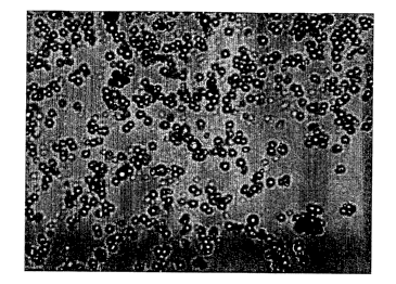
[00351 FIG. 1 gives optical microscope images of anti-Factor VIII monoclonal
antibody microspheres prepared as described in Example 3.
[0036] FIG. 2 provides polarized optical microscope images of anti-Factor VIII
monoclonal antibody microspheres prepared as described in Example 3.
[0037] FIG. 3 provides scanning electron micrographs of anti-Factor VIII
monoclonal
antibody microspheres viewed as described in Example 3.
[0038] FIG. 4 gives gel electrophoresis images of anti-Factor VIII monoclonal
antibody (starting material and dissolved microspheres) as described in
Example 4.
[0039] FIG. 5 gives scanning electron micrographs of anti-Factor VIII
monoclonal
antibody microspheres viewed as described in Example 5.
[0040] FIG. 6 reports particle size distribution by number, surface area and
volume
distribution of anti-Factor VIII monoclonal antibody microspheres as described
in Example 5.
[0041] FIG. 7 provides optical microscope images of anti-CD34 monoclonal
antibody
microspheres prepared as described in Example 6.
[0042] FIG. 8 is an optical microscope image of anti-CD34 monoclonal antibody
microspheres prepared as described in Example 8.
[0043] FIG. 9 is a scanning electron micrograph of anti-CD34 monoclonal
antibody
microspheres prepared as described in Example 6.
[0044] FIG. 10 reports particle size distribution by number distribution of
anti-CD34
monoclonal antibody microspheres prepared as described in Example 6.
[0045] FIG. 11 gives X-ray powder diffraction of anti-CD34 monoclonal antibody
microspheres (with 2 slit configuration) and of hexatriacontane:silicon
mixture as described
in Example 10.
[0046] FIG. 12 reports fluorescence monitoring of conformational stability of
anti-
CD34 monoclonal antibody microspheres during cooling with poloxamer as
described in
Example 7.
[0047] FIG. 13 is a two-dimensional phase diagram plotting active agent
concentration against temperature.
8
CA 02566075 2006-11-08
WO 2005/112893 PCT/US2005/016651
[00481 FIG. 14 is a cooling temperature profile.
[00491 FIG. 15 is an HPLC analysis showing overall maintenance of chemical
stability of insulin when prepared into small spherical particles.
[0050] FIGS. 16a and 16b are schematics demonstrating batch-to-batch
reproducibility.
[0051] FIG. 17a is a circular dichroism (CD) plot for alpha-l-antitrypsin
(AAT).
[0052] FIG. 17b is a plot of activity against storage time at room temperature
in
Example 21.
[0053] FIG. 17c is a plot of activity against storage time at 4 C in Example
21.
[0054] FIGS 18-28b are DSC plots.
[0055] FIG. 29 is a chart showing insulin stability data in HFA-134a.
[0056] FIG. 30 is a chart comparing aerodynamic performance of Insulin using
three
inhalation devices.
[0057] FIG. 31 is a chart of stability data of Insulin small spherical
particles
compared to Insulin starting material stored at 25 C.
[0058] FIG. 32 is a chart of stability data of Insulin small spherical
particles
compared to Insulin starting material stored at 37 C.
[0059] FIG. 33 is a chart of stability data of Insulin small splierical
particles
compared to Insulin starting material stored at 25 C.
[0060] FIG. 34 is a chart of stability data of Insulin small spherical
particles
compared to Insulin starting material stored at 37 C.
[0061] FIG. 35 is a cliart of stability data of Insulin small spherical
particles
compared to Insulin starting material stored at 25 C.
[0062] FIG. 36 is a chart of stability data of Insulin small spherical
particles
compared to Insulin starting material stored at 37 C.
[0063] FIG. 37 is a bar graph of insulin aerodynamic stability using a
Cyclohaler DPI.
[0064] FIG. 38 is a light micrograph of Danes small spherical particles.
[0065] FIG. 39 is a chart of enzymatic activity of DNase.
[0066] FIG. 40 is a light micrograph of SOD small spherical particles.
[0067] FIG. 41 is a chart of enzymatic data for SOD small spherical particles.
[0068] FIGS. 42A-B are schematic illustrations of the continuous
emulsification
reactor, where FIG. 42A is a schematic illustration of the continuous
emulsification reactor
when surface active compound added to the continuous phase or the dispersed
phase before
9
CA 02566075 2006-11-08
WO 2005/112893 PCT/US2005/016651
emulsification, atnd FIG. 42B is a schematic illustration of the continuous
emulsification
reactor when the surface active compound is added after emulsification.
Description of the Preferred Embodiments
[00691 The present invention is susceptible to embodiments in many different
forms.
Preferred embodiments of the invention are disclosed with the understanding
that the present
disclosure is to be considered as exemplifications of the principles of the
invention and are
not intended to limit the broad aspects of the invention to the embodiments
illustrated.
[0070] As required, detailed embodiments of the present invention are
disclosed
herein; however, it is to be understood that the disclosed embodiments are
merely exemplary
of the invention, which may be embodied in various forms. Therefore, specific
details
disclosed herein are not to be interpreted as limiting, but merely as a basis
for the claims and
as a representative basis for teaching one skilled in the art to variously
employ the present
invention in virtually any appropriate manner.
[0071] The present invention is related to coinpositions of substantially
amorphous or
non-crystalline small particles of a active agent that is a protein. Special
application is found
when the active agent has a molecular weight of at least about 25,000 Daltons.
In accordance
with the method of production, the active agent is dissolved in a solvent
containing a
dissolved phase-separation enhancing agent to form a solution that is a single
liquid
continuous phase. The solvent is preferably an aqueous or aqueous-miscible
solvent. The
solution is then subjected to a phase change, for example, by lowering the
temperature of the
solution to below the phase transition temperature of the active agent,
whereby the active
agent goes through a liquid-solid phase separation to form a suspension of
substantially
amorphous or non-crystalline small particles constituting a discontinuous
phase while the
phase-separation enhancing agent remains in the continuous phase.
[0072] The present invention relates to compositions of small particles,
preferably
substantially spherical in shape, of an active agent. The active agents are
preferably high
molecular weight proteins, and more preferably substantially amorphous forms
of high
molecular weight proteins, and most preferably substantially amorphous
monoclonal
antibodies. The invention has the capability of providing injectable or
syringable
compositions of high molecular weight proteins, including monoclonal
antibodies, at high
concentrations, and accordingly provides the ability to deliver a clinically
effective dose of
such active agents with a low volume of coinposition, preferably with 10 ml or
less of
composition, and more preferably with a volume typically found in a standard
syringe.
CA 02566075 2006-11-08
WO 2005/112893 PCT/US2005/016651
[00731 Methods of production and methods of use of these coinpositions of
small
spherical particles of an active agent are also contemplated by this
invention. In accordance
with the method of production, the active agent is dissolved in an aqueous or
aqueous-
miscible solvent containing a dissolved phase-separation enhancing agent
(PSEA) to form a
solution in a single liquid phase. The solution then is subjected to a liquid-
solid phase
separation having the active agent comprising the solid phase and the PSEA and
solvent
comprising the liquid phase. The liquid-solid phase separation can be induced
in numerous
ways, such as changing the temperature of the solution or energy addition. The
method is
most suitable for forming small spherical particles of therapeutic agents
which can be
delivered to a subject in need of the therapeutic agent. The method is also
most suitable for
forming solid, small spherical particles of macromolecules, particularly
macromolecules
which are heat labile, such as proteins, including monoclonal antibody
materials. The
invention has the capability of providing syringable macromolecules.
The Active Agent
[00741 The active agent of the present invention is a protein which can be a
therapeutic agent or a diagnostic agent. Preferred active agents are high
molecular weight
proteins. Preferred agents are amorphous forms of proteins, including
amorphous antibodies.
[0075] When used herein, the term antibody encompasses monoclonal antibodies,
polyclonal antibodies, and antibody fragments, especially the antigen-binding
fractions
generally known as "Fab" fragments or regions, single chain antibodies, as
well as
monoclonal or polyclonal antibodies or other antibodies in recombinant form,
and are what
are currently recognized in the art by the designation "trap molecule".
Antibodies also refers
to any of the aforementioned forms of antibodies that are treated, such as by
coating or
encapsulating, including by approaches as described herein.
[00761 Trap molecules are composed of fusions between two distinct receptor
components and a portion of an antibody molecule referred to as the "Fc
region" resulting in
the generation of growth factor and cytokine blockers with markedly increased
affinity over
that offered by single-component reagents.
[00771 The following references provide further information on trap molecules:
"Cytokine Traps: Multi-Component, High-Affinity Blockers of Cytokine Action";
Economides AN, Carpenter LR, Rudge JS, Wong V, Koehler-Stec EM, Hartnett C,
Pyles EA,
Xu X, Daly TJ, Young MR, Fandl JP, Lee F, Carver S, McNay J, Bailey K,
Ramakanth S,
Hutabarat R, Huang TT, Radziejewski C, Yancopoulos GD, Stahl N; Journal: Nat
Med
11
CA 02566075 2006-11-08
WO 2005/112893 PCT/US2005/016651
(2003); Volume, (Number), Pages: 9(l):47-52. "Vascular Eendothelial Growth
Factor-Trap
Decreases Tumor Burden, Inhibits Ascites, and Causes Dramatic Vascular
Remodeling in an
Ovarian Cancer Model"; Byrne AT, Ross L, Holash J, Nakanishi M, Hu L, Hofinann
JI,
Yancopoulos GD, Jaffe RB; Journal: Clin Cancer Res (2003); Volume, (Number),
Pages:
15;9(15):5721-8. "Prevention of Thecal Angiogenesis, Antral Follicular Growth,
and
Ovulation in the Primate by Treatment with Vascular Endothelial Growth Factor
Trap
R1R2"; Wulff C, Wilson H, Wiegand SJ, Rudge JS, Fraser HM; Journal:
Endocrinology
(2002); Volume, (Number), Pages: 143(7):2797-807. Volume, (Nuinber), Pages:
143(7):2797-807.
[0078] In a preferred embodiment of the present invention, the active agent is
a
monoclonal antibody, which can be natural or synthetic. Examples of monoclonal
antibodies
include, but are not liinited to: adalimutab (available from Abbot under the
tradename
Humira), abciximab (available from Centocor under the tradname ReoPro);
daclizumab,
(available from Roche under the tradname Zenapaz), rituximab (available from
IDEC/Genentech under the tradname Rituxin or Rituxan), basiliximab (available
from
Novartis under the tradname Simulect), palivzumab (available from Medimmune
under the
tradname Synagis), infliximab (available from Centocor under the tradname
Remicade),
trastuxumab (available from Genentech under the tradname Herceptin),
gemtuzumab
(available from IDEC under the tradname Mylotarg), alemzutumab (available from
Millennium/ILEX under the tradname Campath), and ibritumomab (available from
IDEC
under the tradname Zevulin). Garnmagard Liquid (available from Baxter
Healthcare
Corporation, Westlake Village, CA) is a ready-for-use sterile, liquid
preparation of highly
purified and concentrated immunoglobulin G (IgG) antibodies.
[0079] Examples of antibody "Fab" fractions or regions include, but are not
limited
to, the following. TGX-6B4, currently in development by ThromboGenics Ltd of
Dublin,
Ireland, is an antibody to GP1b which inhibits platelet adhesion and is
indicated to be a novel
approach to prevent early steps in arterial thrombosis. Digoxin specific Fab
fragments have
been reported to be beneficial in the treatment of toad venom poisoning.
(Heart.2003; 89: 12-
472, Toxalert, 15: issue 1, 1998). Humanized Fab fragments have been shown to
recognize
the IgE-binding domain of human Fc(epsilon)RIalpha in COS and CHO cells.
(Journal of
Biochemistry, 2001: Vol 129, Issue 1 5-12). Other information concerning Anti-
tumor
Radioimmunotherapy using multivalent Fab' fragments is found in British
Journal of Cancer
(1999) 81, 972-980.
12
CA 02566075 2006-11-08
WO 2005/112893 PCT/US2005/016651
[0080] Examples of other high molecule weight proteins include but are not
limited to
AAT, Dnase, superoxide dismutase, subtilisin and other proteins. Typically,
high molecular
weight indicates a protein having molecular weights on the order of
approximately 25,000,
depending on particular needs or properties of the protein or to its intended
use. Lower
molecular weight proteins can benefit from the invention to the extent same
needs to be
administered, for example by injection, in high concentrations. Such proteins
are known in
the art; see for example U.S. Patent Application No. 10/894,410 filed July 19,
2004 and No.
10/896,326 filed July 21, 2004.
The Microparticles, Small Spherical Particles or Microspheres
[00811 The microparticles or the microspheres of the present invention
preferably
have an average geometric particle size of less than 200 microns, typically
from about 0.01
m to about 200 m, typically not more than about 50 m, more preferably from
0.1 m to
m, even more preferably from about 0.5 gm to about 5 m, and most preferably
from
about 0.5 m to about 3 m, as measured by dynamic light scattering methods
(e.g.,
photocorrelation spectroscopy, laser diffraction, low-angle laser light
scattering (LALLS),
medium-angle laser light scattering (MALLS)), by light obscuration methods
(Coulter
analysis method, for example) or by other methods, such as rheology or
microscopy (light or
electron).
[0082] The small spherical particles or microspheres are substantially
spherical.
What is meant by "substantially spherical" is that the ratio of the lengths of
the longest to the
shortest perpendicular axes of the particle cross section is less than or
equal to about 1.5.
Substantially spherical does not require a line of symmetry. Further, the
particles may have
surface texturing, such as lines or indentations or protuberances that are
small in scale when
coinpared to the overall size of the particle and still be substantially
spherical. More
preferably, the ratio of lengths between the longest and shortest axes of the
particle is less
than or equal to about 1.33. Most preferably, the ratio of lengths between the
longest and
shortest axes of the particle is less than or equal to about 1.25. Surface
contact is minimized
in microspheres that are substantially spherical, which minimizes the
undesirable
agglomeration of the particles upon storage. Many crystals or flakes have flat
surfaces that
can allow large surface contact areas where agglomeration can occur by ionic
or non-ionic
interactions. A sphere permits contact over a much smaller area.
13
CA 02566075 2006-11-08
WO 2005/112893 PCT/US2005/016651
[00831 The microparticles also preferably have substantially the same particle
size.
Particles having a broad size distribution where there are both relatively big
and small
particles allow for the smaller particles to fill in the gaps between the
larger particles, thereby
creating new contact surfaces. A broad size distribution can result in larger
spheres by
creating many contact opportunities for binding agglomeration. This spherical
microparticles
of the invention preferably are within a narrow size distribution, thereby
minimizing
opportunities for contact agglomeration. What is meant by a "narrow size
distribution" is a
preferred particle size distribution that has a ratio of the volume diameter
of the 90th
percentile of the small spherical particles to the volume diameter of the 10t'
percentile less
than or equal to 5. More preferably, the volume diameter of the 90th
percentile of the small
spherical particles to the volume diameter of the 10t' percentile is less than
or equal to 3.
Most preferably, the ratio of the volume diameter of the 90th percentile of
the small spherical
particles to the voluine diameter of the 10th percentile is less than or equal
to 2.
[0084] Geometric Standard Deviation (GSD) can also be used to indicate the
narrow
size distribution. GSD calculations involved determining the effective cutoff
diameter (ECD)
at the cumulative less than percentages of 15.9% and 84.1 %. GSD is equal to
the square root
of the ratio of the ECD less than 84.17% to ECD less then 15.9%. The GSD has a
narrow
size distribution when GSD < 2.5, more preferably less than 1.8.
[0085] In a preferred form of the invention, the active agent in the
microparticle or
microsphere is semi-crystalline or non-crystalline or substantially amorphous.
(0086] The microspheres are preferably comprised of active agents which are
substantially amorphous or non-crystalline, that is they are in an amorphous
or semi-
crystalline form. As used herein, "ainorphous" refers to a generally random
solid form of the
active agent wherein crystalline lattices of the protein(s) or other active
agent(s) within the
microsphere are absent, and "semi-crystalline" refers to a generally random
solid form of
active agent(s) wherein the active agent content of the microsphere is
comprised of less than
50% of crystalline lattice forms of the active agent(s).
[0087] Typically, the microparticles or microspheres are substantially non-
porous and
have a density greater than 0.5 g/cm3, more preferably greater than 0.75 g/cm3
and most
preferably greater than about 0.85 g/cm3. A preferred range for the density is
from about 0.5
to about 2 g/cin3 and more preferably from about 0.75 to about 1.75 g/cin3 and
even more
preferably from about 0.85 g/cm3 to about 1.5 g/cm3. The substantially
amorphous or non-
crystalline microparticles according to the invention are more readily soluble
or exhibit a rate
14
CA 02566075 2006-11-08
WO 2005/112893 PCT/US2005/016651
of dissolution faster than microparticles which are not so constituted, such
as crystalline
microparticles.
[00881 The microparticles or microspheres of the present invention can exhibit
a high
content of the active agent. There is no requirement for a significant
quantity of bulking
agents or similar excipients that are required by many other methods of
preparing
microparticles, although materials in addition to the active agent can be
included as desired to
achieve a particular objective or objectives. For example, in many
applications, the
microspheres comprise equal to or greater than 95% by weight of the particles.
Typically, the
active agent is present from about 20% to 100% by weight of the particle,
preferably from
about 50% to about 100% by weight, more preferably from about 80% to about
100% by
weight, even more preferably from about 90% to about 100% by weight. When
stating
ranges herein, it is meant to include any range or combination of ranges
therein.
[00891 A furtlier aspect of the present invention is that the microparticles
or
microspheres retain the biochemical integrity and the biological activity of
the active agent
with or without the inclusion of excipients.
In vivo Delivery of the Particles
[0090] Microparticles, small spherical particles or microspheres containing
the active
agent in the present invention are suitable for in vivo delivery to a subject
in need of the agent
by an injectable route. A preferred delivery route is injectable, which
includes intravenous,
intramuscular, subcutaneous, intraperitoneal, intrathecal, epidural, intra-
arterial, intra-
articular and the like. Other delivery routes, such as topical, oral, rectal,
nasal, pulmonary,
vaginal, buccal, sublingual, transdermal, transmucosal, otic or intraocular,
could be practiced,
but typically the advantages of the invention are more evident for injection
applications.
Most preferred for the purpose of this invention is the syringable delivery
route. Most
importantly, the microparticles or microspheres can be aspirated into a
syringe and injected
through fine needles despite the high molecular weight of the proteins or
active agents. A
preferred delivery route is injection with a fine needle, which includes
subcutaneous, ocular
and the like. By fine needle is meant needles of at least 20 Gauge size,
typically between
about 22 Gauge and about 30 Gauge and above. Advantageously, the fine needles
can be at
least as fine as 24 Gauge, more advantageously at least as fine as 26 gauge,
and even more
advantageously at least as fine as 28 Gauge.
[0091] The microparticles or microspheres are capable of being injected at a
concentration of at least about 50 mg of protein per ml of the composition
being injected. For
CA 02566075 2006-11-08
WO 2005/112893 PCT/US2005/016651
example, from about 100 to about 800 mg of protein are injectable in a
delivery volume if not
more than about 10 ml, and usually at least about 2 ml for many applications.
Also, the
delivery is made during normal injections time periods. Typically such time
periods are not
more than about 20 seconds or less.
[00921 The present method for particle formation set forth herein provides for
particle
formation with or without excipients or other components or additives as
desired or required.
Fabrication of protein microparticles or microspheres from protein itself with
no additives is
also an approach according to the invention and at time provides superior
advantages for use.
Methods for MakingMicroparticles
The Continuous Phase
[00931 The method of the present invention of preparing microparticles or
microspheres of a active agent begins with providing a solution having the
active agent and a
phase-separation enhancing agent dissolved in a first solvent in a single
liquid phase. The
solution can be an organic system comprising an organic solvent or a mixture
of miscible
organic solvents. The solution can also be an aqueous-based solution
comprising an aqueous
medium or an aqueous-miscible organic solvent or a mixture of aqueous-miscible
organic
solvents or combinations thereof. The aqueous medium can be water, normal
saline, buffered
solutions, buffered saline, and the like. Suitable aqueous-miscible organic
solvents include,
but are not limited to, N-methyl-2-pyrrolidinone (N-methyl-2-pyrrolidone), 2-
pyrrolidinone
(2-pyrrolidone), 1,3-dimethyl-2-imidazolidinone (DMI), dimethylsulfoxide,
dimethylacetamide, acetic acid, lactic acid, acetone, methyl ethyl ketone,
acetonitrile,
methanol, ethanol, isopropanol, 3-pentanol, n-propanol, benzyl alcohol,
glycerol,
tetrahydrofuran (THF), polyethylene glycol (PEG), PEG-4, PEG-8, PEG-9, PEG-12,
PEG-14,
PEG-16, PEG-120, PEG-75, PEG-150, polyethylene glycol esters, PEG-4 dilaurate,
PEG-20
dilaurate, PEG-6 isostearate, PEG-8 palmitostearate, PEG-150 palmitostearate,
polyethylene
glycol sorbitans, PEG-20 sorbitan isostearate, polyethylene glycol monoalkyl
ethers, PEG-3
dimethyl ether, PEG-4 dimethyl ether, polypropylene glycol (PPG),
polypropylene alginate,
PPG-10 butanediol, PPG-10 methyl glucose ether, PPG-20 methyl glucose ether,
PPG-15
stearyl ether, propylene glycol dicaprylate/dicaprate, propylene glycol
laurate, and glycofurol
(tetrahydrofurfuryl alcohol polyethylene glycol ether), alkanes including
propane, butane,
pentane, hexane, heptane, octane, nonane, decane, or a combination thereof.
[00941 The single continuous phase can be prepared by first providing a
solution of
the phase-separation enhancing agent, which is either soluble in or miscible
with the first
16
CA 02566075 2006-11-08
WO 2005/112893 PCT/US2005/016651
solvent. This is followed by adding the active agent to the solution. The
active agent may be
added directly to the solution, or the active agent may first be dissolved in
a second solvent
and then together added to the solution. The second solvent can be the same
solvent as the
first solvent, or it can be another solvent selected from the list above and
which is miscible
with the solution. It is preferred that the active agent is added to the
solution at an ambient
temperature or lower, which is important particularly for heat labile
molecules, such as
certain proteins. What is meant by "ambient temperature" is a temperature of
around room
temperature of about 20 C to about 40 C. However, the system can also be
heated to
increase the solubility of the active agent in the system as long as heating
does not cause
significant reduction in the activity of the agent.
The Phase-Separation EnhancingAgent
[00951 The phase-separation enhancing agent (PSEA) of the present invention
enhances or induces the liquid-solid phase separation of the active agent from
the solution
when the solution is subjected to the step of phase separation in which the
active agent
becomes solid or semi-solid to form a suspension of small spherical particles
as a
discontinuous phase while the phase-separation enhancing agent remains
dissolved in the
continuous phase. The phase-separation enhancing agent reduces the solubility
of the active
agent wllen the solution is brought to the phase separation conditions.
Suitable phase-
separation enhancing agents include, but are not limited to, polymers or
mixtures of polymers
that are soluble or miscible with the solution. Examples of suitable polymers
include linear
or branched polymers, copolymers and block copolymers. These polymers can be
water
soluble, semi-water soluble, water-miscible, or insoluble.
[0096] In a preferred form of the invention, the phase-separation enhancing
agent is
water soluble or water miscible. Types of polymers that may be used include
carbohydrate-
based polymers, polyaliphatic alcohols, poly(vinyl) polymers, polyacrylic
acids, polyorganic
acids, polyamino acids, co-polymers and block co-polymers (e.g., poloxamers
such as
Pluronic F127 or F68), tert-polymers, polyethers, naturally occuring polymers,
polyimides,
surfactants, polyesters, branched and cyclo-polymers, and polyaldehydes.
[0097] Preferred polymers are ones that are acceptable as pharmaceutical
additives
for the intended route of administration of the active agent particles.
Preferred polymers are
pharmaceutically acceptable additives such as polyethylene glycol (PEG) of
various
molecular weights, such as PEG 200, PEG 300, PEG 3350, PEG 8000, PEG 10000,
PEG
17
CA 02566075 2006-11-08
WO 2005/112893 PCT/US2005/016651
20000, etc. and poloxamers of various molecular weigllts such as poloxamer 188
and
Pluronic F127 or Pluronic F68. Yet another preferred polymer is
polyvinylpyrrolidone
(PVP). Yet another preferred polymer is hydroxyethylstarch. Other amphiphilic
polymers
can also be used alone or in combinations. The phase-separation enhancing
agent can also be
a non-polymer such as a mixture of propylene glycol and ethanol.
Liquid-Solid Phase Separation
[00981 A liquid-solid phase separation of the active agent in the solution can
be
induced by any method known in the art, such as change in temperature (either
raising or
lowering), change in pressure, change in pH, change in ionic strength of the
solution, change
in the coricentration of the active agent, change in the concentration of the
phase-separation
enhancing agent, change in osmolality of the solution, combinations of these,
and the like.
[00991 In a preferred enlbodiment of the present invention, the phase change
is a
temperature-induced phase change. In many embodiments the temperature-induced
phase
change is effected by lowering the temperature below the phase transition
temperature of the
active agent in the solution.
[001001 FIG. 1 is a two-dimensional phase diagram 10 for the solution
containing
solvent, a PSEA and an active agent. The diagram plots the active agent
concentration
against the temperature of the solution. The concentration of the PSEA is held
constant.
[001011 The diagram of FIG. 1 has a saturation curve 12; a supersaturation
curve 14; a
metastable area 16 therebetween; a first area 18 below the saturation curve
where the system
is in a homogenous, single liquid phase where all components are in the liquid
phase; and a
second area 20 above the supersaturation curve where the system is a two-phase
system
having a solid phase of the active agent and a liquid phase of the PSEA and
solvent. The
phase diagram is helpful in determining the temperature of the system and the
relative
concentration of components in the pure liquid phase, the liquid-solid phase
and the
conditions surrounding the transition between these two phases.
[001021 As disclosed herein, preparation of microparticles or microspheres of
the
active agent principally involves cooling from an undersaturated solution
(point A' in FIG. 1)
reaching saturation at point A where the solution is in equilibrium with any
solid phase that
may be present. On fu.rther cooling, a state is reached where the solution
contains more
active agent than that corresponding to the equilibrium solubility at the
given temperature;
the solution thus becomes supersatui=ated. Spontaneous formation of the solid
phase does not
occur until point B is reached. Point B is a point on the boundary of the
metastable zone.
18
CA 02566075 2006-11-08
WO 2005/112893 PCT/US2005/016651
The metastable zone width can be expressed either by the maximum attainable
undercooling
AT,,,~ T2-Ti or by the supersaturation OC,,,ax=C*2-C*1. These two expressions
are
thermodynamically equivalent:
. . TZ 8C dC*
OCma, = C2 - Cl = T= OTm.
dT
TI
[001031 The path A'-A-B represents a polythernzal method of preparing a
metastable
solution. In an isothernzal process the starting point would be A". By
increasing the
concentration at constant temperature, saturation will again be achieved at
point A. An
isothermal increase in concentration (by solvent evaporation or by
seeding/addition of the
active agent, for instance) to point C will cause the solution to move into
the metastable
region until the metastability limit is again reached. When the metastable
limit is exceeded,
the solution becomes unstable and a spontaneous formation of the solid phase
immediately
occurs.
[00104] The value (OCmax)T=C*3-C*2 obtained isothermally can be different from
the
corresponding value of AT,,,.=T3-TZ obtained polythermally. As the boundary of
the
metastable zone is approached, the time necessary for the solid particle
forination decreases
until the metastable limit is reached.
[001051 In the polythermal process, the rate of cooling is done at a
controlled rate to
control the size and shape of the particles. What is meant by a controlled
rate is about
0.2 C/minute to about 50 C/ininute, and more preferably from 0.2 C/minute to
30 C/minute.
The rate of change can be at a constant or linear rate, a non-linear rate,
intermittent, or a
programmed rate (having multiple phase cycles). The particles can be separated
from the
PSEA in the solution and purified by washing as will be discussed below.
[001061 The present invention contemplates adjusting the concentration of the
active
agent, the concentration of the PSEA, the temperature or any combination of
these to cause a
phase change where the active agent goes from a liquid state to a solid state
while the PSEA
and solvent do not go through a phase change and remain as liquids. It is also
contemplated
changing the pH, the ionic strength, the osmolality and the like to enhance,
promote, control
or suppress the phase change. For solutions in which the freezing point is
relatively high, or
the freezing point is above the phase transition temperature, the solutions
can include a
freezing point depressing agent, such as propylene glycol, sucrose, ethylene
glycol, alcohols
19
CA 02566075 2006-11-08
WO 2005/112893 PCT/US2005/016651
(e.g., ethanol, methanol) or aqueous mixtures of freezing-point depression
agents to lower the
freezing point of the system to allow the phase change in the system without
freezing the
system. The process can also be carried out such that the temperature is
reduced below the
freezing point of the system. The process described herein is particularly
suitable for
molecules that are heat labile (e.g., proteins).
Optional Excipients
[001071 The microparticles of the present invention may include one or more
excipients. The excipient may imbue the active agent or the microparticles
with additional
characteristics such as increased stability of the microparticles or of the
active agents or of
the carrier agents, controlled release of the active agent from the
microparticles, or modified
permeation of the active agent through biological tissues. Suitable excipients
include, but are
not limited to, carbohydrates (e.g., trehalose, sucrose, mannitol), cations
(e.g., Zn2+, N1gz+,
Ca2+), anions (e.g. SO42-), amino acids (e.g., glycine), lipids,
pliospholipids, fatty acids,
surfactants, triglycerides, bile acids or their salts (e.g., cholate or its
salts, such as sodium
cholate; deoxycholic acid or its salts), fatty acid esters, and polymers
present at levels below
their functioning as PSEA's. When an excipient is used, the excipient does not
significantly
affect the phase diagram of the solution.
Separating and Washing the Particles
[00108] In a preferred embodiment of the present invention, the microparticles
or
microspheres are harvested by separating them from the phase-separation
enhancing agent in
the solution. In yet another preferred embodiment, the method of separation is
by washing
the solution containing the microparticles or microspheres with a liquid
medium in which the
active agent is not soluble in the liquid medium while the phase-separation
enhancing agent is
soluble in the liquid medium. Some methods of washing may be by diafiltration
or by
centrifugation. The liquid medium can be an aqueous medium or an organic
solvent. For
active agents with low aqueous solubility, the liquid medium can be an aqueous
medium or
an aqueous medium containing agents that reduce the aqueous solubility of the
active agent,
such as divalent cations. For active agents with high aqueous solubility, such
as many
proteins, an organic solvent or an aqueous solvent containing a protein-
precipitating agent
such as ammonium sulfate may be used.
[00109] In a preferred embodiment of the present invention, the microparticles
or
microspheres are harvested by separating them from the phase-separation
enhancing agent in
CA 02566075 2006-11-08
WO 2005/112893 PCT/US2005/016651
the solution. In yet another preferred embodiment, the method of separation is
by washing
the solution containing the microparticles or microspheres with a liquid
medium in which the
active agent is not soluble in the liquid medium while the phase-separation
enhancing agent is
soluble in the liquid medium. Some methods of washing may be by diafiltration
or by
centrifugation. The liquid medium can be an aqueous medium or an organic
solvent. For
active agents witli low aqueous solubility, the liquid inedimn can be an
aqueous medium or
an aqueous medium containing agent that reduces the aqueous solubility of the
active agent,
such as divalent cations. For active agents with high aqueous solubility, such
as many
proteins, an organic solvent or an aqueous solvent containing a protein-
precipitating agent
such as ammonium sulfate may be used.
[00110] Examples of suitable organic solvents for use as the liquid medium
include
those organic solvents specified above as suitable for the continuous phase,
and more
preferably methylene chloride, chloroform, acetonitrile, ethylacetate,
methanol, etlianol,
pentane, and the like. It is also contemplated to use mixtures of any of these
solvents. One
preferred blend is methylene chloride or a 1:1 mixture of methylene chloride
and acetone. It
is preferred that the liquid medium has a low boiling point for easy removal
by, for example,
lyophilization, evaporation, or drying.
[00111] The liquid medium also can be a supercritical fluid, such as liquid
carbon
dioxide or a fluid near its supercritical point. Supercritical fluids can be
suitable solvents for
the phase-separation enhancing agents, particularly some polymers, but are
nonsolvents for
protein particles. Supercritical fluids can be used by themselves or with a
cosolvent. The
following supercritical fluids can be used: liquid C02, ethane, or xenon.
Potential cosolvents
can be acetontitrile, dichloromethane, ethanol, methanol, water, or 2-
propanol.
[001121 The liquid medium used to separate the microparticles or microspheres
from
the PSEA described herein, may contain an agent which reduces the solubility
of the active
agent in the liquid medium. It is most desirable that the particles exhibit
minimal solubility
in the liquid medium to maximize the yield of the microparticles or
microspheres. For some
proteins, such as insulin and the decrease in solubility can be achieved by
the adding of
divalent cations, such as ZnZ+ to the protein. Other ions that can be used to
form complexes
include, but are not limited to, Ca2+, Cu2+, Fe2+, Fe3+, and the like. The
solubility of the
complexes are sufficiently low to allow diafiltration of the complex in an
aqueous solution.
[00113] The liquid medium may also contain one or more excipients which may
imbue
the active agent or the microparticles with additional characteristics such as
increased
stability of the mircroparticles and/or of the active or carrier agents,
controlled release of the
21
CA 02566075 2006-11-08
WO 2005/112893 PCT/US2005/016651
active agent from the particles, or modified permeation of the active agent
through biological
tissues as discussed previously. In anotlier form of the invention, the
microparticles or
microspheres are not separated from the PSEA containing solution.
Aqueous-based Process
[001141 In another preferred embodiment, the fabrication process of the
present system
is of an aqueous system including an aqueous or an aqueous-miscible solvent.
Examples of
suitable aqueous-miscible solvents include, but are not limited to, those
identified above for
the continuous phase. One advantage of using an aqueous-based process is that
the solution
can be buffered and can contain excipients that provide biochemical
stabilization to protect
the active agents. This can be especially advantageous when the active agent
is a protein.
Microencapsulation of Pre-fabricated Microparticles
[001151 The microparticles or microspheres of the present invention can
further be
encapsulated within matrices of wall-forming materials to form
microencapsulated particles.
The microencapusulation can be accomplished by any process known in the art.
In a
preferred embodiment, microencapsulation of the microparticles or microspheres
of the
present invention is accomplished by an emulsification/solvent extraction
processes as
described below. The matrix can impart sustained release properties to the
active agent
resulting in release rates that persist from minutes to hours, days or weeks
according to the
desired therapeutic applications. The microencapsulated microparticles or
microspheres can
also produce delayed release formulations of the active agent. Additional
methods of making
microparticles and microspheres are set forth in this application.
[00116] In the emulsification/solvent extraction process, emulsification is
obtained by
mixing two immiscible phases, the continuous phase and the discontinuous phase
(which is
also known as the dispersed phase), to form an emulsion. In a preferred
embodiment, the
continuous phase is an aqueous phase (or the water phase) and the
discontinuous phase is an
organic phase (or the oil phase) to form an oil-in-water (O/W) emulsion. The
discontinuous
phase may further contain a dispersion of solid particles present either as a
fine suspension or
as a fine dispersion forming a solid-in-oil (S/O) phase. The organic phase is
preferably a
water immiscible or a partially water miscible organic solvent. The ratio by
weights of the
orgaiiic phase to the aqueous phase is from about 1:99 to about 99:1, more
preferably from
1:99 to about 40:60, and most preferably from about 2:98 to about 1:3, or any
range or
combination of ranges therein. In a preferred embodiment, the ratio of the
organic phase to
22
CA 02566075 2006-11-08
WO 2005/112893 PCT/US2005/016651
the aqueous phase is about 1:3. This aspect of present invention further
contemplates
utilizing reverse emulsions or water-in-oil emulsion (W/O) where the oil phase
forms the
continuous phase and water phase forms the discontinuous phase. This further
contemplates
utilizing emulsions having more than two phases such as an oil-in-water-in-oil
emulsion
(O/W/O) or a water-in-oil-in-water emulsion (W/O/W).
[001171 In a preferred embodiment of this variation on the invention, the
process of
microencapsulation using the emulsification/solvent extraction process starts
with preparing
pre-fabricated microparticles or microspheres by the methods described earlier
and an
organic phase containing the wall-forming material. The pre-fabricated
microparticles or
microspheres are dispersed in the organic phase of the wall-forming material
to fonn a solid-
in-oil (S/O) phase containing a dispersion of the pre-fabricated
microparticles or
microspheres in the oil phase. In a preferred embodiment, the dispersion is
accomplished by
homogenizing the mixture of the microparticles or microspheres and the organic
phase. An
aqueous medium will form the continuous phase. In this case, the emulsion
system formed
by emulsifying the S/O phase with an aqueous phase is a solid-in-oil-in-water
(S/O/W)
emulsion system.
[00118] The wall-fonning material refers to materials capable of forming the
structural
entity of the matrix individually or in combination. Biodegradable wall-
forming materials
are preferred, especially for injectable applications. Examples of such
materials include but
are not limited to the family of poly-lactide/poly-glycolide polymers
(PLGA's), polyethylene
glycol conjugated PLGA's (PLGA-PEG's), and triglycerides. In the embodiment in
which
PLGA or PLGA-PEG is used, the PLGA preferably has a ratio of poly-lactide to
poly-
glycolide of from 100:0 to 0:100, more preferably from about 90:10 to about
15:85, and most
preferably about 50:50. In general, the higher the ratio of the poly-glycolide
to the poly-
lactide in the polymer, the more hydrophilic are the microencapsulated
particles, resulting in
faster hydration and faster degradation. Various molecular weights of PLGA
also can be
used. In general, for the same ratio of poly-glycolide and poly-lactide in the
polymer, the
higher the molecular weight of the PLGA, the slower is the release of the
active agent, and
the wider the distribution of the size of the microencapsulated particles or
spheres.
[00119] When microencapsulation is practiced, the organic solvent in the
organic
phase (oil phase) of an oil-in-water (O/W) or solid-in-oil-in-water (S/O/W)
emulsion can be
aqueous immiscible or partially aqueous immiscible. What is meant by "water
immiscible
solvents" are those solvents which form an interfacial meniscus when conlbined
with an
aqueous solution in a 1:1 ratio (O/W). Suitable water immiscible solvents
include, but are
23
CA 02566075 2006-11-08
WO 2005/112893 PCT/US2005/016651
not limited to, substituted or unsubstituted, linear, branched or cyclic
alkanes with a carbon
number of 5 or higher, substituted or unsubstituted, linear, branched or
cyclic alkenes with a
carbon number of 5 or higher, substituted or unsubstituted, linear, branched
or cyclic alkynes
with a carbon number of 5 or higher; aromatic hydrocarbons completely or
partially
halogenated liydrocarbons, ethers, esters, ketones, mono-, di- or tri-
glycerides, native oils,
alcohols, aldehydes, acids, amines, linear or cyclic silicones,
hexamethyldisiloxane, or any
combination of these solvents. Halogenated solvents include, but are not
limited to, carbon
tetrachloride, methylene chloride, chloroform, tetrachloroethylene,
trichloroethylene,
trichloroethane, hydrofluorocarbons, chlorinated benzene (mono, di, tri),
trichlorofluoromethane. Particularly suitable solvents are methylene chloride,
chloroform,
diethyl ether, toluene, xylene and ethyl acetate. What is meant by "partially
water miscible
solvents" are those solvents wlZich are water immiscible at one concentration,
and water
miscible at another lower concentration. These solvents are of limited water
miscibility and
capable of spontaneous emulsion formation. Examples of partially water
miscible solvents
are tetrahydrofuran (THF), propylene carbonate, benzyl alcohol, and ethyl
acetate.
[00120] A surface active compound can be added in connection with the
microencapsulation aspect, for example, to increase the wetting properties of
the organic
phase. The surface active compound can be added before the emulsification
process to the
aqueous phase, to the organic phase, to both the aqueous medium and the
organic solution, or
after the emulsification process to the emulsion. The use of a surface active
coinpound can
reduce the number of unencapsulated or partially encapsulated small spherical
particles,
resulting in reduction of the initial burst of the active agent during the
release. The surface
active compound can be added to the organic phase, or to the aqueous phase, or
to both the
organic phase and the aqueous phase, depending on the solubility of the
compound.
[00121] What is meant by the term "surface active conlpounds" are compounds
such as
an anionic surfactant, a cationic surfactant, a zwitterionic surfactant, a
nonionic surfactant or
a biological surface active molecule. The surface active compound should be
present in an
amount by weight of the aqueous phase or the organic phase or the emulsion,
whatever the
case may be, from less than about 0.01 % to about 30%, more preferably from
about 0.01 % to
about 10%, or any range or combination of ranges therein.
[00122 ] Suitable anionic surfactants include but are not limited to:
potassium laurate,
sodium lauryl sulfate, sodium dodecylsulfate, alkyl polyoxyethylene sulfates,
sodium
alginate, dioctyl sodium sulfosuccinate, phosphatidyl choline, phosphatidyl
glycerol,
phosphatidyl inosine, phosphatidylserine, phosphatidic acid and their salts,
glyceryl esters,
24
CA 02566075 2006-11-08
WO 2005/112893 PCT/US2005/016651
sodium carboxymethylcellulose, cholic acid and other bile acids (e.g., cholic
acid,
deoxycholic acid, glycocholic acid, taurocholic acid, glycodeoxycholic acid)
and salts thereof
(e.g., sodium deoxycholate, etc.).
[001231 Suitable cationic surfactants include, but are not limited to,
quaternary
ammonium compounds, such as benzalkonium chloride, cetyltrimethyla.intiionium
bromide,
lauryldimethylbenzylammonium chloride, acyl carnitine hydrochlorides, or alkyl
pyridinium
halides. 'As anionic surfactants, phospholipids may be used. Suitable
phospholipids include,
for example phosphatidylcholine, phosphatidylethanolamine, phosphatidylserine,
phosphatidyl inositol, phosphatidylglycerol, phosphatidic acid,
lysophospholipids, egg or
soybean phospholipid or a combination thereof. The phospholipid may be salted
or desalted,
hydrogenated or partially hydrogenated or natural, semisynthetic or synthetic.
[001241 Suitable nonionic surfactants include: polyoxyethylene fatty alcohol
ethers
(Macrogol and Brij), polyoxyethylene sorbitan fatty acid esters
(Polysorbates),
polyoxyethylene fatty acid esters (Myrj), sorbitan esters (Span), glycerol
monostearate,
polyethylene glycols, polypropylene glycols, cetyl alcohol, cetostearyl
alcohol, stearyl
alcohol, aryl alkyl polyether alcohols, polyoxyethylene-polyoxypropylene
copolymers
(poloxomers), polaxamines, polyvinyl alcohol, polyvinylpyrrolidone, and
polysaccharides
(including starch and starch derivatives such as hydroxyethylstarch (HES),
methylcellulose,
hydroxycellulose, hydroxy propylcellulose, hydroxy propylmethylcellulose, and
noncrystalline cellulose). In a preferred form of the invention, the nonionic
surfactant is a
polyoxyethylene and polyoxypropylene copolymer and preferably a block
copolymer of
propylene glycol and ethylene glycol. Such polymers are sold under the
tradename
POLOXAMER also sometimes referred to as PLURONIC , and sold by several
suppliers
including Spectrum Chemical and Ruger. Among polyoxyethylene fatty acid esters
are
included those having short alkyl chains. One example of such a surfactant is
SOLUTOL
HS 15, polyethylene-660-hydroxystearate, manufactured by BASF
Aktiengesellschaft.
Surface active biological molecules include such molecules as albumin, casein,
heparin,
hirudin, hetastarch or other appropriate biocompatible agents.
[00125] In a preferred form of the microencapsulation option of the invention,
the
aqueous phase includes a protein as the surface active compound. A preferred
protein is
albumin. The protein may also function as an excipient. In embodiments in
which protein is
not the surface active compound, other excipients may be included in the
emulsion, added
either before or after the emulsification process. Suitable excipients
include, but are not
CA 02566075 2006-11-08
WO 2005/112893 PCT/US2005/016651
limited to, saccharides, disaccharides, and sugar alcohols. A preferred
disaccharide is
sucrose, and a preferred sugar alcohol is mannitol.
[001261 In addition, use of clianneling agents, such as polyethylelne glycol
(PEG), can
increase the water permeation rate of the final product, which results in
modification of the
initial release kinetics of the active agent from the matrix, when a matrix is
present, as well as
degradation rate of the matrix and degradation-dependent release kinetics by
modifying the
hydration rate. Using PEG as the channeling agent during encapsulation can be
advantageous
in terms of eliminating parts of the washing process during fabrication of the
small spherical
particles in which PEG is used as the phase-separation enhancing agent. In
addition, salinity
and pH of the continuous phase can be varied to affect properties of the
polymer and the
resulting microparticles or microspheres including the matrix packing density,
surface charge,
wetting, porosity, viscosity, particle size distribution, as well as initial
burst and release
kinetics of the encapsulated therapeutic agent from the matrix. Salinity of
the continuous
phase can also be used to reduce miscibility of the two phases. Suitable salts
include, but not
limited to, water-soluble phosphate, sulfate, acetate, and carbonate salts,
Tris, MES, HEPES.
In the embodiment in which salt is used, the salt concentration ranges from 0
to 10 M, more
preferably from 1 mM to 1 M, and most preferably from 20 to 200 mM. The pH
ranges from
1 to 11, more preferably from 2.5 to 9, and most preferably from 6 to 8.
[001271 After dispersing the microparticles or microspheres in the organic
phase (oil
phase), the continuous phase of the aqueous medium (water phase) then is
vigorously mixed,
for example by homogenization or sonication, with the discontinuous phase of
the organic
phase to form an emulsion containing emulsified droplets of embryonic
microencapsulated
particles. The continuous aqueous phase can be saturated with the organic
solvent used in the
organic phase prior to mixing of the aqueous phase and the organic phase, in
order to
minimize rapid extraction of the organic solvent from the emulsified droplets.
The
emulsification process, when practiced, can be performed at any temperature in
which the
mixture can maintain its liquid properties. The emulsion stability is a
function of the
concentration of the surface active compound in the organic phase or in the
aqueous phase, or
in the emulsion if the surface active compound is added to the emulsion after
the
emulsification process. This is one of the factors that determines droplet
size of the emulsion
system (embryonic microencapsulated particles) and the size and size
distribution of the
microencapsulated particles. Other factors affecting the size distribution of
microencapsulated particles are viscosity of the continuous phase, viscosity
of the
26
CA 02566075 2006-11-08
WO 2005/112893 PCT/US2005/016651
discontinous phase, shear forces during emulsification, type and concentration
of surface
active compound, and the Oil/Water ratio.
[001281 After the emulsification, the emulsion then is transferred into a
hardening
medium. The hardening medium extracts the solvent in the discontinous phase
from the
embryonic microencapsulated particles, resulting in formation of solid
microencapsulated
particles having a solid polymeric matrix around the pre-fabricated
microparticles or
microspheres within the vicinity of the emulsified droplets. In the embodiment
of an O/W or
S/O/W system, the hardening medium is an aqueous medium, which may contain
surface
active compounds, or thickening agents, or other excipients. The
microencapsulated particles
are preferably spherical and have a particle size of from about 0.6 to about
300 m, and more
preferably from about 0.8 to about 60 gm. Additionally, the microencapsulated
particles
preferably have a narrow distribution of particle size. To reduce the
extraction time of the
discontinuous phase, heat or reduced pressure can be applied to the hardening
medium. The
extraction rate of discontinuous phase from the embryonic microencapsulated
particles is an
important factor in the degree of porosity in the final solid
microencapsulated particles, since
rapid removal, e.g., by evaporation (boiling effect), of the discontinuous
phase results in
destruction of the continuity of the matrix.
[001291 In a preferred embodiment of the emulsification process, when same is
practiced, same is performed in a continuous fashion instead of a batch
process. FIG. 41
depicts the design of a continuous emulsification reactor which can be used in
this regard.
[001301 In another embodiment when encapsulation is practiced, the hardened
wall-
forming polymeric matrices, encapsulating the microparticles or microspheres
of the active
agent, are further harvested by centrifugation and/or filtration (including
diafiltration), and
washed with water. The remaining liquid phases can further be removed by a
process such as
lyophilization or evaporation.
[001311 The microparticles or microspheres can be encapsulated in at least one
or more
layers of polyelectrolytes selected to amend the particle properties to
targeted characteristics
by controlling surface functional properties. Such microparticles are immersed
with a
polymer of the opposite charge (polyion) in an aqueous media to affect the
surface properties
of the particle. If the particles have a negative charge as determined by zeta
potential
measurements, then the first layer of the polymer will be positive. If the
particles have an
initial positive charge, then the first polymer layer will be negative. The
deposition of the
first layer can take place in water, or in buffer, or in aqueous solution
containing some water
miscible organic solvent.
27
CA 02566075 2006-11-08
WO 2005/112893 PCT/US2005/016651
[001321 In the case of colloidally unstable protein microparticles, other
solubility
reducing agents can be introduced. Low molecular weight solubility reducing
agents/viscosity increasing agents, such as alcohol, glycerol, etc., as well
as polymers such as
the water soluble polyethylene glycol (PEG), polyvinylpyrrolidone (PVP),
hetastarch,
dextran, etc., can be used for this purpose. Incubation with the coating
material can be
performed at room temperature as well as lower temperature to control particle
solubility.
[001331 Particles may be incubated with the polymer for the required amount of
time
(minutes to hours). The suspension of coated particles may then be isolated
from the excess
of unbound polymer either through centrifugation or diafiltration. The
particles are
preferably washed from residual polymer either in an aqueous solution or in a
solution with
the above-mentioned solubility reducing agents. The temperature is optimized
based on the
solubilities of the active inaterial and coating material.
[00134) After the washing step is complete, the next layer of polymer with an
opposite
charge is introduced. The procedure may be repeated as in the previous cycle.
The number
of deposited polymer layers may depend on the desired application. It can be
varied from, for
example, one layer for surface modification, and 10-20 bilayers for sustained
release
applications dependent on the required dissolution kinetics.
[00135] The polymers that may be used include, but are not limited to, the
following
list: polystyrene sulfonate (PSS) and polyallyl amine hydrochloride (PAH),
polyacrylic acid
(PAA), poly(diallyldimethyl ammonium chloride (PDDA), and biocoinpatible
polymers, such
as dextran sulfate, chitosan, chitosan sulfate, polylysine, gelatin, alginate,
protamine,
protamine sulfate, xanthan gum, etc., and also charged lipids, such as
phosphatidyl choline,
phosphatidyl serine, etc.
[001361 In an alternative layer-by-layer approach, the first layer of the
polyion can also
be added at the end of the controlled precipitation of particles without
removal of the
polymer, such as PEG, that is present in the system. Further, lipid structures
(e.g., liposomes)
can be used in alternate deposition with oppositely charged polymers and the
ratio between
charged and uncharged lipids can be optimized to achieve minimum permeability
of the shell.
In some cases, it may be preferable to carry out the layer by layer assembly
in the presence of
divalent cations, such as zinc.
28
CA 02566075 2006-11-08
WO 2005/112893 PCT/US2005/016651
EXAMPLE 1
[001371 This Example provides a basic procedure for preparation of 500 1
batches of
syringable anti-Factor VIII monoclonal antibody microspheres. Ten batches of
500 1 of the
monoclonal antibody are prepared in eppendorf tubes as follows:
[00138] Preparation of 40mM Ammonium Acetate (AA) Buffer at pH=6.5: 40mM of
AA buffer is prepared by dissalving 3.08 grams of AA (Spectrum) in 1 liter of
deionized
H20. The AA is readily soluble and forms a buffer solution with a pH of - 6.4.
Adjust the pH
to pH=6.5 with dilute ammonium hydroxide.
[00139] Preparation of 500mL of 10% Poloxamer 188 in 40mM AA Buffer solution:
50 gram.s of Poloxamer 188 (BASF) are dissolved in 500mL of 40mM AA Buffer (as
described in step 1), only that pH adjustment is not necessary this time). The
dissolution of
this quantity of Poloxamer can be done in several additions. The final pH is
around pH-6.4.
The solution is filtered with a 0.22 filter and kept refrigerated.
[00140] Buffer exchange: 2 PD 10 desalting columns (Amersham Biosciences) are
used for 5 mL of the protein. Total column volume is 3.5mL, and it is
recommended for
exchange of no more than 2.5mL each. Each column should be rinsed with no less
than 25mL
of 40mM NH4OAc buffer to saturate the column with the buffer. Then, 2.5mL of
the anti-
Factor VIII in phosphate buffer is inserted into the column followed by
additional lmL of the
NH4OAc buffer to fill the column. The protein is collected by injecting an
additional 2.5mL
of the 40n1M NH4OAc buffer.
[00141] A preferred dialysis cassette is a 1 Pierce, with 3-12mL total volume,
and a
MW cutoff of 10,000 Daltons to replace the buffer for 5mL of the protein.
Hydrate the
cassette prior to use with deionized H20, such as by using a 5-10mL syringe
with 18G1/2
needle. First, air is injected into the cassette to separate the membrane
walls. Then, the
sample is injected. Air is withdrawn from the cassette to have a better sample-
membrane
contact. Last, the float is added on, and the cassette spins at low speed. As
a rule of thumb,
each buffer exchange should be at least 10X in volume.
[00142] Protein concentration: Protein concentration is determined by
measuring
absorbance at 280nm and calibration curve. If needed, the protein can be with
the buffer
according to the desired working concentration.
(00143] The pH of the 10% Poloxamer solution is adjusted with acetic acid to
pH=6.0
and pH=6.1. The 5mL of the protein is divided into 10 batches of 500 L. Then,
1000 L of
10% Poloxamer 188 in 40mM AA at pH=6.0 is added to 5 eppendorf tubes, and 1000
L of
29
CA 02566075 2006-11-08
WO 2005/112893 PCT/US2005/016651
10% Poloxamer 188 in 40mM AA at pH=6.1 is added to the other 5 eppendorf
tubes. The
solutions are mixed well by gentle vortexing and hand mixing. The solutions
should look
clear to slightly hazy. The samples are incubated for 1-2 hours (-4C) to
achieve slow
cooling. The samples are cooled with dry ice/ethanol mixture and lyophilized
over night, or
place into a -80C refrigerator, to remove all of the deionized H20.
[003.441 Once all the deionized H20 is removed, 1 mL of MeC12/5% Acetone is
added to
the dry samples in each eppendorf tube. Mixing proceeds, and centrifuging is
carried out at
6000-8000 RPM for 3 minutes. The supernatant is carefully removed from the
centrifuge, and
decanted, and washing is repeated two additional times.
[00145] After the last wash is complete, the supematant is decanted, and
additional
solvent is removed using low, very gentle N2 flow to avoid suspension of the
powder. The
dry tubes are placed on the lyophilizer to remove residual solvent, and
microspheres of anti-
Factor VIII monoclonal antibody are collected.
EXAMPLE 2
[00146] This Example provides a basic procedure for preparation of 500 1
batches of
anti-CD34 monoclonal antibody microspheres in eppendorf tubes:
[00147] Preparation of 40mM Ammonium Acetate Buffer at pH=6.0: 40mM of
Ammonium Acetate (AA) buffer is prepared by dissolving 3.08 grams of AA
(Spectruxn) in 1
liter of deionized diH2O. The AA is readily soluble and forms a buffer
solution with a pH-
6.4. Adjust the pH to pH=6.0 with dilute acetic acid.
[00148] Preparation of 500mL of 15% Poloxamer 188 in 40mM AA Buffer solution:
75 grams of Poloxamer 188 (BASF) are dissolved in 500mL of 40mM AA Buffer (as
described in step 1, only that pH adjustment is not necessary this time). The
dissolution of
this quantity of Poloxamer can be carried out in several additions. The final
pH is around
pH-6.4. The solution is filtered with a 0.22 filter and kept refrigerated.
[001491 Buffer exchange: 2 PD 10 desalting columns (Amersham Biosciences) are
used
for 5 mL of the protein. Total column volume is 3.5mL. Each column is rinsed
with no less
than 25mL of 40mM NH4OAc buffer to saturate the column with the buffer. Then,
2.5mL of
the anti-CD34 in phosphate buffer is inserted into the column followed by
additional 1mL of
the NH4OAc buffer to fill the column. The protein is collected by injecting an
additional
2.5mL of the 40mM NH4OAc buffer. A 1 Pierce dialysis cassette with total
volume 3-12mL,
CA 02566075 2006-11-08
WO 2005/112893 PCT/US2005/016651
and a MW cutoff of 10,000MW is used to replace the buffer for 5mL of the
protein. The
sample is injected. The float is added on, and the cassette is used to spin at
low speed.
[001501 Protein concentration is determined by measuring absorbance at 280nm
and
calibration curve. If needed, the protein is diluted with the buffer according
to the desired
working concentration. The working concentration for this procedure is
determined as 1.8
mg/ml (fmal concentration is 0.9 mg/ml).
[00151] The pH of the 15% Poloxamer solution is adjusted with acetic acid to
pH=5.8
and pH=5.9. The 5mL of the protein is divided into 10 batches of 500 L. 500 L
of 15%
Poloxamer 188 in 40mM AA is added at pH=5.8 to 5 eppendorf tubes, and 500 L of
15%
Poloxamer 188 is added in 40mM AA at pH=5.9 to the other 5 eppendorf tubes.
[001521 The solutions are mixed well by gentle vortexing and hand inixing,
with the
solutions looking clear to slightly hazy, the samples are incubated for 1-2
hours in the 'fish
bowl' (-4C), effectingt slow cooling.
[001531 The samples are rapidly dried ice/ethanol mixture and lyophilized over
night
to remove all of the deionized H20, or the samples are placed in a -80C
refrigerator. Once all
the deionized H20 is removed, 1mL of MeC12/5% Acetone is added to each
eppendorf tube,
followed by mixing well and centrifuging at 6000-8000 RPM for 3 minutes. The
supernatant
is decanted and the washes are repeated two additional times.
[001541 After the last wash is complete, the supematant is decanted, and
additional
solvent is removed using low and gentle N2 flow. The almost dry tubes are
placed on the
lyophilizer to remove residual solvent, and microspheres of anti-Factor VIII
monoclonal
antibody are collected.
EXAMPLE 3
[001551 The Example describes preparation of anti-Factor VIII monoclonal
antibody
microspheres with Poloxamer as solvent and microsphere formation under
cooling. Anti-
Factor VIII monoclonal antibody in 40mM phosphate buffer at pH = 7.0 and at a
concentration of 5.3-5.5 (no sodium chloride) was provided by Baxter
Healthcare
Corporation (Bioscience Division, Hayward, CA). Anti-Factor VIII monoclonal
antibody is
a murine monoclonal antibody with a molecular weight of approximately 150kD,
and is used
for purification purposes. 5mL of this monoclonal antibody at concentration of
5.3 mg/mL
were filtered through 0.22 m and dialyzed against 40inM ammonium acetate
buffer pH = 6.5
using dialysis cassette. Protein concentration was determined by measuring
absorbance at
31
CA 02566075 2006-11-08
WO 2005/112893 PCT/US2005/016651
optical density of 280nm. A 10% solution of Poloxamer 188 NF (Lutrol F68)
available from
BASF Corporation (Florham Park, NJ) was prepared at pH= 6.0 and filtered with
0.22 micron
filter. Ammonium acetate was provided by Spectrum Chemicals (Gardena, CA). A
dialysis
cassette "Slide-A-Lyzer", molecular weight cutoff of 10,000 and sample volume
3-l2mL was
provided by Pierce (Rockford, IL). Aliquots of 0.5mL of the monoclonal
antibody solution
were inserted into twenty 1mL microfuge tubes. 1mL of 10% Poloxamer solution
was added
to each tube containing 0.5mL of the anti-Factor VIII (at 5.3 mg/mL), and the
solution was
mixed gently at room temperature and incubated at 29 C for one-half hour.
[001561 Then, the solutions were incubated at 4 C for 1 hour. During cooling,
the
clear solution became opaque as microspheres comprised of monoclonal antibody
were
formed. The yield of protein incorporation into microspheres was then
determined in the
following way: an aliquot of the microsphere suspension was removed, the
microspheres
were separated from the solution by centrifugation, and the protein
concentration in the
supernatant was determined by measuring absorbance at optical density of
280mn.
Following incubation, the tubes were flash-frozen and lyophilized. After
lyophilization, the
dry powder contained the anti-Factor VIII monoclonal antibody microspheres and
poloxamer.
[001571 The poloxamer was removed by the addition of 1 mL of a solution of 95%
methylene chloride and 5% acetone to each tube, centrifugation and removal of
the
supernatant. The washing procedure was repeated three times. The wet pellets
were dried
using nitrogen gas, and residual solvent was removed using vacuum. The dry
power was
examined under light microscope. The light microscope images (FIG. 1) and
polarized light
microscope images (FIG. 2) show spherical particles in the size range of 0.5-5
microns. The
samples were sent to SEM (Hitachi S4800, Electron Microscopy Center,
Northeastern
University, Boston MA).
[00158] An anti-Factor VIII antibody microsphere sample was attached to the
SEM
specimen mount using a double-sided conductive carbon adhesive tab. A thin (10-
15nm)
conductive layer of Platinum/Palladium 80:20 was applied to the sample via
evaporation
using a Denton DV-502 vacuum evaporator. The sample was then imaged and
digitally
recorded on a Hitachi S-4800 Field Emission SEM using an accelerating voltage
of 2-3 W.
Scanning electron inicrographs (FIG. 3) show spherical particles in the size
range of 0.5-6
microns.
[001591 When a polarized light passes through an isotropic sample, the sample
will
have no effect on the polarized light regardless of how the sample is
oriented, since all crystal
axes are completely equivalent. This effect is known as complete or isotropic
extinction, and
32
CA 02566075 2006-11-08
WO 2005/112893 PCT/US2005/016651
it occurs for crystals that have a high degree of synunetry, such as cubic
systems.
Noncrystalline, amorphous samples yield the same behavior. The polarized
optical
microscope images show the microspheres as dark circles surrounded by a bright
halo. These
images are independent of the sample's orientation and indicate its spherical
shape and
amorphous structure.
EXAMPLE 4
[001601 This Example shows gel electrophoresis of anti-factor VIII monoclonal
antibody microspheres prepared according to Example 3. Tris-Acetate gel, 3-8%,
1.5mmx10
wells, Tris-Acetate SDS running buffer, NuPage LDS sample buffer, Mark 12
molecular
weight Standard, and "SimplyBlue SafeStain" drying solution were provided by
Invitrogen
(Carlsbad, CA). Gel electrophoresis is a widely-used analytical technique for
the separation
and characterization of proteins and peptides, and for the estimation of the
molecular weight
of protein.
[00161] Anti-Factor VIII monoclonal antibody microspheres prepared according
to
Example 3 and dissolved in phosphate buffer saline, pH = 7.4, at 37 C. 40 1 of
three
different batches were run in parallel. 40 1 of the native anti-Factor VIII
solution were run in
parallel as a control. Running time was 1 hour, and voltage was 150mV.
[00162] FIG. 4 presents two gel images showing that the dissolved monoclonal
antibody (released from the microspheres) migrated similarly in the gel when
compared with
the native monoclonal antibody. All samples migrated to the 150kD molecular
weight
marker, which indicates that the protein size has not been changed as a result
of the
formulation. Stain intensity also was similar, and there were no stains in the
gel wells, which
indicate that molecular aggregation was minimal.
EXAMPLE 5
[001631 This Example describes preparation of anti-factor VIII monoclonal
antibody
microspheres with PEG/PVP as solvent and microsphere formation under heating.
Anti-
Factor VIII monoclonal antibody in 40mM phosphate buffer (no sodium chloride)
of Baxter
Healthcare Corporation (Bioscience Division, Hayward, CA) was put into
microsphere form.
A 25% PEG/PVP (w/v) solution in 100mM sodium acetate buffer and pH=5.6 was
prepared,
using polyethylene glycol (PEG) 3350 Daltons, polyvinyl pyrrolidone (PVP),
40,000 Daltons,
and Sodium acetate, available from Spectrum Chemicals (Gardena, CA).
33
CA 02566075 2006-11-08
WO 2005/112893 PCT/US2005/016651
[003.641 400 1 of 25% PEG/PVP solution were added to 800 1 of the anti-Factor
VIII
monoclonal antibody solution at a concentration of 5.3 mg/mL at room
temperature. The
solution was mixed and incubated at 65 C for one-half hour. Following
incubation at 65 C,
the solution was rapidly cooled down (quenched) by incubation in cold water to
approximately 20 C. Upon cooling, the clear solution became turbid as
microspheres
comprised of monoclonal antibody were formed. The suspension was centrifuged
and the
supernatant was removed. Excess PEG/PVP was removed by washes with deionized
water.
[001651 FIG. 5 presents a scanning electron microscope image of microspheres
prepared according to the procedure of this Example. A sample of the
microspheres was
prepared and analyzed by AMRAY AMR-1000 scamiing electron microscope (Electron
Microscopy Center, Northeastern University, Boston, MA). The sample was taped
onto a
carbon tab using carbon-based adhesive, and mounted on the SEM specimen
position. The
sample was coated with Platinum/Palladium 80:20 thin coat under vacuum. The
scanning
electron micrographs presented in FIG. 9 show spherical particles in the size
range of 1-3 m.
[00166] Particle size distribution by laser light scattering (Beckman Coulter
LS 230,
Miami FL) was conducted on an aqueous suspension of microspheres prepared
according to
this Example. The distribution of the particle size was narrow, with more than
90% of the
particles being smaller than 2 m. In addition, particle size distribution by
number, by surface
area, and by volume were superimposed, which indicates that all particles were
of
approximately the same size with no apparent aggregates. See FIG. 6
EXAMPLE 6
[00167] In this Example, anti-CD34 monoclonal antibody microspheres were
prepared
with a Poloxamer solvent, and cooling was used in microsphere formation. Anti-
CD34
nionoclonal antibody is a murine IgGl Lamda monoclonal antibody with molecular
weight of
approximately 146kD. This monoclonal antibody is used for extra-cellular
therapy, such as
stem cell selection, in conjunction with the Isolex 300 and Isolex 300i
Magnetic Cell
Selection System (Baxter Healthcare Corporation). Stem cell selection system
and treatment
is indicated for processing autologous periperhal blood progenitor cell (PBPC)
products to
obtain a CD34+ cell enriched population intended for hematopoietic
reconstitution after
myeloablative therapy in patients with CD34-negative tumors.
[00168] Anti-CD34 monoclonal antibody in 0.02M sodium phosphate buffer with
0.15M sodium chloride and 0.001% Tween 80, at pH = 5.5 and at a concentration
of 2.3-2.5
mg/mL, was provided by Baxter Healthcare Corporation (Bioscience Division,
Hayward,
34
CA 02566075 2006-11-08
WO 2005/112893 PCT/US2005/016651
CA). 5mL of the monoclonal antibody at a concentration of 2.2 mg/mL were
filtered through
0.22 m and dialyzed against 40mM ammonium acetate buffer, pH = 6Ø A 15%
solution of
Poloxamer 188 NF (Lutrol F68), available from BASF Corporation (Florham Park,
NJ), the
solution being at pH= 6.0, was prepared and filtered with 0.22gm filter.
Ammonium Acetate
was provided by Spectrum Chemicals (Gardena, CA). A dialysis cassette "Slide-A-
Lyzer",
molecular weight cutoff of 10,000 and sample volume 3-12mL was provided by
Pierce
(Rockford, IL). Aliquots of 0.5mL of the monoclonal antibody solution were
inserted to
twenty 1 mL microfuge tubes. 0.5mL of the 15% Poloxamer solution was added to
each tube
containing 0.5mL of the anti-CD34, at 2.0 mg/mL and the solution was mixed
gently at room
temperature and incubated at 29 C for one-half hour.
[00169] Then, the solutions were incubated at 4 C for 1 hour. During cooling,
the
clear solution became opaque as microspheres comprised of monoclonal antibody
were
formed. The yield of protein incorporation into microspheres was then
determined in the
following manner: an aliquot of the microsphere suspension was removed, the
microspheres
were separated from the solution by centrifugation, and protein concentration
in the
supematant was determined by measuring absorbance at optical density of 280nm.
Following incubation, the tubes were flash-frozen and lyophilized. After
lyophilization, the
dry powder contained the anti-CD34 monoclonal antibody microspheres and
poloxamer.
[00170] The poloxamer was removed by the addition of ImL of a solution of 95%
methylene chloride and 5% acetone to each tube, followed by centrifugation and
removal of
the supernatant. The washing procedure was repeated three times. The wet
pellets were
dried using nitrogen gas, and residual solvent was removed using vacuum. The
dry power
was examined under light microscope and samples were sent to SEM. Light
microscope
images (FIG. 7) show spherical particles in the size range of 0.5-5 microns.
Scanning
electron micrographs of anti-CD34 monoclonal antibody microspheres were viewed
as
describe in Example 4, above (FIG. 9).
[001711 Particle size distribution by aerodynamic time-of-flight measurement
(TSI
Aerosizer) was conducted on 5mg dry powder of anti-CD34 monoclonal antibody
microspheres prepared according to this Example. The distribution of the
particle size by
number was narrow, with mean size aerodynamic diameter of 1.3 gm, and 95% of
the
particles were smaller than 3.6 m (FIG. 10).
CA 02566075 2006-11-08
WO 2005/112893 PCT/US2005/016651
EXAMPLE 7
(001721 Conformational stability of the anti-CD34 monoclonal antibody
microspheres
of Example 6 was also monitored. In conditions as described in Example 6, 1.5
mL of anti-
CD34 in 40 mM ammonium acetate buffer, at pH = 6.0 and at a concentration of
1.6 mg/mL,
were mixed with 1.5 mL of 15% poloxamer in 40 mM ammonium acetate (pH = 6.0 at
25 C).
3 L of 10 mM solution of fluorescent dye 8-anilinonaphthalene-l-sulphonic acid
(ANS) were
added, and the solution was gently mixed and transferred to fluorescence cell.
[00173] Conformational stability of anti-CD34 antibody was monitored by using
intrinsic fluorescence of the protein tryptophane and tyrosine residues and
extrinsic
fluorescence of the ANS dye. Formation of particles in the fluorescence cell
was followed
using detection of the second overtone at 500 nm of elastic scattering of the
light excited at
250 nm. The Example was performed using a Cary Eclipse Biomelt fluorescence
spectrophotometer equipped with Peltier-thermostated multicell holder
accessory. The
sample was incubated at 25 C for 1 minute, heated to 31 C at the rate 0.06
degrees Celsius
per minute, cooled to 2 C at the rate of 5 degrees Celsius per minute, and
incubated at this
temperature for 1 hour. Control samples included 10 M ANS in 40 mM ammonium
acetate
(pH = 6.0), 10 M ANS in 40 mM ammonium acetate 7.5% poloxamer (pH = 6.0), and
0.8
mg/mL anti-CD34 monoclonal antibody with 10 gM ANS in 40 mM ammonium acetate
(pH
= 6.0).
[00174] FIG. 12 presents fluorescence monitoring results of the conformational
stability of anti-CD34 monoclonal antibody. Fluorescence data support that the
protein
conformation remained intact during microsphere formation.
EXAMPLE 8
[00175] According to this Example anti-CD34 monoclonal antibody microspheres
were prepared with Poloxamer or PEG/PVP solvents, and incorporated heating.
Anti-CD34
monoclonal antibody in 0.02M sodium phosphate buffer with 0.15M sodium
chloride and
0.001 % Tween 80, at pH = 5.5 and at the concentration of 2.3-2.5 mg/mL, was
provided by
Baxter Healthcare Corporation (Bioscience Division, Hayward, CA).Desalting
columns,
sample volume 2.5mL, available form Amersham Bioscience (Piscataway, NJ), were
used to
dialyze 5mL of anti-CD34 monoclonal antibody against 40mM ammonium acetate
buffer
(Spectrum Chemicals, Gardena, CA) at pH = 6.3. Protein concentration was
determined by
measuring absorbance at optical density of 280nm. Aliquots of 0.5mL of the
monoclonal
antibody solution were inserted to ten 1 mL microfuge tubes, and 0.3mL aliquot
of 15%
36
CA 02566075 2006-11-08
WO 2005/112893 PCT/US2005/016651
Poloxainer 188 NF (Lutrol F68 by BASF Corporation, Florham Park, NJ) was added
to a
tube containing 0.5mL of the anti-CD34, at 2.1 mg/mL, and the solution was
mixed gently at
room temperature and incubated at 70 C for one-half hour.
[001761 Following incubation at 70 C, the solution was rapidly cooled down
(quenched) by incubation in cold water to 23 C. Upon cooling, the clear
solution became
turbid as microspheres comprised of monoclonal antibody were formed. The
suspension was
centrifuged, and the supernatant was removed. Excess poloxamer was removed by
washes
with deionized water. Optical microscope image (FIG. 8) sliow spherical
particles in the size
range of 0.5-5 m.
EXAMPLE 9
[00177] In this Example, preparation of anti-CD34 monoclonal antibody
microspheres
is described with PEG/PVP as solvent and microsphere formation under cooling.
Anti-CD34
monoclonal antibody in 0.02M sodium phosphate buffer with 0.15M sodium
chloride and
0.001% Tween 80, at pH = 5.5 and at the concentration of 2.3-2.5 mg/mL, was
provided by
Baxter Healthcare Corporation (Bioscience Division, Hayward, CA). Polyethylene
glycol
(PEG) 3350 Da, Polyvinyl pyrrolidone (PVP) 40,000 Da, and sodium acetate were
all
provided by Spectrum Chemicals (Gardena, CA).
[00178] 25% PEG/PVP solution (pH = 5.6) in 100mM sodium acetate buffer was
prepared and filtered through 0.22 m filter. 5mL of the monoclonal antibody at
the
concentration of 2.2 mg/mL was filtered through 0.22 m filter and dialyzed
using a dialysis
cassette "Slide-A-Lyzer" (molecular weight cutoff of 10,000 and sample volume
3-12 mL,
provided by Pierce (Rockford, IL)). The monoclonal antibody was dialyzed
against 40inM
ammonium acetate buffer, pH = 6Ø Then, 200 1 of 25% PEG/PVP solution (w/v)
was
added to 500 1 of the anti-CD34 monoclonal antibody at concentration of 2.0
mg/mL and the
solution was mixed gently at room temperature and incubated at 29 C for on-
half hour. The
process contined as described in Example 6, but for the removal of PEG/PVP, as
opposed to
poloxamer, by washes with a 95% methylene chloride/5% acetone solution.
EXAMPLE 10
[00179] This Example shows X-ray powder diffraction (XRPD) of monoclonal
antibody microspheres prepared according to Example 6. High resolution X-ray
powder
diffraction (XRPD) analyses were acquired using a Shimadzu XRD-6000 X-ray
powder
37
CA 02566075 2006-11-08
WO 2005/112893 PCT/US2005/016651
diffractometer, equipped with a long fine focus X-ray tube, using Cu Ka
radition (SSCI, West
Lafayette, IN).
[001801 The tube voltage and amperage were set to 40 kV and 40 mA,
respectively.
The divergence and scattering slits were set at 1 and the receiving slit was
set at 0.15 mm.
Alternately, the divergence and scattering slits were set at 0.5 and the
receiving slit was set
at 0.15 mm. Diffracted radiation was detected by a NaI scintillation detector.
A 9-20
continuous scan at 0.5 per minute (4.8 seconds per 0.02 step) from 1 to 20
20 was used. A
silicon standard was analyzed to check the instrument alignment. Data were
collected and
analyzed using XRD-6000 v. 4.1. A low angle standard consisting of an 80:20
mixture of
hexatriacontane:silicon was run to demonstrate the instrumental resolution at
lower angles for
a well-defined 'd' value.
[00181] FIG. 11 presents XRPD patterns of the anti-CD34 monoclonal antibody
microspheres and of the 80:20 mixture of hexatriacontane:silicon. The XRPD of
the
hexatriacontane:silicon mixture has distinctive peaks which are indicative of
the crystalline
state, whereas the XRPD pattern of the antibody microspheres is continuous and
has no
distinct peaks which is typical of the amorphous, non-crystalline state.
Insulin microsphere Volume (mL) Syringeability Injectability
concentration
(mg/mL)
100 1 Yes Yes
200 1 Yes Yes
300 1 Yes Yes
400 1 Yes Partially
[00182] The results show that high concentrations of these protein
microspheres could
be aspirated and injected successfully into a 10 lb piece of ham.
EXAMPLE 11
[00183] Anti-CD34 was formulated into microspheres according to the invention,
generally following Exanple 2. The microspheres were suspended into a solution
of 5%
38
CA 02566075 2006-11-08
WO 2005/112893 PCT/US2005/016651
PEG 3350 at the concentrations shown in Table I. A volume of suspended
microspheres was
aspirated into a syringe and delivered through a 25 Gauge injectability needle
into a 4 lb store
bought pork shoulder. Each injection was carried out in 20 seconds or less,
with no clogging.
The results of the syringeability, which in this Example indicates the ability
to aspirate the
microsphere suspension through the 25 Gauge needle into the syringe and to
fully inject the
syringe contents into the pork, are recorded in Table I.
Table I
Anti-CD 34 Volume (mL) Syringeability Injectability
microsphere
concentration
(mg/mL)
50 0.3 Yes Yes
200 0.15 Yes Yes
[001841 The results reported in Table I show that high concentrations of these
protein
microspheres can be aspirated into a fine (25 Gauge) needle and injected
successfully
therefrom. This provides an indication of injectability in a subcutaneous
environment,
through skin and into muscle.
EXAMPLE 12
[001851 Insulin microspheres containing greater than 90% weight-by-weight
recombinant human insulin were formulated into microspheres according to the
invention.
The insulin microspheres were suspended into a solution of 5% PEG 3350 at the
concentrations shown in Table I. One mL of suspended microspheres was
aspirated into the
syringe and delivered through a 28 Gauge insulin needle into a 10 lb store
bought smoked
ham. Each injection was carried out in 20 seconds or less, with no clogging.
The results of
the syringeability, which in this Example indicates the ability to aspirate
the microsphere
suspension through the 28 Gauge needle into the syringe, and injectability,
which in this
Example indicates the ability to fully inject the syringe contents into the
ham, are recorded in
Table I.
Table I
Insulin microsphere Volume (mL) Syringeability Injectability
concentration
(mg/mL)
200 1 Yes Yes
39
CA 02566075 2006-11-08
WO 2005/112893 PCT/US2005/016651
300 1 Yes Yes
350 1 Yes Yes
400 1 Yes Partially
[001861 The results reported in Table I show that high concentrations of these
protein
microspheres can be aspirated into a fine (28 Gauge) needed and injected
successfully
therefrom into a 10 lb piece of ham. This later step provides a rough
indication of
injectability in a subcutaneous environment. The 300 mg/ml injection was made
with 5.8
newtons of force.
EXAMPLE 13
[001871 Insulin microparticles or microspheres are prepared by a general
method. A
solution buffered at pH 5.65 (0.033M sodium acetate buffer) containing 16.67%
PEG 3350
was prepared. A concentrated slurry of zinc crystalline insulin was added to
this solution
while stirring. The insulin concentration in the fmal solution was 0.83 mg/mL.
The solution
was heated to about 85 to 90 C. The insulin crystals dissolved completely in
this temperature
range within five minutes. Insulin small spherical particles started to form
at around 60 C
when the temperature of the solution was reduced at a controlled rate. The
yield increased as
the concentration of PEG increased. This process yields microparticles or
microspheres with
various size distributions with a mean of 1.4 m.
[001881 The insulin microparticles or microspheres formed were separated from
PEG
by washing the microspheres via diafiltration under conditions in which the
microspheres do
not dissolve. The insulin microspheres were washed out of the suspension using
an aqueous
solution containing Zn2+. The Zn2+ ion reduces the solubility of the insulin
and prevents
dissolution that reduces yield and causes microsphere agglomeration.
EXAMPLE 14
[001891 The present invention can also be used to prepare small spherical
particles of
Alpha-l-Antitrypsin (AAT) which are particularly suitable for the preferred
syringable
delivery route of the invention. AAT has a molecular weight of about 44 kDa.
This Example
reports on jacketed colunm batch preparation of AAT small spherical particles
(10-300mg
scale).
[001901 A solution buffered at pH 6.0 with 10mM ammonium acetate containing
16%
PEG 3350 and 0.02% Pluronic F-68 was mixed with a magnetic stirbar in a
jacketed beaker
and heated to 30 C. The beaker temperature was controlled using a circulating
water bath. A
CA 02566075 2006-11-08
WO 2005/112893 PCT/US2005/016651
concentrated solution of recombinant AAT (rAAT) was added to this solution
while stirring
and the pH was adjusted to 6Ø The rAAT concentration in the final solution
was 2mg/ml.
The rAAT was completely soluble at this temperature in this solution
composition. The
entire contents of the vessel were transferred to a jacketed column and heated
to 25-30 C.
The circulating water bath for the column was set to ramp down to -5 C. The
colurnn and
contents were cooled at approximately 1 C/minute to a temperature of about 4
C. The rAAT
small spherical particles formed during the cooling step. The microsphere
suspension was
frozen in glass crystallizing dishes and lyophilized to remove the water and
buffer.
[001911 In order to extract PEG from the protein small spherical particles
after
lyophilization, the PEG/protein cake was washed with methylene chloride
(MeC12). Another
washing media utilized was methylene chloride:acetone 1:1, or methylene
chloride:pentane
1:1. The washing procedure was repeated for a total of 3 times the original
volume washes.
The final pellet was resuspended in a small volume of acetone or pentane and
dried by either
direct exposure to nitrogen gas or by rotary evaporation.
EXAMPLE 15
[001921 In this Example, AAT small spherical particles (200-2000mg scale)
jacketed
vessel batch preparation. This type of preparation was done using the same
formulation
composition as the jacketed column but capable of accommodating larger volumes
and was
more suitable for scale-up. At this scale, the formulation was mixed at 75 rpm
with an A-
shaped paddle style impeller in a jacketed vessel, usually 500-1000m1, and
heated to 30 C.
The vessel temperature was controlled using a circulating water bath. Keeping
the solution in
the same vessel, the water bath source was switched from a 30 C bath to a 2 C
bath. The
vessel and contents were cooled at approximately 1 C/minute to a temperature
of 4 C. The
rAAT small spherical particles formed during the cooling step. The temperature
was
monitored using a thermocouple, and when the suspension reached 4 C, it was
held close to
this temperature for an additional 30 minutes. After the hold step, the small
spherical particle
suspension was concentrated via diafiltration at around 4 C to remove
approximately 75% of
the polymer and volume. The remaining small spherical particle suspension was
frozen as a
thin layer in a precooled lyophilization tray and lyophilized to remove the
water and
remaining buffer.
[00193] The protein small spherical particles were separated from the
remaining dried
polymer either by centrifugation with organic solvents (as described in
Example 18) or by
supercritical fluid (SCF) extraction. For SCF extraction, the dried material
was transferred
41
CA 02566075 2006-11-08
WO 2005/112893 PCT/US2005/016651
into a high pressure extraction chamber, which was pressurized to 2500psi (at
room
temperature) with CO2. Once operating pressure was reached, ethanol was
introduced to the
inlet fluid stream as a 70:30 C02:ethanol mix. This super critical fluid
dissolved the polymer,
leaving the small spherical particles. At the conclusion of the process, the
system was
flushed of ethanol and slowly decompressed.
EXAMPLE 16
[001941 Small spherical particles were fabricated as described in Examples 15
and 16,
and process yield was determined. After the cooling process was complete, a
small aliquot of
the suspension was removed and filtered through a 0.2 m syringe filter to
remove the solid
small spherical particles. The absorbance of the filtrate, which was the rAAT
remaining in
solution, was determined at 280nm using a UV spectrophotometer. The rAAT
concentration
was then calculated from a standard curve. The % conversion was calculated as:
(Starting rAAT concentration - filtrate rAA T concentration) * 100% = %
conversion
Stai=ting rAAT concentration
Scale % conversion to small spherical particles
100-200mg (n=9, column) 91.7 + 4.4
300mg (n=4, column) 93.4 + 1.6
2g (n=5, vessel) 90.4 + 1.8
[00195] As shown in the above table, a high percentage of the AAT protein was
converted into small spherical particles irrespective of the process scale.
EXAMPLE 17
[00196] This Example shows particle size distribution of AAT particles at
different
process scales Aerosizer data. A sample of the final AAT dry powder small
spherical
particles was analyzed in a TSI Aerosizer 3225, which measures particle size
by time of flight
measurements. From these measurements, different ratios of volume diameters
were
calculated to demonstrate the particle size distribution of the AAT small
spherical particles
and were used to compare to particles fabricated by methods other than that of
the present
invention.
42
CA 02566075 2006-11-08
WO 2005/112893 PCT/US2005/016651
Scale d90/dlO (volume) d80/d20 (volume) (d90-d10)/d50 (volume)
5-10mg (n=12, column) 1.88 + 0.20 1.49 0.10 0.67 0.14
100-200mg (n=5, column) 1.83 0.05 1.41 + 0.05 0.66+ 0.05
300 mg (n=3, column) 2.05 0.17 1.61 + 0.11 0.77 + 0.06
1-2g (n=4, vessel) 2.21 0.30 1.60 0.11 0.86 + 0.19
[001971 A 5-10mg sample was weiglied into a gel capsule and administered into
the
Andersen Cascade Impactor using the Cyclohaler Dry Powder Inhaler at a flow
rate of 60
liters per minute (LPM). Small spherical particles were collected from all
impact or stages,
dissolved in 0.2M Tris-HCl buffer at pH 8.0, and quantitated using reverse
phase HPLC. The
data was analyzed and the geometric standard deviation (GSD) calculated as
described in the
United States Pharmacopeia (USP). The data demonstrated the narrow size
distribution.
Scale GSD
100-200mg (n=5, column) 1.74 0.22
300mg (n=3, column) 1.77 + 0.40
2g (n=5, vessel) 1.70. 0.09
[00198] All of the distribution parameters shown above demonstrated the
excellent
particle size distribution that results from the fabrication method of the
present invention.
EXAMPLE 18
[00199] This Example illustrates retention of AAT bioactivity. To determine
the
specific activity, the rAAT small spherical particles were dissolved in 0.2M
Tris-HC1 pH 8.0
at room temperature. The resulting solution was analyzed by an assay which
measures the
capacity of rAAT to inhibit the ability of porcine pancreatic elastase (PPE)
to hydrolyze
synthetic peptides that contain a p-nitroanilide group at their C-terminus.
The same solution
of rAAT small spherical particles was then assayed for protein concentration
using the
Bicinchoninic Acid (BCA) assay. A control rAAT starting material solution was
also
analyzed in both assays. Because the activity assay was developed to determine
the activity
based on a concentration of 1mg/ml protein per sample, the activity value was
corrected
based on the actual protein concentration as determined by BCA, giving the
specific activity
value:
43
CA 02566075 2006-11-08
WO 2005/112893 PCT/US2005/016651
activity value for sample = specific activity for sainple
actual protein concentration
Inhibition of porcine pancreatic elastase by rAAT
Scale IU/mg small spherical particles N/mg control
100-300 mg (n7-12, column) 64.19 5.01 64.34 + 4.95
200-300mg (n=8, vessel) 62.53 + 5.29 65.87 + 0.98
[00200] The specific activity thus demonstrated the retention of bioactivity
after
fabrication of AAT into small spherical particles.
EXAMPLE 19
[00201] This Example describes preparation of humanized monoclonal antibody
microspheres with PEG or Poloxamer as solvent and microsphere formation under
cooling.
A 1 mL solution of 4 mg/mL humanized monoclonal antibody (anti-CD25 monoclonal
antibody) in 40 mM ammonium acetate buffer at pH = 5.9 was mixed with 1 mL of
30%
(w/v) solution of PEG 3350 Da, available from Spectrum Chemicals (Gardena, CA)
in water.
Alternately, the solution was mixed with 1 mL of 30% (w/v) solution of
poloxamer 188 NF
(Lutrol F68), available from BASF Corporation (Florham Park, NJ), in water.
The mixtures
were incubated in a water bath for 10 minutes at 35 C and then were cooled to
2 C at a rate
of approximately 0.7 degrees Celsius per minute.
[00202] The samples were then viewed in the light microscope at lOX and 100X
magnification, and showed formation of spherical particles using either
polymer. Most of the
microspheres appeared to be about 2 microns in dianleter, but some were
smaller. Few
microspheres were larger than 5 microns in diameter.
EXAMPLE 20
[00203] This Exa.tnple illustrates retention of AAT structural integrity. One
of the
central differentiating points of controlled phase separation (CPS) technology
is the
formation of particles under mild conditions utilizing aqueous systems during
particle
formation and avoiding other stress-inducing conditions such as increased
temperature, shear,
etc. In the particle engineering field, major concerns are the stability of
proteins during the
fabrication and the storage stability. The main degradation pathways such as
oxidation,
deamidation and especially aggregation of proteins are believed to be
responsible for protein
44
CA 02566075 2006-11-08
WO 2005/112893 PCT/US2005/016651
formulation side effects including immunogenicity. Therefore, regulatory
concerns require
an extremely low level of degradation products in final particle formulations.
HPLC,
physical chemical characterization such as CD and DSC were utilized to
determine whether
protein modification occurred during formation.
[002041 Circular Dichroism (CD) is the most commonly used method for
evaluation of
structural changes in a protein subjected to perturbation, or comparison of
the structure of an
engineered protein to the parent protein. The CD method is assessing protein
folding, and
protein secondary and tertiary structure.
[002051 Secondary structure can be determined by CD spectroscopy in the "far-
LTV"
spectral region (190-250 nm). At these wavelengths, the chromophore is the
peptide bond
when it is located in a regular, folded environment. Alpha-helix, beta-sheet,
and random coil
structures each give rise to a characteristic shape and magnitude of CD
spectrum. The
approximate fraction of each secondary structure type that is present in any
protein can thus
be detennined by analyzing its far-UV CD spectrum as a sum of fractional
multiples of such
reference spectra for each structural type.
[002061 The CD spectrum of a protein in the "near-UV" spectral region (250-350
nm)
can be sensitive to certain aspects of tertiary structure. At these
wavelengths the
chromophores are the aromatic amino acids and disulfide bonds, and the CD
signals they
produce are sensitive to the overall tertiary structure of the protein.
Signals in the region
from 250-270 nm are attributable to phenylalanine residues, signals from 270-
290 nm are
attributable to tyrosine, and those from 280-300 nm are attributable to
tryptophan. Disulfide
bonds give rise to broad weak signals throughout the near-UV spectrum.
[002071 Far-UV CD spectra of the rAAT stock solution and AAT released from
small
spherical particles in phosphate buffer (pH 7.4, T=25 C, protein concentration
0.05 mg/mL)
are shown in FIG. 13. Each spectrum represents the average of 10 scans.
[00208] The far-UV CD spectra are indistinguishable, demonstrating that
fabrication of
AAT into small spherical particles upon its subsequent release resulted in AAT
molecules
with a structure identical to that of the starting AAT material.
[002091 Small spherical particles were dissolved in 0.2M Tris-HCl at pH 8.0
and
analyzed by reverse-phase HPLC. When compared to a control solution of
starting rAAT
protein, there is no apparent difference in the appearance of the
chromatograms.
HPLC system:
HPLC Column - Pheomenex Jupiter, 5 micron, C4, 300A, 250x4.6 mm
Waters Alliance 2965 Pump/autosampler
CA 02566075 2006-11-08
WO 2005/112893 PCT/US2005/016651
Wavelength - 280 nm
Injection Volume - 75 ul
Gradient of concentration:
Mobile phase 1: 0.1 % TFA in water
Mobile phase 2: 0.085% TFA in 90% (c/v) acetonitrile in water
Run time - 60 min
Flow rate - 1.0 ml/min
[002101 DSC diagrams were generated. See FIGS. 15-25b.
EXAMPLE 21
[002111 This Example reports storage stability of AAT small spherical
particles
relative to that of AAT starting material. Small spherical particles were
analyzed for
retention of bioactivity (using the assay described in Example 17) after
storage at room
temperature and 4 C for 1 week, 1 month, 2 months, 3 months, 6 months, and 12
months.
(FIGS. 17b and 17c.) The bulk material is rAAT starting solution which has
been dialyzed
and then lyophilized. For each time point and storage condition, there were
duplicate
samples which were each assayed in duplicate.
EXAMPLE 22
[00212] Dnase small spherical particles were prepared. DNase has a molecular
weight
of approximately 38 kDa. Formulation example: A solution of: 0.18mg/ml DNase
(from
stock lmg/ml), 18.2% PEG 3350 (from stock 25%), 9mM ammonium acetate, pH 5.15
(from
stock 1M). This suspension was cooled in the -80 C freezer and, once frozen,
was
lyophilized on a manifold lyophilizer, and subsequently washed by
centrifugation with
MeC12/acetone.
[00213] Initial concentrations tried were 0.lmg/ml DNase and 20% PEG 3350. But
after trying to cool from 37 C to 0 C and not getting a precipitate, another
amount of DNase
was added to get the above concentrations. This solution was cooled in the -80
C freezer
and, once frozen, was lyophilized on the manifold lyophilizer. Washed by
centrifugation
with MeC12/acetone. Initial concentrations tried were 0.lmg/ml DNase and 20%
PEG 3350.
But after trying to cool from 37 C to 0 C and not getting a precipitate,
another amount of
DNase was added to get the above concentrations. This solution was cooled in
the -80 C
freezer and, once frozen, was lyophilized on the manifold lyophilizer. Washed
by
centrifugation with MeC12/acetone. (FIGS. 38, 39).
46
CA 02566075 2006-11-08
WO 2005/112893 PCT/US2005/016651
[002141 Activity (Assay for DNase-I using DNA-Methyl Green, purchased from
Sigma). The theoretical activity for the starting material is listed as
775Ku/mg protein. The
stock solution was determined to be 0.145mg/ml protein. This concentration was
diluted into
ml for a final concentration of 0.0199mg/ml. The activity should be 775 Ku/mg
*
0.0199mg/ml = 15.46 Ku/ml.
[00215]
Kunitz units / ml of solution -A,4640 per min of unknown .X 40X dilution
factor
AA640 per min of known
Ku/ml 0.0004 x 40 x 1/- 0.0011 = 14.55 Ku/ml
[002161 Compare to theoretical: Small Spherical Particles/theorectical* 100%=%
activity:
14.55 Kulml / 15.46 Kulml * 100% = 94.1%
EXAMPLE 23
[002171 Superoxide dismutase (molecular weight of about 32 kDa) small
spherical
particles are prepared. A solution of 0.68mg/ml SOD (from stock 5mg/ml),
24.15% PEG
3350 (from stock 31.25 / ), 9.1mM ammonium acetate (from stock 1M), Final pH =
4.99,
adjusted with ammonium hydroxide and acetic acid. The solution was cooled from
40 C to
0 C over 50 minutes (-0.8 C/min) and precipitation initiated around 25 C. The
suspension
was flash froze in liquid nitrogen, and lyophilized on manifold a lyophilizer,
and
subsequently washed by centrifugation with MeC12/acetone. (FIGS. 40, 41).
[002181 Cooled from 40 C to 0 C over 50 minutes (-0.8 C/min). Started
precipitating
around 25 C. Flash froze in liquid nitrogen, and lyophilized on manifold
lyophilizer.
Washed by centrifu.gation with MeC12/acetone. Small spherical particles were
formed and
the majority of acetone was retained.
EXAMPLE 24
[00219] Subtilisin (molecular weight of about 35, 230 Daltons)-small spherical
particles are prepared using non-polymer phase-separation enhancing agents.
The continuous
phase of the initial system may contain a non-polymer phase-separation
enhancing agent to
47
CA 02566075 2006-11-08
WO 2005/112893 PCT/US2005/016651
induce phase separation of a protein during cooling. Subtilisin small
spherical particles can
be formed according to the present invention using a mixture of propylene
glycol and ethanol
without the use of any polymers. Propylene glycol serves as a freezing point
depression
agent and ethanol serves as the phase-separation enliancing agent in this
system. Propylene
glycol also aids in the formation of a spherical shape of the small spherical
particles.
[00220] A 20 mg/mL subtilisin solution in 35% propylene glycol - 10% Formate -
0.02% CaC12 was prepared. The 35% propylene glycol - subtilisin solution was
then brought
to 67% ethanol while mixing. The solution remained clear at room teinperature.
However,
wlien cooled to -20 C for one hour, a suspension of particles formed. After
centrifugation to
collect the particles and washing with 90% ethanol, Coulter Particle Size
analysis was
performed, with absolute ethanol as the suspension fluid. The particles
yielded Coulter
results consistent with discrete particles having an average diameter of 2.2
microns and 95 %
of the particles were between 0.46 and 3.94 microns. Light microscopy
evaluation confirmed
these results showing substantially spherical particles. SEM analysis of the
particles
confirmed the Coulter results.
[00221] The retention of subtilisin enzyme activity after conversion of
subtilisin in
solution to subtilisin small spherical particles was confirmed by a
colorimetric assay. The
theoretical total units of activity for the small spherical particles were
calculated by
subtracting the total units found in the supematant (after separation of the
subtilisin particles)
from the total units of subtilisin assayed in the ethanol-subtilisin-propylene
glycol solution
prior to cooling. The actual total units found for the subtilisin small
spherical particles
divided by the theoretical units expressed as a percentage represents the
retention of subtilisin
activity after particle formation. By this calculation, 107% of the
theoretical subtilisin
activity was retained after formation of the subtilisin small spherical
particles.
[00222] It is to be understood that the embodiments disclosed herein are
merely
exemplary of the invention, which may be embodied in various forms. Therefore,
specific
details disclosed herein are not to be interpreted as limiting, but merely as
a basis for the
claims and as a representative basis for teaching one skilled in the art to
variously employ the
present invention in virtually any appropriate manner. The embodiments of the
present
invention which have been described are illustrative of some of the
applications of the
principles of the present invention, and modifications may be made, including
those
combinations of features that are individually disclosed or claimed herein.
48