Note: Descriptions are shown in the official language in which they were submitted.
DEMANDE OU BREVET VOLUMINEUX
LA PRESENTE PARTIE DE CETTE DEMANDE OU CE BREVET COMPREND
PLUS D'UN TOME.
CECI EST LE TOME 1 DE 2
CONTENANT LES PAGES 1 A 104
NOTE : Pour les tomes additionels, veuillez contacter le Bureau canadien des
brevets
JUMBO APPLICATIONS/PATENTS
THIS SECTION OF THE APPLICATION/PATENT CONTAINS MORE THAN ONE
VOLUME
THIS IS VOLUME 1 OF 2
CONTAINING PAGES 1 TO 104
NOTE: For additional volumes, please contact the Canadian Patent Office
NOM DU FICHIER / FILE NAME:
NOTE POUR LE TOME / VOLUME NOTE:
CA 02567129 2006-11-17
WO 2005/123126 PCT/US2005/020160
ANTIBODIES AGAINST HUMAN INTERLEUKIN-13
AND USES THEREFOR
CROSS-REFERENCE TO RELATED APPLICATIONS
[0001] This application claims priority to U.S. Patent Application Serial No.
60/578,473, filed on June 9, 2004, Serial No. 60/581,375, filed on June 22,
2004, and
Serial No. 60/578,736, filed on June 9, 2004. The entire contents of all of
which are
hereby incorporated by reference. A U.S. utility patent application, entitled
"Anti-IL13
Antibodies and Complexes," filed on June 9, 2005, naming Parris, K.D. et al.,
and
designated by attorney docket no. 16163-029001/ AM101750 (referred to as
"Application 16163-029001" herein) is also incorporated by reference.
FIELD OF THE INVENTION
[0002] This application relates to antibodies, e.g., humanized antibodies, and
antigen-binding fragments thereof, that bind to interleukin-13 (IL-13),
in.particular, -
human IL-13, and their uses in regulating immune responses mediated by IL-13.
The
antibodies disclosed herein are useful in diagnosing, preventing, and/or
treating a
subject, e.g., a human patient, one or more IL-13-associated disorders, e.g.,
respiratory
disorders (e.g., asthma); atopic disorders (e.g., allergic rhinitis);
inflammatory and/or
autoimmune conditions of the skin (e.g., atopic dermatitis), and
gastrointestinal organs
(e.g., inflammatory bowel diseases (IBD)), as well as fibrotic and cancerous
disorders.
BACKGROUND OF THE INVENTION
[0003] Interleukin-13 (IL-13) is a cytokine secreted by T lymphocytes and mast
cells (McKenzie et al. (1993) Proc. Natl. Acad. Sci. USA 90:3735-39; Bost et
al. (1996)
Immunology 87:663-41). IL-13 shares several biological activities with IL-4.
For
example, either IL-4 or IL-13 can cause IgE isotype switching in B cells
(Tomkinson et
al. (2001) J. Irnnaunol. 166:5792-5800). Additionally, increased levels of
cell surface
CD23 and serum CD23 (sCD23) have been reported in asthmatic patients (Sanchez-
1
CA 02567129 2006-11-17
WO 2005/123126 PCT/US2005/020160
Guererro et al. (1994) Allergy 49:587-92; DiLorenzo et al. (1999) Allergy
Astlama Proc.
20:119-25). In addition, either IL-4 or IL-13 can upregulate the expression of
MHC
class II and the low-affinity IgE receptor (CD23) on B cells and monocytes,
which
results in enhanced antigen presentation and regulated macrophage function
(Tomkinson et al., supra). Importantly, either IL-4 or IL-13 can increase the
expression of VCAM-1 on endothelial cells, which facilitates preferential
recruitment
of eosinophils (and T cells) to the airway tissues (Tomkinson et al., supra).
Either IL-4
or IL-13 can also increase airway mucus secretion, which can exacerbate airway
responsiveness (Tomkinson et al., supra). These observations suggest that
although
IL-13 is not necessary for, or even capable of, inducing Th2 development, IL-
13 may
be a key player in the development of airway eosinophilia and AHR (Tomkinson
et al.,
supra; Wills-Karp et al. (1998) Science 282:2258-61).
SUMMARY OF THE INVENTION
[0004] The present application provides, inter alia, IL- 13 binding agents
that
are IL-13 antagonists, including antibodies and antigen-binding fragments
thereof that
bind to IL-13, in particular, human IL-13, with high affinity and specificity.
The
antibodies and antigen-binding fragments thereof of the present disclosure are
also
referred to herein as "anti-IL-13 antibodies" and "fragments thereof,"
respectively. In
one embodiment, the anti-IL-13 antibody or fragment thereof reduces,
neutralizes,
and/or antagonizes at least one IL- 13 -associated activity. For example, the
anti-IL-13
antibody or fragment thereof can bind to IL-13, e.g., an epitope of IL-13, and
interfere
with an interaction, e.g., binding, between IL-13 and an IL-13 receptor
complex
("IL-13R"), e.g., a complex comprising IL-13 receptor al ("IL-13Ral") and the
interleukin-4 receptor alpha chain ("IL-4Ra"), or a subunit thereof (e.g., IL-
13Ra1 or
IL-4Ra, individually). Thus, the antibodies and fragments thereof described
herein can
be used to interfere with (e.g., inhibit, block or otherwise reduce) an
interaction, e.g.,
binding, between IL-13 and an IL-13 receptor complex, or a subunit thereof,
thereby
interfering with the formation of a functional signaling complex.
2
CA 02567129 2006-11-17
WO 2005/123126 PCT/US2005/020160
[0005] In addition, we have shown that administration of a neutralizing anti-
IL-13 antibody ameliorates, inter alia, antigen-induced lung inflammation,
e.g.,
eosinophilia and bronchoconstriction, in nonhuman primates and sheep,
respectively.
Thus, IL- 13 antagonists, e.g., neutralizing anti-IL- 13 antibodies and
fragrnents thereof,
can be used to ameliorate at least one IL- 13 -associated activity in vivo,
e.g., an
inflammatory condition (e.g., lung inflammation). Additionally, neutralizing
anti-IL-13
antibodies and fragments thereof, may be used to ameliorate the enhanced
sensitivity of
cells from atopic patients to IL-13. Accordingly, the antibodies or fragments
thereof
can be used, e.g., for the treatment of seasonal allergies, e.g., allergic
rhinitis. The anti-
IL- 13 antibodies or fragments thereof (including those described herein) are
useful in
diagnosing, treating and/or preventing, in a subject, e.g., a human patient,
one or more
IL-13-associated disorders, e.g., respiratory disorders (e.g., asthma,
including allergic
and non-allergic asthma, chronic obstructive pulmonary disease (COPD)), as
well as
conditions involving airway inflammation, eosinophilia, fibrosis and excess
mucus
production (e.g., cystic fibrosis and pulmonary fibrosis); atopic disorders
(e.g., allergic
rhinitis); inflammatory and/or autoimmune conditions of, the skin (e.g.,
atopic
dermatitis), gastrointestinal organs (e.g., inflammatory bowel diseases
(IBD)), liver
(e.g., cirrhosis); viral infections; scleroderma and fibrosis of other organs,
such as liver
fibrosis.
[0006] Accordingly, in one aspect, this application features an IL-13 binding
agent such as an IL-13 antagonist. An IL-13 binding agent can be a protein,
e.g., an
antibody or an antigen-binding fragment thereof, a peptide, or a scaffold
domain, that
interacts with, e.g., binds to and/or neutralizes, IL-13, in particular,
mammalian IL-13,
e.g., human, sheep, or nonhuman primate IL-13. The antibody can be an isolated
antibody. In one embodiment, the antibody or fragment thereof is a
neutralizing
antibody, e.g., it reduces and/or inhibits one or more IL- 13 -associated
activities,
including but not limited to, induction of CD23 expression; production of IgE
by
human B cells; phosphorylation of a transcription factor, e.g., STAT protein
(e.g.,
STAT6 protein); antigen-induced eosinophilia in vivo; antigen-induced
bronchoconstriction in vivo; or drug-induced airway hyperreactivity in vivo,
among
others.
3
CA 02567129 2006-11-17
WO 2005/123126 PCT/US2005/020160
[0007] The IL-13 antagonists described herein, e.g., anti-IL-13 antibodies or
fragments thereof, can bind to IL-13 with high affinity, e.g., with a Kd less
than 10-7 M,
10-8, 10"9, 10-10, 10-11 M or better. For example, the anti-IL-13 antibodies
or fragments
thereof can bind to IL-13 with an affinity between 50 and 500 pM, e.g.,
between 90 and
120 pM, e.g., between 95 and 105 pM. In other embodiments, the anti-IL-13
antibodies
or fragments thereof can neutralize one or more IL-13-associated activities
with an IC50
of at least about 50 nM to 5 pM, typically about 100 to 250 pM or stronger. In
other
embodiments, the anti-IL- 13 antibodies or fragments thereof associate with IL-
13 with
kinetics in the range of 103 to 107 M"ls"1, typically 104 to 106 M-ls 1. For
example, the
anti-IL- 13 antibodies or fragments thereof may associate with IL-13 with
kinetics in the
range of 5x 10⁴ to 8x10⁶ M"ls"1. In yet another embodiment, the anti-IL- 13
antibodies
or fragments thereof have dissociation kinetics in the range of 10-2 to 10-6 s
I, typically
10-3 to 10-6 s 1, e.g., slower than 5x10"4 s 1, e.g., 9, 8, 6 X10"5 s 1. In
one embodiment,
the anti-IL-13 antibodies or fragments thereof bind to IL-13, e.g., human IL-
13, with an
affinity and/or kinetics similar to monoclonal antibody 13.2 ("mAb13.2"), or
modified
forms thereof, e.g., chimeric forms (e.g., "chl3.2"), or humanized forms
thereof (e.g.,
"h13.2v1," "h13.2v2" or "h13.2v3"). The affinity and binding kinetics of the
anti-
IL-13 antibody or fragment thereof can be tested using, e.g., biosensor
technology
(BIACORETm (see Example 1.2, below).
[0008] In one embodiment, the anti-IL-13 antibody or fragment thereof (e.g., a
Fab, F(ab')2, Fv, or a single chain Fv fragment). is a monoclonal antibody or
an
antibody with single specificity. The antibody or fragment thereof can also be
a
human, humanized, chimeric, or in vitro-generated antibody. In one embodiment,
the
anti-IL-13 antibody or fragment thereof is a humanized antibody. In one
embodiment,
the antibody is effectively human.
[0009] The heavy and light chains of the anti-IL-13 antibody can be full-
length
(e.g., an antibody can include at least one, and preferably two, complete
heavy chains,
and at least one, and preferably two, complete light chains) or can include an
antigen-
binding fragment (e.g., a Fab, F(ab')2, Fv or a single chain Fv fragment). In
yet other
embodiments, the antibody has a heavy chain constant region chosen from, e.g.,
the
heavy chain constant regions of IgGl, IgG2, IgG3, IgG4, IgM, IgAl, IgA2, IgD,
and
IgE; particularly, chosen from, e.g., the heavy chain constant regions of
IgGl, IgG2,
4
CA 02567129 2006-11-17
WO 2005/123126 PCT/US2005/020160
IgG3, and IgG4, more particularly, the heavy chain constant region of IgGl
(e.g.,
human IgGl). In another embodiment, the antibody has a light chain constant
region
chosen from, e.g., the light chain constant regions of kappa or lambda,
preferably kappa
(e.g., human kappa). In one embodiment, the constant region is altered, e.g.,
mutated,
to modify the properties of the antibody (e.g., to increase or decrease one or
more of:
Fc receptor binding, antibody glycosylation, the number of cysteine residues,
effector
cell function, or complement function). For example, the human IgGl constant
region
can be mutated at one or more residues, e.g., one or more of residues 234 and
237 of
SEQ ID NO:17 (e.g., residues 234 and 237 when the Serine at position no. 1 is
shifted
to residue no. 119 (following, e.g., 118 amino acids of VH chain); as shown in
SEQ ID
NO:17, with Serine at position no. 1, these same residues are nos. 116 and
119). In one
embodiment, the anti-IL-13 antibody comprises the human IgGl constant region
shown
as SEQ ID NO:17. In another embodiment, the anti-IL-13 antibody comprises the
human kappa sequence shown as SEQ ID NO:18.
[0010] In another embodiment, the IL-13 antagonist, e.g., the anti-IL-13
antibody or fragment thereof, specifically binds to IL-13, in particular,
mammalian,
e.g., nonhuman primate, sheep, or human IL-13 (e.g., human IL-13 having an
amino
acid sequence of SEQ ID NO:31 (FIG. 11)), or mature human IL-13 sequence from
about amino acids 20-132 of SEQ ID NO:31 (FIG. 11) (see also SEQ ID NO:32 for
mature human IL-13 sequence numbering), or a sequence that is at least 85%,
90%,
95%, 99% or more identical thereto). In one embodiment, the anti-IL-13
antibody or
fragment thereof binds to a variant of human IL-13, e.g., a variant of human
IL-13
having a Glutamine (Q) instead of an Arginine (R) at position 130 of SEQ ID
NO:31
(FIG. 11). In other embodiments, the antibody or fragment thereof specifically
binds to
a fragment of IL-13, e.g., a fragment of at least 10, 20, 50, 75, 100, 120, or
130
contiguous amino acids of the amino acid sequence set forth in SEQ ID NO:31,
or a
sequence that is at least 85%, 90%, 95%, 99% or more identical thereto. In one
embodiment, the anti-IL-13 antibody or fragment thereof specifically binds to
human
IL- 13 and does not cross-react with IL- 13 from nonhuman species. In other
embodiments, the anti-IL-13 antibody or fragment thereof binds to two or more
forms
of mammalian IL-13, e.g., human, sheep and/or nonhuman primate IL-13.
5
CA 02567129 2006-11-17
WO 2005/123126 PCT/US2005/020160
[0011] In one embodiment, the IL- 13 antagonist, e.g., the anti-IL- 13
antibody
or fragment thereof, specifically binds to an epitope, e.g., a linear or a
conformational
epitope, of IL-13, e.g., in particular, mammalian, e.g., human IL-13. In one
embodiment, the anti-IL-13 antibody or fragment thereof binds to an epitope
comprising residues 81-93 and/or 114-132 of human IL-13 (SEQ ID NO:31), or a
modified form thereof (e.g., a fragment or substituted (e.g., conservatively
substituted)
form thereof). In another embodiment, the anti-IL-13 antibody or fragment
thereof
specifically binds to an epitope of human IL-13 comprising one or more of the
following amino acid residues: Glutamate at position 68 [49], Asparagine at
position 72
[53], Glycine at position 88 [69], Proline at position 91 [72], Histidine at
position 92
[73], Lysine at position 93 [74], and Arginine at position 105 [86] of SEQ ID
NO:31
[position in mature sequence; SEQ ID NO:32], or a conserved amino acid
substitution
thereof.
[0012] In another embodiment, the IL- 13 antagonist, e.g., the anti-IL- 13
antibody or fragment thereof, binds to a complex chosen from, e.g., IL-13 and
IL-13Ra1 ("IL-13 / IL-13Ra1"); IL-13 and IL-4Ra ("IL-13 / IL-4Ra"); and IL-13,
IL-13Ral, and IL-4Rct ("IL-13 / IL-13Ra1 / IL-4Ra"). In other embodiments, the
IL- 13 antagonist, e.g., the antibody or fragment thereof, binds to IL- 13 and
interferes
with (e.g., inhibits, blocks or otherwise reduces) an interaction, e.g.,
binding, between
IL-13 and an IL-13 receptor complex, e.g., a complex comprising IL-13Ra1 and
IL-4Ra. In other embodiments, the IL-13 antagonist, e.g., the anti-IL-13
antibody or
fragment thereof, binds to IL-13 and interferes with (e.g., inhibits, blocks
or otherwise
reduces) an interaction, e.g., binding, between IL- 13 and a subunit of the IL-
13 receptor
complex, e.g., IL-13Ra1 or IL-4Ra, individually. In yet another embodiment,
the
IL-13 antagonist, e.g., the anti-IL-13 antibody or fragment thereof, binds to
IL-13, and
interferes with (e.g., inhibits, blocks or otherwise reduces) an interaction,
e.g., binding,
between IL-13/IL-13Ra1 and IL-4Ra. In another embodiment, the IL-13
antagonist,
e.g., the anti-IL-13 antibody or fragment thereof, binds to IL-13 and
interferes with
(e.g., inhibits, blocks or otherwise reduces) an interaction, e.g., binding,
between
IL-13/IL-4Ra and IL-13Ra1. Typically, the anti-IL-13 antibody or fragment
thereof
interferes with (e.g., inhibits, blocks or otherwise reduces) an interaction,
e.g., binding,
6
CA 02567129 2006-11-17
WO 2005/123126 PCT/US2005/020160
of IL-13/IL-13Ra1 with IL-4Ra. Exemplary antibodies inhibit or prevent
formation of
the ternary complex, IL-13/IL-13Ra1/IL-4Ra.
[0013] Examples of IL- 13 antibodies, that interfere with IL- 13 binding to
IL-13R (e.g., an IL- 13 receptor complex), or a subunit thereof, include "mAb
13.2" and
modified, e.g., chimeric or humanized forms thereof. The amino acid and
nucleotide
sequences for the heavy chain variable region of mAb13.2 are set forth herein
as SEQ
ID NO:13 and SEQ ID NO:5, respectively. The amino acid and nucleotide
sequences
for the light chain variable region of mAb 13.2 are set forth herein as SEQ ID
NO:9 and
SEQ ID NO:1, respectively. An exemplary chimeric form (e.g., a form comprising
the
heavy and light chain variable region of mAb 13.2) is referred to herein as
"ch13.2."
The amino acid and nucleotide sequences for the heavy chain variable region of
ch13.2
are set forth herein as SEQ ID NO:14 (e.g., FIG. 15) and SEQ ID NO:6,
respectively.
The amino acid and nucleotide sequences for the light chain variable region of
chl3.2
are set forth herein as SEQ ID NO:10 (e.g., FIG. 16) and SEQ ID NO:2,
respectively.
A humanized form of mAb13.2, which is referred to herein as "h13.2v1," has
amino
acid and nucleotide sequences for the heavy chain variable region set forth
herein as
SEQ ID NO:15 (FIG. 15) and SEQ ID NO:7, respectively. The amino acid and
nucleotide sequences for the light chain variable region of h13.2v1 are set
forth herein
as SEQ ID NO:11 (FIG. 16) and SEQ ID NO:3, respectively. Another humanized
form
of mAb13.2, which is referred to herein as "h13.2v2," has amino acid and
nucleotide
sequences for the heavy chain variable region set forth herein as SEQ ID NO:16
(FIG.
17) and SEQ ID NO:8, respectively. The amino acid and nucleotide sequences for
the
light chain variable region of h13.2v2 are set forth herein as SEQ ID NO:12
(FIG. 18)
and SEQ ID NO:4, respectively. Another humanized form of mAb 13.2, which is
referred to herein as "h13.2v3," has amino acid and nucleotide sequences for
the heavy
chain variable region set forth herein as SEQ ID NO:36 (FIG. 27) and SEQ ID
NO:34,
respectively. The amino acid and nucleotide sequences for the light chain
variable
region of h13.2v3 are set forth herein as SEQ ID NO:35 (FIG. 28) and SEQ ID
NO:33,
respectively.
[0014] In one embodiment, the IL-13 antagonist, e.g., the antibody or fragment
thereof, binds specifically to IL-13, e.g., human, nonhuman primate, or sheep
IL-13,
and competitively inhibits the binding of a secoiid antibody to IL-13, e.g.,
to a target
7
CA 02567129 2006-11-17
WO 2005/123126 PCT/US2005/020160
epitope on IL-13 (e.g., human, nonhuman primate, sheep IL-13). The second
antibody
can be an antibody chosen from, e.g., mAb13.2, chl3.2, h13.2v1, h13.2v2,
and/or
h13.2v3, as described herein.
[0015] In one embodiment, the IL-13 antibody or fragment thereof can confer a
post-injection protective effect against exposure to Ascaris antigen in a
sheep model at
least six weeks after injection.
[0016] In another embodiment, the antibody or fragment thereof comprises at
least one antigen-binding region, e.g., a variable region, from an antibody
chosen from,
e.g., mAb13.2, chl3.2, h13.2v1, h13.2v2, and/or h13.2v3. In yet another
embodiment,
the antibody or fragment thereof includes at least one, two, three or four
variable
regions from an antibody chosen from, e.g., mAb13.2, chl3.2, h13.2v1, h13.2v2,
and/or h13.2v3, as described herein. In another embodiment, the antibody or
fragment
thereof includes. at least one or two heavy chain variable regions from an
antibody
chosen from, e.g., mAbl3.2, chl3.2, h13.2v1, h13.2v2 and/or h13.2v3, as
described
herein. In another embodiment, the antibody or fragment thereof includes at
least one
or two light chain variable regions from an antibody chosen from, e.g., mAb
13.2,
chl3.2, h13.2v1, h13.2v2, and/or h13.2v3, as described herein. In yet another
embodiment, the antibody or fragment thereof includes at least one, two, or
three
complementarity determining regions (CDRs) from a heavy chain variable region
of an
antibody chosen from, e.g., mAbl3.2, ch13.2, h13.2v1, h13.2v2, and/or h13.2v3,
as
described herein, or at least particularly the amino acids from those CDRs
that contact
IL-13. In yet another embodiment, the antibody or fragment thereof includes at
least
one, two, or three CDRs from a light chain variable region of an antibody
chosen from,
e.g., mAbl3.2, ch13.2, h13.2v1, h13.2v2, and/or h13.2v3, as described herein,
or at
least includes the amino acids from those CDRs that contact IL-13. In yet
another
embodiment, the antibody or fragment thereof includes at least one, two,
three, four,
five, or six CDRs from the heavy and light chaiin variable regions of an
antibody chosen
from, e.g., mAb13.2, ch13.2, h13.2v1, h13.2v2, and/or h13.2v3, as described
herein.
[0017] In one preferred embodiment, the protein includes all six CDR's from
mAbl3.2, chl3.2, h13.2v1, h13.2v2, and/or h13.2v3 or closely related CDRs,
e.g.,
CDRs which are identical or which have at least one amino acid alteration, but
not
8
CA 02567129 2006-11-17
WO 2005/123126 PCT/US2005/020160
more than two, three or four alterations (e.g., substitutions, deletions, or
insertions, e.g.,
conservative substitutions). Optionally, the protein may include any CDR
described
herein.
[0018] In yet another embodiment, the antibody or fragment thereof includes at
least one, two, or three Chothia hypervariable loops from a heavy chain
variable region
of an antibody chosen from, e.g., mAb13.2, chl3.2, h13.2v1, h13.2v2, and/or
h13.2v3,
as described herein, or at least particularly the amino acids from those
hypervariable
loops that contact IL-13. In yet another embodiment, the antibody or fragment
thereof
includes at least one, two, or three hypervariable loops from a light chain
variable
region of an antibody chosen from, e.g., mAbl3.2, ch13.2, h13.2v1, h13.2v2,
and/or
h13.2v3, as described herein, or at least includes the amino acids from those
hypervariable loops that contact IL-13. In yet another embodiment, the
antibody or
fragment thereof includes at least one, two, three, four, five, or six
hypervariable loops
from the heavy and light chain variable regions of an antibody chosen from,
e.g.,
mAbl3.2, chl3.2, h13.2v1, h13.2v2, and/or h13.2v3, as described herein.
[0019] In one preferred embodiment, the protein includes all six'hypervariable
loop's from mAbl3.2, chl3.2, h13.2v1, h13.2v2, and/or h13.2v3 or closely
related
hypervariable loops, e.g., hypervariable loops which are identical or which
have at least
one amino acid alteration, but not more than two, three or four alterations
(e.g.,
substitutions, deletions, or insertions, e.g., conservative substitutions).
Optionally, the
protein may include any hypervariable loop described herein.
[0020] In still another example, the protein includes at least one, two, or
three
hypervariable loops that have the same canonical structures as the
corresponding
hypervariable loop of mAb13.2, chl3.2, h13.2v1, h13.2v2, and/or h13.2v3, e.g.,
the
same canonical structures as at least loop 1 and/or loop2 of the heavy and/or
light chain
variable domains of mAb13.2, chl3.2, h13.2v1, h13.2v2, and/or h13.2v3. See,
e.g.,
Chothia et al. (1992) J. Mol. Biol. 227:799-817; Tomlinson et al. (1992) J.
Mol. Biol.
227:776-798 for descriptions of hypervariable loop canonical structures. These
structures can be determined by inspection of the tables described in these
references.
[0021] In one embodiment, the light or the heavy chain variable framework
(e.g.,
the region encompassing at least FRl, FR2, FR3, and optionally FR4) can be
chosen
9
CA 02567129 2006-11-17
WO 2005/123126 PCT/US2005/020160
from: (a) a light or heavy chain variable framework including at least 80%,
85%, 87%
90%, 92%, 93%, 95%, 97%, 98%, or preferably 100% of the amino acid residues
from
a human light or heavy chain variable framework, e.g., a light or heavy chain
variable
framework residue from a human mature antibody, a human germline sequence, a
human consensus sequence, or a human antibody described herein; (b) a light or
heavy
chain variable framework including from 20% to 80%, 40% to 60%, 60% to 90%, or
70% to 95% of the amino acid residues from a human light or heavy chain
variable
framework, e.g., a light or heavy chain variable framework residue from a
human
mature antibody, a human germline sequence, a human consensus sequence; (c) a
non-
human framework (e.g., a rodent framework); or (d) a non-human framework that
has
been modified, e.g., to remove antigenic or cytotoxic determinants, e.g.,
deimmunized,
or partially humanized. In one embodiment, the light or heavy chain variable
framework region (particularly FRl, FR2 and/or FR3) includes a light or heavy
chain
variable framework sequence at least 70, 75, 80, 85, 87, 88, 90, 92, 94, 95,
96, 97, 98,
99% identical or identical to the frameworks of a VH segment of a human
germline
gene, e.g., DP-54 or DPK9. In one embodiment, the heavy chain variable region
includes human residues or human consensus sequence residues at one or more of
the
following positions (preferably at least five, ten, twelve, or all): (in the
FR of the
variable domain of the light chain) 4L, 35L, 36L, 38L, 43L, 44L, 58L, 46L,
62L, 63L,
64L, 65L, 66L, 67L, 68L, 69L, 70L, 71L, 73L, 85L, 87L, 98L, and/or (in the FR
of the
variable domain of the heavy chain) 2H, 4H, 24H, 36H, 37H, 39H, 43H, 45H, 49H,
58H, 60H, 67H, 68H, 69H, 70H, 73H, 74H, 75H, 78H, 91H, 92H, 93H, and/or 103H
(according to the Kabat numbering).
[0022] In one embodiment, the protein includes at least one non-human CDR,
e.g.,
a murine CDR, e.g., a CDR from mAb 13.2, or a mutant thereof, and at least one
framework which differs from a framework of mAb 13.2 by at least one amino
acid,
e.g., at least 5, 8, 10, 12, 15, or 18 amino acids. For example, the proteins
include one,
two, three, four, five, or six such non-human CDR's and includes at least one
amino
acid difference in at least three of HC FRl, HC FR2, HC FR3, LC FRI, LC FR2,
and
LC FR3.
[0023] In one embodiment, the heavy or light chain variable domain of the
antibody includes an amino acid sequence, which is at least 80%, 85%, 90%,
92%,
CA 02567129 2006-11-17
WO 2005/123126 PCT/US2005/020160
95%, 97%, 98%, 99% or higher identical to a variable region of an antibody
described
herein, e.g., mAbl3.2, ch13.2, h13.2v1, h13.2v2, and/or h13.2v3; or which
differs at at
least 1 or 5 residues, but less than 40, 30, 20, or 10 residues, from a
variable region of
an antibody described herein, e.g., mAb13.2, chl3.2, h13.2v1, h13.2v2, and/or
h13.2v3.
[0024] In one embodiment, one or both of the variable domains include amino
acid
positions in the framework region that are variously derived from both a non-
human
antibody (e.g., a murine antibody such as mAb 13.2) and a human antibody or
germline
sequence. For example, the variable domain will include a number of positions
at
which the amino acid residue is identical to both the non-human antibody and
the
human antibody (or human germline sequence) because the two are identical at
that
position. Of the remaining framework positions where the non-human and human
differ, at least 50, 60, 70, 80, or 90% of the positions of the variable
domain are
preferably identical to the human antibody (or human germline sequence) rather
than
the non-human. For example, none, or at least one, two, three, or four of such
remaining framework position may be identical to the non-human antibody rather
than
to the human. For example, in HC FR1, one or two such positions can be non-
human;
in HC FR2, one or two such positions can be non-human; in FR3, one, two,
three, or
four such positions can be non-human; in LC FR1, one, two, three, or four such
positions can be non-human; in LC FR2, one or two such positions can be non-
human;
in LC FR3, one or two such positions can be non-human.
[0025] In one embodiment, the heavy or light chain variable region of the
protein
includes an amino acid sequence encoded by a nucleic acid sequence described
herein
or a nucleic acid that hybridizes to a nucleic acid sequence described herein
(e.g., a
specific nucleic acid sequence or a nucleic acid sequence that encodes an
amino acid
sequence described herein) or its complement, e.g., under low stringency,
medium
stringency, high stringency, or very high stringency conditions, or other
hybridization
condition described herein.
[0026] In another embodiment, the antibody or fragment thereof comprises at
least one, two, three, or four antigen-binding regions, e.g., variable
regions, having an
amino acid sequence as set forth in Table 3 (SEQ ID NOs:13, 14, 15, 16, or 36
for VH,
11
CA 02567129 2006-11-17
WO 2005/123126 PCT/US2005/020160
and/or SEQ ID NOs:9, 10, 11, 12, or 35 for VL), or a sequence substantially
identical
thereto (e.g., a sequence at least about 85%, 90%, 95%, 99% or more identical
thereto,
or which differs by no more than 1, 2, 5, 10, or 15 amino acid residues from
SEQ ID
NOs:9, 10, 11, 12, 13, 14, 15, 16, 35, or 36). In another embodiment, the
antibody
includes a VH and/or VL domain encoded by a nucleic acid having a nucleotide
sequence as set forth in Table 2 (SEQ ID NOs:5, 6, 7, 8, or 34 for VH, and/or
SEQ ID
NOs:1, 2, 3, 4, or 33 for VL), or a sequence substantially identical thereto
(e.g., a
sequence at least about 85%, 90%, 95%, 99% or more identical thereto, or which
differs by no more than 3, 6, 15, 30, or 45 nucleotides from SEQ ID NOs:1, 2,
3, 4, 5,
6, 7, 8, 33, or 34). In yet another embodiment, the antibody or fragment
thereof
comprises at least one, two, or three CDRs from a heavy chain variable region
having
an amino acid sequence as set forth in Table 1(SEQ ID NOs:22, 23, or 24 for VH
CDRs 1-3, respectively), or a sequence substantially homologous thereto (e.g.,
a
sequence at least about 85%, 90%, 95%, 99% or more identical thereto, and/or
having
one or more substitutions, e.g., conserved substitutions). In yet another
embodiment,
the antibody or fragment thereof comprises at least one, two, or three CDRs
from a
light chain variable region having an amino acid sequence as set forth in
Table 1(SEQ
ID NOs:19, 20, or 21 for VL CDRs 1-3, respectively), or a sequence
substantially
homologous thereto (e.g., a sequence at least about 85%, 90%, 95%, 99% or more
identical thereto, and/or having one or more substitutions, e.g., conserved
substitutions). In yet another embodiment, the antibody or fragment thereof
comprises
at least one, two, three, four, five or six CDRs from heavy and light chain
variable
regions having an amino acid sequence as set forth in Table 1 (SEQ ID NOs:22,
23, 24
for VH CDRs 1-3, respectively; and SEQ ID NO:19, 20, or 21 for VL CDRs 1-3,
respectively), or a sequence substantially homologous thereto (e.g., a
sequence at least
about 85%, 90%, 95%, 99% or more identical thereto, and/or having one or more
substitutions, e.g., conserved substitutions).
[0027] In yet another embodiment, the antibody or fragment thereof comprises
at least one, two, or three Chothia hypervariable loops from a heavy chain
variable
region having an amino acid sequence of VH Chothia hypervariable loops 1-3,
respectively, or a sequence substantially homologous thereto (e.g., a sequence
at least
about 85%, 90%, 95%, 99% or more identical thereto, and/or having one or more
12
CA 02567129 2006-11-17
WO 2005/123126 PCT/US2005/020160
substitutions, e.g., conserved substitutions). In yet another embodiment, the
antibody
or fragment thereof comprises at least one, two, or three Chothia
hypervariable loops
from a light chain variable region having an amino acid sequence of Chothia
hypervariable loops 1-3, respectively, or a sequence substantially homologous
thereto
(e.g., a sequence at least about 85%, 90%, 95%, 99% or more identical thereto,
and/or
having one or more substitutions, e.g., conserved substitutions).
[0028] In another embodiment, the anti-IL-13 antibody or fragment thereof
comprises a human IgGl constant region having an amino acid sequence as set
forth in
SEQ ID NO:17 or a sequence substantially homologous thereto (e.g., a sequence
at
least about 85%, 90%, 95%, 99% or more identical thereto, or which differs by
no more
than 1, 2, 5, 10, 50, or 100 amino acid residues from SEQ ID NO:17), or at
corresponding positions. In another embodiment, the anti-IL-13 antibody
comprises a
human kappa constant chain having an amino acid sequence as set forth in SEQ
ID
NO: 18 or a sequence substantially homologous thereto (e.g., a sequence at
least about
85%, 90%, 95%, 99% or more identical thereto, or which differs by no more than
1, 2,
5, 10, 20, or 50 amino acid residues from SEQ ID NO:18). In yet another
embodiment,
the antibody or fragment thereof comprises a human IgGl constant region and a
human
kappa constant chain, e.g., as described herein.
,[0029] In yet another embodiment, the anti-IL-13 antibody or fragment thereof
comprises a heavy chain variable domain that contacts IL-13, typically human
IL-13,
via hydrogen bonds at at least one, two, three or four residues chosen from,
e.g., Serine
50 (CDR2), Serine 53 (CDR2), Tyrosine 101 (CDR3), Tyrosine 102 (CDR3), or a
conservative substitution thereof, of the heavy chain variable region shown in
FIG. 29
according to the linear sequence numbering scheme (see also, e.g., FIG. 17),
or at
positions that correspond to such amino acid residues in the heavy chain
variable
domain. In one embodiment, the antibody or fragment thereof comprises a heavy
chain
variable region that contacts IL-13, typically human IL- 13, via van der Waals
forces at
at least one, two, three, four, five, six, seven, eight, nine, ten, eleven,
twelve, thirteen,
or fourteen residues chosen from, e.g., Isoleucine 30 (CDR1), Serine 31
(CDR1),
Alanine 33 (CDR1), Tryptophan 47, Serine 50 (CDR2), Serine 52 (CDR2), Serine
53
(CDR2), Tyrosine 58 (CDR2), Leucine 98 (CDR3), Aspartate 99 (CDR3), Glycine
100
(CDR3), Tyrosine 101 (CDR3), Tyrosine 102 (CDR3), Phenylalanine 103 (CDR3), or
a
13
CA 02567129 2006-11-17
WO 2005/123126 PCT/US2005/020160
conservative substitution thereof, of the heavy chain variable region shown in
FIG. 29
according to the linear sequence numbering scheme (see also, e.g., FIG. 17),
or at
positions that correspond to such amino acid residues in the heavy chain
variable
domain. In another embodiment, the antibody or fragment thereof comprises a
heavy
chain variable region that contacts IL-13, typically human IL- 13, via
hydrogen bonds at
at least one, two, three or four residues chosen from, e.g., Serine 50 (CDR2),
Serine 53
(CDR2), Tyrosine 101 (CDR3), Tyrosine 102 (CDR3), or a conservative
substitution
thereof, and via van der Waals forces at at least one, two, three, four, five,
six, seven,
eight, nine, ten, eleven, twelve, thirteen, or fourteen residues chosen from,
e.g.,
Isoleucine 30 (CDR1), Serine 31 (CDR1), Alanine 33 (CDR1), Tryptophan 47,
Serine
50 (CDR2), Serine 52 (CDR2), Serine 53 (CDR2), Tyrosine 58 (CDR2), Leucine 98
(CDR3), Aspartate 99 (CDR3), Glycine 100 (CDR3), Tyrosine 101 (CDR3), Tyrosine
102 (CDR3), Phenylalanine 103 (CDR3), or a conservative substitution thereof,
of the
heavy chain variable region shown in FIG. 29 according to the linear sequence
numbering scheme (see also, e.g., FIG. 17), or at positions that correspond to
such
amino acid residues in the heavy chain variable domain.
[0030] In another embodiment, the anti-IL-13 antibody or fragment thereof
comprises a light chain variable region that contacts IL-13, typically human
IL-13, via
hydrogen bonds at at least one, two, three, four or five residues chosen from,
e.g.,
Asparagine 31 (CDR1), Tyrosine 32 (CDR1), Lysine 34 (CDR1), Asparagine 96
(CDR3), Aspartate 98 (CDR3), or a conservative substitution thereof, of the
light chain
variable region shown in FIG. 30 according to the linear sequence numbering
scheme
(see also, e.g., FIG. 18). In yet another embodiment, the anti-IL-13 antibody
or
fragment thereof comprises a light chain variable region that contacts IL- 13,
typically
human IL-13, via van der Waals forces at at least one, two, three, four, five,
six, or
seven residues chosen from, e.g., Asparagine 31 (CDR1), Tyrosine 32 (CDR1),
Lysine
34 (CDR1), Arginine 54 (CDR2), Asparagine 96 (CDR3), Aspartate 98 (CDR3),
Tryptophan 100 (CDR3), or a conservative substitution thereof, of the light
chain
variable region shown in FIG. 30 according to the linear sequence numbering
scheme
(see also, e.g., FIG. 18). In another embodiment, the antibody or fragment
thereof
comprises a light chain variable region that contacts IL-13, typically human
IL- 13, via
hydrogen bonds at at least one, two, three, four or five residues chosen from,
e.g.,
14
CA 02567129 2006-11-17
WO 2005/123126 PCT/US2005/020160
Asparagine 31 (CDRl), Tyrosine 32 (CDRl), Lysine 34 (CDR1), Asparagine 96
(CDR3), Aspartate 98 (CDR3), or a conservative substitution thereof, of the
light chain
variable region, and via van der Waals forces at at least one, two, three,
four, five, six,
or seven residues chosen from, e.g., Asparagine 31 (CDR1), Tyrosine 32 (CDR1),
Lysine 34 (CDR1), Arginine 54 (CDR2), Asparagine 96 (CDR3), Aspartate 98
(CDR3), Tryptophan 100 (CDR3), or a conservative substitution thereof, of the
light
chain variable region shown in FIG. 30 according to the linear sequence
numbering
scheme (see also, e.g., FIG. 18).
[00311 In another embodiment, the anti-I]L-13 antibody or fragment thereof
comprises heavy and light chain variable regions that contact IL-13, e.g.,
human IL-13,
via hydrogen bonds as described herein. In yet another embodiment, the anti-IL-
13
antibody or fragment thereof comprises heavy, and light chain variable regions
that
contact IL-13, e.g., human IL-13, via van der Waals forces as described
herein. In one
embodiment, the anti-IL-13 antibody or fragment thereof comprises heavy and
light
chain variable regions that contact IL-13, e.g., human IL-13, via hydrogen
bonds and
van der Waals forces as described herein.
[0032] In yet another embodiment, the IL-13 antagonist, e.g., anti-IL-13
antibody or fragment thereof, comprises a heavy chain variable region having
one or
more mutations at positions 13, 19, 40, 42, 44, 75, 77, 83, 87, 92, or 113 of
SEQ ID
NO:14. In another embodiment, the heavy chain variable region of the anti-IL-
13
antibody or fragment thereof further comprises a mutation at position 3 of SEQ
ID
NO:14. In one embodiment, the heavy chain variable region of the anti-IL-13
antibody
or fragment thereof comprises one or more of the following substitutions:
Lysine
replaced by Glutamine at position 3, Lysine replaced by Glutamine at position
13,
Lysine replaced by Arginine at position 19, Threonine replaced by Alanine at
position
40, Glutamate replaced by Glycine at position 42, Arginine replaced by Glycine
at
position 44, Arginine replaced by Lysine at position 75, Isoleucine replaced
by Serine
at position 77, Serine replaced by Asparagine at position 83, Serine replaced
by
Alanine at position 87, Methionine replaced by Valine at position 92, or
Threonine
replaced by Leucine at position 113 of SEQ ID NO:14.
CA 02567129 2006-11-17
WO 2005/123126 PCT/US2005/020160
[0033] In another embodiment, the IL-13 antagonist, e.g., anti-IL-13 antibody
or fragment thereof, comprises a light chain variable region having one or
more
mutations at positions 3, 9, 12, 13, 15, 17, 19, 22, 46, 47, 62, 64, 80, 81,
82, 83, 84, 85,
87, or 108 of SEQ IDNO:10. In another embodiment, the light chain variable
region
of the anti-IL-13 antibody, or fragment thereof, further comprises one or more
mutations at positions 4 or 72 of SEQ ID NO:10. In one embodiment, the light
chain
variable region of the anti-IL-13 antibody or fragment thereof comprises one
or more of
the following substitutions: Valine replaced by Glutamine at position 3,
Leucine
replaced by Methionine at position 4, Alanine replaced by Serine at position
9, Alanine
replaced by Serine at positionl2, Valine replaced by Alanine at position 13,
Leucine
replaced by Valine at position 15, Glutamine replaced by Aspartate at position
17,
Alanine replaced by Valine at position 19, Serine replaced by Threonine at
position 22,
Glutamine replaced by Lysine at position 46, Serine replaced by. Alanine at
position 47,
Isoleucine replaced by Valine at position 62, Alanine replaced by Serine at
position 64,
Arginine replaced by Glycine at position 72, Asparagine replaced by Serine at
position
80, Proline replaced by Serine at position 81, Valine replaced by Leucine at
position
82, Glutamate replaced by Glutamine at position 83, Alanine replaced by
Proline at
position 84, Aspartate replaced by Glutamate at position 85, Valine replaced
by
Phenylalanine at position 87, or Leucine replaced by Valine at position 108 of
SEQ ID
NO:10.
[0034] In another embodiment, the antibody or antigen binding fragment
thereof includes one or more CDRs that has a backbone conformation of a CDR
described in Table 10 (of Application 16163-029001) a root mean square
deviation
(RMSD) of not more than 1.5, 1.2, 1.1, or 1.0 Angstroms, Table 11 (of
Application
16163-029001) + an RMSD of not more than 1.5, 1.2, 1.1, or 1.0 Angstroms, or
Table
12 (of Application 16163-029001) an RMSD of not more than 1.5, 1.2, 1.1, or
1.0
Angstroms. For example, one, two, or three of the CDRs of the heavy chain
variable
domain (e.g., particularly in CDR3, or in at least two CDRs, e.g., CDR1 and
CDR3,
CDR2 and CDR3, or in all three CDRs) have an RMSD of not more than 1.5, 1.2,
1.1,
or 1.0 Angstroms relative to those structures. For example, one, two, or three
of the
CDRs of the light chain variable domain (e.g., particularly in CDR1, or in at
least two
CDRs, e.g., CDR1 and CDR3, CDR1 and CDR2, or in all three CDRs) have an RMSD
16
CA 02567129 2006-11-17
WO 2005/123126 PCT/US2005/020160
of not more than 1.5, 1.2, 1.1, or 1.0 Angstroms relative to those structures.
In
embodiment, the antibody or antigen binding fragment thereof includes a
variable
domain that, as a whole, has a backbone conformation of a CDR described in
Table 10
(of Application 16163-029001) a root mean square deviation (RMSD) of not
more
than 1.5, 1.2, 1.1, or 1.0 Angstroms, Table 11 (of Application 16163-029001)
an
RMSD of not more than 1.5, 1.2, 1.1, or 1.0 Angstroms, or Table 12 (of
Application
16163-029001) an RMSD ofnot more than 1.5, 1.2, 1.1, or 1.0 Angstroms. The
variable domain can also be at least at least 70%, 80%, 85%, 87%, 90%, 92%,
93%,
95%, 96%, 97%, 98%, or 99% identical to an antibody described herein, e.g., in
the
CDR region and/or framework regions.
[0035] In yet another embodiment, the IL- 13 antagonist, e.g., anti-IL- 13
antibody or fragment thereof, comprises a heavy chain variable region having
one or
more mutations at positions 3, 13, 19, 40, 42, 44, 75, 77, 83, 87, 92, or 113
of SEQ ID
NO: 14 (e.g., the mutations as described herein), and a light chain variable
region
having one or more mutations at positions 3, 4, 9, 12, 13, 15, 17, 19, 22, 46,
47, 62, 64,
72, 80, 81, 82, 83, 84, 85, 87, or 108 of SEQ ID NO:10 (e.g., the mutations as
described
herein).
[0036] The IL- 13 antagonist, e.g., anti-IL-13 antibody or fragment thereof
described herein, can be derivatized or linked to another functional molecule,
e.g.,
another peptide or protein (e.g., an Fab fragment). For example, the fusion
protein or
an antibody, or antigen-binding portion, can be functionally linked (e.g., by
chemical
coupling, genetic fusion, noncovalent association or otherwise) to one or more
other
molecular entities, such as an antibody (e.g., a bispecific or a multispecific
antibody),
toxins, radioisotopes, cytotoxic or cytostatic agents, among others.
[0037] In yet another embodiment, the IL-13 antagonist, e.g., the anti-IL-13
antibody or fragment thereof described herein, or a pharmaceutical composition
thereof, is administered alone or in combination therapy, i.e., combined with
other
agents, e.g., therapeutic agents, which are useful for treating IL-13-
associated disorders.
Examples of IL- 13 -associated disorders include, but are not limited to,
disorders
chosen from one or more of: respiratory disorders, e.g., asthma (e.g.,
allergic and
nonallergic asthma (e.g., asthma due to infection with, e.g., respiratory
syncytial virus
17
CA 02567129 2006-11-17
WO 2005/123126 PCT/US2005/020160
(RSV), e.g., in younger children)), chronic obstructive pulmonary disease
(COPD), and
other conditions involving airway inflammation, eosinophilia, fibrosis and
excess
mucus production, e.g., cystic fibrosis and pulmonary fibrosis; atopic
disorders, e.g.,
resulting from an increased sensitivity to IL-13, (e.g., atopic dermatitis,
urticaria,
eczema, allergic rhinitis, and allergic enterogastritis); inflammatory and/or
autoimmune
conditions of, the skin (e.g., atopic dermatitis), gastrointestinal organs
(e.g.,
inflammatory bowel diseases (IBD), such as ulcerative colitis and/or Crohn's
disease),
liver (e.g., cirrhosis, hepatocellular carcinoma), and scleroderma; tumors or
cancers
(e.g., soft tissue or solid tumors), such as leukemia, glioblastoma, and
lymphoma, e.g.,
Hodgkin's lymphoma; viral infections (e.g., from HTLV-1); fibrosis of other
organs,
e.g., fibrosis of the liver, (e.g., fibrosis caused by a hepatitis B and/or C
virus); and
suppression of expression of protective type I immune responses, (e.g., during
vaccination), as described herein.
[0038] The combination therapy can include one or more IL-13 antagonists,
e.g., anti-IL-13 antibodies or fragments thereof, coformulated with, and/or
coadministered with, one or more additional therapeutic agents, e.g., one or
more
cytokine and growth factor inhibitors, immunosuppressants, anti-inflammatory
agents
(e.g., systemic anti-inflammatory agents), metabolic inhibitors, enzyme
inhibitors,
and/or cytotoxic or cytostatic agents, as described in more herein.
j0039] Examples of preferred additional therapeutic agents that can be
coadministered and/or coformulated with one or more IL-13 antagonists, e.g.,
anti-
IL-13 antibodies or fragments thereof, include, but are not limited to, one or
more of:
inhaled steroids; beta-agonists, e.g., short-acting or long-acting beta-
agonists;
antagonists of leukotrienes or leukotriene receptors; combination drugs such
as
ADVAIR ; IgE inhibitors, e.g., anti-IgE antibodies (e.g., XOLAIR );
phosphodiesterase inhibitors (e.g., PDE4 inhibitors); xanthines;
anticholinergic drugs;
mast cell-stabilizing agents such as cromolyn; IL-4 inhibitors; IL-5
inhibitors;
eotaxin/CCR3 inhibitors; and antihistamines. Such combinations can be used to
treat
asthma and other respiratory disorders. Additional examples of therapeutic
agents that
can be coadministered and/or coformulated with one or more anti-IL-13
antibodies or
fragments thereof include one or more of= TNF antagonists (e.g., a soluble
fragment of
a TNF receptor, e.g., p55 or p75 human TNF receptor or derivatives thereof,
e.g., 75 kd
18
CA 02567129 2006-11-17
WO 2005/123126 PCT/US2005/020160
TNFR-IgG (75 kD TNF receptor-IgG fusion protein, ENBREL'M )); TNF enzyme
antagonists, e.g., TNFa converting enzyme (TACE) inhibitors; muscarinic
receptor
antagonists; TGF-(3 antagonists; interferon gamma; perfenidone;
chemotherapeutic
agents, e.g., methotrexate, leflunomide, or a sirolimus (rapamycin) or an
analog thereof,
e.g., CCI-779; COX2 and cPLA2 inhibitors; NSAIDs; immunomodulators; p38
inhibitors, TPL-2, Mk-2 and NFxB inhibitors, among others.
[0040] In another aspect, this application provides compositions, e.g.,
pharmaceutical compositions that include a pharmaceutically acceptable carrier
and at
least one IL-13 antagonist, e.g., anti-IL-13 antibody or fragment thereof
described
herein. In one embodiment, the compositions, e.g., pharmaceutical
compositions,
comprise a combination of two or more one of the aforesaid IL-13 antagonists,
e.g.,
anti-IL-13 antibodies or fragments thereof. Combinations of the IL-13
antagonist, e.g.,
the anti-IL-13 antibody or fragment thereof, and a drug, e.g., a therapeutic
agent (e.g.,
one or more cytokine and growth factor inhibitors, immunosuppressants, anti-
inflammatory agents (e.g., systemic anti-inflammatory agents), metabolic
inhibitors,
enzyme inhibitors, and/or cytotoxic or cytostatic agents, as described herein)
are also
within the scope of the invention.
[0041] This application also features nucleic acids comprising nucleotide
sequences that encode heavy and light chain variable regions of the anti-IL-
13
antibodies, and fragments thereof, as described herein. For example, the
application
features a first and second nucleic acid encoding heavy and light chain
variable regions,
respectively, of an anti-IL-13 antibody chosen from one or more of, e.g.,
mAbl3.2,
chl3.2, h13.2v1, h13.2v2, and/or h13.2v3, as described herein.
[0042] In another aspect, the application features host cells and vectors
containing the nucleic acids described herein.
[0043] The epitope of IL-13, e.g., human IL-13, recognized by one or more of,
e.g., mAbl3.2, ch13.2, h13.2v1, h13.2v2, and/or h13.2v3, is featured. In one
embodiment, the anti-IL-13 antibody or fragment thereof binds to an epitope
comprising residues 81-93 and/or 114-132 of human IL-13 (SEQ ID NO:31), or a
modified form thereof (e.g., a fragment or substituted (e.g., conservatively
substituted)
19
CA 02567129 2006-11-17
WO 2005/123126 PCT/US2005/020160
form thereof). In one embodiment, the epitope of human IL-13 comprises one or
more
of: Glutamate at position 49, Asparagine at position 53, Glycine at position
69, Proline
at position 72, Histidine at position 73, Lysine at position 74, and Arginine
at position
86 of SEQ ID NO:32, or a conserved amino acid substitution thereof.
[0044] In another aspect, this application features a method of modulating,
e.g.,
interfering with (e.g., inhibiting, blocking or otherwise reducing), an
interaction, e.g.,
binding, between IL-13 and a cognate IL-13 binding protein, e.g., an IL-13
receptor
complex, e.g., a complex comprising IL-13Ra1 and IL-4Ra, or a subunit thereof.
The
modulating can be effected in vivo or in vitro. In other embodiments, the IL-
13
antagonist, e.g., the anti-IL- 13 antibody or fragment thereof, binds to IL-
13, and
interferes with (e.g., inhibits, blocks or otherwise reduces) an interaction,
e.g., binding,
between IL-13 and a subunit of the IL-13 receptor complex, e.g., IL-13Ral or
IL-4Ra,
individually. In yet another embodiment, the IL-13 antagonist, e.g., the anti-
IL-13
antibody or fragment thereof, binds to IL-13, and interferes with (e.g.,
inhibits, blocks
or otherwise reduces) an interaction, e.g., binding, between IL- 1 3/IL- 1 3Ra
1 and
IL-4Ra. In another embodiment, the IL-13 antagonist, e.g., the anti-IL-13
antibody or
fragment thereof, binds to IL-13, and interferes with (e.g., inhibits, blocks
or otherwise
reduces) an interaction, e.g., binding, between IL-13/IL-4Ra and IL-13Ra1.
Typically,
the anti-IL-13 antibody or fragment thereof interferes with (e.g., inhibits,
blocks or
otherwise reduces) an interaction, e.g., binding, of IL- 13/IL- 1 3Ra 1 with
IL-4Ra.
[0045] The subject method can be used on cells in vitro (e.g., in a cell-free
system), in culture, e.g. in vitro or ex vivo. For example, IL-13 receptor-
expressing
cells can be cultured in vitro in culture medium and the contacting step can
be effected
by adding one or more anti-IL-13 antibodies or fragments thereof, e.g., anti-
IL-13
antibodies or fragments thereof as described herein, to the culture medium.
Alternatively, the method can be performed on cells present in a subject,
e.g., as part of
an in vivo (e.g., therapeutic or prophylactic) protocol.
[0046] In another aspect, this application features a method of treating
(e.g.,
curing, suppressing, ameliorating, delaying or preventing the onset of, or
preventing
recurrence or relapse of) or preventing an IL-13-associated disorder, in a
subject. The
method includes: administering to the subject an IL-13 binding agent
(particularly an
CA 02567129 2006-11-17
WO 2005/123126 PCT/US2005/020160
antagonist), e.g., an anti-IL-13 antibody or fragment thereof as described
herein, in an
amount sufficient to treat or prevent the IL- 13 -associated disorder. The IL-
13
antagonist, e.g., the anti-IL-13 antibody or fragment thereof, can be
administered to the
subject, alone or in combination with other therapeutic modalities as
described herein.
In one embodiment, the subject is a mammal, e.g., a human suffering from one
or more
IL-13-associated disorders, including, e.g., respiratory disorders (e.g.,
asthma (e.g.,
allergic and nonallergic asthma), chronic obstructive pulmonary disease
(COPD), and
other conditions involving airway inflammation, eosinophilia, fibrosis and
excess
mucus production; atopic disorders (e.g., atopic dermatitis and allergic
rhinitis);
inflammatory and/or autoimmune conditions of, the skin, gastrointestinal
organs (e.g.,
inflammatory bowel diseases (IBD), such as ulcerative colitis and/or Crohn's
disease),
and liver (e.g., cirrhosis, fibrosis); scleroderma; tumors or cancers, e.g.,
Hodgkin's
lymphoma as described herein. Accordingly, the disclosure includes the use of
an IL-
13 binding agent (such as an anti-IL-13 antibody or fragment thereof described
herein)
for a treatment described herein and the use of an IL- 13 binding agent (such
as an anti-
IL-13 antibody or fragment thereof described herein) for preparing a
medicament for a
treatment described herein.
[0047] Examples of IL-13-associated disorders include, but are not limited to,
a
disorder chosen from one or more of= respiratory disorders, e.g., asthma
(e.g., allergic
and nonallergic asthma (e.g., asthma due to infection with, e.g., respiratory
syncytial
virus (RSV), e.g., in younger children)), chronic obstructive pulmonary
disease
(COPD), and other conditions involving airway inflammation, eosinophilia,
fibrosis
and excess mucus production, e.g., cystic fibrosis and pulmonary fibrosis;
atopic
disorders, e.g., resulting from an increased sensitivity to IL-13 (e.g.,
atopic dermatitis,
urticaria, eczema, allergic rhinitis, and allergic enterogastritis);
inflammatory and/or
autoimmune conditions of, the skin (e.g., atopic dermatitis), gastrointestinal
organs
(e.g., inflammatory bowel diseases (IBD), such as ulcerative colitis and/or
Crohn's
disease), liver (e.g., cirrhosis, hepatocellular carcinoma), and scleroderma;
tumors or
cancers (e.g., soft tissue or solid tumors), such as leukemia, glioblastoma,
and
lymphoma, e.g., Hodgkin's lymphoma; viral infections (e.g., from HTLV-1);
fibrosis of
other organs, e.g., fibrosis of the liver, (e.g., fibrosis caused by a
hepatitis B and/or C
21
CA 02567129 2006-11-17
WO 2005/123126 PCT/US2005/020160
virus); and suppression of expression of protective type 1 immune responses,
(e.g.,
during vaccination), as described herein.
[0048] In other embodiments, this application provides a method of treating
(e.g., reducing, ameliorating) or preventing one or more symptoms associated
with a
respiratory disorder, e.g., asthma (e.g., allergic and nonallergic asthma);
allergies;
chronic obstructive pulmonary disease (COPD); a condition involving airway
inflammation, eosinophilia, fibrosis and excess mucus production, e.g., cystic
fibrosis
and pulmonary fibrosis. For example, symptoms of asthma include, but are not
limited
to, wheezing, shortness of breath, bronchoconstriction, airway
hyperreactivity,
decreased lung capacity, fibrosis, airway inflammation, and mucus production.
The
method comprises administering to the subject an IL-13 antagonist, e.g., an IL-
13
antibody or a fragment thereof, in an amount sufficient to treat (e.g.,
reduce,
ameliorate) or prevent one or more symptoms. The IL-13 antibody can be
administered
therapeutically or prophylactically, or both. The IL-13 antagonist, e.g., the
anti-IL-13
antibody, or fragment thereof, can be administered to the subject, alone or in
combination with other therapeutic modalities as described herein. Preferably,
the
subject is a mammal, e.g., a human suffering from an IL-13-associated disorder
as
described herein.
[0049] In another aspect, this application provides a method for detecting the
=presence of IL-13 in a sample in vitro (e.g., a biological sample, such as
serum, plasma,
tissue, biopsy). The subject method can be used to diagnose a disorder, e.g.,
an
immune cell-associated disorder. The method includes: (i) contacting the
sample or a
control sample with the anti-IL-13 antibody or fragment thereof as described
herein;
and (ii) detecting formation of a complex between the anti-IL-13 antibody or
fragment
thereof, and the sample or the control sample, wherein a statistically
significant change
in the formation of the complex in the sample relative to the control sample
is
indicative of the presence of the IL- 13 in the sample.
[0050] In yet another aspect, this application provides a method for detecting
the presence of IL- 13 in vivo (e.g., in vivo imaging in a subject). The
subject method
can be used to diagnose a disorder, e.g., an IL-13-associated disorder. The
method
includes: (i) administering the anti-IL- 13 antibody or fragment thereof as
described
22
CA 02567129 2006-11-17
WO 2005/123126 PCT/US2005/020160
herein to a subject or a control subject under conditions that allow binding
of the
antibody or fragment to IL- 13; and (ii) detecting formation of a complex
between the
antibody or fragment and IL-13, wherein a statistically significant change in
the
formation of the complex in the subject relative to the control subject is
indicative of
the presence of IL-13.
[0051] Preferably, the antibody or fragment thereof is directly or indirectly
labeled with a detectable substance to facilitate detection of the bound or
unbound
antibody. Suitable detectable substances include various enzymes, prosthetic
groups,
fluorescent materials, luminescent materials and radioactive materials.
[0052] Methods for delivering or targeting an agent, e.g., a therapeutic or a
cytotoxic agent, to an IL-13-expressing cell in vivo are also disclosed.
[0053] Kits comprising the IL-13 antagonists described herein, e.g., the anti-
IL- 13 antibodies or fragment thereof, for therapeutic and diagnostic uses are
also within
the scope of the application.
[0054] In another aspect, this application provides methods for providing an
antibody that includes a heavy chain variable domain and a light chain
variable domain.
The methods include preparing an antibody (or a nucleic acid encoding such an
antibody) by using one or more framework regions from DP-54 and DPK-9 or
framework regions at least 75, 80, 82, 85, 88, 90, 92, 94, 95, 96, 97, or 98%
identical to
one or more framework regions of DP-54 and DPK-9. In one embodiment, the
method
includes engineering CDRs or portions of CDRs from a non-human antibody into
the
context of a variable domain that includes DP-54 and DPK-9 frameworks in the
heavy
chain variable domain and the light chain variable domain, respectively, or a
variable
domain that includes framework regions at least 75, 80, 82, 85, 88, 90, 92,
94, 95, 96,
97, or 98% identical to one or more framework regions of DP-54 and DPK-9.
Nucleic
acids that include sequences encoding protein chains that include such
variable
domains can be expressed in maminalian cells, e.g., tissue culture cells.
[0055] In a related aspect, the application features an antibody, e.g., an
artificial
antibody, that includes a heavy chain variable domain and a light chain
variable
domain, wherein the heavy chain variable domain includes using one or more
23
CA 02567129 2006-11-17
WO 2005/123126 PCT/US2005/020160
framework regions from DP-54 or framework regions at least 75, 80, 82, 85, 88,
90, 92,
94, 95, 96, 97, or 98% identical to one or more framework regions of DP-54,
and the
light chain variable domain includes using one or more framework regions from
DPK-9
or framework regions at least 75, 80, 82, 85, 88, 90, 92, 94, 95, 96, 97, or
98% identical
to one or more framework regions of DPK-9. The one or more of the CDRs and/or
hypervariable loops are generally non-human, e.g., from a non-human antibody
such as
a murine antibody. In one embodiment, the antibody binds to a human antigen,
e.g.,
IL- 13 or an antigen other than IL-13.
[0056] Other features and advantages will be apparent from the following
detailed description and claims.
BRIEF DESCRIPTION OF THE DRAWINGS
[0057] FIG. 1: Kinetic parameters of mAbl3:2 binding to human IL 13. The
binding interactions between biotinylated IL-13 immobilized to a 69 RU
streptavidin
chip and monoclonal antibody mAbl3.2, monoclonal antibody mAb 13.4, or
monoclonal antibody mAb13.9 are depicted as resonance units (RU; y-axis) over
time
(x-axis). Kinetic constants for mAb 13.2 also are sllown.
[0058] FIG. 2: Kinetic parameters of mAbl3.2 binding to IL-13. The binding
interaction between various doses of human IL-13 and monoclonal antibody
mAb13.2
immobilized to a BIACORETM' chip is depicted as resonance difference (RU; y-
axis)
over time (x-axis). Kinetic constants for mAb 13.2 also are shown.
[0059] FIG. 3: Monoclonal antibody mAbl3.2 binds to native human IL-13.
The figure shows the average absorbance value (A450; y-axis) of biotinylated
mAb13.2
bound to FLAG-human IL-13 in the presence of increasing concentrations (x-
axis) of
recombinant human IL-13 (4), recombinant murine IL-13 (0, ), or native human
IL-13
(0) isolated from mitogen-activated, Th2-skewed, cord blood mononuclear cells.
[0060] FIG. 4: Monoclonal antibody mAbl3.2 binds to and neutralizes the
ARG-variant form of human IL-13. (A) The response (y-axis) of human IL-13 or a
24
CA 02567129 2006-11-17
WO 2005/123126 PCT/US2005/020160
recombinantly expressed ARG-variant of IL-13 passed over biotinylated mAb13.2
immobilized to a BIACORE"m chip is shown as a function of time (x-axis). (B)
The
proliferation of TF 1 cells (y-axis) incubated with increasing concentrations
of mAb 13.2
(x-axis) and either recombinant human IL-13 or a recombinantly expressed ARG-
variant of IL-13 is shown.
[0061] FIG. 5: Monoclonal antibody mAbl3.2 inhibits the bioactivity of
human IL-13 with an IC50 comparable to that of a soluble IL-13 receptor. The
proliferation of the IL-13-dependent TF1 cell line, as determined by 3H-
thymidine
incorporation, is measured as cpm (y-axis) after 3 days of incubation with IL-
13 and
increasing concentrations (x-axis) of either mAb 13.2 or soluble IL- 13
receptor
(rhuIL-13Ra2).
[0062] FIG. 6: Monoclonal antibody mAbl3.2 inhibits IL 13-mediated CD23
expression, but not IL 4-mediated CD23 expression, on normal human monocytes.
(A)
The percentage of monocytes that expressed cell surface CD23 (y-axis) after
peripheral
blood mononuclear cells (PBMCs) isolated from a healthy donor were treated
overnight
with the indicated concentration (x-axis) of recombinant human IL-13 (=) or IL-
4 (o) is
shown. (B) The percentage of monocytes that expressed cell surface CD23 (y-
axis)
after PBMCs isolated from a healthy donor were treated with IL-13 and
indicated
concentrations of purified mouse mAb13.2 (x-axis) is shown. (C) The percentage
of
monocytes that expressed cell surface CD23 (y-axis) after PBMCs isolated from
a
healthy donor were treated overnight with IL-4 and the indicated
concentrations of
purified mouse mAb 13.2 (x-axis) is shown.
[00631 FIG. 7: Monoclonal antibody mAb13.2 inhibits IL 13-dependent IgE
production by human B cells. The concentration of IgE in supematant isolated
from
PBMCs cultured in media alone or various combinations of PHA, IL-13, control
antibody (ms IgG), mAb13.2, and mAb13.8 (x-axis) is shown as the absorbance at
450
nm (IgE (O.D.450); y-axis).
[0064] FIG. 8: Monoclonal antibody mAb13.2 inhibits IL 13-mediated STAT6
phosphorylation by human epithelial cells. (A) Western blot analysis for
phosphorylated STAT6 in cell lysates isolated from HT-29 cells treated with
the
CA 02567129 2006-11-17
WO 2005/123126 PCT/US2005/020160
indicated concentration of IL-13 is shown. (B) The graph demonstrates the
number of
HT-29 cells as determined by flow cytometric analysis (counts; y-axis), that
demonstrated fluorescence (phospho-STAT6; x-axis) after the cells were
untreated
(filled histogram) or treated (unfilled histogram) with IL-13 and stained with
ALEXA..
Fluor 488-labeled monoclonal antibody to phosphorylated STAT6. (C) The graphs
demonstrate the number of HT-29 cells as determined by flow cytometric
analysis
(counts; y-axis), that demonstrated fluorescence (phospho-STAT6) after the
cells
remained untreated (filled histograms); or were treated (unfilled histograms)
with IL- 13
(upper left graph), IL-13 and mAb 13.8 (lower left graph), IL-13 and mAb 13.2
(upper
right graph), or IL-13 and control antibody (msGl) (lower right graph) and
stained with
ALEXA"m Fluor 488.
[0065] FIG. 9: Monoclonal antibody mAb 13.2 prevents Ascaris-induced lung
eosinophilia in vivo. The figure demonstrates the percentage of eosinophils
found in
bronchoalveolar lavage (BAL) samples (y-axis) taken from untreated cynomolgus
monkeys (ascaris/PBS) (0, '=), cynomolgus monkeys pretreated with mAbl3.2
(ascaris/mAb 13.2) (+,4.) or cynomolgus monkeys previously treated with mAb
13.2 and
challenged with Ascaris suum antigen (ascaris/rechallenge -3 months post-Ab)
prior to challenge (dark symbols) or 24 hours post-challenge or rechallenge
(light
symbols) with Ascaris suum antigen.
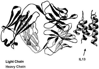
[0066] FIG. 10: Cocrystal structure of mAb13.2 Fab fragment with human
IL-13. X-ray crystallography of the mAb 13.2 Fab fragment reveals the light
chain with
dark shading, and the heavy chain in lighter shading. Also shown is the IL-13
structure
(at right). The figure also depicts interaction of the C-alpha helix of IL- 13
with the
CDR loops of the antibody.
[0067] FIG. 11: Human IL-13 sequence analysis showing mAb13.2 contact
sites. The panel shows the amino acid sequence of human IL-13, wherein the
arrow
indicates the signal peptide cleavage site, the four alpha helices are
underlined, the
antibody contact sites from the mAb 13.2 Fab - IL- 13 cocrystal structure are
highlighted in light boxes, and the ARG-variant residue is highlighted in a
dark box.
26
CA 02567129 2006-11-17
WO 2005/123126 PCT/US2005/020160
[0068] FIG. 12: Fab fragments of mAb13.2 bind to human IL-13. The figure
shows the average absorbance value (450 nm; y-axis) of biotinylated mAbl3.2
bound
to FLAG-human IL-13 in the presence of competing unlabeled mAb13.2 (1),
mAbl3.2
Fab fragments (e), or irrelevant antibody (*) at increasing concentrations (x-
axis)
expressed as (A) pM antibody or (B) pM binding sites.
[0069] FIG. 13: Fab fragments of mAbl3.2 neutralize IL-13-mediated TF1
proliferation and IL-13-mediated CD23 expression by human monocytes. (A) The
graph shows the percentage of the maximum proliferation by IL-13-dependent TF1
cell
line achieved (y-axis) after 3 days of incubation with IL- 13 and increasing
concentrations of competitor binding sites (x-axis) provided by either mAb
13.2 (+) or
mAb13.2 Fab fragments (a). (B) The figure shows the percentage of the maximum
number of monocytes that expressed cell surface CD23 (y-axis) as determined by
flow
cytometric analysis after peripheral blood mononuclear cells (PBMCs) isolated
from a
healthy donor were treated overnight with 1 ng/ml IL- 13 and indicated
concentrations
of competitor binding sites (x-axis) provided by either mAb 13.2 (+) or
mAbl3.2 Fab
fragments (a).
[0070] FIG. 14: Chimeric version (ch13.2) of the mouse monoclonal antibody
mAbl3.2 binds to and neutralizes IL-13. (A) Shown is the absorbance at 450 nm
(A4so;
y-axis) of samples containing IL-13-FLAG and biotinylated mAbl3.2 only (- -)
and
samples containing IL-13-FLAG, biotinylated mAB 13.2 and increasing
concentrations
(x-axis) of mAb 13.8 (o), mAb 13.2 (o), or chimeric mAb 13.2 (ch 13.2; &) as
determined
by ELISA. (B) Shown is the percentage of monocytes that expressed cell surface
CD23 (y-axis) after PBMCs isolated from a healthy donor were treated overnight
with
1 ng/ml IL-13 and indicated concentrations (x-axis) of purified mouse mAb 13.2
(a) or
chimeric mAb 13.2 (chl3.2; 0).
[0071] FIG. 15: Comparison of the human DP-54 germline gene with the
variable heavy (VH) chain amino acid sequences of a chimeric version of
mAbl3.2 and
a partially humanized version of mAb13.2 (h13.2v1). The figure shows the
complementarity determining regions (CDR) (boxed regions) and amino acid
sequences of DP-54, the variable heavy region of a chimeric version of mAbl3.2
(chimeric 13.2), and the variable heavy region of a partially humanized
version of
27
CA 02567129 2006-11-17
WO 2005/123126 PCT/US2005/020160
mAb 13.2 (h13.2v1) as aligned and compared with SEQWEBT'', wherein the amino
acid
substitutions made for the partially humanized version of mAb13.2 (h13.2v1)
are
indicated with shaded boxes and the residues left unchanged are underlined.
[0072] FIG. 16: Comparison of the human DPK9 germline gene with the
variable light (VL) chain amino acid sequences of a chimeric version of
mAbl3.2 and a
partially humanized version of mAbl3.2 (h13.2v1). The figure shows the
complementarity determining regions (CDR) (boxed regions) and amino acid
sequences of DPK-9, the variable light region of a chimeric version of mAb
13.2
(chimeric 13.2), and the variable light region of a partially humanized
version of
mAb 13.2 (h13.2v1) as aligned and compared with SEQWEB'm, wherein the amino
acid
substitutions made for the partially humanized version of mAb 13.2 (h13.2v1)
are
indicated with shaded boxes and the residues left unchanged are underlined.
[0073] FIG. 17: Comparison of the human DP-54 germline gene with the
variable heavy. (VH) chain amino acid sequences of a chimeric version of
mAbl3.2 and
a fully humanized version of mAb13.2 (h13.2v2). The figure shows the
complementarity determining regions (CDR) (boxed regions) and amino acid
sequences of DP-54, the variable heavy region of a chimeric version of mAb13.2
(chimeric 13.2), and the variable heavy region of a fully humanized version of
mAb13.2 (h13.2v2) as aligned and compared with SEQWEBTM, wherein the amino
acid
substitutions made for the fully humanized version of mAb13.2 (h13.2v2) are
indicated
with shaded boxes and the residues left unchanged are underlined.
[0074] FIG. 18: Comparison of the human DPK9 germline gene with the
variable light (VL) chain amino acid sequences of a chimeric version of mAb
13.2 and a
fully humanized version of mAb13.2 (h13.2v2). The figure shows the
complementarity
determining regions (CDR) (boxed regions) and amino acid sequences of DPK-9,
the
variable light region of a chimeric version of mAb 13.2 (chimeric 13.2), and
the variable
light region of a fully humanized version of mAb13.2 (h13.2v2) as aligned and
compared with SEQWEBTM, wherein the amino acid substitutions made for the
fully
humanized version of mAb13.2 (h13.2v2) are indicated with shaded boxes and the
residues left unchanged are underlined.
28
CA 02567129 2006-11-17
WO 2005/123126 PCT/US2005/020160
[0075] FIG. 19: Fully humanized mAbl3.2 (h13.2v2) retains full binding
activity to IL-13. (A) Shown is the absorbance at 450 nm (A45o; y-axis), as
determined
by ELISA, of samples containing IL-13-FLAG and biotinylated mAb 13.2 only (- -
) and
samples containing IL-13-FLAG, biotinylated mAB 13.2 and increasing
concentrations
(x-axis) of mAb13.2 (=), chimeric mAb13.2 (ch13.2; e), partially humanized
mAb13.2
(h13.2v1; A) or fully humanized mAB13.2 (h13.2v2; A). (B) The binding
interaction
between human IL-13 at different dose ranges and fully humanized mAbl3.2
(h13.2v2)
immobilized to a Biacorem chip is depicted as resonance difference (RU; y-
axis) over
time (x-axis). Kinetic constants for h13.2v2 also are shown.
[0076] FIG. 20: Chimeric mAb 13.2 (ch13.2), partially humanized inAB 13.2
(h13.2v1), and fully humanized mAb13.2 (h13.2v2) can neutralize IL-13-mediated
CD23 expression and IL-13-mediated phosphorylation of STAT6. (A) Shown is the
percentage of monocytes that expressed cell surface CD23 (y-axis) after PBMCs
isolated from a healthy donor were treated overnight with 1 ng/ml IL- 13 and
indicated
concentrations (x-axis) of chimeric mAb13.2 (ch13.2; =), partially humanized
mAbl3.2 (h13.2v1; ~), or fully humanized mAb13.2 (h13.2v2; a). (B) The percent
of
HT-29 cells that expressed phosphorylated STAT6 (y-axis) after incubation with
IL-13
and indicated concentration (x-axis) of chimeric mAb13.2 (ch13.2; =),
partially
humanized mAb13.2 (h13.2v1; ~), or fully humanized mAb13.2 (h13.2v2; e) as
determined by flow cytometric analysis is depicted.
[0077] FIG. 21: The ability of fully humanized mAbl3.2 (h13.2v2) to
neutralize human IL- 13 mediated CD23 expression is comparable to the ability
of
mAb13.2 to do the same. The number of gated monocytes that express cell
surface
CD23 after incubation with (A) recombinant human IL-13 or (B) native human IL-
13
and increasing concentrations (x-axis) of mAb13.2 (=) or fully humanized
mAb13.2
(h13.2v2; o) is presented as a percentage of the maximum number of monocytes
that
express cell surface CD23 after incubation with IL-13 alone (% Max. Response;
y-axis).
[0078] FIG. 22: The ability of fully humanized mAb13.2 (h13.2v2) to
neutralize nonhuman primate or sheep IL-13-mediated CD23 expression is
comparable
to the ability of mAb13.2 to do the same. The number of gated monocytes that
express
29
CA 02567129 2006-11-17
WO 2005/123126 PCT/US2005/020160
cell surface CD23 after incubation with (A) recombinant nonhuman primate IL-
13 (rec
NHP IL-13) or (B) recombinant sheep IL- 13 (rec Sheep IL-13) and increasing
concentrations (x-axis) of mAb 13.2 (=) or fully humanized mAb 13.2 (h13.2v2;
) is
presented as a percentage of the maximum number of monocytes that express cell
surface CD23 after incubation with IL-13 alone (% Max. Response; y-axis).
[0079] FIG. 23: Fully humanized mAb13.2 (h13.2v2) reduces late-phase
bronchoconstriction in sheep. The percent airway resistance (y-axis) as
measured 24
hours prior to Ascaris suum challenge (baseline), during challenge (ascaris),
and for
several hours after challenge (x-axis) is shown for sheep that were untreated
(control; +) or prophylactically treated with fully humanized mAbl3.2
(h13.2v2;u) at a
(A) 20 mg/kg or (B) 5 mg/kg dose. Data shown are mean s.d. for a sample size
of
three sheep per group.
[0080] FIG. 24: Fully humanized mAb13.2 (h13.2v2) prevents airway
hyperreactivity in sheep. The percent dose of carbachol required to elicit a
given
magnitude of response (PC400; y-axis) is shown for sheep prophylactically
treated with
(A) 20 mg/kg or (B) 5 mg/kg fully humanized mAb 13.2 (hl 3.2v2; u) or sheep
that
remained untreated (control; ~) pre- and post Ascaris suum challenge (x-axis).
Data
shown are mean s.d. for a sample size of three sheep per group.
[0081] FIG. 25: Fully humanized mAb13.2 (h13.2v2) prevents antigen-induced
lung inflammation in nonhuman primates. The figure shows the total number of
cells
found in bronchoalveolar lavage (BAL) samples (y-axis) taken prior to
challenge (0) or
24 hours post-challenge (24) with Ascaris suum antigen from control cynomolgus
monkeys treated intravenously with saline (0), positive control cynomolgus
monkeys
treated intramuscularly with 2 mg/kg dexamethasone (*), negative control
cynomolgus
monkeys pretreated with 8 mg/kg irrelevant human IgG (IVIG) (A), or cynomolgus
monkeys pretreated intravenously with fully humanized mAbl3.2 (h13.2v2) (v). A
bar
also depicts the mean number of cells found in BAL samples of each group. The
p-values obtained using unpaired T-tests are also shown.
[0082] FIG. 26: Humanization of mAb 13.2 may be based on sequence
homology to other human germline genes in VH Group 3 of V-BASE. Shown is the
CA 02567129 2006-11-17
WO 2005/123126 PCT/US2005/020160
alignment of the amino acid sequence of mAb 13.2 to human germline amino acid
sequences within VH group 3 of the database, V-base. Bolded sequences are
proposed
as those to which humanization of mAb 13.2 may be based.
[0083] FIG. 27: Comparison of the human DP-77 germline gene with the
variable heavy (VH) chain amino acid sequences of a chimeric version of
mAb13.2 and
a fully humanized version of mAb13.2 (h13.2v3). The figure shows the
complementarity determining regions (CDR) (boxed regions) and amino acid
sequences of DP-77, the variable heavy region of a chimeric version of mAb
13.2
(chimeric 13.2), and the variable heavy region of a fully humanized version of
mAb13.2 (h13.2v3) as aligned and compared with SEQWEB'', wherein the amino
acid
substitutions made for the fully humanized version of mAb13.2 (h13.2v3) are
indicated
with shaded boxes and the residues left unchanged are underlined.
[0084] FIG. 28: Comparison of the human B 1 germline gene with the variable
light (VL) chain amino acid sequences of a chimeric version of mAb13.2 and a
fully
humanized version of mAbl3.2 (h13.2v3). The figure shows the complementarity
determining regions (CDR) (boxed regions) and amino acid sequences of B 1, the
variable light region of a chimeric version of mAb 13.2 (chimeric 13.2), and
the variable
light region of a fully humanized version of mAb13.2 (h13.2v3) as aligned and
compared with SEQWEB.", wherein the amino acid substitutions made for the
fully
humanized version of mAb13.2 (h13.2v3) are indicated with shaded boxes and the
residues left unchanged are underlined.
[0085] FIG. 29: The number designation for each residue in the variable heavy
chain amino acid sequence of monoclonal antibody mAb 13.2 according to various
schemes. Shown is the number designation for each residue in the amino acid
sequence
of the variable heavy region of mAb 13.2 according to a linear sequence
numbering
scheme, the Chothia structure numbering scheme, and the Kabat sequence
numbering
scheme.
[0086] FIG. 30: The number designation for each residue in the variable light
chain amino acid sequence of monoclonal antibody mAb13.2 according to various
schemes. Shown is the number designation for each residue in the amino acid
sequence
of the variable light region of mAb 13.2 according to a linear sequence
numbering
31
CA 02567129 2006-11-17
WO 2005/123126 PCT/US2005/020160
scheme, the Chothia structure numbering scheme, and the Kabat sequence
numbering
scheme.
[0087] FIG. 31: Titers of Ascaris-specific IgE at 8 weeks post-Ascaris
challenge decreased in cynomolgus monkeys treated with anti-human IL-13
antibody
(h13.2v2). The graphs depict the relative titers of Ascaris-specific IgE in
cynomolgus
monkeys (y-axis) before and 8 weeks following challenge with Ascaris suum
antigen.
Pre-challenge values were normalized to "100." (A) Animals were treated
intravenously with humanized anti-IL-13 antibody (h13.2v2). (B) Control
animals
were administered saline or irrelevant human IgG (IVIG) intravenously.
[0088] FIG. 32: Anti-IL-13 antibody (h13.2v2) prevented the increase in
basophil sensitivity that follows in vivo allergen challenge. (A) Dose-
dependent
histamine release as a percentage of the maximum (y-axis) from blood of
representative
control and antibody-treated animals is depicted for animals before treatment
and 24
hours and 8 weeks after Ascaris antigen challenge. (B) The total histamine
release over
the dose range shown in (a) was determined for each animal at each time point.
The
figure shows mean and standard error of normalized histamine release at each
time
point for the Control and anti-IL-13 - treated groups. Pre = pre-antibody
treatment.
Time 0= 24 hr post-antibody, pre Ascaris lung challenge. The 24 hour, 8 week
and 4
month time points refer to time post Ascaris challenge.
[0089] FIG. 33: Binding of biotinylated h13.2v2 to IL-13-loaded A375 cells.
The histograms depict the number of A375 human melanoma cells (counts; y-axis)
that
demonstrated binding (fluorescence; x-axis) of biotinylated h13.2v2 (0.1
g.g/ml, 1
g/m1, or 10 gg/m1) following pre-incubation with 3 ng/ml human IL-13, as
determined
by flow cytometric analysis.
[0090] FIG. 34: The wild-type Fc revertant of h13.2v2 mediates ADCC in the
presence of IL-13. (A) The percentage of Chromium-51 release (y-axis) of A375
human melanoma cells treated with the indicated concentration (x-axis) of
h13.2v2
(L234A / G237A; =) or its wild-type Fc revertant (o) is shown. (B) The
percentage of
Chromimum-51 released (y-axis) is shown for IL-13 loaded A375 cells treated
with the
wild-type Fc revertant of h13.2v2 in the presence or absence of IL-13.
32
CA 02567129 2006-11-17
WO 2005/123126 PCT/US2005/020160
[0091] FIG. 35: The wild-type Fc revertant of h13.2v2 mediates ADCC of IL-
13Ra2-expressing A375 cells, but not of IL- 1 3Ra 1 -expressing HT-29 cells.
(A) The
percentage of Chromium-51 release (y-axis) of IL-13-loaded A375 human melanoma
cells treated with the indicated concentration (x-axis) of h13.2v2 (L234A /
G237A; =)
or its wild-type Fc revertant (0) is shown. (B) The percentage of Chromium-51
release
(y-axis) of IL-13-loaded HT-29 human epithelial cells treated with the
indicated
concentration (x-axis) of h13.2v2 (L234A / G237A; =) or its wild-type Fc
revertant (0)
is shown.
DETAILED DESCRIPTION
[0092] IL-13 binding agents, e.g., anti-IL13 antibodies and antigen-binding
fragments thereof, pharmaceutical compositions thereof, nucleic acids encoding
the
aforesaid antibodies, as well as vectors and host cells containing the
aforesaid nucleic
acid sequences, are disclosed. Methods of producing the aforesaid antibodies,
as well
as methods for modulating one or more IL- 13 -associated activities using
antibodies that
bind to IL-13, e.g., human IL-13, and reduce or prevent it from binding to its
receptor
are also disclosed. Anti-IL- 13 antibodies can be used to -mitigate IL- 1 3-
mediated
disorders, e.g., for treating respiratory disorders (e.g., asthma); atopic
disorders (e.g.,
allergic rhinitis); inflammatory and/or autoimmune conditions of, the skin
(e.g., atopic
dermatitis), gastrointestinal organs (e.g., inflammatory bowel diseases
(IBD)), as well
as fibrotic and cancerous disorders. Anti-IL- 13 antibodies can be used alone
or in
combination with other therapies used to treat the same or another disease,
e.g., an
allergic response.
[0093] It has been found, inter alia, that a reduction of IL-13 activity by
using
the antibodies described herein, which interfere with the formation of a
functional
IL-13 signaling complex, reduces airway inflammation in cynomolgus monkeys
naturally allergic to Ascaris suum (Examples 1.4 and 3.5, below). In addition,
the anti-
human IL-13 antibodies described herein prevent late-phase bronchoconstriction
in
sheep naturally allergic to Ascaris suum, and prevent carbachol-induced airway
hyperresponsiveness in sheep (Example 3.5, below). Accordingly, anti-IL-13
33
CA 02567129 2006-11-17
WO 2005/123126 PCT/US2005/020160
antibodies that neutralize one or more IL-13-associated activities may be used
to reduce
one or more IL-13-associated activities in vivo, e.g., to treat or prevent IL-
13-mediated
disorders (e.g., asthma, airway inflammation, eosinophilia, fibrosis, and
excess mucus
production).
[0094] Anti-Human IL-13 Antibodies
[0095] Antibodies that are capable of binding to, neutralizing and/or
inhibiting
one or more IL- 13 -associated activities, particularly the signaling activity
of IL-13, are
useful for treating IL-13-mediated diseases, such as allergic asthma,
nonallergic
asthma, and asthma-related pathologies, such as fibrosis, eosinophilia, and
mucus
production.
[0096] In one embodiment, the anti-IL13 antibodies disclosed herein are
isolated or purified. An "isolated" or "purified" polypeptide or protein,
e.g., an
"isolated antibody," is purified to a state beyond that in which it exists in
nature. For
example, the "isolated" or "purified" polypeptide or protein, e.g., an
"isolated
antibody," refers to a protein that is separated from at least one component
of cellular
material or other contaminatirig proteins from the cell or tissue source from
which the
protein is derived, or separated from at least one component of the chemical
precursors
or other chemicals when chemically synthesized. For example, an isolated
protein can
be substantially free of other proteins, other cellular material, or chemical
precursors.
In some embodiments, the preparation of antibody protein having less than
about 50%
of non-antibody protein (also referred to herein as a "contaminating
protein"), or of
chemical precursors, is considered to be "substantially free." In other
embodiments,
40%, 30%, 20%, 10% and more preferably 5% (by dry weight), of non-antibody
protein, or of chemical precursors is considered to be substantially free.
When the
antibody protein or biologically active portion thereof is recombinantly
produced, it is
also preferably substantially free of culture medium, e.g., the culture medium
represents less than about 30%, 20%, more preferably less than about 10%, and
most
preferably less than about 5% of the volume or mass of the protein
preparation.
Proteins or polypeptides referred to herein as "recombinant" are proteins or
polypeptides produced by the expression of recombinant nucleic acids. Proteins
can be
purified by standard methods (including, e.g., ion exchange and affinity
34
CA 02567129 2006-11-17
WO 2005/123126 PCT/US2005/020160
chromatography) to provide preparations in which a particular protein is at
least 5, 10,
20, 25, 50, 75, 80, 90, 95, 98, 99% pure relative to other proteins or
relative to other
biologically active components.
[0097] The term "antibody" as used herein includes intact antibodies,
fragments
of antibodies, e.g., Fab, F(ab')2 Fd, dAb and scFv fragments, and intact
antibodies and
fragments that have been mutated either in their constant and/or variable
region (e.g.,
mutations to produce chimeric, partially humanized, or fully humanized
antibodies, as
well as to produce antibodies with a desired trait, e.g., enhanced IL-13
binding and/or
reduced FcR binding). Exemplary antibodies bind specifically to IL-13, and
may, for
example, interfere with the formation of a functional IL-13 signaling complex,
and/or
neutralize or inhibit one or more IL-13-associated activities.
[0098] The antibodies described herein can be effectively human. An
"effectively human" antibody is an antibody that includes a sufficient number
of human
amino acid positions such that the antibody does not elicit an immunogenic
response in
a normal human. Preferably, the protein does not evoke a neutralizing antibody
response, e.g., the human anti-murine antibody (HAMA) response. HAMA can be
problematic in a number of circumstances, e.g., if the antibodies are desired
to be
administered repeatedly, e.g., in treatment of a chronic or recurrent disease
condition.
A HAMA response can make repeated antibody administration potentially
ineffective
because of an increased antibody clearance from the serum (see, e.g., Saleh et
al.,
CanceYlnnzmunol. hnmunotheY., 32:180-190 (1990)) and also because of potential
allergic reactions (see, e.g., LoBuglio et al., Hybridoma, 5:5117-5123
(1986)).
[0100] Conservative substitutions typically include the substitution of one
amino acid for another with similar characteristics, e.g., substitutions
within the
following groups: valine, glycine; glycine, alanine; valine, isoleucine,
leucine; aspartic
acid, glutamic acid; asparagine, glutamine; serine, threonine; lysine,
arginine; and
phenylalanine, tyrosine
[0101] Antibodies, also known as immunoglobulins, are typically tetrameric
glycosylated proteins composed of two light (L) chains of approximately 25 kDa
each
and two heavy (H) chains of approximately 50 kDa each. Two types of light
chain,
termed lambda and kappa, may be found in antibodies. Depending on the amino
acid
CA 02567129 2006-11-17
WO 2005/123126 PCT/US2005/020160
sequence of the constant domain of heavy chains, immunoglobulins can be
assigned to
five major classes: A, D, E, G, and M, and several of these may be further
divided into
subclasses (isotypes), e.g., IgGt, IgG2, IgG3, IgG4, IgA1, and IgA2. Each
light chain
includes an N-terminal variable (V) domain (VL) and a constant (C) domain
(CL).
Each heavy chain includes an N-terminal V domain (VH), three or four C domains
(CHs), and a hinge region. The CH domain most proximal to VH is designated as
CHl . The VH and VL domains consist of four regions of relatively conserved
sequences called framework regions (FR1, FR2, FR3, and FR4), which form a
scaffold
for three regions of hypervariable sequences (complementarity determining
regions,
CDRs). The CDRs contain most of the residues responsible for specific
interactions of
the antibody with the antigen. CDRs are referred to as CDR1, CDR2, and CDR3.
Accordingly, CDR constituents on the heavy chain are referred to as H1, H2,
and H3,
while CDR constituents on the light chain are referred to as Ll, L2, and L3.
CDR3 is
typically the greatest source of molecular diversity within the antibody-
binding site.
H3, for example, can be as short as two amino acid residues or greater than 26
amino
acids. The subunit structures and three-dimensional configurations of
different classes
of immunoglobulins are well known in the art. For a review of the antibody
structure,
see Antibodies: A Laboratory Manual, Cold Spring Harbor Laboratory, eds.
Harlow et
al., 1988. One of skill in the art will recognize that each subunit structure,
e.g., a CH,
VH, CL, VL, CDR, FR structure, comprises active fragments, e.g., the portion
of the
VH, VL, or CDR subunit the binds to the antigen, i.e., the antigen-binding
fragment, or,
e.g., the portion of the CH subunit that binds to and/or activates, e.g., an
Fc receptor
and/or complement. The CDRs typically refer to the Kabat CDRs, as described in
Sequences of Proteins of Immunological Interest, US Department of Health and
Human
Services (1991), eds. Kabat et al. Another standard for characterizing the
antigen
binding site is to refer to the hypervariable loops as described by Chothia.
See, e.g.,
Chothia, D. et al. (1992) J Mol. Biol. 227:799-817; and Tomlinson et al.
(1995) EMBO
J. 14:4628-4638. Still another standard is the AbM definition used by Oxford
Molecular's AbM antibody modelling software. See, generally, e.g., Protein
Sequence
and Structure Analysis ofAntibody Variable Dofnains. In: Antibody Engineering
Lab
Manual (Ed.: Duebel, S. and Kontermann, R., Springer-Verlag, Heidelberg).
Embodiments described with respect to Kabat CDRs can alternatively be
implemented
36
CA 02567129 2006-11-17
WO 2005/123126 PCT/US2005/020160
using similar described relationships with respect to Chothia hypervariable
loops or to
the AbM-defined loops.
[0102] Examples of binding fragments encompassed within the term "antigen-
binding fragment" of an antibody include (i) a Fab fragment, a monovalent
fragment
consisting of the VL, VH, CL and CH1 domains; (ii) a F(ab')2 fragment, a
bivalent
fragment comprising two Fab fragments linked by a disulfide bridge at the
hinge
region; (iii) a Fd fragment consisting of the VH and CHl domains; (iv) a Fv
fragment
consisting of the VL and VH domains of a single arm of an antibody, (v) a dAb
fragment (Ward et al. (1989) Nature 341:544-46), which consists of a VH
domain; and
(vi) an isolated complementarity determining region (CDR). Camelid antibodies,
and
camelized antibodies can also be used. Such antibodies, e.g., can include CDRs
from
just one of the variable domains described herein. Furthermore, although the
two
domains of the Fv fragment, VL and VH, are coded for by separate genes, they
can be
joined, using recombinant methods, by a synthetic linker that enables them to
be made
as a single protein chain in which the VL and VH regions pair to form
monovalent
molecules (known as single chain Fv (scFv); see, e.g., Bird et al. (1988)
Science
242:423-26; Huston et al. (1988) Proc. Natl. Acad. Sci. U.S.A. 85:5879-83).
Such
single chain antibodies are also intended to be encompassed within the term
"antigen-
binding fragment" of an antibody. These antibody fragments are obtained using
conventional techniques known to those skilled in the art, and the fragments
are
evaluated for function in the same manner as are intact antibodies.
[0103] Antibody diversity, in a natural system, is created by the use of
multiple
germline genes encoding variable regions and a variety of somatic events. The
somatic
events include recombination of variable gene segments with diversity (D) and
joining
(J) gene segments to make a complete VH region, and the recombination of
variable
and joining gene segments to make a complete VL region. The recombination
process
itself can be imprecise, resulting in the loss or addition of amino acids at
the V(D)J
junctions. These mechanisms of diversity occur in the developing B cell prior
to
antigen exposure. After antigenic stimulation, the expressed antibody genes in
B cells
undergo somatic mutation. Based on the estimated number of germline gene
segments,
the random recombination of these segments, and random VH-VL pairing, up to
1.6 x 10⁷ different antibodies could be produced (Fundamental Immunology, 3rd
ed.
37
CA 02567129 2006-11-17
WO 2005/123126 PCT/US2005/020160
(1993), ed. Paul, Raven Press, New York, NY). When other processes that
contribute
to antibody diversity (such as somatic mutation) are taken into account, it is
thought
that upwards of 1010 different antibodies could be generated (Immunoglobulin
Genes,
2nd ed. (1995), eds. Jonio et al., Academic Press, San Diego, CA). Because of
the
many processes involved in generating antibody diversity, it is unlikely that
independently derived monoclonal antibodies with the same antigen specificity
will
have identical amino acid sequences.
[0104] Thus, this disclosure provides, inter alia, antibodies, and antigen-
binding
fragments thereof, that bind to IL-13 and, e.g., interfere with the fonnation
of a
functional IL-13 signaling complex. The antigen-binding fragments described
herein,
e.g., structures containing a CDR, will generally be an antibody heavy or
light chain
sequence, or an active fragment thereof, in which the CDR is placed at a
location
corresponding to the CDR of naturally occurring VH and VL. The structures and
locations of immunoglobulin variable domains may be determined as described in
Sequences of Proteins of Immunological Interest, U.S. Department of Health and
Human Services (1991), eds. Kabat et al.
[0105] Antibody molecules (including antigen-binding fragments) disclosed
herein, i.e., antibody molecules that bind to IL-13 and interfere with the
formation of a
functional IL-13 signaling complex, include, but are not limited to, murine
monoclonal
antibody, mAb13.2, and its variants, specifically the chimeric variant chl3.2,
the
partially humanized variant h13.2v1, and the fully humanized variants h13.2v2
and
h13.2v3. These antibody molecules may be useful in preventing or treating
asthma
(both allergic and nonallergic), as well as asthma-related pathologies. The
amino acid
sequences of the light chain variable regions of mAb13.2, ch13.2, h13.2v1,
h13.2v2,
and h13.2v3 are set forth in SEQ ID NOs:9, 10, 11, 12, and 35, respectively.
The
amino acid sequences of the heavy chain variable regions of mAb 13.2, ch13.2,
h13.2v1, h13.2v2, and h13.2v3 are set forth in SEQ ID NOs:13, 14, 15, 16, and
36,
respectively. The amino acid sequences of the three complementarity
determining
regions (CDRs) in the variable light chains of mAbl3.2, chl3.2, h13.2v1,
h13.2v2, and
h13.2v3 are set forth in SEQ ID NOs:19, 20, and 21. The amino acid sequences
of the
three CDRs in the variable heavy chains of mAb13.2, chl3.2, h13.2v1, h13.2v2,
and
h13.2v3 are set forth in SEQ ID NOs:22, 23, and 24.
38
CA 02567129 2006-11-17
WO 2005/123126 PCT/US2005/020160
[0106] As described above, the CDRs contain most of the residues responsible
for specific interactions with the antigen, and are contained within the VH
and VL
domains, i.e., the heavy chain variable region and the light chain variable
region,
respectively. Exemplary antibodies include at least one CDR comprising an
amino acid
sequence selected from the amino acid sequences set forth in SEQ ID NOs:l9-24,
or
selected residues, particularly IL-13 contact residues from such CDRs. Such
antibodies
may also bind to IL-13 and, e.g., interfere with the formation of a functional
IL-13
signaling complex. The amino acid sequences of the active fragments of the
CDRs,
i.e., the minimum core CDR sequences, described herein are set forth in SEQ ID
NOs:25-30, and are disclosed in Table 1. An antibody may include one or more
CDRs
of the VL chain as set forth in SEQ ID NOs:19-21 or SEQ ID NOs: 25-27. An
antibody may include one or more CDRs of the VH chain as set forth in SEQ ID
NOs:22-24 or SEQ ID NOs: 28-30. Additionally, an antibody may include one or
more
of the CDRs of the VL and VH chain has an amino acid sequence selected from
the
amino acid sequences set forth in SEQ ID NOs:19-30. As shown, X can be any
amino
acid, e.g., a non-cysteine amino acid, or an amino acid that is has a similar
charge,
hydrophobicity, and/or side chain length, as the amino acid at the
corresponding
position in the left hand column of Table 1.
Table 1. Exemplary CDRs
CDR sequence Minimum core CDR sequence
Light chain CDR1 (Ll)
SEQ ID NO:19 SEQ ID NO:25
24-KASESVDNYGKSLMH-381 24-xxxxxxxNYxKxxxx-38
Light chain CDR2 (L2)
SEQ ID NO:20 SEQ ID NO:26
54-RASNLES-60 54-Rxxxxxx-60
Light chain CDR3 (L3)
SEQ ID NO:21 SEQ ID NO:27
93-QQSNEDPWT-101 93-xxxNxDxWx-101
Heavy chain CDR1 (H1)
SEQ ID NO:22 SEQ ID NO:28
30-ISYAMS-352 30-ISxAxx-35
Heavy chain CDR2 (H2)
SEQ ID NO:23 SEQ ID NO:29
50-SISSGGNTYYPDSVKG-65 50-SxSSxxxxxYxxxxxx-65
Heavy chain CDR3 (H3)
SEQ ID NO:24 SEQ ID NO:30
98-LDGYYFGFAY-107 98-LDGYYIxxxx-107
Numbering for VL CDRs is according to a linear sequence numbering scheme as in
FIG. 30.
39
CA 02567129 2006-11-17
WO 2005/123126 PCT/US2005/020160
Z Numbering for VH CDRs is according to a linear sequence numbering scheme as
in FIG. 29.
[0107] Also described above, an antigen-binding fragment may be an Fv
fragment, which includes VH and VL domains. Thus, an Fv fragment of mAb13.3,
chl3.2, h13.2v1, h13.2v2 or h13.2v3 may constitute an antibody described
herein. It
may bind to IL- 13 and interfere with the formation of a functional IL- 13
signaling
complex. Other fragments include the Fv fragment, e.g., scFv fragments, Fab
fragments, and F(ab')2 fragments of the mAbl3.3, chl3.2, h13.2v1, h13.2v2, or
h13.2v3 antibodies or of an antibody that includes one or more CDRs having an
amino
acid sequence selected from the amino acid sequences set forth in SEQ ID NOs:
19-30.
[0108] Such antibody molecules may be produced by methods known to those
skilled in the art. For example, monoclonal antibodies may be produced by
generation
of hybridomas in accordance with known methods. Hybridomas formed in this
manner
are then screened using standard methods, such as enzyme-linked immunosorbent
assay
(ELISA) and surface plasmon resonance (BiacoreTM) analysis, to identify one or
more
hybridomas that produce an antibody that specifically binds with IL-13,
interferes with
the formation of a functional IL- 13 signaling complex, and neutralizes one or
more
IL-13-associated activities. Recombinant IL-13, naturally occurring IL-13
(i.e., the
processed mature form of IL-13), any variants thereof, and aniigenic peptide
fragments
of IL-13 may be used as the immunogen.
[0109] An antigenic peptide fragment of IL-13 can comprise at least 7
continuous amino acid residues and encompasses an epitope such that an
antibody
raised against the peptide forms a specific immune complex with IL- 13.
Preferably, the
antigenic peptide comprises at least 10 amino acid residues, more preferably
at least 15
amino acid residues, even more preferably at least 20 amino acid residues, and
most
preferably at least 30 amino acid residues. Additionally, it is preferable
that the
antigenic peptide fragment of IL-13 comprises the IL-13 receptor-binding site
or IL-4
receptor binding site.
[0110] As an alternative to preparing monoclonal antibody-secreting
hybridomas, a monoclonal antibody may be identified and isolated by screening
a
recombinant combinatorial immunoglobulin library (e.g., an antibody phage
display
CA 02567129 2006-11-17
WO 2005/123126 PCT/US2005/020160
library) with IL- 13, including variants and/or portions thereof, to thereby
isolate
immunoglobulin library members that bind to IL-13. Techniques and commercially
available kits for generating and screening phage display libraries are known
to those
skilled in the art. Additionally, examples of methods and reagents
particularly
amenable for use in generating and screening antibody display include those
described
in US 5,658,727, US 5,667,988, and US 5,885,793.
[0111] Polyclonal sera and antibodies described herein may be produced by
immunizing a suitable subject with IL- 13, its variants and/or portions
thereof. The
antibody titer in the immunized subject may be monitored over time by standard
techniques, such as an Enzyme Linked Immunosorbent Assay (ELISA), using
immobilized IL-13 or other marker proteins (e.g., FLAG). Antibodies may be
isolated
from an animal or culture media. A variety of methods can be used to purify
antibodies
including well-known techniques, such as use of protein A chromatography to
obtain
an IgG fraction.
[0112] Certain embodiments comprise the VH and/or VL domain of the Fv
fragment of mAb13.2, ch13.2, h13.2v1, h13.2v2 or h13.2v3. Fragments of
antibodies
e.g., Fab, F(ab')2, Fd, and dAb fragments, may be produced by cleavage of the
antibodies or by recombinant engineering. For example, immunologically active
Fab
and F(ab')2 fragments may be generated by treating the antibodies with an
enzyme
such as pepsin.
[0113] Further embodiments comprise one or more complementarity
determining regions (CDRs) of any of these VH and VL domains, as set forth in
SEQ
ID NOs:19-30. One embodiment comprises an H3 fragment of the VH domain of
mAbl3.2, chl3.2, h13.2v1, h13.2v2, or h13.2v3.
[01141 The VH and VL domains described herein, in certain embodiments, can
be germlined, i.e., the framework regions (FRs) of these domains may be
changed
using conventional molecular biology techniques to match human germline genes
or
the consensus amino acid sequences of human germline gene products, at one or
more
positions (e.g., at at least 70, 80, 85, 90, 95, 97, 98, or 99% of framework
positions). In
other embodiments, the framework sequences remain diverged from the gennline.
41
CA 02567129 2006-11-17
WO 2005/123126 PCT/US2005/020160
[0115] Human germline sequences, for example, are disclosed in Tomlinson,
I.A. et al. (1992) J. Mol. Biol. 227:776-798; Cook, G. P. et al. (1995)
bnrnunol. Today
Vol. 16 (5): 237-242; Chothia, D. et al. (1992) J. Mol. Biol. 227:799-817; and
Tomlinson et al. (1995) EMBO J 14:4628-4638. The V BASE directory provides a
comprehensive directory of human immunoglobulin variable region sequences
(compiled by Tomlinson, I.A. et al. MRC Centre for Protein Engineering,
Cambridge,
UK). These sequences can be used as a source of human sequence, e.g., for
framework
regions and CDRs. Consensus human framework regions can also be used, e.g., as
described in US 6,300,064.
[0116] Additionally, chimeric, humanized, and single-chain antibodies
described herein, comprising both human and nonhuman portions, may be produced
using standard recombinant DNA techniques, as described in more detail in the
Examples. Humanized antibodies may also be produced using transgenic mice that
express human heavy and light chain genes, but are incapable of expressing the
endogenous mouse immunoglobulin heavy and light chain genes.
[0117] Additionally, the antibodies described herein also include those that
bind
to IL-13, interfere with the formation of a functional IL-13 signaling
complex, and have
mutations in the constant regions of the heavy and light chains. It is
sometimes
desirable to mutate and inactivate certain fragments of the constant region.
For
example, mutations in the heavy constant region can be made to produce
antibodies
with reduced binding to the Fc receptor (FcR) and/or complement; such
mutations are
well known in the art. An example of such a mutation to the amino sequence of
the
constant region of the heavy chain of IgG is provided in SEQ ID NO:17. Certain
active
fragments of the CL and CH subunits (e.g., CH1) are covalently link to each
other. A
further aspect provides a method for obtaining an antibody antigen-binding
domain
specific for domain of IL-13 that aids formation of a functional IL-13
signaling
complex.
[0118] Exemplary antibodies can include sequences of VL chains as set forth in
SEQ ID NOs:9, 10, 11, 12, or 35, and/or of VH chains as set forth in and SEQ
ID NOs:
13, 14, 15, 16, or 36, but also can include variants of these sequences that
retain
antigen-binding ability. Such variants may be derived from the provided
sequences
42
CA 02567129 2006-11-17
WO 2005/123126 PCT/US2005/020160
using techniques well known in the art. Amino acid substitutions, deletions,
or
additions, can be made in either the FRs or in the CDRs. While changes in the
framework regions are usually designed to improve stability and reduce
immunogenicity of the antibody, changes in the CDRs are usually designed to
increase
affinity of the antibody for its target. Such affinity-increasing changes are
typically
determined empirically by altering the CDR region and testing the antibody.
Such
alterations can be made according to the methods described in Antibody
Engineering,
2nd. ed. (1995), ed. Borrebaeck, Oxford University. Press.
[0119] An exemplary method for making a VH domain, which is an amino acid
sequence variant of a VH domain set out herein, comprises a step of adding,
deleting,
substituting or inserting one or more amino acids in the amino acid sequence
of the
presently disclosed VH domain, optionally combining the VH domain thus
provided
with one or more VL domains, and testing the VH domain or VH/VL combination or
combinations for specific binding to IL-13, and (preferably) testing the
ability of such
antigen-binding domain to modulate one or more IL-13-associated activities.
The VL
domain may have an amino acid sequence that is substantially as set out
herein. An
analogous method may be employed in which one or more sequence variants of a
VL
domain disclosed herein are combined with one or more VH domains.
[0120] A further aspect of the invention provides a method of preparing an
antigen-binding fragment that specifically binds to IL-13. The method
comprises: (a)
providing a starting repertoire of nucleic acids encoding a VH domain that
either
includes a CDR3 to be replaced or lacks a CDR3 encoding region; (b) combining
the
repertoire with a donor nucleic acid encoding a donor CDR comprising an active
fragment of SEQ ID NO:24, e.g., a donor nucleic acid encoding the amino acid
sequence set forth in SEQ ID NO:30, such that the donor nucleic acid is
inserted into
the CDR3 region in the repertoire so as to provide a product repertoire of
nucleic acids
encoding a VH domain; (c) expressing the nucleic acids of the product
repertoire; (d)
selecting a specific antibody or antigen-binding fragment specific for IL- 13;
and (e)
recovering the specific antibody or antigen-binding fragment or nucleic acid
encoding
it.
43
CA 02567129 2006-11-17
WO 2005/123126 PCT/US2005/020160
[0121] In another embodiment, an analogous method may be employed in
which a VL CDR3 (i.e., L3) described herein is combined with a repertoire of
nucleic
acids encoding a VL domain, which either includes a CDR3 to be replaced or
lacks a
CDR3-encoding region. A coding sequence of a CDR described herein (e.g., CDR3)
may be introduced into a repertoire of variable domains lacking a CDR (e.g.,
CDR3),
using recombinant DNA technology. For example, Marks et al. (Bio/Technol gy
(1992) 10:779-83) describes methods of producing repertoires of antibody
variable
domains in which consensus primers directed at or adjacent to the 5' end of
the variable
domain area are used in conjunction with consensus primers to the third
framework
region of human VH genes to provide a repertoire of VH variable domains
lacking a
CDR3. The repertoire may be combined with a CDR3 of a particular antibody.
Using
analogous techniques, the CDR3-derived sequences may be shuffled with
repertoires of
VH or VL domains lacking a CDR3, and the shuffled complete VH or VL domains
combined with a cognate VL or VH domain to provide specific antigen-binding
fragments. The repertoire may then be displayed in a suitable host system such
as the
phage display system of WO 92/01047, so that suitable antigen-binding
fragments can
be selected. Analogous shuffling or combinatorial techniques are also
disclosed by
Stemmer (Nature (1994) 370:389-91). A further alternative is to generate
altered VH
or VL regions using random mutagenesis of one or more selected VH and/or VL
genes
to generate mutations within the entire variable domain. See, e.g., Gram et
al. Proc.
Nat. Acad. Sci. U.S.A. (1992) 89:3576-80.
[0122] Another method that may be used is to direct mutagenesis to CDR
regions of VH or VL genes. Such techniques are disclosed by, e.g., Barbas et
al. (Proc.
Nat. Acad. Sci. U.S.A. (1994) 91:3809-13) and Schier et al. (,T. Mol. Biol.
(1996)
263:551-67). Similarly, one or more, or all three CDRs may be grafted into a
repertoire
of VH or VL domains, or even some other scaffold (such as a fibronectin
domain). The
resulting protein is evaluated for ability to bind to IL- 13.
[0123] In one embodiment, a binding protein that binds to a target is
modified,
e.g., by mutagenesis, to provide a pool of modified binding proteins. The
modified
binding proteins are then evaluated to identify one or more altered binding
proteins
which have altered functional properties (e.g., improved binding, improved
stability,
lengthened stability in vivo). In one implementation, display library
technology is used
44
CA 02567129 2006-11-17
WO 2005/123126 PCT/US2005/020160
to select or screen the pool of modified binding proteins. Higher affinity
binding
proteins are then identified from the second library, e.g., by using higher
stringency or
more competitive binding and washing conditions. Other screening techniques
can also
be used.
[0124] In some embodiments, the mutagenesis is targeted to regions known or
likely to be at the binding interface. If, for example, the identified binding
proteins are
antibodies, then mutagenesis can be directed to the CDR regions of the heavy
or light
chains as described herein. Further, mutagenesis can be directed to framework
regions
near or adjacent to the CDRs, e.g., framework regions, particular within 10,
5, or 3
amino acids of a CDR junction. In the case of antibodies, mutagenesis can also
be
limited to one or a few of the CDRs, e.g., to make step-wise improvements.
[0125] In one embodiment, mutagenesis is used to make an antibody more
similar to one or more germline sequences. One exemplary germlining method can
include: identifying one or more germline sequences that are similar (e.g.,
most similar
in a particular database) to the sequence of the isolated antibody. Then
mutations (at
the amino acid level) can be made in the isolated antibody, either
incrementally, in
combination, or both. For example, a nucleic acid library that includes
sequences
encoding some or all possible germline mutations is made. The mutated
antibodies are
then evaluated, e.g., to identify an antibody that has one or more additional
germline
residues relative to the isolated antibody and that is still useful (e.g., has
a functional
activity). In one einbodiment, as many germline residues are introduced into
an
isolated antibody as possible.
[0126] In one embodiment, mutagenesis is used to substitute or insert one or
more germline residues into a CDR region. For example, the germline CDR
residue
can be from a germline sequence that is similar (e.g., most similar) to the
variable
region being modified. After mutagenesis, activity (e.g., binding or other
functional
activity) of the antibody can be evaluated to determine if the germline
residue or
residues are tolerated. Similar mutagenesis can be performed in the framework
regions.
[0127] Selecting a germline sequence can be performed in different ways. For
example, a germline sequence can be selected if it meets a predetermined
criteria for
CA 02567129 2006-11-17
WO 2005/123126 PCT/US2005/020160
selectivity or similarity, e.g., at least a certain percentage identity, e.g.,
at least 75, 80,
85, 90, 91, 92, 93, 94, 95, 96, 97, 98, 99, or 99.5% identity. The selection
can be
performed using at least 2, 3, 5, or 10 germline sequences. In the case of
CDR1 and
CDR2, identifying a similar germline sequence can include selecting one such
sequence. In the case of CDR3, identifying a similar germline sequence can
include
selecting one such sequence, but may including using two germline sequences
that
separately contribute to the amino-terminal portion and the carboxy-terminal
portion.
In other implementations more than one or two germline sequences are used,
e.g., to
form a consensus sequence.
[0128] In one embodiment, an antibody or fragment thereof has CDR sequences
that differ only insubstantially from those of the antibodies described
herein.
Insubstantial differences include minor amino acid changes, such as
substitutions of 1
or 2 out of any of typically 5-7 amino acids in the sequence of a CDR, e.g., a
Chothia
or Kabat CDR. Typically an amino acid is substituted by a related amino acid
having
similar charge, hydrophobic, or stereochemical characteristics. Such
substitutions
would be within the ordinary skills of an artisan. Unlike in CDRs, more
substantial
changes in structure framework regions (FRs) can be made without adversely
affecting
the binding properties of an antibody. Changes to FRs include, but are not
limited to,
humanizing a nonhuman-derived framework or engineering certain framework
residues
that are important for antigen contact or for stabilizing the binding site,
e.g., changing
the class or subclass of the constant region, changing specific amino acid
residues
which might alter an effector function such as Fc receptor binding (Lund et
al. (1991) J.
Imnaunol. 147:2657-62; Morgan et al. (1995) Irnmunology 86:319-24), or
changing the
species from which the constant region is derived. Antibodies may have
mutations in
the CH2 region of the heavy chain that reduce or alter effector function,
e.g., Fc
receptor binding and complement activation. For example, antibodies may have
mutations such as those described in U.S. Patent Nos. 5,624,821 and 5,648,260.
In the
IgGl or IgG2 heavy chain, for example, such mutations may be made to resemble
the
amino acid sequence set forth in SEQ ID NO:17. Antibodies may also have
mutations
that stabilize the disulfide bond between the two heavy chains of an
immunoglobulin,
such as mutations in the hinge region of IgG4, as disclosed in the art (e.g.,
Angal et al.
(1993) Mol. Immunol. 30:105-08).
46
CA 02567129 2006-11-17
WO 2005/123126 PCT/US2005/020160
[0129] The IL-13 binding proteins can be in the form of intact antibodies,
fragments of antibodies, e.g., Fab, F(ab')2, Fd, dAb, and scFv fragments, and
intact
antibodies and fragrnents that have been mutated either in their constant
and/or variable
region (e.g., mutations to produce chimeric, partially humanized, or fully
humanized
antibodies, as well as to produce antibodies with a desired trait, e.g.,
enhanced IL 13
binding and/or reduced FcR binding).
[0130] In some embodiments, a substantial portion of an immunoglobulin
variable domain can comprise at least one of the CDR regions and, optionally,
their
intervening framework regions from the variable regions as set out herein. The
portion
will also include at least about 50, 60, 70, 80, 85, 87, 88, 90, 92, 94, 95,
96, 97, 98% of
either or both of FRI and FR4. For example, the portion which may be
contiguous or
non-contiguous may include the C-terminal 50% of FRI and the N-termina150 10
of
FR4. Additional residues at the N-terminal or C-terminal end of the
substantial part of
the variable domain may be those not normally associated with naturally
occurring
variable domain regions. For example, construction of specific antigen-binding
fragments made by recombinant DNA techniques may result in the introduction of
N-
or C-terminal residues encoded by linkers introduced to facilitate cloning or
other
manipulation steps. Other manipulation steps include the introduction of
linkers to join
variable domains described herein to further protein sequences, including
immunoglobulin heavy chains, other variable domains (e.g., in the production
of
diabodies) or protein labels as discussed in more detail below.
[0131] Although the embodiments illustrated in the Examples comprise a
"matching" pair of VH and VL domains, the invention also enconlpasses binding
fragments containing a single variable domain derived from either VH or VL
domain
sequences, especially VH domains. In the case of either of the single-chain
specific
binding domains, these domains may be used to screen for complementary domains
capable of forming a two-domain specific antigen-binding domain capable of
binding
IL-13. This may be achieved by phage display screening methods using the so-
called
hierarchical dual combinatorial approach (as disclosed in, e.g., WO 92/01047)
in which
an individual colony containing either an H or L chain clone is used to infect
a
complete library of clones encoding the other chain (L or H) and the resulting
two-
chain specific antigen-binding domain is selected in accordance with phage
display
47
CA 02567129 2006-11-17
WO 2005/123126 PCT/US2005/020160
techniques such as those described in that reference. This technique is also
disclosed in
Marks et al., supra. Antibodies can be conjugated by chemical methods with
radionuclides, drugs, macromolecules, or other agents, or can be made as
fusion
proteins comprising one or more CDRs described herein.
[0132] An antibody fusion protein contains a VH-VL pair where one of these
chains (usually VH) and another protein are synthesized as a single
polypeptide chain.
These types of products differ from antibodies in that they generally have an
additional
functional element: e.g., the active moiety of a small molecule or the
principal
molecular structural feature of the conjugated or fused macromolecule.
[0133] In addition to the changes to the amino acid sequence outlined above,
the antibodies can be glycosylated, pegylated, or linked to albumin or a
nonproteinaceous polymer. For instance, the presently disclosed antibodies may
be
linked to one of a variety of nonproteinaceous polymers, e.g., polyethylene
glycol,
polypropylene glycol, or polyoxyalkylenes, in the manner set forth in U.S.
Patent Nos.
4,640,835; 4,496,689; 4,301,144; 4,670,417; 4,791,192; or 4,179,337. The
antibodies
are chemically modified by covalent conjugation to a polymer to increase their
circulating half-life, for example. Exemplary polymers, and methods to attach
them to
peptides, are also shown in U.S. Patent Nos. 4,766,106; 4,179,337; 4,495,285;
and
4,609,546.
[0134] In other embodiments, the antibody may be modified to have an altered
glycosylation pattern (e.g., altered from the original or native glycosylation
pattern).
As used in this context, "altered" means having one or more carbohydrate
moieties
deleted, and/or having one or more glycosylation sites added to the original
antibody.
Addition of glycosylation sites to the presently disclosed antibodies may be
accomplished by altering the amino acid sequence to contain glycosylation site
consensus sequences; such techniques are well known in the art. Another means
of
increasing the number of carbohydrate moieties on the antibodies is by
chemical or
enzymatic coupling of glycosides to the amino acid residues of the antibody.
These
methods are described in, e.g., WO 87/05330, and Aplin and Wriston ((1981) CRC
Crit. Rev. Bioclaem. 22:259-306). Removal of any carbohydrate moieties present
on the
antibodies may be accomplished chemically or enzymatically as described in the
art
48
CA 02567129 2006-11-17
WO 2005/123126 PCT/US2005/020160
(Hakimuddin et al. (1987) Arch. Biochein. Biophys. 259:52; Edge et al. (1981)
Anal.
Biochem. 118:131; and Thotakura et al. (1987) Meth. Enzymol. 138:350). See,
e.g.,
U.S. 5,869,046 for a modification that increases in vivo half life by
providing a salvage
receptor binding epitope.
[0135] Antibodies described herein may also be tagged with a detectable or
functional label. Detectable labels include radiolabels such as 131I or 99Tc,
which may
be attached to antibodies described herein using conventional chemistry known
in the
art. Labels also include enzyme labels such as horseradish peroxidase or
alkaline
phosphatase. Labels further include chemical moieties such as biotin, which
may be
detected via binding to a specific cognate detectable moiety, e.g., labeled
avidin.
[0136] The binding characteristics of an antibody disclosed herein may be
measured by any suitable methods, including the following methods: Biacore
analysis,
Enzyme Linked hrnnunosorbent Assay (ELISA), x-ray crystallography, sequence
analysis and scanning mutagenesis as described in the Examples below, and
other
methods that are well known in the art. The ability of an antibody described
herein to
neutralize and/or inhibit one or more IL- 13 -associated activities may be
measured by
the following methods: assays for measuring the proliferation of an IL- 13
dependent
cell line, e.g. TFI; assays for measuring the expression of IL-13-mediated
polypeptides,
e.g., flow cytometric analysis of the expression of CD23; assays measuring the
activity
of downstream signaling molecules, e.g., STAT6; assays testing the efficiency
of an
antibody described herein to prevent asthma in a relevant animal model, e.g.,
the
cynomolgus monkey; as described in the Examples below, and other assays that
are
well known in the art.
[0137] The binding interaction of a protein of interest and a target (e.g., IL-
13)
can be analyzed using SPR. SPR or Biomolecular Interaction Analysis (BIA)
detects
biospecific interactions in real time, without labeling any of the
interactants. Changes
in the mass at the binding surface (indicative of a binding event) of the BIA
chip result
in alterations of the refractive index of light near the surface (the optical
phenomenon
of surface plasmon resonance (SPR)). The changes in the refractivity generate
a
detectable signal, which are measured as an indication of real-time reactions
between
biological molecules. Methods for using SPR are described, for example, in
U.S.
49
CA 02567129 2006-11-17
WO 2005/123126 PCT/US2005/020160
Patent No. 5,641,640; Raether (1988) Surface Plasmons Springer Verlag;
Sjolander and
Urbaniczky (1991) Anal. Chem. 63:2338-2345; Szabo et al. (1995) Curr. Opin.
Struct.
Biol. 5:699-705 and on-line resources provide by BIAcore International AB
(Uppsala,
Sweden).
[0138] Information from SPR can be used to provide an accurate and
quantitative measure of the equilibrium dissociation constant (Kd), and
kinetic
parameters, including Kon and Koff, for the binding of a biomolecule to a
target. Such
data can be used to compare different biomolecules. For example, proteins
encoded by
nucleic acid selected from a library of diversity strands can be compared to,
identify
individuals that have high affinity for the target or that have a slow Koff.
This
information can also be used to develop structure-activity relationships
(SAR). For
example, the kinetic and equilibrium binding parameters of matured versions of
a
parent protein can be compared to the parameters of the parent protein.
Variant amino
acids at given positions can be identified that correlate with particular
binding
parameters, e.g., high affinity and slow Koff. This information can be
combined with
structural modeling (e.g., using homology modeling, energy minimization, or
structure
determination by x-ray crystallography or NMR). As a result, an understanding
of the
physical interaction between the protein and its target can be formulated and
used to
guide other design processes.
.[0139] In a further aspect, this disclosure provides a method of selecting
antibodies capable of binding IL-13 and neutralizing and/or inhibiting one or
more
IL- 13 -associated activities. The method comprises: a) contracting a
plurality of
antibodies or antigen binding fragments with IL-13; b) choosing antibodies or
antigen
binding fragments that bind to IL-13; c) testing the ability of chosen
antibodies or
antigen binding fragments to prevent IL-13 from binding to the IL-13 receptor;
and d)
selecting one or more antibodies or antigen binding fragments capable of
preventing
IL-13 from binding to its receptor. One or more antibodies can be further
modified if
desired. One or more such antibodies can be formulated, e.g., as a
pharmaceutical
composition.
[0140] The anti-IL- 13 antibodies disclosed herein are also useful for
isolating,
purifying, and/or detecting IL-13 in supernatant, cellular lysate, or on the
cell surface.
CA 02567129 2006-11-17
WO 2005/123126 PCT/US2005/020160
- _ . _... .. - - ---
Antibodies disclosed herein can be used diagnostically to monitor IL- 13
protein levels
as part of a clinical testing procedure. Additionally, antibodies disclosed
herein can be
used in treatments requiring the neutralization and/or inhibition of one or
more IL-13-
associated activities, e.g. allergic or nonallergic asthma, and related
pathologies. The
present disclosure also provides novel isolated and purified polynucleotides
and
polypeptides related to novel antibodies directed against human IL-13. The
genes,
polynucleotides, proteins, and polypeptides disclosed herein include, but are
not limited
to, a murine antibody to IL-13 (mAb13.2) and variants thereof.
[0141] Anti-IL-13 Antibody Palynucleotides and Polype tp ides
[0142] For example, the disclosure provides purified and isolated
polynucleotides encoding the variable region of a murine antibody to IL-13
that
modulates one or more IL-13-associated activities (e.g., neutralizes IL-13
bioactivity)
(mAb 13.2), a chimeric version of mAb 13.2 (chl 3.2), a partially humanized
version of
mAbl3.2 (h13.2v1) and two fully humanized versions of mAbl3.2 (h13.2v2 and
h13.2v3).
[0143] The nucleotide sequences can include those that encode the light chain
variable regions of mAbl3.2, chl3.2, h13.2v1, h13.2v2, and h13.2v3 and are set
forth
in SEQ ID NO:1, SEQ ID NO:2, SEQ ID NO:3, SEQ ID NO:4, and SEQ ID NO:33,
respectively. The nucleotide sequences can also include those that encode the
heavy
chain variable regions of mAbl3.2, ch13.2, h13.2v1, h13.2v2, and h13.3v3 and
are set
forth in SEQ ID NO:5, SEQ ID NO:6, SEQ ID NO:7, SEQ ID NO:8, and SEQ ID
NO:34, respectively. The polynucleotides can also include polynucleotides that
hybridize under stringent conditions to any of the sequences set forth in SEQ
ID
NOs: 1-8, 33, and 34, or complements thereof, and/or that encode polypeptides
that
retain substantial biological activity (i.e., active fragments) of the
variable regions
encoded by these sequences. The polynucleotides can also include continuous
portions
of the any of the sequences set forth in SEQ ID NOs:1-8, 33, and 34,
comprising at
least 21 consecutive nucleotides. Table 2 summarizes the SEQ ID NOs for the
nucleotide sequences of several exemplary polynucleotides.
51
CA 02567129 2006-11-17
WO 2005/123126 PCT/US2005/020160
Table 2: Sequence Identification Numbers (SEQ ID NOs) of Polynucleotides of
the
Invention
mAB13.2 ch13.2 h13.2v1 h13.2v2 h13.2v3
VLregion SEQ ID NO:1 SEQIDNO:2 SEQIDNO:3 SEQIDNO:4 SEQIDNO:33
VH region SEQ IDNO:5 SEQ IDNO:6 SEQ IDNO:7 SEQ IDNO:8 SEQ IDNO:34
[0144] The amino acid sequences of the light chain variable regions of
mAbl3.2, chl3.2, h13.2v1, h13.2v2, and h13.2v3 are set forth in SEQ ID NOs:9,
10,
11, 12, and 35, respectively. The amino acid sequences of the heavy chain
variable
regions of mAb13.2, chl3.2, h13.2v1, h13.2v2, and h13.2v3 are set forth in SEQ
ID
NOs:13, 14, 15, 16, and 36, respectively. Polypeptides disclosed herein also
include
continuous portions of any of the sequences set forth in SEQ ID NOs:9-16, 35,
and 36
comprising at least 4 consecutive amino acids and that retain substantial
biological
activity (i.e., active fragments) of these variable regions. Preferably,
polypeptides of
the present application include continuous portions of any of the sequences
set forth in
SEQ ID NOs:9-16, 35, and 36 comprising 5-7 amino acids. More preferred
polypeptides of the present application include any continuous portion of the
any of
sequences set forth in SEQ ID NOs:9 and 13, SEQ ID NOs:10 and 14, SEQ ID NOs:1
1
and 15, SEQ ID NOs:12 and 16, and SEQ 1D NOs:35 and 36 that retains
substantial
biological activity of mAb13.2, ch13.2, h13.2v1, h13.2v2, and h13.2v3,
respectively.
Polynucleotides disclosed herein also include, in addition to those
polynucleotides
described above, polynucleotides that encode any of the amino acid sequences
set forth
in SEQ ID NOs:9-16, 35 and 36, or a continuous portion thereof, and that
differ from
the polynucleotides described above only due to the well-known degeneracy of
the
genetic code. Table 3 summarizes the SEQ ID NOs for the amino acid sequences
of
several of the polypeptides disclosed herein. For example, Table 3 summarizes
the
SEQ ID NOs for the amino acid sequences for variable light chains (VL),
variable
heavy chains (VH), constant heavy chains (CH), constant light chains (CL),
CDR1 s of
the variable light chains (L1), CDR2s of the variable light chains (L2), CDR3s
of the
variable light chains (L3), CDRls of the variable heavy chains (Hl), CDR2s of
the
variable heavy chains (H2), and CDR3s of the variable heavy chains (H3) of the
antibodies mAbl3.2, chl3.2, h13.2v1, h13.2v2 and h13.2v3.
52
CA 02567129 2006-11-17
WO 2005/123126 PCT/US2005/020160
Table 3: Sequence ID Numbers (SEQ ID NOs) of exemplary polypeptides
mAb13.2 ch13.2 h13.2v1 h13.2v2 h13.2v3
VL SEQ ID NO:9 SEQ ID NO:10 SEQ ID NO:11 SEQ ID NO:12 SEQ ID NO:35
VH SEQ ID NO:13 SEQ ID NO:14 SEQ ID NO:15 SEQ ID NO:16 SEQ ID NO:36
CH SEQ ID NO:17 SEQ ID NO:17 SEQ ID NO:17 SEQ ID NO:17
CL SEQ ID NO:18 SEQ ID NO:18 SEQ ID NO:18 SEQ ID NO:18
L1 SEQ ID NO:19 SEQ ID NO:19 SEQ ID NO:19 SEQ ID NO:19 SEQ ID NO:19
L2 SEQ ID NO:20 SEQ ID NO:20 SEQ ID NO:20 SEQ ID NO:20 SEQ ID NO:20
L3 SEQ ID NO:21 SEQ ID NO:21 SEQ ID NO:21 SEQ ID NO:21 SEQ ID NO:21
Hl SEQ ID NO:22 SEQ ID NO:22 SEQ ID NO:22 SEQ ID NO:22 SEQ ID NO:22
H2 SEQ ID NO:23 SEQ ID NO:23 SEQ ID NO:23 SEQ ID NO:23 SEQ ID NO:23
H3 SEQ ID NO:24 SEQ ID NO:24 SEQ ID NO:24 SEQ ID NO:24 SEQ ID NO:24
[0145] The isolated polynucleotides disclosed herein may be used as
hybridization probes and primers to identify and isolate nucleic acids having
sequences
identical to or similar to those encoding the disclosed polynucleotides.
Hybridization
methods for identifying and isolating nucleic acids include polymerase chain
reaction
(PCR), Southern hybridization, in situ hybridization and Northern
hybridization, and
are well known to those skilled in the art.
[0146] Hybridization reactions can be performed under conditions of different
stringency. The stringency of a hybridization reaction includes the difficulty
with
which any two nucleic acid molecules will hybridize to one another.
Preferably, each
hybridizing polynucleotide hybridizes to its corresponding polynucleotide
under
reduced stringency conditions, more preferably stringent conditions, and most
preferably highly stringent conditions. Examples of stringency conditions are
shown in
Table 4 below: highly stringent conditions are those that are at least as
stringent as, for
example, conditions A-F; stringent conditions are at least as stringent as,
for example,
conditions G-L; and reduced stringency conditions are at least as stringent
as, for
example, conditions M-R.
TABLE 4
Stringency Poly- Hybrid Length Hybridization Wash Temperature
Condition nucleotide (bp)' Temperature and and Buffer2
Hybrid Buffer2
53
CA 02567129 2006-11-17
WO 2005/123126 PCT/US2005/020160
.... ...... . ....... .... ....... ..... .....
A DNA:DNA > 50 65 C; 1X SSC -or- 65 C; 0.3X SSC
42 C; 1X SSC,
50% formaniide
B DNA:DNA <50 TB*; 1X SSC TB*; 1X SSC
C DNA:RNA >50 67 C; 1X SSC -or- 67 C; 0.3X SSC
45 C; 1X SSC,
50% formamide
D DNA:RNA <50 TD*; 1X SSC TD*; 1X SSC
E RNA:RNA >50 70 C; 1X SSC -or- 70 C; 0.3X SSC
50 C; 1X SSC,
50% formamide
F RNA:RNA <50 TF*; 1X SSC TF*; 1X SSC
G DNA:DNA >50 65 C; 4X SSC -or- 65 C; 1X SSC
42 C; 4X SSC,
50% formamide
H DNA:DNA <50 TH*; 4X SSC TH*; 4X SSC
I DNA:RNA >50 67 C; 4X SSC -or- 67 C; 1X SSC
45 C; 4X SSC,
50% formamide
J DNA:RNA <50 Tj*; 4X SSC Tj*; 4X SSC
K RNA:RNA >50 70 C; 4X SSC -or- 67 C; 1X SSC
50 C; 4X SSC,
50% formamide
L RNA:RNA <50 TL*; 2X SSC TL*; 2X SSC
M DNA:DNA >50 50 C; 4X SSC -or- 50 C; 2X SSC
40 C; 6X SSC,
50% formamide
N DNA:DNA <50 TN*; 6X SSC TN*; 6X SSC
0 DNA:RNA >50 55 C; 4X SSC -or- 55 C; 2X SSC
42 C; 6X SSC,
50% formamide
P DNA:RNA <50 Tp*; 6X SSC Tp*; 6X SSC
Q RNA:RNA >50 60 C; 4X SSC -or- 60 C; 2X SSC
45 C; 6X SSC,
50% formamide
R RNA:RNA <50 TR*; 4X SSC TR*; 4X SSC
'The hybrid length is that anticipated for the hybridized region(s) of the
hybridizing polynucleotides.
When hybridizing a polynucleotide to a target polynucleotide of unknown
sequence, the hybrid length is
assumed to be that of the hybridizing polynucleotide. When polynucleotides of
known sequence are
hybridized, the hybrid length can be determined by aligning the sequences of
the polynucleotides and
identifying the region or regions of optimal sequence complementarity.
2SSPE (1xSSPE is 0.15 M NaCl, 10 mM NaH2PO4i and 1.25 mM EDTA, pH 7.4) can be
substituted for
SSC (1xSSC is 0.15 M NaCI and 15 mIvl sodium citrate) in the hybridization and
wash buffers; waslies
are performed for 15 min after hybridization is complete.
TB* - TR*: The hybridization temperature for hybrids anticipated to be less
than 50 base pairs in length
should be 5-10 C less than the melting temperature (T,,,) of the hybrid, where
Tm is determined according
to the following equations. For hybrids less than 18 base pairs in length, Tm(
C) = 2(# of A + T bases) +
4(# of G + C bases). For hybrids between 18 and 49 base pairs in length, Tm(
C) = 81.5 + 16.6(log10Na)
+ 0.41(%G + C) -(600/N), where N is the number of bases in the hybrid, and Na+
is the concentration of
sodium ions in the hybridization buffer (Na+ for 1X SSC = 0.165 M).
Additional examples of stringency conditions for polynucleotide hybridization
are provided in Sambrook
et al., Molecular Cloning: A Laboratory Manual, Chs. 9 & 11, Cold Spring
Harbor Laboratory Press,
Cold Spring Harbor, NY (1989), and Ausubel et al., eds., Current Protocols in
Molecular Biology, Sects.
2.10 & 6.3-6.4, John Wiley & Sons, Inc. (1995), herein incorporated by
reference.
54
CA 02567129 2006-11-17
WO 2005/123126 PCT/US2005/020160
:t - ,. . I..a - -õ - . ....... ..... k...,. ..,.. .,..,
[0147] The isolated polynucleotides disclosed herein may be used as
hybridization probes and primers to identify and isolate DNA having sequences
encoding allelic variants of the disclosed polynucleotides. Allelic variants
are naturally
occurring alternative forms of the disclosed polynucleotides that encode
polypeptides
that are identical to or have significant similarity to the polypeptides
encoded by the
disclosed polynucleotides. Preferably, allelic variants have at least 90%
sequence
identity (more preferably, at least 95% identity; most preferably, at least
99% identity)
with the disclosed polynucleotides.
[0148] The isolated polynucleotides disclosed herein may also be used as
hybridization probes and primers to identify and isolate DNAs having sequences
encoding polypeptides homologous to the disclosed polynucleotides. These
homologs
are polynucleotides and polypeptides isolated from a different species than
that of the
disclosed polypeptides and polynucleotides, or within the same species, but
with
significant sequence similarity to the disclosed polynucleotides and
polypeptides.
Preferably, polynucleotide homologs have at least 50%, 70%, 75%, 80%, 85%,
87%,
89%, 90%, 91%, 92%, 93%, 94%, 95%, 96%, 97%, 98%, or 99%,, sequence with the
disclosed polynucleotides
[0149] The isolated polynucleotides disclosed herein may also be used as
hybridization probes and primers to identify cells and tissues that express
the antibodies
and the conditions under which they are expressed.
[0150] The isolated polynucleotides disclosed herein may be operably linked to
an expression control sequence for recombinant production of the polypeptides
described herein. A polynucleotide can be operably linked to a nucleotide
sequence
encoding a constant region, e.g., a constant region of one of the various
antibody
isotypes. For example, a polynucleotide that encodes a light chain variable
region
disclosed herein (e.g., any one of those set forth in SEQ ID NOs:1-4, and 33)
may be
operably linked to a nucleotide sequence that encodes the constant region (or
derivatives thereof) of either a kappa light chain (e.g., as set forth in SEQ
ID NO: 18) or
lambda light chain, such that the expression of the linked nucleotides will
result in a
full kappa or lambda light chain with a variable region that specifically
binds to IL-13,
interferes with the formation of a functional IL- 13 signaling complex, and
neutralizes
CA 02567129 2006-11-17
WO 2005/123126 PCT/US2005/020160
one or more IL-13-associated activities. Similarly, a polynucleotide that
encodes a
heavy chain variable region disclosed herein (e.g., any of those set forth in
SEQ ID
NOs:5-8, and 34) may be operably linked to a nucleotide sequence that encodes
the
constant region of a heavy chain isotype (or derivatives thereof), e.g., IgM,
IgD, IgE,
IgG and IgA. General methods of expressing recombinant proteins are well known
in
the art. Such recombinant proteins may be expressed in soluble form for use in
treatment of disorders resulting from IL-13-mediated signaling (e.g., allergic
and
nonallergic asthma).
[0151] The recombinant expression vectors disclosed herein may carry
additional sequences, such as sequences that regulate replication of the
vector in host
cells (e.g., origins of replication) and selectable marker genes. The
selectable marker
gene facilitates selection of host cells into which the vector has been
introduced (see,
e.g., U.S. Patent Nos. 4,399,216, 4,634,665 and 5,179,017). For example,
typically the
selectable marker gene confers resistance to a drug, such as G41 8, hygromycin
or
methotrexate, on a host cell into which the vector has been introduced.
Preferred
selectable marker genes include the dihydrofolate reductase (DHFR) gene (for
use in
dhfr- host cells with methotrexate selection/amplification) and the neo gene
(for G418
selection).
[0152] A number of cell lines are suitable host cells for recombinant
expression.
Mammalian host cell lines include, for example, COS cells, CHO cells, 293T
cells,
A431 cells, 3T3 cells, CV-1 cells, HeLa cells, L cells, BHK21 cells, HL-60
cells, U937
cells, HaK cells, Jurkat cells, as well as cell strains derived from in vitro
culture of
primary tissue and primary explants.
[0153] Alternatively, it may be possible to recombinantly produce polypeptides
in lower eukaryotes such as yeast (e.g., Saccharomyces, Pichia, Kluyveromyces
strains,
and Candida strains) or in prokaryotes (e.g., Escherichia coli, Bacillus
subtilis, and
Salmonella typhimuriurn). Polypeptides made in yeast or bacteria can be
modified,
e.g., glycosylation of appropriate sites
[0154] Polypeptides can also be produced in animal cells, e.g., insect or
mammalian cells. For example, a sequence encoding the polypeptide can be
inserted
56
CA 02567129 2006-11-17
WO 2005/123126 PCT/US2005/020160
into an insect expression vector, such as a baculovirus vector, and used in an
insect cell
expression system (e.g., the 1VIAXBAC kit, Invitrogen, Carlsbad, CA).
[0155] The polypeptides disclosed herein may then be purified from culture
medium or cell extracts using known purification processes, such as gel
filtration and
ion exchange chromatography. Purification may also include affinity
chromatography
with agents known to bind the polypeptides disclosed herein.
[0156] Alternatively, the polypeptides disclosed herein may also be
recombinantly expressed in a form that facilitates purification. For example,
the
polypeptides may be expressed as fusions with proteins such as maltose-binding
protein
(MBP), glutathione-S-transferase (GST), or thioredoxin (TRX) or as fusions to
hexa-
histidine, penta-histidine, or small epitope tags, e.g., the FLAG epitope.
[0157] The polypeptides disclosed herein also encompass molecules that are
structurally different from the disclosed polypeptides (e.g., which have a
slightly
altered sequence), but which have substantially the same biochemical
properties as the
disclosed polypeptides (e.g., are changed only in functionally nonessential
amino acid
residues). Such molecules include naturally occurring allelic variants and
deliberately
engineered variants containing alterations, substitutions, replacements,
insertions, or
deletions. Techniques for such alterations, substitutions, replacements,
insertions, or
deletions are well known to those skilled in the art.
[0158] IL-13 Binding agents
[0159] Also provided are binding agents, other than binding agents that are
antibodies and fragments thereof, that bind to IL-13, particularly binding
agents that
compete with mAb 13.2 and other antibodies described herein for binding to IL-
13. For
example, the binding agents can bind to the same epitope or an overlapping
epitope as
mAb13.2 on IL- 13. The binding agents preferably inhibit or neutralize IL- 13
activity.
For example, the binding agents inhibit binding of IL-13 to IL-4Ra, and, e.g.,
does not
prevent binding of IL-13 to IL-13Ra1. Such binding agents can be used in the
methods described herein, e.g., the methods of treating and preventing
disorders. All
embodiments described herein can be adapted for use with IL-13 binding agents.
57
CA 02567129 2006-11-17
WO 2005/123126 PCT/US2005/020160
[0160] Binding agents can be identified by a number of means, including
modifying a variable domain described herein or grafting one or more CDRs of a
variable domain described herein onto another scaffold. Binding agents can
also be
identified from diverse libraries, e.g., by screening. One method for
screening protein
libraries uses phage display. Particular regions of a protein are varied and
proteins that
interact with IL-13 are identified, e.g., by retention on a solid support or
by other
physical association. To identify particular binding agents that bind to the
same epitope
or an overlapping epitope as mAbl3.2 on IL-13, binding agents can be eluted by
adding
mAb13.2 (or related antibody), or binding agents can be evaluated in
competition
experiments with mAb13.2 (or related antibody). It is also possible to deplete
the
library of agents that bind to other epitopes by contacting the library to a
complex that
contains IL- 13 and mAb13.2 (or related antibody). The depleted library can
then be
contacted to IL- 13 to obtain a binding agent that binds to IL- 13 but not to
IL-13 when it
is bound by mAbl3.2. It is also possible to use peptides from IL-13 that
contain the
mAb13.2 epitope as a target.
[0161] Phage display is described, for example, in U.S. Patent No. 5,223,409;
Smith (1985) Science 228:1315-1317; WO 92/18619; WO 91/17271; WO 92/20791;
WO 92/15679; WO 93/01288; WO 92/01047; WO 92/09690; WO 90/02809; WO
94/05781; Fuchs et al. (1991) Bio/Teclanology 9:1370-1372; Hay et al. (1992)
Hum
AntibodHybridomas 3:81-85; Huse et al. (1989) Science 246:1275-1281; Griffiths
et
al. (1993) EMBO J 12:725-734; Hawkins et al. (1992) JMoI Biol 226:889-896;
Clackson et al. (1991) Nature 352:624-628; Gram et al. (1992) PNAS 89:3576-
3580;
Garrard et al. (1991) Bio/Technology 9:1373-1377; Rebar et al. (1996) Methods
Enzymol. 267:129-49; and Barbas et al. (1991) PNAS 88:7978-7982. Yeast surface
display is described, e.g., in Boder and Wittrup (1997) Nat. Biotechnol.
15:553-557.
Another form of display is ribosome display. See, e.g., Mattheakis et al.
(1994) Proc.
Natl. Acad. Sci. USA 91:9022 and Hanes et al. (2000) Nat Biotechnol. 18:1287-
92;
Hanes et al. (2000) Methods Enzymol. 328:404-30. and Schaffitzel et al. (1999)
J
Inanaunol Methods. 231(1-2):119-3 5.
[0162] Binding agents that bind to IL-13 can have structural features of one
scaffold proteins, e.g., a folded domain. An exemplary scaffold domain, based
on an
antibody, is a "minibody" scaffold has been designed by deleting three beta
strands
58
CA 02567129 2006-11-17
WO 2005/123126 PCT/US2005/020160
from a heavy chain variable domain of a monoclonal antibody (Tramontano et
al.,
1994, J. Mol. Recognit. 7:9; and Martin et al., 1994, The EMBO Journal 13, pp.
5303-
5309). This domain includes 61 residues and can be used to present two
hypervariable
loops, e.g., one or more hypervariable loops of a variable domain described
herein or a
variant described herein. In another approach, the binding agent includes a
scaffold
domain that is a V-like domain (Coia et al. WO 99/45110). V-like domains refer
to a
domain that has similar structural features to the variable heavy (VH) or
variable light
(VL) domains of antibodies. Another scaffold domain is derived from
tendamistatin, a
74 residue, six-strand beta sheet sandwich held together by two disulfide
bonds
(McConnell and Hoess, 1995, J. Mol. Biol. 250:460). This parent protein
includes three
loops. The loops can be modified (e.g., using CDRs or hypervariable loops
described
herein) or varied, e.g., to select domains that bind to IL-13. WO 00/60070
describes a
(3-sandwich structure derived from the naturally occurring extracellular
domain of
CTLA-4 is used as a scaffold
[0163] Still another scaffold domain for an IL-13 binding agent is a domain
based on the fibronectin type III domain or related fibronectin-like proteins.
The overall
fold of the fibronectin type III (Fn3) domain is closely related to that of
the smallest
functional antibody fragment, the variable region of the antibody heavy chain.
Fn3 is a
0-sandwich similar to that of the antibody VH domain, except that Fn3 has
seven [3-
strands instead of nine. There are three loops at the end of Fn3; the
positions of BC, DE
and FG loops approximately correspond to those of CDR1, 2 and 3 of the VH
domain
of an antibody. Fn3 is advantageous because it does not have disulfide bonds.
Therefore, Fn3 is stable under reducing conditions, unlike antibodies and
their
fragments' (see WO 98/56915; WO 01/64942; WO 00/34784). An Fn3 domain can be
modified (e.g., using CDRs or hypervariable loops described herein) or varied,
e.g., to
select domains that bind to IL- 13.
[0164] Still other exemplary scaffold domains include: T-cell receptors; MHC
proteins; extracellular domains (e.g., fibronectin Type III repeats, EGF
repeats);
protease inhibitors (e.g., Kunitz domains, ecotin, BPTI, and so forth); TPR
repeats;
trifoil structures; zinc finger domains; DNA-binding proteins; particularly
monomeric
DNA binding proteins; RNA binding proteins; enzymes, e.g., proteases
(particularly
inactivated proteases), RNase; chaperones, e.g., thioredoxin, and heat shock
proteins;
59
CA 02567129 2006-11-17
WO 2005/123126 PCT/US2005/020160
~~ _., . . =r..= .~,..... -.:. . E,..., ,...: .f,.. .,.,..rt.
and intracellular signaling domains (such as SH2 and SH3 domains). US
20040009530 describes examples of some alternative scaffolds.
[0165] Examples of small scaffold domains include: Kunitz domains (58 amino
acids, 3 disulfide bonds), Cucurbida maxima trypsin inhibitor domains (31
amino acids,
3 disulfide bonds), domains related to guanylin (14 amino acids, 2 disulfide
bonds),
domains related to heat-stable enterotoxin IA from gram negative bacteria (18
amino
acids, 3 disulfide bonds), EGF domains (50 amino acids, 3 disulfide bonds),
kringle
domains (60 amino acids, 3 disulfide bonds), fungal carbohydrate-binding
domains (35
amino acids, 2 disulfide bonds), endothelin domains (18 amino acids, 2
disulfide
bonds), and Streptococcal G IgG-binding domain (35 amino acids, no disulfide
bonds).
Examples of small intracellular scaffold domains include SH2, SH3, and EVH
domains. Generally, any modular domain, intracellular or extracellular, can be
used.
[0166] Exemplary criteria for evaluating a scaffold domain can include: (1)
amino acid sequence, (2) sequences of several homologous domains, (3) 3-
dimensional
structure, and/or (4) stability data over a range of pH, temperature,
salinity, organic
solvent, oxidant concentration. In one embodiment, the scaffold domain is a
small,
stable protein domains, e.g., a protein of less than 100, 70, 50, 40 or 30
amino acids.
The domain may include one or more disulfide bonds or may chelate a metal,
e.g., zinc.
[0167] Still other binding agents are based on peptides, e.g., proteins with
an
amino acid sequence that are less than 30, 25, 24, 20, 18, 15 or 12 amino
acids.
Peptides can be incorporated in a larger protein, but typically a region that
can
independently bind to IL-13, e.g., to an epitope described herein. Peptides
can be
identified by phage display. See, e.g.,
[0168] An IL-13 binding agent may include non-peptide linkages and other
chemical modification. For example, part or all of the binding agent may be
synthesized as a peptidomimetic, e.g., a peptoid (see, e.g., Simon et al.
(1992) Proc.
Natl. Acad. Sci. USA 89:9367-71 and Horwell (1995) Trends Biotechnol.13:132-
4). A
binding agent may include one or more (e.g., all) non-hydrolyzable bonds. Many
non-
hydrolyzable peptide bonds are known in the art, along with procedures for
synthesis of
CA 02567129 2006-11-17
WO 2005/123126 PCT/US2005/020160
1'.._ lSa._= L r .r,.P ro-.4t 11:::11 -.. . B.:<ro r.=!r ..iur. 4rra....ti
peptides containing such bonds. Exemplary non-hydrolyzable bonds include --
[CH2NH]-- reduced amide peptide bonds, --[COCH2]-- ketomethylene peptide
bonds, --
[CH(CN)NH]--(cyanomethylene)amino peptide bonds, --[CH2CH(OH)]--
hydroxyethylene peptide bonds, --[CH2O]--peptide bonds, and --[CH2S]--
thiomethylene peptide bonds (see e.g., U.S. Pat. No. 6,172,043).
j01691 Pharmaceutical Composition, Dosages and Modes of Administration
[0170] Anti-IL- 13 antibodies (or other IL-13 binding agent) can be
incorporated
into a pharmaceutical composition, e.g., by combination with a
pharmaceutically
acceptable carrier. Such a composition may also contain, e.g., various
diluents, fillers,
salts, buffers, stabilizers, solubilizers, and other materials well known in
the art. The
term "pharmaceutically acceptable" means a nontoxic material that does not
interfere
with the effectiveness of the biological activity of the active ingredient(s).
The
characteristics of the carrier can depend on the route of administration.
[0171] The pharmaceutical compositions may also contain other factors, such
as, but not limited to, other anti-cytokine antibodies or other anti-
inflammatory agents
as described in more detail below. Such additional factors and/or agents may
be
included in the pharmaceutical composition to produce a synergistic effect
with an
antibody described herein. For example, in the treatment of allergic asthma, a
pharmaceutical composition may also include, e.g., anti-IL-4 antibodies or
drugs
known to reduce an allergic response.
[0172] In one embodiment, the pharmaceutical composition includes the anti-
IL-13 antibody as the sole biologic (e.g., the sole protein component) or as
the sole
biologically active ingredient. For example, the composition can include less
than 25,
20, 15, 10, 5, 3, 2, 1, 0.4, or 0.1 % other protein components on a w/w basis.
[0173] The pharmaceutical compositions may be in the form of a liposome in
which an antibody is combined, in addition to other pharmaceutically
acceptable
carriers, with amphipathic agents such as lipids that exist in aggregated form
as
micelles, insoluble monolayers, liquid crystals, or lamellar layers while in
aqueous
solution. Suitable lipids for liposomal formulation include, without
limitation,
monoglycerides, diglycerides, sulfatides, lysolecithin, phospholipids,
saponin, bile
61
CA 02567129 2006-11-17
WO 2005/123126 PCT/US2005/020160
S4"" +Fa:r u tr..,...,+ .n.r ,,,:a= , n.,... v::.. ms. n.,. .,.,s
acids, and the like. Preparation of such liposomal formulations is within the
level of
skill in the art, as disclosed, for example, in U.S. Patent Nos. 4,235,871;
4,501,728;
4,837,028; and 4,737,323, all of which are incorporated herein by reference.
[0174] As used herein, the term "therapeutically effective amount" means the
total amount of each active component of the pharmaceutical composition or
method
that is sufficient to show a meaningful patient benefit, e.g., amelioration of
symptoms
of, healing of, or increase in rate of healing of such conditions. When
applied to an
individual active ingredient, administered alone, the term refers to that
ingredient alone.
When applied to a combination, the term refers to combined amounts of the
active
ingredients that result in the therapeutic effect, whether administered in
combination,
serially or simultaneously.
[0175] In practicing an exemplary method of treatment or use described herein,
a therapeutically effective amount of antibody that binds to IL- 13 and
interferes with
the formation of a functional IL-13 signaling complex (and, e.g., neutralizes
or inhibits
one or more IL-13-associated activities), can be administered to a subject,
e.g.,
mammal (e.g., a human). An antibody may be administered in accordance with the
methods described either alone or in combination with other therapies such as
treatments employing cytokines, lymphokines or other hematopoietic factors, or
anti-
inflammatory agents. When coadministered with one or more agents, the antibody
may be administered either simultaneously with the second agent, or
separately, e.g.,
sequentially. If administered separately, e.g., sequentially, the attending
physician will
decide on the appropriate sequence of administering the antibody in
combination with
other agents.
[0176] Administration of a pharmaceutical composition (e.g., a pharmaceutical
composition containing an antibody that binds to IL-13) can be carried out in
a variety
of conventional ways, such as oral ingestion, inhalation, or cutaneous,
subcutaneous, or
intravenous injection. Subcutaneous administration to the patient is
preferred.
[0177] When a therapeutically effective amount of an antibody that binds to
IL- 13 and interferes with the formation of a functional IL-13 signaling
complex is
administered orally, the binding agent will be in the form of a tablet,
capsule, powder,
solution or elixir. When administered in tablet form, a pharmaceutical
composition
62
CA 02567129 2006-11-17
WO 2005/123126 PCT/US2005/020160
Ff 4e= tt t .nrt .rrrt trcr rsnrt itn.- rrr+r ++enrr e.+rr may additionally
contain a solid carrier such as a gelatin or an adjuvant. The tablet,
capsule, and powder contain from about 5 to 95% binding agent, and preferably
from
about 25 to 90% binding agent. When administered in liquid form, a liquid
carrier such
as water, petroleum, oils of animal or plant origin such as peanut oil,
mineral oil,
soybean oil, or sesame oil, or synthetic oils may be added. The liquid form of
the
pharmaceutical composition may further contain physiological saline solution,
dextrose
or other saccharide solution, or glycols such as ethylene glycol, propylene
glycol or
polyethylene glycol. When administered in liquid form, the pharmaceutical
composition contains from about 0.5 to 90% by weight of the binding agent, and
preferably from about 1 to 50% the binding agent.
[0178] When a therapeutically effective amount of an antibody that binds to
IL-13 is administered by intravenous, cutaneous, or subcutaneous injection,
the binding
agent will be in the form of a pyrogen-free, parenterally acceptable aqueous
solution.
The preparation of such parenterally acceptable protein solutions, having due
regard to
pH, isotonicity, stability, and the like, is within the skill in the art. A
preferred
pharmaceutical composition for intravenous, cutaneous, or subcutaneous
injection
should contain, in addition to binding agent an isotonic vehicle such as
Sodium
Chloride Injection, Ringer's Injection, Dextrose Injection, Dextrose and
Sodium
Chloride Injection, Lactated Ringer's Injection, or other vehicle as known in
the art. A
pharmaceutical composition may also contain stabilizers, preservatives,
buffers,
antioxidants, or other additive known to those of skill in the art.
[0179] The amount of an antibody (or other IL- 13 binding agent) in the
pharmaceutical composition can depend upon the nature and severity of the
condition
being treated, and on the nature of prior treatments that the patient has
undergone.
Ultimately, the attending physician will decide the amount of antibody with
which to
treat each individual patient. Initially, the attending physician will
administer low
doses of antibody and observe the patient's response. Larger doses of antibody
may be
administered until the optimal therapeutic effect is obtained for the patient,
and at that
point the dosage is not generally increased further. For example, doses in the
range of
0.1-50 mg/kg, 0.5-50 mg/kg, 1-100 mg/kg, 0.5-25 mg/kg, 0.1-15 mg/kg,=or 1-8
mg/kg
of body weight can be administered. The pharmaceutical composition can be
63
CA 02567129 2006-11-17
WO 2005/123126 PCT/US2005/020160
(t"' .~- IF .- .v.l. 4:"11 +in.l+ -,v. . Ik...r t...r 111-1 n.,v 1111r
administered to normal patients or patients who do not show symptoms, e.g., in
a
prophylactic mode.
[0180] Inhalation
[0181] A composition that includes an IL-13 antibody or fragment thereof can
be formulated for inhalation or other mode of pulmonary delivery. Accordingly,
the
compounds described herein can be administered by inhalation to pulmonary
tissue.
The term "pulmonary tissue" as used herein refers to any tissue of the
respiratory tract
and includes both the upper and lower respiratory tract, except where
otherwise
indicated. An IL-13 antibody or fragment thereof can be administered in
combination
with one or more of the existing modalities for treating pulmonary diseases.
[0182] In one example, the compound is formulated for a nebulizer. In one
embodiment, the compound can be stored in a lyophilized form (e.g., at room
temperature) and reconstituted in solution prior to inhalation.
[0183] It is also possible to formulate the compound for inhalation using a
medical device, e.g., an inhaler (see, e.g., U.S. Patent Nos. 6,102,035 (a
powder
inhaler) and 6,012,454 (a dry powder inhaler). The inhaler can include
separate
compartments for the active compound at a pH suitable for storage and another
compartment for a neutralizing buffer, and a mechanism for combining the
compound
with a neutralizing buffer immediately prior to atomization. In one
embodiment, the
inhaler is a metered dose inhaler.
[0184] The three common systems used to deliver drugs locally to the
pulmonary air passages include dry powder inhalers (DPIs), metered dose
inhalers
(MDIs) and nebulizers. MDIs, used in the most popular method of inhalation
administration, may be used to deliver medicaments in a solubilized form or as
a
dispersion. Typically MDIs comprise a Freon or other relatively high vapor
pressure
propellant that forces aerosolized medication into the respiratory tract upon
activation
of the device. Unlike MDIs, DPIs generally rely entirely on the inspiratory
efforts of
the patient to introduce a medicament in a dry powder form to the lungs.
Nebulizers
form a medicarnent aerosol to be inhaled by imparting energy to a liquid
solution.
Direct pulmonary delivery of drugs during liquid ventilation or pulmonary
lavage using
64
CA 02567129 2006-11-17
WO 2005/123126 PCT/US2005/020160
1~'= - 16- SÃ B.Js 4...R u 14- sL..t: ...ts,. n.>,r. s.a=
a fluorochemical medium has also been explored. These and other methods can be
used to deliver an IL-13 antibody or fragment thereof. In one embodiment, the
IL-13
antibody or fragment thereof is associated with a polymer, e.g., a polymer
that
stabilizes or increases half-life of the compound.
[0185] For example, for administration by inhalation, an IL-13 antibody or
fragment thereof is delivered in the form of an aerosol spray from a pressured
container
or dispenser that contains a suitable propellant or a nebulizer. The compound
may be
in the form of a dry particle or a liquid. Particles that include the compound
can be
prepared, e.g., by spray drying, by drying an aqueous solution of the IL-13
antibody or
fragment thereof with a charge neutralizing agent and then creating particles
from the
dried powder, or by drying an aqueous solution in an organic modifier and then
creating particles from the dried powder.
[0186] The compound may be conveniently delivered in the form of an aerosol
spray presentation from pressurized packs or a nebulizer, with the use of a
suitable
propellant, e.g., dichlorodifluoromethane, trichlorofluoromethane,
dichlorotetrafluoroethane, carbon dioxide, or other suitable gas. In the case
of a
pressurized aerosol, the dosage unit may be determined by providing a valve to
deliver
a metered amount. Capsules and cartridges for use in an inhaler or insufflator
may be
formulated containing a powder mix of an IL-13 antibody or fragment thereof
and a
suitable powder base such as lactose or starch, if the particle is a
formulated particle. In
addition to the formulated or unformulated compound, other materials such as
100%
DPPC or other surfactants can be mixed with the IL-13 antibody or fragment
thereof to
promote the delivery and dispersion of formulated or unformulated compound.
Methods of preparing dry particles are described, for example, in PCT
Publication
WO 02/32406.
[0187] An IL-13 antibody or fragment thereof can be formulated for aerosol
delivery, e.g., as dry aerosol particles, such that when administered it can
be rapidly
absorbed and can produce a rapid local or systemic therapeutic result.
Administration
can be tailored to provide detectable activity within 2 minutes, 5 minutes, 1
hour, or 3
hours of administration. In some embodiments, the peak activity can be
achieved even
more quickly, e.g., within 30 minutes or even within 10 minutes. An IL-13
antibody or
CA 02567129 2006-11-17
WO 2005/123126 PCT/US2005/020160
;t, SS- I4 'l.d' ..A s1.,:4c -p , It,..., t::,rv ,.st,, s,.s= ,en=
fragment thereof can be formulated for longer biological half-life (e.g., by
association
with a polymer such as PEG) and can be used as an alternative to other modes
of
administration, e.g., such that the compound enters the circulation from the
lungs and is
distributed to other organs or to a particular target organ.
[0188] In one embodiment, the IL-13 antibody or fragment thereof is delivered
in an amount such that at least 5% of the mass of the polypeptide is delivered
to the
lower respiratory tract or the deep lung. Deep lung has an extremely rich
capillary
network. The respiratory membrane separating the capillary lumen from the
alveolar
air space is very thin and extremely permeable. In addition, the liquid layer
lining the
alveolar surface is rich in lung surfactants. In other embodiments, at least
2%, 3%, 5%,
10%, 20%, 30%, 40%, 50%, 60%, 70%, or 80% of the composition of an IL-13
antibody or fragment thereof is delivered to the lower respiratory tract or to
the deep
lung. Delivery to either or both of these tissues results in efficient
absorption of the
compound and high bioavailability. In one embodiment, the compound is provided
in a
metered dose using, e.g., an inhaler or nebulizer. For example, the compound
is
delivered in a dosage unit form of at least about 0.02, 0.1, 0.5, 1, 1.5, 2,
5, 10, 20, 40, or
50 mg/puff or more. The percent bioavailability can be calculated as follows:
the
percent bioavailability = (AUCnoninvasive/AUCi.v. or s.c.) x (dosei.v. or
s.c./dosenoninvasive) x 100.
[0189] Although not necessary, delivery enhancers such as surfactants can be
used to further enhance pulmonary delivery. A "surfactant" as used herein
refers to a
compound having hydrophilic and lipophilic moieties that promote absorption of
a drug
by interacting with an interface between two immiscible phases. Surfactants
are useful
with dry particles for several reasons, e.g., reduction of particle
agglomeration,
reduction of macrophage phagocytosis, etc. When coupled with lung surfactant,
a more
efficient absorption of the compound can be achieved because surfactants, such
as
DPPC, will greatly facilitate diffusion of the compound. Surfactants are well
known in
the art and include, but are not limited to, phosphoglycerides, e.g.,
phosphatidylcholines, L-alpha-phosphatidylcholine dipalmitoyl (DPPC) and
diphosphatidyl glycerol (DPPG); hexadecanol; fatty acids; polyethylene glycol
(PEG);
polyoxyethylene-9-; auryl ether; palmitic acid; oleic acid; sorbitan trioleate
(SPATr
66
CA 02567129 2006-11-17
WO 2005/123126 PCT/US2005/020160
E... 11...n 1: . 'k:.h' +....tr 4 11....,tr --- ,,.rt.. ,nn r...r
85); glycocholate; surfactin; poloxomer; sorbitan fatty acid ester; sorbitan
trioleate;
tyloxapol; and phospholipids.
[0190] Stabilization and Retention
[0191] In one embodiment, an IL-13 antibody or fragment thereof is physically
associated with a moiety that improves its stabilization and/or retention in
circulation,
e.g., in blood, serum, lymph, bronchopulmonary or bronchoalveolar lavage, or
other
tissues, e.g., by at least 1.5, 2, 5, 10, or 50 fold. For example, an IL-13
antibody or
fragment thereof can be associated with a polymer, e.g., a substantially
nonantigenic
polymer, such as polyalkylene oxides or polyethylene oxides. Suitable polymers
will
vary substantially by weight. Polymers having molecular number average weights
ranging from about 200 to about 35,000 (or about 1,000 to about 15,000, or
about 2,000
to about 12,500) can be used. For example, an IL-13 antibody or fragment
thereof can
be conjugated to a water-soluble polymer, e.g., hydrophilic polyvinyl
polymers, e.g.
polyvinylalcohol and polyvinylpyrrolidone. A nonlimiting list of such polymers
includes polyalkylene oxide homopolymers such as polyethylene glycol (PEG) or
polypropylene glycols, polyoxyethylenated polyols, copolymers thereof and
block
copolymers thereof, provided that the water solubility of the block copolymers
is
maintained.
[0192] The molecular weight of the polymer can range up to about 500,000 Da,
and preferably is at least about 20,000 Da, or at least about 30,000 Da, or at
least about
40,000 Da. The molecular weight chosen can depend upon the effective size of
the
conjugate to be achieved, the nature (e.g., structure, such as linear or
branched) of the
polymer, and the degree of derivatization. A covalent bond can be used to
attach an
IL- 13 antibody or fragment thereof to a polymer, for example, cross linking
to the
N-terminal amino group of the antibody and epsilon amino groups found on
lysine
residues of the antibody, as well as other amino, imino, carboxyl,
sulfliydryl, hydroxyl
or other hydrophilic groups. Functionalized PEG polymers that can be attached
to an
IL- 13 antibody or fragment thereof are available, e.g., from Shearwater
Polymers, Inc.
(Huntsville, AL). The reaction conditions for coupling PEG and other polymers
may
vary depending on the IL-13 antibody or fragment thereof, the desired degree
of
PEGylation, and the polymer utilized. Some factors involved in the choice of
PEG
67
CA 02567129 2006-11-17
WO 2005/123126 PCT/US2005/020160
{C=. 4.r:Y- . , y- rr.,.lF tÃ,,tF .,,.5'x if+,r:r -r.r.- .r,lt.r 1Sn.11 1M.,1.
derivatives include: the desired point of attachment (such as lysine or
cysteine R-
groups), hydrolytic stability and reactivity of the derivatives, stability,
toxicity and
antigenicity of the linkage, suitability for analysis, etc. Specific
instructions for the use
of any particular derivative are available from the manufacturer.
[0193] The conjugates of an IL-13 antibody or fragment thereof and a polymer
can be separated from the unreacted starting materials, e.g., by gel
filtration or ion
exchange chromatography, e.g., HPLC. Heterologous species of the conjugates
are
purified from one another in the same fashion. Resolution of different species
(e.g.
containing one or two PEG residues) is also possible due to the difference in
the ionic
properties of the unreacted amino acids (see, e.g., WO 96/34015).
[0194] Therapeutic and Prophylactic Uses of Anti-IL-13 Antibodies
[0195] In yet another aspect, this disclosure features a method for
neutralizing
and/or inhibiting one or more associated activities of IL- 13 in vivo by
administering an
antibody that binds to and/or neutralizes IL-13, e.g., an antibody described
herein in an
amount sufficient to inhibit its activity. Such antibodies can also be
administered to
subjects for whom inhibition of an IL-13-mediated inflammatory response is
required.
These conditions include, e.g., airway inflammation, asthma, fibrosis,
eosinophilia and
increased mucus production.
[0196] We have shown that an antibody that binds IL-13 reduces airway
inflammation in cynomolgus monkeys exposed to the allergen Ascaris suum
(Example
3.6). We have also demonstrated upregulation by IL-13 of CD23 in human
peripheral
blood monocytes. Accordingly, antibodies disclosed herein that bind to IL-13,
interfere
with the formation of a functional IL-13 signaling complex, and may neutralize
one or
more IL- 13 -associated activities, can be used to reduce IL-13 mediated
inflammation in
vivo, e.g., for treating or preventing IL-13-associated pathologies, including
asthnia
and/or its associated symptoms and/or atopic disorders (e.g., that result from
increased
sensitivity to IL- 13).
[0197] Accordingly, the antibodies disclosed herein may be used to treat an
IL- 13 -associated disorder, e.g., a disorder chosen from one or more of:
respiratory
disorders, e.g., asthma (e.g., allergic and nonallergic asthma (e.g., asthma
due to
68
CA 02567129 2006-11-17
WO 2005/123126 PCT/US2005/020160
infection with, e.g., respiratory syncytial virus (RSV), e.g., in younger
children)),
chronic obstructive pulmonary disease (COPD), and other conditions involving
airway
inflammation, eosinophilia, fibrosis and excess mucus production, e.g., cystic
fibrosis
and pulmonary fibrosis; atopic disorders, e.g., resulting from an increased
sensitivity to
IL-13, (e.g., atopic dermatitis, urticaria, eczema, allergic rhinitis, and
allergic
enterogastritis); inflammatory and/or autoimmune conditions of, the skin
(e.g., atopic
dermatitis), gastrointestinal organs (e.g., inflammatory bowel diseases (IBD),
such as
ulcerative colitis and/or Crohn's disease), liver (e.g., cirrhosis,
hepatocellular
carcinoma), and scleroderma; tumors or cancers (e.g., soft tissue or solid
tumors), such
as leukemia, glioblastoma, and lymphoma, e.g., Hodgkin's lymphoma; viral
infections
(e.g., from HTLV-1); fibrosis of other organs, e.g., fibrosis of the liver,
(e.g., fibrosis
caused by a hepatitis B and/or C virus); and suppression of expression of
protective
type 1 immune responses, (e.g., during vaccination), as described herein.
[01981 Respiratory Disorders
[0199] IL- 13 antagonists (e.g., an IL-13 binding agent such as an antibody or
antigen binding fragment described herein) can be used to treat or prevent
respiratory
disorders including, but are not limited to asthma (e.g., allergic and
nonallergic asthma
(e.g., due to infection, e.g., with respiratory syncytial virus (RSV), e.g.,
in younger
children)); bronchitis (e.g., chronic bronchitis); chronic obstructive
pulmonary disease
(COPD) (e.g., emphysema (e.g., cigarette-induced emphysema); conditions
involving
airway inflanunation, eosinophilia, fibrosis and excess mucus production,
e.g., cystic
fibrosis, pulmonary fibrosis, and allergic rhinitis.
[0200] Asthma can be triggered by myriad conditions, e.g., inhalation of an
allergen, presence of an upper-respiratory or ear infection, etc. (Opperwall
(2003) Nurs.
Clin. Nortlz Arn. 38:697-711). Allergic asthma is characterized by airway
hyperresponsiveness (AHR) to a variety of specific and nonspecific stimuli,
elevated
serum immunoglobulin E (IgE), excessive airway mucus production, edema, and
bronchial epithelial injury (Wills-Karp, supra). Allergic asthma begins when
the
allergen provokes an immediate early airway response, which is frequently
followed
several hours later by a delayed late-phase airway response (LAR) (Henderson
et al.
(2000) J. Immunol. 164:1086-95). During LAR, there is an influx of
eosinophils,
69
CA 02567129 2006-11-17
WO 2005/123126 PCT/US2005/020160
lymphocytes, and macrophages throughout the airway wall and the bronchial
fluid.
(Henderson et al., supra). Lung eosinophilia is a hallmark of allergic asthma
and is
responsible for much of the damage to the respiratory epithelium (Li et al.
(1999) J.
Immunol. 162:2477-87).
[0201] CD4+ T helper (Th) cells are important for the chronic inflammation
associated with asthma (Henderson et al., supra). Several studies have shown
that
commitment of CD4+ cells to type 2 T helper (Th2) cells and the subsequent
production
of type 2 cytokines (e.g., IL-4, IL-5, IL-10, and IL-13) are important in the
allergic
inflammatory response leading to AHR (Tomkinson et al. (2001) J. Imnaunol.
166:5792-5800, and references cited therein). First, CD4+ T cells have been
shown to
be necessary for allergy-induced asthma in murine models. Second, CD4+ T cells
producing type 2 cytokines undergo expansion not only in these animal models
but also
in patients with allergic asthma. Third, type 2 cytokine levels are increased
in the
airway tissues of animal models and asthmatics. Fourth, Th2 cytokines have
been
implicated as playing a central role in eosinophil recruitment in murine
models of
allergic asthma, and adoptively transferred Th2 cells have been correlated
with
increased levels of eotaxin (a potent eosinophil chemoattractant) in the lung
as well as
lung eosinophilia (Wills-Karp et al., supra; Li et al., supra).
[0202] The methods for treating or preventing asthma described herein include
those for extrinsic asthma (also known as allergic asthma or atopic asthma),
intrinsic
asthma (also known as non-allergic asthma or non-atopic asthma) or
combinations of
both, which has been referred to as mixed asthma. Extrinsic or allergic asthma
includes
incidents caused by, or associated with, e.g., allergens, such as pollens,
spores, grasses
or weeds, pet danders, dust, mites, etc. As allergens and other irritants
present
themselves at varying points over the year, these types of incidents are also
referred to
as seasonal asthma. Also included in the group of extrinsic asthma is
bronchial asthma
and allergic bronchopulmonary aspergillosis.
[0203] Disorders that can be treated or alleviated by the agents described
herein
include those respiratory disorders and asthma caused by infectious agents,
such as
viruses (e.g., cold and flu viruses, respiratory syncytial virus (RSV),
paramyxovirus,
rhinovirus and influenza viruses. RSV, rhinovirus and influenza virus
infections are
CA 02567129 2006-11-17
WO 2005/123126 PCT/US2005/020160
common in children, and are one leading cause of respiratory tract illnesses
in infants
and young children. Children with viral bronchiolitis can develop chronic
wheezing
and asthma, which can be treated using the methods described herein. Also
included
are the asthma conditions which may be brought about in some asthmatics by
exercise
and/or cold air. The methods are useful for asthmas associated with smoke
exposure
(e.g., cigarette-induced and industrial smoke), as well as industrial and
occupational
exposures, such as smoke, ozone, noxious gases, sulfur dioxide, nitrous oxide,
fumes,
including isocyanates, from paint, plastics, polyurethanes, varnishes, etc.,
wood, plant
or other organic dusts, etc. The methods are also useful for asthmatic
incidents
associated with food additives, preservatives or pharmacological agents. Also
included
are methods for treating, inhibiting or alleviating the types of asthma
referred to as
silent asthnia or cough variant asthma.
[0204] The methods disclosed herein are also useful for treatment and
alleviation of asthma associated with gastroesophageal reflux (GERD), which
can
stimulate bronchoconstriction. GERD, along with retained bodily secretions,
suppressed cough, and exposure to allergens and irritants in the bedroom can
contribute
to asthmatic conditions and have been collectively referred to as nighttime
asthma or
nocturnal asthma. In methods of treatment, inhibition or alleviation of asthma
associated with GERD, a pharmaceutically effective amount of the IL- 13
antagonist
can be used as described herein in combination with a pharmaceutically
effective
amount of an agent for treating GERD. These agents include, but are not
limited to,
proton pump inhibiting agents like PROTONIX brand of delayed-release
pantoprazole
sodium tablets, PRILOSEC brand omeprazole delayed release capsules, ACIPHEX
brand rebeprazole sodium delayed release tablets or PREVACID brand delayed
release lansoprazole capsules.
[0205] Atopic Disorders and Symptoms Thereof
[0206] "Atopic" refers to a group of diseases in which there is often an
inherited tendency to develop an allergic reaction. Examples of atopic
disorders
include allergy, allergic rhinitis, atopic dermatitis, asthma and hay fever.
Asthma is a
phenotypically heterogeneous disorder associated with intermittent respiratory
symptoms such as, e.g., bronchial hyperresponsiveness and reversible airflow
71
CA 02567129 2006-11-17
WO 2005/123126 PCT/US2005/020160
obstruction. Immunohistopathologic features of asthma include, e.g.,
denudation of
airway epithelium, collagen deposition beneath the basement membrane, edema,
mast
cell activation, and inflammatory cell infiltration (e.g., by neutrophils,
eosinophils, and
lymphocytes). Airway inflammation can further contribute to airway
hyperresponsiveness, airflow limitation, acute bronchoconstriction, mucus plug
formation, airway wall remodeling, and other respiratory symptoms. An IL-13
antagonist (e.g., an IL-13 binding agent such as an antibody or antigen
binding
fragment described herein) can be administered to ameliorate one or more of
these
symptoms.
[0207] Symptoms of allergic rhinitis (hay fever) include itchy, runny,
sneezing,
or stuffy nose, and itchy eyes. An IL- 13 antagonist can be administered to
ameliorate
one or more of these symptoms. Atopic dermatitis is a chronic (long-lasting)
disease
that affects the skin. Information about atopic dermatitis is available, e.g.,
from NIH
Publication No. 03-4272. In atopic dermatitis, the skin can become extremely
itchy,
leading to redness, swelling, cracking, weeping clear fluid, and finally,
crusting and
scaling. In many cases, there are periods of time when the disease is worse
(called
exacerbations or flares) followed by periods when the skin improves or clears
up
entirely (called remissions). Atopic dermatitis is often referred to as
"eczema," which
is a general term for the several types of inflammation of the skin. Atopic
dermatitis is
the most common of the many types of eczema. Examples of atopic dermatitis
include:
allergic contact eczema (dermatitis: a red, itchy, weepy reaction where the
skin has
come into contact with a substance that the immune system recognizes as
foreign, such
as poison ivy or certain preservatives in creams and lotions); contact eczema
(a
localized reaction that includes redness, itching, and burning where the skin
has come
into contact with an allergen (an allergy-causing substance) or with an
irritant such as
an acid, a cleaning agent, or other chemical); dyshidrotic eczema (irritation
of the skin
on the palms of hands and soles of the feet characterized by clear, deep
blisters that itch
and bum); neurodermatitis (scaly patches of the skin on the head, lower legs,
wrists, or
forearms caused by a localized itch (such as an insect bite) that become
intensely
irritated when scratched ); nunullular eczema (coin-shaped patches of
irritated skin-
most common on the arms, back, buttocks, and lower legs that may be crusted,
scaling,
and extremely itchy); seborrheic eczema (yellowish, oily, scaly patches of
skin on the
72
CA 02567129 2006-11-17
WO 2005/123126 PCT/US2005/020160
scalp, face, and occasionally other parts of the body). Additional particular
symptoms
include stasis dermatitis, atopic pleat (Dennie-Morgan fold), cheilitis,
hyperlinear
palms, hyperpigmented eyelids (eyelids that have become darker in color from
inflammation or hay fever), ichthyosis, keratosis pilaris, lichenification,
papules, and
urticaria. An IL-13 antagonist (e.g., an IL-13 binding agent such as an
antibody or
antigen binding fragment described herein) can be administered to ameliorate
one or
more of these symptoms.
[0208] An exemplary method for treating allergic rhinitis or other allergic
disorder can include initiating therapy with an IL-13 antagonist prior to
exposure to an
allergen, e.g., prior to seasonal exposure to an allergen, e.g., prior to
allergen blooms.
Such therapy can include one or more doses, e.g., doses at regular intervals.
[0209] Cancer
[0210] IL- 13 and its receptors may be involved in the development of at least
some types of cancer, e.g., a cancer derived from hematopoietic cells or a
cancer
derived from brain or neuronal cells (e.g., a glioblastoma). For example,
blockade of
the IL-13 signaling pathway, e.g., via use of a soluble IL-13 receptor or a
STAT6 -/-
deficient mouse, leads to delayed tumor onset and/or growth of Hodgkins
lymphoma
cell lines or a metastatic mammary carcinoma, respectively (Trieu et al.
(2004) Cancer
Res. 64: 3271-75; Ostrand-Rosenberg et al. (2000) J Immunol. 165: 6015-6019).
Cancers that express IL-13Ra2 (Husain and Puri (2003) J Neurooncol. 65:37-48;
Mintz et al. (2003) J. Neurooncol. 64:117-23) can be specifically targeted by
anti-II,-13
antibodies described herein. IL- 13 antagonists, e.g., anti-IL-13 antibodies
and
fragnients thereof, can be useful to inhibit cancer cell proliferation or
other cancer cell
activity. A cancer refers to one or more cells that has a loss of
responsiveness to
normal growth controls, and typically proliferates with reduced regulation
relative to a
corresponding normal cell.
[0211] Examples of cancers against which IL-13 antagonists (e.g., an IL-13
binding agent such as an antibody or antigen binding fragment described
herein) can be
used for treatment include leukemias, e.g., B-cell chronic lymphocytic
leukemia, acute
myelogenous leukemia, and human T-cell leukemia virus type 1(HTLV-1)
transformed
T cells; lymphomas, e.g. T cell lymphoma, Hodgkin's lymphoma; glioblastomas;
73
CA 02567129 2006-11-17
WO 2005/123126 PCT/US2005/020160
pancreatic cancers; renal cell carcinoma; ovarian carcinoma; and AIDS-Kaposi's
sarcoma.
[0212] Fibrosis
[0213] IL-13 antagonists (e.g., an IL-13 binding agent such as an antibody or
antigen binding fragment described herein) can also be useful in treating
inflammation
and fibrosis, e.g., fibrosis of the liver. IL-13 production has been
correlated with the
progression of liver inflammation (e.g., viral hepatitis) toward cirrhosis,
and possibly,
hepatocellular carcinoma (de Lalla et al. (2004) J. Immunol. 173:1417-1425).
Fibrosis
occurs, e.g., when normal tissue is replaced by scar tissue, often following
inflammation. Hepatitis B and hepatitis C viruses both cause a fibrotic
reaction in the
liver, which can progress to cirrhosis. Cirrhosis, in turn, can evolve into
severe
complications such as liver failure or hepatocellular carcinoma. Blocking IL-
13
activity using the IL- 13 antagonists, e.g., anti-IL- 13 antibodies, described
herein can
reduce inflammation and fibrosis, e.g., the inflammation, fibrosis, and
cirrhosis
associated with liver diseases, especially hepatitis B and C.
[0214] Inflammatory Bowel Disease
[0215] Inflammatory bowel disease (IBD) is the general name for diseases that
cause inflammation of the intestines. Two examples of inflammatory bowel
disease are
Crohn's disease and ulcerative colitis. IL-13/STAT6 signaling has been found
to be
involved in inflammation-induced hypercontractivity of mouse smooth muscle, a
model
of inflammatory bowel disease (Akiho et al. (2002) Am. J. Playsiol.
Gastrointest. Liver
Physiol. 282:G226-232). 1L-13 antagonists (e.g., an IL-13 binding agent such
as an
antibody or antigen binding fragment described herein) can be useful in
treating,
preventing, or alleviating inflammatory bowel disease or one or more symptoms
of
inflammatory bowel disease.
[0216] In one embodiment, an antibody described herein, e.g., a pharmaceutical
composition thereof, is administered in combination therapy, i.e., combined
with other
agents, e.g., therapeutic agents, that are useful for treating pathological
conditions or
disorders, such as allergic and inflainmatory disorders. The term "in
combination" in
this context means that the agents are given substantially contemporaneously,
either
74
CA 02567129 2006-11-17
WO 2005/123126 PCT/US2005/020160
simultaneously or sequentially. If given sequentially, at the onset of
administration of
the second compound, the first of the two compounds is preferably still
detectable at
effective concentrations at the site of treatment.
[0217] For example, the combination therapy can include one or more
antibodies described herein, i.e., that bind to IL-13 and interfere with the
formation of a
functional IL-13 signaling complex, coformulated with, and/or coadministered
with,
one or more additional therapeutic agents, e.g., one or more cytokine and
growth factor
inhibitors, immunosuppressants, anti-inflammatory agents, metabolic
inhibitors,
enzyme inhibitors, and/or cytotoxic or cytostatic agents, as described in more
detail
below. Furthermore, one or more anti-IL-13 antibodies described herein may be
used
in combination with two or more of the therapeutic agents described herein.
Such
combination therapies may advantageously utilize lower dosages of the
administered
therapeutic agents, thus avoiding possible toxicities or complications
associated with
the various monotherapies. Moreover, the therapeutic agents disclosed herein
act on
pathways that differ from the IL-13 / IL- 13 -receptor pathway, and thus are
expected to
enhance and/or synergize with the effects of the IL-13 antibodies.
[0218] Examples of preferred additional therapeutic agents that can be
coadministered and/or coformulated with one or more IL-13 antagonists, e.g.,
anti-
IL- 13 antibodies or fragments thereof, include, but are not limited to, one
or more of:
inhaled steroids; beta-agonists, e.g., short-acting or long-acting beta-
agonists;
leukotriene antagonists or leukotriene receptor antagonists; combination drugs
such as
ADVAIR~; IgE inhibitors, e.g., anti-IgE antibodies (e.g., XOLAII2 );
phosphodiesterase inhibitors (e.g., PDE4 inhibitors); xanthines;
anticholinergic drugs;
mast cell-stabilizing agents such as cromolyn; IL-4 inhibitors; IL-5
inhibitors;
eotaxin/CCR3 inhibitors; and antihistamines. Such combinations can be used to
treat
asthma and other respiratory disorders. Additional examples of therapeutic
agents that
can be coadministered and/or coformulated with one or more anti-IL-13
antibodies or
fragments thereof include one or more of: TNF antagonists (e.g., a soluble
fragment of
a TNF receptor, e.g., p55 or p75 human TNF receptor or derivatives thereof,
e.g., 75 kd
TNFR-IgG (75 kD. TNF receptor-IgG fusion protein, ENBRELTT')); TNF enzyme
antagonists, e.g., TNFa converting enzyme (TACE) inhibitors); muscarinic
receptor
antagonists; TGF-(3 antagonists; interferon gamma; perfenidone;
chemotherapeutic
CA 02567129 2006-11-17
WO 2005/123126 PCT/US2005/020160
agents, e.g., methotrexate, leflunomide, or a sirolimus (rapamycin) or an
analog thereof,
e.g., CCI-779; COX2 and cPLA2 inhibitors; NSAIDs; immunomodulators; p38
inhibitors, TPL-2,1VIk-2 and NFxB inhibitors, among others.
[0219] It is also possible to provide kits for carrying out the combined
administration of an IL-13 antibody with one or more other therapeutic
compounds, or
for using the anti-IL- 13 antibodies as a research or therapeutic tool to
determine the
presence and/or level of IL-13 in a biological sample, such as an ELISA kit.
In one
embodiment, the kit comprises one or more anti-IL-13 antibody formulated in a
pharmaceutical carrier, and at least one agent, e.g., a therapeutic agent,
formulated as
appropriate, in one or more separate pharmaceutical preparations.
[0220] Vaccine Formulations
[0221] IL-13 antagonists (e.g., an IL-13 binding agent such as an antibody or
antigen binding fragment described herein) can be used to increase the
efficacy of a
vaccine formulation to immunize an subject. For example, an IL-13 antagonist
can be
administered before, during, and/or after an immunization to increase vaccine
efficacy.
In one embodiment, the vaccine formulation contains one or more IL-13
antagonists
and an antigen, i.e., an immunogen. In another embodiment, the IL-13
antagonist and
the irnmunogen are administered separately, e.g., within one hour, three
hours, one day,
or two days of each other.
[0222] Inhibition of IL-13 can improve the efficacy of, e.g., cellular
vaccines,
e.g., vaccines against diseases such as cancer and viral infection, e.g.,
retroviral
infection, e.g., HIV infection. Induction of CD8+ cytotoxic T lymphocytes
(CTL) by
vaccines is down modulated by CD4+ T cells, likely through the cytokine IL-13.
Inhibition of IL-13 has been shown to enhance vaccine induction of CTL
response
(Ahlers et al. (2002) PYoc. Natl. Acad. Sci. USA 99:13020-10325). An IL 13
antagonist, e.g., anti-IL 13 antibody or fragment thereof, an antibody
described herein,
can be used in conjunction with a vaccine to increase vaccine efficacy. Cancer
and
viral infection (such as retroviral (e.g., HIV) infection) are exemplary
disorders against
which a cellular vaccine response can be effective. Vaccine efficacy is
enhanced by
blocking IL-13 signaling at the time of vaccination (Ahlers et al. (2002)
Proc. Nat.
Acad. Sci. USA 99:13020-25).
76
CA 02567129 2006-11-17
WO 2005/123126 PCT/US2005/020160
[0223] A vaccine formulation may be administered to a subject in the form of a
pharmaceutical or therapeutic composition. Pharmaceutical compositions
comprising
the IL-13 antagonists described herein and an antigen may be manufactured by
means
of conventional mixing, dissolving, granulating, dragee-making, levigating,
emulsifying, encapsulating, entrapping, or lyophilizing processes.
Pharmaceutical
compositions may be formulated in conventional manner using one or more
pharmaceutically acceptable carriers, diluents, excipients, or auxiliaries
that facilitate
processing of the antigens and antagonists described herein into preparations
which can
be used pharmaceutically. Proper formulation is dependent upon the route of
administration chosen. Systemic formulations include those designed for
administration by injection, e.g. subcutaneous, intradermal, intramuscular, or
intraperitoneal injection. For injection, the vaccine preparations may be
formulated in
aqueous solutions, preferably in physiologically compatible buffers such as
Hanks's
solution, Ringer's solution, phosphate buffered saline, or any other
physiological saline
buffer. The solution may contain formulatory agents such as suspending,
stabilizing,
and/or dispersing agents. Alternatively, the proteins may be in powder form
for
constitution with a suitable vehicle, e.g., sterile pyrogen-free water, before
use..
[0224] An effective dose can be estimated initially using animal models. For
example, a dose can be formulated in animal models to achieve an induction of
an
immune response using techniques that are well known in the art. Dosage amount
and
interval may be adjusted individually. For example, when used as a vaccine,
the
vaccine formulations may be administered in about 1 to 3 doses for a 1-36 week
period.
Preferably, 1 or 2 doses are administered at intervals of about 3 weeks to
about 4
months, and booster vaccinations may be given periodically thereafter.
Alternative
protocols may be appropriate for individual animals. A suitable dose is an
amount of
the vaccine formulation that, when administered as described above, is capable
of
raising an immune response in an immunized subject sufficient to protect the
subject
from an infection for at least 4 to 12 months. In general, the amount of the
antigen
present in a dose ranges from about 1 pg to about 100 mg per kg of host,
typically from
about 10 pg to about 1 mg, and preferably from about 100 pg to about 1 g.
Suitable
dose range will vary with the route of injection and the size of the patient,
but will
typically range from about 0.1 mL to about 5 mL.
77
CA 02567129 2006-11-17
WO 2005/123126 PCT/US2005/020160
[0225] Methods for Dia ng osing, Prognosing, and Monitoring Disorders
[0226] Proteins that bind to IL-13, e.g., antibodies, described herein have in
vitro and in vivo diagnostic, utilities. An exemplary method includes: (i)
administering
the IL-13 antibody to a subject; and (ii) detecting the IL-13 antibody in the
subject.
The detecting can include determining location of the IL-13 antibody in the
subject.
Another exemplary method includes contacting an IL-13 antibody to a sample,
e.g., a
sample from a subject.
[0227] In another aspect, the present invention provides a diagnostic method
for
detecting the presence of a IL-13, in vitro (e.g., a biological sample, such
as tissue,
biopsy) or in vivo (e.g., in vivo imaging in a subject). The method includes:
(i)
contacting a sample with IL-13 antibody; and (ii) detecting formation of a
complex
between the IL-13 antibody and the sample. The method can also include
contacting a
reference sample (e.g., a control sample) with the ligand, and determining the
extent of
formation of the complex between the ligand an the sample relative to the same
for the
reference sample. A change, e.g., a statistically significant change, in the
formation of
the complex in the sample or subject relative to the control sample or subject
can be
indicative of the presence of IL-13 in the sample.
[0228] The IL- 13 antibody can be directly or indirectly labeled with a
detectable substance to facilitate detection of the bound or unbound protein.
Suitable
detectable substances include various enzymes, prosthetic groups, fluorescent
materials, luminescent materials, and radioactive materials.
[0229] Complex formation between the IL-13 antibody and IL-13 can be
detected by measuring or visualizing either the ligand bound to the IL-13 or
unbound
ligand. Conventional detection assays can be used, e.g., an enzyme-linked
inimunosorbent assays (ELISA), a radioimmunoassay (RIA) or tissue
immunohistochemistry. Further to labeling the IL- 13 antibody, the presence of
IL- 13
can be assayed in a sample by a competition immunoassay utilizing standards
labeled
with a detectable substance and an unlabeled IL-13 antibody.
78
CA 02567129 2006-11-17
WO 2005/123126 PCT/US2005/020160
[0230] Methods for Diagnosin ,g Prognosing, and Monitoring the Pro erg ss of
Asthma
[0231] It is also possible to diagnose, prognose, and/or monitor the progress
of
asthma and/or atopic disorders (e.g., resulting from increased sensitivity to
IL-13) by
measuring the level of IL-13 in a biological sample. In particular, the
antibodies
disclosed herein can be used in a method of distinguishing whether a patient
is
suffering from allergic or nonallergic asthma.
[0232] Such methods for diagnosing allergic and nonallergic asthma can
include detecting an alteration (e.g., a decrease or increase) of IL- 13 in a
biological
sample, e.g., serum, plasma, bronchoalveolar lavage fluid, sputum, etc.
"Diagnostic" or
"diagnosing" means identifying the presence or absence of a pathologic
condition.
Diagnostic methods involve detecting the presence of IL-13 by determining a
test
amount of IL-13 polypeptide in a biological sample, e.g., in bronchoalveolar
lavage
fluid, from a subject (human or nonhuman mammal), and comparing the test
amount
with a normal amount or range (i.e., an amount or range from an individual(s)
known
not to suffer from asthma) for the IL-13 polypeptide. While a particular
diagnostic
method may not provide a definitive diagnosis of asthma, it suffices if the
method
provides a positive indication that aids in diagnosis.
[0233] Methods for prognosing asthma and/or atopic disorders can include
detecting upregulation of IL-13, at the mRNA or protein level. "Prognostic" or
"prognosing" means predicting the probable development and/or severity of a
pathologic condition. Prognostic methods involve determining the test amount
of IL-13
in a biological sample from a subject, and comparing the test amount to a
prognostic
amount or range (i.e., an amount or range from individuals with varying
severities of
asthma) for IL-13. Various amounts of the IL-13 in a test sample are
consistent with
certain prognoses for asthma. The detection of an amount of IL-13 at a
particular
prognostic level provides a prognosis for the subject.
[0234] The present invention also provides methods for monitoring the course
of asthma and/or atopic disorders by detecting the upregulation of IL-13.
Monitoring
methods involve determining the test amounts of IL-13 in biological samples
taken
from a subject at a first and second time, and comparing the amounts. A change
in
79
CA 02567129 2006-11-17
WO 2005/123126 PCT/US2005/020160
amount of IL- 13 between the first and second time indicates a change in the
course of
asthma and/or atopic disorder, with a decrease in amount indicating remission
of
asthma, and an increase in amount indicating progression of asthma and/or
atopic
disorder. Such monitoring assays are also useful for evaluating the efficacy
of a
particular therapeutic intervention (e.g., disease attenuation and/or
reversal) in patients
being treated for asthma and/or atopic disorder.
[0235] Fluorophore- and chromophore-labeled protein ligands can be prepared.
Since antibodies and other proteins absorb light having wavelengths up to
about 310
nm, the fluorescent moieties should be selected to have substantial absorption
at
wavelengths above 310 nm, and preferably above 400 nm. A variety of suitable
fluorescers and chromophores are described by Stryer (Science (1968) 162:526)
and by
Brand et al. (Annual Rev. Biochefn. (1972) 41:843 868). The protein ligands
can be
labeled with fluorescent chromophore groups by conventional procedures such as
those
disclosed in U.S. Patent Nos. 3,940,475, 4,289,747, and 4,376,110. One group
of
fluorescers having a number of the desirable properties described above is the
xanthene
dyes, which include the fluoresceins and rhodamines. Another group of
fluorescent
compounds are the naphthylamines. Once labeled with a fluorophore or
chromophore,
the protein ligand can be used to detect the presence or localization of the
IL- 13 in a
sample, e.g., using fluoresceni microscopy (such as confocal or deconvolution
microscopy).
[0236] Immunohistochemistry can be performed using the protein ligands
described herein. For example, in the case of an antibody, the antibody can
synthesized
with a label (such as a purification or epitope tag), or can be detectably
labeled, e.g., by
conjugating a label or label-binding group. For example, a chelator can be
attached to
the antibody. The antibody is then contacted to a histological preparation,
e.g., a fixed
section of tissue that is on a microscope slide. After an incubation for
binding, the
preparation is washed to remove unbound antibody. The preparation is then
analyzed,
e.g., using microscopy, to identify if the antibody bound to the preparation.
[0237] The antibody (or other polypeptide or peptide) can be unlabeled at the
time of binding. After binding and washing, the antibody is labeled in order
to render it
detectable.
CA 02567129 2006-11-17
WO 2005/123126 PCT/US2005/020160
[0238] The IL-13 antibody can also be immobilized on a protein array. The
protein array can be used as a diagnostic tool, e.g., to screen medical
samples (such as
isolated cells, blood, sera, biopsies, and the like). The protein array can
also include
other ligands, e.g., that bind to IL-13 or to other target molecules.
[0239] Methods of producing polypeptide arrays are described, e.g., in De
Wildt et al. (2000) Nat. Biotechnol. 18:989-994; Lueking et al. (1999) Anal.
Biocliem.
270:103-111; Ge (2000) Nucleic Acids Res. 28, e3, I-VII; MacBeath and
Schreiber
(2000) Science 289:1760-1763; WO 01/40803 and WO 99/51773A1. Polypeptides for
the array can be spotted at high speed, e.g., using commercially available
robotic
apparati, e.g., from Genetic MicroSystems or BioRobotics. The array substrate
can be,
for example, nitrocellulose, plastic, glass, e.g., surface-modified glass. The
array can
also include a porous matrix, e.g., acrylamide, agarose, or another polymer.
[0240] For example, the array can be an array of antibodies, e.g., as
described in
De Wildt, supra. Cells that produce the protein ligands can be grown on a
filter in an
arrayed format. Polypeptide production is induced, and the expressed
polypeptides are
immobilized to the filter at the location of the cell.
[0241] A protein array can be contacted with a labeled target to determine the
extent of binding of the target to each immobilized polypeptide from the
diversity
strand library. If the target is unlabeled, a sandwich method can be used,
e.g., using a
labeled probed, to detect binding of the unlabeled target.
[0242] Information about the extent of binding at each address of the array
can
be stored as a profile, e.g., in a computer database. The protein array can be
produced
in replicates and used to compare binding profiles, e.g., of a target and a
non-target.
Thus, protein arrays can be used to identify individual members of the
diversity strand
library that have desired binding properties with respect to one or more
molecules.
[0243] The IL-13 antibody can be used to label cells, e.g., cells in a sample
(e.g., a patient sample). The ligand is also attached (or attachable) to a
fluorescent
compound. The cells can then be sorted using fluorescent activated cell sorted
(e.g.,
using a sorter available from Becton Dickinson Immunocytometry Systems, San
Jose
CA; see also U.S. Patent No. 5,627,037; 5,030,002; and 5,137,809). As cells
pass
81
CA 02567129 2006-11-17
WO 2005/123126 PCT/US2005/020160
through the sorter, a laser beam excites the fluorescent compound while a
detector
counts cells that pass through and determines whether a fluorescent compound
is
attached to the cell by detecting fluorescence. The amount of label bound to
each cell
can be quantified and analyzed to characterize the sample.
[0244] The sorter can also deflect the cell and separate cells bound by the
ligand from those cells not bound by the ligand. The separated cells can be
cultured
and/or characterized.
[0245] In still another embodiment, the invention provides a method for
detecting the presence of a IL-13 within a subject in vivo. The method
includes (i)
administering to a subject (e.g., a patient having an IL-13 associated
disorder) an anti-
IL- 13 antibody, conjugated to a detectable marker; (ii) exposing the subject
to a means
for detecting the detectable marker. For example, the subject is imaged, e.g.,
by NMR
or other tomographic means.
[0246] Examples of labels useful for diagnostic imaging include radiolabels
such as 131I1111In, 123I999mTc, 32P333p, 125I, 3H, 14 C, and 188Rh,
fluorescent labels such as
fluorescein and rhodamine, nuclear magnetic resonance active labels, positron
emitting
isotopes detectable by a positron emission tomography ("PET") scanner,
chemiluminescers such as luciferin, and enzymatic markers such as peroxidase
or
phosphatase. Short range radiation emitters, such as isotopes detectable by
short range
detector probes can also be employed. The protein ligand can be labeled with
such
reagents using known techniques. For example, see Wensel and Meares (1983)
Radioimmunoimaging and Radioimmunotlaerapy, Elsevier, New York for techniques
relating to the radiolabeling of antibodies and Colcher et al. (1986) Metla.
Enzymol.
121: 802 816.
[0247] A radiolabeled ligand can also be used for in vitro diagnostic tests.
The
specific activity of a isotopically-labeled ligand depends upon the half life,
the isotopic
purity of the radioactive label, and how the label is incorporated into the
antibody.
[0248] Procedures for labeling polypeptides with the radioactive isotopes
(such
as 14C, 3H, 35S, 99mTc, 125I, 32P, 33p, and 1311) are generally known. See,
e.g., U.S.
4,302,438; Goding, J.W. (Monoclonal antibodies : principles and practice :
production
82
CA 02567129 2006-11-17
WO 2005/123126 PCT/US2005/020160
and application of monoclonal antibodies in cell biology, biochemistry, and
immunology 2nd ed. London; Orlando: Academic Press, 1986. pp 124 126) and the
references cited therein; and A.R. Bradwell et al., "Developments in Antibody
Imaging", Monoclonal Antibodies for Cancer Detection and Therapy, R.W. Baldwin
et
al., (eds.), pp 65 85 (Academic Press 1985).
[0249] IL-13 antibodies described herein can be conjugated to Magnetic
Resonance Imaging (MRI) contrast agents. Some MRI techniques are summarized in
EP-A-0 502 814.
[0250] The differences in these relaxation time constants can be enhanced by
contrast agents. Examples of such contrast agents include a number of magnetic
agents
paramagnetic agents (which primarily alter T1) and ferromagnetic or
superparamagnetic (which primarily alter T2 response). Chelates (e.g., EDTA,
DTPA
and NTA chelates) can be used to attach (and reduce toxicity) of some
paramagnetic
substances (e.g., Fe3+, Mn2+, Gd3). Other agents can be in the form of
particles, e.g.,
less than 10 mm to about 10 nm in diameter and having ferromagnetic,
antiferromagnetic, or superparamagnetic properties.
[0251] The IL-13 antibodies can also be labeled with an indicating group
containing the NMR active 19F atom, or a plurality of such atoms as described
by
Pykett ((1982) Scientific Ainerican 246:78-88) to locate and image IL-13
distribution.
[0252] Also within the scope described herein are kits comprising the protein
ligand that binds to IL- 13 and instructions for diagnostic use, e.g., the use
of the IL- 13
antibody (e.g., antibody or antigen-binding fragment thereof, or other
polypeptide or
peptide) to detect IL-13, in vitro, e.g., in a sample, e.g., a biopsy or cells
from a patient
having an IL-13 associated disorder, or in vivo, e.g., by imaging a subject.
The kit can
further contain a least one additional reagent, such as a label or additional
diagnostic
agent. For in vivo use the ligand can be formulated as a pharmaceutical
composition.
[0253] Kits
[0254] An IL-13 antagonist, e.g., anti-IL-13 antibody or fragment thereof, can
be provided in a kit, e.g., as a component of a kit. For example, the kit
includes (a) an
83
CA 02567129 2006-11-17
WO 2005/123126 PCT/US2005/020160
IL-13 antagonist, e.g., anti-IL- 13 antibody or fragment thereof, e.g., a
composition that
includes an IL-13 antibody or fragment thereof, and, optionally (b)
informational
material. The informational material can be descriptive, instructional,
marketing, or
other material that relates to the methods described herein and/or the use of
an IL-13
antagonist, e.g., anti-IL-13 antibody or fragment thereof, for the methods
described
herein.
[0255] The informational material of the kits is not limited in its form. In
one
embodiment, the informational material can include information about
production of
the compound, molecular weight of the compound, concentration, date of
expiration,
batch, or production site information, and so forth. In one embodiment, the
informational material relates to using the ligand to treat, prevent,
diagnose, prognose,
or monitor a disorder described herein.
[0256] In one embodiment, the informational material can include instructions
to administer an IL-13 antagonist, e.g., anti-IL-13 antibody or fragment
thereof, in a
suitable manner to perform the methods described herein, e.g., in a suitable
dose,
dosage form, or mode of administration (e.g., a dose, dosage form, or mode of
administration described herein). In another embodiment, the informational
material
can include instructions to administer an IL-13 antagonist, e.g., anti-IL-13
antibody or
fragment thereof, to a suitable subject, e.g., a human, e.g., a human having,
or at risk
for, allergic asthma, non-allergic asthma, or an IL-13 mediated disorder,
e.g., an
allergic and/or inflammatory disorder, or HTLV-1 infection. IL-13 production
has
been correlated with HTLV-1 infection (Chung et al., (2003) Blood 102: 4130-
36).
[0257] For example, the material can include instructions to administer an
IL-13 antagonist, e.g., anti-IL-13 antibody or fragment thereof, to a patient,
a patient
with or at risk for allergic asthma, non-allergic asthma, or an IL-13 mediated
disorder,
e.g., an allergic and/or inflammatory disorder, or HTLV-1 infection.
[0258] The kit can include one or more containers for the composition
containing an IL-13 antagonist, e.g., anti-IL-13 antibody or fragment thereof.
In some
embodiments, the kit contains separate containers, dividers or compartments
for the
composition and informational material. For example, the composition can be
contained in a bottle, vial, or syringe, and the informational material can be
contained
84
CA 02567129 2006-11-17
WO 2005/123126 PCT/US2005/020160
in a plastic sleeve or packet. In other einbodiments, the separate elements of
the kit are
contained within a single, undivided container. For example, the composition
is
contained in a bottle, vial, or syringe that has attached thereto the
informational
material in the form of a label. In some embodiments, the kit includes a
plurality (e.g.,
a pack) of individual containers, each containing one or more unit dosage
forms (e.g., a
dosage form described herein) of an IL-13 antagonist, e.g., anti-IL-13
antibody or
fragment thereof. For example, the kit includes a plurality of syringes,
ampules, foil
packets, atomizers, or inhalation devices, each containing a single unit dose
of an IL- 13
antagonist, e.g., anti-IL-13 antibody or fragment thereof, or multiple unit
doses.
[0259] The kit optionally includes a device suitable for administration of the
composition, e.g., a syringe, inhalant, pipette, forceps, measured spoon,
dropper (e.g.,
eye dropper), swab (e.g., a cotton swab or wooden swab), or any such delivery
device.
In a preferred embodiment, the device is an implantable device that dispenses
metered
doses of the ligand.
[0260] The Examples which follow are set forth to aid in the understanding of
the inventions but are not intended to, and should not be construed to, limit
its scope in
any way.
[0261] Example 1: Generation of a murine monoclonal antibody specific for
human IL-13
[0262] Example 1.1: Isolation of a murine monoclonal antibody that binds to
human IL-13 (mAb13.2)
[0263] Polyclonal antisera were prepared by immunization of female BALB/c
mice with recombinant human IL-13 (R&D Systems, Minneapolis, MN). Sera were
screened for binding to human IL-13 by ELISA. Splenocytes from a mouse
demonstrating high serum antibody titers were fused with the P3X63 AG8.653
myeloma (ATCC), and plated in selective media. Fusions were isolated with 3
rounds
of subcloning by limiting dilution and screened for the production of
antibodies that
had a binding affinity to human IL-13. Three monoclonal antibodies were
capable of
binding IL- 13, interfering with the formation of a functional IL- 13
signaling complex
CA 02567129 2006-11-17
WO 2005/123126 PCT/US2005/020160
and neutralizing and/or inhibiting one or more IL-13-associated activities;
antibody
mAb13.2 (IgG1 kappa) was chosen for further study.
[0264] Example 1.2: Murine monoclonal antibody, mAb13.2, binds human
IL-13 with high affinity and specificity
[0265] Several measures were taken to confirm that the murine monoclonal
antibody isolated in Example 1.1, i.e., mAb13.2, binds with high affinity and
specificity
to human IL-13. First, BIACORE77'' analyses of three monoclonal antibodies to
human
IL-13 (mAb 13.2, mAb 13.4, and mAb 13.9) were performed using a 69 RU
streptavidin
chip to which biotinylated IL-13 had been immobilized. The three antibodies
were
individually passed over the chip and each showed rapid binding (FIG. 1). Upon
buffer exchange, dissociation was slow (FIG. 1). Second, BIACORE"" analysis of
mAb 13.2 was performed using a Biacore chip to which mAb13.2 had been
immobilized. Different concentrations of IL-13 were passed over the chip.
Again,
rapid binding and slow dissociation was shown (FIG. 2). Third, analysis by
ELISA
determined that mAb 13.2 bound to all forms of human IL- 13 tested, including
native
IL-13 derived from cord blood T cells (FIG. 3). Plates were coated with anti-
FLAG""
M2 antibody. The binding of FLAG"-human IL-13 was detected with biotinylated
mAb 13.2 and streptavidin-peroxidase. ELISA demonstrated that the binding
between
mAb 13.2 and IL-13 could be competed with native human IL-13 isolated from
mitogen-activated, Th2-skewed, cord blood mononuclear cells and recombinant
human
IL-13 (FIG. 3). There was no detectable binding of niAb13.2 to recombinant
murine
IL-13 (FIG. 3). To confirm the results found by ELISA, the monoclonal
antibody,
mAb13.2, was coated onto a Biacore chip, and solutions containing recombinant
human
IL-13 or a polymorphic form of IL-13 (ARG variant), which is expressed at high
frequency in patients suffering from asthma (Heinzmann et al. (2000) Hum. Mol.
Genet. 9:594), were passed over the chip. Both forms showed rapid binding and
undetectable dissociation from the antibody (FIG. 4A). Finally, during a
preliminary
cross-reactivity study performed under GLP (good laboratory practice)
conditions,
mAb13.2 demonstrated no significant cross-reactivity when screened against a
panel of
37 normal liuman tissues obtained at autopsy or biopsy, i.e., a tissue panel
that included
all the tissues on the "suggested list of human tissues to be used for
immunohistochemical investigations of cross-reactivity" in Annex II of the DC
CPMP
86
CA 02567129 2006-11-17
WO 2005/123126 PCT/US2005/020160
Guideline II1/5271/94 Draft 5, "Production and quality control of monoclonal
antibodies" and all the tissues recommended in Table 2 of the 1997 US FDA/CBER
"Points to Consider in the Manufacture and Testing of Monoclonal Antibody
Products
for Human Use".
[0266] Example 1.3: Murine rnonoclonal antibody mAb13.2 neutralizes IL-13-
associated activities irt vitro
[0267] The ability of mAbl3.2 to neutralize one or more IL-13-associated
activities in vitro was confirmed using the TF1 bioassay, human peripheral
blood
monocytes, and human peripheral blood B cells. Under appropriate conditions,
proliferation of the human TF1 erythroleukemia cell line can be made dependent
on
IL-13. It was first determined whether proliferation of the cytokine-depending
TF1 cell
line induced with suboptimal concentrations of either recombinant human IL- 13
or the
ARG-variant form of human IL-13 could be inhibited by mAbl3.2. FIG. 4B
demonstrates that inAB13.2 inhibited the abilities of both recombinant human
IL- 13
and the ARG-variant form of human IL-13 to stimulate TF1 proliferation.
Second, the
TFI cell line was starved for IL-13, then exposed to a suboptimal
concentration of
recombinant human IL- 13 to induce proliferation in the presence of either
purified
mouse mAb 13.2 or soluble IL- 13 receptor (rhuIL- 1 3Ra2). Cells were
incubated for 3
days, and 3H-thymidine incorporation over the final 4 hours was determined by
liquid
scintillation counting. At suboptimal IL- 13 concentrations, mAb 13.2 caused a
dose-
dependent inhibition of TFI proliferation (FIG. 5). The IC50 for this effect,
250 pM, is
highly comparable to that of soluble rhuIL-13Ra2 (FIG. 5). Exemplary
antibodies
have an IC50 for this effect between about 50 -500 pM, or 120-300 pM, or 240-
350 pM.
[0268] Since human peripheral blood monocytes respond in a dose-dependent
manner to IL-13 or IL-4 by increasing cell-surface expression of low affinity
IgE
receptor (CD23) (FIG. 6A), human monocytes were used to confirm the ability of
mAb 13.2 to neutralize this IL- 13 -associated activity. To determine whether
mAb13.2
could neutralize IL-13-mediated CD23 cell surface expression by monocytes,
peripheral blood mononuclear cells were isolated from a healthy donor and
incubated
with increasing amounts of IL-13 only, increasing amounts of IL-4 only, 1
ng/ml IL-13
and increasing amounts of mAb13.2, or 0.3 ng/ml IL-4 and increasing amounts of
87
CA 02567129 2006-11-17
WO 2005/123126 PCT/US2005/020160
mAb 13.2. The next day, cells were harvested, and stained with CYCHROMETM -
labeled
anti-CDl lb (monocyte marker) and PE-labeled anti-CD23. Gated CD11b+ monocytes
were assayed for CD23 expression by flow cytometry. As expected, mAbl3.2
inhibited IL-13-mediated CD23 expression (FIG. 6B) but did not inhibit IL-4-
induced
CD23 expression (FIG. 6C).
[0269] The effects of mAb13.2 were also tested in a model of IL-13-mediated
IgE production by human peripheral blood B cells. In response to IL-13 and the
T cell
mitogen, PHA, human B cells undergo an Ig isotype switch recombination to IgE,
resulting in higher IgE levels in culture. This effect can be seen as an
increased
frequency of IgE-producing B cells. PBMCs from a healthy donor were cultured
in
microtiter wells in the presence of autologous irradiated PBMCs as feeders,
and
stimulated with PHA and IL-13. After 3 weeks, each well was assayed for IgE
concentrations by ELISA. PHA + IL-13 increased the frequency of IgE-producing
B
cell clones (FIG. 7). This effect was inhibited by mAb13.2, but not by an IL-
13-
specific nonneutralizing antibody (mAb 13.8) or by control mouse IgG (mslgG)
(FIG.
7), demonstrating that mAbl3.2 efficiently blocked IL- 1 3-mediated IgE
isotype
switching by cultured B cells.
[0270] Finally, the ability of mAb13.2 to block an early cellular response to
IL-13 was tested by examining its effects on signal transducer and activator
of
transcription (STAT) 6 phosphorylation. Upon IL-13 interaction with its cell
surface
receptor, STAT6 dimerizes, becomes phosphorylated, and translocates from the
cytoplasm to the nucleus, where it activates transcription of cytokine-
responsive genes
(Murata et al. (1995) J. Biol. Chem. 270:30829-36). Specific antibodies to
phosphorylated STAT6 can detect this activation by Western blot and/or flow
cytometric analysis within 30 minutes of IL-13 exposure.
[0271] The HT-29 human epithelial cell line was used to assay STAT6
phosphorylation. HT-29 cells were incubated in increasing concentrations of IL-
13 for
rninutes at 37 C. Western blot analysis of cell lysates demonstrated dose-
dependent IL-13-mediated phosphorylation of STAT6 (FIG. 8A). Similarly, flow
30 cytometric analysis demonstrated phosphorylated STAT6 in HT-29 cells that
were
treated with a saturating concentration of IL-13 for 30 minutes at 37 C,
fixed,
88
CA 02567129 2006-11-17
WO 2005/123126 PCT/US2005/020160
permeabilized, and stained with . an ALEXA"~ Fluor 488-labeled mAb to phospho-
STAT6 (FIG. 8B). Treated cells stained with an isotype control antibody did
not
demonstrate fluorescence. Finally, when cells were treated with a suboptimal
concentration of IL- 13 alone, IL- 13 and mAb 13.8, IL- 13 and mAB 13.2, or IL-
13 and a
control msIgGl antibody, flow cytometric analysis demonstrated complete
abrogation
of STAT6 phosphorylation only when cells were treated in the presence of
mAbl3.2,
i.e., whereas mAbl3.2 blocked STAT6 phosphorylation, an IL-13-specific
nonneutralizing antibody (mAb13.8) and a control mouse IgGl had no effect
(FIG.
8C). These studies demonstrated that mAbl3.2 inhibited IL-13-mediated STAT6
phosphorylation.
[0272] Example 1.4: Murine monoclonal antibody mAb13.2 neutralizes IL-13-
associated activities in vivo
[0273] The efficacy of mouse mAb 13.2 to neutralize one or more IL-13-
associated activities in vivo was tested using a model of antigen-induced
airway
inflammation in cynomolgus monkeys naturally allergic to Ascaris suum. In this
model, challenge of an allergic monkey with Ascaris suum antigen results in an
influx
of inflammatory cells, especially eosinophils, into the airways. To test the
ability of
mAb 13.2 to prevent this influx of cells, the antibody was administered 24
hours prior to
challenge with Ascaris suum antigen. On the day of challenge, a baseline
bronchoalveolar lavage (BAL) sample was taken from the left lung. The antigen
was
then instilled intratracheally into the right lung. Twenty-four hours later,
the right lung
was lavaged, and the BAL fluid from animals treated intravenously with 8 mg/kg
ascites-purified mAb 13.2 was compared to BAL fluid from untreated animals.
Eosinophil counts increased in 4 of 5 untreated animals following challenge,
as
compared to 1 of 6 animals treated with mAbl3.2 (FIG. 9). The percent BAL
eosinophils was significantly increased for the untreated group (p < 0.02),
but not for
the antibody-treated group. These results confirm that mAb 13.2 effectively
prevents
airway eosinophilia in allergic animals challenged with an allergen.
[0274] The average serum half-life of mouse mAb 13.2 was less than one week
in the monkeys. At the 3-month time point, when all traces of mAb13.2 would
have
been gone from the serum, mAb13.2-treated animals were rechallenged with
Ascaris
89
CA 02567129 2006-11-17
WO 2005/123126 PCT/US2005/020160
suum to confirm the Ascaris-responsiveness of those individuals. Two of six
monkeys
in the treated group were found to be nonresponders.
[0275] Example 1.5: Murine inonoclonal antibody mAb13.2 binds to a region
ofIL-13 that normally binds to IL-4Ra
[0276] IL- 13 -associated activities are presumably mediated through a
receptor
complex consisting of the IL-13Ral and IL-4Ra chains. The cytokine first
undergoes a
relatively low affinity interaction with IL-13Ra1 on the surface of cells. The
IL-13/IL-13Ra1 complex then recruits IL-4Ra to form the complete IL- 13
receptor,
which is bound to its ligand (IL-13) with high affinity (Zurawski et al.
(1993) EMBO J.
12:2663; Zurawski et al. (1995) J. Biol. Chem. 270:23869). The binding of IL-
13 with
the high affinity receptor then sends downstream signals through the IL-4Ra
chain
involving the Janus kinase-signal transducer and activator of transcription
(JAK-STAT)
pathway, e.g., via phosphorylation of STAT6, which can be monitored as one of
the
earliest cellular responses to IL-13 (Murata et al., supra). Several
approaches, such as
epitope mapping, x-ray crystallography, and further BIACORE."' analysis, were
used to
elucidate the interaction between murine mAb 13.2 antibody and human IL-13 and
further examine the basis underlying the ability of mAb13.2 to modulate one or
more
IL-13-associated activities.
[0277] The interaction between mAb13.2 and IL-13 was studied by x-ray
crystallography. Total IgG from ascites was isolated over a protein A colunm
and
digested with papain to generate mAbl3.2 Fab fragments, which were then highly
purified. The Fab fragments themselves were crystallized, and structural
analyses were
obtained at 2.8 A resolution using synchrotron radiation. In addition, mAb
13.2 Fab
fragments were cocrystallized with human IL-13, and this crystal structure
resolved to
1.8 A resolution. The major contact sites between mAbl3.2 and IL-13 were
identified
as clustered mainly at the CDR loops of the antibody and the C-terminal region
of the
C-helix of IL-13 (FIG. 10). According to the numbering sequence shown in FIG.
11
for the mature IL- 13 protein, i.e., the IL- 13 protein from which the signal
peptide has
been cleaved, the major residues of IL-13 that contact mAb13.2 are GLU49,
ASN53,
GLY69, PR072, HIS73, LYS74, and ARG86 of SEQ ID NO:32.
CA 02567129 2006-11-17
WO 2005/123126 PCT/US2005/020160
[0278] The ability of mAb13.2 Fab fragments to bind to human IL-13 was
confirmed by ELISA. The binding of FLAG-human IL-13 to ELISA plates that were
coated overnight with anti-FLAG M2 antibody was detected using biotinylated
mAb13.2. Unlabeled mAbl3.2, isolated mAbl3.2 Fab fragments, or irrelevant
antibody was introduced to compete with biotinylated mAb 13.2 binding to FLAG-
human IL-13. The data demonstrate that unlabeled mAb 13.2 and mAb 13.2 Fab
fragments were able to compete with biotinylated mAb13.2 for binding to FLAG-
human IL-13 (FIG. 12). Additionally, although it appeared that a greater
concentration
of Fab fragments was required to achieve a similar degree of competition as
unlabeled
mAb13.2 (FIG.12A), this discrepancy was resolved when the competition was
analyzed as a function of the concentration of binding sites, e.g., assuming
one binding
site per each isolated Fab fragment and two binding sites per each unlabeled
mAb 13.2
(FIG. 12B). In contrast to unlabeled mAbl3.2 and mAb13.2 Fab fragments,
irrelevant
antibody was unable to compete with biotinylated mAb13.2 for binding to FLAG-
human IL-13 (FIG.12).
[0279] The ability of mAb13.2 Fab fragments to neutralize one or more IL-13-
associated activities in vitro was confirmed by determining the proliferation
of TF1
cells and expression of CD23 by human peripheral blood monocytes, as described
above, in the absence or presence of mAbl3.2 Fab fragments. FIG. 13A
demonstrates
that mAB 13.2 Fab fragments, similar to mAb13.2, inhibited the ability of
recombinant
human IL-13 to stimulate TF 1 proliferation in a binding site concentration-
dependent
manner. Additionally, mAb13.2 Fab fragments, similar to mAbl3.2, inhibited IL-
13-
mediated expression of CD23 in a binding site concentration-dependent manner
(FIG.
13B).
[0280] The x-ray crystallography, epitope mapping, ELISA, TF1 proliferation,
and CD23 expression analyses described above indicated that mAb 13.2 binds to
the C-
terminal region of IL-13 helix, i.e., the IL-4R binding region. To confirm
this analysis,
the interaction between mAb13.2 and IL-13 was analyzed with a BIACORE'N' chip.
This analysis was done in several formats. First, IL-4R was bound to the
BIACORE"m
chip, and a complex of IL-13 prebound to IL-13Ra1 was flowed over the chip. In
absence of mAb 13.2, formation of a trimolecular complex was demonstrated.
However, addition of mAb13.2 to the mixture of IL-13 prebound to IL-13Ra1
91
CA 02567129 2006-11-17
WO 2005/123126 PCT/US2005/020160
prevented binding to IL-4R on the chip. Second, mAb13.2 was immobilized on the
chip and bound IL-13 was added in solution phase. Although IL-13Ral was
detected
to interact with the bound IL-13, no interaction of IL-4R with bound IL-13 was
detected. Third, it was demonstrated that mAb 13.2 could bind to IL-13 that
was bound
to IL-13Ral-Fc or IL-13Ra1 monomer immobilized on the chip. These observations
support the hypothesis that mAb 13.2 does not inhibit IL-13 interaction with
IL-13Ra1
but disrupts the interaction of IL-13Ral with IL-4Ra. This disruption is
thought to
interfere with the formation of a functional IL- 13 signaling complex. These
observations provide a theoretical model for the neutralizing activity of this
antibody.
[0281] The in vitro demonstration of a complex of mAb13.2 with IL-13 and
IL-13Ra1 suggests that mAbl3.2 could potentially be bound to receptor-
associated
IL- 13 at the cell surface. In order to determine whether cell-bound mAb 13.2
could be
detected under conditions of saturating receptor-bound IL-13, the HT-29 human
epithelial cell line was treated with various concentrations of IL- 13 at 4 C
followed by
addition of monoclonal antibody,mAb13.2, mAbl3.8 or control mouse IgGl.
Binding
was detected by flow cytometric analysis using biotinylated anti-mouse IgGl
and PE-
streptavidin. Although HT-29 cells express IL-13Ra1, binding of mAb13.2 to
cell
surface-bound IL-13 was not detected at concentrations of mAbl3.2 up to 2
mg/ml.
This observation, together with the demonstration that mAb13.2 is a potent
neutralizer
of one or more IL- 13 -associated activities, indicates that normal
functioning of the
IL- 13 signaling complex, i.e., the IL- 13 receptor, is disrupted by mAbl3.2.
[0282] The findings described above confirm that murine monoclonal antibody
mAb13.2, binds with high affinity and specificity to IL-13, and exhibits
potent
neutralization activity, i.e., mAb13.2 efficiently blocks every IL-13-
associated activity
that was tested. These observations are correlated with the finding that
mAb13.2 may
interact with the IL-4Ra binding site, and not the IL-13Ral binding site, of
human
IL-13.
92
CA 02567129 2006-11-17
WO 2005/123126 PCT/US2005/020160
[0283] Example 2: Generation of a chimeric mAbl3.2 antibody (chl3.2)
[0284] Example 2.1: Isolation of a chinzenic mAb13.2 antibody (ch13.2)
[0285] The variable heavy (VH) and variable light (VL) genes encoding
mAb13.2 were cloned and sequenced from mRNA isolated from the hybridoma
producing the antibody. The VH sequence was subcloned into the pED6 huIgGl mut
expression vector, which encodes human IgGl containing two point mutations
(L234A
and G237A) to reduce binding to human Fc receptors and complement components
(SEQ ID NO:17; Morgan et al. (1995) Irnmunology 86:319-24; Shields et al
(2001) J.
Biol. Chem. 276:6591-604). The VL sequence of mAb13.2 was subcloned into the
pED6 Kappa expression vector. The expression vectors containing the mAb13.2 VH
and VL sequences were cotransfected into COS-1 cells and the chimeric mAb13.2
antibody (chl3.2) was purified from conditioned medium.
[0286] Example 2.2: Chimeric mAb13.2 antibody (ch13.2) neutralizes IL-13-
associated activities in vitro
[0287] The chimeric antibody, ch13.2, was tested for IL-13 binding.
Monoclonal antibody mAb 13.2, the chimeric form of mAb13.2 (ch13.2), and a
control
antibody (13.8) were tested for their abilities to compete with biotinylated
mouse
mAb13.2 for binding to human IL-13-FLAG immobilized on an ELISA plate with
anti-
FLAG antibody. Chimeric antibody, ch13.2, was able to competitively bind to IL-
13
similarly to mAb 13.2 (FIG. 14A). In another test, human peripheral blood
monocytes
were treated overnight with IL-13 to induce CD23 expression in the presence of
various
concentrations of mouse mAb13.2 or ch13.2. As shown in FIG. 14B, ch13.2
prevented
IL- 13 -mediated CD23 expression by monocytes to the same degree as mAbl3.2.
[0288] Example 3= Generation of partially and fully humanized mAb13.2
antibodies (h13.2v1 and h13.2v2)
[0289] Example 3.1: Isolation of a partially humanized mAb13.2 antibody
(h13.2v1)
[0290] Humanization ofmAbl3.2 was based on amino acid sequence
homology, CDR cluster analysis, frequency of use among expressed human
antibodies,
93
CA 02567129 2006-11-17
WO 2005/123126 PCT/US2005/020160
and available information on the crystal structures of human antibodies.
Humanization
was based on the human DP-54 variable heavy (VH) and DPK-9 variable light (VL)
germline genes (shown, e.g., in FIGs. 16 and 17, respectively). Taking into
account
possible effects on antibody binding, VH-VL pairing, and other factors, murine
residues were mutated to human residues where murine and human framework
residues
were different, with a few exceptions. A comparison of the ch13.2 VH chain
amino
acid sequence to the predicted amino acid sequence of the human DP-54 germline
gene
and the resulting partially humanized h13.2v1 VH chain amino acid sequence is
shown
in FIG. 15. A comparison of the ch13.2 VL chain amino acid sequence to the
predicted amino acid sequence of human DPK-9 and the resulting partially
humanized
h13.2v1 VL chain is shown in FIG. 16. As can be seen in FIGs. 16 and 17, the
h13.2v1 VH and VL chains retained the complementarity determining regions
(CDR),
or antigen-binding regions, of chl3.2 VH and VL chains, respectively.
Additionally,
only one residue in the VH framework and two in the VL framework were kept
murine
to reduce the risk of drastically changing the antigen-binding region and the
VH-VL
pairing (FIGs. 16 and 17).
[0291] Partial humanization of mAb13.2 was achieved by mutating the
nucleotide sequence encoding (murine) mAb 13.2 such that amino acids
corresponding
to the human germline gene would be substituted at the indicated framework
positions.
For each amino acid change to be introduced, appropriate nucleotide
substitutions were
devised, with codons optimized for expression in CHO cells. Humanization of VH
was
done by a process of PCR mutagenesis, in which oligonucleotide primers
incorporating
mutations for one or two amino acids at a time were used to amplify the murine
template gene sequence. Several rounds of PCR mutagenesis were required to
achieve
partial humanization. Humanization of VL was done by PCR using a panel of nine
overlapping oligonucleotides corresponding to the murine VL sequence with
appropriate nucleotide substitutions. The overlapping regions served as
templates, and
PCR was used to fill in the gaps between primers on the sense and antisense
strands.
The resulting partially humanized antibody was named h13.2v1. The nucleotide
sequences encoding the VL and VH of h13.2v1 are designated SEQ ID N0:3 and SEQ
ID N0:7, respectively.
94
CA 02567129 2006-11-17
WO 2005/123126 PCT/US2005/020160
[0292] Example 3.2: Isolation offully humanized mAb13.2 (1a13.20)
[0293] As indicated in FIG. 15 and FIG. 16, h13.2v1 retained 3 murine
residues in the framework regions: one in the VH chain and two in the VL
chain.
Preliminary work and analysis of the cocrystal structure of nmAb13.2 Fab with
human
IL- 13 confirmed that all 3 residues could be mutated to human. These were
residue #3
in the VH chain (K in mouse, Q in human), residue #4 in the VL chain (L in
mouse, M
in human) and residue #72 in the VL chain (#68 in germline; R in mouse, G in
human).
Therefore, to produce the fully humanized version of mAb13.2, h13.2v2, PCR
mutagenesis was used to introduce the mutations K3Q in the VH chain of
h13.2v1, and
L4M and R72G in the VL chain of h13.2v1. The final VH and VL sequences of
h13.2v2 are shown in FIG. 17 and FIG. 18, respectively. Nucleotide sequences
encoding the VL and VH of h13.2v2 are designated SEQ ID NO:4 and SEQ ID NO:8,
respectively.
[0294] According to the sequence shown in FIG. 29 (using the linear
numbering scheme), it was determined that the maj or residues of the mAb 13.2
heavy
chain that make hydrogen bond contacts with IL-13 are SER50 (CDR2), SER53
(CDR2), TYR101 (CDR3), and TYR102 (CDR3). Additionally, the major residues of
the mAbl3.2 heavy chain that make Van der Waals contacts with IL- 13 are ILE30
(CDR1), SER31 (CDR1), ALA33 (CDR1), TRP47, SER50 (CDR2), SER52 (CDR2),
SER53 (CDR2), TYR58 (CDR2), LEU98 (CDR3), ASP99 (CDR3), GLY100 (CDR3),
TYR101 (CDR3), TYR102 (CDR3), and PHE103 (CDR3) (based on FIG. 29 using the
linear numbering scheme).
[0295] According to the sequence shown in FIG. 30 (using the linear
numbering scheme), it was determined that the major residues of mAb 13.2 light
chain
that make hydrogen bond contacts with IL-13 are ASN31 (CDR1), TYR32 (CDR1),
LYS34 (CDR1), ASN96 (CDR3), and ASP98 (CDR3). The major residues of the
mAb13.2 light chain that make Van der Waals contacts with IL-13 are ASN31
(CDR1),
TYR32 (CDR1), LYS34 (CDRI), ARG54 (CDR2), ASN96 (CDR3), ASP98 (CDR3),
and TRP100 (CDR3) (based on FIG. 30 using the linear numbering scheme).
CA 02567129 2006-11-17
WO 2005/123126 PCT/US2005/020160
[0296] Example 3.3: Fully humanized mAb13.2 (1i13. 2v2) retains full binding
activity to IL-13
[0297] The ability of fully humanized mAb 13.2 (h13.2v2) to compete with
biotinylated mAb13.2 for binding to IL-13-FLAG was determined by ELISA as
described in Example 2. The data demonstrated that the chimeric (ch13.2),
partially
humanized (h13.2v1) and fully humanized versions of mAb13.2 were capable of
competing with biotinylated mAb13.2 for binding to FLAG-human IL-13 to similar
degrees (FIG. 19A). BIACORE analysis also confirmed that IL-13 had rapid
binding
to and slow dissociation to immobilized h13.2v2 (FIG. 19B).
[0298] Example 3.4: Fully humanized mAb13.2 (h13.2v2) neutralizes IL-13-
associated activities in vitro
[0299] Additionally, the ability of the chimeric (ch13.2), partially humanized
(h13.2v1) and fully humanized (h13.2v2) versions of mAb13.2 to inhibit IL-13-
mediated cell surface expression of CD23 by human monocytes and reduce IL-13-
induced STAT6 phosphorylation in HT-29 cells was determined by flow cytometric
analysis, as described in Example 1.3. Briefly, human peripheral blood
monocytes
were incubated overnight with a suboptimal concentration of recombinant human
IL-13
in the presence of increasing concentrations of chl3.2, h13.2v1 or h13.2v2.
Monocytes
were gated and assayed for CD23 expression as an indication of IL-13
responsiveness.
Flow cytometric analysis demonstrated that all three versions of mAb13.2,
i.e., ch13.2,
h13.2v1, and h13.2v2, were capable of mitigating IL-13-mediated CD23
expression by
monocytes in a dose dependent manner (FIG. 20A).
[0300] To test for STAT6 phosphorylation, the HT-29 human epithelial cell line
was incubated for 30 minutes at 37 C with a suboptimal dose of recombinant
human
IL-13 and increasing concentrations of chl3.2, h13.2v1, or h13.2v2, then
assayed for
phosphorylated STAT6 expression using ALEXATM' Fluor 488-labeled monoclonal
antibody to phosphorylated STAT6. Flow cytometric analysis demonstrated that
all
three versions of mAb13.2, i.e., chl3.2, h13.2v1, and h13.2v2, were capable of
mitigating IL-13-mediated STAT6 phosphorylation (FIG. 20B).
96
CA 02567129 2006-11-17
WO 2005/123126 PCT/US2005/020160
[0301] Since h13.2v2 was capable of inhibiting recombinant IL-13-associated
and native human IL-13-associated activities in vitro (FIG. 21), it was tested
for its
ability to inhibit IL-13-associated disorders in animal models. In preparation
for testing
fully humanized mAbl3.2, h13.2v2, in nonhuman primate (NHP) and sheep models
of
respiratory disease, the antibody's ability to neutralize recombinant IL- 13
from
cynomolgus monkeys or from sheep was assayed. IL-13 was cloned from cynomolgus
monkeys or from sheep. The NHP IL-13 was expressed in E. coli, purified and
refolded as for recombinant human IL-13. In contrast, the recombinant sheep IL-
13
was expressed in Chinese Hamster Ovary (CHO) cells. The ability of mAb13.2 or
h13.2v2 to inhibit one or more activities associated with the recombinant NHP
cynomolgus monkey form of IL-13 or the recombinant sheep form of IL-13 was
tested
in vitro. FIG. 22 demonstrates that h13.2v2 strongly neutralized the ability
of
cynomolgus monkey NHP II.-13 to induce cell surface expression of CD23 by
human
monocytes. However, h13.2v2 only weakly neutralized sheep IL- 13, as
neutralization
of sheep IL- 13 -associated activity required much higher concentrations of
antibody
(FIG. 22).
[0302] Example 3.5: Fully humanized mAb13.2 (1i13.2v2) neutralizes IL 13
activities in a sheep model of asthma.
[0303] Despite its relatively low potency in neutralizing the bioactivity of
the
sheep form of IL-13, h13.2v2 was tested for efficacy in a sheep model of
Ascaris-
induced airway hyperreactivity. Sheep were sensitized to the roundworm
parasite,
Asearis suum, by natural exposure. When given a lung challenge with the
Ascaris
antigen, the animals undergo bronchoconstriction similar to the response of
asthmatic
humans to antigen challenge. The response consists of an immediate reaction
followed
by a late-phase response, beginning 4-5 hours post-challenge. The early phase
is
thought to be a smooth muscle response, while the late phase is an
inflammatory
reaction.
[0304] To test the ability of h13.2v2 to reduce Ascaris-induced
bronchoconstriction, sheep were administered 20 mg/kg, 5 mg/kg, or 2 mg/kg
antibody
by intravenous (i.v.) infusion. Twenty-four hours post-infusion, the animals
were given
an intratracheal challenge with Ascaris suum antigen. Results indicated that
h13.2v2 at
97
CA 02567129 2006-11-17
WO 2005/123126 PCT/US2005/020160
20 mg/kg or 5 mg/kg attenuated the late-phase response to antigen (FIG. 23).
h13.2v2
at 2 mg/kg had no significant effect on the antigen response compared to the
control,
presumably due to the weak neutralization activity of h13.2v2 on sheep IL-13.
[0305] In addition, the ability of h13.2v2 to reduce airway hyperreactivity
induced by airway challenge with the cholinergic agonist, carbachol, in sheep
was
tested. Carbachol elicits bronchoconstriction, measured as a decrease in
forced
expiratory volume (FEV), and the dose of stimulus required to elicit a given
magnitude
of response (PC400) is typically lower for asthmatics than for healthy
subjects. Sheep
remained untreated or were administered 20 mg/kg, 5 mg/kg, or 2 mg/kg of
h13.2v2
intravenously 24 hours prior to carbachol-inhalation challenge, and PC400 was
determined in the sheep pre- and post-challenge with Ascaris suum. The data
demonstrates that h13.2v2 at 20 mg/kg or 5 mg/kg prevented the drop in PC400
induced by challenge with Ascaris suum (FIG. 24). Similar to results of the
antigen
response experiment, h13.2v2 at 2 mg/kg had no significant effect on PC400
compared
to the control.
[0306] Example 3.6: Fully humanized mAb13.2 (h13.2v2) neutralizes IL-13
activities in a nonhuman primate model of asthma
[0307] To test the ability ofhl3.2v2 to prevent Ascaris-induced lung
inflammation in nonhuman primates, control cynomolgus monkeys were treated
with
saline, 8 mg/kg irrelevant human IgG (IVIG), or 2 mg/kg dexamethasone
intramuscularly (as a positive control). Test cynomolgus monkeys were
administered
10 mg/kg h13.2v2 intravenously. The next day, prechallenged BAL samples were
collected from the left lung, and animals were challenged with Ascaris suum
antigen
intratracheally in the right lung. Twenty-four hours post-challenge, BAL
samples were
collected from the right lung and assayed for cellular infiltrate. Results
showed that
pretreatment of animals with h13.2v2 prevented airway inflammation induced by
the
Ascaris suurn allergen (FIG. 25). In a parallel experiment, IL-5 and eotaxin
were
induced in BAL following Ascaris challenge. Pretreatment of animals with
h13.2v2
reduced the level of induction of both IL-5 and eotaxin.
98
CA 02567129 2006-11-17
WO 2005/123126 PCT/US2005/020160
[0308] Non-human primates sensitized to Ascaris suum develop IgE to Ascaris
antigen. This IgE binds to FcsRI on circulating basophils, such that in vitro
challenge
of peripheral blood basophils with Ascaris antigen induces degranulation and
release of
histamine. Repeated antigen exposure boosts basophil sensitization, resulting
in
enhanced histamine release responses. To test the effects of h13.2v2 on this
process,
cynomolgus monkeys dosed with h13.2v2 or saline or IVIG controls as described
were
bled at 8 weeks post-Ascaris challenge, and levels of total and Ascaris-
specific IgE in
plasma were determined by ELISA. The levels of Ascaris-specific IgE were
increased
at 8 weeks post-challenge in control animals treated with saline or IVIG (FIG.
31B).
In contrast, animals treated with h13.2v2 showed a significant reduction in
levels of
circulating IgE specific for Ascaris (FIG. 31A). There were no significant
change in
total IgE titer for any of the treatment groups.
[0309] To evaluate effects on basophil histamine release, the animals were
bled
at 24 hours, 8 weeks, and 4 months post-Ascaris challenge. Whole blood was
challenged with Ascaris antigen for 30 minutes at 37 C, and histamine
released into
the supernatant was quantitated by ELISA (Beckman Coulter, Fullerton, CA). As
shown in FIG. 32A, these sensitized animals demonstrated some level of Ascaris-
induced basophil histamine release even before the segmental antigen
challenge.
Following challenge, the control animals showed the expected increase in
basophil
responsiveness. In contrast, the animals treated with h13.2v2 failed to
undergo this
increase in basophil sensitization, 'such that at 2-4 months post-challenge,
they had a
significantly lower in vitro histamine release response to Ascaris (FIG. 32B).
Thus, a
single administration of h13.2v2 had long-term disease-modifying activity in
this
model.
[0310] Example 4 Humanization of mAb13.2 based on various human
germline genes
[0311] Example 3 provided a method and result for humanizing mAb13.2 based
on the DP-54 and DPK9 germline genes; in this Example, the humanization of
mAb13.2 based on other germline genes is briefly detailed.
99
CA 02567129 2006-11-17
WO 2005/123126 PCT/US2005/020160
[0312] Additional humanization strategies were designed based on an analysis
of human germline antibody sequences, or a subgroup thereof, that possessed a
high
degree of homology, i.e., sequence similarity, to the actual amino acid
sequence of the
murine antibody variable regions. For example, the VH group 3 of V-BASE showed
a
high degree of sequence similarity to mAb13.2. It was determined that the CDRs
of
heavy chain variable region of mAbl3.2 could be transferred into any one of a
subgroup of germline genes within VH group 3, i.e., 3-53 (DP-42), 3-48 (DP-
51), 3-09
(DP-31), 3-13 (DP-48), 3-15 (DP-38), 3-20 (DP-32), 3-21 (DP-77), 3-23 (DP-47),
3-30
and 3-30.5 (DP-49), 3-64 (DP-45), 3-66 (DP-86), and 3-73 (YAC-9) (FIG. 26).
Also
shown in FIG. 26 is the sequence for DP-61, which may also be used to humanize
mAb13.2. It was determined that the following common amino acid substitutions
could
be introduced into mAb13.2 to convert its VH framework into any of the
proposed
human germlined frameworks: K3Q, K13Q or K13R, K19R, T40A, E42G, R44G,
R75K, 177S or 177T, S83N, S87A or S87D, M92V or M92L, and T113L. The
individual amino acid substitutions for humanization based on a particular
germline
gene were also delineated, e.g., humanization based on DP-47 may involve the
additional mutations of V5L, A49S, and A74S; humanization based on DP-42 may
involve the additional mutations of V121, A49S, and A74S; humanization based
on DP-
51 may involve the additional mutation of A49S; humanization based on DP-48
may
involve the additional mutations of P41T, D72E, and E88G; humanization based
on
DP-53 may involve the additional mutations of E46V and A49S; humanization
based
on DP-32 may involve the additional mutations of Ll 1 V, A49S, and Y94H;
humanization based on DP-38 may involve the additional mutations of A49G,
A74D,
R75S, N76K, R86K, and S87T; humanization based on DP-31 may involve the
additional mutations of G16R, A49S, and R97K; humanization based on DP-61 may
involve the additional mutations of W47Y, A49S, A74S, and T90M; and
humanization
based on DP-45 may involve the additional mutations of E6Q, A24G, A49S, and
T90M. For example, when humanization of mAbl3.2 is based on the human germline
gene DP-47, which has 79% sequence identity, the following mutations, K3Q,
V5L,
K13Q, K19R, T40A, E42G, R44G, A49S, A74S, R75K, I77T, S83N, S87A, M92V,
and T113L, can be introduced into mAb13.2. Introduction of the common amino
acid
substitutions alone, or in combination with the individual amino acid
substitutions
based on a particular human germline gene, into mAb 13.2 during humanization
should
100
CA 02567129 2006-11-17
WO 2005/123126 PCT/US2005/020160
result in an active antibody. For example, an active antibody, h13.2v3,
resulted when
humanization of niAb13.2 was based on 3-21 (DP-77), as described below.
[0313] An alternative humanization of mAbl3.2 was based on the germline
genes DP-77 and B 1. The alignment between the predicted amino acid sequence
of
DP-77, the variable heavy chain amino acid sequence of ch13.2, and the heavy
chain
amino acid sequence of fully humanized mAb13.2 antibody (h13.2v3) is shown in
FIG.
27. The alignment between the predicted amino acid sequence of B 1, the
variable light
chain amino acid sequence of chl3.2, and the variable light chain amino acid
sequence
of hl3.2v3 is shown in FIG. 28. The predicted amino acid sequences of DP-77
and B 1
demonstrated 80.4% amino acid identity with the variable heavy chain of
mAb13.2 and
77.5% amino acid identity with the variable light chain of mAb 13.2,
respectively.
Although amino acid differences between DP-77 and the heavy chain of mAb13.2
(and
the heavy chain of ch13.2, as well), and between B1 and the light chain of
mAb13.2
(and the light chain of ch13.2, as well), were found in both the framework and
complementarity determining regions, the differences in the CDRs remained
unchanged. In contrast, where an amino acid found in the framework region of
the
heavy chain mAb13.2 differed from the amino acid in the same position in DP-
77, the
amino acid of the variable heavy region of mAbl3.2 was changed to the amino
acid in
DP-77. Similarly, where an amino acid found in the framework region of the
light
chain of mAb13.2 differed from the amino acid in the sanle position in B1, the
amino
acid in the variable light chain of mAb 13.2 was changed to the amino acid in
B 1. Two
rounds of PCR mutagenesis, as described above, introduced the changes. After
the first
round of changes in the heavy and light chains, ELISA was performed, as
described in
Example 2, to ensure that the changes did not affect the ability of the
antibody to bind
to IL-13. After the second round of changes, the amino acid sequences of the
framework regions within the heavy and light chains of mAb13.2 were identical
to the
amino acid sequences of the framework regions of DP-77 and B1, respectively.
[0314] The humanized mAb13.2 antibody, h13.2v3, was also capable of
inhibiting IL-13-associated activities in vitro. h13.2v3 was tested by ELISA
and the
TF1 proliferation assay to determine its ability to bind and inhibit one or
more IL-13-
associated activities. It was demonstrated that h13.2v3 could similarly
compete with
mAbl3.2 for binding to IL-13 when compared to chl3.2 and h13.2v1 .
Additionally,
101
CA 02567129 2006-11-17
WO 2005/123126 PCT/US2005/020160
h13.2v3 was capable of inhibiting TF1 proliferation to the same degree as
mAbl3.2
and h13.2v1.
[0315] Example 5: Effector activity mediated by the wild-type Fc revertant of
h13.2v2
[0316] The cell signaling form of the IL-13 receptor, which includes the IL-
13Ra1 and IL-4Ra polypeptides, is found on the surface of cell types that
respond to
the cytokine. IL-13Ra2 is not typically expressed on the surface of IL- 13 -
responsive
cells, but has been reported on tumors of the brain, head, and neck (Kawakami
et al.
(2003) Clin. Cancer Res. 9:6381-8; Mintz et al. (2002) Neoplasia 4:388-99; Liu
et al.
(2000) Cancer Imrnunol. Immunother. 49:319-24). IL-13Ra2 expression may also
be
induced on IFNy-treated primary human monocytes (Daines et al. (2002) J. Biol.
Chem.
277:10387-93), or TNFa- or IL-13-treated primary human fibroblasts (Yoshikawa
et al.
(2003) Biochem. Biophys. Res. Cornmun. 312:1248-55). This receptor is not
competent
to mediate signaling responses to IL-13 (Kawakami et al. (2001) Blood 97:2673-
9), but
instead appears to act as a decoy receptor, competing for productive IL- 13
interactions
with IL-13Ra1 (Feng et al. (1998) Lab. Invest. 78:591-602). IL-13Ra2 has a
high
affinity for IL-13 (Andrews et al. (2002) J. Biol. Chem. 277:46073-8), and
appears to
interact primarily with the C-terrninal region of the cytokine (Madhankumar et
al.
(2002) J. Biol. Claena. 277:43194-205), which is not predicted to include the
h13.2v2
binding site. Therefore, it was examined whether h13.2v2 could interact with
IL-13
captured by IL-13Ra2 on the cell surface. A375 is a human melanoma cell line
that
expresses IL-13Ra2. Recombinant human IL- 13 (3 ng/ml) was contacted to these
cells
at 4 C for 20 minutes. The cells were washed, and tested for binding of
biotinylated
h13.2v2. Results showed a dose-dependent binding of the antibody in to these
cells in
the presence of IL-13, indicating that h13.2v2 bound to IL-13 that was bound
to its
receptor (FIG. 33).
[0317] In order to determine whether h13.2v2 could promote Fc-dependent
effector function, the mutated Fc residues of h13.2v2 (L234A / G237A; residues
116
and 119 of SEQ ID NO: 17) were reverted to wild-type, and the antibodies
expressing
wild-type or mutated Fc were expressed and purified. Effector function was
tested in
an antibody-dependent cellular cytotoxicity assay (ADCC) using IL-13-loaded
A375
102
CA 02567129 2006-11-17
WO 2005/123126 PCT/US2005/020160
cells as targets. A375 target cells were labeled with Chromium-51 and
incubated with
ng/ml recombinant human IL-13. Human PBMC enriched for natural killer (NK)
cells by treatment with ROSETTESEP Human NK Enrichment Cocktail (StemCell
Technologies, Seattle, WA) were used as effectors. Effector and target cells
were
5. incubated together in the presence of increasing concentrations of h13.2v2
or its wild-
type Fc revertant for 5 hours at 37 C. Cytotoxicity was measured as the
release of
Chromium-51 into the supematant, and expressed as a percentage of the maximum
release, determined by lysing the A375 target cells with TRITON X-100.
[0318] Results showed that the wild-type Fc revertant was able to mediate
10 ADCC of the IL-13-loaded A375 target cells, whereas h13.2v2 was not (FIG.
34A).
No cytotoxicity was observed in the absence of IL-13 (FIG. 34B). Similar
results were
seen using effector cells from three different donors. These results indicate
that, due to
the presence of the L234A and G237A Fc mutations, h13.2v2 is unable to mediate
ADCC, even under optimal in vitro conditions.
[0319] Taken together, these observations indicate that h13.2v2 can bind to
cells expressing IL-13Ra2 (FIG. 33), but not to those expressing IL-13Ra1.
Experiments were done to test whether the h13.2v2 wild-type revertant could
mediate
ADCC of the IL-13Ra1-expressing HT-29 cells, even in the absence of detectable
binding. HT-29 cells were used as targets in an ADCC assay performed as
described
above. Results showed that under conditions leading to cytolysis of the IL-
13Ra2-
expressing A375 cells, the IL-13Ra1-expressing HT-29 cells were not lysed
(FIG. 35).
These results also demonstrate that, whereas antibodies that include the L234A
and
G237A Fc mutations do not induce ADCC, antibodies with wild-type Fc effector
function can be useful for the treatment of certain cancers that express IL-
13Ra2.
[0320] Example 6: Expression of humanized 13.2 antibody in COS cells
[0321] In order to evaluate the expression of humanized anti-IL13 antibodies
in
the mammalian recombinant system the variable regions of mouse 13.2 (SEQ ID
NO:9
andSEQID NO:13),hu13.2V1 (SEQ ID NO:11 andSEQID NO:15),andhu13.2V2
(SEQ ID NO:12 and SEQ ID NO:16) were subcloned into a pED6 expression vector
containing human kappa and IgGlmut constant regions. Monkey kidney COS-1 cells
103
CA 02567129 2006-11-17
WO 2005/123126 PCT/US2005/020160
were grown in DME media (Gibco) containing 10% of heat-inactivated fetal
bovine
serum, 1 mM of glutamine and 0.1 mg/ml of Penicillin/ Streptomycin.
Transfection of
COS cells was performed using TRANSITIT -LT1 Transfection reagent (Mirus Bio
Corp., Madison, VVI) according to the protocol suggested by supplier.
Transfected COS
cells were incubated for 24 hours at 37 C in the presence of 10% C02, washed
with
sterile PBS and then grown in serum-free media R1CD1 (Gibco) for 48 hours to
allow
the secretion of the antibody and its accumulation in the conditioned media.
The
expression of 13.2 antibody was quantified by total human IgG ELISA using
purified
human IgGl/kappa antibody as a standard. The chimeric 13.2 antibody as well as
both
humanized versions expressed well in COS cells.
Table 5. Transient expression of humanized 13.2 antibodies in COS cells.
N Construct Expression
/ml, 48 hours
1 Chimeric 13.2 14.5
2 Partially humanized 13.2(V 1) 13.2
3 Fully humanized 13.2.2 (V2) 14.9
104
DEMANDE OU BREVET VOLUMINEUX
LA PRESENTE PARTIE DE CETTE DEMANDE OU CE BREVET COMPREND
PLUS D'UN TOME.
CECI EST LE TOME 1 DE 2
CONTENANT LES PAGES 1 A 104
NOTE : Pour les tomes additionels, veuillez contacter le Bureau canadien des
brevets
JUMBO APPLICATIONS/PATENTS
THIS SECTION OF THE APPLICATION/PATENT CONTAINS MORE THAN ONE
VOLUME
THIS IS VOLUME 1 OF 2
CONTAINING PAGES 1 TO 104
NOTE: For additional volumes, please contact the Canadian Patent Office
NOM DU FICHIER / FILE NAME:
NOTE POUR LE TOME / VOLUME NOTE: