Note: Descriptions are shown in the official language in which they were submitted.
CA 02567282 2006-11-15
WO 2005/115233 PCT/US2005/017163
lllFrExENTIATING ISCHEMIC FROM NON-ISCHEMIC T
WAVE INVERSION
BACKGROUND OF THE INVENTION
Field of the Invention
[0001] The present invention relates to electrocardiography, and, more
particularly, to a system and method for differentiating cardiac memory T-
wave inversion from ischemic inversion.
Related Art
[00021 T-wave inversion (TWI) has a wide range of etiologies, from a normal
variant to hypertrophic cardiomyopathy, pericarditis, and life-threatening
myocardial ischemia. The majority of TWI falls in a category of "nonspecific
ST-T-wave abnormalities" and accounts for 50% to 70% of abnormal tracings
in general hospital populations. Interpretation of these ECGs is based
primarily on correlation with available clinical data.
[0003] Post-pacing precordial T-wave inversions, known as cardiac memory,
mimic anterior myocardial ischemia, and there are no established
electrocardiographic criteria that adequately distinguish between the two.
This
phenomenon is well known to cardiologists. Cardiac memory is usually
exhibited when a heart is paced for some period of time, and then the pacing
is
stopped. The cardiac memory effect usually depends on how long the heart
was paced, and can last anywhere from a few hours to many weeks.
Frequently, the T-wave following the pacing appears inverted. This is
commonly referred to as T-wave inversion, or TWI. A similar TWI effect is
frequently observed in ischemic patients. Specifically, post-pacing precordial
T-wave inversion mimics anterior myocardial ischemia.
[0004] Cardiac memory is one of the benign causes of precordial TWI. ECG
patterns of cardiac memory are manifested upon resumption of a sinus rhythm
after a period of abnormal ventricular activation, such as ventricular pacing,
transient left bundle branch block, ventricular arrhythmias, or WPW (Wolff
CA 02567282 2006-11-15
WO 2005/115233 PCT/US2005/017163
-2-
Parkinson White syndrome). The most common cause of cardiac memory is
ventricular pacing. Because T-wave changes of cardiac memory may persist
for long periods of time after the pacing is discontinued, their causal
relationship is often obscured. Although the benign nature of cardiac memory
TWI is well established, no reliable diagnostic mechanisms have been
described to differentiate pacing-induced cardiac memory from T-wave
inversions resulting from anterior wall ischemia and infarction.
[0005] While the cardiac memory-induced T-wave inversion is a generally
harmless phenomenon that usually disappears over time, ischemia is a serious
problem, normally treated by coronary angioplasty, stenting or coronary
bypass surgery. Ischemia is probably the most dangerous cause of T-wave
inversion.
[0006] Because of the difficulty in distinguishing between the two causes of
TWI, as well as in distinguishing causes of TWI in patients with pacemakers,
many physicians, upon seeing T-wave inversion, are compelled to perform
expensive and unnecessary catheterizations, angiograms, hospital admissions,
time-consuming and costly evaluations to rule out ischemia, and other tests
that would not be preformed had the physician known that the T-wave
inversion is due to cardiac memory, and not ischemia. Most physicians, in
fact, when they see an inverted T-wave, assume the worst. Similarly, much of
the automated diagnostic equipment, upon detection of an inverted T-wave,
gives a diagnosis of possible ischemia.
[0007] Accordingly, there is a need in the art for a simple method of
differentiating between benign cardiac memory-induced T-wave inversion,
and ischemia-induced inversion.
SUMMARY OF THE INVENTION
[0008] The present invention relates to differentiating ischemic from non-
ischemic T-wave inversion that substantially obviates one or more of the
disadvantages of the related art.
CA 02567282 2006-11-15
WO 2005/115233 PCT/US2005/017163
-3-
[0009] More particularly, in an exemplary embodiment of the present
invention, a method of differentiating between ischemic and cardiac memory
inverted T-waves includes sensing an ECG of a patient, identifying inverted T-
waves in at least one precordial lead, identifying non-inverted T-waves in at
least two limb leads, diagnosing ischemia if the at least one precordial lead
comprises inverted T-waves, and diagnosing cardiac memory if the at least
one limb lead comprises non-inverted T-waves. One of the two limb leads
can be lead I, and the other can be lead aVL. The method can further include
identifying T-waves in lead III, confirming ischemic diagnosis if lead III
shows deeper T-waves than maximal T wave inversion in the precordial lead,
and confirming cardiac memory diagnosis otherwise.
[0010] An alternative embodiment of a method for discriminating between
ischemic and cardiac memory effects in a heart includes receiving
electrocardiographic data, calculating, from the ECG data, a direction of a
T-wave vector, diagnosing ischemia if the T-wave vector is between about
+75 degrees -and about +200 degrees (preferably between +90 and +180
degrees), and diagnosing cardiac memory if the T-wave vector is between
about zero degrees and minus 90 degrees.
[0011] The invention also includes a system for differentiating between
ischemic and cardiac memory inverted T-waves including means for
identifying inverted T-waves in at least one precordial lead, means for
identifying T-waves in at least two limb leads, means for diagnosing ischeinia
if the at least one precordial lead comprises inverted T-waves, and means for
diagnosing cardiac memory if the limb lead comprises non-inverted T-waves.
[0012] The system can also optionally include means for identifying T-waves
in lead III, means for confirming ischemic diagnosis if lead III shows deeper
T-waves than maximal T wave inversion in the precordial lead, and means for
confirming cardiac memory diagnosis otherwise.
[0013] Additional features and advantages of the invention will be set forth
in
the description that follows, and in part will be apparent from the
description,
or may be learned by practice of the invention. The advantages of the
CA 02567282 2006-11-15
WO 2005/115233 PCT/US2005/017163
-4-
invention will be realized and attained by the structure particularly pointed
out
in the written description and claims hereof as well as the appended drawings.
[0014] It is to be understood that both the foregoing general description and
the following detailed description are exemplary and explanatory and are
intended to provide further explanation of the invention as claimed.
BRIEF DESCRIPTION OF THE FIGURES
[0015] The accompanying drawings, which are included to provide a further
understanding of the invention and are incorporated in and constitute a part
of
this specification, illustrate embodiments of the invention and together with
the description serve to explain the principles of the invention. In the
drawings:
[0016] FIGs. lA-1F illustrate placements of ECG leads.
[0017] FIG. 2 shows a classification of T-waves.
[0018] FIG. 3A shows a representative ECG of an ischemic patient.
[0019] FIG. 3B shows a representative ECG of a cardiac memory patient.
[0020] FIG. 4 shows T-wave amplitude in the precordial leads (VI-V6).
[0021] FIG. 5 shows T-wave amplitude in the limb leads.
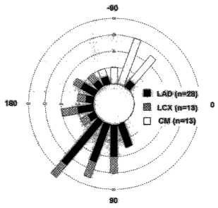
[0022] FIG. 6 shows a circular histogram of frontal plane T axes distribution.
[0023] FIGS. 7A-7B illustrate an exemplary method of the present invention
in flow chart form.
[0024] FIG. 8 shows an exemplary hardware system for differentiating TWI.
DETAILED DESCRIPTION OF THE INVENTION
[0025] Reference will now be made in detail to the preferred embodiments of
the present invention, examples of which are illustrated in the accompanying
drawings.
[0026] FIGs. IA-IE illustrate the terminology used in cardiography, and
FIG. 2 shows exemplary electrocardiogram (ECG) traces.
CA 02567282 2006-11-15
WO 2005/115233 PCT/US2005/017163
-5-
[00271 FIG. 1A illustrates representative ECG waveforms taken from the
twelve standard surface leads, the six limb leads numbered I, II, III, aVR,
aVL
and aVF, and the six chest leads, also known as precordial leads, VI-V6.
FIG. 1B shows positioning of the limb leads I, II and III. FIG. 1C illustrates
the connections for the limb leads I, II and III. Lead I has a horizontal
axis,
going from right to left. Lead aVF has a vertical axis, and goes top to
bottom.
Leads I and II are approximately 30 apart. Lead II is approximately 60
down from right to left. FIG. 1D illustrates the connections for limb lead
aVF. FIG. 1E illustrates the connections for the limb leads aVL and aVR.
FIG. 1F illustrates the placements locations of the precordial leads V1-V6.
Lead aVF points straight down, or towards six o'clock.
[0028] Typical diagnostic equipment that is used in vector cardiography gives
an angle measurement of the T-wave vector (and usually not the magnitude,
since it is the vector direction that is of primary interest). The reader is
referred to, e.g., Dale Dubin, Rapid Interpretation of EKG's, 4th ed., Cover
Publishing Co., 1989, which is incorporated by reference herein, for a more
complete discussion of lead placements. Also, the three arteries in the heart
are usually abbreviated as the LAD artery (left anterior descending), the
circumflex artery (LCX), and the right coronary artery (RCA).
[00291 Panels A-C in FIG. 2 show examples of negative inverted T-waves
(-0.8; -0.2; -0.1 mV, respectively). Panel D shows an isoelectric T-wave (0
mV). Panel E shows a (normal) positive T-wave (+0.2 mV). As shown in
FIG. 2, panel E, in a healthy heart, the QRS complex is followed by the S-T
segment, and then followed by a positive T-wave.
[00301 Based on cardiac memory definition (post-pacing sinus rhythm T
vector approaching direction of the paced QRS), the inventors hypothesized
that cardiac memory resulting from right ventricular pacing would have a
frontal T vector direction different from that of anterior ischemic TWI,
thereby
enabling to discrimination between the two.
[0031] Two groups of patients were studied. The cardiac memory group
consisted of thirteen patients undergoing permanent pacemaker implantation
who had sinus rhythm with 1:1 atrioventricular (AV) conduction at
CA 02567282 2006-11-15
WO 2005/115233 PCT/US2005/017163
-6-
physiologic heart rates. None of the patients had clinical, ECG or biochemical
evidence of active ischemia. Cardiac memory was induced by one week of
AV pacing with a short atrioventricular delay. The extent of the
atrioventricular delay was adjusted individually to allow ventricular
activation
to proceed completely from the endocardial pacemaker electrode positioned in
the right ventricular apex. At one week, a 12-lead ECG was recorded after the
pacemaker was reprogrammed in AAI mode. This ECG was used for analysis.
[0032] T-wave axis, polarity, and amplitude on a 12-lead ECG were compared
between cardiac memory and ischemic patients. The cardiac memory group
included eleven patients with no clinical signs of ischemia, and were
sequentially paced for one week after pennanent pacemaker implantation.
The ischemic patient group consisted of 47 patients with precordial TWI
undergoing LAD (left anterior descending) artery intervention for non-ST
elevation myocardial infarction. Table 1 below shows the baseline patient
data.
Table 1. Distribution of TWI by infarct-related artery in ischemic group.
Vessel TWI No TWI Excluded Total
involved
LAD 28 (47%)* 31 20 79
Proximal 16 (57%) 12 7
Mid, Dl 12 (44%) 15 10
Distal 0$ 4 3
LCX 12 (21 %) 44t 17 73
RCA 7(11%) 56 13 76
Total 47 (26%) 131 50 228
* p < 0.05 vs. LCX and RCA groups
t Including 5 patients with isolated TWI in leads I, aVL
$ p < 0.05 vs. other LAD locations
29 patients with inferior TWI only, 1 patient with TWI in leads I, avL
[0033] Patients with preexisting ECG abnormalities were excluded, e.g.,
patients with secondary TWI, such as pre-existing left bundle branch block or
LVH (left ventricular hypertrophy) manifesting negative T-waves in leads I
CA 02567282 2006-11-15
WO 2005/115233 PCT/US2005/017163
-7-
and aVL, atrial fibrillation and ST elevation infarcts. Patients with voltage
criteria for left ventricular hypertrophy were also excluded, unless upright
precordial T-waves were documented on prior tracings.
[0034] The ischemic patient group had ischemic precordial TWI due to
unstable angina/non-Q wave myocardial infarction, identified retrospectively
among patients undergoing percutaneous coronary intervention (PCI) on one
of the three major coronary arteries (LAD, LCX, RCA). If TWI was present
on more than one ECG, the earliest ECG from index admission was used for
analysis.
[0035] Burdick Space Lab and Marquette MAC-5000 electrocardiographs
were used to record the ECGs, which were analyzed manually. T-wave
amplitude was measured in each lead at T-wave peak/nadir to the baseline
determined by T-P segment. In case of biphasic T-waves (see, e.g., panel C in
FIG. 2), the most negative deflection was taken for the peak and T-wave was
classified as negative. T-wave was classified as isoelectric (amplitude = 0)
if
both positive and negative components were present with an amplitude of less
than 0.05 mV. QT was measured manually over three consecutive RR
intervals in leads available on the rhythm strip (typically, lead II or lead
V5)
and the results were averaged. Frontal plane QRS and T vector angles were
obtained from standard automated ECG printouts.
[0036] Clinical data was obtained from electronic medical records. Left
ventricular ejection fraction, determined as a part of routine clinical
management by echocardiograpy, or contrast left ventriculography, was used
for analysis if it was performed during the index admission (ischemic group)
or within a year prior to the pacemaker implant (cardiac memory group).
[0037] Location of the culprit lesion within LAD system (proximal, mid) and
involvement of the first diagonal branch (D 1 branch) was determined from
angiographic reports and confirmed by visual analysis of digital angiographic
films (if report statements were unclear).
[0038] Continuous variables were expressed as mean + SEM and compared
analysis of variance. Nominal data were compared using a Chi-square test.
Angular variables (frontal plane QRS and T-wave axes) were compared using
CA 02567282 2006-11-15
WO 2005/115233 PCT/US2005/017163
-8-
Watson-Williams F test. P values of less than 0.05 were considered
statistically significant.
[0039] Baseline group characteristics are presented in Table 2 below.
Male/female ratio did not differ between groups. Patients in this ischemic
group were, on average, younger than in the cardiac memory group (65.3 vs.
72.5 years old, p < 0.05). Prior ECGs were available in 13/13 cardiac memory
and 19/47 ischemic patients. There was no statistically significant
differences
in the prevalence of baseline ECG abnormalities between ischemic and cardiac
memory groups.
Table 2. Baseline Clinical Data
Group ischemia Cardiac memory
N 47 13
Male, n (%) 28 (60) 6 (46)
Age, yrs 65.3 2.0 72.5 3.0 *
Prior history of MI, n(%) 12 (25.5%) 3(23%)
History of CABG 8(17%) 2(15%)
Prior ECG Available 19/47 13/13 *
Precordial TWI 4/19 0/13
Right bundle branch block 1/19 3/13
Q waves 4/19 2/13
*-p<0.05
[0040] All patients in the study had endocardial right ventricular apex lead
implants. Other positions within the right ventricle can produce different
pacing QRS vectors with different resulting memory T-waves. Endocardial
pacemaker implants utilize the right ventricular apex, mid-septum, or outflow
tract as sites for the ventricular electrode. The QRS complex produced by
pacing from any of these sites usually has a left axis with varying degree of
superior (right ventricle apex) or inferior (right ventricle outflow tract)
angulation. Therefore, post-pacing TWI will always assume a left frontal axis,
no matter where in the right ventricle the pacing lead is situated. However,
with right ventricle outflow tract pacing, one would not usually see deep T-
wave inversions in inferior leads, which are considered typical for post-
pacing
TWI.
CA 02567282 2006-11-15
WO 2005/115233 PCT/US2005/017163
-9-
[00411 T-wave morphology, polarity and amplitude in precordial leads were
similar between the two patient groups. In the cardiac memory group, T-
waves in both leads I and aVL were positive or isoelectric in 13/13 patients
vs.
0/47 in ischemia (p < 0.001). If present, inferior TWI in ischemic patients
invariably demonstrated a TWI ITII1>1TIII1 pattern (the subscript indicates
the
lead in which the T-wave was observed), whereas cardiac memory uniformly
showed a TWI pattern ITI11I>ITII1= T-wave patterns in limb leads were
consistent with left superior frontal plane T vector in cardiac memory and
rightward in ischemic patients.
[0042] Sixteen patients (57%) had a proximal LAD lesion with ischemic
territory involving the D 1 branch. Twelve patients (44%) had a mid-LAD
lesion or an isolated Dl lesion. No significant differences were found in the
magnitude of T-wave inversions between proximal and mid LAD lesions as
well as between patients with and without D1 territory involvement.
[0043] CK (creatine kinase) levels were available in 27/28 LAD ischemic
patients. In seven patients, CK MB (creatine kinase myocardial branch)
testing was not performed, as the total CK was <100 IU/1. Ten patients had
CK MB within the normal range (<10 ng/ml), seventeen patients (61 %) had
CK MB elevation ranging from 13 to 366 ng/ml (median 46 ng/ml). Twenty
three patients (82%) had troponin I or T results available. Of those, 26
patients (93% of the LAD ischemic patients) had troponin elevation (range 0.2
to >50, median 4.2 ng/ml). All but one patient had results of either CK MB or
troponin available.
[0044] No significant difference was observed in T-wave amplitudes in any of
the limb leads in patients with and without CK MB elevation (<10 ng/ml).
Comparison of precordial T-wave amplitudes showed a trend for deeper T-
waves in patients with normal CK MB, compared to those with positive
enzyme, with differences in leads V3 and V5 reaching statistical significance
(see Table 3 below).
CA 02567282 2006-11-15
WO 2005/115233 PCT/US2005/017163
-10-
Table 3. Precordial T-wave inversion amplitude in ischemic patients with
(MB+, n=17) and without (MB-, n=10) CK MB elevation.
Leads V1 V2 V3 V4 V5 V6
T-wave MB(+ 0.08 0.04 -0.18 0.08 -0.17 0.06 -0.25f0.05 -0.18 0.05 -0.07-L0.04
amplitude, MB(-) 0.040.06 -0.24 0.13 -0.45 0.17* -0.42 0.11 -0.26f0.10* -0.15
0.09
mV
T-F 0.7 0.2 0.02 0.07 0.03 0.07
*- p < 0.05. No relationship was found between EF and the degree of TWI in
the ischemic group.
[0045] The inventors have discovered that cardiac memory and ischemia that
cause indistinguishable precordial TWI can nonetheless be differentiated on
the basis of frontal plane T vector direction. Cardiac memory results in
frontal
T vector projection were opposite to those of anterior ischemia. A
combination of positive TaVL and non-inverted TI was present in all cardiac
memory patients and in none of the ischemic patients, thus discriminating
cardiac memory from ischemia. The presence of positive T-waves in leads I
and aVL provides evidence against ischemic etiology of precordial TWI.
[0046] Representative examples of ECGs are depicted in FIGs. 3A-3B.
FIG. 3A shows a representative ECG of an ischemic patient, while FIG. 3B
shows a representative ECG of a cardiac memory patient. Both cardiac
memory and ischemia traces demonstrate deep T-wave inversion in the
precordial leads Vl-V6 of similar magnitude and morphology. In addition to
precordial TWI, the cardiac memory patient demonstrates deep inferior T-
wave inversion. However, a biphasic T-wave is also present in lead II in the
ischemia tracing. An important difference between recordings is seen in leads
I and aVL, in which ischemia shows T-wave inversions, whereas cardiac
memory manifests positive T-waves.
[0047] Electrocardiographic data is summarized in Table 3 below. The heart
rate was faster in the cardiac memory group (p < 0.05) due to predominant
atrial pacing in this group. Both QT and QTc intervals were not statistically
different between groups.
CA 02567282 2006-11-15
WO 2005/115233 PCT/US2005/017163
-11-
Table 3. Electrocardiographic Data
ischemia Cardiac
Group LAD LCX RCA memory
HR, miri-1 69.4 2.1 74.2:0.1 66.9 3.6 71.7 3.7
QT, ms 44W:10 415 11 438110 417::L10.
QTc 415 14 377-:L16 418 15 371 11.4
Number of 4.0 0.3 3.25 0.5* 2.9 0.3* 4.8 0.3
precordial leads
with TWI
Maximal -0.21+0.10* -0.26:0.11 *
precordial TWI, -0.45 0.06 -0.53+0.06
mV
QRS frontal axis, +20 7 +6 11 6 46 +18:L12
degrees
T wave frontal +128:L10* +146 15* -98 30 -70+5 *
axis, degrees
*- p < 0.05 compared to cardiac memory group
[0048] T-wave amplitudes measured at the peak/nadir of T-wave in precordial
leads were indistinguishable between CM and ISC-LAD groups (see FIG. 4,
discussed below, p > 0.05 for all precordial leads VI-V6). In contrast, all
the
limb leads (with the exception of aVR), showed highly significant differences
in T-wave amplitude as well as polarity between groups (see FIG. 5, discussed
below). The most dramatic difference was observed in lead aVL, where all
cardiac memory patients had positive T-waves compared to only one ischemic
patient (p < 0.01), whose T-wave in lead I was negative. Positive T-wave in
lead I was observed in 11 out of 13 cardiac memory patients, in the remaining
two, the T-wave was isoelectric, and none had negative T-waves No ischemic
patients had the combination of positive TaVL and non-inverted (positive or
isoelectric) TI. This is in contrast to all observed cardiac memory patients.
[0049] The inventors hypothesize that, when a patient is implanted with a
pacemaker, one of the leads goes into the right ventricle. Pacing the heart
from this lead produces negative QRS complexes in all the precordial leads
CA 02567282 2006-11-15
WO 2005/115233 PCT/US2005/017163
-12-
V1-V6. This is the reason why the T-waves are inverted when the pacing is
stopped. By the same token, cardiac memory produces positive T-waves in
leads I and aVL. Ischemia gives the same result in the precordial leads (Vl-
V6), while it gives the opposite result in leads I and aVL. This is also due
to
the fact that ischemia typically affects the left ventricle, and not the right
ventricle. Ischemia therefore gives negative T-waves in leads I and aVL. In
other words, in a patient with cardiac memory-induced T-wave inversion, the
ECG on leads I and aVL looks normal.
[0050] FIGs. 4 and 5 illustrate the data distribution for two patient
populations, the LAD ischemic patients and the cardiac memory patients. The
open triangle symbols represent the cardiac memory patients, and the closed
(dark) triangle symbols represent the ischemic patients. FIG. 4 shows T-wave
amplitude in the precordial leads Vl-V6. No significant difference in
amplitude is observed between groups. The T-wave negativity is particularly
pronounced for leads V2-V6, with both groups exhibiting T-wave negativity.
[0051] FIG. 5 shows T-wave amplitude in the limb leads. Again, closed
symbols are LAD ischemic patients, open symbols are cardiac memory
patients. The difference in amplitude between groups is statistically
significant (p < 0.05 for all leads except aVR). As may be seen in FIG. 5,
leads I and aVL exhibit the greatest contrast in the T-waves between the two
groups. With regard to both leads I and aVL, the T-waves for the cardiac
memory group are either flat or positive, while the T-waves for the ischemic
group are typically negative, and generally less than +0.05 millivolts.
Additionally, ECG from lead III may also be used to discriminate, although
not to the same extent, but lead III T waves are particularly useful for
discriminating RCA ischemia TWI from cardiac memory.
[0052] As shown in the tables and FIGs. 4-5, all cardiac memory patients had
inverted T-waves in leads III with TIII deeper than Vl-V6.
[0053] The reason for the observed differences in limb lead T-wave
amplitudes between groups is best appreciated via vectorcardiography. While
the frontal plane QRS axis in both groups was almost identical (see Table 3
above), T-wave axes differed dramatically (see also FIG. 6), with the mean
CA 02567282 2006-11-15
WO 2005/115233 PCT/US2005/017163
- 13-
angle difference between groups approaching 180 degrees (+128 vs. about 71
degrees, for ischemic and cardiac memory groups, respectively, p < 0.01).
[0054] FIG. 6 is a polar histogram representing the information summarized
in FIGs. 4 and 5. FIG. 6 shows a polar histogram of frontal plane T axes
distribution. Filled bars are LAD ischemic patients, hatched bars are LCX
ischemic patients, and open bars are cardiac memory patients. Each circular
dashed line represents two patients. The histogram shows that a typical
cardiac memory patient will show T-wave vectors generally in the
approximately -90 direction. Ischemic patients, on the other hand, will show
T-wave vectors generally between about +90 (probably from about as low as
+75 ) and about +180 (probably up to about +200 ). The difference in T
vector direction between groups is statistically significant (p < 0.01).
[0055] In the limb leads, the same principle was observed. In the majority of
LAD and LCX patients, T waves were negative in leads I and aVL . Three
LAD/LCX patients had positive T waves in lead I, one patient - in lead aVL
and none in both leads. In vector terms, this translated into left-to-right
direction of the T axis (see Table 4 below and FIG. 6). Limb lead TWI pattern
in RCA group was variable, depending on the relative involvement of lateral
and inferior leads. Four patients with predominantly lateral precordial TWI
(maximal precordial TWI amplitude > maximal inferior lead TWI amplitude)
demonstrated TWI in leads I and/or aVL and left-to-right T vector axis similar
to LAD and LCX groups. Three patients with predominantly inferior lead TWI
(maximal amplitude precordial TWI < TWIIII) had positive T waves in leads I
and aVL.
[0056] T vector in cardiac memory group followed the direction of the paced
QRS complex. RVA pacing produced QRS that was predominantly negative
in precordial leads, negative in inferior leads and invariably positive in
leads I
and aVL. As a result, diffuse TWI in the precordial and inferior leads and
positive T waves in leads I and aVL were characteristic for cardiac memory.
This translated into left superior T vector axis opposite in direction to that
of
LAD, LCX and part of RCA groups. With the exception of the patient with
CA 02567282 2006-11-15
WO 2005/115233 PCT/US2005/017163
-14-
post-implant pericarditis, all cardiac memory patients demonstrated maximal
precordial TWI > TWI III.
[0057] Cardiac memory vs. LAD/LCX: The most dramatic difference between
groups was observed in lead aVL, where all cardiac memory patients had
positive T waves compared to only one ischemia patient, whose T wave in
lead I was negative. Positive T wave in lead I was observed in 11/13 cardiac
memory patients; in the remaining two (both of whom had prior inferior wall
MI) T-waves were isoelectric, and none had negative T waves. The
combination of positive T wave in lead aVL and positive/isoelectric T in lead
I
(criterion I+aVL) was seen in all cardiac memory patients and none of
LAD/LCX patients (see Table 4 below).
[0058] Cardiac memory vs. RCA: Four out of 7 RCA patients conformed to
the pattern of LAD/LCX TWI and criterion I+aVL discriminated them from
cardiac memory. The remaining 3 RCA patients with positive TI and TaVL had
maximal precordial ITWII <ITWI1ziI in contrast to all but one cardiac memory
patients.
Table 4. Lead distribution of TWI in ischemic and cardiac memory groups, n
N.
Group ischemia CM (in=13)
Lead LAD (n=28) LCX (n=12) RCA (n=7)
V1 8 (29) 1 (8) 0 5 (39)
V2 21 (75) 5 (42) 0* 8 (62)
V3 22 (79) 5 (42) 1 (14)* 12 (92)
V4 24 (86) 7 (58) 6 (86) 12 (82)
V5 21 (75) 10 (83) 6 (86) 13 (100)
V6 16 (57)* 11 (92) 7 (100) 13 (100)
I 20 (71)* 11 (91)* 4 (57)* 0
II 8 (29)* 5 (42)* 6 (86) 13 (100)
III 3 (11)* 2 (17)* 4 (57)* 13 (100)
aVR 10 (36)* 0 1 (14) 1 (8)
aVL 23 (82)* 11 (92)* 2 (29) 0
aVF 6 (21)* 4 (33)* 5 (71) 13 (100)
(I+aVL ** 0* 0* 3 (43)* 13 (100)
(I+aVL) and maximal precordial TWI > 0* 12 (92)
TWI III
*- p < 0.05 with cardiac memory group
CA 02567282 2006-11-15
WO 2005/115233 PCT/US2005/017163
-15-
** (I+aVL) -positive T wave in lead aVL, positive or isoelectric T wave in
lead I.
[0059] Based on the obtained results, it is generally sufficient to look at
leads I
and aVL for LAD and LCX ischemia, and to consider the most negative
component of the T-wave. If lead I shows a positive T-wave, and lead aVL
shows positive or flat T-wave, while the precordial leads V1-V6 show inverted
T-waves, then the patient most likely has cardiac memory-induced T-wave
inversion. ("Positive" here is selected, for example, to be represented as
approximately 0.05 millivolts or greater. The signal is generally calibrated
to
millimeters per millivolt on the ECG printout.)
[0060] One embodiment of the invention may be implemented using a
standard diagnostic ECG, 'such as available from Burdick Space Lab or
Marquette, modified to differentiate the two types of TWI according to the
principles described above. Alternatively, although the discussion above is
primarily in terms of using an external ECG (e.g., a standard 12-lead ECG),
the invention is also applicable to implantable devices. For example,
implantable cardiac defibrillators (ICDs) usually have three implanted
electrodes: a pacing electrodesin the right ventricle, a coil (defibrillator)
electrodesin the superior vena cava, and the ICD "can" itself (usually located
in the pectoral area under the skin). Using these electrodes (and, optionally,
using additional electrodes as well, if available), the implantable device can
"reconstruct" the direction of the T-wave vector, and, based on the direction
of
the T-wave vector, as discussed above, discriminate between cardiac memory
TWI and ischemic TWI. Alternatively, the implantable device can perform
mathematical operations on the data from the leads that generally correspond
to discriminating between the two types of TWI in the manner discussed
above, without directly calculating the T-wave vector direction.
[0061] Another exemplary hardware system for differentiating TWI is shown
in FIG. 8. Referring to FIG. 8, an ECG processing system 804 is described.
ECG processing system 804 includes a programmed microcomputer 8040
equipped with an analog-to-digital (A/D) conversion board 8050. The steps of
the method are performed using a software program written in, e.g.,
CA 02567282 2006-11-15
WO 2005/115233 PCT/US2005/017163
-16-
C programming language. The program follows the steps set forth above. It is
believed that any skilled programmer would have no difficulty writing the
code necessary to perform the steps of this invention.
[0062] Microcomputer or computer platform 8040 includes a hardware unit
8041 which includes a central processing unit (CPU) 8042, a random access
memory (RAM) 8043, and an input/output interface 8044. RAM 8043 is also
called a main memory. Computer platform 8040 also typically includes an
operating system 8045. In addition, a data storage device 8046 may be
included. Storage device 8046 may include an optical disk or a magnetic tape
drive or disk.
[0063] Various peripheral components may be connected to computer
platform 8040, such as a terminal 8047, a keyboard 8048, and a printer 8049.
Analog-to-digital (A/D) converter 8050 is used to sample an ECG signal. A/D
converter 8050 may also provide amplification of the ECG signal prior to
sampling.
[0064] FIGS. 7A-7B illustrate an exemplary method of the present invention
in flow chart form. As shown in FIGS. 7A-7B, step 702 includes sensing an
electrocardiogram from a patient. Alternatively, pre-recorded data may be
analyzed. Step 704 includes identifying inverted T-waves in at least some of
precordial leads. Step 706 includes identifying T-waves in leads I and aVL.
Steps 708-710 include diagnosing anterior ischemia if leads I and aVL show
inverted T-waves. Step 712 includes diagnosing possible cardiac memory if
the leads I and aVL show non-inverted T-waves. Optional step 714 includes
identifying T-waves in lead III. Steps 715 and 720 includes confirming
cardiac memory diagnosis if the lead II shows inverted T-waves. Optional
steps 722-724 include confirming ischemic diagnosis if the lead III shows
deeper inverted T-waves than maximum amplitude of precordial TWI. Step
725 includes confirming cardiac memory otherwise.
[0065] It is important to note that T-wave positivity in leads I and aVL is an
active part of cardiac memory development, as an increase in T-wave
amplitude is observed in the leads with a positive paced QRS complex (e.g.,
leads I and aVL).
CA 02567282 2006-11-15
WO 2005/115233 PCT/US2005/017163
-17-
[0066] The pattern of T-wave inversion in the inferior leads, if present, can
also be useful in determining the etiology of TWI. Combined ECG changes in
anterior and inferior leads can be present with wrap-around LAD ischemia.
However, in that case, the T-wave vector maintains a rightward direction,
causing more T-wave negativity in lead II compared to lead III, which is the
opposite of the cardiac memory pattern.
[0067] As demonstrated previously in animal studies, the early stages of
cardiac memory development can be accompanied by T vector rotation in the
frontal plane before T-wave assumes the direction of pacing QRS complex.
Drugs, such as calcium channel blockers and quinidine, affect development of
cardiac memory and T vector shape. At the present time, the clinical
relevance of these observations remains unclear.
[0068] In the above study, the site of the culprit lesion varied between the
proximal and mid-LAD (below D1) and Dl alone. Intuitively, one would
expect that a more lateral LV (left ventricle) spread of ischemia would result
in a more rightward shift of the T-wave axis. Altematively, with a distal LAD
lesion perfusing only the apical-septal left ventricle, the rightward axis
shift
might be absent. The inventors did not observe differences in T-wave patterns
between proximal and mid-LAD lesions, nor between lesions involving and
not involving the Dl region. Therefore, there is no data to suggest that the
location of LAD lesion by itself influences the degree of T-wave negativity in
leads I and aVL. However, no patient in the ischemic group had distal LAD
lesions, and the total number of patients in the study is insufficient to
account
for all possible variations of coronary anatomy.
[0069] Degree of ischemia is another potential factor contributing to the
magnitude of T-wave changes. The majority of ischemic patients in the study
had positive markers for myocardial injury, signifying severe ischemia.
Conceivably, a lesser degree of ischemia could produce smaller T-wave
changes. Counter-intuitively, when ischemic patients were divided into MB+
(myocardial branch (+)) and MB- (myocardial branch (-)) categories, no
difference between the two groups was found in T-wave amplitude in the limb
leads. Moreover, marker-negative patients had deeper precordial TWI than
CA 02567282 2006-11-15
WO 2005/115233 PCT/US2005/017163
-18-
positive ones (see Table 4 above). This finding is in accord with observations
in patients having myocardial infarction who demonstrate an inverse
relationship between TWI magnitude, enzymatic size of MI (myocardial
infarction) and functional recovery, suggesting that T-wave inversions
indicate
the presence of a viable stunned myocardium. Therefore, it seems unlikely
that milder ischemia would alter the T-wave changes in ischemic patients.
[0070] Preliminary observations suggest that cardiac memory does not change
the abnormal T vector associated with these conditions Cardiac memory
development might be altered in patients with prior inferior myocardial
infarction, presumably due to a lack of a viable myocardium adjacent to the
pacing site.
[0071] It is also possible that the frontal plane T vector direction can be
helpful in distinguishing between ischemic and non-ischemic (but other than
cardiac memory) precordial TWI. Several studies using precordial ECG
mapping showed that an I mapping pattern (inverted T-waves in the left upper
quadrant with positive T-waves in the lower right quadrant) is highly
predictive of ischemic TWI. Non-ischemic TWI were characterized by an N
pattern (TWI in lower right quadrant and positive T-waves in left upper
quadrant). These unipolar map patterns would likely correspond to positive
(type N) and negative (type I) T-waves in bipolar leads I, aVL, as
demonstrated in previously published ECGs.
[0072] Note that the present method may not help to separate repolarization
changes associated with LVH, the most frequent confounder of ischemic
changes, as they have similar frontal T-wave axis. Anterior wall ischemia is
generally regarded as the most dangerous form of ischemia. Anterior wall
ischemia is generally associated with LAD (left anterior descending) artery
stenosis.
[0073] It should be noted that different locations of ischemia can result in
different patterns of T-wave inversion. The present invention is particularly
applicable to LAD ischemia, although it is also applicable, to other forms of
ischemia. Of the three arteries in the heart - the LAD artery, the circumflex
artery (LCX), and the right coronary artery (RCA) - in the case of LCX
CA 02567282 2006-11-15
WO 2005/115233 PCT/US2005/017163
-19-
ischemia, sometimes there are negative T-waves in the precordial leads, and
other times, not. Thus, it should be remembered that, compared to LAD
ischemia, the frequency of TWI is less in the case of LCX ischemia. In
approximately 40% of the cases, LCX ischemia is accompanied by T-wave
inversion in the precordial leads.
[0074] In conclusion, the invention includes the advantage of differentiating
precordial ischemic TWI from post-pacing TWI, based on the opposite
directions of the frontal plane T-wave vectors. The inventors demonstrated
that ischemic TWI is characterized by a rightward frontal plane T-wave axis,
whereas in cardiac memory patients, the direction of the T vector points
leftward. Bearing in mind these vector concepts, a simple discriminating rule
has been devised, using standard 12-lead ECG criteria, which is easily
applicable in everyday clinical practice. All cardiac memory patients and only
one ischemic patient had positive T-wave in lead aVL. However, the single
ischemic patient with positive T-wave in lead aVL showed a negative T-wave
in lead I, a pattern not observed in cardiac memory patients. Therefore, the
combination of: 1) positive T-wave in lead aVL and 2) non-inverted (positive
or isoelectric) T-wave in lead I completely discriminated cardiac memory
patients from ischemic patients. Using the most negative point in the T-wave
was usually a better discriminator than using the frontal T-wave axis, which
had minimal overlap between groups. This occurs because calculation of T-
wave axis is based on the total T-wave area (negative and positive
components) in a given lead, which in the case of biphasic T-waves dilutes the
effect of terminal T-wave negativity.
[0075] By applying vectorcardiographic principles to interpretation of a
standard 12-lead ECG, a simple algorithm was developed to discriminate
between ischemic and post-pacing precordial TWI. Use of such
vectorcardiographic information can significantly improve differential
diagnosis of TWI.
[0076] It should also be appreciated that various modifications, adaptations,
and alternative embodiments thereof may be made within the scope and spirit
CA 02567282 2006-11-15
WO 2005/115233 PCT/US2005/017163
-20-
of the present invention. The invention is further defined by the following
claims.