Note: Descriptions are shown in the official language in which they were submitted.
CA 02567860 2012-10-22
Flow Control and Gas Detection and Gas Removal in
An Intravenous Fluid Delivery System
CROSS REFERENCE TO RELATED APPLICATIONS
This application claims the benefit under 35 U.S.C.
119(e) of U.S. Provisional Patent Application No. 60/575,246,
filed May 28, 2004, and U.S. Provisional Patent Application
No. 60/576,258, filed June 2, 2004.
STATEMENT REGARDING FEDERALLY SPONSORED RESEARCH OR
DEVELOPMENT
N/A
BACKGROUND OF THE INVENTION
Devices to deliver fluids intravenously to a patient
involve a number of considerations, such as air or gas bubble
detection, gas removal, and flow rate control.
Air and bubble detection in medical intravenous (IV) fluid
delivery systems is important. Large amounts of air can cause
air embolisms in any part of the body, blocking off blood flow.
Embolisms in the brain can cause severe memory loss and even
death. Air trapped in the heart can also cause death or heart
damage. Ultrasonic, optical, and electrical conductivity methods
are used in the prior art detection of air bubbles in medical IV
fluid lines.
Ultrasonic detectors are the most widely used detectors in
the IV medical fluid delivery field and are based on the fact
that sound is more readily conductive through liquid than
through air. Thus, an air bubble does not "conduct" sound from
-1-
CA 02567860 2006-11-23
WO 2005/118051
PCT/US2005/018659
one side of the tubing wall to the other, while fluid does
conduct sound. Ultrasonic detectors are effective at detecting
small amounts of gas in IV tubing, but have a number of
drawbacks. They are expensive. They require that the tubing be
in direct contact with the ultrasonic transmitter and receiver.
Moreover, the slightest air gap can trigger the detector,
causing a false alarm. Micro bubbles that build up on the
surface of the tubing and are too small to be harmful also can
trigger false alarms, since the micro bubbles, despite their
very small size, still provide a boundary to the ultrasound. In
addition, ultrasonic detectors have a fairly high power
consumption, greater than 100 mW.
Optical detectors are typically inexpensive. Some optical
detectors work using light absorption while others use light
transmission. These methods are, however, fluid dependent and
therefore not very common, because many different fluids are
used for IVs. Also, their performance is dependent on the
optical characteristics of the tubing, and many different tubing
sets, having different optical characteristics, can be used.
Additionally, optical detectors can be subject to interference
from light from other sources.
Electrical conductivity detectors are used the least, as
they require a direct electrical connection to the IV fluid. To
electrically isolate the patient, this connection must have low
leakage current and high dielectric strength. Typically, two or
three electrodes are placed in contact with the fluid and are
excited from an AC or DC source while the current/voltage is
monitored. Gas bubbles do not conduct electricity, but many IV
fluids do. A drawback, however, is that some IV fluids do not
conduct electricity. Another drawback is that a thin film of
fluid connecting one electrode to the other where the electrode
penetrates the tubing wall can give a false detection of fluid
presence.
-2-
CA 02567860 2006-11-23
WO 2005/118051
PCT/US2005/018659
When a fluid is heated, outgassing occurs. In prior art IV
fluid warming devices, outgassing has either been ignored or
handled with elaborate schemes. In one scheme, a hydrophobic
filter has been employed to vent gases. This system is
disadvantageous, because it is difficult and expensive to test
to ensure that the filter does not leak. Also, the check valve
used to prevent air from entering the system can stick, for
example, if the humidity becomes too high or if another fluid
inadvertently drips onto the valve, thereby requiring a greater
pressure to open the valve. In another scheme, a drip chamber is
used to collect the gases. This scheme is disadvantageous,
because the chamber has a fixed volume and once full, the air
can enter the patient unless a manually operated venting drip
chamber is employed. A user must remember to vent such a venting
drip chamber.
Intravenous (IV) fluids need to be delivered at different
rates. Hydration fluids typically are delivered at higher rates,
while drugs are typically delivered at lower rates. Flow rate
control in medical IV fluid devices involves considerations of
flow rate accuracy, errors made by personnel in setting flow
rates, cost, and set up time. Three main types of devices are
used in the control of IV fluid flow rates, namely, roller
clamps, volumetric pumps, either volume displacement pumps or
valve-regulated gravity assist pumps, and in line mechanical
flow regulators.
Roller clamps are the most widely used flow control
device. The roller clamp comprises a wheel trapped within a
housing that compresses the IV tubing as it is slid along a
gradual ramp. The flow rate is calculated by counting drips in a
drip chamber. This device is inexpensive, but has a number of
drawbacks. The setup operator must take time to count drips into
the drip chamber, an iterative process taking up to 15 seconds
for each adjustment. Also, the setup operator must know the size
-3-
CA 02567860 2006-11-23
WO 2005/118051
PCT/US2005/018659
of the drips and must calculate the flow rate and may make a
mistake. Even after it is set up, the IV tubing in the clamp
continues to deform over time, causing the rate to change. The
IV solution must be held above the patient insertion site. Any
changes in height can affect the flow rate, because the roller
clamp is a relative device. The advantages of the roller clamp
are that it does not require any power, it is widely accepted,
and it is inexpensive.
Volumetric pumps are also widely used for drug delivery.
There are two main types of infusion rate control. In a first
type of control, a displacement pump forces fluid through the IV
line at repeatable volumes and adjustable intervals. These pumps
can be reciprocating piston, peristaltic (linear or rotary), or
syringe types. These pumps are typically quite precise, as
required for drug delivery, and are not typically used for
standard IVs. A second type of infusion rate control utilizes
gravity driven fluid. With this type, drips through a drip
chamber are counted, and a variable orifice valve is controlled
based upon the number of drips over time.
A disadvantage of such pumps is their great expense. Also,
the tubing set is typically disposable, which further increases
the cost. These pumps also take up a lot of space. The main
advantages of such pumps are accurate flow control, no change in
flow with change in bag height, reduced setup time, and reduced
chance of error by the operator.
In line mechanical flow regulators, using diaphragms,
needles valves, and the like, are not very common. They are
advantageous in that they require no power and are reasonably
independent of IV fluid bag height. They are, however, dependent
on fluid viscosity. Also, they typically have two flow rate
scales (ml/min and ml/hour), which, while providing versatility,
can also be confused by operating personnel.
-4-
.
CA 02567860 2006-11-23
WO 2005/118051
PCT/US2005/018659
SUMMARY OF THE INVENTION
In one aspect of the present invention, an air or gas
bubble detection system is provided that detects the presence or
absence of liquid in intravenous (IV) tubing. By detecting the
presence of liquid rather than gas, false alarms due to micro
bubbles or small air gaps between the sensor and the tubing are
avoided. This system is independent of fluid type. Additionally,
high electrical isolation is maintained and leakage currents are
acceptably low. Also, power consumption is low with the present
system.
More particularly, the air or gas bubble detection system
employs a circuit that transfers a charge to a sense electrode
adjacent the IV tubing and to a reference capacitor. Two
grounded electrodes are spaced on the sides of the sense
electrode. The three electrodes are aligned parallel with the
length of the IV tubing. The tubing and any fluid (liquid or
gas) therein act as dielectrics. A grounded shield electrode
shapes the electric field toward the tubing and prevents outside
fields from interfering with the sensing process. The circuit
detects the amount of charge transferred to the reference
capacitor, which is indicative of the type of material present.
The presence of a liquid produces a high number, whereas the
absence of a liquid produces a low number. By comparing the
produced number with a limit, the controller determines if an
alarm condition is met.
In another aspect of the present invention, an active gas
removal system is provided to remove air or gas bubbles from an
intravenous fluid prior to infusion into the patient. The gas
removal system employs a drip chamber through which intravenous
fluid is pumped by an upstream pump. The IV fluid exits the
chamber through an outlet in the bottom, while gas or air is
retained in the upper portion of the chamber. A vent valve at
the top of the drip chamber is operative to open upon an
-5-.
CA 02567860 2006-11-23
WO 2005/118051
PCT/US2005/018659
increase of gas pressure within the drip chamber to vent the gas
in the chamber. A controller is provided in communication with a
fluid level sensor in the drip chamber and with a downstream
patient line-occluding valve. The controller is operative to
close the downstream valve upon detection of a specified fluid
level in the drip chamber detected by the fluid level sensor,
whereby a pressure increase in the drip chamber caused by
continued operation of the upstream pump opens the vent valve
and releases gas retained within the drip chamber.
A further aspect of the present invention relates to an
intravenous (IV) flow control system that employs tubing orifice
size data and thermal data from a fluid warming system to
provide closed loop control to maintain a desired flow rate. The
actual flow rate is determined by two techniques, a
geometrically based technique that uses geometric parameters of
the IV tubing system, and a thermally based technique that uses
the power input to an IV fluid warmer and the temperature of the
= IV fluid entering and exiting the warmer.
The system employs a pincer or other moveable element
positioned to compress the tubing, forming an orifice at which
flow can be controlled. The geometry of the tubing at the
orifice can be determined by a force transducer, the data from
which is fed to a system controller that calculates flow rate
based on orifice geometry data. The thermal data from the fluid
warmer is also fed to the controller, which calculates flow rate
from this data also. Based on the combined calculations of the
orifice geometry and the thermal transfer data, the pincer is
controlled to adjust the flow at the orifice to maintain the
desired flow rate.
The system is advantageous, because it can utilize a
standard hospital IV set and standard hospital procedure. It can
adapt to real time changes in the tubing, and it can handle both
standard IV fluids and blood. The system utilizes two
-6-
CA 02567860 2006-11-23
WO 2005/118051
PCT/US2005/018659
independent control loops to calculate and control flow rate,
and can switch to one or the other control loop to suit
circumstances. For example, at higher flow rates, the thermally
based control loop is usually preferred. The system can also be
operated to allow infusion of a bolus of fluid to the patient.
DESCRIPTION OF THE DRAWINGS
The invention will be more fully understood from the
following detailed description taken in conjunction with the
accompanying drawings in which:
Fig. 1 is an isometric view of an air or gas bubble
detection system of the present invention for an intravenous
(IV) infusion system;
Fig. 2 is an isometric side view of the air or gas bubble
detection system of Fig. 1;
Fig. 3 is an isometric bottom view of the air or gas
bubble detection system of Fig. 1;
Fig. 4 is a schematic view of the air or gas bubble
detection system of Fig. 1;
Fig. 5 is a schematic view of the air or gas bubble
detection system of Fig. 1 illustrating field lines when fluid
is present in the tubing;
Fig. 6 is a schematic view of the air or gas bubble
detection system of Fig. 1 illustrating field lines when air is
present in the tubing;
Fig. 7 is an electrical schematic of a circuit operative
to detect capacitance of the tubing and fluid for the air or gas
bubble detection system of Fig. 1;
Fig. 8 is a schematic diagram of an air or gas bubble
removal system of the present invention;
Fig. 9 schematically illustrates a front view of an
intravenous (IV) flow control system of the present invention;
-7-
CA 02567860 2006-11-23
WO 2005/118051
PCT/US2005/018659
Fig. 10 schematically illustrates a side view of the IV
flow control system of Fig. 9;
Fig. 11 schematically illustrates the pincer of the IV
flow control system of Fig. 9 compressing an IV tubing portion;
Fig. 12 is a block diagram illustrating control of the IV
flow control system;
Fig. 13 illustrates cross-sectional
geometric
configurations of IV tubing at various levels of compression by
the IV flow control system of Fig. 9;
Fig. 14 is a graph illustrating force vs. time at the
pincer compressing the tubing;
Fig. 15 is a graph illustrating force vs. distance at the
pincher compressing the tubing;
Fig. 16 is a schematic illustration of a further
embodiment of a flow control system of the present invention
employing a wide pincer and a dielectric tubing detection
mechanism;
Fig. 17 is a schematic illustration of the wide pincer IV
flow control system of Fig. 16 compressing tubing; and
Fig. 18 is a graph illustrating dielectric measurement as
a function of crush at full compression.
DETAILED DESCRIPTION OF THE INVENTION
Gas Bubble Detection System
The present invention relates in one aspect to an air or
gas bubble detection system for use with an intravenous (IV)
fluid infusion system. The system of the present invention
detects the presence or absence of liquid in the IV tubing, not
the presence of gas. If the system detects that liquid is not
present, then gas must be present in the tubing. With this
system, false alarms due to detection of micro bubbles or small
air gaps between the sensor and the tubing are avoided.
-8-
ak 02567860 2006-11-23
WO 2005/118051
PCT/US2005/018659
More particularly, all materials have a physical
dielectric constant. Gasses have a very low dielectric constant,
plastics have a medium dielectric constant, and liquids have a
very high dielectric constant. The present system utilizes
electrodes to act as capacitor plates and the adjacent materials
(the tubing and any fluid therein) as dielectrics. The detected
charge on a reference capacitor is indicative of the type of
material present.
Referring to Figs. 1-3, the system employs a body or
housing 12 having a support member 14, generally L-shaped in
cross-section in the embodiment illustrated, and a holding
member 16. The support member and the holding member provide
generally opposed faces 18, 20 that define a channel 22 or other
suitable recess configured to receive a portion of the IV
tubing.
Referring also to Fig. 4, the support member 14 is made of
a suitable insulating material, such as a plastic. Three
electrodes 32, 34, 36 are mounted on the support member in
series relative to each other and parallel with the portion of
the IV tubing 38 held in the channel. In this manner, the three
electrodes form the channel face 18 of the support member that
contacts the tubing.
The tubing must be held in contact with the three
electrodes. Toward this end, the channel may be sized to provide
a snug fit for the tubing, or the holding member may be
configured to apply a clamping force to the tubing portion, such
as with a movable element or piston, to ensure the tubing
portion is retained in the body. The holding member may include
pads 24 (Figs. 1-3), such as of foam rubber, to hold the tubing
in contact with the opposed face of the support member and
prevent the tubing from shifting or otherwise moving.
The two outer electrodes 34, 36 are grounded. The middle
sense or charge transfer electrode 32 is used to transfer a
-9-
CA 02567860 2006-11-23
WO 2005/118051
PCT/US2005/018659
charge of electrons to its surroundings. Two air gaps or spaces
42 are formed in the support member on each side of the sense
electrode 32 to limit coupling fields from the sense electrode
to the ground electrodes. The electrodes are arranged in such a
way that the electric field is focused toward and over a short
section of the IV tubing. The ground electrodes 34, 36 at
opposite ends of the detector establish the length of tubing
being tested, that is, the length of the sense electrode 32
along the tubing plus the air gaps 42. As noted above, the
electrodes act as capacitor plates and the adjacent materials
(the tubing and any fluid therein) act as dielectrics. A fourth
shield electrode 44 is provided around the support member 16 of
the body. The shield electrode is grounded and shapes or directs
the electric field toward the tubing. The shield electrode also
keeps extraneous outside fields from interfering with the
sensing process. Fig. 5 illustrates electric field lines 52 when
a liquid 46 is present in the tubing, but no gas is present in
the tubing. Fig. 6 illustrates electric field lines 54 when air
56 is present in the tubing.
Referring to Fig. 7, a controller or microprocessor 62 is
in communication with a charge transfer device 64, such as a
capacitance-to-digital converter integrated circuit (IC). Upon
request from the controller, a charge or burst of electrons is
placed on the charge transfer electrode 32 and on a reference
capacitor 66. The charge transfer device measures the amount of
charge transferred to the reference capacitor and converts this
charge to a digital number, which is transmitted back to the
controller. In the embodiment illustrated, the controller and
the charge transfer device are in communication via a line 72 to
transmit a signal from the controller to the device to request a
burst or charge transfer, a line 74 to transmit clock signals, a
data ready line 76 indicating the device is ready to transfer a
-10-
CA 02567860 2006-11-23
WO 2005/118051
PCT/US2005/018659
charge, and a data return line 78 to return data from the device
to the controller.
The digitized signal returned to the controller is a ratio
of the reference capacitor to the "load capacitance," i.e., the
tubing, surrounding plastic, and any fluid (gas or liquid)
present in the tubing. The presence of a liquid produces a high
number, while the absence of a liquid produces a low number. The
controller compares the numbers to limits and determines if an
alarm condition is met.
A suitable charge transfer device 64 to be utilized as a
capacitance-to-digital converter is the integrated circuit (IC)
identified by model QT300, available from Quantum Research
Group. The reference capacitor 66 can be any suitable type, such
as a plastic film or ceramic capacitor. A bypass capacitor 68 is
placed between Vdd and ground for proper operation of the power
supply.
The system can be configured so that the operator, such as
a physician, can set the bubble size or volume limit. For
example, a lesser limit can be set if the patient is a child,
and a greater limit can be set if the patient is an adult. For
example, a gas volume of 0.5 ml or less may be acceptable in a
particular situation. Thus, the system can be set so that only a
detected gas volume larger than 0.5 ml triggers an alarm.
Preferably, when an alarm is triggered indicating
detection of an air bubble, the IV flow is stopped in any
suitable manner. For example, the system can be configured such
that the tubing is compressed to occlude completely if the alarm
is triggered, preventing further infusion to the patient until
the system can be checked by an operator. The system can be used
in conjunction with any valve to occlude the tubing. For
example, the valve could be located at a pump, at a fluid
warmer, or at a gas removal system. The system can be used in
-11-
CA 02567860 2006-11-23
WO 2005/118051
PCT/US2005/018659
conjunction with the IV flow control system of the present
invention, described below.
With the present system, the charge transfer method is
independent of fluid type. Air gaps do not cause false alarms.
Fluid films in the tubing do not cause false alarms. High
electrical isolation is maintained. Very low leakage currents
are present. Power consumption is very low.
The controller 62 can control the frequency of the burst
of electrons to the sense electrode 32 by the charge transfer
device 64. Burst control allows for gas volume measurement when
the IV flow rate is known. Thus, if the flow rate were low,
bursts can be less frequent to detect a particular volume of air
than if the flow rate were higher. Similarly, different tubing
geometries allow for different size bubbles or volumes to be
detected. Thus, if the tubing has a larger diameter, the burst
= rate can be more frequent to detect the same volume of air than
if the tubing had a smaller diameter. The fluid flow rate
through the tubing may be known or can be determined in any
suitable manner, for example, from a flow rate controller such
as that described below. The bubble detection system may be
employed with the flow control system described below, or with
any other suitable flow control system known in the art.
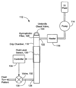
Gas Removal System
The present invention also provides an active air or gas
removal system, illustrated in Fig. 8. The gas removal system
112 is located downstream from an IV fluid pump 114 and, in the
embodiment illustrated, downstream from an IV fluid heater 116.
The system includes a drip chamber 118 that receives IV fluid
through an input port 122 near the upper end of the chamber 118.
A vent valve 124, such as an umbrella or other type of check
valve, is located at the top of the drip chamber. When pressure
in the chamber is increased (as described further below), the
-12-
CA 02567860 2006-11-23
WO 2005/118051
PCT/US2005/018659
vent valve opens to allow gas to escape from the drip chamber.
The vent valve also prevents outside gas from entering the drip
chamber. A hydrophobic filter 126 in front of the vent valve
prevents the IV fluid from passing through the vent valve.
The IV fluid collected in the drip chamber 118 is
introduced to the patient through an output port 128 near the
bottom of the chamber via tubing 130 that delivers fluid to the
patient. A line-occluding valve 132 in the patient line
downstream from the drip chamber is operative to close or reduce
flow of fluid therein. Pressure to open the vent valve is
generated by occluding the patient line via the downstream line-
occluding valve while continuing to run the upstream pump. The
pump forces any gas in the drip chamber to be expelled up
through the hydrophobic filter and through the vent valve.
A controller 134 is in communication with the patient line
valve 132 and with a fluid level sensor 136 that detects the
fluid level in the drip chamber 118. Any suitable fluid level
detector, such as a float sensor or an ultrasonic detector, can
be used, as would be apparent to those of skill in the art. The
controller is operative to cause the patient line-occluding
valve 132 to close upon detection of a determined low fluid
level by the fluid level sensor in the drip chamber, thereby
causing pressure to increase in the drip chamber to open the
vent valve and preventing gas from traveling to the patient.
The hydrophobic filter does not function when contacted by
blood. Thus, the system can include a variable level fluid
sensor or multiple fluid level sensors to detect both a low
fluid level for purging gas and a high fluid level for
preventing contact with the hydrophobic filter. The controller
can be operative upon detection of a high level to signal an
alarm or take other appropriate action to indicate that the
fluid level is too high.
-13-
CA 02567860 2006-11-23
WO 2005/118051
PCT/US2005/018659
Outgassing occurs when a fluid is heated. Henry's Law can
be used to calculate the amount of gas dissolved in a solution
versus pressure and temperature, as is known in the art. For
example, approximately 7 cc of gas comes out of solution per one
liter of fluid at room temperature. Using Henry's Law, it can be
determined that a suitable size for the drip chamber is, for
example, 50 cc. Other sizes can, of course, be provided, as
determined by the application.
The air or gas bubble removal system of the present
invention can be employed in conjunction with the system for the
detection of air or gas bubbles described above, with any other
air or gas bubble detection system, or with any other infusion
system.
Intravenous Flow Control System
The present invention also relates to an intravenous (IV)
flow control system. The IV flow control system works with a
standard hospital IV set and gravity feed and employs a control
valve with tubing measurement capabilities and thermal data from
a fluid warming system to provide closed loop control to
maintain a desired flow rate. More particularly, actual flow
rate is determined by two independent control loops to calculate
and control the fluid flow rate. One control loop is
geometrically based and uses geometric parameters of the IV
system. The other control loop is thermally based and uses the
power input to an IV fluid warmer and temperatures of the IV
fluid entering and exiting the warmer.
Referring to Figs. 9-11, the flow control system employs a
holding or clamping mechanism 212 to retain the tubing in place
and a movable element 213 that squeezes or compresses the IV
tubing 218. In the embodiment illustrated, the movable element
comprises a piston 214 that operates in conjunction with an
anvil 216. A pincer 220 of a determined width is disposed at the
-14-
CA 02567860 2006-11-23
WO 2005/118051
PCT/US2005/018659
end of the piston adjacent the tubing to impinge thereon. The
anvil is fixed and the piston is driven toward the tubing,
thereby squeezing the tubing between the pincer and the anvil.
By driving the piston toward and away from the anvil, the tubing
at the pincer acts as an orifice 222 to allow more or less fluid
flow through the orifice, thereby allowing control of the fluid
flow rate. Any suitable mechanism for driving the piston can be
employed, such as a linear stepper motor 246 (see Fig. 12). A
force transducer 224 mounted between the IV tubing 218 and the
anvil 216 provides a determination of the geometry of the tubing
during the crushing or squeezing operation, discussed further
below. Hold down pads 226, such as of foam rubber, on a support
panel 228 keep the tubing in place so that outside motion does
not affect the force transducer.
Fig. 12 further illustrates operation of the IV flow
control system. The system includes a computer or controller
232. The desired flow rate 234 is input to the controller by the
user. Data 236 from a fluid warmer is also input to the
controller for use in the thermally based flow rate calculation.
The thermal data includes the power to the fluid warmer, the
temperature Tir, of the IV fluid entering the warmer, and the
temperature Tout of the IV fluid exiting the warmer. Preferably,
the data is automatically transferred from the fluid warmer to
the controller. The force transducer 238 provides an indication
of the geometry of the tube at the orifice. The signal from the
force transducer is transmitted to an A/D converter 242 and then
to the controller 232. The controller uses this data for the
geometrically based flow rate calculation. The controller
determines the actual flow rate at the orifice and then sends a
signal via driver 244 to drive the linear stepper motor 246 to
advance or retract the piston an appropriate amount to provide
an orifice size sufficient to adjust the flow rate to the
desired flow rate. A sensor 248 is operative to determine the
-15-
CA 02567860 2006-11-23
WO 2005/118051
PCT/US2005/018659
end of the travel position of the moveable piston to provide a
signal indication thereof to the controller.
The controller is able to calculate and control the flow
rate using both the thermal and the geometric techniques and can
employ one or the other technique as the dominant technique to
suit circumstances. When both techniques are used, they can
provide a check for each other. Also, the flow rates determined
by both techniques can, for example, be averaged to determine a
flow rate.
The geometrically based flow rate calculation can be
derived as follows. With flow rates normally seen in IV fluid
delivery (1 to 20 ml/min), most of the pressure drop occurs
across the orifice made by the pincer crushing the tubing. Fluid
velocity can be determined from the following relationship:
force x orifice length
fluid velocity =
frictional surface area x viscosity
The orifice length is the pincer width plus a decay constant due
to the slope 252 of the tubing (see Fig. 11). The surface area
254 exposed to the fluid is the orifice length multiplied by n
times the inner diameter of the tubing. The force is unknown,
but the typical bag height is known, so a rough pressure drop
can be calculated:
pressure drop = (fluid density) x g x (bag height above patient)
If the area of the tubing can be characterized, then the
approximate flow rate can be calculated by integrating over the
cross sectional area. The main characteristic needed is the
cross sectional area through which the fluid flows. When tubing
is first inserted in between the anvil and the pincer, the
tubing has a round cross-section 262. As the tubing is crushed,
its cross sectional area goes from round 262 to oval 264 and
then begins to buckle 266, looking like a figure eight, as
illustrated in Fig. 13. After the tubing buckles, it takes on
-16-
CA 02567860 2006-11-23
WO 2005/118051
PCT/US2005/018659
more of a rectangular shape 268. The outside diameter, inside
diameter, and "buckle" point can be determined by looking at the
force transducer and moving the pincer in predetermined steps.
These values are determined during calibration at set up when
the system is turned on by driving the piston to fully close and
then open the tubing and measuring the force at the force
transducer at each step that the piston is driven. See Figs. 14
and 15. Thus, if the dimensions of the tubing are known and the
distance from the pincer to the anvil is known, then the cross
sectional area can be calculated.
The flow rate in a round tube can then be calculated by
Poiseulle's law:
orifice diameter 11
pressuredropxrc
2
flow rate =
viscosityx8xorificelength
In this equation, the orifice diameter and length are known. At
low flows, the entire pressure drop can be assumed to be across
the orifice, and therefore roughly 65 mm Hg. IV fluids come in
two dissimilar physical categories, blood and non-blood
solutions. The difference between these fluids is in their
viscosity and specific heat. All IV fluids with the exception of
blood have a viscosity of 1 cP at room temperature, so the
viscosity can be assumed to be 1. Blood has a viscosity varying
from 4 cP to 12 cP, depending on flow rate (it is a non-
Newtonian fluid) and temperature. By using this method, a rough
determination of IV fluid flow rates can be obtained for fluids
other than blood. Other standard formulas or derivations can be
used for the ovals, buckles and rectangles to obtain the flow
rate, as would be known by one of skill in the art. See, for
example, Sears, Zemansky, and Young, University Physics,
Addison-Wesley, 1982, Chapter 13, 13-5, 13-6, "Fluid
Dynamics," pp. 271-276.
-17-
CA 02567860 2012-10-22
The largest variable in the geometrically based technique
is the pressure, which changes according to bag height, and the
second largest variable is the viscosity. These variables are
not used in the thermally based technique, which instead
utilizes the power input to an IV fluid warmer and the input and
output temperatures Tin and Tout of the IV fluid as the fluid
passes through the fluid warmer. Any suitable fluid waLming
system can be used, such as that disclosed in U.S. Patent
Application No. 10/876,824, published as U.S. Patent Publication
No. US 2005-0008354 Al,
Using the thermally based technique, the flow rate can be
calculated from the following formula:
power
flow rate = _________________________________________________
density x specific heat x -
In this case, Tont, Tin/ power, and fluid density are known. Tin
is the temperature at the heat exchanger entrance at the tubing,
and Tout is the temperature at the heat exchanger exit at the
tubing. The specific heat of the fluid is variable. All IV
fluids have a specific heat of 1 cal/gm C while whole blood has
a specific heat about 0.85 cal/gm C.
In practice, blood for IV infusions is refrigerated before
use. The system can determine whether blood or a standard IV
fluid is being used in most cases by looking at Tin. The system
can also determine the fluid type (blood or a standard IV fluid)
by calculating the specific heat using the geometrically based
technique. If the specific heat does not coincide with the
expected constant, then by adjusting the pressure and viscosity
variables in the geometric technique and the specific heat in
the thermal technique, the approximate actual flow rate can be
determined, and the motor can be operated to drive the piston to
the correct position to obtain the desired flow rate.
-18-
CA 02567860 2006-11-23
WO 2005/118051
PCT/US2005/018659
The shape of the tubing can change over time. For example,
the tubing can take on a set, or the tubing material can soften
if, for example, a warm fluid flows through the tubing. Thus,
the controller is operative to continually measure the force
from the transducer and servo the motor to maintain the force at
the initial or desired value.
At low flow rates, accurate control of the pincer valve
can be difficult to achieve. For example, it can be difficult to
measure movement of the piston of 0.001 inch or less. In this
case, the system is operative to open and close the pincer valve
at a duty cycle to obtain average lower flow rates with more
accuracy.
In some situations, only one technique may be used, or one
technique may be used preferentially over the other. For
example, at higher flow rates, at which the orifice pressure
drop is not dominant, the thermally based technique above can be
used as the dominant technique, and the geometrically based
technique can be used to determine whether the IV fluid is or is
not blood. In another example, during system start up, the
geometrically based technique can be used while the temperatures
at the fluid warmer are stabilizing, which can take several
minutes.
The system has additional capabilities. The controller is
operative to determine the approximate volume of an IV fluid
that has been infused by integrating the flow rate over time. If
a squeeze bulb infuser is used, the force transducer can detect
the large pressure change and provide a signal to the controller
to open the pincer valve, thereby allowing the user to give a
large volume of fluid in a short period of time. The system can
provide a bolus feature by which the user can run the system at
an initially high flow rate and then have the system reduce the
flow after a specific amount of time has elapsed or a specific
volume of fluid has been infused. The valve can clamp the tubing
-19-
CA 02567860 2006-11-23
WO 2005/118051
PCT/US2005/018659
to stop flow in the event of a dangerous condition such as the
detection of air in the tubing or an over temperature of the
warmer.
The IV flow control system can also be used in conjunction
with the gas detection system of the present invention described
above. In one embodiment, illustrated in Figs. 16 and 17, if a
sufficiently wide pincer is used, the gas detector can detect
the dielectric of the tubing to determine if the pincer valve is
fully closed, in which case only tubing is present. When closed,
the wide pincer 272 displaces all of the fluid 274 to the sides
of the sense electrode 276 and the only dielectric left is the
tubing 278 and pincer. Fig. 18 shows the dielectric reduction as
the IV fluid is displaced until it reaches a minimum value at
the point where just the plastic pincer and the tubing remain.
The dielectric begins to rise after this point as the tubing is
compressed and the pincer gets closer to the sense electrode. In
this case, the controller determines that the valve is closed
and no fluid is flowing through the orifice. This feature can be
used, for example, to provide bolus control or to clamp the
tubing closed in case sufficient air is detected to trigger an
alarm or if the temperature of the IV fluid exiting the fluid
warmer becomes too great.
The system is beneficial for a number of reasons. It uses
standard hospital IV sets and standard hospital procedure. It
adapts to real time changes in tubing. It handles both standard
IV fluids and blood. The operator only needs to set the desired
flow rate, greatly simplifying operation. The flow rate is
calculated in two independent ways, which overcomes the drawback
of using only one or the other technique.
The invention is not to be limited by what has been
particularly shown and described, except as indicated by the
appended claims.
-20-