Note: Descriptions are shown in the official language in which they were submitted.
CA 02568032 2013-02-07
CA 2,568,032
Blakes Ref: 50586/00047
1 METHOD AND APPARATUS FOR MEASURING GLUCOSE IN BODY FLUIDS USING SUB-
2 DERMAL BODY TISSUE IMPEDANCE MEASUREMENTS
3 Field of Invention
4 The present invention relates to a method and apparatus for
determination of glucose
level in the body fluid of a subject, typically blood glucose level, by
measurement of impedance
6 of a sub-dermal or subcutaneous body tissue.
7 Background of the Invention
8 At the present time, patients with diabetes rely on self-monitoring of
blood glucose, using
9 an invasive blood glucose meter several times every day. This method
typically involves
drawing a small sample of blood, which is then tested directly for glucose
level. There are
11 numerous drawbacks to this method. The patient must draw samples of
blood every day,
12 several times a day at regular intervals. There is some discomfort
associated with drawing blood
13 samples repeatedly. Also, there is a margin for error. For example,
patients may forget to take
14 blood samples when required. It would be of great economical as well as
medical interest to
develop a new device for self monitoring of blood glucose that facilitates
continuous monitoring
16 of blood glucose level, that is reliable and accurate, but does not
negatively impact a patient's
17 quality of life. Health costs would be lowered and the quality of life
for diabetes patients would
18 be greatly improved.
19 There have been attempts at developing non-invasive glucose measurement
techniques
that are able to monitor blood glucose concentration. Non-invasive measurement
systems
21 could minimize the discomfort to patients and also provide more
accurate, risk-free
22 measurement of glucose and the required dosage of insulin. By non-
invasive techniques, it will
23 be understood that the surface of the skin is not broken and/or samples
of body tissues,
24 including bodily fluids such as blood, from patients is not required
Some of these methods involve measurement of impedance of certain types of
26 electromagnetic radiation with or through body tissues. This is also
known as bioimpedance.
27 Impedance measurements have been used previously to evaluate different
types of body
28 conditions. As the total impedance of body tissue depends on a variety
of factors, including
29 cellular structure and the composition of both extra and intra cellular
fluid, it can be a good
1
22341227.1
CA 02568032 2013-02-07
CA 2,568,032
Blakes Ref: 50586/00047
1 diagnostic tool in health care. Body tissue impedance has used in a
number of other
2 applications, including estimation of skin irritation from different
chemicals (Nicander, I. (1998)
3 "Electrical impedance related to experimentally induced changes of human
skin and oral
4 mucosa" PhD thesis, Karolinska Institutet), cardiac monitoring function
(Min, M. et al. "Electrical
Impedance and Cardiac Monitoring - Technology, Potential and Applications."
International
6 Journal of Bioelectromagnetism, v. 5: 53-56, 2003), and skin cancer
detection (Beetner D.G., et
7 al. (2003): "Differentiation among basal cell carcinoma, benign lesions,
and normal skin using
8 electric impedance", IEEE T Bio-Med Eng, 50(8), pp 1020-1025; Aberg P. et
al. (2003)
9 "Minimally invasive electrical impedance spectroscopy of skin exemplified
by skin cancer
assessments", Proc IEEE EMBS, Cancun (MX), 17-21 Sept 2003, pp 3211-3214, ISBN
07803-
11 7790-7).
12 Non-invasive methods for determining blood glucose level, involving
measurement of
13 skin tissue impedance have been described. For example, U.S. Pat. No.
5,036,861 (issued to
14 Sembrowich et al. on Aug. 6, 1991) describes a wrist-mountable device
having an electrode
which measures glucose present in sweat at the skin surface. WO 01/26538 (to
Sisstrunk, et
16 al, published October 13, 2000) describes another wrist-mountable device
for measurement of
17 blood glucose. U.S. Pat. No. 5,222,496 (issued to Clarke et al. on Jun.
29, 1993) describes an
18 infrared glucose sensor mountable, for instance, on a wrist or finger.
U.S. Pat. No. 5,433,197
19 (issued to Stark on Jul. 18, 1995) describes determination of blood
glucose through illuminating
a patient's eye with near-infrared radiation. U.S. Pat. Nos. 5,115,133,
5,146,091 and 5,197,951
21 (issued to Knudson on May 19, 1992, Sep. 8, 1992 and Jan. 19, 1993,
respectively) describe
22 measuring blood glucose within blood vessels of a tympanic membrane in a
human ear through
23 light absorption measurements. WO 9504496 (to Fuller, published February
16, 1995, describes
24 the use of radio frequency spectroscopy to determine concentrations of
blood analytes,
including glucose. W098/04190 (to Elden et al., published February 5, 1998)
and W099/39627
26 (to Elden et al., published August 12, 1999) describe the use of
measuring skin tissue
27 impedance to determine glucose concentration in a body fluid. EP
Application No. 1 437 091, to
28 01!mar et al., published on July 14, 2004 describes a minimally invasive
method and apparatus
29 for measuring skin impedance and correlation with blood glucose level,
by way of an electrode
with micromachined spikes which penetrate the skin surface. Finally, U.S.
5,353,802 (issued to
31 011mar on October 11, 1994) describes a probe with a plurality of
electrodes for detection and
2
22341227.1
CA 025 68032 2013-02-07
CA 2,568,032
Blakes Ref: 50586/00047
1 characterization of surface phenomena in a body tissue, by surface
measurement of the
2 impedance of the tissue.
3 There may be difficulties associated with the correlation of skin
impedance to glucose
4 levels or concentrations in body fluids. For example, the accuracy and
reproducibility of skin
impedance measurements can be affected by several factors, including the
condition of the
6 skin, which may vary between individuals. Such conditions can include,
for example, the
7 thickness of the skin, the location on the body where the impedance
measurement is taken, the
8 presence of dirt and/or oils on the surface and/or the presence of
inflammation or a disease
9 state affecting the skin. The accuracy and reproducibility of skin
impedance measurements is
also affected by the nature of skin tissue. These difficulties may not be
overcome with some
11 prior art devices or methods.
12 Accordingly, there is a need for a more accurate and reproducible method
to allow
13 monitoring of body fluid glucose levels, such as blood glucose levels.
14 Summary of the Invention
It is an object of this invention to overcome the difficulties associated with
measuring
16 glucose levels or concentrations within a bodily fluid, such as blood
via skin impedance. It is
17 another object of the present invention to provide a method and
apparatus for measuring or
18 obtaining glucose levels in a body fluid, typically blood. It is a
further object of this invention to
19 utilize measurements of sub-dermal or subcutaneous body tissue impedance
to determine
glucose levels in a body fluid in an accurate and reproducible manner.
21 In accordance with a broad aspect of the present invention, there is
provided a method
22 for monitoring glucose in a body fluid of a subject, comprising
measuring impedance of a sub-
23 dermal or subcutaneous body tissue between two injection electrodes for
injecting electrical
24 current into said sub-dermal body tissue and two sensing electrodes for
detecting the ensuing
voltage of said sub-dermal or subcutaneous body tissue, wherein said injection
electrodes and
26 said sensing electrodes are in electrically conductive contact with the
a body tissue impedance
27 is measured at a plurality of frequencies in a range of 1 Hz to 10 MHz;
and determining the
28 amount of glucose in the body fluid based upon the measured impedance.
The current injected
29 into the sub-dermal tissue is forced through regardless of electrode and
body tissue impedance
at all frequencies in the above-noted range.
3
22341227.1
CA 025 68032 2013-02-07
CA 2,568,032
Blakes Ref: 50586/00047
1 In a further embodiment of the invention, the method comprises a first
injection electrode
2 and a first sensing electrode which are placed in conductive contact with
a body tissue or at a
3 first position on the subject and a second injection electrode and a
second sensing electrode
4 are placed in conductive contact with a body tissue at a second position
on the subject, and
impedance of the sub-dermal body tissue is measured between the first and
second positions.
6 The injection electrodes can be in electrically conductive contact with,
for example, the skin
7 surface of the subject and the sensing electrodes can be in electrically
conductive contact with a
8 sub-dermal or subcutaneous body tissue of the subject. In yet another
embodiment, the
9 injection electrodes and sensing electrodes can be implanted within the
sub-dermal or
subcutaneous body tissue from which the impedance is to be measured. The sub-
dermal or
11 subcutaneous body tissue can be muscle, adipose (e.g. fat), blood
vessels or blood. The body
12 fluid in which glucose level is to be determined can be blood. It will
be understood that the
13 terms "sub-dermal" or "subcutaneous" refer to tissues below the skin or
dermis, and can include
14 all tissues except for the skin dermis.
In a further aspect of the invention, the method of determining the amount of
glucose
16 includes comparing the measured impedance of sub-dermal or subcutaneous
body tissue with a
17 predetermined relationship between impedance of the sub-dermal body
tissue and blood
18 glucose level.
19 In another aspect of the invention, there is provided an apparatus for
monitoring glucose
in a body fluid of a subject comprising two injection electrodes for injecting
electrical current into
21 said sub-dermal or subcutaneous body tissue, two sensing electrodes for
detecting the voltage
22 of the sub-dermal or subcutaneous body tissue, said injection electrodes
and sensing
23 electrodes being in electrically conductive contact with a body tissue.
There is provided a source
24 of electrical current, an amperometer, and a voltmeter for measuring the
impedance of the sub-
dermal body tissue between said injection electrodes and said sensing
electrodes, wherein said
26 electrical current is provided at a plurality of frequencies in a range
of 1 Hz to 10 MHz, and
27 wherein the amperometer and source of electric current are in operative
connection with the
28 injection electrodes and the voltmeter is in operative connection with
the sensing electrodes,
29 and a microprocessor operatively connected to the means for measuring
impedance for
determining the amount of glucose in the body fluid based upon the impedance
measurement.
4
22341227.1
CA 025 68032 2013-02-07
CA 2,568,032
Blakes Ref: 50586/00047
1 In a further embodiment of the apparatus of the invention, the
microprocessor is
2 operatively connected to an insulin pump and includes means to adjust the
amount of insulin
3 flow via the pump to the subject based on the determined blood glucose
level. In yet a further
4 embodiment, the apparatus comprises means for calibrating the apparatus
against a directly
measured glucose level of said subject. The microprocessor can be programmed
to determine
6 the glucose level of a subject based on a principal component analysis
and a partial least
7 squares regression analysis. There can also be an indicator operatively
connected to the
8 microprocessor for indication of the determined amount of glucose. The
apparatus can be
9 implanted in the sub-dermal body tissue for which the impedance is to be
measured.
Other and further advantages and features of the invention will be apparent to
those
11 skilled in the art form the following detailed description of an
embodiment thereof, taken in
12 conjunction with the accompanying drawings.
13 Brief Description of the Drawings
14 Various objects, features and attendant advantages of the present
invention will become
more fully appreciated and better understood when considered in conjunction
with the
16 accompanying drawings, in which like reference characters designate the
same or similar parts
17 throughout the several views.
18 Figure 1 is a schematic view of the surface of an electrode probe of the
present
19 invention.
Figure 2 is data of the first depth setting obtained with an electrode probe
of Figure 1.
21 Figure 3 is the score plot from a PCA model of the data depicted in
Figure 2.
22 Figure 4 is a raw data and box plot of raw data of the data obtained
using an electrode
23 probe of the present invention.
24 Figure 5a is a loading plot, from 5 Hz to 500 kHz, for impedance data
from and electrode
probe of the present invention.
26 Figure 5b is a loading plot after excluding problematic frequencies,
from 34 Hz to 232
27 kHz for impedance data from an electrode probe of the present invention.
5
22341227.1
CA 025 68032 2013-02-07
CA 2,568,032
Blakes Ref: 50586/00047
1 Figure 6 is a result of the permutation validation of the data of Figure
5b, wherein the y-
2 axis represents R2Yõmand Q2õ, and the x-axis shows the correlation
between the permuted
3 and original glucose values (Y data). The original R2Ymmand Q2cum are the
rightmost values. A
4 = R2Yõmand o = Q2cum =
Figure 7 is a BD error grid, showing the resulting prediction based on the
analysis of the
6 data of Figure 5b and 6, correlating predicted glucose level (based on
impedance
7 measurements) to directly measured glucose level.
8 Figure 8 is a PCA score plot of the two first principle components.
9 Figure 9 is the results of the permutation validation, wherein the y-
axis represents
R211cum and Q2,,, and the x-axis shows the correlation between the permuted
and original
11 glucose values (Y data). The original R2Kum and Q2curn are the rightmost
values. A = R211cum and
12 = Q2cum
13 Figure 10 is the resulting prediction based on every other sample,
presented in a BD
14 error grid to show clinical significance.
Detailed Description of the Preferred Embodiments
16 In order that the invention may be more fully understood, it will now be
described, by
17 way of example, with reference to the accompanying drawings in which
Figures 1 through 10
18 illustrate embodiments of the present invention.
19 A preferred method and apparatus of the invention involves contacting a
body tissue
with one or two pairs of injection electrodes and sensing electrodes, in order
to measure
21 impedance with what is referred to as the "two-point impedance analysis
and "four point
22 impedance analysis". The body tissue can be either skin (e.g. dermal
tissue) or a sub-dermal or
23 subcutaneous tissue (e.g. any body tissue other than the skin). In two
point impedance
24 analysis, on pair of injection and sensing electrodes are used, while in
four point impedance
analysis two pairs of injection and sensing electrodes are used. Such
equipment can be referred
26 to as "2 point equipment" or "4 point equipment".
27 Four point impedance analysis, using implanted electrodes, has been used
to examine
28 cardiac function, by detection of alterations in electrical properties
of heart muscle tissue (Min,
6
22341227.1
CA 025 68032 2013-02-07
CA 2,568,032
Blakes Ref: 50586/00047
1 M. et al. "Electrical Impedance and Cardiac Monitoring - Technology,
Potential and
2 Applications." International Journal of Bioelectromagnetism, v. 5: 53-56,
2003), but until now, it
3 has not been used to measure blood glucose levels in an accurate and
reliable manner.
4 An aspect of the invention utilizes four-point impedance analysis to
measure impedance
of sub-dermal body tissues (i.e. subcutaneous or below the dermis). The use of
four-point
6 impedance analysis of electrical impedance of subcutaneous body tissues
provides
7 reproducible impedance measurements which can be accurately correlated to
blood glucose
8 levels. By measuring the impedance of sub-dermal tissues, such as muscle,
adipose (e.g.fat),
9 blood vessels or blood to determine blood glucose levels, the potential
problems associated
with the accuracy and reproducibility of prior art methods and apparatuses,
including some of
11 the methods and apparatus using skin impedance measurements can be
reduced and possibly
12 avoided.
13 The method and apparatus employing 4-point impedance analysis using two
injection
14 electrodes for injecting electrical current into a body tissue, and two
sensing electrodes for
measuring the voltage or potential of body tissue the body tissue can be skin
or a sub-dermal
16 tissue. It would also be understood that an amperometer for measuring
the amount of applied
17 current, and a voltmeter for measuring the potential of the body tissue
would also be provided.
18 Body tissue as defined herein can be any body tissue. By injecting
electrical current with two
19 injection electrodes, the current can be injected at varying depths
below the surface of the body
tissue to which the injection electrodes have been applied, as described in
U.S. Patent No.
21 5,353,802 to 011mar. An aspect of the method and apparatus of the
invention comprises
22 injecting electrical current into or below the skin (e.g. sub-dermally
or subcutaneously), such
23 that impedance measurements of the underlying or sub-dermal tissue can
be obtained. Such
24 measurements of sub-dermal impedance provides an improvement over
previous methods
since sub-dermal tissues may not be affected by the variables which can affect
the reliability of
26 the impedance measurements taken from skin. As such, impedance
measurements of sub-
27 dermal tissue can be reproducible, accurate and reliable.
28 In an aspect of the invention, an impedance spectrometer equipped with a
2-point or a 4-
29 point depth selective electrode probe for non-invasive measurements on
human skin is used to
obtain impedance measurements of sub-dermal body tissue. For example, the
SciBase
31 depth selective spectrometer (SciBase AB, Huddinge, Sweden) can be used.
An Impedance
7
22341227.1
CA 025 68032 2013-02-07
CA 2,568,032
Blakes Ref: 50586/00047
1 Body Segment Analyzer ("IBSA"), modified to include a 4-point electrode
probe can also be
2 used (Department of Medical Technology, Huddinge University Hospital,
Huddinge, Sweden;
3 011mar, Nicander I. (1995) "Information in multi-frequency measurements
on intact skin", Innov.
4 Tech. Biol. Med. 16: 745-751)). The probe and spectrometer are
operatively connected to a
computer and/or a microprocessor programmed with appropriate software for
presentation and
6 storing of measured impedance data, such as ImpSoftTM (SciBase AB,
Huddinge, Sweden).
7 Impedance is measured at several different frequencies. In a further
aspect of the invention, the
8 method and apparatus uses electrical currents which can between 1 Hz and
10 MHz, and are
9 preferably between 10 Hz and 10 MHz.
A sufficiently high voltage is applied across the injection electrodes to
overcome the
11 impedance of the skin, enabling the current to flow at all frequencies.
An ideal voltage sensor is
12 used that would not draw current through the voltage sensing electrode.
A test current is forced
13 through the injection electrodes and the sensing electrodes detect the
ensuing voltage.
14 In a preferred aspect of the invention, the injection electrodes and the
sensing
electrodes are in electrically conductive contact with skin tissue, and
preferably a skin surface.
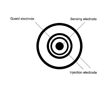
16 The 2-point electrode probe surface can consist of four concentric
electrodes, which are to be
17 applied to a skin surface of a subject, as shown in Figure 1. The two
outermost electrodes 10,
18 11 are referred to injection electrodes as they are used for electricity
injection into the body
19 tissue. The two inner electrodes 12, 13 are referred to as sensing
electrodes as they are used
for receiving and sensing the electrical current, which passes through the
body tissue. By
21 injecting the electricity with two electrodes, a virtual electrode can
be created, which is
22 positioned somewhere in between the two physical injection electrodes.
The depth of
23 penetration of the electrical current into the body tissue is dependent
on the distance between
24 the two injection electrodes, and the amount of electrical current that
is injected through one
injection electrode relative to the other. In the SciBase II electrode probe
noted above, the
26 system is configured to measure at five predetermined depths between 0.1
and 2 mm below the
27 skin surface, but any number of depths can be programmed. The outermost
sensing electrode
28 12 is also known as a guard electrode as it is used to shield the
central sensing electrode 13
29 from currents passing along the skin surface from the injection
electrodes. As these currents
have not passed through any body tissue, they do not represent an impedance
value which can
31 be correlated to blood glucose level.
8
22341227.1
CA 025 68032 2013-02-07
CA 2,568,032
Blakes Ref: 50586/00047
1 In addition to being in contact with the skin, one or more of the
injection electrodes or the
2 sensing electrodes can be in electrically conductive contact with a sub-
dermal or subcutaneous
3 body tissue, for example, muscle tissue from which impedance is to be
measured. All or some
4 of the injection electrodes and the sensing electrodes can also be
implanted within the body of
the subject, typically they are implanted sub-dermally. For example, the
electrodes can be
6 implanted subcutaneously, e.g. on the surface of a large muscle.
Alternatively, the injection
7 electrodes can be in conductive contact with skin tissue, preferably a
skin surface and the
8 sensing electrodes can be in conductive contact with the body tissue from
which impedance is
9 to be measured, such as sub-dermal tissue.
The two injection electrodes can be placed at two different positions on the
body of the
11 subject, in order to measure impedance of one or more body segments
(e.g. a portion of an arm
12 or a leg) or the whole body. In other words, each pair of injection and
sensing electrodes can
13 be located at two different positions. A pair of a first injection
electrode and a first sensing
14 electrode are placed in conductive contact with a skin tissue or a sub-
dermal body tissue at a
first position on the body of the subject, and a second pair of a second
injection electrode and a
16 second sensing electrode are placed in conductive contact with a skin
tissue or a sub-dermal
17 body tissue at a second position on the body of the subject.
18 In a further preferred embodiment of the invention, a first injection
electrode is placed on
19 a skin surface near the knee of a subject, and a second electrode is
placed on a skin surface
near the ankle of the same leg of the subject. The sensing electrodes can be
placed on the skin
21 surface or inserted below the skin. In this configuration, the impedance
of the lower leg, from the
22 knee to the ankle, is measured.
23 When the injection electrodes and/or the sensing electrodes are in
conductive contact
24 with a skin surface, the skin surface can be treated with saline
solution prior to contact with the
electrode to increase electrical coupling between the skin and the electrode.
For example, the
26 skin can be moistened with saline solution for 60 seconds, using a paper
tissue soaked with
27 saline solution. Excess saline solution is then wiped off and the
electrode(s) placed on the
28 treated site. Also, an electrically conductive gel can improve contact
between the skin surface
29 and electrodes to provide more accurate and reproducible measurements.
The gel can be left
on the skin during the impedance measuring step, i.e. while the electrode is
in contact with the
9
22341227.1
CA 025 68032 2013-02-07
CA 2,568,032
Blakes Ref: 50586/00047
1 skin surface. Such electrically conductive gels would be well known to a
person skilled in the
2 relevant art.
3 In a broad aspect of the invention, determination of the amount of
glucose includes
4 comparison of the measured impedance with a predetermined relationship
between impedance
of body tissue and blood glucose level. For example, blood glucose levels can
be determined
6 directly from a blood sample and measuring the blood glucose level with a
commercially
7 available glucometer, e.g. an EliteTM Glucometer (Bayer Diagnostics). At
the same time, body
8 tissue impedance measurements, using the above-noted configurations of
electrodes, can be
9 collected for each directly measured blood glucose level to form a
standard set of data for
calibrating the apparatus of the invention. The injection electrodes and the
sensing electrodes
11 are in operative connection with a computer or a microprocessor
programmed to determine the
12 amount of glucose level based upon the measured impedance.
13 The microprocessor can be programmed to determine the glucose level of
a subject
14 based on a principal component analysis and a partial east squares
regression analysis of the
measured impedance. In principal component analysis ("PCA"), data gathered in
an experiment
16 with a large number of variables is accumulated in a data matrix X of
size n x k, where the rows
17 n represent the different measurements and the columns k signify the
variables, in order to
18 reduce the number of dimensions and therf ore find hidden patterns which
are not detectable by
19 simple analysis of raw data (Eriksson L. et al. Multi- and Mega variate
Data Analysis, Umetrics
AB).
21 The principal component analysis algorithm NIPALS splits the data
matrix into a new
22 data structure as well as a residual matrix in which the noisy part of
the data is gathered. The
23 equation for the PCA decomposition is given by:
X =W31'1421)21+-1- taP:= E
24 (1)
X =TP'+E
where t, is the principal component score vector, pi is the principal
component loading
26 vector, T is the score matrix and P the loading matrix and E the
residual matrix.
27 Partial least squares regression ("PLS") is the projection of latent
structures by means of
28 partial least squares (Eriksson L. et al. (2001) Multi- and Mega variate
Data Analysis, Umetrics
22341227.1
CA 025 68032 2013-02-07
CA 2,568,032
Blakes Ref: 50586/00047
1 AB). The partial least squares-algorithm NIPALS uses an extra loading
weight W, which directly
2 connects to the building relationship of the X and Y. As long as the
dominant structures in X
3 agree with the maximum direction of correlation in Y, the loadings P and
W remain similar.
4 Should they show significant difference this would imply that the
features found in Y do not
correlate well to the dominant characteristics in X (Esbensen K. et al. (1994)
Multivariate
6 Analysis in Practise, Camo AS). The formulas for the decomposition of the
two data matrices
7 with the NIPALS algorithm are given below:
8 X = TP' + E
(2)
9 Y=U01+F
(3)
Y = (W(P'W)-10') X (4)
11 where T, U are the score matrices and P, W, Q the loading matrices and
E, F the residual
12 matrices for X and Y respectively. Y stands for the predicted value from
a PLS model To
13 summarize PLS tries to capture the most variance within each data matrix
X and Y and also
14 take into account that the correlation between the two should be as
large as possible.
Cross-validation will iteratively calculate a value for the closeness of the
fit for the model,
16 called Q2, which is calculated as follows:
17
18 Q2=1¨
_)2
19
(5)
where yi are the observed response values and kothe predicted response values
obtained
21 through cross-validation using only the p.th PLS component and y being
the mean of all the
22 measured responses. The closeness of the fit, Ci for each PLS component
revealing its
23 individual significance. If kip is calculated when all significant p PLS
components are
24 incorporated, a cumulative score for Cf, called Claim is attained
through the equation above. A
Q2cõ of 0.5 is considered to be good and a value of 0.9 is excellent. However,
this value is
26 application specific, depending on what process is modelled. Hence a
lower or higher Q2cõ can
11
22341227.1
CA 025 68032 2013-02-07
CA 2,568,032
Blakes Ref: 50586/00047
1 be considered good or excellent. The explained variance of Y, R2Yc,,,,,
is then compared with
2 Cfc,,.. The results from above-described analysis of the impedance
measurements can then
3 presented in an error grid. In the Examples, a BD grid is employed
(Parkes J.L., et al. (2000), "A
4 new consensus error grid to evaluate the clinical significance of
inaccuracies in the
measurement of blood glucose," Diabetes Care, 23, pp 1143-1148). Other types
of error
6 analysis can be used, such as the Clarke grid (Clarke W.L., et al. (1987)
"Evaluating clinical
7 accuracy of systems for self-monitoring of blood glucose", Diabetes Care,
10, pp 622-628.; Cox
8 D.J., et al. (1989) "Clarification of Error-Grid Analysis", Diabetes
Care, 12, pp 235-236). It will be
9 understood that in view of the above discussion and the cited references,
persons skilled in the
relevant art would understand which multivariate algorithms can be used to
correlate impedance
11 measurements with glucose levels, when provided with directly measured
glucose levels.
12 The computer or microprocessor includes an indicator for indication
of the determined
13 glucose level. This indicator can include a visual display for the
subject to see. The computer of
14 microprocessor can optionally be operatively connected to an insulin
pump and means to adjust
the amount of insulin flow via the pump to the subject, based on the
determined blood glucose
16 level. In the case of implanted electrodes, the electrodes can be
operatively attached to a
17 microprocessor, programmed as described above, and the electrodes and
the microprocessor
18 placed in a housing, similar to a pacemaker device, which allow for
implantation. An insulin
19 pump can also be combined with the implanted electrodes and
microprocessor.
Further details of the preferred embodiments of the invention are illustrated
in the
21 following Examples which are understood to be non-limiting with respect
to the appended
22 claims.
23 Examole 1
24 Skin impedance was measured using the SciBase II skin impedance
spectrometer with
2-point electrode (Figure 1), applied to a skin surface of a subject. To
increase the conductance
26 of the horny layer of the skin, it was moisturized with physiological
saline solution. In this
27 experiment the volar forearms of two volunteers were used. One volunteer
is diagnosed with
28 Diabetes Mellitus type 1, whilst the other subject is not known to
suffer from neither Diabetes
29 nor any other blood or skin related diseases. Impedance was measured
every ten minutes for
approximately 4 hours during several days.
12
22341227.1
CA 025 68032 2013-02-07
CA 2,568,032
Blakes Ref: 50586/00047
1 On the volar forearms of the volunteers, two hairless sites measuring
2x2 cm, located
2 above and below the middle of the arm, were marked. One site at a time
was moistened with
3 saline solution during 60 seconds, using a paper tissue with the
dimensions 4.5x6.5 cm soaked
4 with 2.5 ml of saline solution. Quickly afterwards excess water was wiped
off and the probe was
placed on the marked site. The probe rested on the skin with merely its own
weight for ten
6 seconds in order to establish a good contact. Skin measurements were then
carried out with the
7 impedance spectrometer at five different depth settings using 31
frequencies for each depth.
8 This resulted in 155 magnitude and 155 phase values after a measuring
time of 20 seconds.
9 The data acquired was analysed using PCA to show possible outliers or
drifts within the
measured data. PLS was then applied to the data in order to correlate the
impedance against
11 blood glucose measured in a drop of blood from a fingertip
12 For the non-diabetic subject, the data showed significant fluctuations
between
13 consecutive measurements even without changes in blood glucose level.
The fluctuations were
14 so severe that the hope of finding any correlation was close to
nonexistent. Therefore, the
results presented below originate only from the diabetic subject.
16 Applying PCA on the raw data quickly showed the existence of four
outliers in the score
17 plot (Figure 3). These outliers correspond to the magnitude and phase
curves marked in the raw
18 data plot (Figure 2). They are probably the result of either too much
inundation, excess fluid on
19 the skin surface or long time storage of fluid in the top layers of the
skin.
After removing these outliers from the data, PCA was applied once more. No new
critical
21 outliers were found and the remaining data are considered to be a good
base for further
22 analysis.
23 Example 2
24 In the non-invasive experiment of Example 1, a small correlation was
detected between
glucose concentration and impedance magnitude, suggesting that a correlation
could be
26 detected by placement of the electrodes in direct contact with body
tissue, in the case muscle
27 tissue.
28 Body tissue impedance measurements were obtained with a instrument
obtained by
29 modifying an Impedance Body Segment Analyzer ("IBSA"), Department of
Medical Technology,
13
22341227.1
CA 025 68032 2013-02-07
CA 2,568,032
Blakes Ref: 50586/00047
1 Huddinge University Hospital, Huddinge, Sweden (011mar, Nicander I.
(1995) "Information in
2 multi-frequency measurements on intact skin", lnnov. Tech. Biol. Med. 16:
745-751). The IBSA
3 measures in the frequency range from 5 Hz to 500 kHz and was converted to
4-point
4 impedance measurements for this experiment. Circuitry for separation of
current injection and
voltage detection was added to the IBSA.
6 The measurements were performed in the right leg of one volunteer
suffering from
7 diabetes mellitus type 1. A Styrofoam form was used to stabilize the leg
during the
8 measurement period, since movement will affect the measurements. Two
injection electrodes
9 were placed in conductive contact with the skin surface of the subject's
leg, and two sensing
electrodes, in the form of needles, were inserted below the skin of the
subject's leg. The
11 positions of the needles (sensing electrodes) were decided after studies
of the human anatomy,
12 as well as discussions with medical personnel. One injection electrode
was placed near the
13 knee, and the second injection electrode placed near the ankle. The
sensing electrodes were
14 implanted near the middle of the calf muscle, i.e. approximately mid-way
between the two
injection electrodes.
16 An estimated blood glucose value for each measurement was obtained
using an
17 invasive blood glucose meter (Glucometer Elite XL 3901E, Bayer).
18 The measurements started approximately 60 minutes after the needles
were put into
19 place, in order to ensure a minimum of remaining inflammatory responses
(Koschinsky T.
Heinemann L. (2001) "Sensors for glucose monitoring: technical and clinical
aspects" Diabetes
21 Metab. Res. Rev. 17, 113-123).
22 Impedance values and blood glucose were registered every 10 minutes
for roughly four
23 hours. For each impedance measurement, five registrations were made to
be able to investigate
24 the deviation within each measurement compared to the deviation between
consecutive ones.
To achieve a wide span of blood glucose concentrations in the subject, 75 g of
water-
26 free glucose powder dissolved in water were consumed after the first
hour. When the blood
27 glucose reached a high enough value, insulin was administered yielding a
drop in the glucose
28 value. Lunch was also consumed during the measurement period. At the end
of the experiment
29 the blood glucose of the subject was thoroughly checked for a longer
time to make sure
hypoglycaemia did not set in.
14
22341227.1
CA 025 68032 2013-02-07
CA 2,568,032
Blakes Ref: 50586/00047
1 The collected data was analysed in several steps. First the raw data was
visualized as
2 is, without any data analysis applied. Thereafter statistical methods box
plot, mean value and
3 standard deviation were used to reveal artifacts such as noise. PCA was
used to make sure that
4 no outliers would affect the model.
The raw data gathered from the measurements are shown in Figure 4. Two
injection
6 electrodes in conductive contact with the skin surface of a leg of a
subject, and two surface
7 electrodes implanted below the surface of the skin of the leg. The data
shows signs of
8 interference at specific frequencies. Since all frequencies are cross-
correlated, there should be
9 no apparent discontinuities in the plot. Three distinctive frequency
ranges are affected by noise
(5-10 Hz, 1-2 kHz and 100-500 kHz) which were dropped from the data analysis.
11 After those adjustments, the data set was analysed with PCA to assure
that all five
12 registrations that were done for each measurement showed similar
characteristics area, implies
13 that the total deviation is large within one observation. Although there
was background noise, it
14 is evident from the data analysis that there is a correlation between
sub-dermal tissue
impedance and the directly measured glucose level.
16 Example 3
17 The procedure of Example 2 was repeated, with the subject seated in a
Faraday cage
18 constructed in which the walls, floor and ceiling are covered by metal
plates made of p.-metal.
19 The computer used for analysis of the measurements was a laptop portable
computer, in order
to further reduce noise sources. As soon as the needles were put into place,
the measurements
21 started. The procedure was the same Example 2 where five registrations
were made for each
22 impedance measurement, with one
addition. During the measurements, both lunch and
23 75 g water-free glucose powder was consumed in order to achieve a wide
span of blood
24 glucose values
Since the raw impedance data contained less noise than before, the mean values
of the
26 replicas were considered directly. A PCA model was created based on the
mean values of the
27 magnitudes and possible outliers were identified. Thereafter a PLS model
was constructed to
28 see if a correlation exists between impedance and blood glucose. Every
even sample was used
29 as training data for the model and every other as prediction set. Cross
validation of the model
was also done using SIMCA.
22341227.1
CA 025 68032 2013-02-07
CA 2,568,032
&ekes Ref: 50586/00047
1 To validate the model further, the reference glucose vector was
permutated a number of
2 times and for each permutation new Qc2-õts and RzY.,õ,s were calculated
using the same
3 amount of PLS components to validate the statistical significance of the
model.
4 The first five measurements (i.e. the measurements carried out during
the first 50
minutes) were distorted and were discarded. The first five frequencies (i.e. 5-
23 Hz) were also
6 excluded, since these were disturbed. It is known that low-frequency
measurements are hard to
7 get noise-free. Figure 5a is a loading plot of the impedance data
collected between 5 Hz to 500
8 kHz. As observed in the loading plots (Figures 5a,b), the two highest
frequencies (340 and
9 500kHz) are separated from the rest, indicating that these frequencies
were affected by noise
as well and hence they were excluded. Figure 5b is a loading plot after
excluding problematic
11 frequencies, from 34 Hz to 232 kHz for impedance data from an electrode
probe of the present
12 invention
13 Figure 7 shows the result of the 200 permutations of the Y data. For
each permutation
14 new values for R2 Ymm, and Q2curn have been calculated. The fact that
all the new R2Kõand
C/2õ, values remain beneath the values calculated for the model indicates its
statistical
16 significance. The correlation between blood glucose level and impedance
found in this
17 experiment is quite clear. The Q2cum is 0.516 indicating that the model
must be considered to be
18 significant from a statistical point-of-view. The validation through
permutation of the Y data
19 enforced the significance of the model.. As can be seen the BD error
grid (Figure 7), 7 values
are categorized as A and 5 values are categorized as B (see Parkes J.L., et
al. (2000), "A new
21 consensus error grid to evaluate the clinical significance of
inaccuracies in the measurement of
22 blood glucose," Diabetes Care, 23, pp 1143-1148). From a clinical point-
of-view, the model
23 based on every other sample is significant and the method provides a
correlation between
24 measured impedance and blood glucose level.
26 Example 4
27 The SciBase II was used in place of the modified IBSA of Examples 2 and
3, since it is
28 much less sensitive to background noise. The experiment was carried out
in an unshielded
29 environment. Attempts were made to reduce possible noise sources, such
as turning off nearby
computer screens. The same experimental procedure as Examples 2 and 3 was
used.
16
22341227.1
CA 025 68032 2013-02-07
CA 2,568,032
Blakes Ref: 50586/00047
1 The SciBase Ills a two-point impedance measurement device. As such, the
two needles
2 were placed subcutaneously in the upper and lower part of the left arm of
the same volunteer as
3 in previous experiments. To assure that the needles would not cause more
than an initial
4 inflammatory response, the sharp point of the needles were pushed
through, so that they exited
the skin a few centimeters from the insertion site. Blood samples were taken
from the finger tips
6 of the right hand and were used for direct measurements of blood glucose
concentration.
7 The measurements did not start until two hours after the incision of the
needles. Both
8 glucose level and impedance values were registered simultaneously every
five minutes during
9 the entire experiment, that lasted almost four hours. For each impedance
measurement, two
registrations were done to compare the deviation within each measurement with
the deviation
11 between consecutive ones.
12 Lunch was consumed in the beginning of the measurements, and half way
through 75 g
13 of water-free glucose powder dissolved in a glass of water was consumed
to achieve a wide
14 span of blood glucose concentrations in the subject. Insulin was
administered when the blood
glucose level reached approximately 300 mg/di. At the end of the experiment,
the subject was
16 observed during a longer time period, to assure that the subject
returned to and remained in
17 euglycaemia.
18 The two replicas from the impedance measurements were examined both in a
raw data
19 plot and in a PCA score plot. Possible outliers were also identified in
the score plot. After this, a
PLS model was constructed and R2Y and Q2were calculated using the same cross
validation
21 method as before. Permutations of the Y data were also performed to
validate the model further.
22 In the end a PLS model was constructed from every other sample and the
rest were used as a
23 prediction set.
24 No measurements were discarded due to initial inflammatory response in
this
experiment, since the measurements did not start until two hours after the
insertion of the
26 needles. A PCA score plot was constructed anyhow to reveal possible
outliers later during the
27 experiment (Figure 8). The seven last measurements (sample 40-46) were
found to be outliers
28 and were discarded. No frequencies were found to deviate substantially
from the rest and thus
29 all frequencies were included in the model. Thereafter a PLS model was
created on the first 39
measurements. Cross validation was performed to obtain values for R2 Yõ, and
Q2cum . The
17
22341227.1
CA 025 68032 2013-02-07
CA 2,568,032
Blakes Ref: 50586/00047
1 conclusion was made that four components should be used, since increasing
from four to five
2 PLS components only gave a minor change in Y variance and Q. For further
validation, the Y
3 data was permuted 200 times using the same randomizing seed as before.
New values for
4 R2Yõ, and Q2õ, were calculated for each permutation. As seen in Figure 9,
all the new values
remained well below the original R2 Yõ, and Q20, which is another indication
of an accurate
6 model.
7 Another validation was performed as before, where half the data were
used as a training
8 set and the other half as a prediction set. The result from this
prediction is shown in a BD error
9 grid in Figure 10. Using every second point for building the model and
the rest for prediction
gave a very significant model from both a clinical and statistical point-of-
view. In the BD error
11 grid (Figure 10) only 3 points were categorized outside A and all these
were categorized in B.
12 The Q2õ, calculated for the model by using cross validation was 0.875
and R211c,m0.909
13 for four PLS components. This is a close to excellent model when only
considering these values
14 and thus the correlation between blood glucose concentration and sub-
dermal or sub-cutaneous
impedance is even stronger.
16 The scope of the claims should not be limited by the preferred
embodiments set forth in
17 the examples, but should be give the broadest interpretation consisting
with the description as a
18 whole.
19
18
22341227.1