Note: Descriptions are shown in the official language in which they were submitted.
CA 02568981 2006-12-01
74618-47
1
INTEGRASE-DERIVED HIV-INHIBITING AGENTS
FIELD OF INVENTION
The present invention relates to agents based on integrase
of HIV-1, for inhibiting the proliferation of HIV-1.
BACKGROUND OF THE INVENTION
The integrase (IN) of human immunodeficiency virus type 1
(HIV-1) mediates the integration process. It is also
implicated in different steps during viral life cycle
including reverse transcription and viral DNA nuclear
import.
HIV-1 integrase is encoded by the pol gene. During early
phase of the HIV-1 replication cycle, after virus entry into
target cells, another pol gene product, reverse
transcriptase (RT), copies viral genomic RNA into double-
stranded cDNA which exists within a nucleoprotein
preintegration complex (PIC). The PIC also contains viral
proteins including RT, IN, nucleocapsid (NC, p9), Vpr and
matrix (MA, p17) and this large nucleoprotein complex is
capable of actively translocating into the cell nucleus,
including that of non-dividing cells. This feature is
particularly important for the establishment of HIV-1
replication and pathogenesis in exposed hosts, since the
infection of postmitotic cells including tissue macrophages,
mucosal dendritic cells as well as non-dividing T cells may
CA 02568981 2006-12-01
74618-47
2
be essential not only for viral transmission and
dissemination, but also for the establishment of persistent
viral reservoirs.
HIV-1 IN is composed of three functional domains, an N-
terminal domain, a central catalytic core domain and a C-
terminal domain, all of which are required for a complete
integration reaction. The N-terminal domain harbors an HHCC-
type zinc binding domain and is implicated in the
multimerization of the protein and contributes to the
specific recognition of DNA ends. The core domain of IN
contains the highly conserved DDE. The C-terminal domain was
shown to possess nonspecific DNA binding properties. Fassati
et al EMBO J. 2003 July 15; 22(14): 3675-3685 described
nuclear import assays in primary macrophages using purified
HIV-1 reverse transcription complexes (RTCs) as substrate.
Fassati found that imp7 is a mediator of HIV-1 nuclear
import, that small interfering RNA (siRNA) mediated
depletion of imp7 in cultured cells, and that recombinant IN
could pull down imp7, impa, imp(3 and transportin from HeLa
cell lysates. Fassati also concluded that impf3 alone was
insufficient to sustain significant RTC nuclear import, and
that functional imp7 was necessary.
SUMMARY OF THE INVENTION
The present invention provides an isolated peptide
comprising an IN-derived sequence. The IN-derived sequence
comes from the C-terminal domain of integrase, which is
generally defined by residues 205 to 288 of the HIV-1
integrase shown by example as SEQ ID NO:1. The IN-derived
portion of the peptide has a length of at least 8 amino
acids and no more than 83 amino acids that come from the C-
terminal domain of integrase. The IN-derived portion of the
peptide comprises at least one of the sequences:
CA 02568981 2006-12-01
74618-47
3
KELQKQITK (#211-219 of SEQ ID NO:1), KGPAKLLWK (#236-244 SEQ
ID NO:1), WKGPAKLLWKGEGAVV (#235-250 SEQ ID NO:1), VVPRRKAK
(#259-266 SEQ ID NO:1), KVVPRRKAK (#258-266 SEQ ID NO:l),
and PRRKAKII (#261-268 SEQ ID NO:1).
In one form, in the peptide above, the at least 8 and no
more than 83 consecutive amino acids of integrase comprises
at least one of the sequences: TKELQKQITKLQNFRV (SEQ ID
NO:10), PLWKGPAKLLWKGEGAVV (SEQ ID NO:11), PRRKAKIIRDYGK
(SEQ ID NO:12), KELQKQITKLQNFRVYYRDSRDPLWKGPAKLLWKG (SEQ ID
NO:13), KGPAKLLWKGEGAVVIQDNSDIKVVPRRKAK (SEQ ID NO:14), and
KELQKQITKLQNFRVYYRDSRDPLWKGPAKLLWKGEGAVVIQDNSDIKVVPRRKAK
(SEQ ID NO:15).
In another form, in the peptide above, the at least 8 and no
more than 83 consecutive amino acids of integrase comprises
the sequence KELQKQITK (#211-219 of SEQ ID NO:1) or
KGPAKLLWK (#236-244 SEQ ID NO:1) or both.
In another form, in the peptide above, the at least 8 and no
more than 83 consecutive amino acids of integrase comprises
the sequence KGPAKLLWK (#236-244 SEQ ID NO:1) or VVPRRKAK
(#259-266 SEQ ID NO:1) or both.
In another form, in the peptide above, the at least 8 and no
more than 83 consecutive amino acids of integrase comprises
the sequence KELQKQITK (#211-219 of SEQ ID NO:1), KGPAKLLWK
(#236-244 SEQ ID NO:1), and VVPRRKAK (#259-266 SEQ ID NO:1).
In another form, in any of the peptides above, the peptide
comprises at least 13 and no more than 83 consecutive amino
acids from residues 205 to 288 of an HIV-1 integrase
sequence, and the at least 13 and no more than 83
consecutive amino acids of integrase comprises at least one
of the sequences: KELQKQITK (#211-219 of SEQ ID NO:l),
KGPAKLLWK (#236-244 SEQ ID NO:1), WKGPAKLLWKGEGAVV (#235-250
CA 02568981 2006-12-01
74618-47
4
SEQ ID NO:1), VVPRRKAK (#259-266 SEQ ID NO:1), KVVPRRKAK
(#258-266 SEQ ID NO:1), and PRRKAKII (#261-268 SEQ ID NO:1).
In another form, in any of the peptides above, the IN-
derived portion of the peptide has a length of at least 10
amino acids and no more than 83 amino acids that come from
the C-terminal domain of integrase. Specifically
contemplated are peptides where the IN-derived portion has a
length of at least 10, 12, 14, 18, 22, 28, 34, 40, 50, 60,
70 and 80 amino acids that come from the C-terminal domain
of integrase. Also contemplated are isolated peptides that
consist of IN-derived sequences having a length of at least
10 amino acids and no more than 83 amino acids from the C-
terminal domain of integrase (defined by amino acids 205-288
of SEQ ID NO:1 in one embodiment).
In another form, in any of the peptides above, the peptide
further comprises a heterologous sequence which is fused
with the IN-derived sequence (i.e. derived from residues 205
to 288 of HIV-1 integrase). The heterologous sequence may
be a membrane-translocating sequence, e.g. the HIV Tat
membrane-translocating sequence (SEQ ID NO:9). The
heterologous sequence may also be a reporter sequence.
In another form, any of the peptides above, when expressed
with HIV-1 provirus, renders HIV-1 replication-defective or
infection-defective.
Another aspect of the invention relates to a variant
polypeptide of HIV-1 integrase having a substitution or
deletion in at least one of the following positions of HIV-1
integrase: K211, K215, K219, K236, K240, K244, V249, V250,
K258, R262, R263, K264, K266, and K273.
Another aspect of the invention relates to a variant
polypeptide of HIV-1 integrase having at least one of the
following regions deleted: KELQKQITK (#211-219 of SEQ ID
CA 02568981 2006-12-01
74618-47
NO:1), KGPAKLLWK (#236-244 SEQ ID NO:1), WKGPAKLLWKGEGAVV
(#235-250 SEQ ID NO:1), VVPRRKAK (#259-266 SEQ ID NO:1),
KVVPRRKAK (#258-266 SEQ ID NO:1), and PRRKAKII (#261-268 SEQ
ID NO:1), or having a combination of the above deletions and
5 substitutions where the substitutions occur in at least one
of the following positions of HIV-1 integrase: K211, K215,
K219, K236, K240, K244, V249, V250, K258, R262, R263, K264,
K266, and K273. In one embodiment, the variant polypeptide
comprises residues 205 to 288 of HIV-1 integrase. (This
takes into account the positions and regions other than
those substituted or deleted.)
In other embodiments, the variant polypeptide is a truncated
version of integrase containing the substitutions and/or
deletions described above. The truncated variant has an N-
terminus corresponding to any position of integrase from
amino acid 1 to amino acid 205 and has a C-terminus
corresponding to any position of integrase from amino acid
267 to 288.
In other embodiments, the variant polypeptide is a truncated
version of integrase that lacks at least one region defined
by amino acids 212-288, 240-288, 258-288, 212-266.
Another aspect of the invention relates to a variant
polypeptide described herein that, when expressed with HIV-1
provirus, renders HIV-1 replication-defective or infection-
defective. The variant polypeptide may have impaired
binding to imp7 or impg.
Another aspect of the invention relates to a fusion
polypeptide comprising the variant polypeptide described
herein fused to a heterologous sequence. The heterologous
sequence may be a membrane-translocating sequence such as
the HIV Tat membrane-translocating sequence (SEQ ID NO:9).
CA 02568981 2006-12-01
74618-47
6
Another aspect of the invention relates to an isolated
polynucleotide encoding any of the peptide or variant
polypeptide or fusion polypeptide described above.
Another aspect of the invention relates to a monoclonal
antibody or fragments thereof specifically immunoreactive
against at least one of the sequences: KELQKQITK (#211-219
of SEQ ID NO:1), KGPAKLLWK (#236-244 SEQ ID NO:1),
WKGPAKLLWKGEGAVV (#235-250 SEQ ID NO:1), VVPRRKAK (#259-266
SEQ ID NO:1), KVVPRRKAK (#258-266 SEQ ID NO:1), PRRKAKII
(#261-268 SEQ ID NO:1), TKELQKQITKLQNFRV (SEQ ID NO:10),
PLWKGPAKLLWKGEGAVV (SEQ ID NO:11), PRRKAKIIRDYGK (SEQ ID
NO:12), KELQKQITKLQNFRVYYRDSRDPLWKGPAKLLWKG (SEQ ID NO:13),
KGPAKLLWKGEGAVVIQDNSDIKVVPRRKAK (SEQ ID NO:14), and
KELQKQITKLQNFRVYYRDSRDPLWKGPAKLLWKGEGAVVIQDNSDIKVVPRRKAK
(SEQ ID NO:15). The monoclonal antibody may be a single
chain monoclonal antibody. The monoclonal antibody may
inhibit binding of HIV-1 integrase with imp7 or impg. The
monoclonal antibody fragment or single chain monoclonal
antibody may be fused with a heterologous sequence which may
be a membrane-translocating sequence, e.g. the HIV Tat
membrane-translocating sequence.
Another aspect of the invention relates to a chemically
synthesized double stranded short interfering nucleic acid
(siNA) molecule that directs cleavage via RNA interference
(RNAi) of a HIV RNA encoding amino acids 205 to 288 of HIV-1
integrase, wherein a) each strand of said siNA molecule is
about 18 to about 23 nucleotides in length; and b) one
strand of said siNA molecule comprises nucleotide sequence
having sufficient complementarity to said HIV RNA for the
siNA molecule to direct cleavage of the HIV RNA via RNA
interference. Each strand of the siNA molecule may be about
18 to about 23 nucleotides in length; and one strand of the
siNA molecule may comprise a nucleotide sequence having
CA 02568981 2006-12-01
74618-47
7
sufficient complementarity to SEQ ID NO:16 for the siNA
molecule to direct cleavage of the HIV RNA via RNA
interference. In one embodiment, each strand of said siNA
molecule is about 21 nucleotides in length.
Another aspect of the invention relates to a method of
inhibiting HIV-1 replication in a cell, comprising
transporting into the cell any of the peptide, the variant
polypeptide, the fusion polypeptide, the monoclonal
antibody, or the short interfering nucleic acid (siNA)
molecule described herein.
Another aspect of the invention relates to a method of
inhibiting HIV-1 replication in a cell, comprising
expressing in the cell any of the peptide, the variant
polypeptide, the fusion polypeptide, the monoclonal
antibody, or the short interfering nucleic acid (siNA)
molecule described herein.
Another aspect of the invention relates to a method of
inhibiting HIV-1 infection in a human comprising
administering to the human the peptide, the variant
polypeptide, the fusion polypeptide, the monoclonal
antibody, or the short interfering nucleic acid (siNA)
molecule described herein.
Another aspect of the invention relates to a method for
screening for a compound that affects HIV-1 replication or
infection, the method comprising: (a) incubating, in the
presence of a candidate agent, the IN-derived peptide as
defined herein with imp7 or impfs, under conditions suitable
for binding to occur between the peptide and imp7 or impS;
(b) determining the level of binding between the peptide and
imp7 or impg, wherein detecting a change in the level of
binding between the peptide and imp7 or impg in the presence
of the candidate agent, compared to the level of binding in
CA 02568981 2006-12-01
74618-47
8
the absence of the candidate agent, indicates that said
agent is a compound that affects HIV-1 replication or
infection. The screening method may screen for a compound
that inhibits HIV-1 replication or infection, which would
require detecting a decrease in the level of binding between
the peptide and imp7 or impS in the presence of the
candidate agent, compared to the level of binding in the
absence of the candidate agent.
Another aspect of the invention relates to a method for
screening for a compound that affects HIV-1 replication or
infection, the method comprising: (a) providing a cell that
expresses (i) the IN-derived peptide as defined herein and
(ii) imp7 or impg; (b) providing the cell with a candidate
agent; and (c) determining the level of binding between the
expressed peptide and the expressed imp7 or impS, wherein
detecting a change in the level of binding between the
peptide and imp7 or impS in the presence of the candidate
agent, compared to the level of binding in the absence of
the candidate agent, indicates that said agent is a compound
that affects HIV-1 replication or infection. The screening
method may screen for a compound that inhibits HIV-1
replication or infection, which would require detecting a
decrease in the level of binding between the peptide and
imp7 or impS in the presence of the candidate agent,
compared to the level of binding in the absence of the
candidate agent.
BRIEF DESCRIPTION OF THE DRAWINGS
The present invention will be further understood from the
following description with reference to the drawings, in
which:
Figure 1 shows the interaction of HIV-1 IN and importin 7.
CA 02568981 2006-12-01
74618-47
9
1A) Schematic representation of constructs of IN-YFP, T7-
imp7 and T7-imp8. For IN-YFP, a full-length wild-type HIV-1
IN was fused in frame to the N-terminus of EYFP. For T7-imp7
and imp8, a T7-tag (9 amino acids) was fused in frame to the
N-terminus of imp7 and imp8.
1B) Expression of IN-YFP and T7-imp7 and T7-imp8. Cell
lysates from about 6x10⁵ 293T cells transfected with CMV-YFP,
CMV-IN-YFP or indicated importin expressors was analyzed
with immunoprecipitation with rabbit anti-GFP antibody
followed by western blotting using mouse anti-GFP antibody
(lanes 1 to 3) or immunoprecipitation with mouse anti-T7
antibody followed by western blotting using the same
antibody (lanes 4 to 5).
1C) The in vivo co-IP assay. CMV-IN-YFP was co-transfected
with plasmids for T7-imp7 (lane 3) or T7-imp8 (lane 4) into
2x10⁶ 293T cells. As a control, CMV-YFP also was co-
transfected with each importin expressing plasmid (lane 1,
2). After 48 hr of transfection, cells were lysed by 0.5%
CHAPS buffer and immunoprecipitated with rabbit anti-GFP
antibody. Then, immunoprecipitated complexes were resolved
by 12.5% SDS-PAGE and immunoblotted with either mouse anti-
T7 antibody (upper panel) or mouse anti-GFP antibody (middle
panel). The unbound T7-imp7 and T7-imp8 were also checked by
sequential immunoprecipitation with anti-T7 antibody
followed by immunoblotting with the same antibody (lower
panel ) .
Figure 2 shows that HIV-1 IN interacts with endogenous imp7
and that the interaction between IN and impf3 takes place in
the cells.
2A) The IN-YFP and T7-imp7 plasmids were co-transfected
(lane 2) or transfected individually (lane 3) into 293T
cells. After 48 hrs, cells were mixed accordingly, lysed and
CA 02568981 2006-12-01
74618-47
analyzed with co-immunoprecipitation using the same
procedure as Fig. 1C. Upper panel: co-precipitated imp7
detected by western blot with anti-T7 antibody; Middle
panel: The expression of IN-YFP detected by western blot
5 with mouse anti-GFP antibody; Lower panel: the unbound imp7
visualized by immunoprecipitation and western blot by anti-
T7 antibody.
2B) IN interacts with endogenous imp7. 10x106293T cells were
mock-transfected (lane 1) or transfected with CMV-YFP (lane
10 2) and CMV-IN-YFP (lane 3). After 48 hours of transfection,
cells were lysed by 05%. CHAPS buffer and immunoprecipitated
with rabbit polyclone anti-GFP antibody. Then,
immunoprecipitates were separated in 10% SDS-PAGE followed
by immunoblotting with rabbit anti-importin7 (upper panel)
or monoclonal anti-GFP antibody (lower panel). In parallel,
2x10⁶ of non-transfected 293T cells were lysed with the same
lysis buffer and 10% of celllysates were loaded in SDS-PAGE
as positive control (PC).
Figure 3 shows in vitro interaction between IN and imp7.
3A) GST (lane 1) and GST-imp7 (lane 2) were expressed in E
coli and affinity-purified on amylose resin. The similar
amount of purified protein was directly loaded on a 12.5%
SDS-PAGE followed by the Coomassie Blue staining.
3B) Equal amount of GST (lane 1) and GST-imp7 (lane 2) was
incubated with a purified recombinant HIV-1 IN in 199 medium
(containing 0.1% CHAPS) for 2 hours at 4 C. Then, the
glutathione-sepharose 4B beads were added and incubate for
additional one hour. After incubation, the beads were washed
five times with the same lysis buffer and the protein
complexes bound to glutathione-sepharose 4B beads were
eluted with 10 mM glutathione buffer and loaded onto a 12.5%
CA 02568981 2006-12-01
74618-47
11
SDS-PAGE followed by western blot analysis with rabbit anti-
IN specific antibodies.
Figure 4 shows differential binding ability of HIV-1 MAp17
and IN to cellular importins Rchl and imp7.
4A) HIV-I MAp17G2A, but not IN, binds to T7-Rchl. 293T cells
were co-transfected by CMV-T7-Rchl with YFP (lane 2), IN-YFP
(lane 3) or MAp17G2A-YFP expressor (lane 4). After 48 hrs of
transfection, cells were lysed by CHAPS lysis buffer and
immunoprecipitated with rabbit anti-GFP antibody followed by
western blot with either anti-T7 or mouse anti-GFP
antibodies, as described in the legend for Fig. 1C. Upper
panel shows the co-precipitated T7-Rchl protein. The middle
panel shows the expression of YFP, IN-YFP or Map17C;2A-YFP and
the lower panel reveals the unbound T7-Rchl.
4B) HIV-1 IN, but not MAp17G2A, binds to imp7. 293T cells
were co-transfected with YFP (lane 2), IN-YFP (lane 3) or
MApl7G2A-YFP (lane 4) plasmid with T7-imp7 expressor. Upper
panel indicates the co-precipitated T7-imp7; the middle
panel shows the expression of YFP, IN-YFP or MAp17G2A-YFP and
the lower panel reveals the unbound T7-imp7 in each cell
lysate sample.
Figure 5 indicatess the region(s) of HIV-1 IN that interact
with imp7.
5A) Schematic representation of IN-YFP and YFP-IN truncated
proteins used for binding assay. The IN sequence shown
corresponds to amino acids 210-288 of SEQ ID NO:1.
SB) The N-terminal domain is dispensable for IN:imp7
interaction. The YFP (lane 3), IN-YFP (lane 4) and
IN50-288-YFP (lane 5) were co-expressed with T7-imp7 in 293T
cells. In parallel, YFP and IN-YFP were expressed alone in
293T cells as control (lanes 1 and 2). At 48 hrs of
CA 02568981 2006-12-01
74618-47
12
transfection, cells were lysed and the interaction between
IN-YFP mutants and imp7 was analyzed using anti-GFP
immunoprecipitation and subsequently western blot with anti-
T7 or anti-GFP antibodies, as described in Fig. 1C. The
upper panel reveals the co-precipitated T7-imp7 and the
middle panel shows the expression of YFP, IN-YFP,
IN50_288-YFP, as indicated. The lower panel shows the
detection of unbound T7-imp7 by anti-T7 immunoprecipitation
and western blot.
5C) The C-terminal domain is required for IN:imp7
interaction. The IN full-length protein (lane 3), IN1_212
(lane 4) , IN1_240 (lane 5) and IN,._260 (lane 6) were assayed for
the interaction with imp7 as described before. Upper panel:
co-precipitated T7-imp7. Middle panel: Expression of YFP,
YFP-IN and YFP-IN mutants. Lower panel: unbound T7-imp7.
Figure 6 shows the effect of different IN C-terminal
substitutions on IN:imp7 interaction.
6A) Diagram of HIV-1 IN domain structure and introduced
mutations at the C-terminal domain of the protein. The
position of introduced mutation is shown at the bottom of
sequence. The IN sequence shown corresponds to amino acids
210-288 of SEQ ID NO:1.
6B) Both of KK240,4 and RK263,4 of IN are involved in Imp7
interaction. The YFP (lanes 2 and 7), YFP-INwt (lanes 3 and
8) and different YFP-IN mutant expressors were co-
transfected with T7-Imp7 expressor in 293T cells and after
48 h of infection, cells were lysed with CHAPS lysis buffer
and the IN/Imp7 interaction for each IN mutant was analyzed
by using the same protocol as described in Figure 1C. Upper
panel: co-precipitated T7-Imp7. Middle panel: Expression of
YFP, YFP-INwt and YFP-IN mutants. Lower panel: unbound T7-
Imp7. The position of each immunoprecipitated and co-
CA 02568981 2006-12-01
74618-47
13
precipitated proteins were indicated on the right side of
the gel.
6C) The YFP-IN mutants as in B) were transfected into cells
and visualized by fluorescence.
Figure 7 shows interaction of HIV-1 IN with T7-impg in co-
transfected 293T cells. 293T cells were mock-transfected or
transfected with SVCMVin-YFP, SVCMVin-IN-YFP or SVCMVin-YFP-
IN expressors. Cells were lysed with 199 medium containing
0.25%NP-40 and a protease inhibitor cocktail (Roche), and
clarified by centrifugation at 13,000 rpm for 30 min at 4 C.
The supernatant was subjected to immunoprecipitation with
rabbit anti-GFP antibody and immunoprecipitates were
resolved by 10% SDS-PAGE gel followed by western blot using
mouse anti-T7 (upper panel) or mouse anti-GFP antibodies
(middle panel), respectively. Also, the total T7-ImpR
expression in cell lysates was sequentially
immunoprecipitated with mouse anti-T7 antibody followed by
western blot using the same antibody (lower panel).
Figure 8 shows an immunocomplex of IN-YFP and endogenous
impg and imp7 in 293T cells. 293T cells were transfected
with SVCMVin-YFP, SVCMVin-IN-YFP or SVCMVin-YFP-IN
expressor. After 48 hours of transfection, cells were lysed
by 199 medium with 0.25% NP-40 and immuno-precipitated with
anti-GFP followed by western blot with a rabbit anti-human
ImpR antibody (Cat# SC-11367, Santa Cruz Biotechnology Inc)
(shown in middle panel) and anti-GFP antibody (shown in the
lower panel). Then, the nitrocellular membrane from the
middle panel was stripped with glycine/HC1 buffer (0.1M
glycine, pH. 2.7) and re-processed with western blot with
anti-imp7 antibody (shown in upper panel). The positions of
different proteins are shown at the right side of the gel.
Figure 9 shows interaction of HIV-1 IN with impR in vitro.
CA 02568981 2006-12-01
74618-47
14
9A) Protein expression. Left panel: [35S] methionine-labeled
CAT, T7-IN and T7-Ran protein were expressed in vitro using
TnT T7 coupled reticulocyte lysate system, extracts were
separated on a SDS-PAGE and expressed proteins were detected
by autoradiography. Right panel: Purified GST (Control),
GST-Impa, and GST-impR were verified by directly loading on
a 12.5% SDS-PAGE followed by the Coomassie Blue staining.
9B) ImpR interacts with T7-IN and with T7-Ran, but not with
T7-CAT. A GST pull-down assay was conducted as described
above. Following extensive washing in 199 medium containing
0.25% NP40, the bound protein complexes were eluted with
50 mM glutathione and separation on a SDS-PAGE followed by
autoradiography.
9C) Direct binding of GST-impR and imp7 with purified HIV-
1 IN. Left panel: as above, GST, GST imp7 and impR were
expressed and purified. Purified proteins were separated on
an SDS-PAGE and detected by Coomassie Blue staining. Right
panel: A GST pull-down assay was conducted as described
above, with purified HIV-1 IN protein. Following extensive
washing in 199 medium containing 0.25% NP40, the bound
protein complexes were eluted with 50 mM glutathione and
separation on a SDS-PAGE and finally pulled-down protein was
detected by a western anti-IN antibody.
Figure 10 shows that the C-terminal domain of HIV-1 IN
interacts with impg in co-transfected 293T cells. YFP-IN
and different mutant expressors, as indicated, were co-
transfected with T7-impS in 293T cells. After 48 hours,
cells were lysed, immunopreciptated with anti-GFP followed
by western blot with anti-T7 antibody (upper panel) and
anti-GFP antibody (middle panel). The total amount of T7-
impg was analyzed by sequential IP with anti-T7 antibody
followed by western blot with anti-T7 antibody.
CA 02568981 2006-12-01
74618-47
Figure 11 shows that the HIV-1 IN C-terminal domain alone is
sufficient for binding to imp7 and to inhibit HIV-1
infection.
11A) Schematic representation of CMV-YFP and CMV-YFP-INc205
5 (IN amino acids 205-288) expressors.
11B) intracellular localization of YFP or YFP-INc205 in
HeLa cells.
11C) The HIV-1 IN C-terminal domain alone is sufficient for
binding to imp7. Plasmids expressing YFP, IN-YFP, or YFP-
10 INc205 were cotransfected with CMV-T7-imp7. After 48 hrs of
transfection, imp7-binding was analyzed using anti-GFP
immunoprecipitation and subsequently western blot with anti-
T7 or anti-GFP antibodies.
11D) Over-expression of the HIV-1 IN C-terminal domain
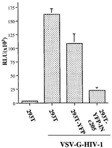
15 alone is sufficient for inhibiting infection of VSV-G-
pseudotyped HIV-1 virus in 293T cells. To test the effect
of YFP-INc205 on HIV-1 infection, each 293T cell line,
including parental 293T cells, was infected with equal
amounts of VSV-G pseudotyped pNLlucOBgII virus (at 5 cpm of
RT activity/cell). Since viruses contain a luciferase (luc)
gene in place of the nef gene, viral infection can be
monitored by using a sensitive luc assay which could
efficiently detect viral gene expression After 48 hours of
infection, equal amounts of cells (1x10⁶ cells) were lysed in
50 l of luc lysis buffer and then, 10 l of cell lysates
was used for measurement of luc activity.
Figure 12 shows that mutations in the C-terminal domain of
IN inhibit HIV single-cycle replication and affect reverse
transcription and nuclear import.
12A) 293T cells were transfected with a RT, IN and Env
deleted HIV-1 provirus NLlucOBglORI with different Vpr-RT-
CA 02568981 2006-12-01
74618-47
16
IN(WT/Mutant) expressors and a VSV-G expressor. Produced
viruses (lane 1 to 3) were lysed and directly loaded in 12%
SDS-PAGE and analyzed by Western blot with human anti-HIV
serum. The positions of HIV-1 Gag , RT and IN proteins are
indicated.
12B) The CD4+ C8166 cells were infected with viruses vWT,
vD64E, and vKKRK viruses. At different time intervals after
infection, the equal amount (1x10⁶) of cells was collected
and cell-associated luciferase activity was measured by
luciferase assay.
12C) Effects of Imp7-binding defect mutants on HIV-1 reverse
transcription and DNA nuclear import. At 24 hours post-
infection, 2x10⁶ cells were gently lysed and fractionated
into the cytoplasmic and the nuclear fractions. The amount
of viral DNA in both fractions were analyzed by PCR using
HIV-1 LTR-Gag primers and Southern blot. Nuc. nuclear
fraction; Cyt. cytoplasmic fraction, The purity and DNA
content of each subcellular fraction were monitored by PCR
detection of human globin DNA and visualized by specific
Southern blot (lower panel).
12D) The total amounts of viral DNA (right panel) and the
percentage of nucleus-associated viral DNA relative to the
total amount of viral DNA (left panel) for each mutant was
also quantified by laser densitometry. Means and standard
deviations from two independent experiments are shown.
Figure 13 shows siRNA-mediated silencing of Imp7 inhibits
HIV-1 infection.
13A) A schematic depiction of the method steps shown as an
example.
13B) siRNA-mediated silencing of Imp7 in 293T and HeLa-R-
Gal-CD4/CCR5 cells. Cells were transfected with 20 nM of
CA 02568981 2006-12-01
74618-47
17
siRNA at 0 and 18 hours. After 48, 72 and 96 hours post
initial transfection, the Imp7 expression levels in the
cells were verified by Western blot with anti-Imp7 antibody
(upper panel). Meanwhile, the expression of (x-tubulin was
also verified (lower panel).
13C) 293Tcells were treated with sc-RNA or si-imp7 once a
day for two days and used to produced VSV-G-pseudotyped
HIV-1 4.3 virus (sc-virus and si-virus). Both viruses were
then used to infect HeLa-R-Gal-CD4/CCR5 cells that have been
treated with Imp7 siRNA or scramble RNA for 72h. Luciferase
activity was measured at 48h post-infection.
13D) sc-RNA or si-imp7 treated HeLa-R-Gal cells were
infected with wild-type enveloped HxBru virus produced from
sc-RNA- or si-imp7-treated HeLa cells. Viral Infection was
evaluated by MAGI assay.
Figure 14 shows subcellular localization of the wild-type
and truncated HIV integrase fused with YFP.
14A) Schematic structure of HIV-1 integrase-YFP fusion
proteins. Full-length (1-288aa) HIV-1 integrase, the N-
terminus-truncated mutant (51-228aa) or the C-terminus-
truncated mutant (1-212aa) was fused in frame at the N-
terminus of YFP protein. The cDNA encoding for each IN-YFP
fusion protein was inserted in a SVCMV expression plasmid.
14B) Expression of different IN-YFP fusion proteins in 293T
cells. 293T cells were transfected with each IN-YFP
expressor and at 48 hours of transfection, cells were lysed,
immunoprecipitated with anti-HIV serum and resolved by
electrophoresis through a 12.5% SDS-PAGE followed by Western
blot with rabbit anti-GFP antibody. The molecular weight
markers are indicated at the left side of the gel.
CA 02568981 2006-12-01
74618-47
18
14C) Intracellular localization of different IN-YFP fusion
proteins. HeLa cells were transfected with each HIV-1 IN-YFP
fusion protein expressor and at 48 hours of transfection,
cells were fixed and subjected to indirect immuno-
fluorescence using rabbit anti-GFP and then incubated with
FITC-conjugated anti-rabbit antibodies. The localization of
each fusion protein was viewed by Fluorescence microscopy
with a 50x oil immersion objective. Upper panel is
fluorescence images and bottom panel is DAPI nucleus
staining.
Figure 15 shows the effect of different IN C-terminal
substitution mutants on IN-YFP intracellular localization.
15A) Diagram of HIV-1 IN domain structure and introduced
mutations at the C-terminal domain of the protein. The
position of lysines in two tri-lysine regions and introduced
mutations are shown at the bottom of sequence. The IN
sequence shown corresponds to amino acids 210-288 of SEQ ID
NO:1.
15B) The expression of the wild-type and mutant IN-YFP
fusion proteins were detected in transfected 293T cells by
using immunoprecipitation with anti-HIV serum and Western
blot with rabbit anti-GFP antibody, as described in
Figure 1. The molecular weight markers are indicated at the
left side of the gel.
15C) Intracellular localization of different HIV-1 IN
mutant-YFP fusion proteins in HeLa cells were analyzed by
fluorescence microscopy with a 50x oil immersion objective.
The nucleus of HeLa cells was simultaneously visualized by
DAPI staining (lower panel).
Figure 16 shows the production of different single-cycle
replicating viruses and their infection in HeLa-CD4-CCR5-S-
Gal cells.
CA 02568981 2006-12-01
74618-47
19
16A) To evaluate the trans-incorporation of RT and IN in
VSV-G pseudotyped viral particles, viruses released from
293T cells transfected with NLlucLBg10RI provirus alone
(lane 6) or cotransfected with different Vpr-RT-IN
expressors and a VSV-G expressor (lane 1 to 5) were lysed,
immunoprecipitated with anti-HIV serum. Immunoprecipitates
were run in 12% SDS-PAGE and analyzed by Western blot with
rabbit anti-IN antibody (middle panel) or anti-RT and anti-
p24 monoclonal antibody (upper and lower panel).
16B) The infectivity of trans-complemented viruses produced
in 293 T cells was evaluated by MAGI assay. HeLa-CD4-CCR5-
LTR-9-Gal cells were infected with equal amounts (at 10
cpm/cell) of different IN mutant viruses and after 48 hours
of infection, numbers of S-Gal positive cells (infected
cell) were monitored by X-gal staining. Error bars represent
variation between duplicate samples and the data is
representative of results obtained in three independent
experiments.
Figure 17 shows the effect of IN mutants on viral infection
in dividing and nondividing C8166 T cells. To test the
effect of different IN mutants on HIV-1 infection in CD4+ T
cells, dividing (panel A) and non-dividing (aphidicolin-
treated, panel B) C8166 T cells were infected with equal
amount of VSV-G pseudotyped IN mutant viruses (at 5
cpm/cell). For evaluation of the effect of different IN
mutants on HIV-1 envelope-mediated infection in CD4+ T
cells, dividing C8166 T cells were infected with equal
amount of HIV-1 envelope competent IN mutant viruses (at 10
cpm/cell) (panel C). After 48 hours of infection, HIV-1 DNA-
mediated luciferase induction was monitored by luciferase
assay. Briefly, the same amount (106cells) of cells was
lysed in 50 ul of luciferase lysis buffer and then, 10 l of
cell lysate was subjected to the luciferase assay. Error
CA 02568981 2006-12-01
74618-47
bars represent variation between duplicate samples and the
data is representative of results obtained in three
independent experiments.
Figure 18 shows the effects of different IN mutants on HIV-1
5 reverse transcription and DNA nuclear import.
Dividing C8166 T cells were infected with equal amounts of
different HIV-1 IN mutant viruses.
18A) At 12 hours post-infection, 1 x 10⁶ cells were lysed and
the total viral DNA was detected by PCR using HIV-1 LTR-Gag
10 primers and Southern blot.
18B) Levels of HIV-1 late reverse transcription products
detected in panel A were quantified by laser densitometry
and viral DNA level of the wt virus was arbitrarily set as
100%. Means and standard deviations from two independent
15 experiments are presented.
18C) At 24 hours post-infection, 2 x 10⁶ cells were
fractionated into cytoplasmic and nuclear fractions as
described in Materials and Methods. The amount of viral DNA
in cytoplasmic and nuclear fractions were analyzed by PCR
20 using HIV-1 LTR-Gag primers and Southern blot (upper panel,
N. nuclear fraction; C. cytoplasmic fraction). Purity and
DNA content of each subcellular fraction were monitored by
PCR detection of human globin DNA and visualized by specific
Southern blot (lower panel).
18D). The percentage of nucleus-associated viral DNA
relative to the total amount of viral DNA for each mutant
was also quantified by laser densitometry. Means and
standard deviations from two independent experiments are
shown.
CA 02568981 2006-12-01
74618-47
21
Figure 19 shows the ffect of IN mutants on HIV-1 proviral
DNA integration. Dividing C8166 T cells were infected with
equal amounts of different HIV-1 IN mutant viruses. At 24
hours post-infection, 1 x 10⁶cells were lysed and serial-
diluted cell lysates were analyzed by two-step Alu-PCR and
Southern blot for specific detection of integrated proviral
DNA from infected cells (Upper panel). The DNA content of
each lysis sample was also monitored by PCR detection of
human 9-globin DNA and visualized by specific Southern blot
(middle panel). The serial-diluted ACH-2 cell lysates were
analyzed for integrated viral DNA and as quantitative
control (lower panel). The results are representative for
two independent experiments.
Figure 20 shows that expression of HIV-1 integrase C-
terminal domain in viral producer cells inhibits subsequent
HIV-1 infection in HeLa-R-Gal-CD4-CCR5 cells and in CD4+ T-
lymphoid MT4 cells. 293T cells were transfected with HIV-1
provirus NL4.3-Nef+/GFP+ and SVCMVin-T7 or SVCMVin-T7-INc2o5-
288 expressor (the IN sequence shown as SEQ ID NO:2). After
48 hours of transfection, viruses were collected from the
supernatant through an ultracentrifugation, and virus titers
were quantified by HIV-1 RT activity assay. Equal amounts
of viruses, as measured by virion-associated reverse
transcriptase activity (A), were used to infect HeLa-R-Gal-
CD4/CCR5 cells (B) or MT4 cells (C). At 48h post-infection,
the viral infection levels were evaluated by MAGI assay (B)
or by counting of GFP-positive cells (C).
Figure 21 shows the amino acid sequence of HIV-1 integrase
(SEQ ID NO:1 derived from HIV-1 pNL4.3 strain) shown as an
example. The C-terminal domain of IN and the two tri-lysine
regions and an arginine/lysine region involved in IN/imp7
and IN/impR interactions are indicated.
CA 02568981 2006-12-01
74618-47
22
Figure 22 shows fusions of Tat peptide (SEQ ID NO:9) with IN
peptides (SEQ ID NOs 10-15) as examples.
Figure 23 shows the siRNA target regions of IN as examples.
The HIV-1 IN RNA sequence from nt 628 to 801 is shown (SEQ
ID NO:16); this sequence encodes amino acids 210 to 267 of
integrase. Also indicated are the RNA sequence encoding the
two tri-lysine regions and the arginine/lysine rich region.
These sequences (siRNA #1-4; SEQ ID NOs 17-20) can be used
for siRNA silencing of IN protein expression during viral
replication.
Figure 24 is an ELISA scheme based on INc205-288 as an
example, for screening of compounds that inhibit IN
interaction with imp7 and/or with impR.
Figure 25 is a schematic depiction of a live cell BRET assay
used as an example for detecting interaction of the C-
terminal domain of HIV-l IN with impR and imp7 in live
cells.
DETAILED DESCRIPTION OF EMBODIMENTS
The invention has to do with using the C-terminal domain of
HIV-1 integrase, and certain peptides derived from specific
regions of this domain, for inhibiting HIV-1 infection.
Without being limited by mechanism, the invention is based
on the finding that these regions of the IN C-terminal
domain interact directly with the nuclear import machinery
of the host cell, specifically with imp7 and impg, and that
these IN regions are necessary for translocating the HIV-1
nucleoprotein pre-integration complex (PIC) into the
nucleus. Thus these IN regions are important for
establishing HIV replication and subsequent infection.
The experiments carried out herein make use of certain
specific materials and techniques. These are set forth
CA 02568981 2006-12-01
74618-47
23
below solely for verifying the experimental findings and
should not limit the scope of the invention.
(1) Construction of different IN expressors and HIV-1 RT/IN
defective provirus: The full-length wild-type HIV-1 IN cDNA
was amplified by polymerase chain reaction (PCR) using HIV-1
HxBru strain [Yao et al. J Virol. 1995;69:7032-7044] as
template and an engineered initiation codon (ATG) was placed
prior to the first amino acid (aa) of IN. The primers are
5'-IN-HindIII-ATG (5'-GCGCAAGCTTGGATAGATGTTTTTAGATGGAA-3';
SEQ ID NO:23) and 3'-IN-Asp718 (5'-CCATGTGTGGTACCTCATCCTGCT-
3'; SEQ ID NO:24). The PCR product was digested with HindIII
and Asp718 restriction enzymes and cloned in frame to 5' end
of EYFP cDNA in a pEYFP-Nl vector (BD Biosciences Clontech)
and generated a IN-YFP fusion expressor. Also, cDNA encoding
for truncated IN (aa 50 to 288 or aa 1 to 212) was amplified
by PCR and also cloned into pEYFP-Nl vector. The primers for
generation of IN50-288 cDNA are IN50-HindIII-ATG-5' (5'-
GCGCAAGCTTGGATAGATGCATGGACAAGTAG-3; SEQ ID NO:25) and 3'-IN-
Asp718 and primers for amplifying IN1-212 cDNA are IN-
HindIII-ATG-5' and IN-212-XmaI-3' (5'-
CAATTCCCGGGTTTGTATGTCTGTTTGC-3; SEQ ID NO:26). IN
substitution mutants INKK215,gAA-YFP, INKK240,4AE-YFP and
INRK263,4AA-YFP, were generated by a two-step PCR-based method
[Yao et al. Gene Ther. 1999;6:1590-1599] by using a 5'-
primer (5'-IN-HindIII-ATG), a 3'-primer (3'-IN-Asp718) and
complementary primers containing desired mutations.
Amplified IN cDNAs harboring specific mutations were then
cloned into pEYFP-N1 vector. To improve the expression of
each IN-YFP fusion protein, all IN-YFP fusing cDNAs were
finally subcloned into a SVCMV vector, which contains a
cytomegalovirus (CMV) immediate early gene promoter [Yao et
al. Gene Ther. 1999;6:1590-1599].
CA 02568981 2006-12-01
74618-47
24
To construct HIV-1 RT/IN defective provirus NLlucLBglLRI, we
used a previously described HIV-1 envelope-deleted
NLlucLBglD64E provirus as the backbone. In this provirus,
the nef gene was replaced by a firefly luciferase gene [Poon
et al. J Virol. 2003;77:3962-3972]. The ApaI/SalI cDNA
fragment in NLlucBg1D64E was replaced by the corresponding
fragment derived from a HIV-1 RT/IN deleted provirus R-/ORI
[Ao et al. J Virol. 2004;78:3170-3177] and generated a RT/IN
deleted provirus NL1uc4BglORI, in which RT and IN gene
sequences were deleted while a 194-bp sequence harboring
cPPT/CTS cis-acting elements was maintained. To restore HIV-
1 envelope gene sequence in NLluc4BglLRI provirus, the
SalI/BamHI cDNA fragment in this provirus was replaced by a
corresponding cDNA fragment from a HIV-1 envelope competent
provirus R-/LRI [Ao et al. J Virol. 2004;78:3170-3177] and
the resulting provirus is named as NLlucLRI. To functionally
complement RT/IN defects of NL1ucOBg14RI, a CMV-Vpr-RT-IN
fusion protein expressor [Ao et al. J Virol. 2004;78:3170-
3177] was used in this study. Co-transfection of
NLlucLBglORI, CMV-Vpr-RT-IN and a vesicular stomatitis virus
G (VSV-G) glycoprotein expressor results in the production
of VSV-G pseudotyped HIV-1 that can undergo for single cycle
replication in different cell types [Ao et al. J Virol.
2004;78:3170-3177]. To investigate the effect of IN mutants
on viral replication, different mutants KK215,9AA,
KK240.4AE, RK263,4AA or D64E were introduced into CMV-Vpr-
RT-IN expressor by PCR-based method as described above and
using a 5'-primer corresponding to a sequence in RT gene and
including a natural NheI site (51-GCAGCTAGCAGGGAGACTAA-31;
SEQ ID NO:27), a 3'-primer (3'-IN-stop-PstI, 5'-
CTGTTCCTGCAGCTAATCCTCATCCTG-3'; SEQ ID NO:28) and the
complementary oligonucleotide primers containing desired
mutations. All IN mutants were subsequently analyzed by DNA
sequencing to confirm the presence of mutations or
deletions.
CA 02568981 2006-12-01
74618-47
(2) Cell lines and reagents: Human embryonic kidney 293T,
HeLa and HeLa-CD4-CCR5-9-Gal cells were maintained in
Dulbecco's Modified Eagles Medium (DMEM) supplemented with
10% fetal calf serum (FCS). Human C8166 T-lymphoid cells
5 were maintained in RPMI-1640 medium. Antibodies used in the
immunofluorescent assay, immunoprecipitation or western blot
are as follows: The HIV-1 positive human serum 162 and anti-
HIVp24 monoclonal antibody used in this study were
previously described [Yao et al. J Virol. 1998;72:4686-
10 46931. The rabbit anti-GFP and anti-IN antibodies were
respectively obtained from Molecular Probes Inc and through
AIDS Research Reference Reagent Program, Division of AIDS,
NIAID, NIH. Aphidicolin was obtained from Sigma Inc.
(3) Cell transfection and immunofluorescence assay: DNA
15 transfection in 293T and HeLa cells were performed with
standard calcium phosphate DNA precipitation method. For
immunofluorescence analysis, HeLa cells were grown on glass
coverslip (12 mm2) in 24-well plate. After 48 h of
transfection, cells on the coverslip were fixed with PBS-4%
20 paraformaldehyde for 5 minutes, permeabilized in PBS-0.2%
Triton X-100 for 5 minutes and incubated with primary
antibodies specific for GFP or HIV-1 IN followed by
corresponding secondary FITC-conjugated antibodies. Then,
cells on the coverslip were viewed using a computerized
25 Axiovert 200 inverted fluorescence microscopy (Becton
Deckson Inc).
(4) Virus production and infection: Production of different
single-cycle replicating virus stocks and measurement of
virus titer were previously described [Ao et al. J Virol.
2004;78:3170-3177]. Briefly, 293T cells were co-transfected
with RT/IN defective NLlucLBglORI provirus, a VSV-G
expressor and each of CMV-Vpr-RT-IN (wt/mutant) expressor.
To produce HIV-1 envelope competent single cycle replicating
CA 02568981 2006-12-01
74618-47
26
virus, 293T cells were co-transfected with NL1ucRI and
different CMV-Vpr-RT-IN (wt/mutant) expressors. After 48
hours of transfection, supernatants were collected and virus
titers were quantified by RT activity assay [Yao et al. Gene
Ther. 1999;6:1590-1599].
To test the effect of IN mutants on virus infection, equal
amounts of virus were used to infect HeLa-CCR5-CD4-9-Gal
cells, dividing and non-dividing C8166 T cells. To compare
the infection of each viral stock in HeLa-CCR5-CD4-9-Gal
cells, numbers of infected cells (9-Gal positive cells) were
evaluated by the MAGI assay 48 hours post-infection (p.i) as
described previously [Kimpton et al. J Virol. 1992;66:2232-
2239]. To infect CD4+ T cells, dividing or aphidicolin-
treated non-dividing C8166 T cells (with 1.3 g/ml of
aphidicolin) were infected with equivalent amounts of single
cycle replicating viruses (5 cpm/cell) for 2 hours. Then,
infected cells were washed and cultured in the absence or
presence of the same concentration of aphidicolin. At 48
hours post-infection, 1 x 10⁶ cells from each sample were
collected, washed twice with PBS, lysed with 50 l of
luciferase lysis buffer (Fisher Scientific Inc) and then, 10
l of cell lysate was subjected to the luciferase assay by
using a TopCount NXTTM Microplate Scintillation &
Luminescence Counter (Packard, Meriden) and the luciferase
activity was valued as relative luciferase units (RLU). Each
sample was analyzed in duplicate and the average deviation
was calculated.
(5) Immunoprecipitation and Western blot analyses: For
detection of IN-YFP fusion proteins, 293T cells transfected
with each IN-YFP expressor were lysed with RIPA lysis buffer
and immunoprecipitated using human anti-HIV serum. Then,
immunoprecipitates were run in 12% SDS-PAGE and analyzed by
Western blot using rabbit anti-GFP antibody. To analyze
CA 02568981 2006-12-01
74618-47
27
virion-incorporation of IN and virus composition, 293T cells
were co-transfected with NLlucOBglARI provirus and each of
CMV-Vpr-RT-IN (wt/mutant) expressors. After 48 hours,
viruses were collected, lysed with RIPA lysis buffer and
immunoprecipitated with human anti-HIV serum. Then,
immunoprecipitates were run in 12% SDS-PAGE and analyzed by
Western blot with rabbit anti-IN antibody and anti-p24
monoclonal antibody or anti-HIV serum.
(6) HIV-1 reverse-transcribed and integrated DNA detection
by PCR and Southern blotting: C8166 T cells were infected
with equal amount of the wt or IN mutant viruses for 2
hours, washed for three times and cultured in RPMI medium.
To detect total viral DNA synthesis, at 12 hours post-
infection, equal number (1 x 10⁶ cells) of cells were
collected, washed twice with PCR washing buffer (20 mM Tris-
HC1, pH 8.0, 100 mM KC1), and lysed in lysis buffer (PCR
washing buffer containing 0.05% NP-40, 0.05% Tween-20).
Lysates were then incubated at 56 C for 30 min with
proteinase K (100 g/ml) and at 90 C for 10 min prior to
phenol-chloroform DNA purification. To detect viral cDNA
from each sample, all lysates were serially diluted 5-fold
and subjected to PCR analysis. The primers used to detect
late reverse transcription products were as follows: 5'-LTR-
U3, 5'-GGATGGTGCTTCAAGCTAGTACC-3' (SEQ ID NO:29; nt position
8807, +1 = start of BRU of transcription initiation); 3'-Gag
5'-ACTGACGCTCTCGCACCCATCTCTCTC-3' (SEQ ID NO:30; nt position
329). The probe for southern blot detection was generated by
PCR with a 5'-LTR-U5 oligonucleotide, 5'-
CTCTAGCAGTGGCGCCCGAACAGGGAC-3' (SEQ ID NO:31; nt position
173) and the 31-Gag oligo. PCR was carried out using lx
HotStar Taq Master Mix kit (QIAGEN, Mississauga, Ontario),
as described previously [Ao et al. J Virol. 2004;78:3170-
3177] .
CA 02568981 2006-12-01
74618-47
28
To analyze nucleus- and cytoplasm-associated viral DNA, a
subcellular fractionation of infected C8166 T cells (2 x 10⁶)
was performed after 24 hours of infection, as described
previously [Simon et al. J Virol. 1996;70:5297-5305].
Briefly, infected cells were pelleted and resuspended in
ice-cold PCR lysis buffer (washing buffer containing 0,1%
NP-40). After a 5-min incubation on ice, the nucleus was
pelleted by centrifugation, washed twice with PCR wash
buffer, and lysed in lysis buffer (0,05% NP-40, 0,05% Tween-
20). Then, both cytoplasmic sample (supernatant from the
first centrifugation) and the nuclear sample were treated
with proteinase K and used for PCR analysis, as described
above.
Integrated proviral DNA was detected in cell lysates by a
modified nested Alu-PCR [Ao et al. J Virol. 2004;78:3170-
3177], in which following the first PCR, a second PCR was
carried-out to amplify a portion of the HIV-1 LTR sequence
from the first Alu-LTR PCR-amplified products. The first PCR
was carried out by using primers including 51-Alu oligo (51-
TCCCAGCTACTCGGGAGGCTGAGG-3'; SEQ ID NO:32) and 3'-LTR oligo
(51-AGGCAAGCTTTATTGAGGGCTTAAGC-3'; SEQ ID NO:33) (nt
position 9194) located respectively in the conserved region
of human Alu sequence and in HIV-1 LTR. The primer used for
both of the second nested PCR and for generating a probe are
5'-NI: 5'-CACACACAAGGCTACTTCCCT-3' (SEQ ID NO:34) and 3'-NI:
5'-GCCACTCCCCAGTCCCGCCC-3' (SEQ ID NO:35). As a control, the
first and second PCR primer pairs were also used in parallel
to detect integrated viral DNA from serially diluted ACH-2
cells, which contain one viral copy/cell, in a background of
uninfected C8166 cellular DNA.
To evaluate the DNA content of extracted chromosomal DNA
preparations, detection of human 9-globin gene was carried-
out by PCR, as described previously [Simon et al. J Virol.
CA 02568981 2006-12-01
74618-47
29
1996;70:5297-5305]. All final PCR products were
electrophoresed through 1.2% agarose gel and transferred to
hybridization transfer membrane (GeneScreen Plus,
PerkinElmer Life Sciences), subjected to Southern
hybridization by using specific PCR DIG-Labeling probes
(Roche Diagnostics, Laval, Que) and visualized by a
chemiluminescent method. Densitometric analysis was
performed using a Personal Molecular Imager (Bio-Rad) and
Quantity One software version 4.1.
(7) Interaction of HIV-1 IN and importin 7. We investigated
the interaction of HIV-1 IN with different cellular nuclear
import factors. We first tested the interaction of HIV-1 IN
with cellular nuclear import receptors Imp7 and Imp8, by
using a cell-based co-immunoprecipitation (co-
immunoprecipitation) assay. SVCMV-T7-Imp7 and T7-Imp8
expressors were constructed by inserting Imp7 and Imp8 cDNAs
into a SVCMV-T7 vector at the 3' end of a T7 tag encoding
sequence (Fig. 1A). The HIV-1 IN-YFP fusion protein,
expressed from expressor CMV-IN-YFP, and YFP expressed from
the CMV-YFP expressor, were used in the study and shown in
Figure 1A. First, expression of these proteins was checked
by transfecting each of these expressors into 293T cells,
and processed using anti-GFP or anti-T7 immmunoprecipitation
(IP), followed by western blot with corresponding
antibodies. Results showed that IN-YFP and YFP were detected
at positions 58 and 27 kDa respectively (Fig. 1B, lanes 2
and 3), while T7-Imp7 and T7-Imp8 were at positions that
ranged between 110 to 130 kDa (Fig. 1B, lanes 4 and 5). To
test whether IN-YFP could bind to different importins, the
YFP or IN-YFP expressor was co-transfected with each
importin expressor in 293T cells, as indicated in Fig. 1C.
After 48 h, cells were lysed with CHAPS lysis buffer (199
medium containing 0.5% CHAPS), and immunoprecipitated using
rabbit anti-GFP antibody. Precipitated complexes were run
CA 02568981 2006-12-01
74618-47
on an SDS-PAGE, followed by western blot with anti-T7
antibody (Fig. 1C, upper panel). Results revealed that,
while YFP protein did not co-precipitate with any importin
(Fig. 1C, upper panel, lane 1, 2), the immunoprecipitation
5 of IN-YFP specifically co-pulled down T7-Imp7 (Fig. 1C, Lane
3), but not T7-Imp8 (Fig. 1C, lanes 4). Meanwhile, the
immunoprecipitated IN-YFP and YFP in each sample
respectively were checked by anti-GFP western blot, and
similar levels of each protein were detected (Fig. 1C;
10 middle panel, lanes 3, 4). To rule out the possibility that
the co-precipitated T7-Imp7 was due to differential levels
of importin expression in each transfection sample, the cell
lysates were processed using sequential immunoprecipitation
with anti-T7 antibody followed by anti-T7 Western blot, and
15 the results showed similar expression levels of each
importin in different samples (Fig. 1C; lower panel). All
of these results indicated that IN specifically interacts
with Imp7, but not with Imp8.
(8) HIV-1 IN interacts with endogenous imp7 and the
20 interaction between IN and impg takes place in the cells. We
asked was whether the IN/Imp7 interaction occurs in the
cells or after cells had been lysed. To address this
question, IN-YFP or T7-Imp7 expressor was individually
transfected into different 293T cell cultures, as indicated
25 in Figure 2A. After 48 hours, cells from two transfected
cultures were mixed, lysed with 0.5% CHAPS lysis buffer and
incubated in 4 C for two hours. Then, the presence of IN/Imp7
interaction in the cell lysate was checked by anti-GFP
immunoprecipitation, followed by anti-T7 western blot. In
30 parallel, cells co-transfected with both IN-YFP and T7-Imp7
expressors were mixed with the same amounts of mock-
transfected cells and processed identically. Strikingly, the
co-precipitated T7-Imp7 was only detected in co-transfected
cell lysate, but not in mixed cell lysate from individually
CA 02568981 2006-12-01
74618-47
31
transfected cell samples (Fig. 2A, upper panel, compare lane
2 with 3). These results clearly indicate that the
interaction of IN-YFP and T7-Imp7 takes place in the cells.
Again, the specific detection of IN/Imp7 complex in co-
transfected cells, was not due to the varying levels of
expression of IN-YFP or T7-Imp7 protein in the different
samples (Fig. 2 A, middle panel and lower panel; lanes 2 and
3). To further test the interaction between IN-YFP and
endogenous Imp7, 293T cells were transfected with CMV-YFP or
CMV-IN-YFP expressor, lysed by 0.5% CHAPS lysis buffer and
immunoprecipitated with anti-GFP. The co-precipitated
endogenous Imp7 was checked by western blot with a rabbit
anti-human Imp7 antibody. Meanwhile, the non-transfected
293T cell lysates were directly loaded into SDS-PAGE as the
positive control (Fig. 2B, lane 1). We found that IN-YFP,
but not YFP, was able to pull down the endogenous Imp7 (Fig.
2B, upper panel, compare lane 4 to lane 3), indicating that
IN-YFP interacts with endogenous Imp7 in 293T cells.
(9) In vitro interaction between IN and imp7. We asked
whether IN binding to Imp7 could be through direct protein
interaction. We produced purified recombinant GST and GST-
Imp7 proteins in an E coli expression system, and the
purified protein in each sample was tested by directly
loading protein samples in an SDS-PAGE, and verified by
Coomassie Blue staining of the gel (Fig. 3A) and by western
blot with specific anti-Imp7 antibody. To test the direct
interaction of IN and Imp7 in vitro, similar amounts of
purified GST and GST-Imp7 were incubated with a purified
recombinant HIV-1 IN in 199 medium containing 0.1% CHAPS for
2 h at 4 C, followed by an additional one hour incubation
with glutathione-sepharose 4B beads. Then, the bound
protein complex was eluted out with 10 mM glutathione, and
loaded onto a 12.5% SDS-PAGE gel, followed by western blot
analysis with anti-IN antibodies. Results showed that the
CA 02568981 2006-12-01
74618-47
32
purified HIV-1 IN, in both of dimer and monomer forms, was
able to specifically interact with GST-Imp7, and not with GST
(Fig. 3B1). Thus, the binding of IN to Imp7 may be through
a direct protein/protein interaction.
(10) Differential binding ability of HIV-1 MAp17 and IN to
cellular importins Rchl and imp7. The importin a/(3 nuclear
translocation pathway has been implicated in assisting with
HIV-1 nuclear import. Several HIV-1 proteins, including
MAp17, Vpr and IN have been shown to be able to interact
with Impa in in vitro binding assays. In this study, we
attempted to test whether HIV-1 IN could interact with Rchl,
a member of the human importin a family, by using co-
immunoprecipitation assay. A T7-tagged Rchl expressing
plasmid (CMV-T7-Rchl), and an HIV-1 MAp17G2A mutant-YFP
fusion protein expressing plasmid (CMV-MAG2A-YFP) were
constructed. In MAp17G2A-YFP, the second amino acid glycine
in MAp17 protein was replaced by alanine, and this MApl7
mutant was previously shown to capable of binding to Rchl in
a cell-based co-immunoprecipitation system. After IN-YFP or
MAG2A-YFP were co-expressed with T7-Rchl in 293T cells, their
interaction with Rchl was analyzed using the same co-
immunoprecipitation and western blot protocols, as described
in Figure 1. MAGZA-YFP was shown to be able to bind to T7-
Rchl (Fig. 4A; lane 4). However, IN-YFP did not show any
interaction with T7-Rchl (Fig. 4A, lane 3). In contrast,
while T7-Imp7 co-precipitated with IN-YFP, no T7-Imp7 was
detected in the immunoprecipitated MAG2A-YFP sample (Fig. 4B,
compare lane 4 to 3). These results suggest that HIV-1 IN
and MAp17 may interact with different cellular nuclear
import factors during HIV-1 replication.
(11) Delineation of necessary region(s) of HIV-1 IN for its
interaction with imp7. To delineate which region(s) within
HIV-1 IN is required for its Imp7-binding, we first tested
CA 02568981 2006-12-01
74618-47
33
an IN N-terminal deletion mutant expressed from the CMV-IN50-
288-YFP expressor (Fig. 5A) for Imp7-binding. Co-
immunoprecipitation analysis revealed that, similar to the
IN-YFP, IN50-288-YFP also bound efficiently to T7-Imp7 (Fig.
5B, compare lane 5 to lane 4), indicating that the N-
terminal domain of IN is not required for IN/Imp7
interaction.
To test the core domain and the C-terminal domain of IN for
their contribution towards Imp7-binding, we constructed
three YFP-IN expressors, including CMV-YFP-INwt and two IN
C-terminal deletion mutants (CMV-YFP-IN1-212 and CMV-YFP-
IN1-240) (Fig. 5A). With the CMV-YFP-INwt expressor, the
PCR-amplified HIV-1 IN full length cDNA, was placed in frame
at the 3' end of the YFP cDNA, while for CMV-YFP-IN1-212 and
CMV-YFP-IN1-240, sequences encoding for the last 76 and 48
aa of IN was removed respectively. Expression of each YFP-
IN fusion protein along with its ability to bind Imp7 was
tested in 293T cells by co-transfecting each YFP-IN fusion
protein expressor with the T7-Imp7 plasmid. The YFP-INwt,
YFP-IN1-212 and CMV-YFP-IN1-240 fusion proteins were
detected at molecular weights ranging approximately from 47
to 58 kDa (Fig. 5C, middle panel, lanes 3 to 5). Co-
immunoprecipitation experiments revealed that while YFP-INwt
efficiently bound to T7-Imp7, the two IN C-terminal deletion
mutants were unable to bind to T7-Imp7 (Fig. SC, upper
panel, compare lane 3 to lanes 4 and 5), suggesting that the
C-terminal region encompassing residues 240 and 288 is
required for IN interacting with Imp7.
(12) Subcellular localization of the wild-type and truncated
HIV integrase fused with YFP. We investigated the
intracellular localization of HIV-1 IN and delineated the
region(s) of IN contributing to its karyophilic property. A
HIV-1 IN-YFP fusion protein expressor (CMV-IN-YFP) was
CA 02568981 2006-12-01
74618-47
34
generated by fusing a full-length HIV-1 IN cDNA (amplified
from HIV-1 HxBru molecular clone, see Yao et al. J Virol.
1995;69:7032-7044) to the 5' end of YFP cDNA in a CMV-IN-YFP
expressor. Transfection of CMV-IN-YFP expressor in 293T
cells resulted in the expression of a 57 kDa IN-YFP fusion
protein (Fig. 14B, lane 2; Fig. 15B, lane 1), whereas
expression of YFP alone resulted in a 27 kDa protein (Fig.
15B, lane 5). Given that HeLa cells have well-defined
morphology and are suitable for observation of intracellular
protein distribution, we tested the intracellular
localization of YFP and IN-YFP by transfecting CMV-IN-YFP or
CMV-YFP expressor in HeLa cells. After 48 hours of
transfection, cells were fixed and subjected to indirect
immunofluorescence assay using primary rabbit anti-GFP
antibody followed by secondary FITC-conjugated anti-rabbit
antibodies. Results showed that, in contrast to a diffused
intracellular localization pattern of YFP (data not shown),
the IN-YFP fusion protein was predominantly localized in the
nucleus (Fig 14C, al), confirming the karyophilic feature of
HIV-1 IN.
We constructed two truncated IN-YFP expressors, CMV-IN50-
288-YFP and CMV-IN1-212-YFP. In CMV-IN50-288-YFP, the N-
terminal HH-CC domain of IN (aa 1-49) was deleted and in
CMV-IN1-212-YFP, the C-terminal domain (aa 213-288) was
removed (Fig. 14A). Transfection of each truncated IN-YFP
fusion protein expressor in 293T cells resulted in the
expression of IN50-288-YFP and IN1-212-YFP at approximately
52 kDa and 48 kDa molecular mass respectively (Fig. 14B,
lanes 3 and 4). We next investigated the intracellular
localization of truncated IN-YFP fusion proteins in HeLa
cells by using indirect immunofluorescence assay, as
described above. Results showed that the IN50-288-YFP was
predominantly localized in the nucleus with a similar
pattern as the wild-type IN-YFP fusion protein (Fig. 14C,
CA 02568981 2006-12-01
74618-47
compare bl to al). However, IN1-212-YFP fusion protein was
excluded from the nucleus, with an accumulation of the
mutant protein in the cytoplasm (Fig 14C, cl). These results
were also further confirmed by using rabbit anti-IN antibody
5 immunofluorescence assay. Taken together, our data show that
the C-terminal domain of HIV-1 IN is required for its
nuclear accumulation.
(13) Effect of different IN C-terminal substitution mutants
on IN:imp7 interaction. We constructed several IN mutants in
10 the IN C-terminal region, in the form of YFP-IN fusion
proteins (Fig. 6A) . Mutants YFP-IN240,4AA, YFP-IN263,4aA and YFP-
INgKRK were designed to target a tri-lysine region
(z35WKGPA240KLLW244KG) , and/or an arginine/lysine rich region
(262RRKAK) . The YFP-IN249, 50AA and YFP-IN258A mutants were
15 constructed to target highly conserved residues valine and
lysine at positions 249, 250 and 258 (Fig. 6A). An IN core
domain mutant YFP-INKR186,7AA was also included in this
study. Each YFP-IN mutant plasmid was co-transfected with
the T7-Imp7 expressor in 293T cells, and processed by the
20 co-immunoprecipitation assay to test each protein's Imp7-
binding ability. Results revealed that while other IN
mutants did not affect the ability to bind Imp7 (Fig. 6B,
lanes 4, 5, 10), the YFP-IN263,4AA mutant significantly
impaired the ability of IN to bind Imp7, and the YFP-INKKRK
25 mutant was unable to interact with imp7 (Fig. 6B, lanes 9
and 10). Thus, all these results indicate that both tri-
lysine region (235WKGPA240KLLW244KG) and the arginine/lysine
rich region (262RRKAK) is required for efficient interaction
between IN and Imp7.
30 (14) Effect of different IN C-terminal substitution mutants
on IN-YFP intracellular localization. The C-terminal domain
of HIV-1 IN contains several regions that are highly
conserved in different HIV-1 strains, including Q, C and N
CA 02568981 2006-12-01
74618-47
36
regions. Regions C(235WKGPAKLLWKGEGAVV; SEQ ID NO:5) and N
(2s9WpRRKAK; SEQ ID NO:6) are conserved in all known
retroviruses and the 211KELQKQITK (SEQ ID NO:3) motif falls
within the so-called glutamine-rich based region (sequence
Q) of lentiviruses. We term the sequences 211KELQKQITK (SEQ
ID NO:3) and 236KGPAKLLWK (SEQ ID NO:4) the proximal tri-
lysine region and distal tri-lysine region, respectively
(Fig. 15A). The lysine residues in these regions are highly
conserved in most HIV-1 strains [Kuiken et al. HIV Sequence
Compendium 2001. Los Alamos National Laboratory. 20011. To
test whether these basic lysine residues could constitute
for a possible nuclear localization signal for IN nuclear
localization, we specifically introduced substitution
mutations for two lysines in each tri-lysine region and
generated INKK215,yAA-YFP and INKK240,4AE-YFP expressors (Fig.
15A). In the conserved N region, there is a stretch of four
basic residues among five amino acids (aa) 262RRKAK. To
characterize whether this basic aa region may contributes to
IN nuclear localization, we replaced an arginine and a
lysine at positions of 263 and 264 by alanines in this
region and generated a mutant (INRK263,4AA-YFP) . The protein
expression of different IN-YFP mutants in 293T cells showed
that, like the wild type IN-YFP, each IN-YFP mutant fusion
protein was detected at similar molecular mass (57 kDa) in
SDS-PAGE (Fig 15B, lanes 1 to 4), while YFP alone was
detected at position of 27 kDa (lane 5). Then, the
intracellular localization of each IN mutant was
investigated in HeLa cells by using similar methods, as
described above. Results showed that, while the wild type
IN-YFP and INRK263,4AA-YFP still predominantly localized to the
nucleus (Fig. 15C, al and dl ), both INKK215, 9AA-YFP and
INKK240,4AE-YFP fusion proteins were shown to distribute
throughout the cytoplasm and nucleus, but with much less
intensity in the nucleus (Fig. 15C, al and bl). These data
suggest that these lysine residues in each tri-lysine
CA 02568981 2006-12-01
74618-47
37
regions are required for efficient HIV-1 IN nuclear
localization.
(15) Interaction of HIV-1 IN with T7-impS in co-transfected
293T cells. We investigated whether HIV-1 IN could also
interact with impR. We constructed the SVCMVin-T7-ImpR
expressor by cloning PCR amplified impR from pET30a-impR.
Then, the SVCMVin-T7-ImpR was co-transfected with SVCMVin-
YFP, SVCMVin-IN-YFP or SVCMVin-YFP-IN expressor in 293T
cells. After 48 hours, cells were lysed with 199 medium
containing 0.25%NP-40 and a protease inhibitor cocktail
(Roche) on ice for 30 min and clarified by centrifugation at
13,000 rpm for 30 min at 4 C. Then, the supernatant was
subjected to immunoprecipitation (IP) with rabbit anti-GFP
antibody and immunoprecipitates were resolved by 10% SDS-
PAGE gel followed by western blot using mouse anti-T7 or
mouse anti-GFP antibodies, respectively. Also, the total
T7-ImpR expression in cell lysates was sequentially
immunoprecipitated with mouse anti-T7 antibody followed by
western blot using the same antibody. Results showed that
immunoprecipitation of IN-YFP and YFP-IN pulled down T7-ImpR
(Fig. 7, lanes 2 and 3), whereas no T7-ImpR was detected in
YFP-transfected or mock-transfected samples (lane 1 and 4).
Again, the specific detection of IN/ImpR complex in co-
transfected cells, was not due to the varying levels of
expression of T7-ImpR protein in the different samples (Fig.
7, lower panel). These results indicate that IN-YFP and YFP-
IN, but not YFP, are capable of binding to T7-ImpR.
(16) Irmnunocomplex of IN-YFP and endogenous impit and imp7 in
293T cells. To rule out the possibility that the IN/ImpR
interaction could be an artifact of overexpression of these
proteins in cells, we tested whether HIV-1 IN (in IN-YFP and
YFP-IN) could interact with endogenous ImpR. SVCMVin-IN-YFP
CA 02568981 2006-12-01
= 74618-47
38
or SVCMVin-YFP-tN expressor was transfected alone in 293T
cells and after 48 hours of transfection, cells were lysed
by 0.25% NP-40 lysis buffer and immunoprecipitated with
anti-GFP followed by western blot with a rabbit anti-human
ImpR antibody (Cat# SC-11367, Santa Cruz Biotechnology Inc).
The co-immunoprecipitation and western blot results revealed
that the endogenous ImpR were co-precipitated with IN-YFP
and YFP-IN (Fig. 8, middle panel, lanes 3 and 4), but not
with YFP alone (lane 2). Meanwhile, the similar amount of
YFP, IN-YFP and YFP-IN were detected through anti-GFP
western blot. These results demonstrated that HIV-1 IN is
capable of binding endogenous ImpR.
We investigated whether endogenous Imp7 could be present in
co-precipitated IN/ImpR complex. We stripped the membrane
(middle panel Figure 8) and re-processed the western blot
with anti-human Imp7 antibody. Results revealed that
endogenous Imp7 could also be detected in IN-YFP and YFP-IN
samples (Fig. 8, upper panel, lanes 3 to 4), but not in
mock-transfected and YFP expressing sample (lanes 1 and 2).
This indicates that endogenous ImpR and Imp7 could be
detected in the same IN-precipitation samples.
(17) Interaction of HIV-1 IN with impo in vitro. To further
study the interaction of HIV-1 IN with ImpR, we constructed
pET21-chloramphenicol acetyltransferase (pET21-T7-CAT),
pET21-T7-IN and pET21-T7-Ran and produced these proteins
using the TnT T7 coupled reticulocyte lysate system (Cat#
L4610, Promega) and labelled them with [35S] methionine
(PerkinElmer). Then, the produced protein samples were
analyzed with SDS-PAGE and each protein was shown at the
corresponding molecular weight (shown in Fig. 9A, left
panel). Also, we produced and purified GST, GST-Impa(Rchl),
GST-imp7 and GST-ImpR from E. coli BL21. Protein expression
CA 02568981 2006-12-01
74618-47
39
was induced by adding isopropyl-l-(3-D-thiogalactopyranoside
(1 mM) for 3 h at 37 C. Bacteria were harvested, suspended in
35 ml of ice-cold column buffer, and broken by sonication
(five 30-s pulses at 100 watts, Sonics & Materials, Inc.)
The resulting lysates were centrifuged for 30 min at 13000
rpm and pass through a glutathione-sepharose 4B column
(Amersham Pharmacia Biotech Inc). After being washing by
column buffer, the bound GST and GST-Imp7 proteins were
eluted by glutathione buffer (100mM reduced glutathione
(Roche), 120mM NaCl, 100mM Tris-HC1 pH 8.5). Finally, the
eluted protein was dialyzed in PBS to remove high
concentration of glutathione. Each purified protein stock
was verified by directly loading on a 12.5% SDS-PAGE
followed by the Coomassie Blue staining (Fig. 9A, right
panel; 9C, left panel).
To test the IN-binding ability of GST, GST-Impa(Rchl), GST-
imp7 and GST-Imp(3, equal amounts of recombinant GST, GST-
Impa(Rchl), GST-imp7 and GST-Imp(3 were incubated with in
vitro-translated [35S]methionine-labeled T7-CAT, T7-IN and
T7-Ran proteins (indicated in Fig. 9B) or a purified HIV-1
recombinant IN protein (Cat No. 9420, obtained through AIDS
Research Reference Reagent Program, Division of AIDS, NIAID,
NIH) in 199 medium containing 0.25% NP40, for 2 hours at
4 C. Then, 100 l of glutathione-sepharose 4B beads were
added and incubated for additional one hour. The beads were
washed and the bound proteins were eluted with 50 mM
glutathione, loaded onto a 12.5% SDS-PAGE and subsequently
analyzed by autoradiography (Fig. 9B) or followed by western
blot analysis with rabbit anti-IN antibodies (Fig. 9C, right
pane l ) .
The in vitro binding results revealed that GST and GST-Impa
did not show any binding to T7-CAT, T7-IN and T7-Ran. There
CA 02568981 2006-12-01
74618-47
was binding of GST-imp(3 to T7-IN and T7-Ran, but not T7-CAT
(Fig. 9B). Moreover, results in Figure 9C showed that both
GST-imp7 and Imp(3, but not GST alone, could pull down
recombinant IN (Fig. 9C, right panel), suggesting a direct
5 interaction between both GST-imp7 and Imp(3 and HIV IN.
(18) The C-terminal domain of HIV-1 IN interacts with impi3
in co-transfected 293T cells. We made different IN C-
terminal deletion mutants to test whether the C-terminal
domain of IN is required for IN interaction with impf3 and
10 which region(s) are necessary for their binding. In CMV-
YFP-INwt expressor, the PCR-amplified HIV-1 IN full length
cDNA was placed in frame at the 3' of YFP cDNA, while in
CMV-YFP-INl-212, CMV-YFP-IN1-240 and CMV-YFP-INl-262, sequences
encoding for last 76, 48 or 26 aa of IN was removed
15 respectively. In YFP-IN50-288, the N-terminal domain (1 to 49
aa) of IN was deleted. To test the expression of each of
YFP-IN fusion proteins along with their abilities to bind
impi3, each YFP-IN fusion protein expressor was co-
transfected with a T7-impS expressor in 293T cells. After 48
20 hours of transfection, cells were lysed with 0.5% NP-40-199
medium and processed using the co-IP assay, as described
above. Results (see Figure 10) show that, like the wt YFP-
IN, the YFP-IN1_240, YFP-INl-262 and YFP-IN50-288 efficiently
bound to T7-impg. However, CMV-YFP-INl_212 lost drastically
25 its binding ability to T7-impS, suggesting a region between
213-239 aa in the C-terminal domain of IN may be important
for this viral/cellular protein interaction.
Our results provide evidence that 1) HIV-1 IN, in both YFP-
IN and IN-YFP forms, is able to interact with endogenous
30 impg and imp7; 2) in vitro binding results suggest that this
IN-impf3 interaction may be through a direct protein-protein
interaction; 3) the C-terminal domain of IN encompassing aa
213-239 may be necessary for its interaction with impg.
CA 02568981 2006-12-01
74618-47
41
(19) The HIV-1 IN C-terminal domain alone is sufficient for
binding to imp7 and to inhibit HIV-1 infection. We
determined that the HIV-1 IN C-terminal domain alone is
sufficient for binding to imp7. We co-transfected YFP, IN-
YFP, or YFP-INc205 expressors with CMV-T7-imp7. After 48
hrs of transfection, imp7-binding was analyzed using anti-
GFP immunoprecipitation and subsequently western blot with
anti-T7 or anti-GFP antibodies (Figure 11C).
We also determined that over-expression of the HIV-1 IN C-
terminal domain alone is sufficient for inhibiting infection
of VSV-G-pseudotyped HIV-1 virus in 293T cells. We tested
the effect of YFP-INc205 (containing the C-terminal IN
residues 205-288) on HIV-1 infection by infecting each 293T
cell line, including the YFP-INc205 expressor cells and the
parental 293T cells, with equal amounts of VSV-G pseudotyped
pNLlucABgII virus (at 5 cpm of RT activity/cell). Since
viruses contain a luciferase (luc) gene in place of the nef
gene, viral infection can be monitored by using a sensitive
luc assay which could efficiently detect viral gene
expression. After 48 hours of infection, equal amounts of
cells (1x10⁶ cells) were lysed in 50 l of luc lysis buffer
and then, 10 l of cell lysates was used for measurement of
luc activity. Results (see Figure 11D) showed that virus
infection in 293T-YFP cells induced 110x103 RLU of luc
activity, which was 33% lower than that in parental 293T
cells. In 293T-YFP-INc205 cells, HIV-1 infection only
induced 24xl 03 RLU of luc activity, that was only
approximately 15% and 21% of levels of that in parental 293T
and 293T-YFP cells. These observations indicate that
expression of the C-terminal region of IN in 293T cells
inhibited HIV-1 infection.
(20) Production of different single-cycle replicating
viruses and their infection in HeLa-CD4-CCR5-1&-Gal cells. To
CA 02568981 2006-12-01
74618-47
42
specifically analyze the effect of IN mutants in early steps
of viral infection, we modified a previously described HIV-1
single-cycle replication system [Ao et al. J Virol.
2004;78:3170-3177] and constructed a RT/IN/Env gene-deleted
HIV-1 provirus NLlucLBglLRI, in which the nef gene was
replaced by a firefly luciferase gene [Poon et al. J Virol.
2003;77:3962-3972]. Co-expression of NLlucOBg14RI provirus
with Vpr-RT-IN expressor and a vesicular stomatitis virus G
(VSV-G) glycoprotein expressor will produce viral particles
that can undergo a single-round of replication, since RT, IN
and Env defects of provirus will be complemented in trans by
VSV-G glycoprotein and Vpr-mediated RT and IN trans-
incorporation [Ao et al. J Virol. 2004;78:3170-3177]. This
single cycle replication system allows us to introduce
different mutations into IN gene sequence without
differentially affecting viral morphogenesis and the
activity of the central DNA Flap. After different IN
mutations KK215,9AA, KK240,4AE and RR263,4AA were introduced
into Vpr-RT-IN expressor, we produced VSV-G pseudotyped
HIV-1 IN mutant virus stocks in 293T cells. In order to
specifically investigate the effect of IN mutants on early
steps during HIV-1 infection prior to integration, an IN
class I mutant D64E was also included as control. After each
viral stock was produced (as indicated in Fig. 16A), similar
amounts of each virus stock (quantified by virion-associated
RT activity) were lysed and virus composition and trans-
incorporation of RT and IN of each virus stock were analyzed
by Western blot analysis with anti-IN and anti-HIV
antibodies. Results showed that all VSV-G pseudotyped IN
mutant viruses had similar levels of Gagp24, IN and RT, as
compared to the wild-type virus (Fig. 16A), indicating that
trans-incorporation of RT and IN as well as HIV-1 Gag
processing were not differentially affected by the
introduced IN mutations.
CA 02568981 2006-12-01
74618-47
43
To test the infectivity of different IN mutant viruses in
HeLa-CD4-CCR5-LTR-S-Gal cells, we first compared the
infectivity of VSV-G pseudotyped wild type virus and the
D64E mutant virus. At 48 hours post-infection with
equivalent amount of each virus stock (at 1 cpm RT
activity/cell), the number of 9-Gal positive cells was
evaluated by MAGI assay, as described in Kimpton et al. J
Virol. 1992;66:2232-2239. Results showed that the number of
infected cells (9-Gal positive cells) for D64E mutant
reached approximately 14% of the wild type level. This
result is consistent with a previous report showing that, in
HeLa MAGI assay, the infectivity level of class I IN
integration-defect mutant was approximately 20 to 22% of
wild type level. It indicates that, even though the IN
mutant D64E virus is defective for integrating viral DNA
into host genome, tat expression from nucleus-associated and
unintegrated viral DNAs can activate HIV-1 LTR-driven 9-Gal
expression in HeLa-CD4-CCR5-LTR-S-Gal cells. Indeed, several
studies have already shown that HIV infection leads to
selective transcription of tat and nef genes before
integration. Therefore, this HeLa-CD4-CCR5-LTR-S-Gal cell
infection system provides an ideal method for us to evaluate
the effect of different IN mutants on early steps of viral
infection prior to integration. We next infected HeLa-CD4-
CCR5-LTR-fs-Ga1 cells with different VSV-G pseudotyped IN
mutant viruses at higher infection dose of 10 cpm RT
activity/cell and numbers of 9-Gal positive cells were
evaluated by MAGI assay after 48 hours of infection.
Interestingly, results showed that the IN mutant D64E virus
infection induced the highest level of 9-Gal positive cells,
whereas infection with viruses containing IN mutants
KK215,9AA, KK240,4AE or RK263,4AA yielded much lower levels
of 9-Gal positive cells, which only reached approximately
11%, 5% or 26% of the level of D64E virus infection (Fig.
16B). Based on these results, we reasoned that these IN C-
CA 02568981 2006-12-01
74618-47
44
terminal mutants blocked infection mostly by affecting
earlier steps of HIV-1 life cycle, such as reverse
transcription and/or viral DNA nuclear import steps, which
are different from the action of D64E mutant on viral DNA
integration.
(21) Effect of IN mutants on viral infection in dividing and
nondividing C8166 T cells. To further test whether these C-
terminal mutants could induce similar phenotypes in CD4+ T
cells, we infected dividing and non-dividing (aphidicolin-
treated) C8166 CD4+ T cells with equal amounts of VSV-G
pseudotyped IN mutant viruses (at 5 cpm of RT
activity/cell). Since all IN mutant viruses contain a
luciferase (luc) gene in place of the nef gene, viral
infection can be monitored by using a sensitive luc assay
which could efficiently detect viral gene expression from
integrated and unintegrated viral DNA [Poon et al. J Virol.
2003;77:3962-3972]. After 48 hours of infection, equal
amounts of cells were lysed in 50 l of luc lysis buffer and
then, 10 l of cell lysates was used for measurement of luc
activity, as described in Materials and Methods. Results
showed that the D64E mutant infection in dividing C8166 T
cells induced 14.3 x 10⁴ RLU of luc activity (Fig. 17A),
which was approximately 1000-fold lower than that in the
wild type virus infection. This level of luc activity
detected in D64E mutant infection is mostly due to nef gene
expression from the unintegrated DNA. In agreement with the
finding by MAGI assay described in Figure 16, the Luc
activity detected in KK215,9AA, KK240,4AE and RK263,4AA
mutant samples were approximately 13%, 5% and 36% of level
of D64E mutant infection (Fig. 17A). In parallel, infection
of different IN mutants in non-dividing C8166 T cells was
also evaluated and similar results were observed (Fig. 17B).
CA 02568981 2006-12-01
74618-47
To test whether these IN mutants had similar effects during
HIV-1 envelope-mediated single cycle infection, we produced
virus stocks by co-transfecting 293T cells with a HIV-1
envelope-competent NLlucRI provirus with each Vpr-RT-IN
5 mutant expressor. Then, dividing CD4+ C8166 cells were
infected with each virus stock (at 10 cpm RT
activity/cells). At 48 hours post-infection, cells were
collected and measured for luc activity. Results from Figure
4C showed that, similar to results obtained from VSV-G
10 pseudotyped virus infection (Fig. 17A), the Luc activity
detected in cells infected by HIV-1 envelope competent
KK215,9AA, KK240,4AE and RK263,4AA mutant viruses were
approximately 13.5%, 6% and 29% of level of D64E mutant
infection (Fig. 17C). All of these results confirm the data
15 from HeLa-CD4-CCR5-LTR-9-Gal infection (Fig. 16) by using
either VSV-G- and HIV-1 envelope-mediated infections and
suggest again that the significantly attenuated infection of
KK215,9AA, KK240,4AE and RK263,4AA mutant viruses may be due
to their defect(s) at reverse transcription and/or viral DNA
20 nuclear import steps.
(22) Effects of different IN mutants on HIV-1 reverse
transcription, DNA nuclear import, and proviral DNA
integration. To directly assess the effect of IN C-terminal
mutants KK215,9AA, KK240,4AE and RK263,4AA on each early
25 step during viral infection, we analyzed the viral DNA
synthesis, their nuclear translocation and integration
following each IN mutant infection in dividing C8166 cells.
Levels of HIV-1 late reverse transcription products were
analyzed by semi-quantitative PCR after 12 hours of
30 infection with HIV-1 specific 5'-LTR-U3/3'-Gag primers and
Southern blot, as previously described [Ao et al. J Virol.
2004;78:3170-3177 and Simon et al. J Virol. 1996;70:5297-
53051. Also, intensity of amplified HIV-1 specific DNA in
each sample was evaluated by laser densitometric scanning of
CA 02568981 2006-12-01
74618-47
46
bands in Southern blot autoradiograms (Fig. 18A). Results
showed that total viral DNA synthesis in both KK215,9AA and
RK263,4AA infection reached approximately 61% and 46% of
that of the wild type (wt) virus infection (Fig. 18A and
18B). Strikingly, in KK240,4AE sample, detection of viral
DNA synthesis was drastically reduced, which only reached
21% of viral DNA level in WT sample (Fig. 18A and 18B).
These results indicate that all three C-terminal mutants
negatively affected viral reverse transcription during viral
infection and KK240,4AE mutant exhibited most profound
effect.
Meanwhile, the nucleus- and cytoplasm-associated viral DNA
levels were analyzed at 24 hours post-infection in C8166 T
cells. The infected cells were first gently lysed and
separated into nuclear and cytoplasmic fractions by using a
previously described fractionation technique [Simon et al. J
Virol. 1996;70:5297-5305]. Then, levels of HIV-1 late
reverse transcription products in each fraction were
analyzed by semi-quantitative PCR, as described above.
Results revealed differential effects of C-terminal mutants
on HIV-1 DNA nuclear import. In the wt, D64E and RK263,4AA
virus-infected samples, there were respectively 70%, 72% and
68% of viral DNA associated with nuclear fractions (Fig. 18C
upper panel, lanes 1 and 2; 3 and 4; 9 and 10 and Fig. 18D).
For KK240,4AE mutant, approximately 51% of viral DNA was
nucleus-associated (Fig. 18C upper panel, lanes 7 and 8 and
Fig. 18D). Remarkably, in KK215,9AA infected sample, viral
cDNA was found predominantly in the cytoplasm and only
approximately 21% of viral DNA was associated with the
nuclear fraction (Fig. 18C upper panel, lanes 5 and 6; and
Fig. 18D). Meanwhile, the integrity of fractionation
procedure was validated by detection of 9-globin DNA, which
was found solely in the nucleus and levels of this nucleus-
CA 02568981 2006-12-01
74618-47
47
associated cellular DNA were similar in each nuclear sample
(Fig. 18C, lower panel).
Even though the C-terminal mutants were shown to
significantly affect HIV-1 reverse transcription and/or
nuclear import, the various low levels of nucleus-associated
viral DNA during the early stage of replication (Fig. 18C)
may still be accessible for viral DNA integration. To
address this question, 1 x 10⁶ dividing C8166 T cells were
infected with equivalent amounts of each single cycle
replicating virus stock (5 cpm/cell), as indicated in Figure
6 and after 24 hours of infection, the virus integration
level was checked by using a previously described sensitive
Alu-PCR technique [Ao et al. J Virol. 2004;78:3170-3177].
Results revealed that, while the wt virus resulted in an
efficient viral DNA integration (Fig. 19, upper panel; lanes
1 and 2), there was no viral DNA integration detected in
D64E mutant (lanes 3 to 4) and in all three C-terminal
mutant infection samples (lanes 5 to 10), although similar
levels of cellular 9-globin gene were detected in each
sample (Fig. 19, middle panel). These results suggest that,
in addition to affecting HIV-1 reverse transcription and
nuclear import, all three C-terminal IN mutants tested in
this study also negatively affected viral DNA integration.
Overall, all of these results indicate that all three IN C-
terminal mutants belong to class II mutants, which affect
different early steps during HIV-1 replication. Among these
mutants, KK240,4AE showed the most profound inhibition on
reverse transcription; KK215,9AA, and to a lesser extent
KK240,4AE, impaired viral DNA nuclear translocation during
early HIV-1 infection in C8166 T cells.
(23) Mutations in the C-terminal domain of IN inhibits HIV
single-cycle replication and affect reverse transcription
and nuclear import. To test the effect of the IN mutant
CA 02568981 2006-12-01
74618-47
48
(INKKRK) on HIV-1 replication, the mutant was introduced
into a VSV-G pseudotyped HIV-1 single-cycle replication
system (see Ao et al. 2004 J Virol. 78:3170-7). Briefly,
the INKKRK mutant was first introduced into a CMV-Vpr-RT-IN
expressor. Then, the VSV-G pseudotyped HIV-1 single cycle
replicating virus (vKKRK) was produced in 293T cells by co-
transfection with CMV-Vpr-RT-INKKRK, an RT/IN-deleted HIV
provirus NLlucABgl/ARI and a VSV-G expressor. In parallel,
the VSV-G pseudotyped wild type virus (vINwt) and IN class I
mutant D64E virus (vD64E), were also produced in parallel as
controls. After each virus stock was harvested, the trans-
incorporation of RT and IN as well as the Gag composition in
the viral particle was analyzed using western blot with a
human anti-HIV positive serum. Results showed that similar
amounts of RT, IN and Gagp24 were detected in each virus
preparation (Fig. 12A). Then, equal amount of each virus
stock (as adjusted by amounts of HIV-1 Gagp24) was used to
infect CD4+ C8166 cells. At different time intervals, the
luciferase (luc) activity in equal amounts of cells was
measured, as shown in Figure 12B. Since D64E mutant virus
(vD64E) is unable to mediate viral DNA integration, its
infection expressed very low luc activity (Fig. 12B). The
luc activity detected from the vKKRK virus infection was
considerably lower than that of the D64E mutant virus at
different time points (Fig. 12B), indicating that the vKKRK
virus lost its replication ability in CD4+ C8166 cells.
To test which step of the infection was affected in the IN
mutants, the cytoplasm- and nucleus-associated viral DNA
levels were analyzed at 24 hours post-infection, using semi-
quantitative PCR and southern blot. For the vKKRK virus
infection, the level of total viral DNA (including the
cytoplasm- and nucleus-associated viral DNA levels) was
reduced by approximately 60%, compared to the total viral
DNA level detected from the wt virus infection (Fig. 12C,
CA 02568981 2006-12-01
74618-47
49
upper panel, compare lanes 5 and 6 to lanes 1 and 2, and D,
left panel). Moreover, results indicated that for the wt
and vD64E virus infections, approximately 73 and 77% of
viral DNA were associated with nuclear fractions (Fig. 12C
(upper panel, lanes 1 to 4) and D, right panel). However,
during vKKRK infection, only 44% of viral DNA was nucleus-
associated (Fig. l2C (upper panel, lanes 5 and 6) and D,
right panel). The integrity of the fractionation procedure
was also validated by detection of R-globin DNA, which was
found solely in the nucleus, and levels of this cellular DNA
were similar in each nuclear sample (Fig. 12C, lower panel).
Taken together, all of these results indicate that the Imp7-
binding defect mutant virus vKKRK was unable to replicate in
C8166 cells and displayed impairment at both viral reverse
transcription and nuclear import.
(24) Expression of HIV-1 integrase C-terminal domain in
viral producer cells inhibits subsequent HIV-1 infection in
HeLa-P-Ga1-CD4-CCR5 cells and in CD4+ T-lymphoid MT4 cells.
It is possible that expression of the IN C-terminal domain
in the late stage of HIV-1 replication inhibits HIV-1
infection. To test this, the IN C-terminal domain was co-
transfected with NL4.3-Nef+/GFP+ provirus in 293T cells.
After 48 hours of transfection, viruses were harvested and
equal amounts of viruses (as measured by virion-associated
reverse transcriptase activity (Figure 20A)) were used to
infect HeLa-R-Gal-CD4/CCR5 cells (Figure 20B) or CD4+
lymphoid MT4 cells (Figure 20C). Results showed that the
virus infection was significantly impaired when HeLa-R-Gal-
CD4/CCR5 cells or MT4 cells were infected by viruses which
were produced by 293T cells expressing the C-terminal domain
of IN (Fig. 20B and 20C). The results suggest that
expression of IN peptides derived from the C-terminal domain
CA 02568981 2006-12-01
74618-47
of IN during the late stage of HIV-1 replication will impair
HIV-1 infection.
(25) SiRNA-mediated silencing of Imp7 inhibits HIV-1
infection. We investigated the effect of small interfering
5 RNA (siRNA)-mediated Imp7-knockdown on HIV-1 replication.
First, we tested the efficiency of Imp7 knockdown, the Imp7-
siRNA (100 pmol) was introduced into 293T and HeLa-R-Gal-
CD4/CCR5 cells once a day for two days (Fig. 13A). Briefly,
293T cells and HeLa-R-gal-CD4/CCR5 cells were plated at 2x10⁵
10 cells/well in 6-well plates and transfected at the next day
with 100 pmol of Imp7-specific small interfering RNA (siRNA)
duplex (IP07-HSS116173) with LipofectamineTM RNAiMAX Reagent
(Invitrogen). After 18 h of first transfection, another
Imp7 siRNA duplex (IP07-HSS116174) was transfected again
15 into cells. These two Imp7-siRNA duplexes (Stealth RNAi),
IP07-HSS116173 and IP07-HSS116174, were synthesized by
Invitrogen Inc and the targeting sequence are respectively
corresponding to Imp7 mRNA nucleotides 1990-2013 (5'-
UAAGCAGAUUCCCUCAAGCUGUUGG-3'; SEQ ID NO:21), and to Imp7
20 mRNA nucleotides 610-633 (sense 5'-
AAUGCUGCAUUGCUGGCUACCAAUGG-3'; SEQ ID NO:22). In parallel,
transfection of a scramble RNA (sc-RNA) (purchased from
Santa Cruz Biotechnology) was used as control. At different
time intervals, equal amounts of cells (0.5 x10⁶ cells) were
25 collected and monitored for Imp7 expression. Western blot
results revealed that Imp7 protein expression were
progressively decreased over the course of the experiments.
At 48 hours following the first Imp7-siRNA transfection, the
Imp7 protein level was reduced to approximately 30%, and at
30 96 hours, the level of Imp7 expression was reduced to <10%
in both 293T and HeLa-R-Gal-CD4/CCR5 cells (Fig. 13B).
Next, we tested the effect of Imp7 knockdown on HIV-1
infection. To avoid the possibility that Imp7 might have
CA 02568981 2006-12-01
74618-47
51
effect on the late stage of viral replication and/or be
packaged into viral particles and thus playing a role in
subsequent viral infection, we first produced a VSV-G
pseudotyped HIV-1 (NL4.3-BruLBgl/luc+) from Imp7-siRNA- or
scramble RNA (sc-RNA)-transfected 293T cells. The Imp7
protein expression in siRNA transfection cells was to <10%
(at 96 hours of siRNA transfection) when the viruses were
collected. Then, viruses (si-virus and sc-virus) produced
from Im7-siRNA- or scRNA-transfected 293T cells were
normalized by HIV Gagp24 levels and used to infect siRNA-
treated and sc-RNA-treated HeLa-R-Gal-CD4/CCR5 cells (target
cells) (Fig. 13A). Results in Figure 7C showed that there
were no significant luc activity differences detected in sc-
RNA- and si-RNA-treated target cells after being infected
with sc-virus (Fig. 13C, bars 1 and 2) or in the sc-RNA-
treated cells being infected by si-virus (Fig. 7C, bar 3).
However, when siRNA-treated HeLa-R-Gal-CD4/CCR5 cells were
infected with si-virus, the luc activity was reduced to
approximately 37% of the wt infection level (Fig. 13C,
compare bar 4 to bar 1).
These observations were further extended to HIV-1 envelope-
mediated viral infection. HIV-1 envelope competent si-HxBru
and sc-HxBru viruses were produced in Imp7-siRNA and sc-RNA-
treated HeLa-R-Gal-CD4/CCR5 cells by transfecting with a
HIV-1 HxBru provirus and used to infect the Imp7-siRNA and
sc-RNA-treated HeLa-R-Gal-CD4/CCR5 cells at 72 h post-
transfection. The numbers of R-Gal positive cells were
evaluated by MAGI assay at 48h post infection. As expected,
when Imp7-siRNA-treated HeLa-R-Gal-CD4/CCR5 cells were
infected with si-virus, the R-Gal positive cell level was
significantly reduced to approximately 27% of the wild type
infection level (Fig. 13D, compare bar 4 to bar 1).
Whereas, the R-Gal positive cell levels for sc-virus
CA 02568981 2006-12-01
74618-47
52
infection in siRNA-treated cells and for si-RNA virus
infection in the control cells were slightly decreased to
76% and 70% of the wt infection level (Fig. 13D, compare
bars 2 and 3 to bar 1). All of these results indicate that
the knockdown of Imp7 in both HIV-1 producing and target
cells impaired HIV-1 infection.
(26) Description of terms and expressions
The following definitions are provided as an aid to
understanding the invention.
The terms "polypeptide," "peptide" and "protein" are used
herein to refer to a polymer of amino acid residues. The
terms apply to amino acid polymers in which one or more
amino acid residue is an analog or mimetic of a
corresponding naturally occurring amino acid, as well as to
naturally occurring amino acid polymers. The polypeptide
sequences are displayed herein in the conventional N-
terminal to C-terminal orientation. The term "amino acid"
refers to naturally occurring and synthetic amino acids, as
well as amino acid analogs and amino acid mimetics that
function in a manner similar to the naturally occurring
amino acids. Naturally occurring amino acids are those
encoded by the genetic code, as well as those amino acids
that are later modified, e.g., hydroxyproline,
carboxyglutamate, and 0-phosphoserine. Amino acid analogs
refers to compounds that have the same basic chemical
structure as a naturally occurring amino acid, i.e., an a
carbon that is bound to a hydrogen, a carboxyl group, an
amino group, and an R group, e.g., homoserine, norleucine,
methionine sulfoxide, methionine, and methyl sulfonium. Such
analogs have modified R groups (e.g., norleucine) or
modified peptide backbones, but retain the same basic
chemical structure as a naturally occurring amino acid.
Amino acid mimetics refers to chemical compounds that have a
CA 02568981 2006-12-01
74618-47
53
structure that is different from the general chemical
structure of an amino acid, but that functions in a manner
similar to a naturally occurring amino acid.
For amino acid and nucleic acid sequences, individual
substitutions, deletions or additions that alter, add or
delete an amino acid or nucleotide in the sequence create a
variant sequence. A "conservatively modified variant" means
the alteration results in the substitution of an amino acid
with a chemically similar amino acid. Conservative
substitution tables providing functionally similar amino
acids are well known in the art. Such conservatively
modified variants are in addition to and do not exclude
polymorphic variants and alleles of the invention.
"Conservatively modified variants" applies to both peptide
and nucleic acid sequences. With respect to particular
nucleic acid sequences, conservatively modified variants
refers to those nucleic acids which encode identical or
essentially identical amino acid sequences, or where the
nucleic acid does not encode an amino acid sequence, to
essentially identical sequences. Specifically, degenerate
codon substitutions may be achieved by generating sequences
in which the third position of one or more selected (or all)
codons is substituted with mixed-base and/or deoxyinosine
residues. Because of the degeneracy of the genetic code, a
large number of functionally identical nucleic acids encode
any given protein. For instance, the codons GCA, GCC, GCG
and GCU all encode the amino acid alanine. Thus, at every
position where an alanine is specified by a codon in an
amino acid herein, the codon can be altered to any of the
corresponding codons described without altering the encoded
polypeptide. Such nucleic acid variations are "silent
variations," which are one species of conservatively
modified variations. Every nucleic acid sequence herein
CA 02568981 2006-12-01
74618-47
54
which encodes a polypeptide also describes every possible
silent variation of the nucleic acid. One of skill will
recognize that each codon in a nucleic acid (except AUG,
which is ordinarily the only codon for methionine, and TGG,
which is ordinarily the only codon for tryptophan) can be
modified to yield a functionally identical molecule.
Accordingly, each silent variation of a nucleic acid which
encodes a polypeptide is implicit in each described
sequence. The following groups each contain amino acids that
are conservative substitutions for one another:
1) Alanine (A), Glycine (G);
2) Serine (S), Threonine (T);
3) Aspartic acid (D), Glutamic acid (E);
4) Asparagine (N), Glutamine (Q);
5) Cysteine (C), Methionine (M) ;
6) Arginine (E), Lysine (K), Histidine (H);
7) Isoleucine (I), Leucine (L), Valine (V); and
8) Phenylalanine (F), Tyrosine (Y), Tryptophan (W).
"Nucleic acid" refers to deoxyribonucleotides or
ribonucleotides andpolymers thereof in either single- or
double-stranded form. The term encompasses nucleic acids
containing known nucleotide analogs or modified backbone
residues or linkages, which are synthetic, naturally
occurring, and non-naturally occurring, which have similar
binding properties as the reference nucleic acid, and which
are metabolized in a manner similar to the reference
nucleotides. Examples of such analogs include, without
limitation, phosphorothioates, phosphoramidates, methyl
phosphonates, chiral-methyl phosphonates, 2-0-methyl
ribonucleotides, peptide-nucleic acids (PNAs).
The phrases "coding sequence," "structural sequence," and
"structural nucleic acid sequence" refer to a physical
structure comprising an orderly arrangement of nucleic
CA 02568981 2006-12-01
74618-47
acids. The nucleic acids are arranged in a series of nucleic
acid triplets that each form a codon. Each codon encodes for
a specific amino acid. Thus, the coding sequence, structural
sequence, and structural nucleic acid sequence encode a
5 series of amino acids forming a protein, polypeptide, or
peptide sequence. The coding sequence, structural sequence,
and structural nucleic acid sequence may be contained within
a larger nucleic acid molecule, vector, or the like. In
addition, the orderly arrangement of nucleic acids in these
10 sequences may be depicted in the form of a sequence listing,
figure, table, electronic medium, or the like.
The phrases "DNA sequence," "nucleic acid sequence," and
"nucleic acid molecule" refer to a physical structure
comprising an orderly arrangement of nucleic acids. The DNA
15 sequence or nucleic acid sequence may be contained within a
larger nucleic acid molecule, vector, or the like. In
addition, the orderly arrangement of nucleic acids in these
sequences may be depicted in the form of a sequence listing,
figure, table, electronic medium, or the like.
20 The term "expression" refers to the transcription of a gene
to produce the corresponding mRNA and translation of this
mRNA to produce the corresponding gene product (i.e., a
peptide, polypeptide, or protein).
The term "expression of antisense RNA" refers to the
25 transcription of a DNA to produce a first RNA molecule
capable of hybridizing to a second RNA molecule. Formation
of the RNA--RNA hybrid inhibits translation of the second
RNA molecule to produce a gene product.
The phrase "heterologous" refers to the relationship between
30 two or more nucleic acid or protein sequences that are
derived from different sources. For example, a promoter is
heterologous with respect to a coding sequence if such a
CA 02568981 2006-12-01
74618-47
56
combination is not normally found in nature. In addition, a
particular sequence may be "heterologous" with respect to a
cell or organism into which it is inserted (i.e., does not
naturally occur in that particular cell or organism). A
heterologous sequence in a fusion protein means the
heterologous sequence is different from the reference
sequence (the reference sequence being the sequence of
interest); e.g. a heterologous sequence in fusion with HIV
integrase means the heterologous sequence is a sequence
other than HIV integrase; a heterologous sequence in fusion
with an antibody sequence (fragment or single chain
antibody) means the heterologous sequence is not an antibody
sequence. Usually the heterologous sequence is used to
facilitate purification of the protein of interest (e.g.
fusions with His tag allows purification using an antibody
against the His tag), to facilitate monitoring of the
protein of interest (e.g. fusions with a reporter sequence
such as GFP or YFP), or to target the protein of interest
(e.g. fusions with a signal sequence allows the protein to
be directed to the secretory pathway; a membrane-
translocating sequence mediates crossing of the membrane by
the fusion protein.)
A "reporter sequence" refers to a nucleic acid or
polypeptide of a gene product that can be expressed in the
cell of interest and is used for ease of assay and
detection. The reporter gene must be sufficiently
characterized such that it can be operably linked to the
promoter. Reporter genes used in the art include the LacZ
gene from E. coli, the CAT gene from bacteria, the
luciferase gene from firefly, the GFP gene from jellyfish,
the YFP gene, galactose kinase (encoded by the galK gene),
and 9-glucosidase (encoded by the gus gene).
CA 02568981 2006-12-01
74618-47
57
The phrase "operably linked" refers to the functional
spatial arrangement of two or more nucleic acid regions or
nucleic acid sequences. For example, a promoter region may
be positioned relative to a nucleic acid sequence such that
transcription of a nucleic acid sequence is directed by the
promoter region. Thus, a promoter region is "operably
linked" to the nucleic acid sequence.
The term "promoter" or "promoter region" refers to a nucleic
acid sequence, usually found upstream (5') to a coding
sequence, that is capable of directing transcription of a
nucleic acid sequence into mRNA. The promoter or promoter
region typically provide a recognition site for RNA
polymerase and the other factors necessary for proper
initiation of transcription. As contemplated herein, a
promoter or promoter region includes variations of promoters
derived by inserting or deleting regulatory regions,
subjecting the promoter to random or site-directed
mutagenesis, etc. The activity or strength of a promoter may
be measured in terms of the amounts of RNA it produces, or
the amount of protein accumulation in a cell or tissue,
relative to a promoter whose transcriptional activity has
been previously assessed.
The term "recombinant vector" refers to any agent such as a
plasmid, cosmid, virus, autonomously replicating sequence,
phage, or linear single-stranded, circular single-stranded,
linear double-stranded, or circular double-stranded DNA or
RNA nucleotide sequence. The recombinant vector may be
derived from any source; is capable of genomic integration
or autonomous replication.
"Regulatory sequence" refers to a nucleotide sequence
located upstream (5'), within, or downstream (3') to a
coding sequence. Transcription and expression of the coding
CA 02568981 2006-12-01
74618-47
58
sequence is typically impacted by the presence or absence of
the regulatory sequence.
As used herein, the term "substantially purified" refers to
a molecule separated from substantially all other molecules
normally associated with it in its native state. More
preferably a substantially purified molecule is the
predominant species present in a preparation. The term
"substantially purified" is not intended to encompass
molecules present in their native state. Similarly, the term
"isolated" refers to material, such as a nucleic acid or a
protein, which is: (1) substantially or essentially free
from components which normally accompany or interact with
the material as found in its naturally occurring environment
or (2) if the material is in its natural environment, the
material has been altered by deliberate human intervention
to a composition and/or placed at a locus in the cell other
than the locus native to the material.
The term "recombinant" when used with reference, e.g., to a
cell, or nucleic acid, protein, or vector, indicates that
the cell, nucleic acid, protein or vector, has been modified
from the natural state. Thus, for example, recombinant cells
express genes that are not found within the native
(naturally occurring) form of the cell or express a second
copy of a native gene that is otherwise normally or
abnormally expressed, under expressed or not expressed at
all.
An "expressor" is a genetic construct to express a nucleic
acid of interest. It is generated recombinantly or
synthetically, with a series of specified nucleic acid
elements that permit transcription of a the nucleic acid of
interest in a host cell, and optionally integration or
replication of the expressor in a host cell. The expressor
can be part of a plasmid, virus, or nucleic acid fragment,
CA 02568981 2006-12-01
74618-47
59
of viral or non-viral origin. Typically, the expressor
comprises a nucleic acid to be transcribed operably linked
to a promoter.
(27) Integrase-derived peptides and variants
The invention relates to peptides and variants derived from
HIV-1 integrase. An example of an HIV-1 integrase is set
out in SEQ ID NO:1 and Figure 21 but others are known in the
art. See Kuiken et al. HIV Sequence Compendium 2001. Los
Alamos National Laboratory. 2001. Other HIV-1 IN sequences
having variations in the C-terminal domain include sequences
identified by NCBI accession numbers AA061870, AAL01917,
AAL01984, AA061859, Q73368, AA061895.
The peptides of the invention are based on the finding that
certain specific regions of HIV-1 IN are important for HIV-1
replication and infection, possibly because these regions
are important for nuclear translocation of the PIC into the
nucleus. The regions of interest are found largely within
amino acids 205-288 of IN, i.e. generally the C-terminal
domain. Certain specific regions within the C-terminal
domain are important and are generally within the tri-lysine
proximal region, the tri-lysine distal region, and the
arginine/lysine region. The peptides containing these
regions are therefore useful at least as antagonists to the
full-length, naturally occurring HIV-1 integrase. Without
being bound by theory or mechanism, we think the peptides
derived from these regions of the IN C-terminal domain
compete with the naturally occurring IN, thereby inhibiting
entry of HIV-1 PIC into the nucleus, and/or inhibiting
assembly of the HIV-1 complex at early or late stages of
infection.
In a similar manner to the IN-derived peptides, variants of
IN can be used as antagonists to the full-length, naturally
CA 02568981 2006-12-01
74618-47
occurring HIV-1 integrase. Without being bound by theory or
mechanism, we think the IN variants containing substitution
or deletion mutations in the imp7-binding or impS-binding
regions of the IN C-terminal domain compete with the
5 naturally occurring IN, thereby inhibiting assembly of the
HIV-1 complex at early or late stages of infection.
(28) Synthesis and recombinant expression of IN-derived
peptides and variants
Chemical synthesis, especially solid-phase synthesis may be
10 used for short (e.g., less than 50 residues) peptides or
those containing unnatural or unusual amino acids such as D-
Tyr, ornithine, amino-adipic acid, and the like.
Recombinant procedures are usually better for longer
polypeptides.
15 Peptides can be synthesized chemically by such commonly used
methods as t-BOC or FMOC protection of alpha-amino groups.
Both methods involve stepwise syntheses whereby a single
amino acid is added at each step starting from the carboxyl-
terminus of the peptide (See, Coligan et al., Current
20 Protocols in Immunology, Wiley Interscience, 1991, Unit 9).
Peptides of the invention can also be synthesized by the
solid phase peptide synthesis methods well known in the art.
(Merrifield, J. Am. Chem. Soc., 85:2149, 1962), and Stewart
and Young, Solid Phase Peptides Synthesis, Pierce, Rockford,
25 Ill. (1984)). Peptides can be synthesized using a
copoly(styrene-divinylbenzene) containing 0.1-1.0 mmol
amines/g polymer. On completion of chemical synthesis, the
peptides can be deprotected and cleaved from the polymer by
treatment with liquid HF-10% anisole for about 0.25 to 1
30 hour at 0 C. After evaporation of the reagents, the peptides
are extracted from the polymer with 1% acetic acid solution
which is then lyophilized to yield the crude material.
CA 02568981 2006-12-01
74618-47
61
The crude material can typically be purified by such
techniques as gel filtration on Sephadex G-15 using 5%
acetic acid as a solvent, by high pressure liquid
chromatography, and the like. Lyophilization of appropriate
fractions of the column will yield the homogeneous peptide
or peptide derivatives, which can then be characterized by
such standard techniques as amino acid analysis, thin layer
chromatography, high performance liquid chromatography,
ultraviolet absorption spectroscopy, molar rotation,
solubility, and assessed by the solid phase Edman
degradation. Automated synthesis using FMOC solid phase
synthetic methods can be achieved using an automated peptide
synthesizer.
"Amino acid cleavage site" refers to an amino acid or amino
acids that serve as a recognition site for a chemical or
enzymatic reaction such that the peptide chain is cleaved at
that site by the chemical agent or enzyme. Amino acid
cleavage sites include those at aspartic acid-proline (Asp-
Pro), methionine (Met), tryptophan (Trp) or glutamic acid
(Glu). "Acid-sensitive amino acid cleavage site" as used
herein refers to an amino acid or amino acids that serve as
a recognition site such that the peptide chain is cleaved at
that site by acid. Particularly preferred is the Asp-Pro
cleavage site which may be cleaved between Asp and Pro by
acid hydrolysis.
Fusion polypeptides containing IN-derived sequences may
contain a linking amino acid or amino acids for cleaving the
specific IN-derived sequence from the polypeptide. For
example, the IN-derived sequence may be produced as a fusion
protein where the IN-derived sequence is fused to a
heterologous polypeptide such as the commercially available
His-tag, and where an amino acid cleavage site is placed
between the IN-derived sequence and the heterologous
CA 02568981 2006-12-01
74618-47
62
peptide. The linking amino acid or amino acids are
incorporated between the IN-derived sequence of interest and
the remainder of the fusion in such a way that one or more
cleavage reactions separate each polypeptide species to the
degree necessary for intended applications.
As used herein, a fusion polypeptide is one that contains an
IN-derived sequence fused at the N- or C-terminal end to a
polypeptide unrelated to integrase, i.e. a heterologous
polypeptide. A simple way to obtain such a fusion
polypeptide is by translation of an in-frame fusion of the
polynucleotide sequences, i.e., a hybrid gene. The hybrid
gene encoding the fusion polypeptide is inserted into an
expression vector which is used to transform or transfect a
host cell. Alternatively, the polynucleotide sequence
encoding the IN-derived sequence is inserted into an
expressor in which the polynucleotide encoding the
heterologous polypeptide is already present. Such vectors
and instructions for their use are commercially available,
e.g. the pMal-c2 or pMal-p2 system, in which the
heterologous polypeptide is a maltose binding protein, the
glutathione-S-transferase system, or the His-Tag system.
These and other expression systems provide convenient means
for further purification of the desired IN-derived sequence.
Amino acids that may be used to link the IN-derived sequence
of interest to the remainder of the polypeptide include
aspartic acid-proline, asparagine-glycine, methionine,
cysteine, lysine-proline, arginine-proline, isoleucine-
glutamic acid-glycine-arginine, and the like. Cleavage may
be effected by exposure to the appropriate chemical reagent
or cleaving enzyme. It should be recognized that cleavage
may not be necessary for every IN-derived sequence or fusion
polypeptide that is constructed. A cleavage site could be
incorporated, or absent.
CA 02568981 2006-12-01
74618-47
63
The invention also encompasses a method of producing a
desired IN-derived sequence of high purity comprising the
steps of transforming a compatible host with a vector
suitable for expressing a fusion polypeptide containing the
IN-derived sequence, culturing the host, isolating the
fusion polypeptide by selective binding to an affinity
matrix such as a carrier linked to an antibody specific for
the heterologous polypeptide, and cleaving off the desired
IN-derived sequence either directly from the carrier-bound
fusion polypeptide or after desorption from the carrier.
A necessary condition to permit such cleavage of the
produced polypeptide is that it contains a unique cleavage
site which may be recognized and cleaved by suitable means.
Such a cleavage site may be a unique amino-acid sequence
recognizable by chemical or enzymatic means and located
between the desired portion of the polypeptide and remainder
of the fusion polypeptide to be produced. Such a specific
amino acid sequence must not occur within the desired
portion.
Examples of enzymatic agents include proteases, such as
collagenase, which in some cases recognizes the amino acid
sequence NH2 --Pro--X--Gly--Pro--COOH, wherein X is an
arbitrary amino acid residue, e.g. leucine; chymosin
(rennin), which cleaves the Met-Phe bond; kallikrein B,
which cleaves on the carboxyl side of Arg in X--Phe--Arg--Y;
enterokinase, which recognizes the sequence X--(Asp)n --Lys--
Y, wherein n=2-4, and cleaves it on the carboxyl side of
Lys; thrombin which cleaves at specific arginyl bonds.
Examples of chemical agents include cyanogen bromide (CNBr),
which cleaves after Met; hydroxylamine, which cleaves the
Asn-Z bond, wherein Z may be Gly, Leu or Ala; formic acid,
which in high concentration (about 70%) specifically cleaves
Asp-Pro. Thus, if the desired portion does not contain any
CA 02568981 2006-12-01
74618-47
64
methionine sequences, the cleavage site may be a methionine
group which can be selectively cleaved by cyanogen bromide.
Chemical cleaving agents may be preferred in certain cases
because protease recognition sequences may be sterically
hindered in the produced polypeptide.
The techniques for introducing DNA sequences coding for such
amino acid cleavage sites into the DNA sequence coding for
the polypeptide are well-known in the art.
As mentioned above, cleavage may be effected either with the
fusion polypeptide bound to the affinity matrix or after
desorption therefrom. A batch-wise procedure may be carried
out as follows. The carrier having the fusion polypeptide
bound thereto, e.g. IgG-Sepharose where the IgG is specific
against the heterologous polypeptide, is washed with a
suitable medium and then incubated with the cleaving agent,
such as protease or cyanogen bromide. After removal of the
carrier material having the heterologous polypeptide bound
thereto, a solution containing the cleaved desired
polypeptide and the cleavage agent is obtained, from which
the former may be isolated and optionally further purified
by techniques known in the art such as gel filtration, ion-
exchange etc.
Where the fusion polypeptide comprises a protease
recognition site, the cleavage procedure may be performed in
the following way. The affinity matrix-bound fusion
polypeptide is washed with a suitable medium, and then
eluted with an appropriate agent which is as gentle as
necessary to preserve the desired IN-derived sequence. Such
an agent may, depending on the particular IN-derived
sequence, be a pH-lowering agent such as a glycine buffer.
The eluate containing the pure fusion polypeptide is then
passed through a second column comprising the immobilized
protease, e.g. collagenase when the cleavage site is a
CA 02568981 2006-12-01
74618-47
collagenase susceptible sequence. When passing therethrough
the fusion polypeptide is cleaved into the desired IN-
derived sequence and the heterologous polypeptide. The
resulting solution is then passed through the same affinity
5 matrix, or a different affinity matrix, to adsorb the
heterologous polypeptide portion of the solution.
Recombinant procedures may be used to produce longer IN-
derived peptides and variants. Expression system vectors,
which incorporate the necessary regulatory elements for
10 protein expression, as well as restriction endonuclease
sites that facilitate cloning of the desired sequences into
the vector, are known to those of skill in the art. A number
of these expression vectors are commercially available, e.g.
pGEX-3X (Amersham Pharmacia, Piscataway N.J.) which
15 comprises a nucleotide sequence encoding a fusion protein
including glutathione-S-transferase.
Alternately, cell-free systems known to those of skill in
the art can be chosen for expression of the desired IN-
derived sequence.
20 The purified IN-derived peptide, variant and fusion produced
by the expressor system or by chemical synthesis can then be
administered to the target cell, where the membrane-
translocating sequence mediates the import of the fusion
protein through the cell membrane of the target cell into
25 the interior of the cell.
An expressor system can be chosen from among a number of
such systems that are known to those of skill in the art. In
one embodiment of the invention, the fusion protein can be
expressed in Escherichia coli. In alternate embodiments of
30 the present invention, fusion proteins may be expressed in
other bacterial expression systems, viral expression
systems, eukaryotic expression systems, or cell-free
CA 02568981 2006-12-01
74618-47
66
expression systems. Cellular hosts used by those of skill in
the art include, but are not limited to, Bacillus subtilis,
yeast such as Saccharomyces cerevisiae, Saccharomyces
carlsbergenesis, Saccharomyces pombe, and Pichia pastoris,
as well as mammalian cells such as 3T3, HeLa, and Vero. The
expression vector chosen by one of skill in the art will
include promoter elements and other regulatory elements
appropriate for the host cell or cell-free system in which
the fusion protein will be expressed. In mammalian
expression systems, for example, suitable expression vectors
can include DNA plasmids, DNA viruses, and RNA viruses. In
bacterial expression systems, suitable vectors can include
plasmid DNA and bacteriophage vectors.
Examples of specific expression vector systems include the
pBAD/gIII vector (Invitrogen, Carlsbad, Calif.) system for
protein expression in E. coli, which is regulated by the
transcriptional regulator AraC. Dose-dependent induction
enables identification of optimal expression conditions for
the specific target protein to be expressed. By inserting
the polynucleotide sequence of the membrane translocating
sequence of the present invention either 5' or 3' to the
polynucleotide sequence of a target protein, this vector can
be used to express a number of fusion proteins for which
optimal expression conditions may vary. Furthermore, the
vector encodes the polyhistidine (6xHis) sequence and an
epitope tag to allow rapid purification of the fusion
protein with a nickel-chelating resin, along with protein
detection with specific antibodies to detect the presence of
the secreted protein.
An example of a vector for mammalian expression is the
pcDNA3.l/V5-His-TOPO eukaryotic expression vector
(Invitrogen). In this vector, the fusion protein can be
expressed at high levels under the control of a strong
CA 02568981 2006-12-01
74618-47
67
cytomegalovirus (CMV) promoter. A C-terminal polyhistidine
(6xHis) tag enables fusion protein purification using
nickel-chelating resin. Secreted protein produced by this
vector can be detected using an anti-His (C-term) antibody.
A baculovirus expression system can also be used for
production of the IN-derived peptide, variant and fusion. A
commonly used baculovirus is AcMNPV. Cloning of the
MTS/target protein DNA can be accomplished by using
homologous recombination. The MTS/target protein DNA
sequence is cloned into a transfer vector containing a
baculovirus promoter flanked by baculovirus DNA,
particularly DNA from the polyhedrin gene. This DNA is
transfected into insect cells, where homologous
recombination occurs to insert the MTS/target protein into
the genome of the parent virus. Recombinants are identified
by altered plaque morphology.
Proteins as described above can also be produced in the
method of the present invention by mammalian viral
expression systems. The Sindbis viral expression system, for
example, can be used to express the fusion protein at high
levels, such as pSinHis (Invitrogen, Carlsbad, Calif.). In
vitro transcribed RNA molecules encoding the IN-derived
peptide, variant and fusion, and the Sindbis proteins
required for in vivo RNA amplification can be electroporated
into baby hamster kidney (BHK) cells using methods known to
those of skill in the art. Alternatively, the RNA encoding
the fusion protein and Sindbis proteins required for in vivo
RNA amplification can be cotransfected with helper RNA that
permits the production of recombinant viral particles. Viral
particles containing genetic material encoding the IN-
derived peptide, variant and fusion can then be used to
infect cells of a wide variety of cell types, including
mammalian, avian, reptilian, and Drosophila.
CA 02568981 2006-12-01
74618-47
68
An ecdysone-inducible mammalian expression system
(Invitrogen, Carlsbad, Calif.) can also be used to express
the IN-derived peptide, variant and fusion. With the
ecdysone-inducible system, higher levels of protein
production can be achieved by use of the insect hormone 20-
OH ecdysone to activate gene expression via the ecdysone
receptor.
Yeast host cells, such as Pichia pastoris, can also be used
for the production of a IN-derived peptide, variant and
fusion by the method of the present invention. Expression of
proteins from plasmids transformed into Pichia has
previously been described. Vectors for expression in Pichia
are commercially available as part of a Pichia Expression
Kit (Invitrogen, Carlsbad, Calif.).
Purification of heterologous protein produced in Pichia has
been described in U.S. Pat. No. 5,004,688, and techniques
for protein purification from yeast expression systems are
well known to those of skill in the art. In the Pichia
system, commercially available vectors can be chosen from
among those that are more suited for the production of
cytosolic, non-glycosylated proteins and those that are more
suited for the production of secreted, glycosylated
proteins, or those directed to an intracellular organelle,
so that appropriate protein expression can be optimized for
the target protein of choice.
The IN-derived peptides and variants may be in the form of
fusion proteins which can contain cellular targeting tags
for directing the agent to the cell membrane or cellular
organelles e.g. the nucleus. Such tags can be used to
mediate crossing of the membrane by the fusion protein.
Suitable protein uptake tags include, for example and
without limitation:
CA 02568981 2006-12-01
74618-47
69
(1) poly-arginine and related peptoid tags (Chen et al.
(2001) Chem. Biol. 8: 1123-1129, Wender et al. (2000) Proc.
Natl. Acad. Sci. 97: 13003-13008);
(2) HIV TAT protein, its Protein Transduction Domain (PTD)
spanning approximately amino acids 47-57, or synthetic
analogs of the PTD (Becker-Hapak et al (2001) Methods 24:
247-256, Ho et al. (2001) Cancer Res. 61: 474-477);
(3) Drosophila Antennapedia protein, the domain spanning
approximately amino acids 43-58 also called Helix-3 or
Penetratin-1, or their synthetic analogs (Derossi et al
(1998) Trends Cell Biol. 8: 84-87, Prochiantz (1996) Curr.
Opin. Neurobiol. 6: 629-63);
(4) Herpesvirus VP22 protein, the domain spanning
approximately amino acids 159-301, or portions or synthetic
analogs thereof (Normand et al (2001) J. Biol. Chem. 276:
15042-15050, Phelan et al (1998) Nat. Biotech. 16: 440-443);
(5) Membrane-Translocating Sequence (MTS) from Kaposi
fibroblast growth factor or related amino acid sequences
(Rojas et al (1998) Nat. Biotech. 16: 370-375, Du et al
(1998) J. Peptide Res. 51: 235-243);
(6) Pep-i, MPG, and similar peptides (Morris et al. (2001)
Nat. Biotech. 19: 1173-1176, Morris et al. (1999) Nuc. Acid.
Res. 27: 3510-3517);
(7) Transportan, Transportan 2, and similar peptides (Pooga
et al. (1998) FASEB J. 12: 67-77; Pooga et al. (1998) Ann.
New York Acad. Sci. 863: 450-453);
(8) Amphipathic model peptide and related peptide sequences
(Scheller et al. (2000) Eur. J. Biochem. 267: 6043-6049,
Scheller et al. (1999) J. Pept. Sci. 5: 185-194);
CA 02568981 2006-12-01
74618-47
(9) Tag protein to be delivered with approximately amino
acids 1-254 of Bacillus anthracis lethal factor (LF), and
administer along with B. anthracis protective antigen (PA)
to deliver the tagged protein into cells, or similar methods
5 (Leppla et al (1999) J. App. Micro. 87: 284, Goletz et al.
(1997) Proc. Natl. Acad. Sci. 94: 12059-12064); and
(10) Folic acid (Leamon and Low (2001) Drug Discov. Today 6:
44-51, Leamon et al (1999) J. Drug Targeting 7: 157-169).
Methods for attaching uptake tags to the proteins employ
10 standard methods and will be recognized by one of skill in
the art.
Examples of IN-derived peptides fused to the TAT membrane-
translocating sequence are shown in Figure 22.
Suitable conditions for protein import into the cell
15 mediated by the membrane-translocating peptide of the
present invention may include, as previously reported with
NIH3T3 cells, incubating the cells in an extracellular
concentration of fusion protein in the 20 M range at 37 C.
for 30 minutes, to accomplish the import of approximately
20 0.5-1x10⁶ molecules of transported protein per cell.
Effective concentrations, however, may vary with differing
proteins and cell types, and may be considered as amounts
sufficient to result in import of fusion proteins into the
cell, with protein import exhibiting dose-dependence.
25 Methods for providing sufficient concentration to achieve
protein import are known to those of skill in the art.
Suitable import temperatures include temperatures in a
preferred range between 22 C and 37 C.
The IN-derived sequences produced by the method of the
30 present invention may be administered in vitro by any of the
standard methods known to those of skill in the art, such as
addition of fusion protein to culture medium, or other known
CA 02568981 2006-12-01
74618-47
71
methods. Furthermore, the IN-derived peptide, variant and
fusions produced by this method may be delivered in vivo by
standard methods utilized for protein/drug delivery.
Administration of IN-derived peptide, variant and fusions
produced by the method of the present invention may be
performed for a time length of from 30 minutes to 18 hours,
particularly when administration is accomplished by addition
of IN-derived peptide, variant and fusions to culture media
for in vitro use. For in vivo or in vitro use, effective
administration time for a fusion protein produced by the
method of the present invention may be readily determined by
one of skill in the relevant art.
(29) Antibodies
The peptides of the invention are based on the regions
largely within amino acids 205-288 of IN, i.e. generally the
C-terminal domain, that are important for HIV-l replication
and infection. Thus, encompassed by the invention are
antibodies that are specifically reactive against these
specific regions; i.e. these regions constitute the
antigenic epitopes. Such antibodies specifically bind to
the respective polypeptides via the antigen-binding sites of
the antibody (as opposed to non-specific binding). Thus, the
IN-derived peptides, variants and fusions as set forth above
may be employed as "immunogens" in producing antibodies
immunoreactive therewith, and specifically react with the
regions of the IN C-terminal domain that interact with imp7
or impf3. Some of these antibodies, by their specific
binding to IN at the regions important for interaction with
imp7 and impg, should inhibit IN binding to imp7 and impg,
thereby inhibiting HIV-1 proliferation. Such antibodies
immunoreactive against the IN C-terminal regions which
define the reactive epitope are in isolated form and are not
intended to encompass antibodies that may have been raised
CA 02568981 2006-12-01
74618-47
72
against integrase in the past, either as a means to obtain
an antibody for research or commercial use, or in
individuals infected with HIV.
An antibody of the invention is either polyclonal or
monoclonal. Monospecific antibodies may be recombinant,
e.g., chimeric (e.g., constituted by a variable region of
murine origin associated with a human constant region),
humanized (a human immunoglobulin constant backbone together
with hypervariable region of animal, e.g., murine, origin),
and/or single chain. Both polyclonal and monospecific
antibodies may also be in the form of immunoglobulin
fragments, e.g., F(ab)'2 or Fab fragments. Both polyclonal
and monoclonal antibodies may be prepared by conventional
techniques.
Hybridoma cell lines that produce monoclonal antibodies
specific for the polypeptides of the invention are also
contemplated herein. Such hybridomas may be produced and
identified by conventional techniques. One method for
producing such a hybridoma cell line comprises immunizing an
animal with a polypeptide or a DNA encoding a polypeptide;
harvesting spleen cells from the immunized animal; fusing
said spleen cells to a myeloma cell line, thereby generating
hybridoma cells; and identifying a hybridoma cell line that
produces a monoclonal antibody that binds the polypeptide.
The monoclonal antibodies may be recovered by conventional
techniques.
The monoclonal antibodies of the present invention include
chimeric antibodies, e.g., humanized versions of murine
monoclonal antibodies. Such humanized antibodies may be
prepared by known techniques and offer the advantage of
reduced immunogenicity when the antibodies are administered
to humans. In one embodiment, a humanized monoclonal
antibody comprises the variable region of a murine antibody
CA 02568981 2006-12-01
74618-47
73
(or just the antigen binding site thereof) and a constant
region derived from a human antibody. Alternatively, a
humanized antibody fragment may comprise the antigen binding
site of a murine monoclonal antibody and a variable region
fragment (lacking the antigen-binding site) derived from a
human antibody. Procedures for the production of chimeric
and further engineered monoclonal antibodies are known in
the art.
Antigen-binding fragments of the antibodies, which may be
produced by conventional techniques, are also encompassed by
the present invention. Examples of such fragments include,
but are not limited to, Fab and F(ab')2 fragments. Antibody
fragments and derivatives produced by genetic engineering
techniques are also provided.
In one embodiment, the antibodies are specific for epitopes
defining the imp7 and impg binding regions within the C-
terminal domain of HIV-1 integrase and do not cross-react
with other proteins. Screening procedures by which such
antibodies may be identified are well known, and may involve
immunoaffinity chromatography, for example.
The antibodies of the invention can be used in assays to
detect the presence of the polypeptides or fragments of the
invention, either in vitro or in vivo. The antibodies also
may be employed in purifying polypeptides or fragments of
the invention by immunoaffinity chromatography.
In one embodiment of the invention, antibodies to epitopes
defining the imp7 and impg binding regions within the C-
terminal domain of HIV-1 integrase are antagonistic, that
is, they bind to the specific regions within IN and prevent
the binding of a receptor to IN, specifically imp7 and impg.
Such antagonistic antibodies would be useful for inhibiting
HIV-1 proliferation.
CA 02568981 2006-12-01
74618-47
74
The antibodies of the invention are produced and identified
using standard immunological assays, e.g., Western blot
analysis, dot blot assay, or ELISA (see, e.g., Coligan et
al., Current Protocols in Immunology (1994) John Wiley &
Sons, Inc., New York, NY). The antibodies are used in
diagnostic methods to detect the presence of IN in a sample,
such as a biological sample. The antibodies are also used
in affinity chromatography for purifying a polypeptide or
polypeptide derivative of IN.
Briefly, for making monoclonal antibodies, somatic cells
from the a host animal immunized with with antigen, with
potential for producing antibody, are fused with myeloma
cells, forming a hybridoma of two cells by conventional
protocol. Somatic cells may be derived from the spleen,
lymph node, and peripheral blood of transgenic mammals.
Somatic cell-myeloma cell hybrids are plated in multiple
wells with a selective medium, such as HAT medium. Selective
media allow for the detection of antibodyproducing
hybridomas over other undesirable fused-cell hybrids.
Selective media also prevent growth of unfused myeloma cells
which would otherwise continue to divide indefinitely, since
myeloma cells lack genetic information necessary to generate
enzymes for cell growth. B lymphocytes derived from somatic
cells contain genetic information necessary for generating
enzymes for cell growth but lack the "immortal" qualities of
myeloma cells, and thus, last for a short time in selective
media. Therefore, only those somatic cells which have
successfully fused with myeloma cells grow in the selective
medium. The infused cells were killed off by the HAT or
selective medium.
A screening method can be used to isolate potential
antibodies derived from hybridomas grown in the multiple
wells, where the antibodies are specifically reactive with
CA 02568981 2006-12-01
74618-47
the IN-derived peptides described herein. Multiple wells are
used in order to prevent individual hybridomas from
overgrowing others. Screening methods used to identify such
antibodies include enzyme immunoassays, radioimmunoassays,
5 plaque assays, cytotoxicity assays, dot immunobinding
assays, fluorescence activated cell sorting (FACS), and
other in vitro binding assays.
Hybridomas which test positive for antibodies having the
desired immunoreactivity are maintained in culture and may
10 be cloned in order to produce monoclonal antibodies.
Alternatively, desired hybridomas can be injected into a
histocompatible animal of the type used to provide the
somatic and myeloma cells for the original fusion. The
injected animal develops tumors secreting the specific
15 monoclonal antibody produced by the hybridoma.
The monoclonal antibodies secreted by the selected hybridoma
cells are suitably purified from cell culture medium or
ascites fluid by conventional immunoglobulin purification
procedures such as, for example, protein A-SEPHAROSE
20 hydroxylapatite chromatography, gel electrophoresis,
dialysis, or affinity chromatography.
Contemplated within the scope of the invention are single
chain antibodies that are specifically reactive with the
imp7 or impg binding regions of IN. Specifically
25 contemplated are single chain antibodies fused to the
cellular targeting tags and sequences described above,
especially the TAT membrane-translocating sequence, to
facilitate uptake of the single chain antibody into the
cell.
30 Single chain antibodies are formed by linking the heavy and
light chain fragments of the Fv region via an amino acid
bridge, resulting in a single chain polypeptide. To create
CA 02568981 2006-12-01
= 74618-47
76
a single chain antibody (scFv), the VH- and VL-encoding DNA
fragments are operably linked to another fragment encoding a
flexible linker, e.g., encoding the amino acid sequence
(Gly4-Ser)3, such that the VH and VL sequences can be
expressed as a contiguous single-chain protein, with the VL
and VH regions joined by the flexible linker. The single
chain antibody may be monovalent, if only a single VH and VL
are used, bivalent, if two VH and VL are used, or
polyvalent, if more than two VH and VL are used.
Recombinant expression of an antibody, or fragment,
derivative or analog thereof, (e.g., a heavy or light chain
of an antibody or a single chain antibody), requires
construction of an expressor containing a polynucleotide
that encodes the antibody. Once a polynucleotide encoding an
antibody molecule or a heavy or light chain of an antibody,
or portion thereof (preferably containing the heavy or light
chain variable domain) has been obtained, the vector for the
production of the antibody molecule may be produced by
recombinant DNA technology using techniques well known in
the art. Thus, methods for preparing a protein by expressing
a polynucleotide encoding the desired polypeptide as
described above are applicable for recombinant expression of
monoclonal and single chain antibodies. Methods which are
well known to those skilled in the art can be used to
construct expressors containing antibody coding sequences
and appropriate transcriptional and translational control
signals. These methods include, for example, in vitro
recombinant DNA techniques, synthetic techniques, and in
vivo genetic recombination. The invention, thus, provides
replicable vectors comprising a nucleotide sequence encoding
an antibody molecule of the invention, or a heavy or light
chain thereof, or a heavy or light chain variable domain,
operably linked to a promoter. Such vectors may include the
nucleotide sequence encoding the constant region of the
CA 02568981 2006-12-01
74618-47
77
antibody molecule and the variable domain of the antibody
may be cloned into such a vector for expression of the
entire heavy or light chain.
Antibody fragments and single chain antibodies specific
against the IN-derived sequences may be produced
recombinantly as fusions with heterologous sequences. Such
antibody fusions can be produced as described above for
fusion polypeptides. In particular, the antibody fragments
and single chain antibodies may be in the form of fusion
proteins which can contain cellular targeting tags for
directing the antibody fragments and single chain antibodies
to the cell membrane or cellular organelles. As described
above, suitable protein uptake tags include for example
poly-arginine and related peptoid tags, HIV TAT protein, its
Protein Transduction Domain (PTD) spanning approximately
amino acids 47-57, or synthetic analogs of the PTD;
Drosophila Antennapedia protein, the domain spanning
approximately amino acids 43-58 also called Helix-3 or
Penetratin-l, or their synthetic analogs; herpesvirus VP22
protein, the domain spanning approximately amino acids 159-
301, or portions or synthetic analogs thereof; membrane-
translocating sequence (MTS) from Kaposi fibroblast growth
factor or related amino acid sequences; Pep-1, MPG, and
similar peptides; Transportan, Transportan 2, and similar
peptides; amphipathic model peptide and related peptide
sequences; Tag protein to be delivered with approximately
amino acids 1-254 of Bacillus anthracis lethal factor (LF),
and administer along with B. anthracis protective antigen
(PA) to deliver the tagged protein into cells; and Folic
acid. It is noted that conjugation of mouse IgG at the Fc
domain to a 17-mer synthetic peptide incorporating the
membrane-translocating (MTS) and nuclear import sequence of
HIV-1 TAT promoted internalization and nuclear uptake of the
IgG, and that immunoreactivity was preserved in the tat-MTS-
CA 02568981 2006-12-01
74618-47
78
IgG fusion. See Hu et al 2006 Eur J Nuclear Med Molec
Imaging 33(3):301-310, the sections relating to cell
membrane import being the most relevant.
Once an antibody molecule of the invention has been produced
by an animal, chemically synthesized, or recombinantly
expressed, it may be purified by any method known in the art
for purification of an immunoglobulin molecule, for example,
by chromatography (e.g., ion exchange, affinity,
particularly by affinity for the specific antigen after
Protein A, and sizing column chromatography),
centrifugation, differential solubility, or by any other
standard technique for the purification of proteins. In
addition, the antibodies that bind to a Therapeutic protein
and that may correspond to a Therapeutic protein portion of
an albumin fusion protein of the invention or fragments
thereof can be fused to heterologous polypeptide sequences
described herein or otherwise known in the art, to
facilitate purification.
(30) Anti-sense sequences
The inventors found that certain specific regions of HIV-1
IN are important for HIV-1 replication and infection. The
regions of interest are found largely within amino acids
205-288 of IN. It is contemplated that providing antisense
molecules and ribozymes to degrade and/or inhibit
translation of IN mRNA specifically at these regions,
proliferation of HIV-1 will be inhibited.
Therefore, in alternative embodiments, the invention
provides antisense molecules and ribozymes, targeted to the
C-terminal domain of IN, specifically within the sequences
encoding amino acids 205-288 of IN and the imp7 and impf3
binding regions, for exogenous administration to effect the
degradation and/or inhibition of the translation of IN mRNA.
CA 02568981 2006-12-01
74618-47
79
Examples of therapeutic antisense oligonucleotide
applications, include: U.S. Pat. No. 5,135,917, issued Aug.
4, 1992; U.S. Pat. No. 5,098,890, issued Mar. 24, 1992; U.S.
Pat. No. 5,087,617, issued Feb. 11, 1992; U.S. Pat. No.
5,166,195 issued Nov. 24, 1992; U.S. Pat. No. 5,004,810,
issued Apr. 2, 1991; U.S. Pat. No. 5,194,428, issued Mar.
16, 1993; U.S. Pat. No. 4,806,463, issued Feb. 21, 1989;
U.S. Pat. No. 5,286,717 issued Feb. 15, 1994; U.S. Pat. No.
5,276,019 and U.S. Pat. No. 5,264,423; BioWorld Today, Apr.
29, 1994, p. 3.
Preferably, in antisense molecules, there is a sufficient
degree of complementarity to mRNA encoding amino acids 205-
288 of IN and the imp7 and impg binding regions, to avoid
non-specific binding of the antisense molecule to non-target
sequences under conditions in which specific binding is
desired, such as under physiological conditions in the case
of in vivo assays or therapeutic treatment or, in the case
of in vitro assays, under conditions in which the assays are
conducted. The target mRNA for antisense binding may include
not only the information to encode a protein, but also
associated ribonucleotides, which for example form the 5'-
untranslated region, the 31-untranslated region, the 5' cap
region and intron/exon junction ribonucleotides. A method of
screening for antisense and ribozyme nucleic acids that may
be used to provide such molecules as Shc inhibitors of the
invention is disclosed in U.S. Patent No. 5,932,435.
Antisense molecules (oligonucleotides) of the invention may
include those which contain intersugar backbone linkages
such as phosphotriesters, methyl phosphonates, short chain
alkyl or cycloalkyl intersugar linkages or short chain
heteroatomic or heterocyclic intersugar linkages,
phosphorothioates and those with CH2--NH--O--CH2, CH2--N(CH3) -
-O--CH2 (known as methylene(methylimino) or MMI backbone),
CA 02568981 2006-12-01
74618-47
CH2--O--N(CH3) --CH2, CH2--N(CH3) --N(CH3) --CH2 and O--N(CH3) --
CH2 --CH2 backbones (where phosphodiester is O--P--O--CH2)
Oligonucleotides having morpholino backbone structures may
also be used (U.S. Pat. No. 5,034,506). In alternative
5 embodiments, antisense oligonucleotides may have a peptide
nucleic acid (PNA, sometimes referred to as "protein nucleic
acid") backbone, in which the phosphodiester backbone of the
oligonucleotide may be replaced with a polyamide backbone
wherein nucleosidic bases are bound directly or indirectly
10 to aza nitrogen atoms or methylene groups in the polyamide
backbone (Nielsen et al., 1991, Science 254:1497 and U.S.
Pat. No. 5,539,082). The phosphodiester bonds may be
substituted with structures which are chiral and
enantiomerically specific. Persons of ordinary skill in the
15 art will be able to select other linkages for use in
practice of the invention.
Oligonucleotides may also include species which include at
least one modified nucleotide base. Thus, purines and
pyrimidines other than those normally found in nature may be
20 used. Similarly, modifications on the pentofuranosyl portion
of the nucleotide subunits may also be effected. Examples of
such modifications are 2'-0-alkyl- and 2'-halogen-
substituted nucleotides. Some specific examples of
modifications at the 2' position of sugar moieties which are
25 useful in the present invention are OH, SH, SCH3, F, OCN,
O( CH2 ) n NH2 or O( CHZ ) n CH3 where n is from 1 to about 10; C1 to
Clo lower alkyl, substituted lower alkyl, alkaryl or aralkyl;
Cl; Br; CN; CF3 ; OCF3 ; 0-, S-, or N-alkyl; 0-, S-, or N-
a l kenyl ; SOCH3 ; SO2 CH3; ONO2 ; NOZ ; N3; NH2;
30 heterocycloalkyl; heterocycloalkaryl; aminoalkylamino;
polyalkylamino; substituted silyl; an RNA cleaving group; a
reporter group; an intercalator; a group for improving the
pharmacokinetic properties of an oligonucleotide; or a group
for improving the pharmacodynamic properties of an
CA 02568981 2006-12-01
74618-47
81
oligonucleotide and other substituents having similar
properties. One or more pentofuranosyl groups may be
replaced by another sugar, by a sugar mimic such as
cyclobutyl or by another moiety which takes the place of the
sugar.
In some embodiments, the antisense oligonucleotides in
accordance with this invention may comprise from about 5 to
about 100 nucleotide units. As will be appreciated, a
nucleotide unit is a base-sugar combination (or a
combination of analogous structures) suitably bound to an
adjacent nucleotide unit through phosphodiester or other
bonds forming a backbone structure.
(31) siNA
The inventors found that certain specific regions of HIV-1
IN are important for HIV-1 replication and infection. The
regions of interest are found largely within amino acids
205-288 of IN. It is contemplated that providing antisense
molecules and ribozymes to degrade and/or inhibit
translation of IN mRNA specifically at these regions,
proliferation of HIV-1 will be inhibited. Therefore it is
contemplated that expression of integrase may be inhibited
or prevented using RNA interference (RNAi) targeted at the
specific regions of IN identified by the inventors. RNAi
may be used to create a pseudo "knockout", i.e. a system in
which the expression of the product encoded by a gene or
coding region of interest is reduced, resulting in an
overall reduction of the activity of the encoded product in
a system. As such, RNAi may be performed to target a
nucleic acid of interest or fragment or variant thereof, to
in turn reduce its expression and the level of activity of
the product which it encodes. Such a system may be used for
functional studies of the product, as well as to treat
disorders related to the activity of such a product. RNAi
CA 02568981 2006-12-01
74618-47
82
is described in for example US publication 20050191618.
Reagents and kits for performing RNAi are available
commercially from for example Ambion Inc. (Austin, TX, USA)
and New England Biolabs Inc. (Beverly, MA, USA).
The initial agent for RNAi in some systems is thought to be
dsRNA molecule corresponding to a target nucleic acid. The
dsRNA is then thought to be cleaved into short interfering
RNAs (siRNAs) which are 21-23 nucleotides in length (19-21
bp duplexes, each with 2 nucleotide 3' overhangs). The
enzyme thought to effect this first cleavage step has been
referred to as "Dicer" and is categorized as a member of the
RNase III family of dsRNA-specific ribonucleases.
Alternatively, RNAi may be effected via directly introducing
into the cell, or generating within the cell by introducing
into the cell a suitable precursor (e.g. vector encoding
precursor(s), etc.) of such an siRNA or siRNA-like molecule.
An siRNA may then associate with other intracellular
components to form an RNA-induced silencing complex (RISC).
The RISC thus formed may subsequently target a transcript of
interest via base-pairing interactions between its siRNA
component and the target transcript by virtue of homology,
resulting in the cleavage of the target transcript
approximately 12 nucleotides from the 3' end of the siRNA.
Thus the target mRNA is cleaved and the level of protein
product it encodes is reduced.
RNAi may be effected by the introduction of suitable in
vitro synthesized siRNA or siRNA-like molecules into cells.
RNAi may for example be performed using chemically-
synthesized RNA. Alternatively, suitable expression vectors
may be used to transcribe such RNA either in vitro or in
vivo. In vitro transcription of sense and antisense strands
(encoded by sequences present on the same vector or on
separate vectors) may be effected using for example T7 RNA
CA 02568981 2006-12-01
74618-47
83
polymerase, in which case the vector may comprise a suitable
coding sequence operably-linked to a T7 promoter. The in
vitro-transcribed RNA may in embodiments be processed (e.g.
using E. coli RNase III) in vitro to a size conducive to
RNAi. The sense and antisense transcripts are combined to
form an RNA duplex which is introduced into a target cell of
interest. Other vectors may be used, which express small
hairpin RNAs (shRNAs) which can be processed into siRNA-like
molecules. Various methods for introducing such vectors
into cells, either in vitro or in vivo are known in the art.
Accordingly, in an embodiment integrase expression may be
inhibited by introducing into or generating within a cell an
siRNA or siRNA-like molecule corresponding to a nucleic acid
encoding the C-terminal domain of IN (amino acids 205-288)
or imp7/impf3 -binding fragments thereof, or to an nucleic
acid homologous thereto. "siRNA-like molecule" refers to a
nucleic acid molecule similar to an siRNA (e.g. in size and
structure) and capable of eliciting siRNA activity, i.e. to
effect the RNAi-mediated inhibition of expression. In
various embodiments such a method may entail the direct
administration of the siRNA or siRNA-like molecule into a
cell, or use of the vector-based methods described above.
In an embodiment, the siRNA or siRNA-like molecule is less
than about 30 nucleotides in length. In a further
embodiment, the siRNA or siRNA-like molecule is about 21-23
nucleotides in length. In an embodiment, siRNA or siRNA-
like molecule comprises a 19-21 bp duplex portion, each
strand having a 2 nucleotide 3' overhang. In embodiments,
the siRNA or siRNA-like molecule is substantially identical
to a nucleic acid encoding the C-terminal domain of IN
(amino acids 205-288) or imp7/impi3 -binding fragments
thereof. In embodiments, the sense strand of the siRNA or
siRNA-like molecule is substantially identical to a sequence
found in HIV-1 which encodes SEQ ID NO:2 or a fragment
CA 02568981 2006-12-01
74618-47
84
thereof (RNA having U in place of T residues of the DNA
sequence).
A siNA of the invention can be unmodified or chemically-
modified. It can be chemically synthesized, expressed from
a vector or enzymatically synthesized. The use of
chemically-modified siNA improves various properties of
native siNA molecules through increased resistance to
nuclease degradation in vivo and/or through improved
cellular uptake.
A siNA molecule can comprise a sense region and an antisense
region, where the antisense region comprises sequence
complementary to a HIV RNA sequence and the sense region
comprises sequence complementary to the antisense region. A
siNA molecule can be assembled from two nucleic acid
fragments wherein one fragment comprises the sense region
and the second fragment comprises the antisense region of
said siNA molecule. The sense region and antisense region
can be connected via a linker molecule, including covalently
connected via the linker molecule. The linker molecule can
be a polynucleotide linker or a non-nucleotide linker.
The siNA may be a double-stranded molecule that down-
regulates expression of the HIV-1 IN gene, wherein the siNA
molecule comprises about 15 to about 28 base pairs. The siNA
molecule may comprise a first and a second strand; each
strand of the siNA molecule is about 18 to about 28
nucleotides in length; the first strand of the siNA molecule
comprises nucleotide sequence having sufficient
complementarity to the HIV RNA for the siNA molecule to
direct cleavage of the HIV RNA via RNA interference; and the
second strand of said siNA molecule comprises nucleotide
sequence that is complementary to the first strand. The
siNA molecule may comprise no ribonucleotides, or comprise
one or more ribonucleotides. One strand of the double-
CA 02568981 2006-12-01
74618-47
stranded siNA molecule may comprise a nucleotide sequence
that is complementary to a nucleotide sequence of the HIV IN
gene at the C-terminal domain, and a second strand of the
double-stranded siNA molecule may comprise a nucleotide
5 sequence substantially similar to the nucleotide sequence or
a portion of the HIV IN RNA.
In some embodiments, each strand of the siNA molecule may
comprise about 18 to about 23 nucleotides, and wherein each
strand comprises at least about 19 nucleotides that are
10 complementary to the nucleotides of the other strand. The
siNA molecule may also comprise an antisense region
comprising a nucleotide sequence that is complementary to a
nucleotide sequence of the HIV IN gene at the C-terminal
domain, and further comprising a sense region; the sense
15 region may comprise a nucleotide sequence substantially
similar to the nucleotide sequence of the HIV IN gene at the
C-terminal domain or a portion thereof. The sense region
may be connected to the antisense region via a linker
molecule which may be a polynucleotide linker or a non-
20 nucleotide linker. The pyrimidine nucleotides in the sense
region may be 21-0-methyl pyrimidine nucleotides, and the
purine nucleotides in the sense region may be 2'-deoxy
purine nucleotides. The pyrimidine nucleotides present in
the sense region may also be 2'-deoxy-2'-fluoro pyrimidine
25 nucleotides.
In some embodiments, the siNA molecule comprises a sense
region and an antisense region. The antisense region
comprises a nucleotide sequence that is complementary to a
nucleotide sequence of RNA encoded by the HIV IN gene at the
30 C-terminal domain, or a portion thereof, and the sense
region comprises a nucleotide sequence that is complementary
to the antisense region.
CA 02568981 2006-12-01
74618-47
86
In some embodiments, the siNA molecule is assembled from two
separate oligonucleotide fragments. One fragment comprises
the sense region and a second fragment comprises the
antisense region of the siNA molecule. The fragment
comprising the sense region may include a terminal cap
moiety at a 5'-end, a 3'-end, or both of the 5' and 3' ends
of the fragment comprising the sense region. The terminal
cap moiety may be an inverted deoxy abasic moiety. The
pyrimidine nucleotides of the antisense region may be 2'-
deoxy-2'-fluoro pyrimidine nucleotides. The purine
nucleotides of the antisense region may be 2'-O-methyl
purine nucleotides. The purine nucleotides present in the
antisense region may comprise 2'-deoxy-purine nucleotides.
The antisense region may comprise a phosphorothioate
internucleotide linkage at the 3' end of the antisense
region. The antisense region may comprise a glyceryl
modification at a 3' end of the antisense region. Each of
the two fragments of the siNA molecule may comprise about 21
nucleotides, and about 19 or 21 nucleotides of the antisense
region may be base-paired to the nucleotide sequence of the
IN RNA encoding the C-terminal domain of IN a portion
thereof. Also, about 19 or 21 nucleotides of each fragment
of the siNA molecule may be base-paired to the complementary
nucleotides of the other fragment of the siNA molecule and
at least two 3' terminal nucleotides of each fragment of the
siNA molecule may be not base-paired to the nucleotides of
the other fragment of the siNA molecule. Each of the two 3'
terminal nucleotides of each fragment of the siNA molecule
may be 2'-deoxy-pyrimidines which may be 2'-deoxy-thymidine.
The 5'-end of the fragment comprising the antisense region
may include a phosphate group.
A siNA specifically contemplated has RNAi activity against
HIV-1 RNA. In one embodiment the siNA molecule comprises a
CA 02568981 2006-12-01
74618-47
87
sequence complementary to the RNA sequence set out in Figure
23 and SEQ ID NOs 16-20.
(32) Compositions and formulations
The IN-derived peptide, variant, fusions, antibodies, and
nucleic acids of the invention may be used in compositions
and formulations for treating conditions related to HIV-1
infection. The invention provides corresponding methods of
treatment, in which a therapeutic dose of the IN-derived
peptide, variant, fusion, antibodies, and nucleic acid is
administered in a pharmacologically acceptable formulation,
e.g. to a patient or subject in need thereof. Accordingly,
the invention also provides therapeutic compositions
comprising IN-derived peptide, variant, fusion, antibody,
and nucleic acid, and a pharmacologically acceptable
excipient or carrier. In one embodiment, such compositions
include the IN-derived compounds in a therapeutically or
prophylactically effective amount sufficient to treat a
condition related to HIV-1 infection. The therapeutic
composition may be soluble in an aqueous solution at a
physiologically acceptable pH.
A"therapeutically effective amount" refers to an amount
effective, at dosages and for periods of time necessary, to
achieve the desired therapeutic result, such as a reduction
of HIV-1 infection and in turn a reduction in disease
progression. A therapeutically effective amount may vary
according to factors such as the disease state, age, sex,
and weight of the individual, and the ability of the
compound to elicit a desired response in the individual.
Dosage regimens may be adjusted to provide the optimum
therapeutic response. A therapeutically effective amount is
also one in which any toxic or detrimental effects of the
compound are outweighed by the therapeutically beneficial
effects. A "prophylactically effective amount" refers to an
CA 02568981 2006-12-01
74618-47
88
amount effective, at dosages and for periods of time
necessary, to achieve the desired prophylactic result, such
as preventing or inhibiting the rate of disease onset or
progression. A prophylactically effective amount can be
determined as described above for the therapeutically
effective amount. For any particular subject, specific
dosage regimens may be adjusted over time according to the
individual need and the professional judgement of the person
administering or supervising the administration of the
compositions.
As used herein "pharmaceutically acceptable carrier" or
"excipient" includes any and all solvents, dispersion media,
coatings, antibacterial and antifungal agents, isotonic and
absorption delaying agents, and the like that are
physiologically compatible. In one embodiment, the carrier
is suitable for parenteral administration. Alternatively,
the carrier can be suitable for intravenous,
intraperitoneal, intramuscular, sublingual or oral
administration. Pharmaceutically acceptable carriers
include sterile aqueous solutions or dispersions and sterile
powders for the extemporaneous preparation of sterile
injectable solutions or dispersion. The use of such media
and agents for pharmaceutically active substances is well
known in the art. Except insofar as any conventional media
or agent is incompatible with the active compound, use
thereof in the pharmaceutical compositions of the invention
is contemplated. Supplementary active compounds can also be
incorporated into the compositions.
Therapeutic compositions typically must be sterile and
stable under the conditions of manufacture and storage. The
composition can be formulated as a solution, microemulsion,
liposome, or other ordered structure suitable to high drug
concentration. The carrier can be a solvent or dispersion
CA 02568981 2006-12-01
74618-47
89
medium containing, for example, water, ethanol, polyol (for
example, glycerol, propylene glycol, and liquid polyethylene
glycol, and the like), and suitable mixtures thereof. The
proper fluidity can be maintained, for example, by the use
of a coating such as lecithin, by the maintenance of the
required particle size in the case of dispersion and by the
use of surfactants. In many cases, it will be preferable to
include isotonic agents, for example, sugars, polyalcohols
such as mannitol, sorbitol, or sodium chloride in the
composition. Prolonged absorption of the injectable
compositions can be brought about by including in the
composition an agent which delays absorption, for example,
monostearate salts and gelatin. Moreover, the IN-derived
peptide, variant, fusion, antibody, and nucleic acid of the
invention can be administered in a time release formulation,
for example in a composition which includes a slow release
polymer. The active compounds can be prepared with carriers
that will protect the compound against rapid release, such
as a controlled release formulation, including implants and
microencapsulated delivery systems. Biodegradable,
biocompatible polymers can be used, such as ethylene vinyl
acetate, polyanhydrides, polyglycolic acid, collagen,
polyorthoesters, polylactic acid and polylactic,
polyglycolic copolymers (PLG). Many methods for the
preparation of such formulations are patented or generally
known to those skilled in the art.
Sterile injectable solutions can be prepared by
incorporating the active IN-derived peptide, variant,
fusion, antibody, and nucleic acid, in the required amount
in an appropriate solvent with one or a combination of
ingredients enumerated above, as required, followed by
filtered sterilization. Generally, dispersions are prepared
by incorporating the active compound into a sterile vehicle
which contains a basic dispersion medium and the required
CA 02568981 2006-12-01
74618-47
other ingredients from those enumerated above. In the case
of sterile powders for the preparation of sterile injectable
solutions, the preferred methods of preparation are vacuum
drying and freeze-drying which yields a powder of the active
5 ingredient plus any additional desired ingredient from a
previously sterile-filtered solution thereof. In accordance
with an alternative aspect of the invention, a IN-derived
peptide, variant, fusion, antibody, and nucleic acid may be
formulated with one or more additional compounds that
10 enhance the solubility of the IN-derived peptide, variant,
fusion, antibody, and nucleic acid.In accordance with
another aspect of the invention, therapeutic compositions of
the present invention, comprising a IN-derived peptide,
variant, fusion, antibody, and nucleic acid, may be provided
15 in containers or commercial packages which further comprise
instructions for use of the IN-derived peptide, variant,
fusion, antibody, and nucleic acid for the inhibition of
HIV-1 replication or infection.
Accordingly, the invention further provides a commercial
20 package comprising a IN-derived peptide, variant, fusion,
antibody, and nucleic acid or the above-mentioned
composition together with instructions for the prevention
and/or treatment of HIV-1-related disease.
The invention further provides a use of a IN-derived
25 peptide, variant, fusion, antibody, and nucleic acid for
inhibition of HIV-1 proliferation. The invention further
provides a use of a IN-derived peptide, variant, fusion,
antibody, and nucleic acid for the preparation of a
medicament.
30 There are known in the art means to improve uptake of agents
including peptides, proteins, and nucleic acids. One method
utilizes agents that assist in cellular uptake such
as chemicals that modify cellular permeability, liposomes
CA 02568981 2006-12-01
74618-47
91
for encapsulation of the agent. Neutral or anionic
liposomes, microspheres, ISCOMS, or virus-like-particles
(VLPs) are usually used to facilitate delivery of protein
agents. These compounds are readily available to one
skilled in the art; for example, see Liposomes: A Practical
Approach, RCP New Ed, IRL press (1990). See also US patent
6,417,326. In one recent method, agent uptake into the cell
is via use of neutral liposomes conjugated with an arginine-
rich membrane translocating peptide, e.g. from HIV-TAT,
Antennapedia, and octaarginine (Cryan et al. Mol Pharm. 2006
Mar-Apr;3 (2) :104-12) .
(33) Screening assays
In another aspect, the invention relates to the use of IN-
derived peptides, variants and fusions, specifically the C-
terminal domain of IN (residues 205-288) as a target in
screening assays that may be used to identify compounds that
are useful for inhibiting HIV-1 replication and infection.
In some embodiments, such an assay may comprise the steps of
a) providing a test compound;
b) providing a source of the IN-derived peptides of the
invention;
c) providing a source of imp7 or impf3; and
d) measuring the binding of the IN-derived peptides to
imp7 or impi3 in the presence versus the absence of the test
compound. A lower measured binding in the presence of the
test compound indicates that the compound is an inhibitor of
the interaction and may be useful for the prevention and/or
treatment of disease related to HIV-1 infection.
The assay methods of the invention may further be used to
identify compounds capable of inhibiting HIV-1 proliferation
CA 02568981 2006-12-01
74618-47
92
in a biological system. Such an assay may further comprise
the step of assaying the compounds for the reduction,
abrogation or reversal of HIV-1 infection or persistence in
an animal model.
The above-noted methods and assays may be employed either
with a single test compound or a plurality or library (e.g.
a combinatorial library) of test compounds. In the latter
case, synergistic effects provided by combinations of
compounds may also be identified and characterized. The
above-mentioned compounds may be used as lead compounds for
the development and testing of additional compounds having
improved specificity, efficacy and/or pharmacological (e.g.
pharmacokinetic) properties. In certain embodiments, one or
a plurality of the steps of the screening/testing methods of
the invention may be automated.
Such assay systems may comprise a variety of means to enable
and optimize useful assay conditions. Such means may
include but are not limited to: suitable buffer solutions,
for example, for the control of pH and ionic strength and to
provide any necessary components for optimal binding and
stability, temperature control means for optimal binding
and/or stability, and detection means to enable the
detection of the binding. A variety of such detection means
may be used, including but not limited to one or a
combination of the following: radiolabelling (e.g. 32P),
antibody-based detection, fluorescence, chemiluminescence,
spectroscopic methods (e.g. generation of a product with
altered spectroscopic properties), various reporter enzymes
or proteins (e.g. horseradish peroxidase, green fluorescent
protein), specific binding reagents (e.g.
biotin/(strept)avidin), and others. Binding may also be
analysed using generally known methods in this area, such as
electrophoresis on native polyacrylamide gels, as well as
CA 02568981 2006-12-01
74618-47
93
fusion protein-based assays such as the yeast 2-hybrid
system or in vitro association assays, or proteomics-based
approaches.
The assay may be carried out in vitro utilizing a source of
IN-derived peptides, imp7 or impS which may comprise
naturally isolated or recombinantly produced IN-derived
peptides, imp7 or impg, in preparations ranging from crude
to pure. Recombinant IN-derived peptides, imp7 or impg may
be produced in a number of prokaryotic or eukaryotic
expression systems which are well known in the art. Such
assays may be performed in an array format. In certain
embodiments, one or a plurality of the assay steps are
automated.
The assay may in an embodiment be performed using an
appropriate host cell as a source of IN-derived peptides,
imp7 or impg. Such a host cell may be prepared by the
introduction of DNA encoding the IN-derived peptides, imp7
or impS into the host cell and providing conditions for the
expression of the IN-derived peptides, imp7 or impS. Such
host cells may be prokaryotic or eukaryotic, bacterial,
yeast, amphibian or mammalian.
The above-described assay methods may further comprise
determining whether any compounds so identified can be used
for the prevention or treatment of disease related to HIV
infection, such as examining their effect(s) on disease
symptoms in suitable disease animal model systems.
Specific screening methods are known in the art and along
with integrated robotic systems and collections of chemical
compounds/natural products are extensively incorporated in
high throughput screening so that large numbers of test
compounds can be processed within a short amount of time.
These methods include homogeneous assay formats such as
CA 02568981 2006-12-01
74618-47
94
fluorescence resonance energy transfer, fluorescence
polarization, time-resolved fluorescence resonance energy
transfer, scintillation proximity assays, reporter gene
assays, fluorescence quenched enzyme substrate, chromogenic
enzyme substrate and electrochermiluminescence, as well as
more traditional heterogeneous assay formats such as enzyme-
linked immunosorbant assays (ELISA) or radioimmunoassays.
Also comprehended herein are cell-based assays, for example
those utilizing reporter genes, as well as functional assays
that analyze the effect of an antagonist or agonist on
biological function(s).
Moreover, combinations of screening assays can be used to
find molecules that affect the interaction between IN-
derived peptides and imp7 or impg . Molecules that regulate
the biological activity of a polypeptide may be useful as
agonists or antagonists of the peptide. In using
combinations of various assays, it is usually first
determined whether a candidate molecule binds to the IN-
derived peptides, or to imp7 or impf5, by using an assay that
is amenable to high throughput screening. Binding candidate
molecules identified in this manner are then added to a
biological assay to determine if there are effects on the
biologically relevant interactions. Molecules that bind and
that have an agonistic or antagonistic effect on biologic
activity will be useful in treating or preventing disease or
conditions with which the polypeptide(s) are implicated.
Homogeneous assays are mix-and-read style assays that are
very amenable to robotic application, whereas heterogeneous
assays require separation of free from bound analyte by more
complex unit operations such as filtration, centrifugation
or washing. These assays are utilized to detect a wide
variety of specific biomolecular interactions (including
protein-protein, receptor-ligand, enzyme-substrate, and so
CA 02568981 2006-12-01
74618-47
on), and the inhibition thereof by small organic molecules.
These assay methods and techniques are well known in the
art. The screening assays of the present invention are
amenable to high throughput screening of chemical libraries
5 and are suitable for the identification of small molecule
drug candidates, antibodies, peptides, and other antagonists
and/or agonists, natural or synthetic.
One such assay is based on fluorescence resonance energy
transfer (FRET) between two fluorescent labels, an energy
10 donating long-lived chelate label and a short-lived organic
acceptor.
Another useful assay is BRET (Bioluminescence Resonance
Energy Transfer). BRET is a protein-protein interaction
assay based on energy transfer from a bioluminescent donor
15 to a fluorescent acceptor protein. The BRET signal is
measured by the amount of light emitted by the acceptor to
the amount of light emitted by the donor. The ratio of these
two values increases as the two proteins are brought into
proximity. The BRET assay has been described in the
20 literature. See, e.g., U.S. Pat. Nos. 6,020,192; 5,968,750;
and 5,874,304; and Xu et al. (1999) Proc. Natl. Acad. Sci.
USA 96:151-156. BRET assays may be performed by genetically
fusing a bioluminescent donor protein and a fluorescent
acceptor protein independently to two different biological
25 partners to make partner A-bioluminescent donor and partner
B-fluorescent acceptor fusions. Changes in the interaction
between the partner portions of the fusion proteins,
modulated, e.g., by ligands or test compounds, can be
monitored by a change in the ratio of light emitted by the
30 bioluminescent and fluorescent portions of the fusion
proteins. A schematic depiction of the BRET assay, based on
the 205-288 IN C-terminal domain for detecting interaction
CA 02568981 2006-12-01
74618-47
96
with impR and imp7 in live cells as one example, is shown in
Figure 24.
Another assay that will be useful in the screening methods
of the invention is FlashPlate (Packard Instrument Company,
IL). This assay measures the ability of compounds to inhibit
protein-protein interactions. FlashPlates are coated with a
first protein (e.g. either imp7 or impg), then washed to
remove excess protein. For the assay, compounds to be tested
are incubated with the second protein (e.g. the C-terminal
domain of IN or the IN-derived peptides and fusions of the
invention) and 1125 labeled antibody against the second
protein are added to the plates. After suitable incubation
and washing, the amount of radioactivity bound is measured
using a scintillation counter.
Another useful assay is AlphaScreen, which an "Amplified
Luminescent Proximity Homogeneous Assay" method utilizing
latex microbeads (250 nm diameter) containing a
photosensitizer (donor beads), or chemiluminescent groups
and fluorescent acceptor molecules (acceptor beads). Upon
illumination with laser light at 680 nm, the photosensitizer
in the donor bead converts ambient oxygen to singlet-state
oxygen. The excited singlet-state oxygen molecules diffuse
approximately 250 nm (one bead diameter) before rapidly
decaying. If the acceptor bead is in close proximity to the
donor bead (i.e., by virtue of the interaction of imp7 or
impg with the C-terminal domain of IN), the singlet-state
oxygen molecules reacts with chemiluminescent groups in the
acceptor beads, which immediately transfer energy to
fluorescent acceptors in the same bead. These fluorescent
acceptors shift the emission wavelength to 520-620 nm,
resulting in a detectable signal. Antagonists of the
interaction of imp7 or impg with the C-terminal domain of IN
CA 02568981 2006-12-01
74618-47
97
will thus inhibit the shift in emission wavelength, whereas
agonists of this interaction would enhance it.
Various modifications and variations of the described
invention will be apparent to those skilled in the art
without departing from the scope and spirit of the
invention. Although the invention has been described in
connection with specific embodiments, it should be
understood that the invention as claimed should not be
unduly limited to such specific embodiments. Various
modifications of the above-described modes for carrying out
the invention which are clear to those skilled in the field
of genetics and molecular biology or related fields are
intended to be within the scope of the following claims.
CA 02568981 2006-12-01
74618-47
98
SEQUENCE LISTING
(1) GENERAL INFORMATION:
(i) APPLICANT: UNIVERSITY OF MANITOBA
(ii) TITLE OF INVENTION: INHIBITORS OF HIV INFECTION
(iii) NUMBER OF SEQUENCES: 35
(iv) CORRESPONDENCE ADDRESS:
(A) ADDRESSEE: SMART & BIGGAR
(B) STREET: P.O. BOX 2999, STATION D
(C) CITY: OTTAWA
(D) STATE: ONT
(E) COUNTRY: CANADA
(F) ZIP: K1P 5Y6
(v) COMPUTER READABLE FORM:
(A) MEDIUM TYPE: Floppy disk
(B) COMPUTER: IBM PC compatible
(C) OPERATING SYSTEM: PC-DOS/MS-DOS
(D) SOFTWARE: ASCII (text)
(vi) CURRENT APPLICATION DATA:
(A) APPLICATION NUMBER: CA
(B) FILING DATE:
(C) CLASSIFICATION:
(vii) PRIOR APPLICATION DATA:
(A) APPLICATION NUMBER:
(B) FILING DATE:
(viii) ATTORNEY/AGENT INFORMATION:
(A) NAME: SMART & BIGGAR
(B) REGISTRATION NUMBER:
(C) REFERENCE/DOCKET NUMBER: 74618-47
(ix) TELECOMMUNICATION INFORMATION:
(A) TELEPHONE: (613)-232-2486
(B) TELEFAX: (613)-232-8440
(2) INFORMATION FOR SEQ ID NO.: 1:
(i) SEQUENCE CHARACTERISTICS
(A) LENGTH: 288
(B) TYPE: amino acid
(C) STRANDEDNESS:
(D) TOPOLOGY:
(ii) MOLECULE TYPE: polypeptide
(vi) ORIGINAL SOURCE:
(A) ORGANISM: HIV- 1
(xi) SEQUENCE DESCRIPTION: SEQ ID NO.: 1:
Phe Leu Asp Gly Ile Asp Lys Ala Gln Glu Asp His Glu Lys Tyr His
1 5 10 15
Ser Asn Trp Arg Ala Met Ala Ser Asp Phe Asn Leu Pro Pro Val Val
20 25 30
Ala Lys Glu Ile Val Ala Ser Cys Asp Lys Cys Gln Leu Lys Gly Glu
35 40 45
CA 02568981 2006-12-01
74618-47
99
Ala Met His Gly Gln Val Asp Cys Ser Pro Gly Ile Trp Gln Leu Asp
50 55 60
Cys Thr His Leu Glu Gly Lys Ile Ile Leu Val Ala Val His Val Ala
65 70 75 80
Ser Gly Tyr Ile Glu Ala Glu Val Ile Pro Ala Glu Thr Gly Gln Glu
85 90 95
Thr Ala Tyr Phe Ile Leu Lys Leu Ala Gly Arg Trp Pro Val Lys Thr
100 105 110
Ile His Thr Asp Asn Gly Ser Asn Phe Thr Ser Thr Thr Val Lys Ala
115 120 125
Ala Cys Trp Trp Ala Gly Ile Lys Gln Glu Phe Gly Ile Pro Tyr Asn
130 135 140
Pro Gln Ser Gln Gly Val Val Glu Ser Met Asn Lys Glu Leu Lys Arg
145 150 155 160
Ile Ile Gly Gln Val Arg Asp Gln Ala Glu His Leu Lys Thr Ala Val
165 170 175
Gln Met Ala Val Leu Ile His Asn Phe Lys Arg Lys Gly Gly Ile Gly
180 185 190
Gly Tyr Thr Ala Gly Glu Arg Ile Val Asp Ile Ile Ala Thr Asp Ile
195 200 205
Gln Thr Lys Glu Leu Gln Lys Gln Ile Thr Lys Leu Gln Asn Phe Arg
210 215 220
Val Tyr Tyr Arg Asp Ser Arg Asp Pro Leu Trp Lys Gly Pro Ala Lys
225 230 235 240
Leu Leu Trp Lys Gly Glu Gly Ala Val Val Ile Gln Asp Asn Ser Asp
245 250 255
Ile Lys Val Val Pro Arg Arg Lys Ala Lys Ile Ile Arg Asp Tyr Gly
260 265 270
Lys Gln Met Ala Gly Asp Asp Cys Val Ala Gly Arg Gln Asp Glu Asp
275 280 285
(2) INFORMATION FOR SEQ ID NO.: 2:
(i) SEQUENCE CHARACTERISTICS
(A) LENGTH: 84
(B) TYPE: amino acid
(C) STRANDEDNESS:
(D) TOPOLOGY:
(ii) MOLECULE TYPE: polypeptide
(vi) ORIGINAL SOURCE:
(A) ORGANISM: HIV-1
(xi) SEQUENCE DESCRIPTION: SEQ ID NO.: 2:
Ala Thr Asp Ile Gln Thr Lys Glu Leu Gln Lys Gin Ile Thr Lys Leu
1 5 10 15
CA 02568981 2006-12-01
= 74618-47
100
Gln Asn Phe Arg Val Tyr Tyr Arg Asp Ser Arg Asp Pro Leu Trp Lys
20 25 30
Gly Pro Ala Lys Leu Leu Trp Lys Gly Glu Gly Ala Val Val Ile Gln
35 40 45
Asp Asn Ser Asp Ile Lys Val Val Pro Arg Arg Lys Ala Lys Ile Ile
50 55 60
Arg Asp Tyr Gly Lys Gln Met Ala Gly Asp Asp Cys Val Ala Gly Arg
65 70 75 80
Gln Asp Glu Asp
(2) INFORMATION FOR SEQ ID NO.: 3:
(i) SEQUENCE CHARACTERISTICS
(A) LENGTH: 9
(B) TYPE: amino acid
(C) STRANDEDNESS:
(D) TOPOLOGY:
(ii) MOLECULE TYPE: polypeptide
(vi) ORIGINAL SOURCE:
(A) ORGANISM: HIV-1
(xi) SEQUENCE DESCRIPTION: SEQ ID NO.: 3:
Lys Glu Leu Gln Lys Gln Ile Thr Lys
1 5
(2) INFORMATION FOR SEQ ID NO.: 4:
(i) SEQUENCE CHARACTERISTICS
(A) LENGTH: 9
(B) TYPE: amino acid
(C) STRANDEDNESS:
(D) TOPOLOGY:
(ii) MOLECULE TYPE: polypeptide
(vi) ORIGINAL SOURCE:
(A) ORGANISM: HIV-1
(xi) SEQUENCE DESCRIPTION: SEQ ID NO.: 4:
Lys Gly Pro Ala Lys Leu Leu Trp Lys
1 5
(2) INFORMATION FOR SEQ ID NO.: 5:
(i) SEQUENCE CHARACTERISTICS
(A) LENGTH: 16
(B) TYPE: amino acid
(C) STRANDEDNESS:
(D) TOPOLOGY:
(ii) MOLECULE TYPE: polypeptide
(vi) ORIGINAL SOURCE:
(A) ORGANISM: HIV-1
(xi) SEQUENCE DESCRIPTION: SEQ ID NO.: 5:
Trp Lys Gly Pro Ala Lys Leu Leu Trp Lys Gly Glu Gly Ala Val Val
1 5 10 15
CA 02568981 2006-12-01
. , =
74618-47
101
(2) INFORMATION FOR SEQ ID NO.: 6:
(i) SEQUENCE CHARACTERISTICS
(A) LENGTH: 8
(B) TYPE: amino acid
(C) STRANDEDNESS:
(D) TOPOLOGY:
(ii) MOLECULE TYPE: polypeptide
(vi) ORIGINAL SOURCE:
(A) ORGANISM: HIV-1
(xi) SEQUENCE DESCRIPTION: SEQ ID NO.: 6:
Val Val Pro Arg Arg Lys Ala Lys
1 5
(2) INFORMATION FOR SEQ ID NO.: 7:
(i) SEQUENCE CHARACTERISTICS
(A) LENGTH: 9
(B) TYPE: amino acid
(C) STRANDEDNESS:
(D) TOPOLOGY:
(ii) MOLECULE TYPE: polypeptide
(vi) ORIGINAL SOURCE:
(A) ORGANISM: HIV-1
(xi) SEQUENCE DESCRIPTION: SEQ ID NO.: 7:
Lys Val Val Pro Arg Arg Lys Ala Lys
1 5
(2) INFORMATION FOR SEQ ID NO.: 8:
(i) SEQUENCE CHARACTERISTICS
(A) LENGTH: 8
(B) TYPE: amino acid
(C) STRANDEDNESS:
(D) TOPOLOGY:
(ii) MOLECULE TYPE: polypeptide
(vi) ORIGINAL SOURCE:
(A) ORGANISM: HIV-1
4 0 (xi) SEQUENCE DESCRIPTION: SEQ ID NO.: 8:
Pro Arg Arg Lys Ala Lys Ile Ile
1 5
(2) INFORMATION FOR SEQ ID NO.: 9:
(i) SEQUENCE CHARACTERISTICS
(A) LENGTH: 12
(B) TYPE: amino acid
(C) STRANDEDNESS:
(D) TOPOLOGY:
(ii) MOLECULE TYPE: polypeptide
(vi) ORIGINAL SOURCE:
(A) ORGANISM: HIV-1
(xi) SEQUENCE DESCRIPTION: SEQ ID NO.: 9:
Ala Tyr Gly Arg Lys Lys Arg Arg Gln Arg Arg Arg
1 5 10
(2) INFORMATION FOR SEQ ID NO.: 10:
(i) SEQUENCE CHARACTERISTICS
CA 02568981 2006-12-01
. = + '
74618-47
102
(A) LENGTH: 16
(B) TYPE: amino acid
(C) STRANDEDNESS:
(D) TOPOLOGY:
(ii) MOLECULE TYPE: polypeptide
(vi) ORIGINAL SOURCE:
(A) ORGANISM: HIV-1
(xi) SEQUENCE DESCRIPTION: SEQ ID NO.: 10:
Thr Lys Glu Leu Gln Lys Gln Ile Thr Lys Leu Gln Asn Phe Arg Val
1 5 10 15
(2) INFORMATION FOR SEQ ID NO.: 11:
(i) SEQUENCE CHARACTERISTICS
(A) LENGTH: 18
(B) TYPE: amino acid
(C) STRANDEDNESS:
(D) TOPOLOGY:
(ii) MOLECULE TYPE: polypeptide
(vi) ORIGINAL SOURCE:
(A) ORGANISM: HIV-1
(xi) SEQUENCE DESCRIPTION: SEQ ID NO.: 11:
Pro Leu Trp Lys Gly Pro Ala Lys Leu Leu Trp Lys Gly Glu Gly Ala
1 5 10 15
Val Val
(2) INFORMATION FOR SEQ ID NO.: 12:
(i) SEQUENCE CHARACTERISTICS
(A) LENGTH: 13
(B) TYPE: amino acid
(C) STRANDEDNESS:
(D) TOPOLOGY:
(ii) MOLECULE TYPE: polypeptide
(vi) ORIGINAL SOURCE:
(A) ORGANISM: HIV-1
4 0 (xi) SEQUENCE DESCRIPTION: SEQ ID NO.: 12:
Pro Arg Arg Lys Ala Lys Ile Ile Arg Asp Tyr Gly Lys
1 5 10
(2) INFORMATION FOR SEQ ID NO.: 13:
(i) SEQUENCE CHARACTERISTICS
(A) LENGTH: 35
(B) TYPE: amino acid
(C) STRANDEDNESS:
(D) TOPOLOGY:
(ii) MOLECULE TYPE: polypeptide
(vi) ORIGINAL SOURCE:
(A) ORGANISM: HIV-1
(xi) SEQUENCE DESCRIPTION: SEQ ID NO.: 13:
Lys Glu Leu Gln Lys Gln Ile Thr Lys Leu Gln Asn Phe Arg Val Tyr
1 5 10 15
Tyr Arg Asp Ser Arg Asp Pro Leu Trp Lys Gly Pro Ala Lys Leu Leu
20 25 30
CA 02568981 2006-12-01
74618-47
103
Trp Lys Gly
5 (2) INFORMATION FOR SEQ ID NO.: 14:
(i) SEQUENCE CHARACTERISTICS
(A) LENGTH: 31
(B) TYPE: amino acid
(C) STRANDEDNESS:
10 (D) TOPOLOGY:
(ii) MOLECULE TYPE: polypeptide
(vi) ORIGINAL SOURCE:
(A) ORGANISM: HIV-1
(xi) SEQUENCE DESCRIPTION: SEQ ID NO.: 14:
Lys Gly Pro Ala Lys Leu Leu Trp Lys Gly Glu Gly Ala Val Val Ile
1 5 10 15
Gln Asp Asn Ser Asp Ile Lys Val Val Pro Arg Arg Lys Ala Lys
20 25 30
(2) INFORMATION FOR SEQ ID NO.: 15:
(i) SEQUENCE CHARACTERISTICS
(A) LENGTH: 56
(B) TYPE: amino acid
(C) STRANDEDNESS:
(D) TOPOLOGY:
(ii) MOLECULE TYPE: polypeptide
(vi) ORIGINAL SOURCE:
(A) ORGANISM: HIV-1
(xi) SEQUENCE DESCRIPTION: SEQ ID NO.: 15:
Lys Glu Leu Gln Lys Gln Ile Thr Lys Leu Gln Asn Phe Arg Val Tyr
1 5 10 15
Tyr Arg Asp Ser Arg Asp Pro Leu Trp Lys Gly Pro Ala Lys Leu Leu
20 25 30
Trp Lys Gly Glu Gly Ala Val Val Ile Gln Asp Asn Ser Asp Ile Lys
35 40 45
Val Val Pro Arg Arg Lys Ala Lys
55
(2) INFORMATION FOR SEQ ID NO.: 16:
(i) SEQUENCE CHARACTERISTICS
(A) LENGTH: 174
(B) TYPE: nucleic acid
(C) STRANDEDNESS:
(D) TOPOLOGY:
(ii) MOLECULE TYPE: RNA
(vi) ORIGINAL SOURCE:
(A) ORGANISM: HIV-1
(xi) SEQUENCE DESCRIPTION: SEQ ID NO.: 16:
ACUAAAGAAU UACAAAAACA AAUUACAAAA AUUCAAAAUU UUCGGGUUUA UUACAGGGAC 60
AGCAGAGAUC CAGUUUGGAA AGGACCAGCA AAGCUCCUCU GGAAAGGUGA AGGGGCAGUA 120
GUAAUACAAG AUAAUAGUGA CAUAAAAGUA GUGCCAAGAA GAAAAGCAAA GAUC 174
CA 02568981 2006-12-01
74618-47
104
(2) INFORMATION FOR SEQ ID NO.: 17:
(i) SEQUENCE CHARACTERISTICS
(A) LENGTH: 21
(B) TYPE: nucleic acid
(C) STRANDEDNESS:
(D) TOPOLOGY:
(ii) MOLECULE TYPE: RNA
(vi) ORIGINAL SOURCE:
(A) ORGANISM: HIV-1
(xi) SEQUENCE DESCRIPTION: SEQ ID NO.: 17:
UUACAAAAAC AAAUUACAAA A 21
(2) INFORMATION FOR SEQ ID NO.: 18:
(i) SEQUENCE CHARACTERISTICS
(A) LENGTH: 21
(B) TYPE: nucleic acid
(C) STRANDEDNESS:
(D) TOPOLOGY:
(ii) MOLECULE TYPE: RNA
(vi) ORIGINAL SOURCE:
(A) ORGANISM: HIV-1
(xi) SEQUENCE DESCRIPTION: SEQ ID NO.: 18:
UGGAAAGGAC CAGCAAAGCU C 21
(2) INFORMATION FOR SEQ ID NO.: 19:
(i) SEQUENCE CHARACTERISTICS
(A) LENGTH: 21
(B) TYPE: nucleic acid
(C) STRANDEDNESS:
(D) TOPOLOGY:
(ii) MOLECULE TYPE: RNA
(vi) ORIGINAL SOURCE:
(A) ORGANISM: HIV-1
(xi) SEQUENCE DESCRIPTION: SEQ ID NO.: 19:
GCAAAGCUCC UCUGGAAAGG U 21
(2) INFORMATION FOR SEQ ID NO.: 20:
(i) SEQUENCE CHARACTERISTICS
(A) LENGTH: 21
(B) TYPE: nucleic acid
(C) STRANDEDNESS:
(D) TOPOLOGY:
(ii) MOLECULE TYPE: RNA
(vi) ORIGINAL SOURCE:
(A) ORGANISM: HIV-1
(xi) SEQUENCE DESCRIPTION: SEQ ID NO.: 20:
GUGCCAAGAA GAAAAGCAAA G 21
(2) INFORMATION FOR SEQ ID NO.: 21:
(i) SEQUENCE CHARACTERISTICS
(A) LENGTH: 25
(B) TYPE: nucleic acid
(C) STRANDEDNESS:
(D) TOPOLOGY:
(ii) MOLECULE TYPE: RNA
(vi) ORIGINAL SOURCE:
(A) ORGANISM: artificial
CA 02568981 2006-12-01
74618-47
105
(ix) FEATURE
(C) OTHER INFORMATION: imp7 primer 1
(xi) SEQUENCE DESCRIPTION: SEQ ID NO.: 21:
UAAGCAGAUU CCCUCAAGCU GUUGG 25
(2) INFORMATION FOR SEQ ID NO.: 22:
(i) SEQUENCE CHARACTERISTICS
(A) LENGTH: 26
(B) TYPE: nucleic acid
(C) STRANDEDNESS:
(D) TOPOLOGY:
(ii) MOLECULE TYPE: RNA
(vi) ORIGINAL SOURCE:
(A) ORGANISM: artificial
(ix) FEATURE
(C) OTHER INFORMATION: imp7 primer 2
(xi) SEQUENCE DESCRIPTION: SEQ ID NO.: 22:
AAUGCUGCAU UGCUGGCUAC CAAUGG 26
(2) INFORMATION FOR SEQ ID NO.: 23:
(i) SEQUENCE CHARACTERISTICS
(A) LENGTH: 32
(B) TYPE: nucleic acid
(C) STRANDEDNESS:
(D) TOPOLOGY:
(ii) MOLECULE TYPE: DNA
(vi) ORIGINAL SOURCE:
(A) ORGANISM: artificial
(ix) FEATURE
(C) OTHER INFORMATION: IN primer 1
(xi) SEQUENCE DESCRIPTION: SEQ ID NO.: 23:
GCGCAAGCTT GGATAGATGT TTTTAGATGG AA 32
(2) INFORMATION FOR SEQ ID NO.: 24:
(i) SEQUENCE CHARACTERISTICS
(A) LENGTH: 24
(B) TYPE: nucleic acid
(C) STRANDEDNESS:
(D) TOPOLOGY:
(ii) MOLECULE TYPE: DNA
(vi) ORIGINAL SOURCE:
(A) ORGANISM: artificial
(ix) FEATURE
(C) OTHER INFORMATION: IN primer 2
(xi) SEQUENCE DESCRIPTION: SEQ ID NO.: 24:
CCATGTGTGG TACCTCATCC TGCT 24
(2) INFORMATION FOR SEQ ID NO.: 25:
(i) SEQUENCE CHARACTERISTICS
(A) LENGTH: 32
(B) TYPE: nucleic acid
(C) STRANDEDNESS:
(D) TOPOLOGY:
(ii) MOLECULE TYPE: DNA
(vi) ORIGINAL SOURCE:
(A) ORGANISM: artificial
(ix) FEATURE
(C) OTHER INFORMATION: IN truncation primer 1
CA 02568981 2006-12-01
74618-47
106
(xi) SEQUENCE DESCRIPTION: SEQ ID NO.: 25:
GCGCAAGCTT GGATAGATGC ATGGACAAGT AG 32
(2) INFORMATION FOR SEQ ID NO.: 26:
(i) SEQUENCE CHARACTERISTICS
(A) LENGTH: 28
(B) TYPE: nucleic acid
(C) STRANDEDNESS:
(D) TOPOLOGY:
(ii) MOLECULE TYPE: DNA
(vi) ORIGINAL SOURCE:
(A) ORGANISM: artificial
(ix) FEATURE
(C) OTHER INFORMATION: IN truncation primer 2
(xi) SEQUENCE DESCRIPTION: SEQ ID NO.: 26:
CAATTCCCGG GTTTGTATGT CTGTTTGC 28
(2) INFORMATION FOR SEQ ID NO.: 27:
(i) SEQUENCE CHARACTERISTICS
(A) LENGTH: 20
(B) TYPE: nucleic acid
(C) STRANDEDNESS:
(D) TOPOLOGY:
(ii) MOLECULE TYPE: DNA
(vi) ORIGINAL SOURCE:
(A) ORGANISM: artificial
(ix) FEATURE
(C) OTHER INFORMATION: IN mutant primer 1
(xi) SEQUENCE DESCRIPTION: SEQ ID NO.: 27:
GCAGCTAGCA GGGAGACTAA 20
(2) INFORMATION FOR SEQ ID NO.: 28:
(i) SEQUENCE CHARACTERISTICS
(A) LENGTH: 27
(B) TYPE: nucleic acid
(C) STRANDEDNESS:
(D) TOPOLOGY:
(ii) MOLECULE TYPE: DNA
(vi) ORIGINAL SOURCE:
(A) ORGANISM: artificial
(ix) FEATURE
(C) OTHER INFORMATION: IN mutant primer 2
(xi) SEQUENCE DESCRIPTION: SEQ ID NO.: 28:
CTGTTCCTGC AGCTAATCCT CATCCTG 27
(2) INFORMATION FOR SEQ ID NO.: 29:
(i) SEQUENCE CHARACTERISTICS
(A) LENGTH: 23
(B) TYPE: nucleic acid
(C) STRANDEDNESS:
(D) TOPOLOGY:
(ii) MOLECULE TYPE: DNA
(vi) ORIGINAL SOURCE:
(A) ORGANISM: artificial
(ix) FEATURE
(C) OTHER INFORMATION: RT primer 1
(xi) SEQUENCE DESCRIPTION: SEQ ID NO.: 29:
GGATGGTGCT TCAAGCTAGT ACC 23
CA 02568981 2006-12-01
. = + '
74618-47
107
(2) INFORMATION FOR SEQ ID NO.: 30:
(i) SEQUENCE CHARACTERISTICS
(A) LENGTH: 27
(B) TYPE: nucleic acid
(C) STRANDEDNESS:
(D) TOPOLOGY:
(ii) MOLECULE TYPE: DNA
(vi) ORIGINAL SOURCE:
(A) ORGANISM: artificial
(ix) FEATURE
(C) OTHER INFORMATION: RT primer 2
(xi) SEQUENCE DESCRIPTION: SEQ ID NO.: 30:
ACTGACGCTC TCGCACCCAT CTCTCTC 27
(2) INFORMATION FOR SEQ ID NO.: 31:
(i) SEQUENCE CHARACTERISTICS
(A) LENGTH: 27
(B) TYPE: nucleic acid
(C) STRANDEDNESS:
(D) TOPOLOGY:
(ii) MOLECULE TYPE: DNA
(vi) ORIGINAL SOURCE:
(A) ORGANISM: artificial
(ix) FEATURE
(C) OTHER INFORMATION: Southern probe
(xi) SEQUENCE DESCRIPTION: SEQ ID NO.: 31:
CTCTAGCAGT GGCGCCCGAA CAGGGAC 27
(2) INFORMATION FOR SEQ ID NO.: 32:
(i) SEQUENCE CHARACTERISTICS
(A) LENGTH: 24
(B) TYPE: nucleic acid
(C) STRANDEDNESS:
(D) TOPOLOGY:
(ii) MOLECULE TYPE: DNA
(vi) ORIGINAL SOURCE:
(A) ORGANISM: artificial
(ix) FEATURE
(C) OTHER INFORMATION: 51-Alu oligo
(xi) SEQUENCE DESCRIPTION: SEQ ID NO.: 32:
TCCCAGCTAC TCGGGAGGCT GAGG 24
(2) INFORMATION FOR SEQ ID NO.: 33:
(i) SEQUENCE CHARACTERISTICS
(A) LENGTH: 26
(B) TYPE: nucleic acid
(C) STRANDEDNESS:
(D) TOPOLOGY:
(ii) MOLECULE TYPE: DNA
(vi) ORIGINAL SOURCE:
(A) ORGANISM: artificial
( ix ) FEATURE
(C) OTHER INFORMATION: 3'-LTR oligo
(xi) SEQUENCE DESCRIPTION: SEQ ID NO.: 33:
AGGCAAGCTT TATTGAGGGC TTAAGC 26
(2) INFORMATION FOR SEQ ID NO.: 34:
(i) SEQUENCE CHARACTERISTICS
CA 02568981 2006-12-01
74618-47
108
(A) LENGTH: 21
(B) TYPE: nucleic acid
(C) STRANDEDNESS:
(D) TOPOLOGY:
(ii) MOLECULE TYPE: DNA
(vi) ORIGINAL SOURCE:
(A) ORGANISM: artificial
(ix) FEATURE
(C) OTHER INFORMATION: Nested PCR primer 1
(xi) SEQUENCE DESCRIPTION: SEQ ID NO.: 34:
CACACACAAG GCTACTTCCC T 21
(2) INFORMATION FOR SEQ ID NO.: 35:
(i) SEQUENCE CHARACTERISTICS
(A) LENGTH: 20
(B) TYPE: nucleic acid
(C) STRANDEDNESS:
(D) TOPOLOGY:
(ii) MOLECULE TYPE: DNA
(vi) ORIGINAL SOURCE:
(A) ORGANISM: artificial
(ix) FEATURE
(C) OTHER INFORMATION: Nested PCR primer 2
(xi) SEQUENCE DESCRIPTION: SEQ ID NO.: 35:
GCCACTCCCC AGTCCCGCCC 20