Note: Descriptions are shown in the official language in which they were submitted.
CA 02569027 2006-11-09
WO 2005/113033 PCT/US2005/017565
MEDICAL DEVICES AND METHODS OF MAKING THE SAME
TECHNICAL FIELD
The invention relates to medical devices and methods of making the devices.
BACKGROUND
The body includes various passageways such as arteries, other blood vessels,
and
other body lumens. These passageways, such as a coronary artery, sometimes
become
constricted or blocked (for example, by plaque). When this occurs, a
constricted passageway
can be widened in an angioplasty procedure using a balloon catheter, which
includes a
medical balloon carried by a catheter shaft.
In an angioplasty procedure, the balloon catheter can be used to treat a
stenosis, or a
narrowing of the body vessel, by collapsing the balloon and delivering it to a
region of the
vessel that has been narrowed to such a degree that fluid (e.g., blood) flow
is restricted. The
balloon can be delivered to a target site by passing the catheter shaft over
an emplaced
guidewire and advancing the catheter to the site. In some cases, the path to
the site ca11 be
rather tortuous and/or narrow. Upon reaching the site, the balloon is then
expanded, e.g., by
injecting a fluid into the interior of the balloon. Expanding the balloon can
expand the
stenosis radially so that the vessel can permit an acceptable rate of fluid
flow. After use, the
balloon is collapsed, and the catlieter is withdrawn.
In some cases, re-stenosis, which is the renarrowing of the vessel, can occur
after an
angioplasty procedure. To treat restenosis, the vessel can be reopened or
reinforced, or even
replaced, with a medical endoprosthesis. An endoprosthesis is typically a
tubular member
that is placed in a lumen in the body. Examples of endoprosthesis include
stents, covered
stents, and stent-grafts.
Endoprostheses can be delivered inside the body by a balloon catheter that
supports
the endoprosthesis in a compacted or reduced-size form as the endoprosthesis
is transported
to a treatinent site. The balloon can be inflated to deform and to fix the
expanded
endoprosthesis at a predetermined position in contact with the vessel wall.
The balloon can
then be deflated, and the catheter withdrawn.
-1-
CA 02569027 2006-11-09
WO 2005/113033 PCT/US2005/017565
During angioplasty, stent placement, or other percutaneous methods, plaque,
thrombus or other material (e.g., from a lesion) may break loose and drift
along the vessel.
Under some circumstances, such as when these procedures are performed on
saphenous vein
grafts, embolism may result from the breaking off of thrombus. To reduce the
occurrence of
embolism, an intravascular filter can be placed within the body vessel, for
example, distally
of the treatment site. The filter ca.n be used to filter plaque, thrombus and
other debris
released into the blood stream treatment, and subsequently be removed.
SUMMARY
The invention relates to medical devices and methods of making the devices.
In one aspect, the invention features medical devices that include a
structure, such as
a membrane, including carbon nanotubes. The carbon nanotubes can include inter-
nanotube
bonds that connect the nanotubes together. The resulting structure can be
relatively strong
and tough, while also being thin and flexible. The inter-nanotube bonds can be
formed by
irradiating the nanotubes with electrons and/or ions.
In another aspect, the invention features a method of malcing a medical
device. The
method includes irradiating carbon nanotubes, and incorporating the carbon
nanotubes into
the medical device.
Embodiments may include one or more of the following features. The nanotubes
are
irradiated with electrons and/or ions. The method further includes contacting
the nanotubes
with a polymer; functionalizing the nanotubes; aligning the nanotubes, e.g.,
magnetically;
and/or wrapping a plurality of nanotubes with polymer. The nanotubes include
single-walled
carbon nanotubes.
In another aspect, the invention features a method of making a medical device,
including forming bonds between carbon nanotubes, and incorporating the
nanotubes into the
medical device.
Embodiments may include one or more of the following features. The bonds
consist
essentially of carbon atoms. Forming bonds includes irradiating the carbon
nanotubes, e.g.,
with ions and/or electrons. The nanotubes include single-walled carbon
nanotubes. The
method further includes contacting the nanotubes with a polymer;
functionalizing the
-2-
CA 02569027 2006-11-09
WO 2005/113033 PCT/US2005/017565
nanotubes; aligning the nanotubes, e.g., magnetically; and/or wrapping a
plurality of
nanotubes with polymer.
In another aspect, the invention features a medical device, including a first
carbon
nanotube chemically bonded to a second carbon nanotube.
Embodiments may include one or more of the following features. The carbon
nanotubes are bonded by a bond consisting essentially of carbon atoms. The
device includes
a layer having carbon nanotubes chemically bonded with other carbon nanotubes.
The layer
is corrugated. The device further includes a layer having a polymer. The
device includes a
carbon nanotube wrapped with a polymer. The carbon nanotubes are aligned. The
carbon
nanotubes include an organic functional group bonded to the nanotubes. The
carbon
nanotubes include single-walled carbon nanotubes.
In another aspect, the invention features a medical device, including a
composite
having irradiated nanoparticles and a polymer.
Embodiments may include one or more of the following features. The
nanoparticles
include carbon nanotubes, such as single-walled carbon nanotubes. The
nanoparticles are
bonded by a bond consisting essentially of carbon atoms. The device includes
carbon
nanotubes chemically bonded with other carbon nanotubes. The device includes a
corrugated
structure. The nanoparticles include a carbon nanotube wrapped with a polymer.
The
nanoparticles are aligned. The nanoparticles include an organic fiuictional
group bonded to
the nanoparticles.
In another aspect, the invention features a medical device, including a first
layer
including nanoparticles, such as carbon nanoparticles or nanotubes, and a
second layer
including a polymer adjacent to the first layer. The nanoparticles need not be
irradiated or
crosslinlced.
Embodiments may include one or more of the following features. The
nanoparticles
include single-walled carbon nanotubes. The nanoparticles are bonded by a bond
consisting
essentially of carbon atoms. The device includes carbon nanotubes chemically
bonded with
other carbon nanotubes. The device includes a corrugated structure. The
nanoparticles
include a carbon nanotube wrapped with a polymer. The nanoparticles are
aligned. The
nanoparticles include an organic functional group bonded to the nanoparticles.
The device
includes a plurality of alternating first and second layers.
-3-
CA 02569027 2006-11-09
WO 2005/113033 PCT/US2005/017565
The devices described herein can be an intravascular filter, a medical
balloon, a stent
graft, a catheter, or a catheter sheath.
Other aspects, features and advantages of the invention will be apparent from
the
description of the preferred embodiments and from the claims.
DESCRIPTION OF DRAWINGS
Fig. 1 is an illustration of an intravascular filter.
Fig. 2 is a cross-sectional diagram of a membrane of the filter of Fig. 1,
taken along
line 2-2.
Fig. 3 is a detailed illustration of a portion of the membrane of Fig. 2.
Fig. 4 is a flow chart of a method of malcing a nanotube-containing structure.
Fig. 5 is an illustration of a nanotube-containing structure.
Fig. 6 is an illustration of a stent graft.
Fig. 7 is an illustration of a balloon catheter.
DETAILED DESCRIPTION
Referring to Fig. 1, an intravascular filter 20 includes a support shaft 22, a
deformable
frame 24 carried by the support shaft, and a membrane 26 supported by the
support shaft and
the frame. Membrane 26 includes a plurality of openings (not shown) extending
through the
membrane to allow bodily fluid to pass through the meinbrane. Meinbrane 26 can
be
connected to shaft 22 and frame 24, for example, by an adhesive or by solvent
casting
methods. During use, filter 20 can be delivered to and from a target site
through a catheter
28 having a radiopaque band 30. Intravascular filters are further described,
for example, in
Daniel et al., U.S. Patent No. 6,171,327, and exemplified by the FilterWire
EXTM Embolic
Protection System, available from Boston Scientific Corporation.
Membrane 26 includes carbon nanotubes, such as single-walled carbon nanotubes
and
multiwalled carbon nanotubes. Referring to Fig. 2, membrane 26 includes one or
more (as
shown, four) relatively porous, nanotube-containing layers 38, sometiines
called "bucky
paper" or "nanotube paper", between two or more polymer layers 40. Polymer
layer(s) 40
can enhance the strength of membrane 26. In some embodiments, membrane 26
includes one
polymer layer (an outer layer or an inner layer). Each nanotube-containing
layer 38 includes
-4-
CA 02569027 2006-11-09
WO 2005/113033 PCT/US2005/017565
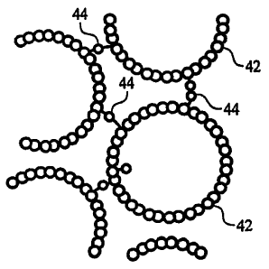
a mat of entangled carbon nanotubes. In preferred einbodiments, referring to
Fig. 3,
nanotube-containing layer 38 includes nanotubes 42 that are chemically bonded
to adjacent
nanotubes, for example, by irradiating the nanotubes with ions, such as argon,
and/or
electrons. The bonds 44, such as covalent carbon-carbon bonds, serve as
molecular links that
directly connect nanotubes 42 to form a continuous, three-dimensional
structure, and to
enhance the mechanical properties, such as strength, of nanotube-containing
layer 38.
Referring to Fig. 4, a method 46 of making membrane 26 is shown. Method 46
includes providing a mixture containing nanotubes (step 48), and forming a
layer containing
nanotubes (step 50), for example, by casting the mixture on an appropriately
shaped
substrate. Next, solvent from the mixture is removed from the fonned layer to
leave a layer
of nanotubes on the substrate (step 52). Inter-nanotube bonds are then formed
in the layer of
nanotubes (step 54), for example, by irradiating the nanotubes. Next, steps
50, 52, and 54 are
repeated as desired to build layer 38 of a predetermined thiclcness (step 56).
As described
below, in some einbodiments, the nanotubes are fiinctionalized with one or
more chemical
moieties, for example, to enhance compatibility and/or adhesion with polymer
layers 40 (step
58). Polymer layers 40 can be formed on nanotube-containing layers 38, for
example, by
injection molding (step 60). Other embodiments of membrane 26, such as a
plurality of
alternating nanotube-containing layers and polyiner layers, can be formed.
The mixture containing nanotubes includes a suspension of carbon nanotubes in
a
solvent. The nanotubes include bioinert, hollow single-walled carbon nanotubes
(SWNTs)
and/or bioinert, hollow multiwalled carbon nanotubes (sometimes called "bucky
tubes")
having at least one dimension less than about 1000 nm.
The physical dimensions of the nanotubes can be expressed as units of length
and/or
as a length to width aspect ratio. The nanotubes can have an average length of
from about
0.1 micron to about 20 microns. For example, the length can be greater than or
equal to
about 0.1 micron, 0.5 micron; 1 micron, 5 microns, 10 microns, or 15 microns;
and/or less
than or equal to about 20 microns, 15 microns, 10 microns, 5 microns, 1
micron, or 0.5
micron. The nanotubes can have an average width or diameter of from about 0.5
nm to about
150 nm. For example, the width or diameter can be greater than or equal to
about 0.5 nm, 1
mn, 5 mn, 10 mn, 25 mn, 50 nm, 75 mn, 100 mn, or 125 nm; and/or less than or
equal to
about 150 mn, 125 nm, 100 mn, 75 nm, 50 mn, 25 nm, 10 mn, 5 mn, or 1 nm.
Alternatively
-5-
CA 02569027 2006-11-09
WO 2005/113033 PCT/US2005/017565
or in addition, the nanotubes can be expressed as having a length to width
aspect ratio of
from about 10:1 to about 50,000:1. The length to width aspect ratio can be
greater than or
equal to about 10:1, 100:1, or 1,000:1; 2,500:1; 5,000:1; 10,000:1; 20,000:1;
30,000:1; or
40,000:1; and/or less than or equal to about 50,000:1; 40,000:1; 30,000:1;
20,000:1;
10,000:1; 5,000:1; 2,500:1; 1,000:1, or 100:1. The nanotubes preferably have
long lengths
and small diameters.
The nanotubes are commercially available or they can be synthesized. Carbon
nanotubes are available, for example, in a mixture from Rice University
(Houston, TX).
Synthesis of carbon nanotubes is described, for example, in Bronikowski et
al., J Vac. Sci.
Technol. A, 19(4), 1800-1805 (2001); and Davis et al., Macromolecules 2004,
37, 154-160.
Dispersion of carbon nanotubes in solvents, for example, to form a film, is
described in
Ausman et al., J. Phys. Chem. B, 2000, 104(38) 8911-8915; Sreekumar et al.,
Chem. Mater.
2003, 15, 175-178
To form a layer or mat of nanotubes, sometimes called "buclcy paper" (step
50), the
nanotubes in the mixture can be separated from the solvent by filtration. For
example,
approximately four grams of a 0.6 mg/ml nanotube suspension, which can be
further diluted
by about 80 ml of deionized water, can be vacuum filtered through a
polytetrafluoroethylene
(PTFE) filter (Millipore LS) or a Whatman Anodisc 47 filter (20 mn pore size).
The filtered
nanotubes can be washed with 2 x 100 ml of deionized water and 100 ml of
methanol. The
washed nanotubes can then dried under vacuum at 70 C for twelve hours to
remove any
residual solvent (step 52) to yield a flexible mat of aggregated nanotubes. In
some
embodiments, the mat of nanotubes is from about 15 to about 35 microns thick,
with a bulk
density of about 0.3-0.4 g/cc. In other embodiments, the mat can have a
thickness as little as
two contacting nanotubes.
Next, bonds are formed between the nanotubes in the mat (step 54). The bonds
can
be formed by irradiating the nanotubes with ions, such as argon ions or carbon
ions, and/or
electrons, such as in a transmission electron microscope. Without wishing to
be bound by
theory, it is believed that when particles of high enough energies collide
with carbon atoms,
the incident particles can displace atoms in the nanotubes, and form defects,
such as
vacancies, and dangling bonds among the nanotubes. The defects and dangling
bonds can
mediate covalent bonds (such as carbon-carbon bonds) between adjacent
nanotubes. These
-6-
CA 02569027 2006-11-09
WO 2005/113033 PCT/US2005/017565
bonds serve as molecular junctions that fuse or weld the nanotubes together
into a continuous
matrix, thereby enhancing the strength and toughness of the mat, while
allowing the mat to
be flexible. As a result, the tliickness of membrane 26 can be reduced without
compromising
performance. The reduced thickness, in turn, reduces the profile of the
filter, thereby
increasing its accessibility to relatively narrow body vessels. The formation
of the inter-
nanotube bonds can also help keep the bucky paper in the shape that it is in
during bond
formation. Thus, membrane 26 can be folded or compacted during delivery, and
subsequently be returned to its shape during bond formation. This shape
recovery can be
useful when membrane 26 or nanotube-containing layer 38 is used, for example,
with a stent
or a balloon. In some embodiments, only selected portions of the nanotube-
containing mat
are irradiated. The portions that are not irradiated can be more flexible than
the irradiated
portions and can correspond to portions that are folded during use.
Irradiation can be performed with ions and/or electrons. The energy of the
incident
ions or electrons is preferably high enough to penetrate through the closest
nanotubes asld
displace carbon atoms of the next closest nanotubes. The nanotubes can be
irradiated, for
example, witli argon ions of about 0.1 keV to about 3 keV (e.g., from about
0.4 to about 1
keV, such as 100 eV, 200 eV, or 400 eV), with irradiation doses from about 5 x
1015 to about
3 x 10¹⁶ Ar/cm, depending, for example, on the diameters of the nanotubes. The
nanotubes
can also be irradiated with energetic electrons, e.g., up to 1.25 MeV. In some
embodiments,
the energy of the incident radiation can be chosen to penetrate the entire
thickness of the
bucky paper, e.g., by using carbon ions (e.g., C3) with energies of 10 MeV.
Irradiation can
be perfonned at room temperature or at elevated temperatures, such as about
800 C, to
facilitate migration and annealing of the defects. The ion flux can be from
about 8.2 x 10¹³ to
about 4.9 x 10¹⁴ Ar/cm2s, and the irradiation time can be from about one
minute to about six
minutes. In some embodiments, the prior to irradiation, the nanotubes can be
amzealed at
about 900 C for about 30 minutes or at about 470 C for about 50 minutes in
air, e.g., to burn
off carbonaceous residues and purify the nanotubes. Irradiation of nanotubes
is fiirther
described, for exainple, in Krasheninnikov et al., Plays. Rev. B, 66, 245403
(2002);
Krasheninnikov et al., Phys. Rev. B 63, 245405 (2001); and Krasheninnikov et
al.,
Krasheniuulikov et al., Phys. Rev.B 65, 165423 (2002).
-7-
CA 02569027 2006-11-09
WO 2005/113033 PCT/US2005/017565
After the inter-nanotubes bonds are created, additional mats of nanotubes can
be
formed on the first mat to increase the thickness of layer 38 (step 56).
Additional layers can
be formed by filtering the nanotube-containing mixture over the first mat
(steps 48 and 50),
removing any residual solvent (step 52), and forming inter-nanotube bonds in
the newly
formed layers (step 54), as described above. The fmal thickness of layer 38
can be a function
of the specific medical device in which the layer is incorporated. The formed
nanotube-
containing layer 38 can be peeled away from the filter.
Still referring to Fig. 4, in some embodiments, the nanotubes in layers 38 are
modified to enhance interactions between the nanotubes and polyiner layers 40
(step 58).
The nanotubes can be chemically modified with one or more functional groups
that increase
interactions with polymer layer 40, e.g., to enhance compatibility or
adhesion. For example,
the nanotubes can be chemically treated to include carboxylic acid and/or
amine chemical
moieties, which can interact well witli polymers. Functionalization of carbon
nanotubes are
described, for example, in Bahr et al., J. Am. Chem. Soc. 2001, 123, 6536-
6542, Hainon et
al., Adv. Mater. 1999, 11, No. 10, 834-840, and U.S. Patent Application
Publication
2003/0093107. Alternatively or in addition, the nanotubes can be wrapped with
a polymer,
such as polyvinyl pyrrolidone (PVP) and/or polystyrene sulfonate (PSS), to
enhance
compatibility with polymer layers 40 and/or to enhance solubility (e.g., in
water). Polymer
wrapping of single-walled carbon nanotubes is described in O'Connell et al.,
Clzemical
Physics Letters 342 (2001) 265-271. Modification(s) of the nanotubes can be
performed
before and/or after bonding of the nanotubes.
After a predetermined number of nanotube-containing layers 38 are formed and
placed together, polymer layers 40 are formed over the nanotube-contaiiling
layers 38 to
yield membrane 26 (step 60). Polymer layers 40 can include materials used in
medical
devices, for example, thermoplastics and thermosets. Examples of
thermoplastics include
polyolefins, polyamides, such as nylon 12, nylon 11, nylon 6/12, nylon 6, and
nylon 66,
polyesters, polyethers, polyurethanes, polyureas, polyvinyls, polyacrylics,
fluoropolyiners,
copolyiners and block copolymers thereof, such as block copolymers of
polyether and
polyamide, e.g., Pebax ; and mixtures thereof. Examples of thermosets include
elastomers
such as EPDM, epichlorohydrin, nitrile butadiene elastomers, silicones, etc.
Thermosets,
such as expoxies and isocyanates, can also be used. Biocompatible thermosets
may also be
-8-
CA 02569027 2006-11-09
WO 2005/113033 PCT/US2005/017565
used, and these include, for example, biodegradable polycaprolactone,
poly(dimethylsiloxane) containing polyurethanes and ureas, and polysiloxanes.
Polymer
layers 40 can also include photocurable resins, such as those used in the
dental field, e.g.,
bisphenol-A-glycidyldimethacrylate (Bis-GMA), triethylenglycol-dimethacrylate
(TEGDMA), urethane-dimethacrylate (UDMA), and polycarbonate dimethacrylate,
(PCDMA). The photocurable resin can be cured during irradiation of the
nanotubes. Other
polymers are described in commonly assigned U.S.S.N. 10/645,055, filed August
21, 2003.
Mixture 37 can include one or more polymers 40. Polymer layers 40 can be
formed by
injection molding, casting, spraying, and/or micro-drop techniques.
In some embodiments, polymer layers 40 can include one or more additives. For
example, polymer layers 40 can include one or more coupling or compatibilizing
agents,
dispersants, stabilizers, plasticizers, surfactants, and/or pigments, that
enhance interactions
between the nanotubes and the polymer. Examples of additive(s) are described
in U.S.
Patent Application Publication 2003/0093107. Alternatively or in addition,
polymer layers
40 can include carbon nanotubes, for example, to enhance the strength of the
polymer layers.
The nanotubes can be bonded together or not bonded together. Polymer layers 40
can
include from about 0.1% to about 60% of na.notubes by weight. Polymer layers
40 can be
loaded with a high concentration of nanotubes using a layer-by-layer method
described
below. Methods of making nanotube-containing mixtures are described, for
example, in
Biercuk, et al., Applied Physics Letters, 80, 2767 (2002); and Sandler et al.,
Mat. Res. Soc.
Syrnp. Proc. Vol. 706, 2002 Z4.7. 1 -Z4.7.6.
In some embodiments, one or more polymer layers 40 include one or more
releasable
therapeutic agents or a pharmaceutically active compounds, such as described
in U.S. Patent
No. 5,674,242, and commonly-assigned U.S.S.N. 09/895,415, filed July 2, 2001.
The
therapeutic agents or pha.rmaceutically active compounds can include, for
exa.inple, anti-
thrombogenic agents, antioxidants, anti-inflainmatory agents, anesthetic
agents, anti-
coagulants, and/or antibiotics. A specific example includes heparin, which can
reduce
throinbus formation on the surface of the medical device, particularly long
ternn implants
such as a septal defect device or a pulmonary filter.
Other methods of forming membrane 26 include a layer-by-layer technique in
which
a multilayer structure is forlned having a plurality of alternating,
oppositely charged layers,
-9-
CA 02569027 2006-11-09
WO 2005/113033 PCT/US2005/017565
as described in U.S.S.N. [Client Ref. No. 04-0048], entitled "Layer-by-Layer
Assembly of Multilayer Regions for Medical Devices" and filed on the same day
as this
application. The structure can include, for example, a plurality of layers
containing charged
nanoparticles alternating with a plurality of layers containing charged
polyelectrolytes.
Charging can be provided, for example, using an electrical potential, by
covalently attaching
functional groups, and/or by exposing the layers to one or more charged
amphiphilic
substances. Exemplary materials and techniques are described in U.S.S.N.
[Client
Ref. No. 04-0048].
The formed membrane 26 can then be used to fonn filter 20. For exainple,
membrane
1 o 26 can be folded over an appropriately shaped template, such as a conical
mandrel, and
opposing edges of the membrane can be secured, for example, with an adhesive.
Membrane
26 can be attached to frame 24 by solvent casting methods (e.g., wherein
liquid membrane
polymer is dipped over the frame and allowed to cure and solidify), or by
adhesive (such as a
cyanoacrylates). Openings 28 can be formed, for example, using excimer laser
or other
ablation techniques, as described in Weber, U.S. Patent No. 6,517,888. The
fitsing of the
nanotubes can hold the nanotubes together, e.g., so that they are not flushed
out through
openings 28.
In embodiments, membrane 26 consists of nanotube-containing layer(s) 38, and
does
not include polymer layers 40. The interconnections among the nanotubes can
enhance the
strength of the membrane and allow the nanotubes to withstand forces, e.g.,
fluid flow,
within the body.
Use of intravascular filter 20 is described, for example, in Daniel et al.,
U.S. Patent
No. 6,171,327.
Other embodiments of nanotube-containing layer 38 and membrane 26 can be
formed. Referring to Fig. 5, a membrane 70 includes a plurality of corrugated
nanotube-
containing layers 72 and polymer 74. Nanotube-containing layers 72 can be
formed by
filtering a mixture containing nanotubes through a corrugated filter, similar
to the procedure
described above. The corrugation of layers 72 provides membrane 70 with
anisotropic
mechanical properties such that, for example, the meinbrane is more crush
resistant in
direction A than in direction B. Membrane 70 can be formed similar to the
methods
-10-
CA 02569027 2006-11-09
WO 2005/113033 PCT/US2005/017565
described above, for example, by spraying, dipping, and/or injection molding
polymer 74
between nanotube-containing layers 72.
In certain embodiments, the nanotubes can be aligned, for example, prior to
inter-
nanotube bond formation. Aligning the nanotubes can be used to control the
porosity of the
nanotube-containing layers. Aligning the nanotubes, e.g., parallel to each
other, can also
efficiently pack the nanotubes, thereby increasing the density of the nanotube-
containing
layer and increasing the likelihood of inter-nanotube formation. Aligning the
nanotubes can
fiirther enhance the homogeneity of the nanotube-containing layer, which can
reduce the
occurrence of localized defective or wealc spots. The nanotubes can be aligned
inagnetically.
1 o For example, to form a nanotube-containing layer, a inixture containing
the nanotubes can be
placed on a filter that is not under vacuuin but exposed to a magnetic field.
The magnetic
field, e.g., from about one to about twenty Tesla, is capable of aligning the
nanotubes while
the nanotubes are dispersed in the solvent above the filter. A vacuum is then
applied across
the filter to separate the solvent from the aligned nanotubes to form an
aligned bucky paper.
Magnetic alignment of nanotubes is described, for example, in Choi et al., J.
ofAppl. Phys.,
Vol. 94, No. 9, 1 Noveinber 2003, 6034-6039. In some embodiments, the magnetic
alignment of nanotubes changes, e.g., is reoriented in the major plane of the
layer, from one
layer to an adjacent layer (e.g., by about 90 degrees).
Embodiments of the membranes or nanotube-containing layers described above can
also be used in other medical devices.
For example, embodiments of the membrane or nanotube-containing layer can be
formed into a cylindrical tube that can be used as a strong, thin and flexible
synthetic
vascular graft. The graft can be used to replace a damaged or dysfunctional
body vessel
(e.g., at the site of an aneurysm or an occlusion), to bypass or divert blood
flow around a
damaged region, or to create a shunt between an artery and a vein (e.g., for
multiple needle
access for hemodialysis access). Vascular grafts are described, for example,
in U.S. Patent
No. 5,320,100.
Referring to Fig. 6, a cylindrical tube of the membrane or the nanotube-
containing
layer 80 can be used as a strong, thin and flexible graft with a stent 82 to
form a stent-graft
84, or a covered stent (as shown, on a support 86 such as a catheter shaft or
a balloon
catheter). The thinness of the membrane or the nanotube-containing layer
reduces resistance
-11-
CA 02569027 2006-11-09
WO 2005/113033 PCT/US2005/017565
to blood flow in the vessel. In some embodiments, one or more of the polymer
layers, such
as the outermost polyiner layer, can include a releasable therapeutic agent or
a
pharmaceutically active compound, such as described in U.S. Patent No.
5,674,242,
commonly-assigned U.S.S.N. 09/895,415, filed July 2, 2001, and U.S.S.N
10/112,391, filed
March 28, 2002. The therapeutic agents or pharnnaceutically active compounds
can include,
for example, anti-thrombogenic agents, antioxidants, anti-inflammatory agents,
anesthetic
agents, anti-coagulants, and antibiotics. The polymer layer(s) can include,
for exainple, a
biocompatible, non-porous or semi-porous polymer matrix made of
polytetrafluoroethylene
(PTFE), expanded PTFE, polyethylene, urethane, or polypropylene.
Stent 82 can be of any desired shape and size (e.g., coronary stents, aortic
stents,
peripheral stents, gastrointestinal stents, urology stents, and neurology
stents). Depending on
the application, stent 82 can have an expanded diameter of between, for
example, 1 inin to 46
inm. In certain embodiments, a coronary stent casi have an expanded diameter
of from about
2 mm to about 6 mm. In some embodiments, a peripheral stent can have an
expanded
diameter of from about 5 mm to about 24 mm. In certain einbodiments, a
gastrointestinal
and/or urology stent can have an expanded diameter of from about 6 mm to about
30 mm. In
some embodiments, a neurology stent can have an expanded diameter of from
about 1 mm to
about 12 mm. An abdominal aortic aneurysm (AAA) stent and a thoracic aortic
aneiurysm
(TAA) stent can have an expanded diameter from about 20 mm to about 46 mm.
Stent 82
can be balloon-expandable, self-expandable, or a coinbination of both (e.g.,
as described in
U.S. Patent No. 5,366,504).
Stent-graft 84 can be used, e.g., delivered and expanded, using a balloon
catheter
system. Suitable catheter systems are described in, for example, Wang U.S.
5,195,969, and
Hamlin U.S. 5,270,086. Suitable stents and stent delivery are also exemplified
by the NIR on
Ranger system, available from Boston Scientific Scimed, Maple Grove, MN.
Embodiments of the membranes or nanotube-containing layers can be incorporated
into a medical balloon. For example, referring to Fig. 7, a tube of the
membrane or
nanotube-containing layer 90 can be placed over a balloon 92 to serve as a
strong, flexible,
thin and non-compliant constraint. The tube can be placed over the balloon,
for example, as
a preformed sleeve, or wrapped around the balloon as a sheet and joined at
opposing edges.
The structure of the balloon itself can include nanotubes, for example, by
forming
-12-
CA 02569027 2006-11-09
WO 2005/113033 PCT/US2005/017565
embodiments of membrane 26 described herein and shaping the membrane into the
balloon.
Balloon 92 is carried by a catheter shaft 94, which, as described below, can
include
nanotubes.
In some embodiments, the membrane or nanotube-containing layer can also be
formed into the shape of a medical balloon to completely encapsulate the
balloon. For
example, the membrane or nanotube-containing layer can be formed on a
dissolvable (e.g.,
water soluble) substrate shaped as a medical balloon, and the substrate can be
subsequently
removed. Prior to applying a nanotube-containing mixture to the substrate, a
mist can be
sprayed over the substrate to dissolve the outer layer of the substrate and
make it sticky. A
mixture containing nanotubes can be applied, e.g., sprayed on the substrate to
form a layer.
After a desired thickness of nanotubes is formed, the nanotubes can be
irradiated as described
above and the substrate can be dissolved. An example of a dissolvable
substrate is
degradable polyvinyl alcohol, described, for example, in Cooper et al.,
Proceedings of th.e 8'72
Annual Global Plastics Environmental Conference, Society of Plastics
Engineers, Detroit
MI, 360, 14 February 2002.
Balloon catheter systems are described, for example, in Wang, U.S. 5,915,969;
Hamlin, U.S. 5,270,086; and exemplified by the Maverick or Symbiot catheter
systems
available from Boston Scientific Corp.-Scimed Life Systems, Inc. (Maple Grove,
MN).
Embodiments of the membranes or nanotube-containing layers can be sized and
shaped to form a variety of catheters. Examples of catheters include guide
catheters (e.g., as
described in U.S. 6,595,952), tumor ablation catheters, aneurysm catheters,
urology catheters,
and perfusion catheters (e.g., as described iii U.S. 6,503,224). The tube can
be foimed into
an introducer sheath or a restraiuzing sheath for a stent delivery system, for
example, as
described in U.S. Patent No. 6,488,694 and Raeder-Devens et al., US
2003/0050686. The
catheters can include one or more nanotube-containing layers for strength, and
one or more
layers carrying a marker for fluoroscopic, ultrasound, and/or magnetic
resonance detection,
as described, for example, in commoi-Ay assigned U.S.S.N. 10/390,202, filed
March 17, 2003.
The nanotubes having inter-nanotube bonds can also be incorporated in bone
cements. For example, the nanotubes can be blended with polymethylmethacrylate
(PMMA), bisphenol A diglycerol ether dimethacrylate, triethylene glycol
diinethacrylate
(TEGDMA), poly(ethylene glycol) methacrylate (PEGMA), N,N-dimethyl-p-
toluidine,
-13-
CA 02569027 2006-11-09
WO 2005/113033 PCT/US2005/017565
strontiuin-containing hydroxylapatite, and/or fumed silica. The nanotubes can
enhance the
strength of the polymers.
All publications, applications, and patents referred to in this application
are herein
incorporated by reference to the same extent as if each individual publication
or patent was
specifically and individually indicated to be incorporated by reference in
their entirety.
Other embodiments are within the claims.
-14-