Note: Descriptions are shown in the official language in which they were submitted.
CA 02569067 2011-11-29
UNAGGLOMERATED CORE/SHELL NANOCOMPOSITE PARTICLES
BACKGROUND OF THE INVENTION
Field of the Invention
[0002] The present invention relates to nanocomposite particles. More
particularly, the
present invention provides a method for synthesizing stable, well dispersed,
unagglomerated
core/shell nanocomposite particles of varying sizes that may be used for a
wide variety of
applications.
Description of Related Art
[00031 One of the most important developments in the field of chemical
technologies is
that of nanostructuring. Nanostructured materials are assemblies of nano-sized
units that
display unique, characteristic properties at a macroscopic scale. The size
range of such units
lies within the colloidal range, where the individual properties are different
to both those of
atoms/molecules and to those of the bulk. The properties of the nanostructured
assemblies,
therefore, can be tuned by varying the colloidal properties of the
constituents, mainly particle
size, surface properties, interparticle interactions and interparticle
distance.
[00041 The use of nanoparticles in biomedical applications is a major focus of
numerous
research groups today. Nanoparticles possess several qualities that make them
useful in
biomedical applications, such as diagnostic bioimaging, drug delivery, and
gene therapy.
Nanoparticles also can be used as bioimaging agents to label cells in
cultures, tissues, or
intact organisms.
100051 Current nanoparticle technologies used in bioimaging applications
include magnetic
nanoparticles, ferrofluids, and quantum dots (QDs). Recently, there have been
numerous
advances in the development of colloidal fluorescent semiconductor
nanocrystals, a class of
QDs used for biological labeling (marketed as Q-dotsTM), such as ZnS shell-
CdSe core
nanoparticles. (Haugland, R.P. Molecular Probe, 6:320-328, 1998). Researchers
are in the
process of developing bioconjugation schemes and applying such probes to
biological assays,
and nanocrystals can be of particular benefit as biological labels when
compared to existing
organic dyes. Quantum dots have been widely tested in a range of bioimaging
applications.
1
CA 02569067 2006-11-29
WO 2005/118702 PCT/US2005/019239
[0006] Semiconductor nanocrystals have several problems associated with their
use, such
as solubility, physicochemical stability and quantum efficiency of the
semiconductor
nanocrystals. Additionally, QD emissions are strongly intermittent and
agglomeration can
limit their effectiveness as a bioimaging tool. Other problems associated with
QDs include
surface electronic defects and toxicological effects, as surface oxidation can
cause
degradation of the QD shell, releasing toxic metals into the body, and poor
crystallinity,
which makes the interpretation of the physical properties of QDs very
difficult. Furthermore,
the routine application of fluorescent nanoparticles as biolabels is
controversial, particularly
because of general environmental concerns regarding the use of highly toxic
compounds,
such as cadmium, in biomedical diagnostics. Moreover, methods for designing
nanometer-
sized structures and controlling their shape to yield new materials with novel
electronic,
optical, magnetic, transport, photochemical, electrochemical and mechanical
properties are
rarely found and present a potentially rewarding challenge.
[0007] Nanoparticles also can function as a mechanism for drug delivery, which
permits
the utilization of numerous water-insoluble and unstable drugs. Additionally,
nanoparticles
can find use in drug targeting and extended release applications based on
resorbable shell
technologies. In addition, nanocomposites can be used in gene therapy for the
delivery of
genetic materials. Current nanoparticle drug and gene "carriers" include
polymeric micelles,
liposomes, low-density lipoproteins, polymeric nanospheres, dendrimers, and
hydrophilic
drug-polymer complexes.
[0008] Over the past ten years, extensive research has been carried out in the
field of
fabrication of nanoscale composite particles because of their unique
properties and potential
applications in electronics and photonics. Silicon dioxide (Si02)-shell
metallic-core
structured nanocomposite particles were first synthesized and reported by
Mulvaney et al.
(Langmuir, 12:4329-4335, 1996), and by Adair et al. (Materials Sci. & Eng. R.,
23:139-242,
1998). Most of the SiO2 coated nanocomposite particles having a core-shell
architecture fall
into two categories based on the synthetic method used. The approach developed
by
Mulvaney et al. involved the modification of metal cluster surfaces with the
silane-coupling
agent 3-aminopropyltriethoxysilane (APS) before the formation of the silica
shell. APS is
used as an adhesion promoter between the vitreophobic metal cluster core and
the Si02. The
state of dispersion for the nanocomposites in suspension, however, was not
examined by
Mulvaney et al. or Adair et al. Adair et al. were successful in coating
metallic and CdS
2
CA 02569067 2006-11-29
WO 2005/118702 PCT/US2005/019239
clusters with Si02 via simple hydrolysis and condensation of tetrethoxysilane
(TEOS) in a
cyclohexane/Igepal/water tertiary system having an aqueous phase. This system
allows for a
very uniform silica-shell coating along with a tunable thickness of both the
core and the shell
due to the confining of water droplets in oil.
[00091 Major limitations in the use of nanoparticles in therapeutic agent
delivery
applications involve the lack of colloidal stability in nanoparticle
suspensions, agglomeration,
polydispersity in size and shape, swelling, and leakage. Other problems
include difficulty of
synthesis and processing techniques, inadequate loading inside the carrier
particle, and lack
of applicability to a variety of medical agents. Residual precursor materials
present in
unwashed nanosuspensions can also have detrimental effects for both targeted
delivery and
toxic effects on the physiological system. In a typical chemical synthetic
method, dispersion
of nanoparticles essentially begins with the washing of the freshly prepared
nanoparticles.
However, washing and dispersion of nanoparticles is a challenge because of the
strong van
der Waals attraction between adjacent nanoparticles. For this reason,
nanoparticle
suspensions usually are stabilized with surface coatings of surfactant, which
effectively
balances the interaction forces with a high repulsion potential created by the
surfactant
molecules. It is necessary, however, to minimize the surfactant dispersants in
order to
achieve better performance for nanoparticle-based applications and devices.
This is because
surfactant additives are transferred to the subsequent process steps and can
negatively impact
the homogeneity of the arrays assembled from the nanoparticles. Furthermore,
when
protective surfactants are removed with conventional washing techniques, such
as
centrifugation, the nanoparticles tend to undergo agglomeration. The presence
of
agglomerates also can compromise the effective yield of particles. If
nanometer-size primary
particles are desired, the presence of agglomerates that are generally an
order of magnitude or
larger in size must be avoided. For example, prior art conventional methods
for fabricating
nanocomposite particles, such as filtration methods, as disclosed in U.S.
Patent No.
6,548,264, 2003 to Tan, W. et al., and discussed in more detail below, result
in
nanocomposite particles that are irreversibly agglomerated with an
agglomeration size of
about 250 nm.
[0010) Considering the current limitations in nanomedicine, there is a need
for a
universally applicable nanoparticle with controlled time-release, high loading
of therapeutic
agent(s), ease of preparation, stability, and up-scaling capabilities. The
formulation of a
3
CA 02569067 2006-11-29
WO 2005/118702 PCT/US2005/019239
stable, non-aggregating colloid to deliver active-medical-agents has the
potential to transform
the medical field by providing universal, controlled, targeted, systemic
delivery for a variety
of bioimaging and therapeutic agents.
SUMMARY OF THE INVENTION
[00111 The present invention provides methods for the preparation of stable,
unagglomerated, well dispersed, active-medical-agent core/shell nanocomposite
particles
having silane coupling agents such as but not limited to alkylamine or
alkylcarboxylic acid
silane coupling agents attached. A detailed discussion of the graft mechanism
has been
described elsewhere (Plueddemann, E.P., "Silane coupling agent," pp 29-48,
Plenum Press,
NY, 1982; Ung, T. et al., Langmuir, 14:3740-3748, 1998; Chiang, C.H., et al.,
J. Colloid
Interface Sci., 74(2):396-403, 1980; Chiang, C.H. et al., J. Colloid Interface
Sci., 86(1):26-34,
1982). The dispersion of the nanocomposites is achieved preferably by using
high
performance liquid chromatography (HPLC) to simultaneously wash and disperse
the
nanocomposite particles, in place of other techniques that involve sequential
washing and
dispersal steps.
[00121 The present invention also provides for the preparation of stable,
dispersed
nanocomposite particles to be used in vivo under physiological conditions,
i.e. isotonic
environment, by surface modification such as a carbodiimide-mediated
polyethylene glycol
(PEG) coupling agent to the silane coupling agent, which maintains their
dispersed state.
Other surface modification methods include dendrimers, amphiphilic agents, and
charged
adsorbates such as citrate as is known in the art. The present invention
further provides for
the attachment of binders, such as antibodies, thus enabling the nanocomposite
particles to
target specific sites for intracellular drug delivery.
[00131 The nanocomposite particles can include a variety of medically-active
substances,
such as organic fluorophores and therapeutic drugs, doped inside silica,
titania, calcium
phosphate or calcium phospho-silicate matrices. The synthesis techniques also
can be
modified to produce nanoparticles containing combinations of fluorophores and
therapeutic
medicinal agents. The intended biomedical application for the colloid of
nanocomposite
particles dictates the selection of core and shell-matrix materials.
[0014] The stable, well dispersed and unagglomerated active-medical-agent
nanoparticles
can be used for a variety of applications, such as, without limitation,
pigments, fluorescent
4
CA 02569067 2006-11-29
WO 2005/118702 PCT/US2005/019239
labeling, inks, slow release formulations, bioimaging, drug delivery, gene
therapy and
combinations thereof For example, and within limitation, the nanoparticles of
the present
invention can be used as calcium deposition transporters, for medical
diagnosis, and for
medical therapeutics for cancer, infectious diseases, diabetes, cystic
fibrosis and other
diseases and disorders.
[0015] The fluorescent nanocomposite particles possess several qualities that
make them
particularly attractive for imaging and pigment applications, such as precise
tunability of
emission peaks, extended fluorescence lifetimes relative to traditional
organic fluorophores,
negligible photobleaching and self-quenching with the benefit of
biocompatibility.
Additionally, the nanoparticulate fluorescent emissions are not intermittent.
Within the
nanoparticle, direct contact between dye molecules and the environment is
avoided,
eliminating photodegradation of the fluorophore presumably because of
absorption of the
most energetic, and therefore, damaging of the excitation photons. As a
result, the
nanocomposite particles exhibit extended fluorescence lifetimes relative to
traditional organic
fluorophores or quantum dots as shown in Figures 3 through 5.
[0016] Nanocomposite particles can be used as a drug delivery system based on
the
encapsulation of a therapeutic agent in either a metal oxide shell with
controlled porosity
and/or a soluble outer shell/coating that on dissolution releases the
therapeutic agent in the
immediate vicinity of the afflicted area as shown in Figures 6 through 9. The
protection of
the therapeutic agent provided by the shell-matrix material allows for the
delivery of drugs
that are highly water-insoluble or unstable in physiological solutions.
Furthermore,
dissolution kinetics of the shell-matrix materials can be engineered to
provide sustained
release of therapeutic agents at target sites for extended periods of time.
[0017] The nanocomposite suspensions can also be used to deliver drugs
including, but not
limited to, ceramide, AZT, and dobutamine. Insulin can also be encapsulated in
a soluble
shell material such as calcium phosphate or calcium phospho-silicate. The
surface can be
modified to permit the nanocomposite particles to cross physiological
membranes such as the
gastro-intestinal tract, the blood-brain barrier, and cellular membranes. The
targeted, time-
controlled release can be used to deliver therapeutic agents such as insulin
from the core-shell
nanoparticles.
[0018] The nanocomposites also can be used in gene therapy for the delivery of
therapeutic
DNA to cells. The nanocomposite particles can offer increased stability of
genetic material
CA 02569067 2006-11-29
WO 2005/118702 PCT/US2005/019239
through encapsulation and improved uptake into target cells. Additionally, the
nanoparticles
can deliver genetic therapeutic agents in transcriptionally active forms while
maintaining
small sizes of less than about 200 nm.
[00191 Small particle size along with enhanced nanoparticle surface chemistry
and
dispersion contribute to the effectiveness of the nanocomposite particles. The
small size
achieved allows for evasion of capture by the reticuloendothelial system (RES)
of animal
models or the human body, permitting the nanoparticles to function in
biological systems by
crossing membranes such as the intestinal wall and the blood-brain barrier.
Numerous
biological barriers can be passed by the small nanocomposite particles, which
allows for high
concentrations of therapeutic agents to be delivered to target sites. In
addition, the dispersed
nanocomposite particles can be readily functionalized to deliver therapeutic
agents directly to
the targeted cells or tissues in the human body. Agglomeration compromises all
the above
biomedical applications as well as non-biomedical uses.
[00201 The synthesis of the nanocomposite particles is achieved using a
reverse micelle
system that includes water, a surfactant and a solvent, such as cyclohexane.
The resultant
size of the nanocomposites depends on the water-surfactant ratio, wherein a
higher water-
surfactant ratio produces larger nanocomposites. The present invention,
therefore, provides
for the synthesis of nanocomposite particles having a diameter of between
about 1.0 to 100
nm, with a preferred diameter between about 1 and 20 nm, which is small enough
to cross
biological cell membranes. However, as defined below, the primary particle
size obtained by
the synthesis is not the actual size particle in suspension. Heretofore,
nanocomposite
particles prepared in reverse micelles have been agglomerated. The
agglomeration occurs
after synthesis during washing and collection operations for the
nanoparticles. Thus,
synthesis alone of primary nanoparticles is insufficient in providing well
dispersed
suspensions.
BRIEF DESCRIPTION OF THE DRAWINGS
[0021]x FFig. 1 is a schematic of the reverse-micelle synthesis, washing and
collection
approach of the present invention;
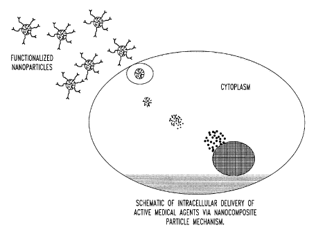
[00221 Fig. 2 is a schematic of intracellular delivery of active medical
agents via
nancomposite particle mechanisms;
[00231 Fig. 3 shows fluorescent emission scans of fluorescein/Si02
nanoparticles;
6
CA 02569067 2006-11-29
WO 2005/118702 PCT/US2005/019239
[0024] Fig. 4 shows fluorescent emission timescans of 1 M rhodamine WT/Si02
nanocomposite particles and 10-5 M rhodamine WT stock solution;
[0025] Fig. 5 illustrates nanocomposite particle photodecay experimental
results;
[0026] Fig. 6A-D shows phase contrast and fluorescent images of smooth muscle
cells and
rat stellate ganglia. Fig. 6A-B is a phase contrast and a fluorescent image,
respectively, of
cultured vascular A7r5 smooth muscle cells that have taken up fluorescein/Si02
nanoparticles; and Fig. 6C-D is a phase image and a fluorescent image,
respectively, of
acutely isolated rat stellate ganglia neurons five days following intracardial
injection of the
nanoparticles;
[0027] Fig. 7A-E shows phase contrast and fluorescent images of rat stellate
ganglia. Fig.
7A-B is a phase contrast and a fluorescent image, respectively, of in vitro
(15 minute)
exposure of stellate ganglion (SG) neurons to 0.002% 10"2 M rhodamine B/Si02
nanoparticles; Fig. 7C-D shows a phase image and a fluorescent image,
respectively, of in
vitro (15 minute) exposure of SG neurons to 0.002% 10-2 M fluorescein/Si02
nanoparticles;
and Fig. 7E is a fluorescent image of fluorescein/Si02-containing neurons in
SG neurons
seven days post-injection;
[0028] Fig. 8 is a schematic of a functionalized nanocomposite particle
containing an
organic fluorophore and a therapeutic drug;
[0029] Fig. 9 is a bar graph illustrating that ceramide/Calo(P04)6(OH)2
nanoparticles
induce human coronary artery smooth muscle cell growth inhibition;
[0030] Fig. 10 is a flow sheet of a Ag/Si02 nanocomposite suspension obtained
from
reverse micelle synthesis and washing with various methods;
[0031] Fig. 11 is a schematic setup of the HPLC system for washing and
dispersion of a
Ag/SiO2 nanocomposite ethanol/water suspension based on size exclusion
chromatography.
The UV-vis detector wavelength is set at 405 nm for the Ag/Si02
nanocomposites. The size
of the HPLC column is HR 5/5 (5 x 50 mm);
[0032] Fig. 12A-C is a schematic of the HPLC system used to wash and disperse
nanopartices. Fig. 12A is the HPLC system; Fig. 12B is a TEM of Ag/SiO2
nanoparticles;
and Fig. 12C is a SEM of spherical silica beads in the stationary phase;
[0033] Fig. 13A-B illustrates a Ag/Si02 nanocomposite suspension (R=2, H=100,
X=1)
washed with centrifugation. Fig. 13A shows TEM analysis of the primary
particle size.
Fig. 13B shows the particle size distribution by dynamic light scattering;
7
CA 02569067 2006-11-29
WO 2005/118702 PCT/US2005/019239
[0034] Fig. 14A-B illustrates a Ag/Si02 nanocomposite suspension (R=2, H=100,
X=1)
washed with Soxhlet extraction. Fig. 14A shows TEM analysis of the primary
particle size.
Fig. 14B shows the particle size distribution by dynamic light scattering;
[0035] Fig. 15A-B illustrates a Ag/Si02 nanocomposite suspension (R=2, H=100,
X=1)
washed with sedimentation. Fig. 15A shows TEM analysis of the primary particle
size.
Fig. 15B shows the particle size distribution by dynamic light scattering;
[0036] Fig. 16A-B illustrates a Ag/Si02 nanocomposite suspension (R=2, H=100,
X=1)
washed with filtration. Fig. 16A shows TEM analysis of the primary particle
size. Fig. 16B
shows the particle size distribution by dynamic light scattering;
[0037] Fig. 17A-B illustrates the morphology of a Ag/Si02 nanocomposite
suspension
(R=2, H=100, X=1) washed with a conventional method. Fig. 17A shows TEM images
of
the core-shell structure. Fig. 17B shows the particle size distribution
obtained from the TEM
image.
[0038] Fig. 18 shows an SEM image of Si02 microspheres used as the stationary
phase in
the HPLC system. The Si02 microspheres were treated with APS (90 g Si02 mixed
with
0.336 mL APS, 1.5 mL glacial acetic acid, 7.5 mL DI water and 150 mL ethanol,
stirred
overnight and dried at 70 C). The surface area is 425 m2/g with anaverage pore
size of 6.5
nm.;
[0039] Fig. 19 is an HPLC spectrum of an Ag/Si02 ethanol/water suspension
(R=2,
H=100, X=1) washed with the HPLC system. Spectrum was acquired at 405 nm
(surface
plasmon resonance of Ag quantum dots) by UV-vis detector. The washing solvent
is an
ethanol/water solution (volume ratio 7:3), and the flow rate is 2 mL/min. The
spectrum was
deconvoluted by PEAKFIT , the central positions of the peaks are 95.1, 112.2
and 150.7 s;
[0040] Fig. 20A-B illustrates the morphology of Ag/Si02 nanocomposite
ethanol/water
(7:3 vol) suspensions washed with HPLC. Fig 20A is two suspensions of
nanocomposites, A
and B. Fig. 20B shows digital images: suspension A, R=2, H=100, X=1,
suspension, D50=30
rim, SD=1.2 rim. B, R=8, H=100, X=1, D50=20.3 nm, SD= 1.5 nm. (With 95%
confidence
interval.) TEM images show the size, core-shell architecture and the state of
dispersion of
the suspensions A and B;
[0041] Fig. 21 shows AFM images of a Ag/Si02 nanocomposite (R=2, H=100, X=1)
suspension obtained with the HPLC washing. Images were obtained by TAPPING
MODETM. The samples were prepared by placing a drop of the Ag/Si02
ethanol/water
8
CA 02569067 2006-11-29
WO 2005/118702 PCT/US2005/019239
suspension on a freshly cleaved mica substrate and spin coated at 1500 rpm for
30 s. The 3D
image shoes the aggregate size of about 60 nm;
[0042] Fig. 22 illustrates the particle size distribution of Ag/Si02
nanocomposites (R=8,
H=300, X=1) measured by dynamic light scattering (DLS) and TEM analysis.
D50=18.6 nm,
SD=1.5 nm for DLS, the particle size from TEM analysis is 20.3 1.5 nm (30
particles were
counted). Noted is the close match between DLS and TEM analysis with AAN
approximately equal to 1, indicating a well dispersed suspension;
[0043] Fig. 23 illustrates the zeta potential of Ag/Si02 nanocomposite (R=2,
H=100, X=1)
ethanol/water suspensions as a function of pH and APS concentration. Noted is
the
conversion of negative to positive charge after the addition of APS. The pHs
of the
suspensions were measured using a Sentron pH meter calibrated against standard
aqueous
buffer solutions. Error bars are the 95% confidence interval;
[0044] Fig. 24 illustrates the zeta potential of Si02 microspheres as a
function of pH and
APS concentration. The average size of Si02 is 20 gm with a surface area of
425 m2/g and a
6.5 nm average pore size. The Si02 microspheres were dispersed in
ethanol/water (7:3 vol)
solution. The pHs of the suspensions were measured using a Sentron pH meter
calibrated
against standard aqueous buffer solutions. Error bars are the 95% confidence
interval;
[0045] Fig. 25 shows an AFM image of a Ag/Si02 nanocomposite (R=2, H=100, X=1)
aggregation formed on the surface of a Si02 microsphere. APS surface coating
only was
applied to the Ag/Si02 nanocomposites (average size 30 nm), and the Si02
microspheres
were used without further treatment (average size 20 gm). Si02 microspheres
from a blocked
HPLC column were placed on a freshly cleaved mica substrate;
[0046] Fig. 26A-B illustrates the effect of pH on the state of dispersion for
Ag/Si02
nanocomposite ethanol/water suspensions (R=8, H=100, X=1); Fig. 26A shows the
particle
size distribution by dynamic light scattering. Fig. 26B is TEM images taken by
placing a
drop of the suspension on a lacy carbon grid and dried at 25 C. Noted is the
dissolution of
the Si02 shell at pH 9.7, which gives rise to the bimodal distribution;
[0047] Fig. 27A-B is a transmission electron micrograph (TEM) image showing
the size
and morphology of organic core/silica shell nanocomposite particles containing
rhodamine B
as the organic core material at (A) low and (B) high; and
[0048] Table 1 provides a list of silane coupling agents which are used for
surface
modification of the Si02-based nanocomposite particles.
9
CA 02569067 2006-11-29
WO 2005/118702 PCT/US2005/019239
DESCRIPTION OF THE PREFERRED EMBODIMENTS
[00491 The present invention provides for the first time a method for the
synthesis of
unagglomerated, highly dispersed, stable core/shell nanocomposite particles.
Preferably, the
nanocomposite particles have dispersing agents such as alkylamine or
alkylcarboxylic acid
silane coupling agents attached thereon, or the dispersing agent may be
selected from the
group consisting of citrate, oxalate, succinate and phosphonates, particularly
in the synthesis
of calcium-based nanoparticles. The dispersion of the nanocomposite particles
is achieved by
using a size exclusion high performance liquid chromatography (HPLC) system to
simultaneously wash and disperse the nanocomposite particles.
[00501 The present invention also provides for the preparation of
unagglomerated, well
dispersed, stable nanocomposite particle suspensions for use in vivo under
physiological
conditions, i.e., isotonic environments, by surface modification such as a
carbodiimide-
mediated polyethylene glycol (PEG) coupling agent.
[00511 The present invention further provides for the formation of calcium-
based shells
such as calcium phosphate and calcium phospho-silicate shells onto the
organic. and/or
inorganic cores that render the shells resorbable or biodegradable in vivo.
The underlying
shells can be porous or dense.
[00521 The present invention still further provides for the attachment of
binders, such as
antibodies and other functional groups including amine, carboxylate and
synthetic polymers,
with a wide variety of functionalities, thus enabling the nanoparticles to
serve in delivery
applications such as specific sites for intracellular drug delivery.
Additionally, other moieties
can be added to the surfaces of the nanocomposite particles, such as, without
limitation,
organic groups, metals, enzymes, macromolecules or plasmids. For example, a
nanocomposite particle may contain a chemotherapeutic drug encapsulated in a
time release
shell surrounded by a target material such as folate that binds to cancer
cells. The target
material also can be used to transport proteins, enzymes, DNA, RNA and other
compounds,
which can enter the cells and/or nucleus of cells, which then are released
therein.
[00531 The preparation of the well dispersed nanocomposite particles is
achieved
according to the methods of the present invention using a reverse micelle
system which
includes a surfactant, , a hydrophobic solvent, an aqueous-based core
precursor, such as an
aqueous-based active-medical-agent precursor, fluorescent molecule, pigment,
metal or other
CA 02569067 2006-11-29
WO 2005/118702 PCT/US2005/019239
desired core materials, and a washing and dispersion method preferably using a
size
exclusion HPLC system (Fig. 1, 11 and 12). The resultant size of the primary
nanocomposite
particles depends on the water-surfactant ratio, wherein a higher water-
surfactant ratio
produces larger nanocomposites. In particular, the size of the primary
nanocomposite
particles can be modified through the manipulation of processing parameters
including the
molar ratio of water to surfactant, the molar ratio of water to the shell
precursor material, and
for silica shell particles the molar ratio of base to the shell precursor
material. The present
invention, therefore, provides for the synthesis of nanocomposites having a
diameter of
between about 1.0 to 100 nm, preferably between about 1 to 20 nm, and most
preferably
about 10-20 rim. For example, a spherical Si02 nanocomposite particle
approximately 10 nm
in diameter can be synthesized when R = [water]/[surfactant] = 2, H =
[water]/[TEOS] = 100,
and X = [NH4OH]/[TEOS] = 1 is applied to the cyclohexane/water/Igepal CO-520
system.
It is believed that nanocomposite particles with diameters of about 20 nm or
less are small
enough to cross biological cell membranes, including the blood-brain barrier.
[0054] As used herein, the term "nanosize" refers to a special state of
subdivision implying
that a particle has an average dimension smaller than about 100 nm and
exhibits properties
not normally associated with a bulk phase, e.g., quantum optical effects.
[0055] As used herein, the terms "nanocomposite particles," "nanocomposites"
and
"nanoparticles" are interchangeable.
[0056] As used herein, the term "agglomeration" refers to the formation of an
aggregate (a
cohesive mass consisting of particulate subunits) in a suspension through
physical (van der
Waals, hydrophobic) or electrostatic forces. The resulting structure is called
an
"agglomerate."
[0057] As used herein, the term "unagglomeration," the antonym of
"agglomeration,"
refers to a state of dispersion of an aggregate in a suspension.
[0058] As used herein, the term "aggregate" refers to a cohesive mass
consisting of
particulate subunits.
[0059] As used herein, the phrase "primary particles" refers to the smallest
identifiable
subdivision in a particulate system. Primary particles can also be subunits of
aggregates.
[0060] An exemplary surfactant which can be used according to the methods of
the present
invention includes, without limitation, poly(oxyetheylene)nonylphenyl ether
(Igepal CO-
520); surfactants, in combination with hydrophobic solvents and aqueous
solutions that can
11
CA 02569067 2006-11-29
WO 2005/118702 PCT/US2005/019239
also have low molecular weight hydrophobic solvents such as ethanol present
that form
water-in-oil, reverse micelles, are considered exemplary.
[0061] Exemplary shell precursors include, without limitation, Si02, Ti02,
ZnO, Fe203,
Zr02, NiO and Ge02, Sn, Pg, Ag and Au, tetraethoxysilane (TEOS), titanium (IV)
isopropoxide, CaPO,t, CaCO3, or calcium phospho-silicates having the general
formula
Ca,(P04)3(OH),(SiO2)a, where x, y, z and a can vary from zero to larger
values.
[0062] Additionally, the core material can contain drug agents including, but
not limited to,
dobutamine, AZT, antibiotics, and ceramide and thus be used for infectious
microorganisms,
cancer or other foreign substances. Additionally, core materials can be
composed of
dehydrated hydrogels based on materials such as without limitation polyvinyl
alcohol,
polymethyl methacrylic acid, and 2- hydroxyethyl methacrylic acid. Further,
the core
material can be comprised of silica with a calcium phosphate coating. The
calcium
phosphate shell/silica core material can be well dispersed using agents such
as citrate at
physiological pH values (pH 6.5 to 7.4), thus, enabling the nanoparticles to
be used as a
therapeutic agent for tooth sensitivity via the release of calcium and
phosphate into dentinal
tubules upon delivery of the well-dispersed nanoparticles in suspension to the
dentin of the
tooth.
[0063] Additionally, the shell precursor material of the nanocomposite
particles can
contain cytotoxic agents selected from the group consisting of polyvinyl
alcohol, polymethyl
methacrylate and 2-hydroxylethyl methacrylate, and thus used as targets for
infectious
microorganisms, cancer or other foreign substances. Further, the shell
precursor material can
be comprised of a silica shell coating that includes calcium phosphate
stabilized with citrate,
thus enabling the nanoparticles to be used as a therapeutic for tooth
sensitivity via the release
of calcium into dentinal tubules upon delivery of the nanoparticles to the
dentin of the tooth.
[0064] Exemplary aqueous core precursors also include, without limitation,
metals such as
Au, Ag, Co, Ni, Cu and Pt; semiconductors such as CdS; organic pigments;
organic dyes;
organic fluorophores, such as the sodium salt of fluorescein; rhodamine 123;
rhodamine WT;
rhodamine B and rhodamine B derivatives, such as rhodamine 123, fluorescein,
fluorescein
derivatives and luciferin; and/or active-medical-agents, such as therapeutic
agents including
those described in the penultimate paragraph above.
[0065] An exemplary therapeutic agent includes, without limitation, a genetic
therapeutic
agent, which can be used in gene therapy for delivery of therapeutic DNA or
RNA (or any
12
CA 02569067 2006-11-29
WO 2005/118702 PCT/US2005/019239
nucleic acids) to cells. The nanocomposite particles of the present invention
offers increased
stability of the genetic material via encapsulation and improved uptake into
target cells (Fig.
2). Additionally, the nanocomposite particles can deliver genetic therapeutic
agents in
transcriptionally active forms while maintaining small diameter sizes of less
than 100 nm.
[0066] Exemplary fluorescein and fluorescein derivatives include, without
limitation,
BDCECF; BCECF-AM; Calcien-AM; 5,(6)-carboxy-2',7'-dichlorofuorescein; 5,(6)-
carboxy-
2'7'-dichlorofuorescein diacetate N-succinimidyl ester; 5,(6)-carboxyeosin;
5,(6)-
carboxyeosin diacetate; 5,(6)-carboxyfluorescein; 5-carboxyfluorescein; 6-
carboxyfluorescein; 5,(6)-carboxyfluorescein acetate; 5,(6)-carboxyfluorescein
acetate N-
succinimidyl ester; 5,(6)-carboxyfluorescein N-succinimidyl ester; 5(6)-
carboxyfluorescein
octadecyl ester; 5,(6)-carboxynaphthofluorescein diacetate; eosin-5-
isothiocyanate; eosin-5-
isothiocyanate diacetate; fluorescein-5(6)-carboxamidocaproic acid;
fluorescein-5(6)-
carboxamidocaproic acid N-succinimidyl ester; fluorescein isothiocyanate;
fluorescein
isothiocyanate isomer 1; fluorescein isothiocyanate isomer 2; fluorescein
isothiocyanate
diacetate; fluorescein octadecyl ester; fluorescein sodium salt;
napthofluorescein;
napthofluorescein diacetate; or N-octadecyl-N'-(5 fluoresceinyl) thiourea
(F18);
[0067] Exemplary rhodamine and rhodamine derivatives include, without
limitation,
5,(6)carboxytetramethylrhodamine; 5-carboxytetramethylrhodamine N-succinimidyl
ester; 6-
carboxytetramethy1rhodamine N-succinimidyl ester; 5,(6)-
carboxytetramethylrhodamine N-
succinimidyl ester; 5,(6)-carboxy-X-rhodamine; dihydrorhodamine 123;
dihydrorhodamine
6G; lissamine rhodamine; rhodamine 110 chloride; rhodamine 123, rhodamine B
hydrazide;
rhodamine B; and rhodamine WT.
[0068] Exemplary organic pigments and dyes include, without limitation,
hematoporphyrin
dyes, such as 7,12-bis(1-hydroxyethyl)-3,8,13,17-tetramethyl-21 H,23H-porphine-
2 and 18-
dipropanoic acid, and cyanine dyes and derivatives, such as indocyanine green;
indoine blue;
R-phycoerythrin (PE), PE-Cy 5; PE-Cy 5.5; PE-Texas Red; PE-Cy 7; Cy 3 NHS
ester; Cy 3
maleimide and hydrazide; Cy 3B NHS ester; Cy 3.5 NHS ester; Cy 3 amidite; Cy 5
NHS
ester; Cy-5; Cy 5 amidite; Cy 5.5; Cy-5.5 NHS ester; Cy 5.5 annexin V; Cy 7;
Cy 7 NHS
ester; Cy 7Q NHS ester; allophycocyanin (APC); APC-Cy 7; APC Cy 5.5; propidium
iodide
(PI); crystal violet lactone; patent blue VF; brilliant blue G; or cascade
blue acetyl azide.
[0069] Exemplary silane coupling agents that can be added to the nanocomposite
particles
according to the method of the present invention include, without limitation,
3-
13
CA 02569067 2006-11-29
WO 2005/118702 PCT/US2005/019239
aminopropyltrimethoxysilane (APS) (pH window 2.0-9.0; widely used to modify
the surface
of Si02); 3-aminopropylsilsesquioxane (pH window 2.0-6.5; adhesion promoter
between
silica particles and Ag nanoparticles); 3-glycidoxypropyltrimethoxysilane
(GPS) (pH window
< 9.0; used to modify Si02 nanoparticles); trimethoxysilylpropyl-
diethylenetriamine (DETA)
(optimal pH 6.8; used for surface coating of Si02 nanoparticles); or 3-
trimethoxysilypropylsuccinic anhydride (pH window > 8.0; used to provide
negative charges
on silica surfaces).
[0070] Additionally, alkylcarboxylic acid silane coupling agents can be added
to the
nanocomposite particles, such as amide-linked carboxyl groups (pH window <
7.0; used to
functionalize open end carbon nanotubes).
[0071] Further, surface modification such as a carbodiimide-mediated
polyethylene glycol
(PEG) coupling agent can be added to the silane coupling agent in order to use
the
nanocomposite particles in vivo in an animal or human. Dendrimer surface
modification can
also be used to promote the use of the nanocomposite particles in
physiological
environments.
[0072] Still further, binders, such as antibodies, can be attached, thus
enabling the
nanocomposite particles to target specific sites for intracellular drug and
nucleic acid
delivery.
[0073] The degree of successful dispersion of the nanoparticles, i.e.,
unagglomeration, can
be estimated by computing an average agglomeration number (AAN) for a
particular
nanocomposite suspension (V.A. Hackley and Chiara F. Ferraris, "The use of
nomenclature
in dispersion science and technology," Special Publ. 960-3, National Institute
of Standards
and Technology, U.S. Department of Commerce, pp. 7-8, August 2001,
Superintendent of
Documents, U.S. Government Printing Office, Mail Stop SSOP, Washington, DC,
20402-
0001). The AAN is defined as the average number of primary particles contained
within an
agglomerate. It can be calculated as the ratio of the median particle volume
determined via
quasi-electric light scattering (QELS) to the microscopic particle size volume
determined
through transmission electron microscopy (TEM) characterization. In
particular, AAN is
calculated from the ratio of the median particle size, as determined by, for
example, dynamic
light scattering, sedimentation or electrical zone sensing techniques, to the
average equivalent
spherical volume (VBET) given by the BET gas adsorption method, such that:
14
CA 02569067 2006-11-29
WO 2005/118702 PCT/US2005/019239
3
AAN = V50 _ D50 = SSA = p
VBET 6
where V50 is the equivalent spherical volume calculated from the median
diameter, D50 is p.m,
SSA is the specific surface area in m2/g and p is the particle density in
g/cm3. Based on our
experience, an AAN less than 10 is a well dispersed suspension; an AAN from
>10 to 30 is a
moderately dispersed suspension, and an AAN greater than 30 is a poorly
dispersed
suspension. A discrepancy sometimes can be observed in particle sizes provided
by QELS
and TEM due to QELS measurement of the electrical double layer surrounding
each of the
nanocomposite particles, however a standard protocol for QELS characterization
of
nanocomposite colloidal suspensions has been developed by the inventors to
minimize
hydrodynamic radius effects due to double layer sensitivity. As shown in
Figures 20-22, the
the present technology permits the reliable preparation of nanocomposite
particle suspensions
with AAN<10.
[0074] In one embodiment of the present invention, rhodamine B/Si02 and
fluorescein/Si02 nanocomposites are synthesized according to the methods of
the present
invention which have the ability to circumvent various functional limitations
encountered by
traditional organic dyes in biotechnical applications. For example, they
exhibit strong size-
dependent emission spectra due to quantum size effects (Figs. 3 and 4,
respectively). Further,
the nanoparticles have virtually continuous excitation spectra above the
threshold for
absorption. As a result, the nanocomposite particles may be used as
exceptional luminescent
probes in biological tagging technologies. Indeed, the rhodamine B/Si02 and
fluorescein/Si02 nanoparticles are superior to existing organic chromophores
in many arenas.
For example, the rhodamine B/Si02 and fluorescein/Si02 nanoparticles possess
high
quantum yield and high resistance to photodegradation (Fig. 5). Additionally,
photoluminescence from the nanocomposite particles may be detected at
concentrations
equivalent to concentrations encountered for organic dyes, thus enabling the
use of
conventional fluorescence methods, with the added benefit of biocompatibility.
The
nanocomposite particles of the present invention thus can alleviate the
inadequacies of
current medical technologies. For example, one application for the fluorescent
nanoparticles
of the present invention is to illuminate the interior of living cells, such
as smooth muscle
cells (Fig. 6A, B) or neurons (Figs. 6C, D and 7A-E). Live-cell staining using
organic dyes is
a widely accepted practice, but the cells need to be saturated with a large
amount of dye
CA 02569067 2006-11-29
WO 2005/118702 PCT/US2005/019239
molecules, otherwise the stain eventually bleaches due to photophysical
degradation. The
rhodamine 123/SiO2 and fluorescein/Si02 nanoparticles solve this problem
because they
possess a high resistance to photodegradation due to the protection of the
fluorescent core
provided by the silica shell. It is believed, without being bound by the
theory, that the
inorganic shell is responsible for the maintenance of nanoparticle
fluorescence, which lasts
up to and beyond seven months. Further, the nanocomposite particles
synthesized according
to the methods of the present invention exhibit inhibited and/or severely
reduced
photobleaching, as shown in Figure 3, as well as having lifetimes in excess of
about six
months.
[0075] Additionally, nanocomposite particles may be used as "tracer bullets"
for chemical
assays. For example, the nanocomposite particles can contain dessicated
hydrogels that are
encapsulated with calcium phosphate, which can circulate throughout the body
and swell
with water. Nanocomposite particles that are approximately 20 nm in diameter
are able to
pass through cell membranes, such as glomerular cells of Bowman's capsule of
the kidney.
Thus, they can be excreted in the urine, after which their contents may be
analyzed.
[0076] The nanocomposite particles of the present invention can be used for
systemic
delivery of hydrophobic therapeutic drugs, which normally are not
transportable through the
circulation (Fig. 8). Furthermore, because nanocomposite particles of 20 nm or
less in
diameter can cross the blood-brain barrier, the delivery of drugs directly
into the central
nervous system can be achieved. Nanocomposite particles comprised of a porous
silica
coating or calcium phosphate can be used to encapsulate hormones, such as
insulin. Such
nanocomposite particles can cross the microvilli of the intestinal lumen but
do not cross
through glomerular cells of tubules. The nanocomposite particles thus can
provide a
feedback mechanism for insulin release, in which pyruvate produced as a result
of glucose
metabolism binds to the calcium phosphate shell of the nanoparticles and
promotes insulin
release from the nanoparticles. Furthermore, the nanocomposite particles can
be targeted
specifically for pancreatic cells, thereafter releasing insulin within the
pancreas.
[0077] Another application for the nanocomposite particles of the present
invention
includes the use of calcium phosphate (CP) or calcium phospho-silicate (CPS)-
coated silica
particles as agents to induce biomineralization. For example, the use of CP or
CPS shell-
silica core particles can be used in toothpaste for incorporation in exposed
dentinal tubules,
16
CA 02569067 2006-11-29
WO 2005/118702 PCT/US2005/019239
which will induce biomineralization and promote closure of the tubules, thus
mitigating
hypersensitivity in teeth.
[0078] Other examples of applications for the resorbable shell nanocomposite
particles
include incorporation of drugs such as dobutamine, 3'-azido-3'-deoxythymidine
(AZT) used
in AIDS therapy, and ceramide used as a chemotherapeutic agent for cancers or
for inhibiting
coronary smooth muscle cell growth for either systemic or targeted delivery to
specific cells
or tissue (Figure 9).
[0079] Other applications for the fluorescent nanoparticles of the present
invention include,
without limitation, fluorescent tagging to examine capillary flow; defining
neuronal cell
connectivity; studying dye translocation through gap junctions; and use for
tracking septic
disposal systems as well as other water transport studies.
[0080] In another embodiment of the present invention, a method of preparing
nanocomposite particles in suspension using a water-in-oil synthesis protocol
is provided
comprised of preparing a reverse micelle microemulsion containing
nanocomposite particles,
treating the reverse micelle microemulsion with a silane coupling agent,
breaking the
microemulsion to form a suspension of the nanocomposite particles by adding an
acid/alcohol
solution, such as acetic acid/ethanol, to the microemulsion maintained at a
desired pH of
between about 6 and 8, and simultaneously washing and dispersing the
suspension of
nanocomposite particles. The reverse micelle is prepared by forming a mixture
comprised of
an amphiphilic surfactant such as poly(oxyethylene) nonylphenylether (marketed
as Igepal
CO-520), a hydrophobic solvent such as cyclohexane, and an aqueous based
solution (with or
without hydrophilic organic co-solvent present as desired), with the mixture
agitated at 25
degrees C. for a time sufficient to produce a stable homogeneous, water-in-
oil, reverse
micelle. Typical times for agitation range from 5 minutes to 24 hours with a
preferred time
of 30 minutes. If desired, particularly for silica and titania shell
materials, base including but
not limited to NH4OH or tetraethylammonium hydroxide is added with the
resulting
suspension agitated for additional time from 2 minutes to 24 hours. The
optimal maturation
time for the nanocomposite materials depends on the shell material. For silica
and titania
shell nanocomposites, 24 hours is preferred; for calcium phosphate, calcium
phosphosilicate,
and other calcium-based dispersal methods for the nanocomposite particles must
begin after a
preferred time of 2 minutes to prevent irreversible agglomeration. A
dispersing agent, such
as APS, is then added to the suspension to modify the surface charge of the
nanoparticles.
17
CA 02569067 2006-11-29
WO 2005/118702 PCT/US2005/019239
The microemulsion is broken by rapidly stirring with a solution that breaks
the reverse
micelles forming a reasonably homogeneous solution. A preferred breaking
solution is
composed of acetic acid and ethanol. It has been determined that an aqueous
solution cannot
be used effectively to break microemulsions of the cyclohexane/Igepal CO-
520/water
system of the present invention.
[0081] In a further embodiment of the present invention, a method is provided
for
synthesizing titania nanocomposite particles, comprised of preparing a reverse
micelle
microemulsion by forming a mixture comprised of the surfactant, such as
poly(oxyetheylene)nonylphenyl ether (Igepal CO-520), hydrophobic solvents
such as
cyclohexane and an aqueous precursor at room temperature, stirring the mixture
for a
preferred time of about 30 minutes, adding base to form a suspension, stirring
the suspension
for about 15 minutes and adding the shell precursor titanium (IV) isopropoxide
(TIPO). Full
maturity of the micelles occurs about 24 hours after the addition of TIPO. A
silane coupling
agent or simple adsorbate such as citrate solution is added to the suspension
to modify
nanoparticle surface charge prior to breaking the microemulsion with an acetic
acid/ethanol
solution.
[0082] In still another embodiment of the present invention, a method is
provided for
synthesizing calcium phosphate nanocomposite particles, comprised of an active-
medical-
agent core and a Caio(PO4)6(OH)2 shell in which two separate microemulsions
are prepared
with Igepal CO-520, cyclohexane and an aqueous solution containing the
precursor serving
as the basis for the microemulsions. Calcium chloride dihydrate and sodium
dihydrogenphosphate serve as the precursors for the calcium phosphate shell.
Sodium
metasilicate is added to induce nucleation in selected systems. In particular,
two
microemulsions, each containing specific amounts of Igepal CO-520,
cyclohexane and
aqueous precursor solutions, are prepared by rapidly mixing at ambient
temperature. The
microemulsions are allowed to mix for about 5 minutes. Microemulsion #2 is
added drop
wise to Microemulsion #1. The micelles are allowed to mature for about 2
minutes. A
dispersant in the form of a silane coupling agent or citrate solution is added
to the suspension
to modify nanoparticle surface charge.
[0083] In a further embodiment of the present invention, a method is provided
for
synthesizing calcium phospho-silicate (CPS) nanocomposite particles, comprised
of an
active-medical-agent core and a Calo(PO4)6(OH)2 shell in which two separate
microemulsions
18
CA 02569067 2006-11-29
WO 2005/118702 PCT/US2005/019239
are prepared with Igepal CO-520, cyclohexane and an aqueous solution
containing the
precursor serving as the basis for the microemulsions. Calcium chloride
dihydrate and
sodium dihydrogenphosphate serve as the precursors for the calcium phosphate
shell.
Sodium metasilicate is added to induce nucleation in selected systems. In
particular, two
microemulsions, each containing specific amounts of Igepal CO-520,
cyclohexane and
aqueous precursor solutions, are prepared by rapidly mixing at ambient
temperature. The
microemulsions are allowed to mix for about 5 minutes. Microemulsion #2 is
added drop
wise to Microemulsion #1. The micelles are allowed to mature for about 5
minutes, after
which NH4OH and then TEOS is added. A silane coupling agent then is added to
the
suspension to modify the surface charge. The microemulsion is broken while
rapidly stirring
with 50 mL of 0.02 M acetic acid/ethanol solution. The microemulsion is broken
while
rapidly stirring with an acetic acid/ethanol solution.
[00841 The methods of the present invention also include simultaneously
washing and
dispersion of the nanocomposite particles comprised of using a size-exclusion
HPLC system
which includes an HPLC column of approximately 5 x 50 mm, packed with
spherical silica
beads of about 1 gm to about 100 gm, preferably about 20 gm in diameter.
Dehydrated
ethanol, which is pH adjusted to the particular nanoparticulate system, is
pumped through the
HPLC to wet the column packing before the nanoparticle suspension is
introduced. The
nanoparticle suspension then is pumped into the HPLC system through a
stationary phase that
can be comprised of microspheres treated with a silane coupling agent at a
flow rate of about
1 mL/min to about 100 mL/min, preferably about 1 mL/min. The HPLC packed
column out-
flow is connected to detectors in order to measure changes in UV absorbance or
fluorescence.
The detectors monitor and distinguish when the column is fully saturated with
nanoparticles.
The particles then are eluted and redispersed using an ethanol/ distilled
water solution of up
to about 250 v/o water, preferably about 70 v/o water.
[00851 Washing involves the removal of residual precursor materials and excess
active-
medical-agents while maintaining nanoparticle dispersion. Washed particles are
more easily
and accurately characterized due to the absence of residual organics
interfering with
techniques such as transmission electron microscopy (TEM) and quasi-elastic
light scattering
(QELS). Washing nanoparticles for biological applications is a critical step
since surfactants
and other organic materials have detrimental toxicological effects. The
dispersion scheme
involves the application of protection-dispersion theory to the nanoparticle
suspensions.
19
CA 02569067 2006-11-29
WO 2005/118702 PCT/US2005/019239
Dispersion of the nanocomposite particles is further enhanced by the use of
size-exclusion
high performance liquid chromatography (HPLC) to simultaneously wash and
disperse the
nanocomposite particles.
[0086] The size-exclusion HPLC system and method as taught in the present
invention
generates unagglomerated, stable nanocomposite particle suspensions superior
to other
particle recovery techniques such as sedimentation, centrifugation or Soxhlet
extraction. The
HPLC washing procedure is a modification of the analytical technique used for
the separation
of complex liquids. The HPLC washing method thus allows for the automated
removal of
surfactants, residual precursor materials, and unencapsulated active-medical-
agents. The
separation of the nanoparticles from the waste-containing carrier solution is
achieved due to
differences in the interactions of the mobile and stationary phases.
[0087] The HPLC washing and dispersion process is influenced by variables
including
surface modification of the mobile and stationary phases, suspension pH,
elutant solution
composition, flow rate and column dimensions. Typically, nanocomposite
particle
suspensions between about 10 to 20 w/o solids loading are obtained after HPLC
washing, as
measured by acoustic methods (Anton Paar, DMA 35N, Graz, Austria).
[0088] The present invention is more particularly described in the following
examples,
which are intended to be illustrative only, because numerous modifications and
variations
therein will be apparent to those skilled in the art.
Example 1-Synthesis and Dispersion of Ag/Si02 Nanocomposite Particles Using
HPLC
Compared to Four Conventional Techniques
1. Materials and Methods
Synthesis
[0089] The method used to synthesize the Ag/SiO2 nanocomposite particles has
been
described previously by Li, T. et al. (Langmuir, 15[13]:4328-4334, 1999). All
chemicals
involved were used as received. Nonionic surfactant poly (oxyethylene)
nonylphenyl ether
(Igepal CO-520), cyclohexane, silver nitrate, tetraethoxysilane (TEOS), silane
coupling agent
3-aminopropyltriethoxysilane (APS), hydrazine and NH4OH (28-30%), were all
purchased
from Aldrich Chemicals Co. (Milwaukee, WI). Dehydrated ethanol (200 proof,
Pharmca
Products, Inc., Brookfield, CT) and glacial acetic acid (J. T. Baker
Chemicals) were used
without further purification. All aqueous stock solutions were prepared with
deionized water
(specific conductivity = 0.4 x 10"7 S/m).
CA 02569067 2006-11-29
WO 2005/118702 PCT/US2005/019239
[0090] Briefly, the reverse micelle was formed by mixing 10 ml of cyclohexane
and 4 ml
of Igepal CO-520 followed by adding a certain amount of 0.01 M AgNO3 aqueous
solution
under vigorous stirring according to the R ratio. The Ag+ ions were reduced to
metallic Ag
by adding a drop of hydrazine into the microemulsion. An appropriate amount of
TEOS was
added to coat the metal cluster based on the H ratio, and a drop of NH4OH
aqueous solution
was introduced as catalyst to ensure the hydrolysis of TEOS in an alkaline pH
range. The
microemulsion was sealed and settled for 24 hours for the completion of the
Si02 coating
under stirring. The reverse micelle microemulsions containing the Ag/Si02
nanocomposite
particles were then treated with an APS-ethanol solution. The surfaces of the
Ag/SiO2
nanocomposite particles were positively charged (-30 mV) at a pH lower than
about 7.0 due
to the surface grafting of APS. The microemulsion was broken with 50 ml of
0.02 M acetic
acid/ethanol stock solution with rigorous stirring to maintain the pH below
7Ø The
suspension was processed further with ethanol using an HPLC washing technique.
Four
other conventional techniques, centrifugation, soxhlet extraction,
sedimentation and filtration,
also were used to compare the effectiveness of the HPLC technique with respect
to dispersion
of the nanocomposite particles. The complete process is shown in Fig. 10. The
residual
concentration of surfactant Igepal CO-520 was monitored by UV-vis spectra.
Using the
Beer-Lambert law, a calibration curve for Igepal CO-520 was constructed by
measuring the
absorbance at 280 rim as a function of concentration.
Washing with HPLC
[0091] Reverse micelle microemulsions containing Ag/Si02 nanocomposite
particles were
treated first with an APS-ethanol solution (1 w/o, 15 g of APS mixed with 15
mL anhydrous
ethanol, 0.15 mL glacial acetic acid and 0.75 L DI water). The surfaces of
the Ag/Si02
nanocomposite particles were positively charged (-30 mV) at a pH lower than
7.0 due to the
surface grafting of APS. The microemulsion was broken with 50 ml 0.02 acetic
acid/ethanol
stock solution with rigorous stirring to maintain the pH below pH 7Ø The
suspension then
was pumped into the HPLC system (Waters Delta Preparation 3000 HPLC system,
Milford,
MA.). An empty HR 5/5 column was purchased from Amersham Pharmacia Biotech,
Piscataway, NJ). The column was packed with 20 m APS-treated spherical silica
beads
(Stellar Phases, Inc., PA) at a rate of 2 mL/min. The terminal of the column
was connected to
a UV-vis spectrum detector set at a wavelength of 405 nm, the wavelength of
the surface
plasmon peak of the Ag quantum dot core. Dehydrated ethanol was pumped through
the
21
CA 02569067 2006-11-29
WO 2005/118702 PCT/US2005/019239
HPLC system as the washing solvent and the nanocomposites were collected with
an ethanol-
water solution (volume ratio 7:3). A fraction collector was utilized to
collect elute from the
HPLC column during the entire washing procedure. A sketch of the configuration
for the
HPLC system is shown in Fig. 11. A schematic of the HPLC system is shown in
Fig. 12A,
which includes a transmission electron micrograph (TEM) of Ag/Si02
nanoparticles (Fig.
12B) and a scanning electron micrograph (SEM) of the spherical silica beads in
the stationary
phase
(Fig. 12C).
Characterization
100921 The zeta potentials of Ag/Si02 nanocomposite suspensions were measured
by a
Zeta PALS Analyzer based on the dynamic light scattering principle (Brookhaven
Instruments Co., NY). The pH was adjusted by 0.1 M HNO2 and 0.1 M KOH aqueous
solutions. The morphology and dispersibility of the Ag/SiO2 suspensions were
first
examined with an atomic force microscope (AFM) (MultiMode, Digital
Instruments) with the
tapping mode. The samples for AFM experiments were prepared by placing drops
of
Ag/Si02 suspension on a freshly cleaved mica substrate and spin coating the
substrate at 1500
rpm for 30 sec. Image analysis was performed on a high-resolution transmission
electron
microscope (HRTEM) (HF 2000, Hitachi, Japan and JEOL 2010F, Tokyo, Japan). A
drop of
freshly prepared suspension was added on a carbon film supported on a copper
grid and dried
overnight in a vacuum oven. A state-of-the-art Malvern Nanosizer (Malvern
Instruments,
UK) was used to determine the state of dispersion for the Ag/SiO2 suspension.
The
morphology of as-received Si02 microspheres was obtained by a scanning
electron
microscope (SEM, Hitachi S-3000H, Japan). The surface structure of Si02
microspheres was
examined by AFM after a washing and dispersion cycle. All the pH measurements
were
carried out with a Sentron pH meter (Argus IP 65 ISFET probe, Sentron, Inc.,
WA) calibrated
against standard aqueous buffer solutions.
2. Results and Discussion
Dispersion and Morphology SiO2 Nanocomposite Particles Using Four Conventional
Techniques
(a) Centrifugation
[00931 The state of dispersion for Ag/SiO2 nanocomposites washed and
redispersed with
centrifugation is shown in Fig. 11A-B. For the sample with R=2, H = 100 and X
= 1, the
22
CA 02569067 2006-11-29
WO 2005/118702 PCT/US2005/019239
primary particle size by TEM was 30 1.2 nm (Fig. 11A). The collective
particle size
distribution measured by dynamic light scattering was 233 nm (Fig. 11B). Using
the average
agglomeration number (AAN) concept developed by Adair et al. (Advances in
Ceramics,
111, pp 142-156, The American Ceramic Society, OH, 1984), the AAN of the
Ag/Si02
suspension was calculated by taking a volume ratio of the light scattering
size (DLS) to the
microscopic size (TEM). For the centrifugation protocol, the AAN was estimated
to be 468,
indicating a considerably aggregated suspension. This was consistent with the
TEM
observation.
(b) Soxhlet Extraction
[0094] Soxhlet extraction offers a pathway to wash and extract materials in a
continuous
manner, which improves the efficiency of the washing solvent. Ag/SiO2
nanocomposites
were washed and collected with a soxhlet extractor. The washing solvent was
heated to its
boiling point and evaporated from the solvent reservoir, then condensed down
to the thimble
which contained as-prepared Ag/Si02 nanocomposites, and finally flowed back
into the
reservoir. The washing cycle lasts for about 40 min. Fig. 4A-B shows a TEM
image as well
as particle size distribution of the Ag/SiO2 nanocomposites washed with the
soxhlet extractor.
The AAN was determined to be around 106, which was confirmed by TEM analysis
(Fig.
12A). A portion of the Ag/SiO2 nanocomposites displayed a particle size less
than 10 nm,
which may have been caused by the dissolution of the SiO2 shell during washing
(Fig. 12B).
(c) Sedimentation
[0095] The Ag/Si02 nanocomposites were washed with sedimentation after APS
coating.
Compared to the particle size from TEM (Fig. 13A), the AAN was estimated to be
921.
Although the AAN was high relative to that of centrifugation, the dispersion
may be further
improved by using filtration to remove the nanocomposite agglomeration.
However, this
washing procedure is usually time-consuming even though the protocol requires
little
instrumentation. The nanocomposite suspension demonstrated bimodal
distribution
according to the light scattering analysis, with a primary mode at around 25
nm and a
secondary mode at 2 m (Fig. 13B).
(d) Filtration
[0096] The filtration washing method follows the protocol reported by Tan, W.
et al. (U.S.
Patent No. 6,548,264, 2003, entitled "Coated Nanoparticles") and Zhao, X. et
al. (Adv.
Mater., 16:173-176, 2004). The microemulsion was broken and coagulated with
acetone, and
23
CA 02569067 2006-11-29
WO 2005/118702 PCT/US2005/019239
then the nanocomposites were filtered (2 m filter, Millipore, Bedford, MA)
and washed with
acetone and ethanol three times. The Tan et al. protocol does not control pH
levels of the
nanocomposite suspension to below pH 7, and preferably to between about pH 6
to 7, which
is necessary to prevent agglomeration. The Tan et al. protocol therefore
resulted in an
irrreversible agglomeration of the nanocomposite particles, as shown in Fig.
14A. The
resultant particle size distribution measured by dynamic light scattering was
bimodal
(Fig. 14B), and the agglomeration size was about 250 nm. The AAN for the
Ag/Si02
nanocomposite ethanol suspension was about 318. The agglomeration most likely
occurred
because of the coagulation induced by the acetone, which allowed the
nanocomposite
contacts to form and grow.
[0097] Fig. 15A-B shows the morphology of Ag/Si02 nanocomposites derived from
water-
in-oil reverse micelle synthesis using four conventional washing protocols.
The formation
mechanism and chemical kinetics of nanocomposites in the
cyclohexane/Igepal/water reverse
micelle system has been discussed previously in detail by Arriagada, F.J. et
al. (J. Colloid
Interface Sci., 211:210-220, 1999; Colloids and Surfaces A, 154:311-326, 1999;
J. Colloid
Interface Sci., 218:68-76, 1999). The conventional methods used to wash and
collect as-
synthesized nanocomposite particles was unable to prevent agglomeration
induced by van der
Waals forces between particles, as clearly illustrated in the TEM images (Fig.
15A).
[0098] The sizes and shapes of the nanocomposites generated from the reverse
micelle
synthesis depend on the molar ratio of water to surfactant R and the ratio of
water to TEOS
H. The general trend for the growth of Ag/SiO2 nanocomposites is that the
silver core
diameter is proportional to R, while the silica shell thickness decreases as H
increases. With
R=2, H=100, and X=1 ([NH4OH] to [TEOS]), the diameter of the Ag/Si02
nanocomposites
obtained through reverse micelle synthesis is about 30 1.2 rim and the
silver quantum dot is
about 5 0.6 nm (with 95% confidence interval). The Si02 layer thickness
would then be
about 12 nm (Fig. 15B).
[0099] The formation step for agglomeration during the synthesis of the
nanocomposites
has not been identified. It is known, however, that nanocomposite particles
trapped in the
reverse micelle do not agglomerate because of the protective layer of
surfactant, thus,
washing out the surfactant layer is believed to induce agglomeration.
Therefore, in order to
synthesize unagglomerated nanocomposite particles, it is important and
necessary to wash
and disperse the nanocomposites simultaneously.
24
CA 02569067 2006-11-29
WO 2005/118702 PCT/US2005/019239
Dispersion and Morphology of Ag/SiO2 Nanocomposite Particles Using HPLC
[00100] A size exclusion HPLC system was employed to simultaneously wash and
disperse nanocomposite particles in order to produce well-dispersed Ag/Si02
suspensions.
Fig. 16 shows the morphology of silica microspheres used as a stationary phase
in the HPLC
system. The silica particles were uniform spheres with a mean particle size of
20 gm and a
pore size of 65 A (surface area 425 m2/g). A random packing density of 57% was
obtained
when the silica microspheres were dry-packed in the HPLC column. This
generated a
column porosity as high as 43%, which could form multiple micro-channels for
nanocomposites to migrate during HPLC operation. The silica microspheres were
treated
with APS to produce positive charges, which prevented the positively charged
Ag/Si02
nanocomposites from sticking on the surface of the stationary phase silica, a
critical step in
the HPLC protocol.
[00101] The spectrum shown in Fig. 17 reflects the washing process of Ag/Si02
inside the
HPLC column. Elution of Ag/Si02 from the HPLC column took about 3 minutes when
the
extraction solvent (ethanol/water, volume ratio 7:3) was being pumped at 2
mL/min. The
spectral intensity increased significantly at the onset point where Ag/Si02
nanocomposite
particles passed through the detector, and a stable suspension was
continuously collected at
the HPLC terminal. The HPLC spectrum appeared to be a relatively narrow band
with a high
intensity (recorded as voltage because of the HPLC detection setup)
accompanied by a
secondary shoulder observed in the range of the washing cycles, which was the
basis for
collecting a well-washed Ag/Si02 suspension. Deconvolution of the spectrum by
PEAKFIT
yielded three discrete peaks (area ratio of the three peaks was 1.1:1.8:1, and
the central
positions of the peaks were 95.1 s, 112,2 s and 150.7 s), which might
correspond to the
resolution of the HPLC column for individual Ag/SiO2 nanocomposite particles
and their
aggregates (doublets, triplets, etc.). This suggested that the concentration
of Ag/Si02
nanocomposite particles was very high based on the Beer-Lambert law.
[00102] The entire washing procedure took about 45 minutes, including an
actual elute
collection time of about 3 minutes. This is much more efficient than
conventional washing
procedures, such as centrifugation and sedimentation. The profile of the
spectra suggested
that the majority of the Ag/Si02 nanocomposite particles traveled through the
HPLC column
at a constant rate, which allowed the nanocomposite particles to continuously
move inside the
interstitial channels and thereby reduced the chance that the nanocomposites
would aggregate
CA 02569067 2006-11-29
WO 2005/118702 PCT/US2005/019239
and deposit on the surface of the stationary Si02 microspheres. The
asymmetrical profile of
the spectra, however, indicated that a small number of Ag/Si02 nanocomposite
particles
needed a longer time to go through the column due to the variation of particle
size, as stated
by the chromatography principle that smaller particles tend to take more time
to elute
compared to larger particles. This could indicate that a few doublet, triplet
or even larger
clusters were formed in the washing process, along with individual Ag/Si02
nanocomposite
particles.
[001031 Fig. 18A-B shows the morphology of Ag/Si02 nanocomposite particles
washed by
the HPLC method. Fig. 18A are two suspensions of Ag/Si02 nanocomposite
particles. Fig.
18B are three digital images of the Ag/Si02 nanocomposite particles. According
to the UV-
vis analysis from 200 to 600 nm, the Ag/Si02 nanocomposite particles were free
of surfactant
within instrumental detection limits because the characteristic 280 nm
absorption band for
Igepal CO-520 was not identified. The zeta potential, +30 mV, strongly
indicates robust
surface grafting with APS even after the HPLC washing. The average size of the
Ag/Si02
nanocomposite particles remained the same as was determined by TEM prior to
the HPLC
washing, with a Ag core of 5 + 0.6 nm and an overall diameter of about 30
1.2 nm (R=2,
H=100, X=1). An average size of 20.3 1.5 nm was observed for R=8, H=300 and
X=1 after
HPLC washing. Along with individual nanocomposites, nanoscale clusters formed
by two,
three or four particles also were observed in HRTEM. Fortunately, aggregations
with
continuous inter-particle connections were not found. This implies that the
HPLC method
breaks the nanocomposite aggregation down to a size that allows the
nanocomposites to
penetrate through the interstitial channels.
[00104] An AFM image of Ag/Si02 ethanol/water suspension (R=2, H=100, X=1)
spin-
coated onto a freshly cleaved mica substrate is shown in Fig. 19. At a spin
rate of 1500 rpm,
nanocomposite particles were sparsely distributed on the surface of the mica.
Further
analysis indicated that the mean particle size was around 60 nm, which derived
an AAN of
about 2 if the particle size of 30 nm in HRTEM is taken as the primary unit.
Furthermore, the
Ag/SiO2 suspension treated with HPLC turned out to be very stable as confirmed
experimentally by sedimentation tests over a span of one month.
[00105] The state of dispersion of the Ag/Si02 ethanol/water suspension was
determined
by a dynamic light scattering method. The hydrodynamic size distribution of
the Ag/Si02
nanocomposite particles is shown in Fig. 20 (R=8, H=300, X=1). A collective
average size
26
CA 02569067 2006-11-29
WO 2005/118702 PCT/US2005/019239
of 18.6 1.5 nm was found for the Ag/Si02 nanocomposite particles with an
extremely
narrow monomode distribution. The light scattering result was in very good
agreement with
the particle size measured by TEM (20.3 1.5 nm). The AAN for the HPLC-washed
sample
was around 1Ø This implies that the Ag/Si02 nanocomposite particles were
well dispersed
in ethanol/water cosolvent after HPLC washing, because light scattering is
more accurate for
revealing the collective state of dispersion for colloidal suspensions. The
consistency
between the dynamic light scattering data and the TEM size analysis indicates
that the
hydrodynamic effect is compressed, which reflects the presence of a certain
amount of ions in
the as-prepared suspension.
APS Grafting
[001061 Silane coupling agents are frequently used to modify the surface of
silica-based
nanoparticles. Table I summarizes some of the silane coupling agents utilized
for a number
of applications. APS is one of the most commonly used silane coupling agents.
The
effectiveness of APS surface grafting is illustrated in Fig. 21. Ag/Si02
nanocomposite
particles without an APS coating showed a weak negative charge when the pH is
higher than
pH 2 in an ethanol/water solution. The zeta potential curve became flat and a
plateau was
reached when the pH was above 7.0, with a peak value of about -30 mV. However,
the APS-
grafted Ag/Si02 nanocomposite particles gained a relatively high surface
charge and
converted from negative to positive. In the acidic region below pH 7.0, the
APS-coated
Ag/Si02 nanocomposite particles showed zeta potentials as high as 30 mV, and
no significant
APS concentration effect was observed. By contrast, the Si02 microspheres
exhibited a
noticeable increase of zeta potential when the APS concentration reached 1.5
w/o (Fig. 22).
Most likely, this is due to the high surface area and porous nature of the
Si02 microspheres.
A detailed discussion of the graft mechanism has been described elsewhere
(Plueddemann,
E.P., "Silane coupling agent," pp 29-48, Plenum Press, NY, 1982; Ung, T. et
al., Langmuir,
14:3740-3748, 1998; Chiang, C.H. et al., J. Colloid Interface Sci., 74(2):396-
403, 1980;
Chiang, C.H. et al., J. Colloid Interface Sci., 86(1):26-34, 1982). The main
reaction has been
described as follows:
3Si -OH(surface)+NH2C3H6Si(OC2H5)3 -* (Si - 0)3 -S1C3H6NH2 +3C2HSOH
This reaction implies that the silane groups on the surface of the Ag/Si02
nanocomposite
particles were replaced by siloxane groups and ethanol was released as a
consequence of the
Si-O bond formation. This configuration was exclusively desired in which the
amine group
27
CA 02569067 2006-11-29
WO 2005/118702 PCT/US2005/019239
tail points to the solvent and protonates at pH < 7Ø As a result, a positive
charge is readily
accomplished in an acidic pH range owing predominantly from the positively
charged amine
groups from the APS. The zeta potential measurements indicate that the surface
charge for
the treated samples was unanimously positive at pH < 7Ø A further decrease
in pH to below
pH 6 would rapidly increase the zeta potential to a plateau value of around +
30mV, which
supports the proposed theoretical interpretation.
[00107] The importance of surface grafting of both the Ag/SiO2 nanocomposite
particles
and the Si02 microspheres with APS is shown in Fig. 15, which shows an AFM
image of an
untreated Si02 nanocomposite after the HPLC washing. The HPLC column was
easily
blocked in the washing process prior to surface grafting of Ag/SiO2 with APS.
This can be
explained if the surface morphology shown in Fig. 23 is taken into account. A
three-
dimensional view of the silica microsphere indicated that the previously
smooth surface was
significantly compromised due to the interaction with the Ag/Si02
nanocomposite particles.
The morphology of the silica microspheres prior to the HPLC washing was
uniformly
spherical with a very low surface roughness according to SEM (Fig. 16) and AFM
observations. However, the surface was compromised during HPLC washing and
agglomerations of Ag/SiO2 nanocomposite particles were attached onto the
surface of the
large silica micropheres. One of the aggregates that was stuck on the silica
microsphere was
about 0.5 m across, thus consisting of at least 150 individual nanocomposite
particles, if the
primary particle size is about 30 nm. Although the exact mechanism for the
aggregation of
Ag/SiO2 nanocomposite particles onto the surface of the Si02 micropheres is
not well
understood, the columbic interaction between APS-grafted Ag/Si02 nanocomposite
particles
and negatively charged spherical Si02 microspheres plays a significant role in
the evolution
of agglomeration. As the HPLC washing proceeds in the weak acidic pH range (pH
5.0-7.0),
the nanocomposites gain a positive charge because of the APS coating, while
the Si02
microspheres are negatively charged in this pH range. When the two oppositely
charged
nanocomposites approach each other, the tendency of aggregation is enhanced
due to the
electrostatic attraction. This probably is the mechanism that causes the HPLC
column to
block, thus causing the initial washing and collection attempts to fail.
Therefore, the same
surface grafting was applied to the SiO2 microspheres as was applied to the
Ag/SiO2
nanocomposite particles, in order to control the surface potential and thereby
eliminate
nanocomposite agglomeration. This protocol proved to be the critical step in
the HPLC
28
CA 02569067 2006-11-29
WO 2005/118702 PCT/US2005/019239
washing of the Ag/Si02 nanocomposite particles based on size exclusion
chromatography.
Stable suspensions of Ag/Si02 nanocomposite particles in ethanol/water solvent
were
routinely synthesized when this protocol was followed precisely. Prior methods
for
synthesizing nanocomposite particles, such as those employed by Tan, W. et al.
described
above, do not use the key features of silane coupling agents, dilution of the
nanocomposites
in an ethanol/water solution, and a size exclusion HPLC or sedimentation and
filtration
system.
Washing Solvent
[00108] Along with the surface modification of the nanocomposite particles
with
dispersing agent for both the mobile and the stationary phase, the selection
of a proper
extraction solvent also is important for successful operation of the HPLC
washing protocol.
It has been found that selection of the washing solution is important to
prevent
agglomeration. For example, the Ag/Si02 nanocomposite particles remain
clustered in the
upper part of the HPLC column when DI water, pure anhydrous ethanol,
isopropanol and
acetone solvents were used. When the preferred ethanol/water co-solvent
(ethanol:water =
7:3 vol) was pumped in, the clusters started to migrate downward and
eventually eluted out of
the column as well dispersed nanoparticles.
Suspension pH
[00109] Control over suspension pH is an important parameter when making well-
dispersed nanocomposite particle dispersions. Fig. 24A shows the particle size
distribution
by dynamic light scattering. As shown in Fig. 24B, at a pH of 2.8, the Ag/Si02
nanocomposite suspension had considerable agglomeration based on the TEM and
dynamic
light scattering analysis, and the AAN was about 15 due to the bimodal
particle size
distribution. In the alkaline range with a measured pH of 9.7, the dissolution
of the Si02 shell
leads to the formation of a Ag metal core contact and a small particle size.
The poorly
dispersed suspension had an AAN of 0.02, which indicates damage to the
nanocomposite
architecture. A well-dispersed Ag/Si02 nanocomposite suspension with an AAN of
about 1
was obtained when the pH is adjusted to around pH 6Ø Dynamic light
scattering data of this
sample suggests that the particle size distribution is monomodal, which is
consistent with the
TEM analysis. The corresponding AAN was around 1, indicating a well-dispersed
suspension at a pH of about 6Ø
29
CA 02569067 2006-11-29
WO 2005/118702 PCT/US2005/019239
[00110]
Column Length
[00111] Another parameter that affects the efficiency of the HPLC washing is
the size of
the HPLC column. More washing solvent and time are required for a large
dimension
column. Three types of HPLC columns having different specific size dimensions
were
studied. Columns HR 16 (16 x 500 mm) and HR 10/10 (10 x 100 mm) were too long
for the
Ag/Si02 nanocomposite particles to elute, whereas the short column HR 5/5 (5 x
50 mm)
proved to be suitable for Ag/Si02 nanocomposite dispersion. The total
accessible pore
volume (V) to nanocomposite with size R0 is expressed as follows:
V =,r.(Rp -R0)2x(L-R0)
where Rp is the pore radius, and L is the column length. For Ag/Si02
nanocomposite
particles, the condition L>>Ro is satisfied and the total volume V is
proportional to L. Hence,
a larger column length L definitely increases the washing time.
3. Conclusions
[00112] The silane coupling agent, APS, effectively reacted with the Ag/Si02
nanocomposite particles, thereby increasing the surface charge of the Ag/Si02
nanocomposite
particles to ensure that the Ag/Si02 nanocomposite particles could diffuse
through the
positively charged spherical Si02 stationary phase matrix during HPLC
operation. Size
exclusion chromatography based on this principle eliminated agglomeration and
deposition of
Ag/Si02 nanocomposite particles on the silica microspheres. Thus, elute
dispersion
generated by this HPLC method demonstrated excellent homogeneity and stability
with a zeta
potential up to +30 mV. The resulting ethanol/water suspension is an ideal
precursor for
colloid chemistry-based "bottom-up" nanoscale assembly for macroscopic
devices.
Processing parameters, such as surface modification of both mobile and
stationary phases,
solvent, suspension pH and column dimension, are of great importance to the
HPLC
dispersion protocol. In the separation process, this approach could be
extended to many
similar nanoparticulate systems where surfactant-free dispersion is a major
concern.
Example 2 - Rhodamine B/Si02 Nanocomposite Particle, AAN=1.
[00113] To perform the nanocomposite particle syntheses using a water-in-oil
synthesis
method, polyoxyethylene(5)nonphenyl ether (Igepal CO-520), cyclohexane,
tetraethoxysilane (TEOS), 3-aminopropyltrimethoxysilane (APS), ammonium
hydroxide, and
acetic acid were purchased from Aldrich Chemical Co. (Milwaukee, WI). Each of
the
CA 02569067 2006-11-29
WO 2005/118702 PCT/US2005/019239
chemicals utilized in the microemulsion synthesis process were used as
received. The
aqueous stock solutions used were prepared with deionized (DI) water (specific
conductivity = 0.4x10⁷ S/m). A variety of organic fluorophore dyes can be
encapsulated in
the nanocomposite particles including the sodium salt of fluorescein,
Rhodamine 123,
Rhodamine B, Indocyanine Green, (Aldrich Chemical Co., Milwaukee, WI),
Rhodamine WT
(Presto Dye Chem Co., Philadelphia, PA), cascade blue acetyl azide (Molecular
Probes, Inc.
Eugene, OR), Cy 3 amidite, and Cy 5 amidite (Amersham Biosciences, Piscataway,
NJ).
[00114] To form a reverse micelle microemulsion, 4 mL Igepal CO-520, 10 mL
cyclohexane and 0.325 mL 10"2 M Rhodamine B solution were combined in a sealed
falcon
cup at room temperature. The solution then underwent stirring at moderate
speed for 30
minutes. Next, 0.05 mL NH4OH was added and the suspension was stirred for 15
minutes.
Subsequently, 0.08 mL TEOS was added. The micelles were allowed to mature for
approximately 24 hours, followed by the addition of 0.013 mL of the silane
coupling agent
APS. The microemulsion was broken while rapidly stirring with 50 mL of 0.02 M
acetic
acid/ethanol solution.
[00115] ' The state of dispersion of the nanocomposite Rhodamine B/SiO2
suspension was
analyzed using the average agglomeration number (AAN) approach. The sample
parameters
were R=4, H=100, X=1. QELS characterization provided a particle size of D50=
32.0 nm and
a standard deviation = 6.9 nm with 95% confidence interval. Characterization
by TEM gave
a particle size of 25 nm 5 nm (30 particles were counted). The AAN for the
Rhodamine
B/Si02 nanosuspension was 1. Thus, the suspension can be classified as well-
dispersed. Fig.
27A-B shows TEM images of organic core/silica shell nanocomposite particles in
which the
organic core material is rhodamine B at two different magnifications.
Example 3 - Synthesis of Calcium Phosphate Nanocomposite Particles
[00116] The synthesis of calcium phosphate shell nanocomposite particles
includes the
preparation of two separate microemulsions. The nonionic surfactant,
poly(oxyethylene)
nonylphenyl ether (Igepal CO-520, Aldrich Chemical Co.), cyclohexane (Aldrich
Chemical
Co.) and distilled water serve as the basis of the microemulsions. The
surfactant was used
without further purification. The size of the resulting nanoparticles was
controlled by varying
the ratio of water to surfactant (R=[water]/[surfactant]. Calcium chloride
dehydrate (CaCl2,
99+%, Aldrich Chemical Co.) and sodium hydrogenphosphate (Na2HPO4, 99+%,
Aldrich
Chemical Co.) serve as the precursors for the calcium phosphate shell. Sodium
metasilicate
31
CA 02569067 2006-11-29
WO 2005/118702 PCT/US2005/019239
(SiO2, 44-47%), tetraethoxysilane (TEOS, 99%), and ammonium hydroxide (NH4OH)
(29%)
(all from Fisher Scientific) were used as received.
[00117] Two microemulsions each of 10 mL total volume, consisting of 2 mL
Igepal, 5
mL cyclohexane and deionized water, containing therapeutic agent/drug/organic
fluorescent
material or deionized water containing sodium metasilicate, respectively, were
prepared at
ambient temperature. The microemulsions were rapidly mixed. Once the solutions
were
uniform in appearance, the precursor materials were added and stored for
approximately 15
minutes. The microemulsions were then slowly and continuously combined using a
separatory funnel. A dispersant in the form of a silane coupling agent or a
citrate solution
was added to the suspension to modify particle surface charge. The
microemulsion was
broken using an acid/alcohol solution and washed and dispersed using HPLC.
Calcium
phosphate silicate nanocomposite particles can be synthesized through the
combination of
Examples 2 and 3. First, as described in Example 2, a core-Si02 shell
nanocomposite particle
can be synthesized in a manner such that the shell is 1-3 nm thick. Then, the
particles can be
integrated into one of the precursor microemulsions present in Example 3 and
treated
accordingly.
Example 4 - Rhodamine WT/CaloUQ-4)6(OH)2 Nanocomposite Particle, AAN = 1
[00118] All materials were received and prepared as described in Example 2.
[00119] Two separate microemulsions, each containing Igepal CO-520,
cyclohexane and
aqueous precursor solutions were prepared by rapidly mixing the components in
a sealed
falcon cup at ambient temperature. Microemulsion #1 consisted of 4 mL Igepal
CO-520,
mL cyclohexane, 1.2 mL 10-2 M calcium chloride solution, and 1 mL 10-3 M
Rhodamine
WT solution. Microemulsion #2 consisted of 4 mL Igepal CO-520, 10 mL
cyclohexane,
1.2 mL 6 x 10"3 M sodium dihydrogenphosphate solution, and 500 ppm sodium
metasilicate
in 1 mL DI H2O. Both of the microemulsions were allowed to mix for five
minutes.
Microemulsion #2 then was slowly added drop wise to Microemulsion #1 using a
disposable
plastic pipette. The micelles were allowed to age for two minutes. The silane
coupling
agent, trimethoxysilylpropyl-diethylenetriamine (DETA) was added to the
suspension to
modify nanoparticle surface charge. The microemulsion then was immediately
broken while
rapidly stirring with 50 mL of 0.02 M acetic acid/ethanol solution.
[00120] The state of dispersion of a nanocomposite Rhodamine
WT/CaIO(PO4)6(OH)2
suspension was analyzed using the average agglomeration number (AAN) approach.
The
32
CA 02569067 2012-07-31
sample parameters were R=11, H=100, X=1. QELS characterization provided a
particle size
of D50 = 67.0 nm. Characterization by TEM gave a particle size of 60 rim 10
rim. The
AAN for the Rhodamine WT/Ca10(PO4)6(OH)2 nanosuspension was 1. Thus, the
suspension can be classified as well-dispersed.
1001211 The scope of the claims should not be limited by the preferred
embodiments
set forth in the examples, but should be given the broadest interpretation
consistent with the
description as a whole. The claims are not to be limited to the preferred or
exemplified
embodiments of the invention.
33