Note: Descriptions are shown in the official language in which they were submitted.
CA 02569375 2006-11-30
WO 2006/085908 PCT/US2005/019021
DEVICES AND METHODS FOR MEASURING AND ENHANCING DRUG OR
ANALYTE TRANSPORT TO/FROM MEDICAL IMPLANT
Cross-Reference to Related Applications
This application claims the benefit of LT.S. Provisional Applications No.
60/575,946, filed June 1, 2004; No. 60/635,780, filed December 13, 2004; No.
60/593,832, filed February 17, 2005; and No. 60/655,785, filed February 24,
2005. The
applications are incorporated herein by reference in their entirety.
Background of the Invention
This invention is generally in the field of implantable medical devices. In
particular, the invention relates to apparatus and methods for measuring and
modulating
mass transport of drug or analyte through a tissue capsule structure to/from
an
implanted medical device, and for controlling tissue/implant interactions for
improved
function, integration, and useful life of the implant.
A variety of medical devices have been or are being developed for implantation
into human and animal patients. Examples include drug delivery devices,
biosensors,
orthopedic prosthesis, and the like. Implantation of medical devices can
induce
inflammation and fibrosis when the body responds to the foreign object.
Fibrosis
results in the formation of a fibrous tissue capsule in the proximity of the
device. Such
capsules can vary in their composition, including extent of vascularity, water
and
cellular content, and the degree of crosslinking of collagen, which is
typically their
primary material. Thickness of capsules can range from a few microns to
several
millimeters.
During the lifetime of an implanted drug delivery device or biosensor, the
structure of the fibrous tissue capsule may change, and such changes may
adversely
affect the transport of drug from the device, or the transport of an analyte
to the device.
For instance, a drug that needs to be delivered as a daily bolus (e.g., pulse)
to be
effective, such as parathyroid hormone to treat osteoporosis, could have its
release
3o slowed to a sub-therapeutic, or even a detrimental, rate of release. In
addition, drugs
that are cleared rapidly from the circulation, such as prostacyclins, may not
be able to
achieve therapeutic concentrations if they are released through the tissue
capsule too
slowly. Similarly, a tissue capsule may slow the diffusion of analytes or
other
substances to sensors contained in or on the implanted device. Slowing the
diffusion
CA 02569375 2006-11-30
WO 2006/085908 PCT/US2005/019021
rate of an analyte to a sensor will increase the time required to detect
changes in the
analyte or decrease the sensitivity of the sensor, either of which may render
the sensor
ineffective for analyte or therapeutic drug monitoring. For example, a tissue
capsule
may slow the rate of glucose transport to a glucose sensor, which introduces a
time lag
and results in a discrepancy between the actual and measured glucose level in
the body.
If the time lag becomes too large, the measured glucose level is no longer
indicative of
the actual glucose level. In this case, if a Type I diabetic were to make
decisions on
insulin dosing using the measured glucose level, they would be at risk of over
or under
dosing themselves, which could lead to a dangerous condition such as
hypoglycemia.
It therefore would be desirable to provide methods, devices, compositions, or
combinations thereof, to negate the diffusion rate-slowing effect of tissue
capsules, for
example so that effective drug release rates can be maintained over time from
an
implanted drug delivery device or so that implanted sensors can maintain their
effectiveness.
Researchers have attempted to modify the structure of tissue capsules using
various means as a way of characterizing and improving molecular transport
through
capsules. Current methodologies have been more or less limited to (1) iya
vitro tests
(e.g., where the tissue capsule is removed from the animal, placed in a
diffusion cell,
and the transport through the 'non-living' capsule is measured) or (2)
infusion of
2o markers into the animal (e.g., the marker is infused into the animal, the
animal is
sacrificed, the tissue capsule is removed and frozen, and the capsule is
analyzed for
marker content and location) limiting analysis to only one time point per
animal, which
is highly inefficient and wasteful. These methods do not allow multiple or
real time
quantitative measurements to occur in situ or ira vivo, which would provide
the most
realistic and reliable data. There remains a need to improve sensor
biocompatibility
and long term reliability and functionality, and to this end there remains a
need to
obtain ira situ measurements of molecular transport across tissue capsules.
Summary of the Invention
Methods and devices have been developed for enhancing mass transport
through fibrous tissue capsules that may form around an implanted medical
device
following implantation, for enhancing vascularization around the implanted
devicewhich also will aid in mass transport to/from the device, or both.
CA 02569375 2006-11-30
WO 2006/085908 PCT/US2005/019021
In one embodiment, a method is provided for enhancing the transport of a drug
from an implanted drug delivery device across a tissue capsule. In this
embodiment,
the method includes controllably releasing a drug formulation from a plurality
of
discrete reservoirs located in medical device implanted in a patient; and
controllably
releasing an effective amount of a transport enhancer from said medical device
implanted in a patient, to facilitate transport of the released drug
formulation through a
fibrous tissue capsule, if any, which exists around the device at the site of
implantation.
In various embodiments, the release of the enhancing agent may be from one or
more reservoirs located in the device, from a surface coating on the device,
or from
both of these locations. Release of the transport enhancer rnay occur
concurrently with
or temporally separate from release of the drug formulation. Release of the
transport
enhancer may occur continuously or at discrete intervals.
In one embodiment, the drug formulation further comprises the transport
enhancer, and the drug formulation and the transport enhancer are released
from the
same reservoirs.
In one embodiment, the transport enhancer comprises a solvent or co-solvent
for
the drug. In another embodiment, the transport enhancer comprises a
surfactant.
Dimethylsulfoxide or N-methylpyrrolidone are examples. In still another
embodiment,
the drug molecules comprises charged molecules and the transport enhancer
comprises
ion-pairing counter-ions.
In one embodiment, the transport enhancer comprises molecules which dissolve
or degrade components of the tissue capsule. Examples include collagenase,
thrombin,
fibrinolysin, hyaluronidase, trypsin, and combinations thereof.
In one embodiment, the device further includes means for mechanically driving
the drug formulation out of the reservoir and through the tissue capsule.
For example, the means for mechanically driving the drug formulation may
include a
piston, a water-swellable material, or a combination thereof.
In still another embodiment, the device further includes an angiogenic coating
or angiogenic molecules for release. Vascular endothelial growth factor is an
example
3o of such a material. In another embodiment, the device further includes an
anti
inflammatory agent, which is released from the reservoirs or from a coating on
the
device or both from the reservoirs and the coating. Dexamethasone is an
example of
such an agent.
CA 02569375 2006-11-30
WO 2006/085908 PCT/US2005/019021
In another aspect, a method is provided for enhancing the transport of drug
from
an implanted drug delivery device and across a tissue capsule, wherein the
method .
includes the steps of controllably releasing a drug formulation, which
comprises
charged drug molecules, from a plurality of discrete reservoirs of a medical
device
implanted into a patient, the release of the drug and the release of the
enhancing agent
being from one or more reservoirs located in the device; and utilizing an
electromotive
method to enhance transport of the charged drug molecules through a tissue
capsule, if
any, surrounding the implanted medical device. In one example, the
electromotive
method includes
iontophoresis. In one embodiment, an external surface of the medical device is
charged
by an electronic component therein, or thereon, creating a driving force
effective to
propel the drug molecules through tissue capsule surrounding the implanted
medical
device.
In another aspect, a method is provided for enhancing the transport of an
analyte
to a sensor device implanted in a patient. In one embodiment, the method
includes the
step of controllably releasing an effective amount of a transport enhancer
from the
implanted sensor device, wherein the device has a plurality of discrete
reservoirs
having sensors located therein. In one embodiment, the device further includes
reservoir caps, and means for rupturing the reservoir caps.
In another aspect, an implantable medical device is provided that includes a
body portion; two or more reservoirs located in and defined by the body
portion;
reservoir contents in the reservoirs; and means for enhancing mass transport,
of all or a
portion of the reservoir contents or of an environmental component intended
for contact
with all or a portion of the reservoir contents, through any fibrous tissue
capsule that
may form around the device following implantation. In one embodiment, the
reservoir
contents include a drug formulation. In another embodiment, the reservoir
contents
include a sensor or sensor component. In one embodiment, the means for
enhancing
mass transport includes a transport enhancer, an electromotive device, a
positive
displacement mechanism, or a combination thereof. The device optionally can
include
an angiogenic coating or angiogenic molecules for release. For example, the
angiogenic coating, angiogenic molecules for release, or both, may include a
vascular
endothelial growth factor. In one embodiment, the device further includes an
ariti-
inflammatory agent, which is released from the reservoirs or from a coating on
the
- ~m the reservoirs and the coating.
CA 02569375 2006-11-30
WO 2006/085908 PCT/US2005/019021
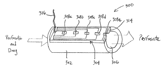
In another aspect, an implantable device is provided for testing drug or
analyte
transport through a tissue capsule. In one embodiment, the device includes a
primary
body having an outer surface, a perfusate fluid inlet, a perfusate fluid
outlet, and a fluid
conduit extending between the inlet and the outlet; a substrate attached to
the primary
body; at least one reservoir defined in and extending through the substrate,
the reservoir
having a first opening in the fluid conduit and a second opening which can be
open to
the outer surface of the device; at least one reservoir cap covering the
second opening
of the reservoir; and means for selectively disintegrating or removing the
reservoir cap.
The device typically would include a first flexible tubing connected to the
perfusate
fluid inlet, a second flexible tubing connected to the perfusate fluid outlet,
and a means
for flowing perfusate thorough the fluid conduit and the flexible tubings. In
one
embodiment, the device further includes a semipermeable barrier structure
blocking
bulk fluid flow through one or both of the reservoir openings following
reservoir cap
disintegration or removal.
Brief Description of the Figures
FIG. 1 is a cross-sectional and perspective view of one embodiment of a device
body with reservoirs and reservoir caps for opening by electrothermal
ablation.
FIG. 2 is a cross-sectional view of a single reservoir of a device undergoing
a
2o process for loading the reservoir with a drug formulation and transport
enhancing
solvent.
FIG. 3 is a cross-sectional and partial view of one embodiment of implanted
drug delivery device using electromotive driving means to drive a charged drug
out of
the device reservoirs and into/through a surrounding fibrous tissue capsule
and
microvasculature.
FIG. 4 is a perspective and partially exploded view of one embodiment of an
implantable multi-reservoir medical device, as described herein.
FIG. 5 is a perspective view of a second embodiment of an implantable multi-
reservoir medical device, as described herein.
FIG. 6 is a perspective and partially exploded view of one embodiment of an
implantable mufti-reservoir medical device having a protective mesh structure
over the
reservoir caps.
FIGS. 7A-B are cross-sectional and perspective views illustrating prior art
drug
a"r~ analvta r~PrfnSipn processes through a semi-permeable membrane in tube
form.
CA 02569375 2006-11-30
WO 2006/085908 PCT/US2005/019021
FIG. 8A is a perspective and cross-sectional view, and FIG. 8B is a cross-
sectional view, of one embodiment of a testing device for use in measuring
cross-tissue
capsule transport.
FIG. 9 is a cross-sectional view of another embodiment of a testing device,
which includes a semi-permeable tube inside the primary perfusate flow tube,
for use in
measuring cross-tissue capsule transport.
FIG. 10 is a cross-sectional view of another embodiment of a testing device,
which includes a semi-permeable plug disposed in the reservoir opening, for
use in
measuring cross-tissue capsule transport.
FIG. 11 is a plan view of one embodiment of a testing device described herein
for in vivo measurement of cross-tissue capsule transport
FIG. 12 is a perspective and cross-sectional view of still another embodiment
of
a testing device, which includes a plurality of individually openable
reservoirs, for use
in measuring cross-tissue capsule transport.
FIG. 13 is a perspective view of yet another embodiment of a testing device,
which includes a plurality of individually openable reservoirs, for use in
measuring
cross-tissue capsule transport.
FIG. 14 is a cross-sectional view of the testing device shown in FIG. 13 being
used to measure analyte flow through a tissue capsule.
FIG. 15 is a perspective view of a laboratory equipment set up/process for
leak
testing one of the testing devices for use in measuring cross-tissue capsule
transport.
FIG. 16 is a perspective view of a laboratory equipment set up/process for in
vitro testing one of the testing devices for use in measuring cross-tissue
capsule
transport.
Detailed Description of the Invention
In one aspect, methods and devices have been developed for enhancing mass
transport through any fibrous tissue capsule that may form around an implanted
medical device following implantation, and/or for enhancing vascularization
around the
implanted device, which also will aid in mass transport to/from the device.
In one embodiment, an implantable medical devices is provided that include a
body portion; one or more reservoirs located in and defined by the body
portion;
reservoir contents; and a means for enhancing mass transport through any
fibrous tissue
capsule that may form around the device following implantation. Methods and
devices
CA 02569375 2006-11-30
WO 2006/085908 PCT/US2005/019021
are provided to enhance the transport of drug molecules and analytes across
tissue
capsules expected to develop around these devices following their implantation
into a
patient, and to reduce capsule tissue growth in the first instance.
In preferred embodiments, the device comprises a plurality of reservoirs, the
contents of which may contain (i) a drug formulation for short- or long-term,
controlled
drug delivery, (ii) sensors for analyte or therapeutic drug monitoring, or
(iii) both drug
and sensors. In one embodiment, the device includes a drug formulation stored
in and
selectively released from the reservoirs and the means for enhancing mass
transport of
drug (released from the reservoirs) across and out of the tissue capsule. In
another
to embodiment, the device includes a sensor and the means for enhancing mass
transport
enhances the transport of an analyte across the tissue capsule.
In another aspect, devices and methods have been developed for isolating the
effect of tissue encapsulation. The devices advantageously allow access to the
inside of
intact tissue capsules in situ (in the animal), which, significantly, makes it
possible to
obtain detailed ih situ measurements of molecular transport across tissue
capsules. The
devices will allow one to assess methods for modifying/modulating the
properties or
structure of the tissue capsule (e.g. thickness, vascularity, density,
porosity,
permeability, etc.) and to make quantitative comparisons of different
strategies for
improving transport for example comparing two tissue capsules that have been
formed in different ways or under the influence of different conditions. The
devices
also permit one to test different materials or. device configurations. In one
embodiment,
the purpose of accessing the inside of the capsule is to test a device. For
instance, one
could compare devices that include means of opening a pathway into the device
at a
specific time, e.g., by mechanically rupturing, by electrochemically or
electrothernzally
disintegrating, or by otherwise removing, a reservoir cap from an opening in
the device
body. In another embodiment, the purpose is to test a bulk material that is
being
considered for an implant material and observe what kind of capsule forms and
whether
such a material/device possibly would be useful as or in a drug delivery or
biosensing
device.
As used herein, the terms "comprise," "comprising," "include," and "including"
are intended to be open, non-limiting terms, unless the contrary is expressly
indicated.
The Imulantable Medical Device and Components Thereof
The medical device includes a body portion; one or more reservoirs located in
ie body portion; reservoir contents; and a means for enhancing mass
CA 02569375 2006-11-30
WO 2006/085908 PCT/US2005/019021
transport (of all or a portion of the reservoir contents or of an
environmental component
intended for contact with all or a portion of the reservoir contents) through
any fibrous
tissue capsule that may form around the device following implantation. In one
embodiment, the reservoir contents comprise a drug formulation, the reservoirs
store
the drug formulation, and control means control release of the drug
formulation
therefrom. In another embodiment, the reservoir contents comprise a sensor,
the
reservoirs store and protect the sensor, and control means control the time at
which the
sensor is exposed to the body (e.g., to a physiological fluid ifa vivo).
The control means can take a variety of forms. In one embodiment, each
reservoir has an opening covered by a reservoir cap that can be selectively
ruptured
(e.g., disintegrated) to initiate release of the drug from the reservoir. For
example, the
reservoir cap can comprise a metal film that is disintegrated by
electrothermal ablation
as described in U.S. Patent Application Publication No. 2004/0121486 Al .
Other
reservoir opening and release control methods are described in U.S. Patent
Application
Publication Nos. 2002/0072784 A1, 2002/0099359 Al, 2002/0187260 Al,
2003/0010808 Al, 2004/0106914 A1, and 2005/0055014 Al; and U.S. Patent Nos.
5,797,898; 6,123,861; 6,527,762; 6,551,838; 6,773,429; 6,808,522 all of which
are
incorporated by reference herein.
Device Body and Reservoirs
The device comprises a body portion, i.e., a substrate, that includes one or
more
reservoirs. A reservoir is a well, a recess, or a cavity, located in a solid
structure and
suitable for containing a quantity of another material and/or a small device.
In a
preferred embodiment, the device includes a plurality of the reservoirs
located in
discrete positions across at least one surface of the body portion.
In various embodiments, the body portion comprises silicon, a metal, a
ceramic,
a polymer, or a combination thereof. Examples of suitable substrate materials
include
metals, ceramics, semiconductors, glasses, and degradable and non-degradable
polymers. Preferably each reservoir is formed of hermetic materials (e.g.,
metals,
silicon, glasses, ceramics) and is hermetically sealed by a reservoir cap.
Biocompatibility of the substrate material is preferred for ira vivo device
applications.
For biocompatible and non-biocompatible materials, the substrate, or portions
thereof,
may be coated, encapsulated, or otherwise contained in a biocompatible
material, such
as polyethylene glycol), polytetrafluoroethylene-like materials, diamond-like
carbon,
tanium, and the like, before use. In one embodiment, the substrate is
CA 02569375 2006-11-30
WO 2006/085908 PCT/US2005/019021
hermetic, that is impermeable (at least during the time of use of the
reservoir device) to
the molecules to be delivered and to surrounding gases or fluids (e.g., water,
blood,
electrolytes or other solutions). In another embodiment, the substrate is made
of a
material that degrades or dissolves over a defined period of time into
biocompatible
components. Examples of such materials include biocompatible polymers, such as
poly(lactic acids, poly(glycolic acids, and poly(lactic-co-glycolic acids, as
well as
degradable poly(anhydride-co-imides).
The substrate may consist of only one material, or may be a composite or multi-
laminate material, that is, composed of several layers of the same or
different substrate
materials that are bonded together. In one embodiment, the substrate comprises
layers
of silicon and Pyrex bonded together. In another embodiment, the substrate
comprises
multiple silicon wafers bonded together. In yet another embodiment, the
substrate
comprises a low-temperature co-fired ceramic (LTCC). In one embodiment, the
body
portion is the support for a microchip device. In one example, this substrate
is formed
of silicon.
The body portion can have a variety of shapes, or shaped surfaces. It can, for
example, have a release side (i.e., an area having reservoir caps) that is
planar or
curved. The substrate may, for example, be in a shape selected from circular
or ovoid
disks, cylinders, or spheres. In one embodiment, the release side can be
shaped to
2o conform to a curved tissue surface or into a body lumen. In another
embodiment, the
back side (distal the release side) is shaped to conform to an attachment
surface. In
various embodiments, the body portion is in the form of a chip, a disk, a
tube, or a
sphere. The body portion can be flexible or rigid.
Total substrate thickness and reservoir volume can be increased by bonding or
attaching wafers or layers of substrate materials together. The device
thickness may
affect the volume of each reservoir and/or may affect the maximum number of
reservoirs that can be incorporated onto a substrate. The size and number of
substrates
and reservoirs can be selected to accommodate the quantity and volume of
reservoir
contents needed for a particular application, manufacturing limitations,
and/or total
device size limitations to be suitable for implantation into a patient,
preferably using
minimally invasive procedures.
The substrate can have one, two, or preferably many, reservoirs. In various
embodiments, tens, hundreds, or thousands of reservoirs are arrayed across the
c"~,~,,."+o ~.... :~..tance, one embodiment of an implantable drug delivery
device
CA 02569375 2006-11-30
WO 2006/085908 PCT/US2005/019021
includes between 250 and 750 reservoirs, where each reservoir contains a
single dose of
a drug for release, which for example could be released daily over a period of
several
months to two years. More or less frequent dosing schedules and shorter or
longer
treatment durations are possible. In one sensing embodiment, the number of
reservoirs
in the device is determined by the operation life of the individual sensors.
For example,
a one-year implantable glucose monitoring device having individual sensors
that
remain functional for 30 days after exposure to the body would contain at
least 12
reservoirs (assuming one sensor per reservoir).
In one sensor embodiment, the distance between the sensor surface and the
reservoir opening means is minimized, preferably only a few microns. In this
case, the
volume of the reservoir is primarily determined by the surface area of the
sensor. For
example, the electrodes of a typical enzymatic glucose sensor may occupy a
space that
is 400 ~,m by 800 Vim.
In one embodiment, the reservoirs are microreservoirs. As used herein, the
terns
"microreservoir" refers to a concave-shaped solid structure suitable for
releasably
containing a material, wherein the structure is of a size and shape suitable
for filling
with a microquantity of the material, which comprises a drug. In one
embodiment, the
microreservoir has a volume equal to or less than 500 ~L (e.g., less than 250
~,L, less
than 100 ~,L, less than 50 ~L, less than 25 ~L, less than 10 pL, etc.) and
greater than
about 1 nL (e.g., greater than 5 nL, greater than 10 nL, greater than about 25
nL, greater
than about 50 nL, greater than about 1 ~L, etc.). The shape and dimensions of
the
microreservoir can be selected to maximize or minimize contact area between
the drug
material and the surrounding surface of the microreservoir. As used herein,
the term
"microquantity" refers to small volumes between 1 nL and 10 ~,L. In one
embodiment,
the microquantity is between 1 nL and 1 ~L. In another embodiment, the
microquantity is between 10 nL and 500 nL.
In other embodiments, the reservoirs are larger than microreservoirs and can
contain a quantity of drug formulation larger than a microquantity. For
example, the
volume of each reservoir can be greater than 10 ~,L (e.g., at least 20 p,L, at
least 50 ~,L,
at least 100 ~,L, at least 250 pL, etc.) and less than 10,000 ~L (e.g., less
than 5000 ~,L,
less than 1000 p,L, less than 750 ~L, less than 500 ~L, less than 100 ~,L,
etc.). These
may be referred to as macro-reservoirs and macro-quantities, respectively.
Unless
explicitly indicated to be limited to either micro- or macro-scale
volumes/quantities, the
CA 02569375 2006-11-30
WO 2006/085908 PCT/US2005/019021
term "reservoir" is intended to include both.
Reservoirs can be fabricated in a structural body portion using any suitable
fabrication technique known in the art. Representative fabrication techniques
include
MEMS fabrication processes or other micromachining processes, various drilling
techniques (e.g., laser, mechanical, and ultrasonic drilling), and build-up
techniques,
such as LTCC (low temperature co-fired ceramics), as well as molding
processes. See,
for example, U.S. Patent Nos. 6,123,861 and 6,808,522, as well as U.S. Patent
Application Publication Nos. 2004/0106914 and 2005/0055014. The surface of the
reservoir optionally can be treated or coated to alter one or more properties
of the
1o surface. Examples of such properties include hydrophilicity/
hydrophobicity, wetting
properties (surface energies, contact angles, etc.), surface roughness,
electrical charge,
release characteristics, and the like.
In one embodiment, the device comprises a microchip chemical delivery device.
In another embodiment, the device includes polymeric chips or devices composed
of
15 non-silicon based materials that might not be referred to as "microchips."
Examples of
various substrate and device configurations are described in U.S. Patent
Application
Publication No. 2004/0121486 A1. In one embodiment, the device comprises an
osmotic pump, for example, the DUROSTM osmotic pump technology (Alza
Corporation) included in commercial devices such as VIADUR~ (Bayer Healthcare
2o Pharmaceuticals and Alza Corporation). In another embodiment, the device
comprises
a LTCC body. In one embodiment, the body portion is the support for a
microchip
device. In one example, this substrate can be formed of silicon.
Means For Enhancing Mass Transport
The implantable device may include one or a combination of components useful
25 for enhancing the rate of mass transport through a tissue capsule. These
include the use
of enzymes, co-solvents, surfactants, or combinations thereof, useful in
making highly
concentrated, stable formulations, counter-ion drug formulations, enzymatic
degradation, and electromotive devices. Another means for enhancing mass
transport
from the device reservoirs and through a tissue capsule includes the positive
30 displacement and/or accelerated release techniques described in PCT WO
2004/026281
Al. Yet another means for enhancing mass transport involves enhancing the
vascularity of the tissue capsule, such as with one or more angiogenic agents
coated on
or released from the device, which will facilitate drug or analyte transport
therethrough.
11
CA 02569375 2006-11-30
WO 2006/085908 PCT/US2005/019021
In various embodiments, combinations of these different means, materials, and
techniques are used to enhance transport of drug or analyte through the tissue
capsule.
In one embodiment, the device releases from the reservoirs, and/or is coated
with, one or more anti-inflammatory agents. In one specific embodiment, the
anti-
s inflammatory agent is dexamethasone. In these embodiments, the anti-
inflammatory
agent reduces inflammation following implantation, which can decrease the
overall
thickness of the fibrous capsule. In another specific embodiment, the device
releases
from the reservoirs, and/or is coated with, a combination of dexamethasone and
VEGF,
which can reduce inflammation and increase vascularity around the implanted
device.
See Norton, et al., "Dual Release of VEGF and Dexamethasone from Microspheres
Incorporated in Anti-fouling Hydrogels" p. 357, Proceedings 7th World
Biomaterials
Congress (Sydney, Australia) May 2004.
As used herein, the term "transport enhancers" refers to and includes
solvents,
co-solvents, and surfactants that alter tissue permeability, and enzymes that
degrade
tissue capsules, and thereby help drug molecules to penetrate the tissue
capsule and
reach their targets at effective (e.g., therapeutic) concentrations and rates
or help
analytes penetrate the tissue capsule to reach a sensor material at
clinically/diagnostically useful concentrations and rates.
Solvef2tlSurfactant Formulations
2o In one embodiment, the means for enhancing mass transport comprises
reservoir contents that include a material effective to alter tissue capsule
permeability.
Altered permeability of the tissue capsule can permit greater mass transport
of drug or
analyte therethrough. In one embodiment, the drug formulation includes one or
more
solvents, co-solvents, surfactants, or combinations thereof, useful in making
highly
concentrated, stable formulations and/or useful in altering tissue
permeability. The
small dose volumes of the reservoir devices advantageously permit an active
ingredient,
i.e., a drug, to be dissolved or physically mixed with powerful solvents, co-
solvents,
and/or surfactants that otherwise would cause tissue irntation or damage if
used in
larger volumes. Examples of such solvents and the Permitted Daily Exposure
limits are
provided in ICH Guideline Q3C. Although these limits were intended for
residual
solvents remaining in drug product from processing operations, many of the
listed
solvents could be contained in device reservoirs without exceeding these
recommended
limits. For example, the Center for Drug Evaluation and Research (CDER) at the
T T__ .. _ , n. . r d and Drug Administration (FDA) has classified
nitromethane as a
12
CA 02569375 2006-11-30
WO 2006/085908 PCT/US2005/019021
Class 2 solvent with a Per Day Exposure (PDE) limit of 500 pg (440 nL), which
is the
most stringent, recommended limit for Class 2 solvents. In one embodiment of
the
present devices, each reservoir contains 200 nL of a Class 2 Solvent, and if
only one
reservoir is released per day, then exposure to the solvent would be less than
half the
PDE.
Representative examples of suitable solvents include dimethylsulfoxide
(DMSO), N-methylpyrrolidone (NMP), as well as Dimethylpyrrolidone (DMP),
dimethylformamide (DMF), dimethylacetamide, acetonitrile, and other polar,
aprotic
solvents that alter the structure of collagen and collagen networks. In one
embodiment, the small volumes per dose (_< 200 nL/dose) are well below the
daily
exposure limits for the Class 2 solvents. Representative examples of suitable
surfactants include polysorbates, Spans, monoalkylpolyoxythenes,
dialkylpolyoxyethylenes, polyoxyethylene monoesters, polyoxyethylene diesters,
and
polyoxyethylene-polyoxyethylene block copolymers. To derive a suitable
formulation
one can, for example, (1) identify a solvent or surfactant, alone or in
combination, that
provides the required drug solubility and stability, and (2) screen for
penetration
enhancement by comparing the rate of drug movement across a sample of the
appropriate capsular tissue using a device such as a Franz cell or mufti-well
microdialysis unit (as described below). By selecting appropriate solvents, co-
solvents,
and surfactants, one is able to produce small molecule, peptide, and protein
drug
formulations that are highly concentrated (for example, >_ 100mg/mL or 10% w/v
or
10% v/v) and stable at body temperature.
Furthermore, the use of concentrated solutions of drug enables one to limit
reservoir size and thus device size, which in turn advantageously could limit
implant
mobility, which may reduce tissue capsule growth. High concentrations
advantageously can keep the implantable device relatively small in size
(because there
is no need for the device to be sized to contain extra solvent volume), which
can make
their placement in the patient safer and less obtrusive. In addition, a
smaller implant
with suture anchoring desirably would be less mobile than a larger device.
Mobility
has been identified as a contributor to tissue capsule growth.
In one embodiment, a transport enhancer is released from some of the
reservoirs. The reservoirs can contain one or more transport enhancers alone
or in
combination with a drug formulation for release. In another embodiment, the
device
13
CA 02569375 2006-11-30
WO 2006/085908 PCT/US2005/019021
includes a coating, such as a controlled-release coating, comprising the
transport
enhancer. In one variation, the transport enhancer is provided in the device
in one or
more reservoirs separate from the reservoirs containing the drug formulation
for
release. For example, in a device comprising a substrate having an array of
reservoirs
located therein, the transport enhancer and the drug formulation could be
stored in
alternating reservoirs, and release of the transport enhancer could be
directed to closely
or immediately precede release of the drug formulation. Alternatively, release
of the
transport enhancer could be simultaneous with, or could follow, release of the
drug
formulation.
In one embodiment, a transport enhancer is contained in one or more reservoirs
near to, but separate, from reservoirs containing glucose sensors. The release
of the
transport enhancer can be timed with respect to exposure of the glucose
sensor, or the
transport enhancer could be released from the reservoirs on a regular
schedule. The
latter situation would work to keep the capsule at the same level of
permeability over a
long implantation period.
Enzynaatic Degradation
In yet another approach, the implant device include molecules that are known
to
dissolve or degrade the components of a tissue capsule can be used to decrease
the
transport limiting effects of the tissue capsule. For example, a composition
comprising
collagenase, thrombin, fibrinolysin, trypsin, hyaluronidase, or a combination
thereof,
could be included in, on, or with the device. The enzyme could be packaged in
reservoirs, attached to the surface of the device, or incorporated into a
release-
modifying matrix.
In one embodiment, the enzyme for enhancing transport is contained in one or
more reservoirs of the device. Reservoirs can be opened prior to releasing a
drug from
one or more neighboring reservoirs, such that the enzyme dissolves at least a
portion of
the capsule near the drug reservoir, thereby reducing the capsule's barrier
properties
and minimizing the capsule's effect on the drug release rate when the drug
reservoir is
opened. A similar strategy can be used with biosensing devices to increase the
permeability of a capsule to an analyte such as glucose. If the tissue capsule
comprises
significant amounts of fibrin or fibrinogen, then the device could release
thrombin,
fibrinolysin, trypsin, or another effective enzyme.
Counter Ion Drug Formulations
14
CA 02569375 2006-11-30
WO 2006/085908 PCT/US2005/019021
In another embodiment, the drug is comprised of charged molecules, and ion-
pairing counterions are included in the drug formulation to modulate drug
transport
through a tissue capsule. The ability of counterions to alter binding of
charged
molecules, including peptides and drugs, to materials is well known as
illustrated by
RP-HPLC and the influence of the Hofmeister series ions on protein structure
and ion .
exchange chromatography retentions. Ion pair formation has been used in
organic
chemistry to facilitate the movement of reactants and products between organic
and
aqueous phases to provide in situ reaction compartmentalization. Ion pairing
of drugs to
vary their performance by changing lipid solubility, particle size, or micelle
formation
has been examined (Choi, et al., Irat'l J. Pharmaceutics 203(1):193-202
(2000);
Kendrick, et al., Arch. Biochena. Biophys. 347:113-18 (1997); Meyer, et al.,
Plaar~m.
Res._15(2):188-93 (1998)). It may also be possible to disrupt or modify the
hydrogen
bonds or ionic bonds within the capsule's collagen structure by interaction or
exchange
of the drug counterion with the collagen.
ElectYOmotive Devices and Methods
In yet another embodiment, the drug can be charged and iontophoretic or other
electromotive methods known in the art are used to enhance transport through a
tissue
capsule. For example, an exposed surface of the implanted device can be
charged by
its internal electronics to create a driving force that would propel drug
molecules
possessing the same charge through a capsule. In one embodiment, a pair of
oppositely
charged electrodes are located on the same device or surface of the device,
but are
positioned sufficiently far apart from one another so that the path of least
electrical
resistance through the tissue barrier. In another embodiment, a counter
electrode,
separate from the drug delivery component, is located outside of the tissue
capsule.
Positiye Displacement Devices and Methods
In one embodiment, positive displacement mechanisms are used to drive a drug
formulation out of the reservoirs. These same mechanisms can also be employed
to
drive or push the drug formulation through the tissue capsule. In one
embodiment, an
osmotic pressure generating material or other swellable material drives a
piston to force
a drug formulation out of the reservoir. This and other embodiments are
detailed in
PCT WO 2004/026281 A1.
In one embodiment, the reservoir contents are sealed in a gas-tight or
hermetic
reservoir under compression or sealed under conditions to create a positive
pressure
---'- -' - ' ent so that contents are expelled upon reservoir cap activation.
CA 02569375 2006-11-30
WO 2006/085908 PCT/US2005/019021
In another embodiment, the device comprises three substrate portions packaged
together, with the reservoir spaces aligned and defining three compartments
(reservoirs). The bottom compartment includes a dry swellable gel, the middle
compartment includes a liquid material (or at least liquid at body
temperature), and the
top compartment includes a drug formulation for release. Membranes (or
reservoir
caps) between the layers separate the adjacent compartments, until release is
intended.
After or simultaneously with disintegration of a reservoir cap over the
exterior opening
of a drug-loaded reservoir, the membrane over the swellable gel is opened to
allow
liquid from the middle compartment to contact the gel, causing the swellable
gel to
expand. The gel materials) would be selected to have an expanded volume that
exceeded the combined volumes of the reservoir compartments. Optionally,
expansion
could be augmented by a controlled temperature change (e.g., with a resistive
heater
element disposed in the reservoir) where the gel is one known in the art to
expand/contract with temperature. The membrane over the liquid is opened to
allow
the swelling gel to displace the drug formulation from the top compartment.
An~i~efzic Matef~ials aT~d APehts
In one embodiment, the device is provided with a coating that comprises one or
more angiogenic materials or factors to promote vascularization around the
implanted
device. This would be useful with both implantable drug delivery devices and
2o implantable analyte monitoring devices (e.g., glucose sensors). As used
herein, the
term "angiogenic" refers to a material or molecules that promote and maintain
the
development of blood vessels and microcirculation around the implanted device.
In
one embodiment example, the device releases or is coated with a vasoinductive
or
angiogenic agent such as a vascular growth factor. Suitable growth factors of
this type
include as vascular endothelial growth factor (VEGF), platelet growth factor,
vascular
permeability factor, fibroblast growth factor, and transforming growth factor
beta.
In another embodiment, the device includes an exterior membrane or coating
layer that itself exhibits angiogenic properties. These layers can be made for
example
of expanded polytetrafluoroethylene (ePTFE), hydrophilic polyvinylidene
fluoride,
mixed cellulose esters, and/or other polymers.
Device Sup ace Modification a~ad Sca oldiyag
In still another embodiment, the implantable medical device includes tissue
scaffolding or other physical surface modification effective to promote
transport over a
an unmodified surface. Underlying material properties that may affect
16
CA 02569375 2006-11-30
WO 2006/085908 PCT/US2005/019021
tissue capsule deposition include alternations in surface hydrophilicity,
surface area,
porosity (both percent and diameter of individual pores), and degree of
nanometer-scale
roughness. These concepts are understood in the art, and those in the art can
adapt
conventional approaches for use with the implantable mufti-reservoir devices
described
herein. As used herein, the term "scaffolding" refers to a three-dimensional
surface
topography that may include nanometer scale features such as roughness or
porosity.
That is, the topography may serve as a scaffold on which cell adhesion occur
in a
controlled fashion, reducing formation of avascular tissue and resulting n a
tissue
quality more likely to permit cross-tissue transport. The surface could, in
turn, be
modified by deposition of an additional layer of a different material or with
chemical or
biochemical decoration that could affect cell adhesion.
Reservoir Control Means
The reservoir control means comprises the structural components) for
controlling the time at which release or exposure of the reservoir contents is
initiated.
In a preferred embodiment, the reservoir control means includes reservoir caps
and the
hardware, electrical components, and software needed to control and deliver
electric
energy from a power source to selected reservoirs) for actuation, e.g.,
reservoir
opening.
Reservoir Cams
2o As used herein, the term "reservoir cap" includes a discrete membrane or
other
structure suitable for separating the contents of a reservoir from the
environment
outside of the reservoir. It generally is self supporting across the reservoir
opening,
although caps having additional structures to provide mechanical support to
the cap can
be fabricated. See, e.g., U.S. Patent No. 6,875,208. Selectively removing the
reservoir
cap or making it permeable will then "expose" the contents of the reservoir to
the
environment (or selected components thereof) surrounding the reservoir. In
preferred
embodiments, the reservoir cap is selectively disintegrated. As used herein,
the term
"disintegrate" includes degrading, dissolving, rupturing, fracturing or some
other form
of mechanical failure, as well as a loss of structural integrity due to a
chemical reaction
(e.g., electrochemical degradation) or phase change (e.g., melting) in
response to a
change in temperature, unless a specific one of these mechanisms is indicated.
In
several preferred embodiments, removal of the reservoir cap primarily involves
a
chemical reaction or phase change component, as opposed to a mechanically
activated
dying on prestressed, brittle membranes being fractured by a
17
CA 02569375 2006-11-30
WO 2006/085908 PCT/US2005/019021
mechanical force from a piezoelectric member, or gas pressure generated
mechanical
rupture).
In one specific embodiment, the disintegration is by an electrochemical
activation technique, such as described in U.S. Patent No. 5,797,898, which is
incorporated herein by reference. For example, the reservoir cap can be a thin
metal
film which is impermeable to the surrounding environment (e.g., body fluids or
another
chloride containing solution). In one variation, a particular electric
potential is applied
to the metal reservoir cap, which is then oxidized and disintegrated by an
electrochemical reaction, to release the drug from the reservoir. Examples of
suitable
1o reservoir cap materials include gold, silver, copper, and zinc.
In another specific embodiment, the disintegration is by thermal activation
technique, such as described in U.S. Patent No. 6,527,762, which is
incorporated herein
by reference. For example, the reservoir cap can be heated (e.g., using
resistive heating
from a separate resistive heater) to cause the reservoir cap to melt and be
displaced
15 from the reservoir opening, to open the reservoir. This latter variation
could be used,
for example, with reservoir caps formed of a metal or a non-metal material,
e.g., a
polymer. In yet another variation, the reservoir cap is formed of a polymer or
other
material that undergoes a temperature-dependent change in permeability such
that upon
heating to a pre-selected temperature, the reservoir is rendered permeable to
the drug
2o and bodily fluids to permit the drug to be released from the reservoir
through the
reservoir cap.
In a preferred embodiment, the "disintegration" is by an electro-thermal
ablation technique, as described in U.S. Patent Application Publication No.
2004/0121486 A1, which is incorporated herein by reference. For example, the
25 reservoir cap is formed of a conductive material, such as a metal film,
through which an
electrical current can be passed to electrothermally ablate it. Representative
examples
of suitable reservoir cap materials include gold, copper, aluminum, silver,
platinum,
titanium, palladium, various alloys (e.g., Au-Si, Au-Ge, Pt-Ir, Ni-Ti, Pt-Si,
SS 304, SS
316), and silicon doped with an impurity to increase electrical conductivity,
as known
30 in the art. In one embodiment, the reservoir cap is in the form of a thin
metal film. In
one embodiment, the reservoir cap is part of a multiple layer structure, for
example, the
reservoir cap can be made of multiple metal layers, such as a mufti-
layer/laminate
structure of platinum/titanium/ platinum. The reservoir cap is operably (i.e.,
iected to an electrical input lead and to an electrical output lead, to
18
CA 02569375 2006-11-30
WO 2006/085908 PCT/US2005/019021
facilitate flow of an electrical current through the reservoir cap. When an
effective
amount of an electrical current is applied through the leads and reservoir
cap, the
temperature of the reservoir cap is locally increased due to resistive
heating, and the
heat generated within the reservoir cap increases the temperature sufficiently
to cause
the reservoir cap to be electrothermally ablated and ruptured.
In a preferred embodiment, a discrete reservoir cap completely covers a single
reservoir opening. In another embodiment, a discrete reservoir cap covers two
or more,
but less than all, of the reservoir's openings.
In passive release devices, the reservoir cap is formed from a material or
mixture of materials that degrade, dissolve, or disintegrate over time, or
that do not
degrade, dissolve, or disintegrate, but are permeable or become permeable to
drug or
analyte molecules. Representative examples of reservoir cap materials include
polymeric materials, and non-polymeric materials such as porous forms of
metals,
semiconductors, and ceramics. Passive semiconductor reservoir cap materials
include
nanoporous or microporous silicon membranes.
Characteristics can be different for each reservoir cap to provide different
times
of release of drug formulation. For example, any combination of polymer,
degree of
crosslinking, or polymer thickness can be modified to obtain a specific
release time or
rate. Any combination of passive and/or active release reservoir cap can be
present in a
single delivery device. For example, the reservoir cap can be removed by
electrothermal ablation to expose a passive release system that only begins
its passive
release after the reservoir cap has been actively removed. Alternatively, a
given device
can include both passive and active release reservoirs.
In one embodiment, the device includes (i) active release reservoirs
containing a
drug formulation, and (ii) passive release reservoirs containing one or more
transport
enhancers. In one method with this embodiment, the transport enhancement
molecules
are continuously released from the passive release reservoirs to maintain a
constant
capsule permeability, while the active release reservoirs are opened on a
schedule
determined by the type of drug therapy prescribed by the physician.
In another embodiment, the device includes (i) active release reservoirs
containing sensors, and (ii) passive release reservoirs containing one or more
transport
enhancers. In one method with this embodiment, the transport enhancement
molecules
are continuously released from the passive release reservoirs to maintain a
constant
ility, while the active release reservoirs are opened as needed
19
CA 02569375 2006-11-30
WO 2006/085908 PCT/US2005/019021
(depending, for example, upon fouling of the sensor) or as dictated by a
predetermined .
schedule.
In yet another embodiment, the device includes (i) active release reservoirs
containing a drug formulation, and (ii) active release reservoirs containing
one or more
transport enhancers. In one method with this embodiment, the transport
enhancement
molecules are released periodically or on a schedule to coincide with or
precede the
opening of the drug-containing active release reservoirs, which are opened on
a
schedule, determined by the prescribed drug therapy.
Other Cofnponents
1o The control means can provide intermittent or effectively continuous
release of
the drug formulation and/or the transport enhancer, and/or selective exposure
of
sensors. The particular features of the control means depend on the mechanism
of
reservoir cap activation described herein. For example, the control means can
include
an input source, a microprocessor, a timer, a demultiplexer (or multiplexer),
and a
power source. As used herein, the term "demultiplexer" also refers to
multiplexers.
The power source provides energy to activate the selected reservoir, e.g., to
trigger
release of the drug formulation from the particular reservoir desired for a
given dose.
For example, the operation of the reservoir opening system can be controlled
by an on-
board microprocessor (e.g., the microprocessor is within an implantable or
insertable
device). The microprocessor can be programmed to initiate the disintegration
or
permeabilization of the reservoir cap at a pre-selected time or in response to
one or
more of signals or measured parameters, including receipt of a signal from
another
device (for example by remote control or wireless methods) or detection of a
particular
condition using a sensor such as a biosensor. In another embodiment, a simple
state
machine is used, as it typically is simpler, smaller, and/or uses less power
than a
microprocessor. The device can also be activated or powered using wireless
means, for
example, as described in U.S. 2002/0072784 A1 to Sheppard et al.
In one embodiment, the device includes a substrate having a two-dimensional
array of discretely spaced reservoirs arranged therein, a drug formulation
contained in
3o the reservoirs, anode reservoir caps covering a semi-permeable membrane for
each of
the reservoirs, cathodes positioned on the substrate near the anodes, and
means for
actively controlling disintegration of the reservoir caps. The means includes
a power
source and circuitry to control and deliver an electrical potential; the
energy drives a
selected anodes and cathodes. Upon application of a potential
CA 02569375 2006-11-30
WO 2006/085908 PCT/US2005/019021
between the electrodes, electrons pass from the anode to the cathode through
the
external circuit causing the anode material (reservoir cap) to oxidize and
dissolve into
the surrounding fluids, exposing and releasing the drug formulation. The
microprocessor directs power to specific electrode pairs through a
demultiplexer as
directed by an EPROM, remote control, or biosensor.
In another embodiment, the activation energy initiates a thermally driven
rupturing or permeabilization process, for example, as described in U.S.
Patent No.
6,527,762. For example, the means for controlling release can actively
disintegrate or
permeabilize a reservoir cap using a resistive heater. The resistive heater
can cause the
reservoir cap to undergo a phase change or fracture, for example, as a result
of thermal
expansion of the reservoir cap or release system, thereby rupturing the
reservoir cap
and releasing the drug from the selected reservoir. 'The application of
electric current to
the resistor can be delivered and controlled using components as described
above for
use in the electrochemical disintegration embodiment. For example, a
microprocessor
can direct current to select reservoirs at desired intervals.
In a preferred embodiment, control means controls electro-thermal ablation of
the reservoir cap. For example, the drug delivery device could include a
reservoir cap
formed of an electrically conductive material; an electrical input lead
connected to the
reservoir cap; an electrical output lead connected to the reservoir cap; and a
control
means to deliver an effective amount of electrical current through the
reservoir cap, via
the input lead and output lead, to locally heat and rupture the reservoir cap,
for example
to release the drug formulation or expose the sensor located therein. In one
embodiment, the reservoir cap and conductive leads are formed of the same
material,
where the temperature of the reservoir cap increases locally under applied
current
because the reservoir cap is suspended in a medium that is less thermally
conductive
than the substrate. Alternatively, the reservoir cap and conductive leads are
formed of
the same material, and the reservoir cap has a smaller cross-sectional area in
the
direction of electric current flow, where the increase in current density
through the
reservoir cap causes an increase in localized heating. The reservoir cap
alternatively
3o can be formed of a material that is different from the material forming the
leads,
wherein the material forming the reservoir cap has a different electrical
resistivity,
thermal diffusivity, thermal conductivity, and/or a lower melting temperature
than the
material forming the leads. Various combinations of these embodiments can be
cribed in U.S. Patent Application Publication No. 2004/0121486 A1.
21
CA 02569375 2006-11-30
WO 2006/085908 PCT/US2005/019021
The implantable devices typically are hermetically sealed, e.g., in a titanium
encasement, which exposes substantially only the reservoir caps.
In one embodiment, the control means includes a microprocessor, a timer, a
demultiplexer, and an input source (for example, a memory source, a signal
receiver, or
a biosensor), and a power source. The timer and demultiplexer circuitry can be
designed and incorporated directly onto the surface of the microchip during
electrode
fabrication, or may be incorporated in a separate microchip. The
microprocessor
translates the output from memory sources, signal receivers, or biosensors
into an
address for the direction of power through the demultiplexer to a specific
reservoir on
to the device. Selection of a source of input to the microprocessor such as
memory
sources, signal receivers, or biosensors depends on the microchip device's
particular
application and whether device operation is preprogrammed, controlled by
remote
means, or controlled by feedback from its environment (i.e., biofeedback). For
example, a microprocessor can be used in conjunction with a source of memory
such as
15 erasable programmable read only memory (EPROM), a timer, a demultiplexer,
and a
power source such as a battery or a biofuel cell. A programmed sequence of
events
including the time a reservoir is to be opened and the location or address of
the
reservoir is stored into the EPROM by the user. When the time for exposure or
release
has been reached as indicated by the timer, the microprocessor sends a signal
2o corresponding to the address (location) of a particular reservoir to the
demultiplexer.
The demultiplexer routes an input, such as an electric potential or current,
to the
reservoir addressed by the microprocessor.
Reservoir Contents
The reservoirs contain a drug formulation, a sensing device, a transport
25 enhancer, or a combination thereof.
Drug
The drug formulation is a composition that comprises a drug. As used herein,
the term "drug" includes any therapeutic or prophylactic agent (e.g., an
active
pharmaceutical ingredient or API). In one embodiment, the drug is provided in
a solid
3o form, particularly for purposes of maintaining or extending the stability
of the drug
over a commercially and medically useful time, e.g., during storage in a drug
delivery
device until the drug needs to be administered. The solid drug matrix may be
in pure
form or in the form of solid particles of another material in which the drug
is contained,
~persed. In one embodiment, the drug is formulated with an excipient
22
CA 02569375 2006-11-30
WO 2006/085908 PCT/US2005/019021
material that is useful for accelerating release, e.g., a water-swellable
material that can
aid in pushing the drug out of the reservoir and through any tissue capsule
over the
reservoir.
The drug can comprise small molecules, large (i.e., macro-) molecules, or a
combination thereof. In one embodiment, the large molecule drug is a protein
or a
peptide. In various other embodiments, the drug can be selected from amino
acids,
vaccines, antiviral agents, gene delivery vectors, interleukin inhibitors,
immunomodulators, neurotropic factors, neuroprotective agents, antineoplastic
agents,
chemotherapeutic agents, polysaccharides, anti-coagulants (e.g., LMWH,
pentasaccharides), antibiotics (e.g., immunosuppressants), analgesic agents,
and
vitamins. In one embodiment, the drug is a protein. Examples of suitable types
of
proteins include, glycoproteins, enzymes (e.g., proteolytic enzymes), hormones
or other
analogs (e.g., LHRH, steroids, corticosteroids, growth factors), antibodies
(e.g., anti-
VEGF antibodies, tumor necrosis factor inhibitors), cytokines (e.g., cc-, (3-,
or y-
interferons), interleukins (e.g., IL-2, IL-10), and diabetes/obesity-related
therapeutics
(e.g., insulin, exenatide, PYY, GLP-1 and its analogs). In one embodiment, the
drug is
a gonadotropin-releasing (LHRH) hormone analog, such as leuprolide. In another
exemplary embodiment, the drug comprises parathyroid hormone, such as a human
parathyroid hormone or its analogs, e.g., hPTH(1-~4) or hPTH(1-34). In a
further
embodiment, the drug is selected from nucleosides, nucleotides, and analogs
and
conjugates thereof. In yet another embodiment, the drug comprises a peptide
with
natriuretic activity. In still another embodiment, the drug is selected from
diuretics,
vasodilators, inotropic agents, anti-arrhythmic agents, Ca+ channel blocking
agents,
anti-adrenergics/ sympatholytics, and renin angiotensin system antagonists. In
one
embodiment, the drug is a VEGF inhibitor, VEGF antibody, VEGF antibody
fragment,
or another anti-angiogenic agent. Examples include an aptamer, such as
MACUGENTM (Pfizer/Eyetech) (pegaptanib sodium)) or LUCENTISTM
(Genetech/Novartis) (rhuFab VEGF, or ranibizumab), which could be used in the
prevention of choroidal neovascularization. In yet a further embodiment, the
drug is a
prostaglandin, a prostacyclin, or another drug effective in the treatment of
peripheral
vascular disease.
In still another embodiment, the drug is an angiogenic agent, such as VEGF. In
a further embodiment, the drug is an anti-inflammatory, such as dexamethasone.
In one
23
CA 02569375 2006-11-30
WO 2006/085908 PCT/US2005/019021
embodiment, a device includes both angiogenic agents and anti-inflammatory
agents.
In various embodiments, the drug is a bone morphogenic protein (BMP), a
growth factor (GF), or a growth or differentiation factor (GDF).
Representative
examples include BMP-2, OP-1 (osteogenic protein-1, i.e, BMP-7), morphogenic
proteins (CDMP), osteogenin, BMP-2/4, BMP-3, BMP-9, BMP-10, BMP-15, and
BMP-16, GDF-5 or rhGDF-5, Epidermal Growth Factors (EGF), Platelet-Derived
Growth Factors (PDGF), Fibroblast Growth Factors (FGFs), Transforming Growth
Factors cc & [3 (TGF-a and TGF-(3),
Erythropoietin (EPO), Insulin-Like Growth Factor-I and -II (IGF-I and IGF-II),
Tumor Necrosis Factors-a & -(3 (TNF-a, and TNF-(3), Colony Stimulating Factors
(CSFs), and Neuronal Growth Factor (NGF).
The reservoirs in one device can include a single drug or a combination of two
or more drugs, and/or two or more transport enhancers, and can further include
one or
more pharmaceutically acceptable Garners. Two or more transport enhancers,
angiogenic agents, anti-inflammatory agents, or combinations thereof, can be
stored
together and released from the same one or more reservoirs or they can each be
stored
in and released from different reservoirs.
Excipients and Matrix Materials
The drug, the transport enhancer, or both, can be dispersed in a matrix
material,
to further control the rate of release of drug, transport enhancer, or both.
This matrix
material can be a "release system," as described in U.S. Patent No. 5,797,898,
the
degradation, dissolution, or diffusion properties of which can provide a
method for
controlling the release rate of the chemical molecules.
The release system may include one or more pharmaceutical excipients.
Suitable pharmaceutically acceptable excipients include most carriers approved
for
parenteral administration. Other excipients may be used to maintain the drug
in
suspensions as an aid to reservoir filling, stability, or release. Depending
on the
properties of the drug, such excipients may be aqueous or non-aqueous,
hydrophobic or
hydrophilic, polar or non-polar, erotic or aprotic. See, e.g., U.S. Patent No.
6,264,990.
The release system optionally includes stabilizers, antioxidants,
antimicrobials,
preservatives, buffering agents, surfactants, and other additives useful for
storing and
releasing molecules from the reservoirs in vivo.
The release system may provide a temporally modulated release profile (e.g.,
24
CA 02569375 2006-11-30
WO 2006/085908 PCT/US2005/019021
pulsatile release) when time variation in plasma levels is desired or a more
continuous
or consistent release profile when a constant plasma level as needed to
enhance a
therapeutic effect, for example. Pulsatile release can be achieved from an
individual
reservoir, from a plurality of reservoirs, or a combination thereof. For
example, where
each reservoir provides only a single pulse, multiple pulses (i.e. pulsatile
release) are
achieved by temporally staggering the single pulse release from each of
several
reservoirs. Alternatively, multiple pulses can be achieved from a single
reservoir by
incorporating several layers of a release system and other materials into a
single
reservoir. Continuous release can be achieved by incorporating a release
system that
degrades, dissolves, or allows diffusion of molecules through it over an
extended
period. Continuous release also can be controlled from reservoirs by
incorporating a
rate-limiting semi-permeable membrane in or over the reservoir opening(s). In
addition, continuous release can be approximated by releasing several pulses
of
molecules in rapid succession ("digital" release). The active release systems
described
herein can be used alone or on combination with passive release systems, for
example,
as described in U.S. Patent No. 5,797,898. For example, the reservoir cap can
be
removed by active means to expose a passive release system, or a given
substrate can
include both passive and active release reservoirs.
In one embodiment, the drug formulation within a reservoir comprises layers of
drug and non-drug material. After the active release mechanism has exposed the
reservoir contents, the multiple layers provide multiple pulses of drug
release due to
intervening layers of non-drug.
SerasinQ Deyice
In some embodiments, a sensing component or device may be provided in one,
or preferably several, of the reservoirs of the device. In a preferred
embodiment, two or
more reservoirs contain a biosensor that can be used to detect the presence,
absence, or
change in a chemical or ionic species or energy at a site ira vivo. For
example, the
sensor could monitor the concentration of glucose, urea, calcium, or a hormone
present
in the blood, plasma, interstitial fluid, or other bodily fluid of the
patient.
3o Types of sensors include biosensors, chemical sensors, physical sensors, or
optical sensors. Examples of biosensors that could be adapted for use in/with
the
reservoir devices described herein include those taught in U.S. Patent No.
6,486,588;
No. 6,475,170; and No. 6,237,398, which are incorporated herein by reference.
Other
ire described in LT.S. Patent No. 6,551,838 and in LT.S. Patent
CA 02569375 2006-11-30
WO 2006/085908 PCT/US2005/019021
Application Publication No. 2005/0096587 Al, which are incorporated herein by
reference. As used herein, the term "biosensor" includes sensing devices that
transduce
the chemical potential of an analyte of interest into an electrical signal, as
well as
electrodes that measure electrical signals directly or indirectly (e.g., by
converting a
mechanical or thermal energy into an electrical signal). For example, the
biosensor
may measure intrinsic electrical signals (EKG, EEG, or other neural signals),
pressure,
temperature, pH, or loads on tissue structures at various ira vivo locations.
The
electrical signal from the biosensor can then be measured, for example by a
microprocessorlcontroller, which then can transmit the information to a remote
1o controller, another local controller, or both. For example, the system can
be used to
relay or record information on the patient's vital signs or the implant
environment, such
as drug concentration.
Several options exist for receiving and analyzing data obtained with the
sensing
device. Devices may be controlled by local microprocessors or remote control.
Biosensor information may provide input to the controller to determine the
time and
type of activation automatically, with human intervention, or a combination
thereof.
For example, the operation of an implantable drug delivery system (or other
controlled
release/controlled reservoir exposure system) can be controlled by an on-board
microprocessor (i.e., within the package of the implantable device). The
output signal
from the device, after conditioning by suitable circuitry if needed, will be
acquired by
the microprocessor. After analysis and processing, the output signal can be
stored in a
writeable computer memory chip, and/or can be sent (e.g., wirelessly) to a
remote
location away from the implantable device. Power can be supplied to the
implantable
device locally by a battery or remotely by wireless transmission. See, e.g.,
U.S. Patent
Application Publication No. 2002/0072784.
In one embodiment, a device is provided having reservoir contents that include
drug molecules for release and a sensor/sensing component. For example, the
sensor or
sensing component can be located in a reservoir or can be attached to the
device
substrate. The sensor can operably communicate with the device, e.g., through
a
microprocessor, to control or modify the drug release variables, including
dosage
amount and frequency, time of release, effective rate of release, selection of
drug or
. drug combination, and the like. The sensor or sensing component detects (or
not) the
species or property at the site of ira vivo implantation and further may relay
a signal to
26
CA 02569375 2006-11-30
WO 2006/085908 PCT/US2005/019021
the microprocessor used for controlling release from the device. Such a signal
could
provide feedback on and/or finely control the release of a drug.
In one embodiment, the device contains one or more sensors for use in glucose
monitoring and insulin control. Information from the sensor could be used to
actively
control insulin release from the same device or from a separate insulin
delivery device
(e.g., a conventional insulin pump, either an externally worn version or an
implanted
version).
Use of the Implantable Medical Device
The implantable medical device can take a variety of forms and be used in a
to variety of therapeutic and/or diagnostic applications. The implantable
device
comprising the reservoir means, reservoir contents, reservoir control means,
and
transport enhancing means can be integrated into another medical system or
device.
Examples include implantable controlled drug delivery devices, drug pumps
(such as an
implantable osmotic or mechanical pump), and combinations thereof.
15 Methods of using and operating the devices are further described in U.S.
Patents
5,797,898; 6,527,762; 6,491,666; 6,551,838; and 6,875,208; as well as U.S.
Patent
Application Publication Nos. 2002/0099359 A1, 2004/0082937 A1, 2004/0127942
A1,
2004/0106953 Al, and 2005/0096587 A1, all of which are incorporated by
reference
herein.
20 Illustrative Embodiments
The present devices and methods can be further understood with reference to
the appended drawings, where like numbers refer to the same device or
component.
FIG. 1 shows one embodiment of the reservoirs and reservoir caps of a multi-
reservoir, implantable medical device. Device 10 (shown only in part)
comprises body
25 portion 12, which includes a first substrate portion 18 and a second
substrate portion
16. Reservoirs 14 are defined in the body portion. (Two are located in the
body portion
in this illustration, but only one can be seen from the cut-away of part of
the first
substrate portion.) The release opening of the reservoirs are covered by
reservoir caps
20a and 20b. Metal conductors 22a and 22b are electrically connected to the
reservoir
30 caps, for delivering electric current to the reservoir caps (for reservoir
opening by
electrothermal ablation). Dielectric layer 25 is provided on the outer surface
of the first
substrate portion and is underneath the conductors. The reservoirs include
reservoir
contents (not shown), such as a drug formulation or sensor, and the device
includes one
~ enhance cross-tissue capsule transport.
27
CA 02569375 2006-11-30
WO 2006/085908 PCT/US2005/019021
FIG. 2 shows in a cross-sectional view one embodiment of a reservoir in the
body portion and shows the reservoir being loaded with a drug formulation. The
substrate 30 includes reservoir 31, which has release opening 33 covered by
reservoir
cap 38. (Although not shown here, the wider fill-side of the reservoir will be
sealed
following completion of the drug loading and formulating processes described
herein.)
Metal conductors 36 can deliver electric current through reservoir cap 38 at
the desired
time of opening the reservoir to initiate release of drug formulation 46.
Dielectric layer
32 and top passivation layer 34 are also shown. The drug formulation 46 is
loaded into
the reservoir by depositing a fluid drug solution or suspension 40 into the
reservoir,
1o lyophilizing or drying the solution to leave a dried drug matrix 42 in the
reservoir, and
then back-filling the matrix with an excipient material 44, such as a solvent
that
enhances tissue capsule transport.
FIG. 3 illustrates one embodiment of an implanted drug delivery device 50,
which includes body portion (or substrate) 52, reservoirs 54 loaded with a
drug
formulation and covered by reservoir caps 60. A polarized electrode 56 is
located
inside the reservoirs distal the opening. Electrode 56 is charged with the
same charge
as the charged drug molecules (e.g., both are shown "positively" charged) in
the
formulation. The device is surrounded by a fibrous tissue capsule and
microvasculature/capillaries, and a positively charged drug molecule is
released from
2o the reservoir (on the right side), and polarized electrodes drive the
charged drug out,
with the direction of the drug molecules being generally toward the oppositely
charged
electrode 58 located some distance away from the opened reservoir.
FIG. 4 and FIG. 5 illustrate two possible configurations of implantable drug
storage and delivery devices. On the left, FIG. 4 shows the exterior of device
62,
which includes a titanium hermetic enclosure 63, and the release side/surface
of the
body portion 64 that includes the reservoirs containing a drug formulation. On
the
right, FIG. 4 shows the interior portion 65 of the device, which includes
microprocessor 66, battery 67, and wireless telemetry antenna 68. FIG. 5 shows
another embodiment of the device which includes a first portion 72 that
includes the
reservoirs containing the drug formulation, and a second portion 70 that
includes all of
the control elements (e.g., electronics, power supply, wireless telemetry,
etc.).
Representative examples of implantable devices that could be adapted for use
with the drug formulations described herein include implantable pumps (e.g.,
ps like those made by Medtronic-MiniMed and Arrow, or osmotic
28
CA 02569375 2006-11-30
WO 2006/085908 PCT/US2005/019021
pumps like DUROSTM or ViadurTM), and microchip chemical delivery devices and
microchip biosensor devices (e.g., U.S. Patent No. 5,797,898, U.S. Patent No.
6,527,762, U.S. Patent No. 6,491,666, U.S. Patent No. 6,551,838, and U.S.
Patent No.
6,849,463).
In one embodiment, the device includes a protective mesh located over at least
the surface of the device in which the reservoirs are provided. The protective
mesh can
protect the reservoir caps from premature rupture, for example due to random
mechanical forces directed against the face of the device before, during, or
after
implantation. The protective mesh should be substantially rigid to provide
this
1o protective function. For example, it can be formed of a biocompatible
metal, polymer,
or ceramic. Optionally, the protective mesh may be coated with one or more
agents
that promote vascularization, and/or minimize capsule thickness, at this
reservoir
portion of the device. In one embodiment, for example, the mesh is coated with
angiogenic agents, anti-inflammatory agents, or both angiogenic agents and
anti-
15 inflammatory agents. One example is shown in FIG. 6, wherein the device 80
includes a titanium housing 82 (which contains inside the electronics, power
source,
and telemetry components), a drug delivery body portion 84 (which includes the
reservoirs and drug or other molecules for storage and controlled release), a
hermetic
feed through 86, and a protective mesh 88. The protective mesh, although shown
20 separated from the housing, will be attached in a secure manner, e.g., by
welding, to the
housing or to the hermetic feed through. The pores in the mesh could be
tailored to
induce tissue ingrowth as well.
Measurement of Cross-Tissue Capsule Transport
In another aspect, testing devices and methods have been developed as a means
25 to better understand the impact of different formulation and device
modifications on
cross-tissue capsule transport. Importantly, the devices and methods can
isolate and
test the effect of tissue encapsulation-without the need to disrupt an intact
tissue
capsule. The devices advantageously allow access to the inside of intact
tissue capsules
in situ (in the animal), which, significantly, makes it possible to obtain
detailed ifa situ
30 measurements of molecular transport across tissue capsules. The devices
allow one to
assess methods for modifying/modulating the properties or structure of the
tissue
capsule (e.g. thickness, vascularity, density, porosity, permeability, etc.)
and to make
quantitative comparisons of different strategies for improving transport for
example
29
CA 02569375 2006-11-30
WO 2006/085908 PCT/US2005/019021
comparing two tissue capsules that have been formed in different ways or under
the
influence of different conditions.
The devices also permit one to test different materials or device
configurations. .
In one embodiment, the purpose of accessing the inside of the capsule is to
test a
device. For instance, one could compare devices that include means of opening
a
pathway into the device at a specific time, e.g., by mechanically rupturing,
by
electrochemically or electrothermally disintegrating, or by otherwise
removing, a
reservoir cap from an opening in the device body. In another embodiment, the
purpose
is to test a bulk material that is being considered for an implant material
and observe
what kind of capsule forms and whether such a material/device possibly would
be
useful as or in a drug delivery or biosensing device.
While these devices and methods are particularly applicable for assessing the
impact on a reservoir-based (particularly microreservoir) implantable medical
device,
the devices and methods are not dependent, for example, on the reservoir
contents, e.g.,,
construction or membrane. The devices and methods can be used or adapted to
measure transport in either direction: into the reservoir (e.g., for sensing
applications)
or from the reservoir (e.g., for drug delivery applications).
The test devices and methods are derived from the generic concept of
microdialysis where a perfusate is flowed through tubing constructed of a semi-
permeable membrane material, and a select molecular species flows to or from
the
perfusate through the tubing. See FIGS. 7A-B. The transported molecular
species can
be collected or measured, for example by measuring the amount of the molecular
species (e.g., glucose or other analyte) that is in the perfusate after
flowing through the
tube, or by measuring how much of the molecular species (e.g., drug) that has
left the
perfusate after flowing through the tube. However, this conventional
microdialysis
process is unsuitable for extended periods of transport observations, because
after a few
days or weeks, a tissue capsule will form around the semi-permeable membrane
tubing
and will grow into the pores of the membrane. This encapsulation and ingrowth
hinders transport through the membrane and can facilitate degradation of
sensor
enzymes contained in or just below the membrane, resembling the biofouling of
sensor
membranes that is one of the two primary reasons for failure of implantable
sensors.
(The other primary reason is time lag introduced by the capsule.)
In one embodiment of the present testing devices and methods, the device
nation of a reservoir-based implantable medical device and an
CA 02569375 2006-11-30
WO 2006/085908 PCT/US2005/019021
impermeable tubing, so that all transport to/from the perfusate is via the
reservoir
openings in the medical device, as illustrated in the non-limiting examples
shown in
FIGS. 8-14. In an alternative embodiment, the device includes a semipermeable
material in combination with a reservoir-based implantable medical device, as
illustrated in the non-limiting examples shown in FIGS. 9-10. For example, the
semi-
permeable membrane may be a typical microdialysis membrane that blocks
convection
while allowing diffusion of species with molecular weights less than about 20
kDa.
The membrane could be inside or outside of the device, depending upon how long
it is
expected to function following implantation. Whether the apparatus needs a
semi-
1o permeable membrane depends for example on how well the tissue capsule is
adhered to
the device and/or how flexible or elastic the tissue capsule is, factors that
impact fluid
flow resistance from the tube to the into the capsule.
In some embodiments of the testing devices and methods, the openings in the
tubes may be referred to herein as "reservoirs" even when they are not
sealed/closed at
both surfaces, e.g., where the interior opening is open to the tube lumen.
That is, the
term "reservoir" can be a closed volume in which another material is stored,
or it can
simply be the opening in the tube wall through which the inside of the capsule
is
accessed. Material transport from these reservoirs is designed to be essential
diffusion-
based with no or minimal convection.
FIGS. 8A-B illustrate one embodiment of a portion of the testing device.
Device 110 includes tubing 112 which is attached to sensor package 113, which
has
outer surface 124. Perfusate flows in channel 122 which extends between the
tubing
112 and the package 113. The sensor package includes substrate 114, reservoirs
118
(one is shown), and reservoir cap 116. The tubing 112 is secured to the
substrate with
impermeable, biocompatible material 120, which can be an adhesive or other
sealing
material known in the art. See, e.g., U.S. Patent No. 6,730,072, U.S. Patent
No.
6,827,250, and PCT Publication No. WO 2005/010240. The tubing can be
essentially
any analyte- or metabolite-impermeable, biocompatible material. It preferably
is
relatively flexible and suitable for implantation into an animal.
In one embodiment, the device includes a semipermeable barrier structure
blocking bulk fluid flow through the reservoir openings following reservoir
cap
disintegration or removal. FIGS. 9 and 10 include a semipermeable material
interposed between the perfusate and the reservoir cap and thus, when the
reservoir cap
;d (i.e., disintegrated) the semipermeable material is interposed
31
CA 02569375 2006-11-30
WO 2006/085908 PCT/US2005/019021
between the perfusate and the environment. In FIG. 9, the semipermeable
material is
in the form of a tube 130 inside channel 122, and perfusate flows through the
interior
space 132 of tube 130 and not through any other space within channel 122. In
FIG. 10,
the semipermeable material is in the form of a plug 140 disposed in the
reservoir
opening 118. In another embodiment, the semi-permeable membrane could be
incorporated directly into the substrate during the substrate fabrication
process. For
example, the membrane could be a porous silicon membrane.
The outer surface of the package (particularly adjacent the reservoir
openings)
optionally can be coated, textured, or otherwise modified to affect the tissue
capsule
structural properties. Exemplary modification strategies include (1)
approaches to
reduce protein adsorption; (2) hydrogel modifications employing adhesion
ligands,
growth factors, and tissue response modifiers, such as cytokines, heparin,
metabolic
intermediates, neutralizing antibodies, NSAIDs, and TGFB; and (3) local drug
delivery
strategies. In one embodiment, one or more angiogenic agents known in the art
are
attached to or otherwise coated on the device to enhance the vascularity of
the tissue
capsule. In another embodiment, a portion of the reservoirs of the transport
measuring
device are loaded with angiogenic agent, and/or anti-inflammatory agent, and
then
sealed, while another portion of the reservoirs remain unfilled and open to
provide
transport information. The angiogenic agent and/or anti-inflammatory agent
then
would be released during the healing process. As used herein, the term
"angiogenic"
refers to a material or molecules that promote and maintain the development of
blood
vessels and microcirculation around the implanted device. In one example, the
device
releases or is coated with a vasoinductive or angiogenic agent such as a
vascular growth
factor. Suitable growth factors of this type include as vascular endothelial
growth
factor (VEGF), platelet growth factor, vascular permeability factor,
fibroblast growth
factor, and transforming growth factor beta. In another embodiment, the device
includes an exterior membrane or coating layer that exhibits angiogenic
properties.
These layers can be made for example of tetrafluoroethylene, hydrophilic
polyvinylidene fluoride, mixed cellulose esters, and/or other polymers. In
another
embodiment, the device is coated with one or more anti-inflammatory agents,
such
dexamethasone, which for example can decrease the overall thickness of the
fibrous
capsule. In a further embodiment, the device is coated with a combination of
dexamethasone and VEGF which can reduce inflammation and increase vascularity
rated device.
32
CA 02569375 2006-11-30
WO 2006/085908 PCT/US2005/019021
FIG. 11 is a plan view of a testing device 200 illustrating the perfusate flow
therethrough. The dashed line represents the ifs vivo region, and shows which
portions
of the device would be implanted into an animal for testing and which parts
would
remain external in typical embodiments. Device 200 includes inlet tubing 212
and
outlet tubing 214, tube/microchip component 213, with reservoir cap 216,
electrical
traces 226 and external wiring 227 for activating reservoir cap
disintegration. In this
embodiment, drug released from the device into the animal can be measure by
periodic
sampling of the animal's blood and/or urine. In a sensing study, one could
give the
animal a bolus of glucose, for example, and monitor how quickly the glucose
permeates
to the capsule and reaches the sensor.
FIG. 12 shows part of a tube/microchip component 300 of a testing device. It
includes tube body 302 and drug delivery package 304 attached thereto.
Perfusate
comprising a drug flows in channel 306. The drug delivery package includes
substrate
314, reservoirs covered by reservoir caps 308a-e, electrical traces to/from
the reservoir
15 caps, and external wiring 316. A linear array of five reservoirs is shown.
In a testing
operation, a reservoir cap is disintegrated, thereby creating a flow path out
of the
device. Drug diffuses from the perfusate and toward/through adjacent tissue
capsule in
the body of the test animal.
FIGS. 13 and 14 show another embodiment of a testing device 400 which
20 includes microchip portion 402, rigid tube body 404, and flexible
inlet/outlet tubing
408a and 408b. Tube body can be made of a biocompatible metal (e.g., stainless
steel,
titanium, etc.) or polymer. Reservoir membranes 410 are disposed in the
reservoir
openings. In operation, analyte 405 flows from the tissue capsule 401, through
the
reservoir openings, and into the perfusate, where the analyte can be measured.
25 In a typical test, one would allow the capsule to form for a specific
period, and
then open the reservoirs, and then make measurements for some period of time
(e.g.,
from a few days up to two weeks), maybe longer depending on how rapidly
capsule
tissue begins to grow into and clog the opening. While the devices and methods
are
primarily intended for use in non-human mammals, the devices and methods
described
30 herein could be used to test capsule permeability in humans.
The perfusate can be essentially any one suitable for a particular test
application. For instance, it could be designed to simulate the drug contents
of
reservoir, or it could be a simple aqueous fluid receptive to diffusion of
glucose.
" - ' ' samples of perfusate include PBS, another physiologic fluid or buffer,
33
CA 02569375 2006-11-30
WO 2006/085908 PCT/US2005/019021
and a drug solution in a non-aqueous, biocompatible solvent. The perfusate can
be
pumped through the tubing of the device using essentially any pumping means
known
in the art including syringes, metering pumps, and the like.
In practice, the tubes of the testing device can, in one embodiment, be placed
completely under the skin of the test animal at the time of implantation, and
the skin at
that site allowed to heal, e.g., to reduce the chance of infection at the site
where the
tubes are externalized. Then, when it is time to access the tubes and run an
experiment,
a small incision is made to access the tubes, e.g., the tubes could be
partially extracted.
For the duration (e.g., few days, or week) of the experiment, it is necessary
to keep the
site clean to avoid infection as with any other wound healing. In an
alternative
embodiment, the tubes remain externalized throughout the implantation period.
The present testing devices and methods are useful for studying the effect ih
vivo of tissue capsule formation on an implanted medical device.
Advantageously, the
devices and methods will isolate the effect of the encapsulation and would be
independent of the particular reservoir contents of the implanted device. The
devices
and methods would for example enable one to quantify the glucose (or other
analyte or
drug) passing through both the tissue capsule that forms around the implanted
medical
device. The testing devices and methods can be designed to minimize any effect
the
device would have on molecular transport so that the results are indicative of
the
2o capsule.
The present devices and methods can be further understood by reference to the
following non-limiting examples.
Example 1: Testing Device Design
The testing device would be in the form of a closed loop implant test system.
In
this design, a microchip device will be attached along a length of metabolite
impermeable tubing, substantially as shown in FIG. 13. The microchip will
contain
active reservoir caps that can be selectively disintegrated at any time
following
implantation. The design may include the placement of suture loops if
necessary or the
placement of surgical mesh to reduce implant motion which will disrupt the
normal
wound healing response. The microchip will be sealed to the test loop system,
and will
include the electrical system, used to activate the membranes and open the
reservoirs
and the percutaneous connectors.
The testing device will be implanted into the subcutaneous space of the animal
icision allowed to heal for a pre-determined period of time. The
34
CA 02569375 2006-11-30
WO 2006/085908 PCT/US2005/019021
wiring and tubing will be accessible through a percutaneous connector. Then,
at
selected times, the reservoir caps will be disintegrated by electrothermal
ablation, in
concert with a subcutaneous injection of a metabolite. The test loop system
(i.e., the
interior fluid) will be exposed to in vivo environment via the exposed
reservoir opening.
The metabolite will subsequently be transported between the in vivo
environment and
the test loop system, substantially as shown in FIG. 14. A saline solution
will be
pumped through the test loop system, removing the metabolite from the device
under
the reservoirs. The outlet saline solution will then be tested for the
metabolite
concentration.
Example 2: Leak Testing
In vitro testing of the testing device to be used in animals is important
prior to
implantation. A system leak test will be performed. The device will be placed
in a
saline solution. The membranes of the device will remain intact throughout the
experiment. An easily detectable compound (e.g. radio-labeled mannitol) will
be
pushed through the system using a pump. At pre-determined time-points, the
saline
will be sampled and analyzed for any evidence of the molecule pushed through
the
system. This experiment must be repeated on multiple devices to ensure proper
device
assembly. FIG. 15 illustrates the test set up.
Example 3: La Vitro Testing
Prior to any in vivo studies, the device will be tested in vitro to ensure
device
functionality. The device will be placed in a saline solution. A saline
solution will be
pumped through the system. The microchip reservoirs will then be ablated,
opening
access to the test loop system. A specified amount of an easily detected
compound will
be injected into the saline solution in which the device is immersed. Saline
solution
will be pumped through the system and collected at certain time intervals. The
outlet
saline will be analyzed for the compound concentration at predetermined time-
points.
This experiment should be repeated on multiple devices to ensure proper device
function. The ira vitro test will provide a best case experiment for
comparison purposes.
FIG. 16 illustrates the test set up. Repeatable results should be obtained
prior to in
vivo experimentation.
In addition to testing functionality, these test methods will be useful for
assessing the performance of a device or a particular transport enhancement
feature.
CA 02569375 2006-11-30
WO 2006/085908 PCT/US2005/019021
For instance, when the device is immersed in a solution having a concentration
of Y,
then one can determine the concentration that is recovered in the solute as a
function of
the flow rate, or for a given flowrate, which is driven by a hydrostatic
pressure
difference between the inlet and the outle, how much perfusate is lost through
the
reservoir opening.
Example 4: In Vivo Testing
The test system will be implanted into the subcutaneous space. Post-
implantation, the system will continuously be filled with a saline solution
and
periodically flushed out. When desired, the reservoir caps covering the
microchip
reservoirs will be electrically ablated. Prior to this on the testing day, a
subcutaneous
glucose sensor will be implanted subcutaneously to serve as a fresh control.
Optionally, another mufti-reservoir transport measuring device can be
implanted the
day of or the day before to get day "0" and day "x"readings. Immediately after
ablation, an injection of the desired metabolite will be given subcutaneously
(SC),
intravenously (1V), intramuscularly (IM), and/or intraperitonealy (IP). At
predetermined time-points after injection, the fluid in the system (~ 1-2 ml)
will be
completely replaced with fresh solution. At these time-points, blood will be
drawn and
the metabolite level analyzed. This is to serve as a second control. After the
last
2o system sample is taken, the animal will be euthanized and the device and
tissue capsule
will be removed. A histological assessment of the capsule along with capsule
vascularity quantification will be performed. This information will be
compared to the
transport quantities obtained after the metabolite injection.
The study design will include groups of multiple animals each appointed to a
specific testing time-point. Each individual study will run for six months
since capsule
properties do not change after 3 to 6 months of implantation, if the implant
properties
and animal health remain the same throughout the implant period.
Initial studies will be conducted using a simple system of tubing, stainless
steel
and a silicon microchip. The data obtained provides a baseline without capsule
modifiers. Once the baseline in. vivo study is completed, multiple studies
using various
fibrous capsule modifiers will be conducted. For example, the impact of porous
materials and the impact of local delivery of VEGF (injection or hydrogel) on
capsule
formation will be investigated. At all time-points, the injection test and
histology data
c:riy~ ~a rnmr~arat~ to the baseline implant study data.
36
CA 02569375 2006-11-30
WO 2006/085908 PCT/US2005/019021
Publications cited herein and the materials for which they are cited are
specifically incorporated by reference. Modifications and variations of the
methods
and devices described herein will be obvious to those skilled in the art from
the
foregoing detailed description. Such modifications and variations are intended
to come
within the scope of the appended claims.
37