Note: Descriptions are shown in the official language in which they were submitted.
DEMANDES OU BREVETS VOLUMINEUX
LA PRESENTE PARTIE DE CETTE DEMANDE OU CE BREVETS
COMPREND PLUS D'UN TOME.
CECI EST LE TOME 1 DE 2
NOTE: Pour les tomes additionels, veillez contacter le Bureau Canadien des
Brevets.
JUMBO APPLICATIONS / PATENTS
THIS SECTION OF THE APPLICATION / PATENT CONTAINS MORE
THAN ONE VOLUME.
THIS IS VOLUME 1 OF 2
NOTE: For additional volumes please contact the Canadian Patent Office.
CA 02569561 2006-12-05
WO 2006/085918
PCT/US2005/020254
RABBIT MONOCLONAL ANTIBODIES TO HEPATITIS B SURFACE
ANTIGENS AND METHODS OF USING THE SAME
TECHNICAL FIELD
[0001] The present invention pertains generally to hepatitis B virus (HBV). In
particular, the invention relates to rabbit monoclonal antibodies directed
against HBV
surface antigens and methods of use thereof for diagnosis of HBV infection.
BACKGROUND
[0002] Hepatitis B virus (HBV) is a member of a group of small DNA-containing
viruses that cause persistent noncytopathic infections of the liver. HBV
infection in
humans can cause severe jaundice, liver degeneration and death. HBV enters
predominantly by the parenteral route, has a characteristic incubation period
of 60 to
160 days, and may persist in the blood for years in chronic carriers. HBV is
of great
medical importance because it is one of the most common causes of chronic
liver
disease, such as hepatocellular carcinoma, in humans. Infected hepatocytes
continually secrete viral particles that accumulate to high levels in the
blood.
Moreover, it is estimated that about 6 to 7% of the human population is
infected, with
the level of infection being as high as 20% of the population in certain
regions of
Southeast Asia and sub-Sahara Africa.
[0003] Several tests have been employed to detect the presence of HBV
constituents in serum and other body fluids. These tests are primarily
immunological
in principle and depend on the presence of antibodies produced in humans or
animals
to detect specific viral proteins such as the hepatitis B surface antigen
(HBsAg),
hepatitis B core (nucleocapsid) antigen (HBcAg) or hepatitis B "E" antigen
(HBeAg).
However, there are increasing concerns about the contribution of variant
HBsAgs
relative to the production of false negatives in serological HBsAg diagnosis
or blood
screening assays.
[0004] In particular, HBV, due to its mode of replication by reverse
transcription of
its pre-genomic RNA, has a high rate of mutation relative to other DNA
viruses.
Amino acid substitutions have been described in all HBV DNA-encoded viral
proteins such as polymerase, HBcAg and HBsAg. The group-specific "a"
determinant region of HBV (amino acids 124-147, numbered relative to the S
portion
of HBsAg) has attracted the most attention, because mutations in this region
have
-1-
CA 02569561 2006-12-05
WO 2006/085918
PCT/US2005/020254
been found in 10-20% of vaccine escapees and have resulted in the misdiagnoses
of
variant HBVs, even using the most current serological assays on the market.
Thus,
there is a need for the development of reliable diagnostic tests to detect HBV
in
viremic samples, in order to prevent transmission of the virus through blood
and
plasma derivatives or by close personal contact.
[0005] Rabbit-rabbit and rabbit-mouse hybridomas have been used in an attempt
to
generate monoclonal antibodies with increased immunoreactivity. See, e.g.,
U.S.
Patent Nos. 4,977,081; 4,859,595; 5,472,868; 5,675,063; Spieker-Polet et al.,
Proc.
Natl. Acad. Sci. USA (1995) 92:9348-9352. Rabbit monoclonal antibodies are
desirable for several reasons. First, rabbits may recognize antigens and
epitopes that
are not immunogenic in mice or rats, the two species from which monoclonal
antibodies are usually generated. Additionally, rabbit antibodies are
generally of high
affinity. U.S. Patent No. 4,859,595 describes the production of rabbit
monoclonal
antibodies to HBsAg using rabbit-rabbit fusions.
[0006] However, there remains a need for improved immunoassays using
monoclonal antibodies with broader immunoreactivity against the various HBsAg
mutants. The wide-spread availability of reagents for use in an accurate and
efficient
assay for HBV infection would be highly desirable.
SUMMARY OF THE INVENTION
[0007] The present invention provides highly immunoreactive monoclonal
antibodies for the simple, accurate and efficient diagnosis of HBV infection.
The
antibodies are produced from rabbit hybridomas and are immunoreactive against
various mutant HBV strains. Thus, assay methods using the rabbit monoclonal
antibodies are more accurate and the number of false negatives seen with other
serological tests is reduced. Assays using the antibodies therefore allow the
detection
of HBV infection caused by a variety of HBV mutants and, if infection is
detected,
the individual can be given appropriate treatment in adequate time to help
prevent
liver damage and death.
[0008] Accordingly, in one embodiment, the invention is directed to an anti-
HBV
rabbit monoclonal antibody that recognizes an HBsAg mutant with a mutation in
the
"a" determinant region, or an immunoreactive fragment thereof, such as a Fab,
F(a1302, Fv or an sFy fragment. In certain embodiments, the antibody
recognizes more
-2-
CA 02569561 2006-12-05
WO 2006/085918
PCT/US2005/020254
than one HBsAg mutant with a mutation in the "a" determinant region. In
additional
embodiments, the antibody also recognizes a wild-type HBsAg. In yet further
embodiments, the HBsAg mutant is a mutant sAg, such as a mutant that comprises
the
sequence of the "a" determinant region of F134A, F134S, G145R, S143L, P142S or
Q129R/M133T. In further embodiments, the antibody recognizes an HBsAg and/or
one or at least two mutant HBSAg selected from the group consisting of F134A,
F1345, G145R, S143L, P142S and Q129R/M133T. Any of the antibodies above can
be produced using a rabbit-rabbit hybridoma or a rabbit-mouse hybridoma.
[0009] In additional embodiments, the invention is directed to hybridomas 99S6
(ATCC Accession number PTA-6015) and 99S9 (ATCC Accession number PTA-
6014), and antibodies produced by these hybridomas. In further embodiments,
the
invention is directed to a rabbit monoclonal antibody that recognizes the same
epitope
as an antibody produced by hybridoma 99S6 and/or 99S9.
[0010] In further embodiments, the invention is directed to a method of
detecting
HBV surface antigens in a biological sample. The method comprises:
contacting the biological sample with at least one rabbit monoclonal antibody
according to any of the embodiments above, under conditions which allow HBV
antigens, when present in the biological sample, to bind to the antibody to
form an
antibody/antigen complex; and
detecting the presence or absence of the antibody/antigen complex,
thereby detecting the presence or absence of HBV surface antigens in the
sample.
[0011] In preferred embodiments of the above method, the at least one rabbit
monoclonal antibody is the antibody produced by the hybridoma 99S6 or the
hybridoma 99S9. In certain embodiments, the at least one rabbit monoclonal
antibody
is detectably labeled. In certain embodiments, the method further comprises
reacting
the biological sample with one or more additional antibodies directed against
a wild-
type HBsAg or an HBsAg mutant with a mutation in the "a" determinant region.
The
one or more additional antibodies may comprise an additional monoclonal
antibody,
such as a mouse monoclonal antibody.
[0012] In additional embodiments, the invention is directed to an
immunodiagnostic test kit for detecting HBV infection. The test kit comprises:
at least one rabbit monoclonal antibody, or immunoreactive fragment thereof
according to any of the embodiments above; and
-3-
CA 02569561 2006-12-05
WO 2006/085918
PCT/US2005/020254
instructions for conducting the immunodiagnostic test.
[0013] In preferred embodiments of the above method, the at least one rabbit
monoclonal antibody is the antibody produced by the hybridoma 99S6 or the
hybridoma 99S9. In certain embodiments, the test kit further comprises one or
more
additional antibodies directed against a wild-type HBsAg or an HBsAg mutant
with a
mutation in the "a" determinant region. The one or more additional antibodies
may
comprise an additional monoclonal antibody, such as a mouse monoclonal
antibody.
[0014] In further embodiments, the invention is directed to a solid support
comprising at least one rabbit monoclonal antibody or immunoreactive fragment
thereof according to any of the above embodiments. In preferred embodiments of
the
solid support, the at least one rabbit monoclonal antibody is the antibody
produced by
the hybridoma 99S6 or the hybridoma 99S9. In certain embodiments, the support
further comprises one or more additional antibodies directed against a wild-
type
HBsAg or an HBsAg mutant with a mutation in the "a" determinant region. The
one
or more additional antibodies may comprise an additional monoclonal antibody,
such
as a mouse monoclonal antibody. In additional embodiments, the solid support
further comprises at least two internal controls, wherein one of the controls
defines
the lower detection limit for a positive result in an immunoassay using the
solid
support and the other control defines a highly positive result in an
immunoassay using
the solid support. In some embodiments, the solid support is a nitrocellulose
strip.
[0015] In yet additional embodiments, the invention is directed to an
immunodiagnostic test kit for detecting HBV. The test kit comprises:
(a) a solid support according to any of the above embodiments; and
(b) instructions for conducting the immunodiagnostic test.
In a further embodiment, the invention is directed to a method of detecting
the
presence of HBV surface antigens in a biological sample. The method comprises:
(a) providing a biological sample;
(b) providing a solid support as described above;
(c) contacting the biological sample with the solid support, under conditions
which allow HBV surface antigens, if present in the biological sample, to bind
with at
least one of the rabbit monoclonal antibodies to form an antibody/antigen
complex;
and
(d) detecting the presence of the antibody/antigen complex,
-4-
CA 02569561 2006-12-05
WO 2006/085918
PCT/US2005/020254
thereby detecting the presence of HBV surface antigens in the biological
sample.
In certain embodiments, the method further comprises:
(e) removing unbound HBV antigens;
(f) providing one or more moieties capable of associating with the
antibody/antigen complex; and
(g) detecting the presence of the one or more moieties,
thereby detecting the presence of HBV surface antigens in the biological
sample.
[0016] In additional embodiments of the method, the one or more moieties
comprises a detectably labeled HBV antibody, such as a detectably labeled
rabbit
monoclonal antibody that recognizes an HBsAg mutant with a mutation in the "a"
determinant region, or an immunoreactive fragment thereof. The detectable
label can
be an enzyme. Additionally, the biological sample can be from a human blood
sample.
[0017] In further embodiments, the invention is directed to a method of
detecting
the presence of anti-HBsAg antibodies in a biological sample. The method
comprises:
(a) providing a solid support as described above;
(b) contacting the solid support with one or more HBsAgs, under conditions
which allow the one or more HBsAgs to bind with at least one of the rabbit
monoclonal antibodies to form an antibody/antigen complex;
(d) contacting the solid support having the antibody/antigen complex with a
biological sample, under conditions which allow anti-HBsAg antibodies, if
present in
the biological sample, to bind with the antibody/antigen complex to form an
antibody/antigen/antibody complex; and
(e) detecting the presence of the antibody/antigen/antibody complex,
thereby detecting the presence of anti-HBsAg antibodies in the biological
sample.
In further embodiments, the method further comprises:
(f) removing unbound antibodies;
(g) providing one or more moieties capable of associating with the
antibody/antigen/antibody complex, such as one or more moieties comprising a
detectably labeled immunoglobulin molecule; and
(h) detecting the presence of the one or more moieties,
thereby detecting the presence of anti-HBsAg antibodies in the biological
sample.
-5-
CA 02569561 2006-12-05
WO 2006/085918
PCT/US2005/020254
[0018] In additional embodiments, the invention is directed to a method of
preparing a blood supply comprising whole blood, platelets, plasma or serum,
substantially free of HBV. The method comprises:
(a) screening aliquots of whole blood, platelets, plasma or serum from
collected blood samples by a method above;
(b) eliminating any samples in which an HBV antigen is detected; and
(c) combining samples in which no HBV antigen is detected to provide a
blood supply substantially free of HBV.
[0019] In further embodiments, the invention is directed to a method of
preparing a
blood supply comprising whole blood, platelets, plasma or serum, substantially
free of
HBV. The method comprises:
(a) screening aliquots of whole blood, platelets, plasma or serum from
collected blood samples by a method above;
(b) eliminating any samples in which an anti-HBsAg antibody is detected; and
(c) combining samples in which no anti-HBsAg antibody is detected to
provide a blood supply substantially free of HBV.
[0020] In yet additional embodiments, the invention is directed to a method of
screening a donated tissue or organ prior to transplantation to provide a
tissue or
organ substantially free of HBV. The method comprises:
(a) screening a sample from the tissue or organ by a method above;
(b) eliminating a tissue or organ in which an HBV antigen is detected to
provide a tissue or organ substantially free of HBV.
[0021] In further embodiments, the invention is directed to a method of
screening a
donated tissue or organ prior to transplantation to provide a tissue or organ
substantially free of HBV. The method comprises:
(a) screening a sample from the tissue or organ by a method above;
(b) eliminating a tissue or organ in which an anti-HBsAg antibody is detected
to provide a tissue or organ substantially free of HBV.
[0022] In still further embodiments, the invention is directed to a method of
preparing an anti-HBV rabbit monoclonal antibody. The method comprises:
(a) immunizing a rabbit with an HBsAg mutant with a mutation in the "a"
determinant region;
-6-
CA 02569561 2006-12-05
WO 2006/085918
PCT/US2005/020254
(b) fusing cells that produce antibodies against the HBsAg mutant from the
rabbit with a cell from an immortalized cell line to produce a hybridoma;
(c) selecting for the hybridoma;
(d) culturing the selected hybridoma; and
(e) collecting the antibody secreted by the cultured hybridoma.
[0023] In certain embodiments, the immunizing step comprises immunizing a
rabbit with more than one HBsAg mutant. In certain embodiments, the antibody-
producing cells are rabbit splenocytes. The splenocytes can be fused with a
cell from
an immortalized rabbit cell line, such as a rabbit plasmacytoma, to produce a
rabbit-
rabbit hybridoma, or with a cell from an immortalized mouse cell line, to
produce a
rabbit-mouse hybridoma.
[0024] In certain embodiments, of the method above, the HBsAg mutant is a
mutant sAg, such as a mutant that comprises the sequence of the "a"
determinant
region of F134A, F134S, G145R, S143L, P142S or Q129R/M133T. In other
embodiments, the HBsAg mutant comprises F134A, F134S, G145R, S143L, P142S or
Q129R/M133T. In yet further embodiments, the rabbit is immunized with at least
two HBsAg mutants with different mutations in the "a" determinant region, such
as
with at least two HBsAg mutants selected from the group consisting of F134A,
F134S, G145R, S143L, P142S or Q129R/M133T. In additional embodiments, the
rabbit is immunized with HBsAg mutants F134A, F1345, G145R, 5143L, P142S and
Q129R/M133T. In further embodiments, the rabbit is additionally immunized with
a
wild type HBsAg.
[0025] In additional embodiments, the invention is directed to an anti-HBV
rabbit
monoclonal antibody produced by the methods above.
[0026] In yet further embodiments, the invention is directed to a method of
preparing a rabbit-rabbit hybridoma. The method comprises:
(a) immunizing a rabbit with an HBsAg mutant with a mutation in the "a"
determinant region;
(b) fusing splenocytes that produce antibodies against the HBsAg mutant from
the rabbit with cells from a rabbit plasmacytoma;
(c) selecting for cells that secrete the antibodies.
-7-
CA 02569561 2006-12-05
WO 2006/085918
PCT/US2005/020254
[0027] In additional embodiments, the invention is directed to a
polynucleotide
encoding a rabbit monoclonal antibody or an immunoreactive fragment thereof
such
as a Fab, F(ab')2, Fv or an sFy fragment, as described above.
[0028] These and other embodiments of the subject invention will readily occur
to
those of skill in the art in view of the disclosure herein.
BRIEF DESCRIPTION OF THE FIGURES
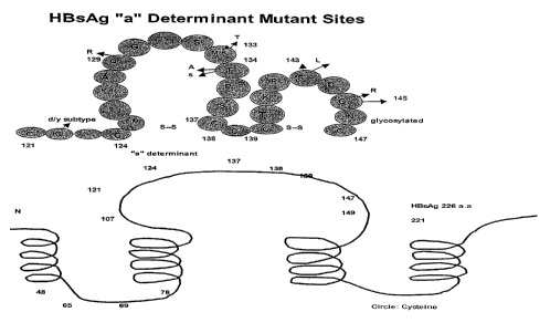
[0029] Figure 1 shows a-schematic of the HBV surface antigen depicting the
highly
conformational structure of the protein (lower panel, solid line) and the
amino acid
sequence (SEQ ID NO:1) around the "a" determinant (from aa 121-147, upper
panel,
in circles). The arrows indicate the position and substitution of various
known HBsAg
variants.
[0030] Figures 2A and 2B (SEQ ID NOS:2 and 3) show the amino acid sequence
for the sAg wild-type adw and ayw antigens, respectively.
[0031] Figures 3A-3D show the immunoreactivities of rabbit monoclonal
antibodies from 99S6 (Figure 3B) and 99S9(Figure 3D), in comparison with the
mouse antibodies mMAbl (Figure 3C) and mMAb2 (Figure 3A) against the HBV
mutant panel described above.
DETAILED DESCRIPTION OF THE INVENTION
[0032] The practice of the present invention will employ, unless otherwise
indicated, conventional methods of virology, chemistry, biochemistry,
recombinant
DNA techniques and immunology, within the skill of the art. Such techniques
are
explained fully in the literature. See, e.g., Fundamental Virology, 3rd
Edition, vol. I
& II (B.N. Fields and D.M. Knipe, eds.); Handbook of Experimental Immunology,
V ols. I-IV (D.M. Weir and C.C. Blackwell eds., Blackwell Scientific
Publications);
T.E. Creighton, Proteins: Structures and Molecular Properties (W.H. Freeman
and
Company, 1993); A.L. Lehninger, Biochemistry (Worth Publishers, Inc., current
addition); Sambrook, et al., Molecular Cloning: A Laboratory Manual (2nd
Edition,
1989); Methods In Enzymology (S. Colowick and N. Kaplan eds., Academic Press,
Inc.).
[0033] The following amino acid abbreviations are used throughout the text:
-8-
CA 02569561 2006-12-05
WO 2006/085918
PCT/US2005/020254
Alanine: Ala (A) Arginine: Arg (R)
Asparagine: Asn (N) Aspartic acid: Asp (D)
Cysteine: Cys (C) Glutamine: Gln (Q)
Glutamic acid: Glu (E) Glycine: Gly (G)
Histidine: His (H) Isoleucine: Ile (I)
Leucine: Leu (L) Lysine: Lys (K)
Methionine: Met (M) Phenylalanine: Phe (F)
Proline: Pro (P) Serine: Ser (S)
Threonine: Thr (T) Tryptophan: Trp (W)
Tyrosine: Tyr (Y) Valine: Val (V)
I. DEFINITIONS
[0034] In describing the present invention, the following terms will be
employed,
and are intended to be defined as indicated below.
[0035] It must be noted that, as used in this specification and the appended
claims,
the singular forms "a", "an" and "the" include plural referents unless the
content
clearly dictates otherwise. Thus, for example, reference to "a rabbit
monoclonal
antibody" includes a mixture of two or more such polypeptides, and the like.
[0036] The terms "polypeptide" and "protein" refer to a polymer of amino acid
residues and are not limited to a minimum length of the product. Thus,
peptides,
oligopeptides, dimers, multimers, and the like, are included within the
definition.
Both full-length proteins and fragments thereof are encompassed by the
definition.
The terms also include postexpression modifications of the polypeptide, for
example,
glycosylation, acetylation, phosphorylation and the like. Furthermore, for
purposes of
the present invention, a "polypeptide" refers to a protein which includes
modifications, such as deletions, additions and substitutions (generally
conservative in
nature), to the native sequence, so long as the protein maintains the desired
activity.
These modifications may be deliberate, as through site-directed mutagenesis,
or may
be accidental, such as through mutations of hosts which produce the proteins
or errors
due to PCR amplification.
[0037] The term "antigen" refers to a polypeptide, whether native, recombinant
or
synthetic, which includes one or more epitopes that recognize an antibody. The
-9-
CA 02569561 2006-12-05
WO 2006/085918
PCT/US2005/020254
antigen in question need not include the full-length amino acid sequence of
the
reference molecule but can include only so much of the molecule as necessary
in
order to generate an immunological reaction (i.e., when the antigen is used
for
generating antibodies) or to react with the HBV antibody of interest (i.e.,
where the
antigen is being detected in an assay). Thus, only one or few epitopes of the
reference
molecule need be present. Furthermore, the antigen may comprise a fusion
protein
between the full-length reference molecule or a fragment of the reference
molecule,
and another protein such as another HBV antigen and/or a protein that does not
disrupt the reactivity of the HBV antigen. It is readily apparent that the
antigen may
therefore comprise the full-length sequence, fragments, truncated and partial
sequences, as well as analogs, muteins and precursor forms of the reference
molecule.
The term also intends deletions, additions and substitutions to the reference
sequence,
so long as the antigen retains the ability to stimulate antibody production
and/or to
react with HBV antibodies.
[0038] In this regard, natural variation will occur from isolate to isolate
within a
particular HBV strain. Thus, the term is intended to encompass such variation
and, in
particular, an antigen that varies in its amino acid composition by not more
than about
20 number percent, more preferably by not more than about 10 to 15 number
percent,
and most preferably, by not more than about 5 number percent, from the
reference
antigen. Proteins having substantially the same amino acid sequence as the
reference
molecule, but possessing minor amino acid substitutions that do not
substantially
affect the antibody binding capabilities of the antigen, are therefore within
the
definition of the reference polypeptide.
[0039] An antigen "derived from" an HBV strain or isolate intends an antigen
which comprises a sequence of one or more regions or portions of regions of an
antigen encoded by the reference HBV genome. Typically, the antigen is
composed
of regions or portions of regions that include epitopes, and will generally
have an
amino acid sequence substantially homologous to the reference polypeptide, as
defined below. Thus, the term "derived from" is used to identify the original
source
of a molecule but is not meant to limit the method by which the molecule is
made
which can be, for example, by chemical synthesis or recombinant means.
[0040] The terms "analog" and "mutein" refer to biologically active
derivatives of
the reference molecule, that retain desired activity, such as immunoreactivity
in
-10-
CA 02569561 2006-12-05
WO 2006/085918
PCT/US2005/020254
assays described herein. In general, the term "analog" refers to compounds
having a
native polypeptide sequence and structure with one or more amino acid
additions,
substitutions (generally conservative in nature) and/or deletions, relative to
the native
molecule, so long as the modifications do not destroy immunogenic activity and
which are "substantially homologous" to the reference molecule as defined
below. A
number of conserved and variable regions are known between the various
isolates
and, in general, the amino acid sequences of epitopes derived from these
regions will
have a high degree of sequence homology, e.g., amino acid sequence homology of
more than 50%, generally more than 60%-70%, when the two sequences are
aligned.
The term "mutein" refers to peptides having one or more peptide mimics (e.g.,
"peptoids"). Preferably, the analog or mutein has at least the same
immunoreactivity
as the native molecule. Methods for making polypeptide analogs and muteins are
known in the art and are described further below.
[0041] The terms "analog" and "mutein" also encompass purposeful mutations
that
are made to the reference molecule. Particularly preferred analogs include
substitutions that are conservative in nature, i.e., those substitutions that
take place
within a family of amino acids that are related in their side chains.
Specifically,
amino acids are generally divided into four families: (1) acidic -- aspartate
and
glutamate; (2) basic -- lysine, arginine, histidine; (3) non-polar -- alanine,
valine,
leucine, isoleucine, proline, phenylalanine, methionine, tryptophan; and (4)
uncharged
polar -- glycine, asparagine, glutamine, cysteine, serine threonine, tyrosine.
Phenylalanine, tryptophan, and tyrosine are sometimes classified as aromatic
amino
acids. For example, it is reasonably predictable that an isolated replacement
of
leucine with isoleucine or valine, an aspartate with a glutamate, a threonine
with a
serine, or a similar conservative replacement of an amino acid with a
structurally
related amino acid, will not have a major effect on the biological activity.
For
example, the antigen of interest may include up to about 5-10 conservative or
non-conservative amino acid substitutions, or even up to about 15-25, 50 or 75
conservative or non-conservative amino acid substitutions, or any integer
between
5-75, so long as the desired function of the molecule remains intact. One of
skill in
the art can readily determine regions of the molecule of interest that can
tolerate
change by reference to Hopp/Woods and Kyte-Doolittle plots, well known in the
art.
-11-
CA 02569561 2006-12-05
WO 2006/085918
PCT/US2005/020254
[0042] By "antigen fragment" is intended an antigen consisting of only a part
of the
intact full-length antigen polypeptide sequence and structure. The fragment
can
include a C-terminal deletion, an N-terminal deletion, and/or an internal
deletion of
the native polypeptide. By "immunogenic fragment" is meant a fragment of a
polypeptide that includes one or more epitopes and thus elicits one or more of
the
immunological responses described herein. An "immunogenic fragment" of a
particular HBV protein will generally include at least about 5-10 contiguous
amino
acid residues of the full-length molecule, preferably at least about 15-25
contiguous
amino acid residues of the full-length molecule, and most preferably at least
about
20-50 or more contiguous amino acid residues of the full-length molecule, that
define
an epitope, or any integer between 5 amino acids and the full-length sequence,
provided that the fragment in question retains the ability to elicit an
immunological
response as defined herein.
[0043] By "HBsAg" is meant an HBV surface antigen derived from any of the
various HBV strains and isolates. The term intends surface antigens which
include a
substantially complete S domain of an HBsAg polypeptide (termed "sAg" herein),
as
well as immunogenic fragments thereof. An S domain of HBsAg is "substantially
complete" if it contains the native sequence of the polypeptide with or
without minor
deletions of one or a few amino acids from either the N-terminal or C-terminal
regions or within the polypeptide. For example the HBsAg S domain can be
truncated by a few amino acids, i.e., up to about 3, 5, 7, or 10 amino acids,
without
greatly affecting its antigenicity. An HBsAg antigen for use herein will
generally
include a region corresponding to the "a" determinant, found at amino acid
positions
124-147, numbered relative to the sAg. This region is described further below.
The
term also intends an antigen that includes the preS2 (formerly called preS)
domain in
addition to the S domain, or both the preS2 and preS1 domains of HBsAg, in
addition
to the S domain. Valenzuela, et al. (1982) Nature 298:347-350, describes the
gene for
a representative HBsAg. See, also, Valenzuela, et al. (1979) Nature 280:815-
819.
[0044] A "mutant" HBsAg molecule, as used herein, refers to analogs of wild-
type
HBsAgs, as defined above. For the purpose of this invention, by "wild-type
HBsAgs"
is meant HBsAgs from the ayw and adw subtypes. These analogs may arise by
natural mutational events, e.g., in the case of escape mutants, or may be
purposefully
created. Representative mutant HBsAg sequences are shown in Figure 1 herein.
-12-
CA 02569561 2006-12-05
WO 2006/085918
PCT/US2005/020254
Additional naturally occurring mutants are known in the art and the nucleotide
sequences and corresponding amino acid sequences for surface antigens from
these
mutants have been deposited with GenBank. See, e.g., NCBI accession numbers
AY341335 (naturally occurring surface mutant with multiple mutations in the
"a"
determinant of sAg), X59795 (naturally occurring mutant from the ayw subtype);
AF01360 and AF013629 (naturally occurring mutants from the adw subtype) and
Zuckerman et al. 1999 (J. Med Virol. 58:193).
[0045] By "immunogenic" sequence of an HBsAg is meant an HBsAg molecule
that includes an amino acid sequence with at least one epitope such that the
molecule
is capable of stimulating the production of antibodies in an appropriate host.
By
"epitope" is meant a site on an antigen to which specific B cells and/or T
cells
respond, rendering the HBV epitope in question capable of stimulating antibody
production. The term is also used interchangeably with "antigenic determinant"
or
"antigenic determinant site." An epitope can comprise 3 or more amino acids in
a
spatial conformation unique to the epitope. Generally, an epitope consists of
at least 5
such amino acids and, more usually, consists of at least 8-10 such amino acids
or
more.
[0046] Regions of a given polypeptide that include an epitope can be
identified
using any number of epitope mapping techniques, well known in the art. See,
e.g.,
Epitope Mapping Protocols in Methods in Molecular Biology, Vol. 66 (Glenn E.
Morris, Ed., 1996) Humana Press, Totowa, New Jersey. For example, linear
epitopes
may be determined by e.g., concurrently synthesizing large numbers of peptides
on
solid supports, the peptides corresponding to portions of the protein
molecule, and
reacting the peptides with antibodies while the peptides are still attached to
the
supports. Such techniques are known in the art and described in, e.g., U.S.
Patent No.
4,708,871; Geysen et al. (1984) Proc. Natl. Acad. Sci. USA 81:3998-4002;
Geysen
et al. (1985) Proc. Natl. Acad. Sci. USA 82:178-182; Geysen et al. (1986)
Molec.
Immunol. 23:709-715. Similarly, conformational epitopes are readily identified
by
determining spatial conformation of amino acids such as by, e.g., x-ray
crystallography and 2-dimensional nuclear magnetic resonance. See, e.g.,
Epitope
Mapping Protocols, supra. Antigenic regions of proteins can also be identified
using
standard antigenicity and hydropathy plots, such as those calculated using,
e.g., the
Omiga version 1.0 software program available from the Oxford Molecular Group.
-13-
CA 02569561 2006-12-05
WO 2006/085918
PCT/US2005/020254
This computer program employs the Hopp/Woods method, Hopp et al., Proc. Natl.
Acad. Sci USA (1981) 78:3824-3828 for determining antigenicity profiles, and
the
Kyte-Doolittle technique, Kyte et al., 1 MoL Biol. (1982) 157:105-132 for
hydropathy
plots.
[0047] An "immunogenic composition" is a composition that comprises at least
one
immunogenic polypeptide (e.g., an HBsAg antigen or antibody).
[0048] "Substantially purified" generally refers to isolation of a substance
(compound, polynucleotide, protein, polypeptide, polypeptide composition) such
that
the substance comprises the majority percent of the sample in which it
resides.
Typically in a sample a substantially purified component comprises 50%,
preferably
80%-85%, more preferably 90-95% of the sample. Techniques for purifying
polynucleotides and polypeptides of interest are well-known in the art and
include, for
example, ion-exchange chromatography, affinity chromatography and
sedimentation
according to density.
[0049] By "isolated" is meant, when referring to a polypeptide, that the
indicated
molecule is separate and discrete from the whole organism with which the
molecule is
found in nature or is present in the substantial absence of other biological
macro-molecules of the same type. The term "isolated" with respect to a
polynucleotide is a nucleic acid molecule devoid, in whole or part, of
sequences
normally associated with it in nature; or a sequence, as it exists in nature,
but having
heterologous sequences in association therewith; or a molecule disassociated
from the
chromosome.
[0050] By "equivalent antigenic determinant" is meant an antigenic determinant
from different isolates or strains of HBV which antigenic determinants are not
necessarily identical due to sequence variation, but which occur in equivalent
positions in the HBV sequence in question. In general the amino acid sequences
of
equivalent antigenic determinants will have a high degree of sequence
homology, e.g.,
amino acid sequence homology of more than 30%, usually more than 40%, such as
more than 60%, and even more than 80-90% homology, when the two sequences are
aligned.
[0051] "Homology" refers to the percent identity between two polynucleotide or
two polypeptide moieties. Two nucleic acid, or two polypeptide sequences are
"substantially homologous" to each other when the sequences exhibit at least
about
-14-
CA 02569561 2006-12-05
WO 2006/085918
PCT/US2005/020254
50% , preferably at least about 75%, more preferably at least about 80%-85%,
preferably at least about 90%, and most preferably at least about 95%-98%
sequence
identity over a defined length of the molecules. As used herein, substantially
homologous also refers to sequences showing complete identity to the specified
sequence.
[0052] In general, "identity" refers to an exact nucleotide-to-nucleotide or
amino
acid-to-amino acid correspondence of two polynucleotides or polypeptide
sequences,
respectively. Percent identity can be determined by a direct comparison of the
sequence information between two molecules (the reference sequence and a
sequence
with unknown % identity to the reference sequence) by aligning the sequences,
counting the exact number of matches between the two aligned sequences,
dividing
by the length of the reference sequence, and multiplying the result by 100.
Readily
available computer programs can be used to aid in the analysis, such as ALIGN,
Dayhoff, M.O. in Atlas of Protein Sequence and Structure M.O. Dayhoff ed., 5
Suppl.
3:353-358, National biomedical Research Foundation, Washington, DC, which
adapts
the local homology algorithm of Smith and Waterman Advances in Appl. Math.
2:482-489, 1981 for peptide analysis. Programs for determining nucleotide
sequence
identity are available in the Wisconsin Sequence Analysis Package, Version 8
(available from Genetics Computer Group, Madison, WI) for example, the
BESTFIT,
FASTA and GAP programs, which also rely on the Smith and Waterman algorithm.
These programs are readily utilized with the default parameters recommended by
the
manufacturer and described in the Wisconsin Sequence Analysis Package referred
to
above. For example, percent identity of a particular nucleotide sequence to a
reference sequence can be determined using the homology algorithm of Smith and
Waterman with a default scoring table and a gap penalty of six nucleotide
positions.
[0053] Another method of establishing percent identity in the context of the
present
invention is to use the MPSRCH package of programs copyrighted by the
University
of Edinburgh, developed by John F. Collins and Shane S. Sturrok, and
distributed by
IntelliGenetics, Inc. (Mountain View, CA). From this suite of packages the
Smith-Waterman algorithm can be employed where default parameters are used for
the scoring table (for example, gap open penalty of 12, gap extension penalty
of one,
and a gap of six). From the data generated the "Match" value reflects
"sequence
identity." Other suitable programs for calculating the percent identity or
similarity
-15-
CA 02569561 2006-12-05
WO 2006/085918
PCT/US2005/020254
between sequences are generally known in the art, for example, another
alignment
program is BLAST, used with default parameters. For example, BLASTN and
BLASTP can be used using the following default parameters: genetic code =
standard;
filter = none; strand = both; cutoff= 60; expect = 10; Matrix = BLOSUM62;
Descriptions = 50 sequences; sort by = HIGH SCORE; Databases = non-redundant,
GenBank + EMBL + DDBJ + PDB + GenBank CDS translations + Swiss protein +
Spupdate + PlR. Details of these programs are readily available.
[0054] Alternatively, homology can be determined by hybridization of
polynucleotides under conditions which form stable duplexes between homologous
regions, followed by digestion with single-stranded-specific nuclease(s), and
size
determination of the digested fragments. DNA sequences that are substantially
homologous can be identified in a Southern hybridization experiment under, for
example, stringent conditions, as defined for that particular system. Defining
appropriate hybridization conditions is within the skill of the art. See,
e.g., Sambrook
et al., supra; DNA Cloning, supra; Nucleic Acid Hybridization, supra.
[0055] "Recombinant" as used herein to describe a nucleic acid molecule means
a
polynucleotide of genomic, cDNA, viral, semisynthetic, or synthetic origin
which, by
virtue of its origin or manipulation is not associated with all or a portion
of the
polynucleotide with which it is associated in nature. The term "recombinant"
as used
with respect to a protein or polypeptide means a polypeptide produced by
expression
of a recombinant polynucleotide. In general, the gene of interest is cloned
and then
expressed in transformed organisms, as described further below. The host
organism
expresses the foreign gene to produce the protein under expression conditions.
[0056] An "antibody" intends a molecule that "recognizes," i.e., specifically
binds
to an epitope of interest present in an antigen. By "specifically binds" is
meant that
the antibody interacts with the epitope in a "lock and key" type of
interaction to form
a complex between the antigen and antibody, as opposed to non-specific binding
that
might occur between the antibody and, for instance, components in a mixture
that
includes the test substance with which the antibody is reacted. Thus, an anti-
HBV
antibody is a molecule that specifically binds to an epitope of an HBV
protein. The
term "antibody" as used herein includes antibodies obtained from both
polyclonal and
monoclonal preparations, as well as, the following: hybrid (chimeric) antibody
molecules (see, for example, Winter et al., Nature (1991) 349:293-299; and
U.S.
-16-
CA 02569561 2006-12-05
WO 2006/085918
PCT/US2005/020254
Patent No. 4,816,567); F(ab')2 and F(ab) fragments; Fv molecules (non-covalent
heterodimers, see, for example, Inbar et al., Proc Natl Acad Sci USA (1972)
69:2659-2662; and Ehrlich et al., Biochem (1980) 19:4091-4096); single-chain
Fv
molecules (sFv) (see, for example, Huston et al., Proc Natl Acad Sci USA
(1988)
85:5879-5883); dimeric and trimeric antibody fragment constructs; minibodies
(see,
e.g., Pack et al., Biochem (1992) 31:1579-1584; Cumber et al., J Immunology
(1992)
149B:120-126); humanized antibody molecules (see, for example, Riechmann et
al.,
Nature (1988) 332:323-327; Verhoeyan et al., Science (1988) 239:1534-1536; and
U.K. Patent Publication No. GB 2,276,169, published 21 September 1994); and,
any
functional fragments obtained from such molecules, wherein such fragments
retain
immunological binding properties of the parent antibody molecule.
[0057] As used herein, the term "monoclonal antibody" refers to an antibody
composition having a homogeneous antibody population. The term is not limited
regarding the species or source of the antibody, nor is it intended to be
limited by the
manner in which it is made. The term encompasses whole immunoglobulins as well
as fragments such as Fab, F(ab1)2, Fv, and other fragments, as well as
chimeric and
humanized homogeneous antibody populations, that exhibit immunological binding
properties of the parent monoclonal antibody molecule.
[0058] As used herein, the term "rabbit monoclonal antibody" refers to a
monoclonal antibody, as defined above, produced by immunizing a rabbit with an
antigen of interest (e.g., a mutant HBsAg). A "rabbit monoclonal antibody" can
be
produced using rabbit-rabbit hybridomas (i.e., fusions between an antibody-
producing
cell from the immunized rabbit with an immortalized cell from a rabbit),
rabbit-mouse
hybridomas (i.e., fusions between an antibody-producing cell from the
immunized
rabbit with an immortalized cell from a mouse), and the like, described more
fully
below.
[0059] A "mouse monoclonal antibody" refers to a monoclonal antibody, as
defined above, produced by immunizing a mouse, with an antigen of interest
(e.g., a
mutant HBsAg). A "mouse monoclonal antibody" is produced using conventional
methods well known in the art, from mouse-mouse hybridomas, described more
fully
below.
[0060] As used herein, a "solid support" refers to a solid surface to which a
macromolecule, e.g., an antibody, protein, polypeptide, peptide,
polynucleotide can be
-17-
CA 02569561 2006-12-05
WO 2006/085918
PCT/US2005/020254
attached, such as a magnetic bead, latex bead, microtiter plate well, glass
plate, nylon,
agarose, polyacrylamide, silica particle, nitrocellulose membrane, and the
like.
[0061] "Immunologically reactive" means that the antibody in question will
react
specifically with HBV antigens present in a biological sample from an HBV-
infected
individual.
[0062] An "immunoreactive fragment" of an antibody, is a molecule consisting
of
only a portion of the intact antibody sequence and structure, and that is
immunologically reactive as defined above. Non-limiting examples of such
immunoreactive fragments include F(ab')2, Fv, and sFy molecules, that are
capable of
exhibiting immunological binding properties of the parent antibody molecule
from
which they are derived.
[0063] "Immune complex" intends the combination formed when an antibody
binds to an epitope on an antigen.
[0064] As used herein, a "biological sample" refers to a sample of tissue or
fluid
isolated from a subject such as, but not limited to, blood, plasma, platelets,
serum,
fecal matter, urine, bone marrow, bile, spinal fluid, lymph fluid,
cerebrospinal fluid,
samples of the skin, secretions of the skin, respiratory, intestinal, and
genitourinary
tracts, tears, saliva, milk, blood cells, organs, biopsies and also samples of
in vitro cell
culture constituents including but not limited to conditioned media resulting
from the
growth of cells and tissues in culture medium, e.g., recombinant cells, and
cell
components. The samples detailed above need not necessarily be in the form
obtained
directly from the source. For example, the sample can be treated prior to use,
such as,
for example, by heating, centrifuging, etc. prior to analysis.
[0065] As used herein, the terms "label" and "detectable label" refer to a
molecule
capable of detection, including, but not limited to, radioactive isotopes,
fluorescers,
semiconductor nanocrystals, chemiluminescers, chromophores, enzymes, enzyme
substrates, enzyme cofactors, enzyme inhibitors, dyes, metal ions, metal sols,
ligands
(e.g., biotin, streptavidin or haptens) and the like. The term "fluorescer"
refers to a
substance or a portion thereof which is capable of exhibiting fluorescence in
the
detectable range. Particular examples of labels which may be used under the
invention include, but are not limited to, horse radish peroxidase (HRP),
fluorescein,
FITC, rhodamine, dansyl, umbelliferone, dimethyl acridinium ester (DMAE),
Texas
red, luminol, NADPH and a-P-galactosidase.
-18-
CA 02569561 2006-12-05
WO 2006/085918
PCT/US2005/020254
ILMODES OF CARRYING OUT THE INVENTION
[0066] Before describing the present invention in detail, it is to be
understood that
this invention is not limited to particular formulations or process parameters
as such
may, of course, vary. It is also to be understood that the terminology used
herein is
for the purpose of describing particular embodiments of the invention only,
and is not
intended to be limiting.
[0067] Although a number of methods and materials similar or equivalent to
those
described herein can be used in the practice of the present invention, the
preferred
materials and methods are described herein.
[0068] The present invention is based on the discovery that novel rabbit
monoclonal antibodies, directed against mutant HBsAgs, are far more
immunoreactive in assays for detecting HBV infection than conventional mouse
monoclonal antibodies. The rabbit monoclonal antibodies of the present
invention are
reactive with a broader range of HBsAg mutants than conventional mouse
monoclonal antibodies. Moreover, the rabbit monoclonal antibodies of the
present
invention are typically also reactive with wild-type HBsAgs. Indeed, a single
rabbit
monoclonal antibody according to the present invention is as effective as the
use of
multiple mouse monoclonal antibodies for detecting the presence of HBV
antigens,
which can be indicative of HBV infection. Thus, the rabbit monoclonal
antibodies of
the present invention decrease the number of false negatives obtained with
assays
using, e.g., mouse monoclonal antibodies and are therefore useful in
diagnostic
methods for accurately detecting HBV infection. The assays of the present
invention
can also utilize additional antibodies, such as additional mouse monoclonal
antibodies, to provide the ability to diagnose HBV infection from a wide
variety of
isolates and escape mutants.
[0069] The methods are useful for detecting HBV infection in humans, as well
as
for detecting HBV infection in blood samples, including without limitation, in
whole
blood, serum, platelets, and plasma, as well as in tissues and organs for
transplantation, in particular by detecting the presence of HBV antigens or
HBV
antibodies. Thus, the methods can be used to diagnose HBV infection in a
subject,
such as a human subject, as well as to detect HBV contamination in donated
blood
samples. Aliquots from individual donated samples or pooled samples can be
-19-
CA 02569561 2006-12-05
WO 2006/085918
PCT/US2005/020254
screened for the presence of HBV and those samples or pooled samples
contaminated
with HBV can be eliminated before they are combined. In this way, a blood
supply
substantially free of HBV contamination can be provided. Similarly, samples
from
tissues and organs to be used in transplantation can also be screened in order
to
eliminate contaminated specimens.
[0070] In order to further an understanding of the invention, a more detailed
discussion is provided below regarding HBV antigens, antibodies and diagnostic
methods for use with the subject invention.
HBV Surface Antigens
[0071] The hepatitis B surface antigens are made up of three size classes of
proteins that share carboxy-terminal sequences. These proteins include large
(L, the
preS2 domain), medium (M, the preS1 domain), and small (S, the sAg domain).
All
three proteins are found in infectious virions (often referred to as Dane
particles)
recovered as 42 nm spheres from the serum of infected patients. Serum samples
also
contain empty spherical particles averaging 22 nm, which contain primarily the
S
class of proteins (sAg). Mammalian cell lines transfected exclusively with DNA
encoding the sAg protein release 20 nm empty spheres similar to those from
infected
cells. Moreover, yeast cells transformed with the same gene form analogous
spheres,
which are found to be equally immunogenic as the 22 nm spheres from infected
cells.
See, e.g., "HBV Vaccines - from the laboratory to license: a case study" in
Mackett,
M. and Williamson, J.D., Human Vaccines and Vaccination, pp. 159-176, for a
discussion of HBV structure; and U.S. Patent Nos. 4,722,840, 5,098,704,
5,324,513,
5,965,140, Beames et al., J. ViroL (1995) 69:6833-6838, Birnbaum et al., 1
ViroL
(1990) 64:3319-3330, Zhou et al., J. ViroL (1991) 65:5457-5464, for
descriptions of
the recombinant production of various HBV particles.
[0072] Thus, as explained above, HBsAgs for use in producing the rabbit
monoclonal antibodies of the present invention can include immunogenic regions
of
sAg, preS1 and/or preS2, as well as immunogenic regions from any combination
of
the above, such as sAg/preS1, sAg/preS2, and sAg/preS1/preS2. Optionally, an
HBsAg polypeptide can comprise more than one sAg, preS1, or preS2 polypeptide.
Additionally, the sAg, preS1, and preS2 polypeptides may be derived from the
same
or different isolates of HBV. These polypeptides may also be provided as a
fusion
-20-
CA 02569561 2006-12-05
WO 2006/085918
PCT/US2005/020254
protein or as separate polypeptides. The sequences of HBsAgs from hundreds of
different HBV isolates are known and can be readily obtained from the NCBI
database.
[0073] A preferred HBsAg for use in the invention comprises at least the
sequence
of amino acids of the "a" determinant region of HBV (amino acids 124-147,
numbered relative to the sAg). Representative wild-type sequences for this
region are
CTTPAQGNSMFPSCCCTKPSDGNC (SEQ ID NO:4, adw wild-type); and
CMTTAQGTSMYPSCCCTKPSDGNC (SEQ ID NO:5, ayw wild-type). Mutations
in this region of sAg have been found in a large number of HBV vaccine
escapees.
For descriptions of a number of HBsAg variants, see, Ashton-Richardt PG,
Murray
K. (1989) Mutants of the hepatitis B virus surface antigen and define some
antigenically essential residues in the immunodominant "a" region. J. Medical
Virology 29: 196-203; Norder H. Courouce A_M, Magnius L (1992) Molecular basis
of hepatitis B virus serotype variation within the four major subtype. J.
General
Virology 73: 3141-3145; Carman WF, Zanetti AR, et.al.(1990) Vaccine-induced
escape mutant of hepatitis B virus. Lancet 336: 325-329; Fujii H. Moriyama K.
et al.
Gly 145 to Arg substitution in HBs antigen of immune escape mutant of
hepatitis B
virus. Biochem. Biophys Res Comm 184: 1152-1157; Carman,W. Vaccine-
associated mutants of hepatitis B virus. Viral Hepatitis and Liver Disease
(1945) pp:
243-247, Eds: K. Nishioka, H. Suzuki, S. Mishiro T. Oda)
[0074] Thus, HBsAgs including mutations in this region are particularly useful
herein. Representative mutants for this region include F134A, F134S, G145R,
S143L, P142S and Q129R/M133T. In each of the mutant designations, the number
indicates the position of the substituted amino acid, the letter before the
number
indicates the amino acid at that position in the WT sequence and the letter
following
the number indicates the amino acid at that position in the mutant. These
mutants are
merely representative and it is to be understood that a large number of
additional
naturally occurring mutants exist, which mutants will find use with the
present
invention. Additionally, synthetic mutants with mutations in the "a"
determinant
region will also find use herein. Variants having mutations in regions other
than the
"a" determinant region, as defined above, may also find use in the present
invention.
For example, the variant having a substitution of Q for P at amino acid
position 120
(P120Q), finds use as antigen for generating monoclonal antibodies.
-21-
CA 02569561 2006-12-05
WO 2006/085918
PCT/US2005/020254
[0075] Antigens for use with the present invention can be obtained using
standard
techniques. The HBV antigens are conveniently generated using recombinant
methods, well known in the art. See, e.g., U.S. Patent Nos. 4,722,840,
5098,704,
5324,513, 5,965,140 and 6,306,625, for descriptions of the recombinant
production of
HBV antigens. For example, the HBsAg S protein coding sequence can be isolated
by phenol extraction of DNA from Dane particles present in infected human
serum,
using methods known in the art, such as described in U.S. Patent No.
4,710,463. The
isolated DNA can then be digested with a restriction endonuclease. The choice
of
endonuclease will depend, in part, on the particular Dane particles. For
example, the
HBsAg coding sequence of HBV DNA of certain Dane particles of the adw serotype
can be isolated as a single BamHI fragment; the HBsAg coding sequence of HBV
DNA of certain Dane particles of the ayw serotype can be isolated as a Hhal
fragment. HBV DNA of Dane particles of the same serotype may also exhibit
different patterns of restriction sites.
[0076] Oligonucleotide probes can be devised based on the known sequences of
the
HBV genome and used to probe genomic or cDNA libraries for HBV genes encoding
for the antigens useful in the present invention. The genes can then be
further isolated
using standard techniques and, if desired, restriction enzymes employed to
mutate the
gene at desired portions of the full-length sequence. See, e.g., Sambrook et
al., supra,
for a description of techniques used to obtain and isolate DNA.
[0077] Finally, the genes encoding the HBV antigens can be produced
synthetically, based on the known sequences. The nucleotide sequence can be
designed with the appropriate codons for the particular amino acid sequence
desired.
In general, one will select preferred codons for the intended host in which
the
sequence will be expressed. The complete sequence is generally assembled from
overlapping oligonucleotides prepared by standard methods and assembled into a
complete coding sequence. See, e.g., Edge, Nature (1981) 292:756; Nambair et
al.,
Science (1984) 223:1299; Jay et al., 1 Biol. Chem. (1984) 259:6311.
[0078] Polynucleotides can comprise coding sequences for the various
polypeptides which occur naturally or can include artificial sequences which
do not
occur in nature. These polynucleotides can be ligated to form a coding
sequence for a
fusion protein, if desired, using standard molecular biology techniques.
-22-
CA 02569561 2006-12-05
WO 2006/085918
PCT/US2005/020254
[0079] Once coding sequences have been prepared or isolated, such sequences
can
be cloned into any suitable vector or replicon. Numerous cloning vectors are
known
to those of skill in the art, and the selection of an appropriate cloning
vector is a
matter of choice. Suitable vectors include, but are not limited to, plasmids,
phages,
transposons, cosmids, chromosomes or viruses which are capable of replication
when
associated with the proper control elements. The coding sequence is then
placed under
the control of suitable control elements, depending on the system to be used
for
expression. Thus, the coding sequence can be placed under the control of a
promoter,
ribosome binding site (for bacterial expression) and, optionally, an operator,
so that
the DNA sequence of interest is transcribed into RNA by a suitable
transformant. The
coding sequence may or may not contain a signal peptide or leader sequence
which
can later be removed by the host in post-translational processing. See, e.g.,
U.S.
Patent Nos. 4,431,739; 4,425,437; 4,338,397.
[0080] If present, the signal sequence can be the native leader found in
association
with the IIBV antigen of interest. Alternatively, a heterologous signal
sequence can
be present which can increase the efficiency of secretion. A number of
representative
leader sequences are known in the art and include, without limitation, the
yeast a-
factor leader, the TPA signal peptide, the Ig signal peptide, and the like.
Sequences
for these and other leader sequences are well known in the art.
[0081] In addition to control sequences, it may be desirable to add regulatory
sequences which allow for regulation of the expression of the sequences
relative to
the growth of the host cell. Regulatory sequences are known to those of skill
in the
art, and examples include those which cause the expression of a gene to be
turned on
or off in response to a chemical or physical stimulus, including the presence
of a
regulatory compound. Other types of regulatory elements may also be present in
the
vector. For example, enhancer elements may be used herein to increase
expression
levels of the constructs. Examples include the SV40 early gene enhancer
(Dijkema et
al. (1985) EMBO J. 4:761), the enhancer/promoter derived from the long
terminal
repeat (LTR) of the Rous Sarcoma Virus (Gorman et al. (1982) Proc. Natl. Acad.
Sci.
USA 79:6777) and elements derived from human CMV (Boshart et al. (1985) Cell
41:521), such as elements included in the CMV intron A sequence (U.S. Patent
No.
5,688,688). The expression cassette may further include an origin of
replication for
-23-
CA 02569561 2006-12-05
WO 2006/085918
PCT/US2005/020254
autonomous replication in a suitable host cell, one or more selectable
markers, one or
more restriction sites, a potential for high copy number and a strong
promoter.
[0082] An expression vector is constructed so that the particular coding
sequence is
located in the vector with the appropriate regulatory sequences, the
positioning and
orientation of the coding sequence with respect to the control sequences being
such
that the coding sequence is transcribed under the "control" of the control
sequences
(i.e., RNA polymerase which binds to the DNA molecule at the control sequences
transcribes the coding sequence). Modification of the sequences encoding the
molecule of interest may be desirable to achieve this end. For example, in
some cases
it may be necessary to modify the sequence so that it can be attached to the
control
sequences in the appropriate orientation; i.e., to maintain the reading frame.
The
control sequences and other regulatory sequences may be ligated to the coding
sequence prior to insertion into a vector. Alternatively, the coding sequence
can be
cloned directly into an expression vector which already contains the control
sequences
and an appropriate restriction site.
[0083] Any suitable expression vector can be constructed or utilized to
express any
form of HBsAg of the invention. An exemplary vector is pCMVII, a pUC19-based
cloning vector designed for expression in mammalian cells. pCMVII comprises
the
following elements: human CMV rE enhancer/promoter, human CMV intron A, a
human tissue plasminogen activator (tPA) leader, a bovine growth hormone poly
A
terminator (BGHt), a Co1E1 origin of replication, and an Amp R ampicillin
resistance
gene. For example, pCMVII-pS2-sAg can be used for expression of preS2-sAg. In
this vector, the coding sequences for the sAg and preS2 domains of HBsAg have
been
inserted into pCMVII between CMV intron A and BGHt. This vector can also be
modified by, e.g., removing the preS2 domain or adding the coding sequence for
the
preS1 domain. These vectors are provided by way of example and are not
intended to
limit the scope of the invention. The above vectors are described in detail in
U.S.
Patent No. 6,740,323.
[0084] As explained above, it may also be desirable to produce mutants or
analogs
of the polypeptide of interest. Mutants or analogs of HBV polypeptides for use
in the
subject compositions may be prepared by the deletion of a portion of the
sequence
encoding the molecule of interest, by insertion of a sequence, and/or by
substitution of
one or more nucleotides within the sequence. Techniques for modifying
nucleotide
-24-
CA 02569561 2006-12-05
WO 2006/085918
PCT/US2005/020254
sequences, such as site-directed mutagenesis, and the like, are well known to
those
skilled in the art. See, e.g., Sambrook et al., supra; Kunkel, T.A. (1985)
Proc. Natl.
Acad. Sci. USA (1985) 82:448; Geisselsoder et al. (1987) BioTechniques 5:786;
Zoller
and Smith (1983) Methods EnzymoL 100:468; Dalbie-McFarland et al. (1982) Proc.
Natl. Acad. Sci USA 79:6409.
[0085] The molecules can be expressed in a wide variety of systems, including
insect, mammalian, bacterial, viral and yeast expression systems, all well
known in
the art. For example, insect cell expression systems, such as baculovirus
systems, are
known to those of skill in the art and described in, e.g., Summers and Smith,
Texas
Agricultural Experiment Station Bulletin No. 1555 (1987). Materials and
methods for
baculovirus/insect cell expression systems are commercially available in kit
form
from, inter alia, Invitrogen, San Diego CA ("MaxBac" kit). Similarly,
bacterial and
mammalian cell expression systems are well known in the art and described in,
e.g.,
Sambrook et al., supra. Yeast expression systems are also known in the art and
described in, e.g., Yeast Genetic Engineering (Barr et al., eds., 1989)
Butterworths,
London.
[0086] A number of appropriate host cells for use with the above systems are
also
known. For example, mammalian cell lines are known in the art and include
immortalized cell lines available from the American Type Culture Collection
(ATCC), such as, but not limited to, Chinese hamster ovary (CHO) cells, HeLa
cells,
baby hamster kidney (BHK) cells, monkey kidney cells (COS), human embryonic
kidney cells, human hepatocellular carcinoma cells (e.g., Hep G2), Madin-Darby
bovine kidney ("MDBK") cells, as well as others. Similarly, bacterial hosts
such as E.
coli, Bacillus subtilis, and Streptococcus spp., will find use with the
present
expression constructs. Yeast hosts useful in the present invention include
inter alia,
Saccharomyces cerevisiae, Candida albicans, Candida maltosa, Hansenula
polymorpha, Kluyveromyces fragilis, Kluyveromyces lactis, Pichia
guillerimondii,
Pichia pastoris, Schizosaccharomyces pombe and Yarrowia lipolytica. Insect
cells for
use with baculovirus expression vectors include, inter alia, Aedes aegypti,
Autographa californica, Bombyx mori, Drosophila melanogaster, Spodoptera
frugiperda, and Trichoplusia ni.
[0087] Nucleic acid molecules comprising nucleotide sequences of interest can
be
stably integrated into a host cell genome or maintained on a stable episomal
element
-25-
CA 02569561 2006-12-05
WO 2006/085918
PCT/US2005/020254
in a suitable host cell using various gene delivery techniques well known in
the art.
See, e.g., U.S. Patent No. 5,399,346.
[0088] Depending on the expression system and host selected, the molecules are
produced by growing host cells transformed by an expression vector described
above
under conditions whereby the protein is expressed. The expressed protein is
then
isolated from the host cells and purified. If the expression system secretes
the protein
into growth media, the product can be purified directly from the media. If it
is not
secreted, it can be isolated from cell lysates. The selection of the
appropriate growth
conditions and recovery methods are within the skill of the art.
[0089] The HBV antigens can also be synthesized using chemical polymer
syntheses such as solid phase peptide synthesis. Such methods are known to
those
skilled in the art. See, e.g., J. M. Stewart and J. D. Young, Solid Phase
Peptide
Synthesis, 2nd Ed., Pierce Chemical Co., Rockford, IL (1984) and G. Barany and
R.
B. Merrifield, The Peptides: Analysis, Synthesis, Biology, editors E. Gross
and J.
Meienhofer, Vol. 2, Academic Press, New York, (1980), pp. 3-254, for solid
phase
peptide synthesis techniques.
[0090] The HBV antigens, obtained as described above, are then used to produce
rabbit monoclonal antibodies for use in diagnostics.
Anti-HBV Antibodies
[0091] The HBV antigens can be used to produce HBV-specific polyclonal and
monoclonal antibodies for use in diagnostic and detection assays. HBV-specific
polyclonal and monoclonal antibodies specifically bind to HBV antigens. In
particular, the HBV antigens can be used to produce polyclonal antibodies by
administering the HBV antigen to a mammal, such as a mouse, a rat, a rabbit, a
goat,
or a horse. Serum from the immunized animal is collected and the antibodies
are
purified from the plasma by, for example, precipitation with ammonium sulfate,
followed by chromatography, preferably affinity chromatography. Techniques for
producing and processing polyclonal antisera are known in the art.
[0092] Rabbit and mouse monoclonal antibodies directed against HBV-specific
epitopes present in the proteins can also be readily produced. In order to
produce
such monoclonal antibodies, the mammal of interest, such as a rabbit or mouse,
is
immunized, such as by mixing or emulsifying the antigen in saline, preferably
in an
-26-
CA 02569561 2006-12-05
WO 2006/085918
PCT/US2005/020254
adjuvant such as Freund's complete adjuvant ("FCA"), and injecting the mixture
or
emulsion parenterally (generally subcutaneously or intramuscularly). The
animal is
generally boosted 2-6 weeks later with one or more injections of the antigen
in saline,
preferably using Freund's incomplete adjuvant ("FIA"). In one embodiment, the
animal is immunized with one or more HBsAg mutants, preferably a mixture of 2
to 5
different HBsAg mutants is used. Wild-type HBsAgs may also be included in the
immunogen. In a preferred regime, the animal, preferably a rabbit, is
initially
immunized with a wild type HBsAg, and thereafter boosted with one or more
HBsAg
mutants. Particularly useful as immunogen are HBsAg mutants which have been
found to occur naturally, e.g., D3, D2, D1, Y1, Y2, described further below.
Antibodies may also= be generated by in vitro immunization, using methods
known in
the art. See, e.g., James et al., 1 Immunol. Meth. (1987) 100:5-40.
[0093] Polyclonal antisera is then obtained from the immunized animal.
However,
rather than bleeding the animal to extract serum, the spleen (and optionally
several
large lymph nodes) is removed and dissociated into single cells. If desired,
the spleen
cells (splenocytes) may be screened (after removal of nonspecifically adherent
cells)
by applying a cell suspension to a plate or well coated with the antigen. B-
cells,
expressing membrane-bound immunoglobulin specific for the antigen, will bind
to the
plate, and are not rinsed away with the rest of the suspension. Resulting B-
cells, or all
dissociated splenocytes, are then induced to fuse with cells from an
immortalized cell
line (also termed a "fusion partner"), to form hybridomas. Typically, the
fusion
partner includes a property that allows selection of the resulting hybridomas
using
specific media. For example, fusion partners can be
hypoxanthine/aminopterin/thymidine (HAT)-sensitive.
[0094] If rabbit-rabbit hybridomas are desired, the immortalized cell line
will be
from a rabbit. Such rabbit-derived fusion partners are known in the art and
include,
for example, cells of lymphoid origin, such as cells from a rabbit
plasmacytoma as
described in Spieker-Polet et al., Proc. Natl. Acad. Sci. USA (1995) 92:9348-
9352 and
U.S. Patent No. 5,675,063, or the TP-3 fusion partner described in U.S. Patent
No.
4,859,595. If a rabbit-mouse hybridoma or a rat-mouse or mouse-mouse
hybridoma,
or the like, is desired, the mouse fusion partner will be derived from an
immortalized
cell line from a mouse, such as a cell of lymphoid origin, typically from a
mouse
-27-
CA 02569561 2006-12-05
WO 2006/085918
PCT/US2005/020254
myeloma cell line. A number of such cell lines are known in the art and are
available
from the ATCC.
[0095] Fusion is accomplished using techniques well known in the art.
Chemicals
that promote fusion are commonly referred to as fusogens. These agents are
extremely
hydrophilic and facilitate membrane contact. One particularly preferred method
of
cell fusion uses polyethylene glycol (PEG). Another method of cell fusion is
electrofusion. In this method, cells are exposed to a predetermined electrical
discharge that alters the cell membrane potential. Additional methods for cell
fusion
include bridged-fusion methods. In this method, the antigen is biotinylated
and the
fusion partner is avidinylated. When the cells are added together, an antigen-
reactive
B cell-antigen-biotin-avidin-fusion partner bridge is formed. This permits the
specific
fusion of an antigen-reactive cell with an immortalizing cell. The method may
additionally employ chemical or electrical means to facilitate cell fusion.
[0096] Following fusion, the cells are cultured in a selective medium (e.g.,
HAT
medium). In order to enhance antibody secretion, an agent that has secretory
stimulating effects can optionally be used, such as IL-6. See, e.g., Liguori
et al.,
Hybridoma (2001) 20:189-198. The resulting hybridomas can be plated by
limiting
/
dilution, and are assayed for the production of antibodies which bind
specifically to
the immunizing antigen (and which do not bind to unrelated antigens). The
selected
monoclonal antibody-secreting hybridomas are then cultured either in vitro
(e.g., in
tissue culture bottles or hollow fiber reactors), or in vivo (e.g., as ascites
in mice).
For example, hybridomas producing HBV-specific antibodies can be identified
using
RIA or ELISA and isolated by cloning in semi-solid agar or by limiting
dilution.
Clones producing HBV-specific antibodies can isolated by another round of
screening.
[0097] An alternative technique for generating the rabbit monoclonal
antibodies of
the present invention is the selected lymphocyte antibody method (SLAM). This
method involves identifying a single lymphocyte that is producing an antibody
with
the desired specificity or function within a large population of lymphoid
cells. The
genetic information that encodes the specificity of the antibody (i.e., the
immunoglobulin VH and VL DNA) is then rescued and cloned. See, e.g., Babcook
et
al., Proc. Natl. Acad. Sci. USA (1996) 93:7843-7848, for a description of this
method.
-28-
CA 02569561 2006-12-05
WO 2006/085918
PCT/US2005/020254
[0100] For further descriptions of rabbit monoclonal antibodies and methods of
making the same from rabbit-rabbit and rabbit-mouse fusions, see, e.g., U.S.
Patent
Nos. 5,675,063 (rabbit-rabbit); 4,859,595 (rabbit-rabbit); 5,472,868 (rabbit-
mouse);
and 4,977,081 (rabbit-mouse). For a description of the production of
conventional
mouse monoclonal antibodies, see, e,g., Kohler and Milstein, Nature (1975)
256:495-497.
[0101] It may be desirable to provide chimeric antibodies. Chimeric antibodies
composed of human and non-human amino acid sequences may be formed from the
monoclonal antibody molecules described above to reduce their immunogenicity
in
humans (Winter et al. (1991) Nature 349:293; Lobuglio et al. (1989) Proc. Nat.
Acad.
Sci. USA 86:4220; Shaw et al. (1987) J Immunol. 138:4534; and Brown et al.
(1987)
Cancer Res . 47:3577; Riechmann et al. (1988) Nature 332:323; Verhoeyen et al.
(1988) Science 239:1534; and Jones et al. (1986) Nature 321:522; EP
Publication No.
519,596, published 23 December 1992; and U.K. Patent Publication No. GB
2,276,169, published 21 September 1994).
[0102] Antibody molecule fragments, e.g., F(ab')2, Fv, and sFy molecules, that
are
capable of exhibiting immunological binding properties of the parent
monoclonal
antibody molecule can be produced using known techniques. Inbar et al. (1972)
Proc.
Nat. Acad. Sci. USA 69:2659; Hochman et al. (1976) Biochem 15:2706; Ehrlich et
al.
(1980) Biochem 19:4091; Huston et al. (1988) Proc. Nat. Acad. Sci. USA
85(16):5879; and U.S. Patent Nos. 5,091,513 and 5,132,405, to Huston et al.;
and
4,946,778, to Ladner et al.
[0103] In the alternative, a phage-display system can be used to expand
monoclonal
antibody molecule populations in vitro. Saiki, et al. (1986) Nature 324:163;
Scharf et
al. (1986) Science 233:1076; U.S. Patent Nos. 4,683,195 and 4,683,202; Yang et
al.
(1995) J Mol Biol 254:392; Barbas, III et al. (1995) Methods: Comp. Meth
Enzymol
8:94; Barbas, III et al. (1991) Proc Natl Acad Sci USA 88:7978.
[0104] Once generated, the phage display library can be used to improve the
immunological binding affinity of the Fab molecules using known techniques.
See,
e.g., Figini et al. (1994) J. Mol. Biol. 239:68. The coding sequences for the
heavy and
light chain portions of the Fab molecules selected from the phage display
library can
be isolated or synthesized, and cloned into any suitable vector or replicon
for
-29-
CA 02569561 2006-12-05
WO 2006/085918
PCT/US2005/020254
expression. Any suitable expression system can be used, including those
described
above.
[0105] Polynucleotide sequences encoding the rabbit monoclonal antibodies and
immunoreactive fragments thereof, described above, are readily obtained using
standard techniques, well known in the art, such as those techniques described
above
with respect to the HBsAgs.
[0106] Antibodies which are directed against HBV epitopes, are particularly
useful
for detecting the presence of HBV or HBV antigens in a sample, such as a serum
sample from an HBV-infected human. An immunoassay for an HBV antigen may
utilize one antibody or several antibodies either alone or in combination with
HBV
antigens. An immunoassay for an HBV antigen may use, for example, a monoclonal
antibody directed towards an HBV epitope, a combination of monoclonal
antibodies
directed towards epitopes of one HBV polypeptide, monoclonal antibodies
directed
towards epitopes of different HBV polypeptides, polyclonal antibodies directed
towards the same HBV antigen, polyclonal antibodies directed towards different
HBV
antigens, or a combination of monoclonal and polyclonal antibodies. For
example,
both rabbit and mouse monoclonal antibodies can be used in the subject assays.
Immunoassay protocols may be based, for example, upon competition, direct
reaction,
or sandwich type assays using, for example, labeled antibody and are described
further below. The labels may be, for example, fluorescent, chemiluminescent,
or
radioactive.
[0107] The anti-HBV antibodies may further be used to isolate HBV particles or
antigens by immunoaffinity columns. The antibodies can be affixed to a solid
support
by, for example, adsorption or by covalent linkage so that the antibodies
retain their
immunoselective activity. Optionally, spacer groups may be included so that
the
antigen binding site of the antibody remains accessible. The immobilized
antibodies
can then be used to bind HBV particles or antigens from a biological sample,
such as
blood or plasma. The bound HBV particles or antigens are recovered from the
column matrix by, for example, a change in pH.
[0108] Preferred anti-HBV antibodies are those produced by the hybridoma cell
lines 99S9 (CMCC #12336) and 99S6 (CMCC #12337) (ATCC Accession Nos.
PTA-6014 and PTA-6015, respectively) as well as antibody fragments (e.g. Fab,
F(ab')2, Fv, sFv) and chimeric or humanized antibodies derived thereform.
-30-
CA 02569561 2006-12-05
WO 2006/085918
PCT/US2005/020254
HBV Diagnostic Assays
[0109] As explained above, the anti-HBV antibodies produced as described
above,
can be used in assays to identify HBV infection. The anti-HBV antibodies can
be
used as either the capture component and/or the detection component in the
assays, as
described further below. Thus, the presence of HBV in a biological sample can
be
determined by the presence of HBV antigens and/or anti-HBV antibodies as an
indicator of HBV in the sample. The monoclonal antibodies can be used for
detecting
HBV in blood samples, including without limitation, in whole blood, serum,
platelets,
and plasma. The antibodies can be used to detect HBV infection in a subject,
such as
a human subject, as well as to detect HBV contamination in donated blood
samples by
detecting the presence of HBV antigens, particularly HBsAgs, and HBV
antibodies,
depending on the assay used. Thus, aliquots from individual donated samples or
pooled samples can be screened for the presence of HBV and those samples or
pooled
samples contaminated with HBV can be eliminated before they are combined. In
this
way, a blood supply substantially free of HBV contamination can be provided.
By
"substantially free of HBV" is meant that the presence of HBV is not detected
using
the assays described herein. Similarly, the methods of the present invention
can be
used to screen potential tissue and organ samples for transplantation and
contaminated
tissues and organs can be discarded.
[0110] Assays for use herein include Western blots; agglutination tests;
enzyme-labeled and mediated immunoassays, such as ELISAs; biotin/avidin and
biotin-streptavidin type assays; protein A- or protein G-mediated
immunoassays;
radioimmunoassays; immunoelectrophoresis; immunoprecipitation, strip
immunoblot
assays, and the like. The reactions generally include detectable labels such
as
fluorescent, chemiluminescent, radioactive, enzymatic labels or dye molecules,
or
other methods for detecting the formation of a complex between the HBV antigen
present in the sample and antibody or antibodies contacted therewith.
[0111] The aforementioned assays generally involve separation of unbound
antibodies or antigen in a liquid phase from a solid phase support to which
antigen-antibody complexes are bound. Solid supports which can be used in the
practice of the invention include substrates such as nitrocellulose (e.g., in
membrane
or microtiter well form); polyvinylchloride (e.g., sheets or microtiter
wells);
-31-
CA 02569561 2006-12-05
WO 2006/085918
PCT/US2005/020254
polystyrene latex (e.g., beads or microtiter plates); polyvinylidine fluoride;
diazotized
paper; nylon membranes; activated beads, magnetically responsive beads, and
the
like.
[0112] In one aspect of the invention, the anti-HBsAg antibodies, such as the
rabbit
monoclonal antibodies described herein, are used for capture or detection or
both of
HBV antigens, particularly HBsAgs, in a sample. Antibodies to the HBsAgs,
produced as described above, can be used for the capture or detection or both
of HBV
antigens in a sample. By "capture" of an analyte ( here HBV antigens in a
sample) is
meant that the analyte can be separated from other components of the sample by
virtue of the binding of the capture molecule. Typically, the capture molecule
is
associated with a solid support, either directly or indirectly. Typically, the
detection
molecule is associated with a detectable label, either directly or indirectly.
[0113] Typically, a solid support is first reacted with a solid phase
component (e.g.,
one or more of the anti-HBV antibodies) under suitable binding conditions such
that
the component is sufficiently immobilized to the support. Sometimes,
immobilization
to the support can be enhanced by first coupling to a protein with better
binding
properties. Suitable coupling proteins include, but are not limited to,
protein A or
protein G, macromolecules such as serum albumins including bovine serum
albumin
(BSA), keyhole limpet hemocyanin, immunoglobulin molecules, thyroglobulin,
ovalbumin, and other proteins well known to those skilled in the art.
Alternatively, a
streptavidin- or avidin-coated solid support can be used to immobilize a
biotinylated
antibody. Other molecules that can be used to bind the antibody to the support
include polysaccharides, polylactic acids, polyglycolic acids, polymeric amino
acids,
amino acid copolymers, and the like. Such molecules and methods of coupling
these
molecules are well known to those of ordinary skill in the art. See, e.g.,
Brinkley,
M.A. Bioconjugate Chem. (1992) 3:2-13; Hashida et al., J. AppL Biochem. (1984)
6:56-63; and Anjaneyulu and Staros, International 1 of Peptide and Protein
Res.
(1987) 30:117-124.
[0114] After reacting the solid support with the solid phase component, any
non-immobilized solid-phase components are removed from the support by
washing,
and the support-bound component is then contacted with a biological sample
suspected of containing the analyte (e.g., HBV antigens) under suitable
binding
conditions. After washing to remove any non-bound analyte, a secondary binder
-32-
CA 02569561 2006-12-05
WO 2006/085918
PCT/US2005/020254
moiety can be added under suitable binding conditions, wherein the secondary
binder
is capable of associating selectively with the bound ligand. The presence of
the
secondary binder can then be detected using techniques well known in the art.
[0115] More particularly, an ELISA method can be used, wherein the wells of a
microtiter plate are coated, directly or indirectly, with the rabbit anti-HBV
antibodies
according to the present invention. Rabbit anti-HBV antibodies directed
against one
or more HBV mutants as described above can be used. Preferably the rabbit
monoclonal antibodies produced by the hybridoma line 99S9 or 99S6 are used.
Additionally, other anti-HBV antibodies directed against wild-type HBsAgs can
also
be present, as can additional mouse monoclonal antibodies directed against a
wild-
type HBsAg or an HBsAg mutant. A biological sample containing or suspected of
containing HBV antigens is then added to the coated wells. After a period of
incubation sufficient to allow antigen-antibody binding, the plate(s) can be
washed to
remove unbound moieties and a detectably labeled secondary binding molecule
added. The secondary binding molecule is allowed to react with any captured
sample,
the plate washed and the presence of the secondary binding molecule detected
using
methods well known in the art.
[0116] In one particular format, an ELISA antigen sandwich format is used. In
this
case, the solid support is coated with anti-HBV antibodies directed against
one or
more HBV mutants as described above. Anti-HBV antibodies directed against wild-
type HBsAgs can also be present. The sample is then contacted with the support
under conditions that allow HBV antigens, if present, to bind one or more or
the
antibodies to form an antigen/antibody complex. Unbound antigens are removed
and
an enzyme-labeled antibody that reacts with the bound antigen/antibody
complex,
such as a labeled anti-HBsAg antibody, is added. An enzyme substrate is used
to
generate a signal. In this particular embodiment, the anti-HBV antibodies that
are
coated on the solid support can be rabbit monoclonal antibodies of the present
invention, preferably the antibodies produced by hybridoma 99S9 or hybridoma
99S6,
or both. Alternatively, or in addition, the detectably labeled antibody can be
a rabbit
monoclonal antibody of the present invention, preferably the antibodies
produced by
hybridoma 99S9 or hybridoma 99S6, or both.
[0117] In another embodiment, the presence of bound HBV analytes from a
biological sample can be readily detected using a secondary binder comprising
an
-33-
CA 02569561 2006-12-05
WO 2006/085918
PCT/US2005/020254
antibody directed against the antigen ligands. A number of anti-human
immunoglobulin (Ig) molecules are known in the art which can be readily
conjugated
to a detectable enzyme label, such as horseradish peroxidase, alkaline
phosphatase or
urease, using methods known to those of skill in the art. An appropriate
enzyme
substrate is then used to generate a detectable signal. In other related
embodiments,
competitive-type ELISA techniques can be practiced using methods known to
those
skilled in the art.
[0118] The rabbit anti-HBV antibodies of the invention can also be used in an
indirect ELISA, for example, an indirect IgG ELISA, as follows. The antibodies
specific for HBV surface antigens are attached to a solid support. Protein A
or protein
G can be used to immobilize the antibodies on the solid support. The support
is then
contacted with HBsAg under conditions that allow binding to the anti-HBV
antibodies bound to the support to form antibody/antigen complexes. Unbound
antigens are removed and the support is contacted with a sample to be tested
for the
presence of human IgG to HBV under conditions that allow binding of human anti-
HBV IgG, if present, to the antigens in the antibody/antigen complexes. The
presence
of bound anti-HBV IgG can be detected using a detectably labeled anti-human
IgG
antibody. In like manner, the presence of human IgM to HBV can be detected by
using labeled anti-human IgM to bind to the antibody/antigen complexes.
[0119] The rabbit anti-HBV antibodies of the invention can also be used in a
capture ELISA, for example, an IgM capture ELISA, as follows. Anti-human IgM
antibodies (e.g., goat anti-human IgM antibodies) are attached to a solid
support, the
support is contacted with a sample to be tested for the presence of human IgM
to
HBV, under conditions that would allow the binding of the anti-HBV IgM, if
present,
to one or more of the anti-human IgM antibodies attached to the solid support,
to form
antibody/antibody complexes. The HBsAgs (e.g., mutant and/or wild-type) are
added
under conditions that would allow binding to the anti-HBV IgM in the
antibody/antibody complexes forming an antibody/antibody/antigen complex.
Unbound antigens are removed and detectably labeled anti-HBV antibodies,
produced
as described above, are added under conditions that allow binding to the bound
antigens. The presence of IgM to HBV in the sample is determined by the
presence
of the detectably labeled anti-HBV antibodies to the bound anti-human IgM
Ab/human anti-HBV IgM/ antigen complexes attached to the solid support.
-34-
CA 02569561 2006-12-05
WO 2006/085918
PCT/US2005/020254
[0120] While some of the foregoing assay formats are termed "ELISA" (Enzyme
Linked ImmunoSorbant Assay) assays, it will be apparent to one of skill in the
art that
the use of a detectable label other than an "enzyme linked" binding moiety is
possible
and may be desirable in many situations. Other suitable detectable labels are
described herein and are well known in the art.
[0121] Assays can also be conducted in solution, such that the HBV antigens or
antibodies and ligands specific for these molecules form complexes under
precipitating conditions. In one particular embodiment, the molecules can be
attached
to a solid phase particle (e.g., an agarose bead or the like) using coupling
techniques
known in the art, such as by direct chemical or indirect coupling. The coated
particle
is then contacted under suitable binding conditions with a biological sample
suspected
of containing HBV antibodies or antigens. Cross-linking between bound
antibodies
causes the formation of complex aggregates which can be precipitated and
separated
from the sample using washing and/or centrifugation. The reaction mixture can
be
analyzed to determine the presence or absence of complexes using any of a
number of
standard methods, such as those immunodiagnostic methods described above.
[0122] In yet a further embodiment, an immunoaffinity matrix can be provided,
wherein, for example, a polyclonal population of antibodies from a biological
sample
suspected of containing HBV antibodies is immobilized to a substrate. An
initial
affinity purification of the sample can be carried out using immobilized
antigens. The
resultant sample preparation will thus only contain anti-HBV moieties,
avoiding
potential nonspecific binding properties in the affinity support. A number of
methods
of immobilizing immunoglobulins (either intact or in specific fragments) at
high yield
and good retention of antigen binding activity are known in the art. For
example,
protein A or protein G can be used to immobilize immunoglobulin molecules to
the
solid support. Once the immunoglobulin molecules have been immobilized to
provide an immunoaffinity matrix, HBV antigens, such as HBsAgs, are contacted
with the bound antibodies under suitable binding conditions. After any
non-specifically bound HBV antigen has been washed from the immunoaffinity
support, the presence of bound antigen can be determined by assaying for label
using
methods known in the art. For example, an enzymatically labeled antibody that
reacts
with the bound antigen/antibody complex, such as a labeled anti-HBsAg
antibody,
-35-
CA 02569561 2006-12-05
WO 2006/085918
PCT/US2005/020254
produced as described above, is added. An enzyme substrate is used to generate
a
signal.
[0123] In another embodiment of the invention, a strip immunoblot assay (SIA)
is
used to detect HBV antigens in a biological sample. For example, one or more
of the
rabbit monoclonal antibodies described above, and optionally mouse monoclonal
antibodies directed against an HBsAg, can be immobilized on the test strip as
capture
reagents. SIA techniques are well known in the art and combine traditional
western
and dot blotting techniques, e.g., the RIBA (Chiron Corp., Emeryville, CA)
SIA. In
these assays, the antibodies are immobilized as individual, discrete portions,
e.g., as
bands or dots, on a membranous support, or may be immobilized as a mixture in
a
single portion. Thus, by "discretely immobilized" on a membrane support is
meant
that the antibodies are present as separate components and not mixed, such
that
reactivity or lack thereof with each of the capture reagents present can be
assessed.
A biological sample suspected of containing HBV antigens is then reacted with
the
test membrane. Visualization of reactivity in the biological sample can be
accomplished using anti-HBV antibody enzyme-conjugates in conjunction with a
colorimetric enzyme substrate. Alternatively, the rabbit monoclonal antibodies
described above can be used for visualization of the bound antibody-antigen
complexes. The test strip for this alternative embodiment may be prepared
using, e.g.,
mouse monoclonal antibodies directed against HBsAg. The assay can be performed
manually or used in an automated format.
[0124] Solid supports which can be used in the practice of the strip
immunoblot
assays include, but are not limited to, membrane supports derived from a
number of
primary polymers including cellulose, polyamide (nylon), polyacrylonitrile,
polyvinylidene difluoride, polysulfone, polypropylene, polyester, polyethylene
and
composite resins consisting of combinations or derivatives of the above.
Particularly
preferred are supports derived from cellulose, such as nitrocellulose
membranes, as
well as nylon membranes. The substrate generally includes the desired membrane
with an inert plastic backing as a support.
[0125] The above-described assay reagents, including the rabbit monoclonal
antibodies and/or the HBsAgs described herein, the solid supports with bound
reagents, as well as other detection reagents, can be provided in kits, with
suitable
instructions and other necessary reagents, in order to conduct the assays as
described
-36-
CA 02569561 2006-12-05
WO 2006/085918
PCT/US2005/020254
above. The kit may also include control formulations (positive and/or
negative),
labeled reagents when the assay format requires same and signal generating
reagents
(e.g., enzyme substrate) if the label does not generate a signal directly.
Instructions
(e.g., written, tape, VCR, CD-ROM, etc.) for carrying out the assay usually
will be
included in the kit. The kit can also contain, depending on the particular
assay used,
other packaged reagents and materials (i.e. wash buffers and the like).
Standard
assays, such as those described above, can be conducted using these kits.
III. Experimental
[0126] Below are examples of specific embodiments for carrying out the present
invention. The examples are offered for illustrative purposes only, and are
not
intended to limit the scope of the present invention in any way.
[0127] Efforts have been made to ensure accuracy with respect to numbers used
(e.g., amounts, temperatures, etc.), but some experimental error and deviation
should,
of course, be allowed for.
Example 1
Preparation of Rabbit Monoclonal Antibodies
[0128] To produce monoclonal antibodies that confer sufficient
immunoreactivities
to the various mutants of interest as well as to the wild-type HBsAg, three
white
female adult New Zealand rabbits were immunized with wild-type HBsAg (adw) and
(ayw) antigens (shown in Figures 2A and 2B, respectively), followed by boosts
every
two weeks with a cocktail of the wild-type antigens and five major HBV
recombinant
mutant antigens, designated D1 (having an A for F substitution at amino acid
position
134), D2 (having an S for F substitution at amino acid position 134), D3
(having an R
for G substitution at amino acid 145), Y1 (having an L for S substitution at
amino
acid 143, and Y2 (having an R for Q substitution at amino acid 129 and a T for
M
substitution at amino acid 133).
[0129] The immune B cells from the spleen of the most immunoreactive rabbit
were fused to rabbit plasmacytoma cells to produce hybridomas, essentially as
described in Spieker-Polet et al., Proc. Natl. Acad. Sci. USA (1995) 92:9348-
9352.
Briefly, 1.5-3 x 10⁸ lymphocytes from an immunized rabbit were fused with a
fusion
partner derived from a rabbit plasmacytoma cell line (for example, 240E 1-1-2,
described in U.S. Patent No. 5,675,063; Spieker-Polet et al., Proc. Natl.
Acad. Sci.
-37-
CA 02569561 2006-12-05
WO 2006/085918
PCT/US2005/020254
USA (1995) 92:9348-9352) at a ratio of 2:1 with 50% PEG 4000 at 37C in serum-
free
medium. The cells were distributed in 96-well cell culture plates at
approximately 1 x
105 lymphocytes per well, in medium with 15% FBS (or FCS). After 48 hr, HAT
medium was added. Medium was changed 2-3 times before screening. Usually,
hybridoma colonies were ready for screening in 3-5 weeks. Supernatants were
tested
for the presence of antibody specific for the immunogen, by ELISA.
Immunohistochemistry was used as a secondary screening assay. The hybridomas
were sub-cloned by limit dilution. For feeder cells, the fusion partner at 2 x
104 cells
per well was used.
[0130] A total of 3000 clones were screened, and 38 clones (Table 1) were
identified as potential candidates for further study. These clones were then
subcloned
and tested for the production of antibodies with the best reactivity against
all seven
HBV surface antigens used for the antigen cocktail, both mutant and wild-types
(see
Table 1). The results indicated that of the 38 clones evaluated, 4 clones (as
highlighted in Table 1) had the broadest immunoreactivities to HBsAg mutants.
Thus, these 4 HBV rabbit hybridomas were scaled up, purified and further
evaluated.
[0131] After extensive analyses of the selected 38 HBV hybridoma clones, two
clones, 99S9 (ATCC Accession No.PTA-6014) and 99S6 (ATCC Accession No. PTA
6015), were demonstrated to produce antibodies with very broad
immunoreactivities
against the HBV surface Ag mutant panel. In particular, an ELISA was used to
compare the immunoreactivity of two mouse monoclonal antibodies, mMAbl and
mMAb2 (mouse monoclonal antibodies directed against HBsAg) and the rabbit
monoclonal antibodies produced by the 99S9 and 99S6 hybridoma cell lines with
the
various mutant HBsAgs described above. Additionally, the mouse monoclonal
antibodies and rabbit monoclonal antibodies from hybridomas 99S6 and 99S9 were
tested using a BIACORE 3000 system (Biacore AB, Piscataway, NJ). This system
provides real-time biomolecular interaction analysis (BIA) using surface
plasmon
resonance (SPR) technology. SPR-based biosensors monitor interactions by
measuring the mass concentration of biomolecules close to a surface. The
surface is
made specific by attaching one of the interacting partners. Sample containing
the
other partner(s) flows over the surface. When molecules from the sample bind
to the
interactant attached to the surface, the local concentration changes and an
SPR
response is measured. The response is directly proportional to the mass of
molecules
-38-
CA 02569561 2006-12-05
WO 2006/085918
PCT/US2005/020254
that bind to the surface. In this case, Goat anti-rabbit IgG antibody (to test
immunoreactivity of the rabbit monoclonal antibodies) or goat anti-mouse IgG
(to test
immunoreactivity of the mouse monoclonal antibodies) was immobilized on a
sensor
chip to provide the surface-capturing antibodies. The rabbit or mouse
monoclonal
antibodies described above (ligands) were then captured by the surface
capturing
antibodies. The mutant and wild-type HBsAgs (analytes) were then passed
through
this surface and the antibody/antigen interactions were detected and measured
using a
BIACORE SPR optical device.
[0132] Figures 3A-3D and Table 2 summarize the BIACORE analysis results of
rabbit monoclonal antibodies from 99S6 (Figure 3B) and 99S9(Figure 3D), in
comparison with the mouse antibodies mMAbl (Figure 3C) and mMAb2 (Figure 3A).
For these and the following studies, a number of different mouse monoclonal
antibodies to HBsAg were used (mMAbl, mMAb2, mMAb3, mMAb4, mMAb5, and
mMAb6). Several commercially available mouse monoclonal antibodies were also
used. As shown in Figures 3A-3D, while mouse monoclonal antibody mMAb2 was
deficient in binding to the mutant D3, and mMAbl was deficient in binding to
mutants D3 and Y1, both rabbit hybridoma clones 99S9 and 99S6 produced
antibodies with significant binding activities to all 7 antigens, including to
D3 and Y1.
In addition, the BIACORE results also evidenced that binding of rabbit
monoclonal
antibodies (from hybridoma clones 99S9 and 99S6) to the mutants D3 and Y1 was
very stable (Figure 3). These results demonstrated that the rabbit monoclonal
antibodies from clones 99S9 and 99S6 had much stronger immunoreactivities
against
mutant antigens D3 and Y1 while retaining immunoreactivities for the other
antigens,
and thus demonstrated that the rabbit antibodies had much broader
immunoreactivities
for the various HBsAg mutants compared to the two mouse monoclonal antibodies.
[0133] To further demonstrate the advantages of the rabbit monoclonal
antibodies
for HBV variant (i.e., mutant) detection, the rabbit monoclonal antibody from
99S9
was tested in comparison with the mouse monoclonal antibodies as the capture
or as
the detection antibody in sandwich ELISA assays for detection of the wild-type
and
variants of HBsAg. In these experiments, two sets of ELISA plates were coated
with
either single rabbit monoclonal antibody (99S9) or a cocktail of two anti-
HBsAg
mouse monoclonal antibodies, mMAb2 and 160S11 (BD Biosciences Pharmingen
(San Diego, CA). These two sets of capture plates were paired with a single
99S9
-39-
CA 02569561 2006-12-05
WO 2006/085918
PCT/US2005/020254
horseradish peroxidase (HRP) detection system, or paired with a mouse
monoclonal
antibody cocktail containing a mixture of 4 HRP-labeled anti-HBsAg MAbs
(mMAbl, M01077 (Fitzgerald Industries International, Concord, MA), M01079
(Fitzgerald Industries International, Concord, MA), and mMAb3).
[0134] The results were consistent with the results from the BIACORE analysis
study. The rabbit monoclonal antibody from 99S9 had broader immunoreactivities
compared with the mouse monoclonal antibodies. When 99S9 was tested as the
sole
capture and detection antibody (99S9 as the capture antibody and HRP- 99S9 as
the
detection antibody), it was capable of detecting all 7 antigens, while the
mouse
monoclonal antibody cocktails (mMAb2/160S11 as the capture antibody and HRP-
mMAbl/M77/M79/2D11 as the detection antibody) failed to detect D3 antigen
(Table
3). These results showed that a single rabbit monoclonal antibody could
sufficiently
replace the multiple mouse monoclonal antibodies in ELISA assays used for
detection
of some major escaped mutants.
[0135] To summarize the above experiments, rabbit monoclonal antibody 99S9 had
affinity to 7 antigens tested and showed much slower off-rates for all of
them,
especially for D3 and Y1 (Figure 3 and Table 2). Although the mouse monoclonal
mMAbl seemed to have significantly higher affinity for mutant D1, D2, and wild-
types adw and ayw, it was incapable of binding to mutant antigens D3 and Y1
(Figure
3 and Table 2). The mouse monoclonal antibody mMAb2 had overall lower
affinities
for the most HBV mutant antigens and was incapable of binding to mutant
antigen D3
(Figure 3 and Table 2). The rabbit monoclonal antibody 99S9 alone was
sufficient to
replace the combinations of multiple mouse monoclonal antibodies for more
effective
capture and/or detection of mutant D3 (Table 3).
[0136] To test the ability of the rabbit monoclonal antibodies to detect other
HBsAg
variants, additional ELISAs were carried out using either only mouse
monoclonal
antibodies against HBsAg (mMAb ELISA) or a combination of mouse monoclonals
and rabbit monoclonals against HBsAg (rMAb ELISA). For the mMAb ELISA, 5
different anti-HBsAg mouse monoclonal antibodies were used, 2 for capture
(mMAb2 and mMAb4) and 3 for detection (mMAbl, mMAb5 and mMAb6). For the
rMAb ELISA, 2 of the mouse monoclonal antibodies used for detection (mMAb5 and
mMAb6) were replaced by a single rabbit monoclonal antibody (99S9). The
capture
antibodies were biotinylated and immobilized on streptavidin-coated wells. The
-40-
CA 02569561 2012-05-07
detection antibodies were conjugated with horseradish peroxidase (HRP). Table
4 shows the
results with a number of HBsAg variants. The rMAb ELISA detected all of the
variants that
were detected in the mMAb ELISA and, additionally, detected 2 variants that
were not
detected in the mMAb ELISA, the P120Q variant and the P142S variant. Thus, the
ELISA
using the rabbit monoclonal antibody (99S9) was able to detect more HBsAg
variants using
fewer antibodies than the ELISA using only the mouse monoclonal antibodies.
[0137] Therefore, rabbit monoclonal antibodies provide a powerful tool for
better
detection of HBV variant antigens.
101381 Thus, novel monoclonal antibodies and methods for detecting HBV
infection are disclosed. Although preferred embodiments of the subject
invention have
been described in some detail, it is understood that obvious variations can be
made without
departing from the scope of the invention as described herein.
- 41 -
CA 02569561 2006-12-05
WO 2006/085918
PCT/US2005/020254
Table 1
HBV Mutant Antigens HBV Wild-type Antigens
ID D1 D2 D3 Y1 Y2 ADW AYW
27S-2 2.95 2.96 2.75 0.70 1.06 3.30 0.75
64S1 0.14 0.13 0.07 0.00 0.00 0.10 0.00
64S2 0.00 0.00 0.00 0.00 0.00 0.00 0.00
64S3 0.05 0.04 0.01 0.00 0.00 0.02 0.00
64S4 0.00 0.00 0.00 0.01 0.00 0.00 0.00
71S1 2.24 1.90 1.40 0.05 0.06 1.70 0.07
71S4 3.07 2.90 2.70 0.21 0.22 2.80 0.24
71S5 3.38 3.40 3.20 0.29 0.35 3.00 0.35
71S7 0.00 0.00 0.00 0.00 0.00 0.00 0.00
71S8 0.00 0.00 0.00 0.00 0.00 0.00 0.00
71S12 0.00 0.00 0.00 0.00 0.00 0.00 0.00
71S14 3.06 0.00 3.20 0.32 0.32 3.00 0.34
71S15 2.93 3.10 3.00 0.28 0.26 2.70 0.30
71S16 3.13 3.00 2.98 0.32 0.35 3.30 0.40
71S17 3.33 3.30 2.77 0.38 0.48 3.00 0.53
96S1 3.45 3.40 2.87 3.10 3.30 2.70 2.92
99S1 3.37 2.80 2.90 1.40 1.50 2.90 1.70
99S4 2.78 2.70 2.40 0.73 0.80 3.00 1.20
99S6 3.01 3.20 2.60 1.05 1.20 3.30 1.45
99S7 2.21 1.99 1.80 0.40 0.54 2.40 0.78
99S8 2.58 2.30 2.30 0.60 0.66 2.70 0.90
99S9 3.62 2.80 2.80 1.40 1.40 2.80 1.68
99S10 2.50 2.40 2.30 0.80 0.90 2.50 1.06
116S1 3.20 3.30 0.00 2.50 2.40 2.30 2.80
116S2 2.30 1.90 0.01 1.70 1.60 2.44 1.99
116S3 2.70 2.60 0.00 2.40 1.98 2.60 2.70
116S4 2.80 2.60 0.00 2.20 1.87 2.80 2.45
116S5 1.90 1.70 0.00 1.60 1.30 2.01 2.03
121S2 3.07 2.96 2.90 0.33 0.00 3.07 2.40
121S4 3.70 3.10 2.90 0.36 0.00 3.20 2.60
121S5 3.01 2.70 2.90 0.31 0.00 2.80 2.10
121S6 3.70 3.40 2.80 0.42 0.00 3.10 2.60
121S8 2.80 3.00 3.00 1.48 0.10 2.90 2.70
121S9 3.20 3.20 2.90 0.27 0.06 2.90 1.80
123S3 1.65 2.10 0.01 0.00 1.70 0.00 0.00
123S4 1.45 1.70 0.01 0.00 1.40 0.00 0.00
123S8 1.00 1.30 0.00 0.02 1.10 0.00 0.00
123S12 1.05 1.40 0.00 0.02 1.10 0.00 0.00
Capture: ELISA plates were coated with the 7 different antigens.
Sample Size: 200 ill (1:100 dilution of cell culture in the specimen diluent).
Detection: Goat anti rabbit (fab)'2-HRP conjugate.
-42-
CA 02569561 2006-12-05
WO 2006/085918 PCT/US2005/020254
Table 2
ELISA BIACORE
Mouse mAb Rabbit mAb
HBV
Mutation mMAb2 (mMAbl) (mMAb2) (99S6) (99S9)
recombinant Site /mMAbl
D1 F134A ++ +++++ + ++ +
D2 F134S ++ +++++ ++ ++ +
D3 G145R (-, +/-, -) - or +/- - + +
Y1 S143L (-9-) - + + +
Y2 129Q / 133T + + + no result no result no result no result
HBV Wild- Mutation mMAb2 / (mMAbl) (mMAb2) (99S6) (99S9)
type Site mMAbl
adw +++ +++++ ++ + ++
ayw + +++++ + + +
Table 3
Capture: 2 Mouse MnAbs mMAb2/160S111 Rabbit MnAb99S9
Detection 4 Mouse MnAbs 1 Rabbit MnAb 4 Mouse
MnAbs 1 Rabbit MnAb
mMAbl/ M77/ M79/ 99S9-HRP mMAbl/ M77/ M79/ 99S9-HRP
inMAb3 mMAb3
Testing samples OD OD OD OD
Adw (3 ng/ test) 2.84 0.73 2.45 0.53
Ayw (3 ng/ test) 2.64 0.31 2.67 0.31
D1 (3 ng/ test) 2.35 0.56 2.02 0.37
D2 (3 ng/ test) 1.90 0.56 1.69 0.46
D3 (3 ng/ test) 0.02 0.08 0.01 0.05
Y1 (3 ng/ test) 0.87 0.30 0.81 0.21
Y2 (3 ng/ test) 1.69 0.32 1.25 0.20
P120Q (A)(3 1.65 0.17 1.00 0.07
ng/test)
Cut Off 0.07 0.09 0.01 0.03
_ Testing samples S/C S/C S/C S/C
adw (3 ng/ test) 40.6 8.1 244.5 17.8
ayw (3 ng/ test) 38.2 3.5 266.7 10.2
D1 (3 ng/ test) 33.6 6.2 201.9 12.2
D2 (3 ng/ test) 27.2 6.2 168.8 15.2
D3 (3 ng/ test) 0.3 0.9 0.9 1.8
Y1 (3 ng/ test) 12.4 3.3 80.6 7.0
Y2 (3 ng/ test) 24.1 3.5 125.3 6.6
P120Q (A)(3 ng/ 23.6 1.9 99.7 2.3
test)
Capture 1: rabbit Monoclonal 99S9 Capture 2: Mouse Monoclonals mMAb2, 160S11
Detection 1: mMnAb-HRP (mMAbl, M01077, M01079, mMA133) Detection 2: rabbit
MnAb-HRP 99S9
-43-
CA 02569561 2006-12-05
WO 2006/085918
PCT/US2005/020254
Table 4 ALU
Sample Mouse MAb Assay Rabbit MAb Assay
_
Positive Control 127.15 143.06
Negative Control 3.34 4.42
adw 0.5 ng/ml 327.62 334.99
ayw 0.5 ng/ml 299.61 302.06
G145R 1 ng/ml 167.36 145.65 _
S143L 1 ng/ml 33.59 39.67
P120Q 1 ng/ml 33.06 68.81
P142L 0.2 ng/ml 21.01 35.77
D144A 0.2 ng/ml 48.40 37.54
F134S 0.2 ng/ml 85.83 88.22
F134A 0.2 ng/ml 140.45 145.80
Y118K 1:9000 26.83 28.68
Y118S 1:9000 46.65 49.10
Y131A 1:9000 85.89 86.72
T126N (Neat) 156.28 166.39
Q129H (Neat) 222.93 291.02
M133D (Neat) 51.13 66.07
P142S (1:10 Dil) 41.48 223.58
D144N (1:600 Dil) 61.55 81.05
'
-44-
CA 02569561 2006-12-05
WO 2006/085918
PCT/US2005/020254
Deposits of Strains Useful in Practicing the Invention
[0139] A deposit of biologically pure cultures of the following strains was
made
with the American Type Culture Collection (ATCC), 10801 University Boulevard,
Manassas, VA. The accession number indicated was assigned after successful
viability testing, and the requisite fees were paid. The deposits were made
under the
provisions of the Budapest Treaty on the International Recognition of the
Deposit of
Microorganisms for the Purpose of Patent Procedure and the Regulations
thereunder
(Budapest Treaty). This assures maintenance of viable cultures for a period of
thirty
(30) years from the date of deposit and at least five (5) years after the most
recent
request for the furnishing of a sample of the deposit by the depository. The
organisms
will be made available by the ATCC under the terms of the Budapest Treaty,
which
assures permanent and unrestricted availability of the cultures to one
determined by
the U.S. Commissioner of Patents and Trademarks to be entitled thereto
according to
35 U.S.C. 122 and the Commissioner's rules pursuant thereto (including 37
C.F.R.
1.12). Upon the granting of a patent, all restrictions on the availability to
the public
of the deposited cultures will be irrevocably removed.
[01401 These deposits are provided merely as convenience to those of skill in
the
art, and are not an admission that a deposit is required under 35 U.S.C. 112.
The
nucleic acid sequences of these plasmids, as well as the amino acid sequences
of the
polypeptides encoded thereby, are incorporated herein by reference and are
controlling in the event of any conflict with the description herein. A
license may be
required to make, use, or sell the deposited materials, and no such license is
hereby
granted.
Hybridoma Deposit Date ATCC No.
99S6 (CMCC #12337) May 26, 2004 PTA-6015
99S9 (CMCC #12336) May 26, 2004 PTA-6014
-45-
DEMANDES OU BREVETS VOLUMINEUX
LA PRESENTE PARTIE DE CETTE DEMANDE OU CE BREVETS
COMPREND PLUS D'UN TOME.
CECI EST LE TOME 1 DE 2
NOTE: Pour les tomes additionels, veillez contacter le Bureau Canadien des
Brevets.
JUMBO APPLICATIONS / PATENTS
THIS SECTION OF THE APPLICATION / PATENT CONTAINS MORE
THAN ONE VOLUME.
THIS IS VOLUME 1 OF 2
NOTE: For additional volumes please contact the Canadian Patent Office.