Note: Descriptions are shown in the official language in which they were submitted.
CA 02569590 2006-12-05
4.
-1-
DESCRIPTION
DRUG HAVING REGULATORY CELL LIGAND CONTAINED IN LIPOSOME
TECHNICAL FIELD
The present invention relates to a drug having a regulatory
cell ligand contained in a liposome, and more particularly relates
to a drug for immune diseases such as allergic diseases and
autoimmune diseases.
BACKGROUND ART
Immune diseases such as allergic diseases, autoimmune
diseases and graft-versus-host diseases (GVHD) are the disease
caused by abnormality or incompatibility of the immune system.
Among them, patients with some illness of allergic disease tend to
increase year by year, and it has been reported that 70% of
Japanese people have already affected with some allergic disease.
A category of the allergic diseases is broad and includes asthma,
atopic dermatitis, pollinosis, food allergy and allergodermia.
Many of the patients with allergy are known to develop various
allergic diseases sequentially, which is referred to as allergy
march. In recent years in Japan, the patients with pollinosiss or
pediatric atopic asthma complicated with allergic rhinitis or
allergic conjunctivitis have increased markedly. As a reason for
this, it has been thought that change of life environment,
particularly the change of immunological environment (decrease of
bacterial infection, increase of house dust density in an airtight
house) in infant in which the immune system is formed may increase
the production of IgE antibody. It is evident that narrowly
defined allergic diseases such as allergic rhinitis, allergic
conjunctivitis and atopic asthma are caused by type I allergic
reaction in which the IgE antibody and Th2 cells which induce the
production of the antibody are involved. It has been frequently
reported that the IgE antibody and the predominant Th2 cells are
CA 02569590 2006-12-05
-2-
deeply involved during the stage of occurrence of other various
allergic diseases other than them. From the above, it is predicted
that depressed production of the IgE antibody which is responsible
for the type I allergic reaction and inhibition of Th2 cell
differentiation can be promising procedures for therapy of the
allergic diseases. For the patients with allergic disease
predicted to further increase in the future, a causal therapy by
medicaments made based on allergy occurrence mechanisms or a
preventive (vaccine) method which reduces the allergy from
occurring are thought to be somehow effective. It is necessary to
assure high safety profile (low side effect) for remedy.
A humanized anti-IgE antibody (rhuMAb-E25, Genentech Inc.)
has been shown to be highly effective in clinical trials with the
patients with atopic asthma (see Non-patent literature 1). In an
attempt to inhibit the production of an antigen specific IgE
antibody using an artificial compound, an immune response of Thl
type was induced in BALB/c mice immunized with a plasmid DNA in
which cedar pollen antigen Cry jl gene had been incorporated. As a
result, an IgG2a antibody was produced, and even when the Cry jl
antigen and alum were boosted, the production of IgG1 and IgE
antibodies was suppressed (see Non-patent literature 2). When the
mouse was immunized with an OVA-IL-12 fusion protein, the immune
response of OVA specific Thl type was induced. Its efficiency was
much higher than in the case of being immunized with a mixture
solution of OVA and IL-12, and the OVA specific IgG2a antibody was
produced (see Non-patent literature 3). This report indicates that
the response can be biased to the Thl type by the immunization with
a complex of the antigen and a cytokine inducer and along with it
the antigen specific production of the IgE antibody can be
suppressed.
To prevent the allergic disease or lead it to cure, it can
be an effective procedure to control regulatory cells which
suppress the differentiation, proliferation and functions of Th
=CA 02569590 2006-12-05
,
-3-
cells and IgE antibody producing B cells. An NKT cell is believed
to be one of the regulatory cells which plays an important role in
cancer cells, parasites and protozoa, and for eliminating
intracellularly infected bacteria such as Listeria and tuberculosis
germs (see Non-patent literature 4). It has been demonstrated that
the NKT cell is an intermediate TCR cell (TCR int cell) which
expresses a T cell receptor (TCR) moderately, and is the cell
analogous to an Natural Killer (NK) cell in points of exhibiting a
large granular lymphocyte (LGL)-like morphology, constitutively
expressing IL-2R p chain on the surface and having perforin
granules, but is absolutely different from the NK cell in point of
having TCR (see Non-patent literature 5). A Vale NKT cell is one
of subsets of the above NKT cells, many of the Vale NKT cells
express Val4Ja281 mRNA and have this as TCR a chain. A Va24JaQ
chain, a human homolog which is homologous to the murine Val4Ja281
chain is present at 20 to 50% in peripheral blood CD4-/CD8- T cells
in healthy donors (see Non-patent literature 6).
a-Galactosyl ceramide which is a ligand compound of these
NKT cells induces the cytokine production of both IFN-y and IL-4.
Thus, it has been shown that the NKT cell is the regulatory cell
for the differentiation of Thl/Th2 (see Non-patent literature 7).
When a-galactosyl ceramide was administered to C57BL/6 mice, the
production of IgE antibody induced by DNP-OVA and alum was
inhibited. In the same experiment using mice deleting the Va14-NKT
cells, the production of IgE antibody was not inhibited (see Non-
patent literature 8). In the experiments in which a-galactosyl
ceramide compound was administered to NOD mice, a type I diabetes
model, the symptomatic improvement was observed. Thus, the
possibility has been suggested that the Va14-NKT cell augments the
immune response via Th2 cells (see Non-patent literature 9).
However, the effect obtained by a-galactosyl ceramide compound
alone is limited, and further improvement of medicinal efficacy has
been required.
= CA 02569590 2006-12-05
-4-
Meanwhile, substances of P-galactosyl ceramide and P-glycosyl
ceramide are present in vivo, but it has been shown that they have
much lower activity compared with immunopotentiation and anti-tumor
effects of a-galactosyl ceramide compound (see Non-patent
literatures 10 to 12, and Patent document 1).
Additionally, the NKT cell has been known to effectively
serve for autoimmune diseases (see Non-Patent literatures 13 to 16).
Therefore, if immunosuppressive functions, e.g., the production of
IL-10 in the NKT cells can be selectively augmented, it is thought
to be effective for the treatment of not only the allergic diseases
but also the other immune diseases such as autoimmune diseases and
GVHD. However, no ligand which alone can selectively augment the
immunosuppressive function of the NKT cell has been known. No
liposome has been used for such a purpose.
Patent document 1: JP Hei-1-93562 A, Publication;
Non-patent literature 1: Immunopharmacology, 48:307 (2000);
Non-patent literature 2: Immunology, 99:179(2000);
Non-patent literature 3: J. Immunol., 158:4137 (1997);
Non-patent literature 4: Olin. Immunol., 28, 1069 (1996);
Non-patent literature 5: J. Immunol., 155, 2972 (1995);
Non-patent literature 6: J. Exp. Med., 182, 1163(1995);
Non-patent literature 7: Nakayama. T., et al., Int. Arch. Allergy
Immunol., 124,:38-42 (2001);
Non-patent literature 8: J. Exp. Med., 190,783-792, (1999);
Non-patent literature 9: Nat. Med., 7:1052-1056 (2001);
Non-patent literature 10: Biochem. Biophys. Acta, 280, 626 (1972);
Non-patent literature 11: Biochem. Biophys. Acta, 316, 317 (1973);
Non-patent literature 12: Biol. PhaLm. Bull., 18, 1487 (1995);
Non-patent literature 13: J.Exp.Med.,186:677 (1997);
Non-patent literature 14: J. Immunol., 166:62 (2001);
Non-patent literature 15: J. Exp. Med.,194:1801 (2001); and
Non-patent literature 16: Nature, 413:531(2001).
CA 02569590 2013-02-14
-5-
DISCLOSURE OF INVENTION
Certain exemplary embodiments provide a pharmaceutical
composition for prevention or treatment of a rejection upon
transplantation, the composition comprising a liposome
containing KRN7000 as the active ingredient together with one
or more pharmaceutically acceptable excipient, carrier or
diluent, wherein the KRN7000 is of the formula:
OH OH
(CH2)23CH3
0
HO '
NH OH
OH
(CH2)13CH3
OH
KRN7000
Other certain exemplary embodiments provide a regulatory cell-
inducing agent comprising a liposome containing KRN7000.
CA 02569590 2012-08-27
=
*
-5a-
It is an object of the present invention to provide a drug
targeting a regulatory cell in vivo, mainly a drug for immune
diseases including but not limited to allergic diseases and
autoimmune diseases.
The present inventors have found that a composition having a
regulatory cell ligand such as P-galactosyl ceramide and a-
galactosyl ceramide compounds contained in a liposome has an
inducible action of IL-10-producing T cells and an inhibitory
action on IgE antibody production which are not exerted by a
solution of these compound alone and is effective as a preventive
or therapeutic agent for the immune diseases such as allergic
diseases. The present inventors have further found that a
composition having a-galactosyl ceramide contained in a liposome
can inhibit differentiation and proliferation of pathogenic T cells
by selectively augmenting immunosuppressive functions of NKT cells
and thus is effective as a preventive or therapeutic agent for
autoimmune diseases and graft-versus-host disease, and have
completed the present invention.
That is, the present invention is as follows.
[1] Drugs comprising a liposome containing a regulatory cell
ligand, as an active ingredient.
[2] The drugs of [1] wherein the regulatory cell is an NET
cell.
[3] The drugs of [1] or [2] wherein the regulatory cell
ligand is P-galactosyl ceramide substances.
[4] The drugs of [1] or [2] wherein the regulatory cell
ligand is a-galactosyl ceramide substances.
[5] The drugs of any of [1] to [4] wherein the liposome
further contains CpG oligonucleotide or imiquimod.
[6] The drugs of any of [1] to [5] wherein the liposome
further contains allergen(s).
[7] The drugs of any of [1] to [6] which is a preventive
CA 02569590 2006-12-05
-6-
agent or a therapeutic agent for immune diseases.
[8] The drugs of [7] wherein the immune diseases are
allergic diseases.
[9] The drugs of [8] wherein the allergic diseases are
atopic bronchial asthma, allergic rhinitis, pollinosis or atopic
dermatitis.
[10] The drug of [4] which is a preventive agent or a
therapeutic agent for autoimmune diseases or graft-versus-host
disease.
[11] A regulatory cell-inducing agent comprising a liposome
containing a regulatory cell ligand, as an active ingredient.
BRIEF DESCRIPTION OF THE DRAWINGS
FIG. 1 shows results of in vitro cytokine production
experiments in which a Lipo-P composition or other liposome
compositions or saline was added to a culture system of CD11e DC
from spleen of BALB/c mice. A vertical axis shows concentrations
of various cytokines in culture supernatants after the addition.
FIG. 2 shows results of in vitro cytokine production
experiments in which the Lipo-P composition or the other liposome
compositions or saline was added to the culture system of CD11c+ DC
from spleen of C575L/6 mice. The vertical axis shows the
concentrations of various cytokines in the culture supernatants
after the addition;
FIG. 3 shows results of in vitro cytokine production
experiments in which the Lipo-P composition or the other liposome
compositions or saline was added to the culture system of CD11c+ DC
from spleen of BALB/c mice. The vertical axis shows the
concentrations of IL-10 in the culture supernatants after the
addition;
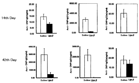
FIG. 4 shows the results of measuring the production of DNP-
OVA specific antibody in plasma by ELISA. BDF1 mice were
administered with Lipo-P or saline, then immunized with DNP-OVA and
CA 02569590 2006-12-05
-7-
alum followed by being boosted with DNP-OVA alone. ELISA was
performed after the primary immunization and after the boosting.
FIG. 5 shows the results of measuring the concentrations of
cytokines in culture supernatants after culturing CD11c+ cells
obtained from spleen of BALB/c mice 7 days after Lipo- P or saline
(negative control) was administered and CD4+ T cells derived from
D011.10 mice (transgenic mice transfected with OVA specific TCRaP)
in the presence of OVA peptide for 4 days.
FIG. 6 shows the results of measuring antibody titers in
blood on the 14th days after immunizing with DNP-OVA and alum after
the cells which proliferated in the experiments in FIG. 5 were
adoptively transferred in BALB/c mice.
FIG. 7 shows the results of measuring the in vitro
production of cytokines. Medium, an aqueous solution of a-
galactosyl ceramide (a-GalCer), a liposome composition as the
control (Lipo-(-)) or an a-galactosyl ceramide-containing liposome
(Lipo-aGC) was added to the cultures of whole spleen cells (upper
panels) and the spleen cells to which anti-CD1d neutralization
antibody had been added or in which NKT cells had been deleted
(lower panels) in C57BL/6 mice. The horizontal axis represents the
concentration of each cytokine in the culture supernatant 2 days
after the addition.
FIG. 8 shows the results of analyzing the numbers of Va14-
NKT cells in the spleen by flow cytometry 3 days or 7 days after
saline, Lipo-(-) or Lipo-aGC was administered to C57BL/6 mice. The
horizontal axis and the vertical axis represent fluorescence
intensity of FITC-labeled anti-TCRP antibody and PE-labeled CD1d
tetramer+ a-GalCer, respectively.
FIG. 9 shows antibody titers of anti-NP-IgE, anti-NP-IgG1
and anti-NP-IgG2a in blood. Saline, a-GalCer, Lipo-(-) or Lipo-aGC
was administered to C57BL/6 mice (upper panels) or IL-10 gene-
deficient mice (lower panels), after 3 days, which were immunized
with DNP-OVA and alum, and after 14 days, the titers were measured.
CA 02569590 2006-12-05
-8-
FIG. 10 shows antibody titers of anti-DNP-IgE, anti-DNP-IgGl,
anti-DNP-IgG2a, and levels of total IgE, total IgG1 and total IgG2a.
Saline, a-GalCer, Lipo-(-) or Lipo-aGC was administered to BDF1
mice, after 3 days (day 0), which were immunized with DNP-OVA and
alum, and boosted with DNP-OVA alone on day 55. The titers were
measured on days 0, 14, 55 and 64.
FIG. 11 shows the results of measuring cell proliferation
ability by MTT method. Saline, a-GalCer, Lipo-(-) or Lipo-aGC was
administered to BDF1 mice, after 3 days, which were immunized with
DNP-OVA and alum. After 7 days, spleen CDC T cells and radiation-
irradiated whole spleen cells from intact BDF1 mice were stimulated
with NDP-OVA or PMA/ionomycin. After 48 hours, the cell
proliferation ability was measured. In the left figure, the
horizontal axis and the vertical axis represent the concentration
of DNP-OVA and absorbance at a wavelength of 570 nm, respectively.
FIG. 12 shows the results of analyzing the cells by flow
cytometry. Saline, a-GalCer, Lipo-(-) or Lipo-aGC was administered
to BALB/c mice. After 3 days, the spleen cells were stained with
anti-CD11c antibody and anti-CD45RB antibody.
FIG. 13 shows the number of CD11c1 wCD45RBhigh cells and the
number of CD1lchi ghCD45RB1 w cells obtained by multiplying a cell
number ratio obtained in flow cytometry memory analysis in FIG. 11
by the number of whole spleen cells.
FIG. 14 shows the results of measuring cytokines in culture
supernatants. Lipo-aGC was administered to BALB/c mouse. After 3
days, the CD11cl'CD45RBhi gh cells and the CD1lchighCD45RB1' cells
separated from the spleen cells were stimulated with LPS. After 2
days, the culture supernatants were analyzed. The horizontal axis
represents the concentration of the cytokine in the culture
supernatant.
FIG. 15 shows the results of measuring the cell
proliferation ability by MTT method. The CD11c1 wCD45RBhigh cells or
the CD1lchighCD45RB1 w cells pulsed with 0VA323_339 peptide were co-
CA 02569590 2006-12-05
. .
-9-
cultured with CD4+ T cells derived from spleen of D011.10 mouse,
and after 48 hours, the cell proliferation ability was assayed.
The horizontal axis represents the absorbance at a wavelength of
570 rim.
FIG. 16 shows the results of flow cytometry analysis using
anti-CD4 antibody, and anti-CD25 antibody, anti-CD28 antibody,
anti-CD152 antibody or anti-ICOS antibody. Cells proliferated by
co-culturing the CD11cl0wCD45RBhigh cells or the CD1lchighCD45RB1'
cells pulsed with 0VA323-339 peptide with the CD4+ T cells derived
from spleen of D011.10 mouse were analyzed.
FIG. 17 shows the results of analyzing intracellular
cytokine expression by flow cytometry. The cells proliferated by
co-culturing the CD11c 1 wCD45RBhi gh cells or the CD1lchi ghCD45RB1'w
cells pulsed with 0VA323-339 peptide with the CD4+ T cells derived
from spleen of D011.10 mouse were stimulated with PMA and ionomycin,
and analyzed by flow cytometry. The upper panels represent
intracellular staining patterns by the corresponding isotype
control antibody. The lower panels represent the intracellular
staining patterns by the cytokine-specific antibody.
FIG. 18 shows the results of analyzing the intracellular
cytokine expression by flow cytometry. The CD4+ T cells from the
spleen of BDF1 mouse administered with Lipo-aGC or Lipo-aGC + OVA
were in vivo cultured with radiation irradiated spleen cells from
the same BDF1 in the presence of OVA, and after 6 days, the
cultured cells were stimulated with PMA and ionomycin. The upper
panels represent the intracellular staining patterns of CD4+ T
cells from the spleen of the mouse administered with Lipo-aGC, and
the lower panels represent the intracellular staining pattern of
CD4+ T cells from the spleen of the mouse administered with Lipo-
aGC + OVA.
FIG. 19 shows antibody titers of anti-DNP-IgE, anti-DNP-IgGl,
anti-DNP-IgG2a (upper panels), and levels of total IgE, total IgG1
and total IgG2a (lower panels) in blood. BDF1 mice were immunized
CA 02569590 2006-12-05
-10-
with DNP-OVA and alum, on days 21, 28 and 35, a liposome alone
(vehicle), Lipo-aGC or Lipo-aGC + OVA was administered, and then on
day 42, the mice were boosted with DNP-OVA antigen alone. On day
48, the antibodies were assayed. * p<0.05, ** p<0.005, *** p<0.001
MODES FOR CARRYING OUT THE INVENTION
Herein, "regulatory cells" includes but is not limited to
NKT cells (natural killer T cells), IL-10-producing Trl cells and
dendritic cells (DC), and among them, the NKT cell is particularly
preferable.
A "regulatory cell ligand" is not particularly limited as
long as the ligand is bound to a cell surface receptor on the above
regulatory cell to facilitate differentiation/proliferation or
activation of the regulatory cell, and includes the followings.
But, the regulatory cell ligand is not limited thereto.
(i) Galactosyl ceramides such as a-galactosyl ceramide and p-
galactosyl ceramide substance which are the ligands of the NKT
cells.
(ii) Vitamin D3, dexamethasone, TGF-P and IL-10 which serve
for the differentiation/proliferation of regulatory dendritic cells
(DC).
(iii) Substances which induce the expression of IL-10 or
FoxP3 which serves for the differentiation/proliferation of
regulatory T cells.
A "regulatory cell-inducing agent" of the present invention
refers to a medicament which induces the
differentiation/proliferation or the activation of the regulatory
cells. The facilitation of the differentiation/proliferation or
the activation of the regulatory cells can be identified, for
instance, as described in Examples, by using spleen CD11c+ DC and
measuring the proliferation of the NKT cells or the IL-10-producing
Trl cells contained therein, or quantifying cytokines (IFN-y, IL-10,
IL-4) produced by NKT cells and the IL-10-producing Tr1 cells.
CA 02569590 2006-12-05
-11-
As a "liposome containing the regulatory cell ligand" of the
present invention, those inducing the NKT cells and the IL-10
producing Trl cells which are the regulatory cells, further having
an activity to suppress the activation of helper T cells and having
an inhibitory action on the production of IgE antibody released
from B cells are preferable. Specifically, those containing the
"regulatory cell ligand" as the above in the liposome are
preferable, and among them a composition including a-galactosyl
ceramide or P-galactosyl ceramide in a lipid double membrane of the
liposome is preferable. The "liposome containing the regulatory
cell ligand" of the present invention may contain two or more
"regulatory cell ligands".
The "liposome containing the regulatory cell ligand" of the
present invention may further contain TLRs (Toll-like receptor)
family ligands in addition to the regulatory cell ligand. The
addition of the TLRs family ligands can increase the production of
cytokines which regulate the action of the "regulatory cells" and
further enhances the effect. The TLRs family ligands include CpG
oligonucleotide (CpGODN) and imiquimod (1-(2-methylprory1)-1H-
imidazo[4,5-c] quinolin-4-amine).
The "liposome containing the regulatory cell ligand" may
also contain allergens. The allergen refers to substances which
cause the allergy, and preferably includes a pollen antigen and a
mite antigen. The allergen specifically includes OVA (ovalbumin).
The present invention provides the liposome in which the
regulatory cell ligand as the above, preferably a lipid-soluble
compound such as galactosyl ceramide has been incorporated as a
water soluble macromolecular substance. Herein, one having a
vesicular structure where a micelle (water soluble particle
obtained by aggregating amphipathic molecules including a
hydrophilic region and a hydrophobic region) has been closed is
referred to as the liposome. A liposome component may be any ones
as long as it is the amphipathic molecule which can faun the
CA 02569590 2006-12-05
-12-
micelle by known methods, and preferably includes lipids. The
lipid in the present invention includes phospholipids such as
dipalmitoylphosphatidylcholine (DPPC), dioleylphosphatidylcholine
(DOPC) and dioleylphosphatidyl ethanolamine (DOPE),
sphingoglycolipid and glyceroglycolipid. These are used for making
the liposome, alone or in combination of two or more or in
combination with a lipid derivative where a non-polar substance
such as cholesterol or a water soluble polymer such as polyethylene
glycol has been bound to the lipid.
The liposome can be prepared in accordance with publicly
known methods. For example, the methods described in Liposome
Technology, vol. 1, 2'd edition (by Gregory Gregoriadis (CRC Press,
Boca Raton, Ann Arbor, London, Tokyo), Chapter 4, pp67-80, Chapter
10, pp167-184 and Chapter 17, pp261-276 (1993)) can be used. More
specifically, the methods include, but are not limited to, a
sonication method, an ethanol injection method, a French press
method, an ether injection method, a cholic acid method, a calcium
fusion method, a lyophilization method and a reverse phase
evaporation method. A size of the liposome of the present
invention is not particularly limited, and typically is preferably
100 to 200 nm and more preferably 100 to 150 nm in average. The
structure of the liposome is not particularly limited, and may be
any liposome such as unilamella and multilamella. As a solution
encapsulated inside the liposome, it is possible to use buffer and
saline and others in addition to water. It is also possible to add
a water soluble organic solvent (e.g., glycerine) in an appropriate
amount thereto and use it.
The liposome used for the drug of the present invention may
be those obtained by modifying the liposome surface for targeting
the "liposome containing the regulatory cell ligand" to a target
site. The target site includes, for example, liver, spleen, lymph
node, bone marrow, lung, eye, skin and nose.
The substance which modifies the liposome surface includes
CA 02569590 2006-12-05
-13-
low molecular compounds, high molecular compounds, nucleic acids,
peptides, proteins and sugar chains. The high molecular compound
includes polyethylene glycol (see Patent No. 2948246). The nucleic
acid includes, for example, single strand RNA and single strand DNA
which recognize TLR-7 or TLR-9 of the Toll-like receptor in the
target cell, and derivatives of these nucleic acids. The protein
includes, for example, antibodies and receptors which recognize the
molecules expressed specifically on the surface of the target cells
such as dendritic cells (DC) which are antigen presenting cells or
precursor cells thereof. The modification with the sugar chain
includes the modification with mannose bound lipid which can be
bound to a mannose receptor expressed on the surface of DC (e.g.,
see Copland, M. J., et al., (2003)Liposome delivery of antigen to
human dendritic cells, Vaccine, 21:883-890).
Inclusion of the ligand into the liposome can be performed
by ordinary methods. For example, as shown in Examples, the
liposome containing the regulatory cell ligand can be obtained by
separately dissolving the liposome component and the ligand in the
organic solvent, mixing these and adding water. But the method for
producing the liposome containing the regulatory cell ligand is not
limited to the above.
The "liposome containing the regulatory cell ligand" can be
used as the active ingredient of the drug.
That is, the drug of the present invention is effective as
the preventive agent or the therapeutic agent for the allergic
diseases caused by IgE antibody because the "liposome containing
the regulatory cell ligand" induces the NKT cells or the IL-10-
producing Trl cells which are the regulatory cells, has the
activity to suppress the activation of the helper T cells and has
the inhibitory action on the production of the IgE antibody
released from B cells. The IgE antibody is particularly deeply
associated with the allergic diseases, and thus by suppressing the
production (secretion ) thereof, it is possible to obtain the
CA 02569590 2006-12-05
-14-
preventive or therapeutic effect on the type I allergic diseases.
The allergic diseases associated with the IgE antibody include
atopic bronchial asthma, atopic dermatitis and nasal allergy such
as allergic rhinitis and pollinosis. In the present invention, the
prevention of the allergic disease encompasses not only making
mammalian animals including human beings who have not had the
allergic disease free from the disease but also making the patients
(mammalian animals including human beings) with allergic disease
who have not had the symptom temporarily free from the symptom.
The drug of the present invention is also effective as the
preventive agent or the therapeutic agent for the disease such as
fulminant hepatitis because the "liposome containing the regulatory
cell ligand" has the action to suppress the activation of the T
cells.
The drug containing the liposome containing a-galactosyl
ceramide as the active ingredient is effective as the drug having
an immunosuppressive ability because the liposome containing a-
galactosyl ceramide has the effect to selectively augment the
immunosuppressive function of the NKT cells. Specifically, the
drug is effective as the drug for autoimmune diseases such as
rheumatoid, multiple sclerosis, systemic lupus erythematosus and
collagen disease and the drug for rejection upon transplantation
such as GVHD.
a-Galactosyl ceramide is not particularly limited as long as
it is bound to the surface receptor of the NKT cell to selectively
augment the immunosuppressive function of the NKT cell, but is
preferably one bound to the receptor composed of Va24JaQ in human
or Val4Ja281 in mouse. The molecular weight thereof is preferably
400 to 2,000.
Meanwhile, the molecular weight of P-galactosyl ceramide used
for the present invention is preferably 400 to 2,000.
As another embodiment of the present invention, the drug
comprising the liposome containing imiquimod, as the active
CA 02569590 2006-12-05
-15-
ingredient is provided. By containing imiquimod in the liposome,
the production amounts of IL-10 and IFNa are enhanced thereby
activating the NKT cells compared with the case of using imiquimod
alone. Therefore, the drug comprising the liposome containing
imiquimod as the active ingredient is useful for the prevention or
the treatment of the allergic diseases as described above.
For an administration route of the drug of the present
invention, the drug can be administered both orally or parenterally,
and the route is optionally selected by a physician. The "liposome
containing the regulatory cell ligand" as the active ingredient can
be administered alone or in combination with a carrier usually used.
When the drug of the present invention is orally
administered, a foLm of the drug includes solid formulations such
as tablets, coated tablets, powdered agents, granules, capsules and
pills, liquid foLmulations such as liquid agents (e.g., eye drops,
nose drops), suspension, emulsion and syrup, inhales such as
aerosol agents, atomizers and nebulizers, and liposome inclusion
agents.
When the drug of the present invention is parenterally
administered, the form of the drug includes injectable agents
(liquid agents, suspensions) used for intravenous injection,
subcutaneous injection, intraperitoneal injection, intramuscular
injection and intraperitoneal injection, liquid agents, suspensions,
emulsions and dripping agents.
When the drug of the present invention is the liquid
foLmulation, the drug may be stored in a frozen state or
lyophilized by removing the water. Injectable distilled water is
added to the lyophilized formulation to re-dissolve the formulation
before use.
As pharmaceutically acceptable carriers utilized for the
drug of the present invention, it is possible to exemplify binders,
disintegrants, surfactants, absorption accelerators, moisture
retention agents, absorbers, lubricants, fillers, extenders,
CA 02569590 2006-12-05
. =
-16-
moisture imparting agents, preservatives, stabilizers, emulsifiers,
solubilizing agents, salts which control osmotic pressure, diluting
agents such as buffers and excipients usually used depending on the
use form of the formulation. These are optionally selected and
used depending on the unit dosage of the resulting folmulation.
Additionally, coloring agents, preserving agents, perfumes,
flavors and sweeteners, and other pharmaceutical articles can be
contained in the drug of the present invention as needed to prepare
as the agent.
An effective amount of the "liposome containing the
regulatory cell ligand" can be easily determined by those skilled
in the art with reference to the conventional art, and is, for
example, about 0.1 to 100 mg per 1 kg of body weight and preferably
about 1 to 10 mg, and this can be administered by dividing into 1
to 3 times daily. It is preferable to optionally regulate the
dosage depending on the foLm of each foLmulation, a gender, an age
and a disease condition of the patient.
EXAMPLES
The present invention will be described with reference to
the following Examples, but the present invention is not limited to
these Examples, and it goes without saying that usual changes in
the art of the present invention can be made.
Example 1
<Preparation of ligand-containing liposome and measurement of
activity>
1. Preparation of P-galactosyl ceramide-containing liposome (Lipo-
i3)
L-a-Phosphatidylethanolamine, dioleoyl (DOPE; Wako Pure
Chemical #166-16183, 0.77 mg), 0.83 mg of cholesteryl 313-N-
(dimethylaminoethyl)carbonate hydrochloride (DC-Chol; SIGMA-
Aldrich) and 0.029 mg of 1,2-distearoyl-sn-glycero-3-
phosphoethanolamine-N-[methoxy(polyethylene glycol)-2000] (AVANTI
CA 02569590 2006-12-05
-17-
POLAR-LIPIDS, INC. #i88653) were dissolved in 250 pL of
chloroform/methanol (1:1) solvent. P-Galactosyl ceramide (ceramide
P-D-galactoside; Sigma-Aldrich #C4905, 0.16 mg) was separately
dissolved in 250 pL of chloroform/methanol (1:1) solvent. Both
were mixed and evaporated using an evaporator, and subsequently
dried overnight in a desiccator under vacuum. Then, 800 pL of
water was added, the mixture was treated with a sonicator for one
minute, then particle sizes were selected by filtration with
pressure using an extruder (AVESTIN; LiposoFast-Basic), and the
particles were sterilized with a membrane having a pore size of
0.22 pm. This liposome composition (Lipo-P) was adjusted to a
final concentration of 200 pL/mL. By the same method, a liposome
composition containing no P-galactosyl ceramide (Lipp-0) was
prepared. An eluted product collected through a salting out column
NAP-10 after mixing oligonucleotide CpGODN (1668) (supplied from
SIGMA GENOSIS) with Lipo-P at a weight ratio of 5:1 was rendered
Lipo-P-CpG.
2. Preparation of imiquimod-containing liposome
L-a-Phosphatidylethanolamine, dioleoyl (DOPE; Wako Pure
Chemical #166-16183, 0.77 mg), 0.83 mg of cholesteryl 33-N-
(dimethylaminoethyl)carbonate hydrochloride (DC-Chol; SIGMA-
Aldrich) and 0.029 mg of 1,2-Distearoyl-sn-Glycero-3-
Phosphoethanolamine-N-[Methoxy(polyethylene glycol)-2000] (AVANTI
POLAR-LIPIDS, INC. #i88653) were dissolved in 250 pL of
chloroform/methanol (1:1) solvent. Imiquimod (Sequoia Research
Products Ltd; SRP0058i, 0.16 mg) was separately dissolved in 250 pL
of chlorofolm/methanol (1:1) solvent. Both were mixed and
evaporated using an evaporator, and subsequently dried overnight in
a desiccator under vacuum. Then, 800 pL of water was added, the
mixture was treated with the sonicator for one minute, then
particle sizes were selected by filtration with pressure using the
extruder (AVESTIN; LiposoFast-Basic), and the particles were
= CA 02569590 2006-12-05
-18-
sterilized with the membrane having a pore size of 0.22 pm. This
liposome composition (Lipo-Imq) was adjusted to a final
concentration of 200 pL/mL. By the same method as in the above
composition, a liposome composition (Lipo-Imq-PGC) containing
ceramide P-D-galactoside (Sigma-Aldrich #C4905) was prepared. An
eluted product collected through the salting out column NAP-10
after mixing oligonucleotide CpGODN (1668) (supplied from SIGMA
GENOSIS) with Lipo-Imq at a weight ratio of 5:1 was rendered Lipo-
Imq-CpG.
3. Measurement of in vitro activity of ligand-containing liposome
for dendritic cells (DC)
Collagenase D (1 mg/mL, Roche) was injected into spleen from
BALB/c or C57BL/6 mouse, which was then incubated in a CO2
incubator for 45 minutes. Subsequently, cells were collected from
the spleen, suspended in 3 'TILL of HistoDenz (14.1%, SIGMA), and then
X-VIVO 15 (Takara Bio) containing 50 M 2-mercaptoethanol (2ME) was
overlaid thereon. After centrifuging at 1,500 rpm for 5 minutes,
the cells in an intermediate layer were collected and incubated in
X-VIVO 15 medium containing 50 pM 2ME, 0.5% fetal calf serum and 20
ng/mL rmGM-CSF (Pharmingen) in the CO2 incubator for one and a half
hours. After pipetting gently, the suspended cells were removed,
the X-VIVO 15 medium containing 50 M 2ME, 0.5% fetal calf serum
and 20 ng/mL rmGM-CSF (Pharmingen) was added, and the cells were
incubated in the CO2 incubator for 18 hours. The suspended cells
were collected, and the cells bound to anti-CD11c antibody-magnetic
microbeads (Miltenyi) were collected to render spleen CD11c+ DC.
The CD11c+ DC at 1 x 10⁴ cells were suspended in 200 L of RPMI
medium containing 10% fetal calf serum in a 96-well round bottom
microtiter plate, the liposome composition at a final concentration
of 1 pg/mL was added thereto, and the plate was incubated in the
incubator containing 5% CO2 at 37 C. After 48 hours, culture
supernatants were collected, and levels of IFN-a, IL-10 and IL-12
CA 02569590 2006-12-05
. .
-19-
were measured by ELISA (FIGS. 1 and 2). The levels of IL-10 and
IFN-a were high whereas the levels of IL-12 were low in Lipo-P and
Lipo-Imq groups. Conversely, in Lipo-P-CpG and Lipo-Imq-CpG groups,
the levels of IL-10 and IFNa were low whereas the levels of IL-12
were high. Meanwhile, in the non-addition group (control), Lipo-0
and the P-galactosyl ceramide solution (P-GalCer) groups, the
production of all cytokines was not detected or was very low. In
the same evaluation method, the production levels of IL-10 in
CD11c+ DC by Lipo-Imq-PGC were measured. As a result, it was found
that Lipo-Imq-PGC induced IL-10 production at much higher levels
than Lipo-P alone or Lipo-Imq alone (FIG. 3).
Example 2
<Inhibitory effect of Lipo-P on in vivo production of IgE antibody>
Lipo-P (2 g/mouse) was intraperitoneally injected in BDF1
mice (5 mice/group), after 7 days (day 0), which were primarily
immunized with 0.1 g of DNP-OVA (Cosmobio) and 10 mg of alum. On
the 14th day after the primary immunization, blood was collected
from orbital venous plexus, and antibody titers of ant-DNP-IgGl,
anti-DNP-IgE and anti-DNP-IgG2a in plasma were measured by ELISA
(14th day in FIG. 4). The mice were boosted with DNP-OVA alone on
the 35th day after the primary immunization, and 7 days thereafter,
the antibody titers of anti-DNP-IgG1 and anti-DNP-IgE in the plasma
from the blood collected from the orbital venous plexus were
measured by ELISA (42nd day in FIG. 4). In the Lipo-P group, on
the day 14, the production of IgG antibody and IgE antibody tended
to be already inhibited, and on the day 42, the increase of IgG
antibody and IgE antibody was completely inhibited after the boost
immunization.
Example 3
<Induction of regulatory T cells by dendritic cells (DC) derived
from mice administered with Lipo-P>
CA 02569590 2006-12-05
,
-20-
1. Evaluation of in vitro activation ability of T cells
Lipo-P or saline (2 L/mouse) was intraperitoneally
administered to BALB/c mice, and after 7 days, the spleen was
removed. Collagenase D (1 mg/mL, Roche) was injected into the
spleen, which was then incubated in the CO2 incubator for 45
minutes. Subsequently, cells were collected from the spleen,
suspended in 3 mL of HistoDenz (14.1%, SIGMA), and then X-VIVO 15
containing 50 M 2-mercaptoethanol (2ME) was overlaid thereon.
After centrifuging at 1,500 rpm for 5 minutes, the cells in the
intermediate layer were collected and incubated in the X-VIVO 15
medium containing 50 M 2ME, 0.5% fetal calf serum and 20 ng/mI
rmGM-CSF (PharMingen) in the CO2 incubator for one and a half hours.
After pipetting gently, the suspended cells were removed, the X-
VIVO 15 medium containing 50 M 2ME, 0.5% fetal calf serum and 20
ng/mL rmGM-CSF (PharMingen) was added, and the cells were incubated
in the CO2 incubator for 18 hours. The suspended cells were
collected, and the cells bound to anti-CD11c antibody-magnetic
microbeads (Miltenyi) were collected to render spleen CD11c+ DC.
CDC T cells were collected from OVA specific TCRaP transgenic
mouse D011.10 (given by Dr. Toshinori Nakayama, Graduate School of
Medicine, Chiba University; Science, 1990, vol. 250, p1720) using
antibody-magnetic microbeads (Miltenyi). Subsequently, CD11c+ DC at
2 x 10⁴ cells and CD4+ T cells at 1 x 10⁵ cells were cultured in the
presence of the OVA peptide in the CO2 incubator for 4 days, then
the culture supernatant was collected, and the levels of IFNI', IL-4
and IL-10 were measured by ELISA (FIG. 5). As a result, when DC
from the spleen of the mouse administered with Lipo-P were used and
when DC from the spleen of the mouse administered with saline
(normal) were used, no difference was observed in the levels of IL-
4 and IFN-7 production. However, the production of IL-10 was
observed only at OVA peptide concentrations of 3 nM and 30 nM when
DC from the spleen of the mouse administered with Lipo-P were used.
Simultaneously, the proliferation of the regulatory cells was also
= CA 02569590 2006-12-05
-21-
identified.
2. Evaluation of inhibitory effect on in vivo IgE antibody
production by adoptive transfer method
D011.10-CD4+ T cells which had proliferated at OVA peptide
concentrations of 3 nM or 30 nM and DC from the spleen of the mouse
administered with Lipo-P in the above 1. in vitro experiment were
collected, and 1 x 10⁶ thereof were intraperitoneally transferred
into BALB/c mice (3 mice/group). After one hour, the mice were
primarily immunized with DNP-OVA (10 g) and alum (10 mg), and on
the 14th day, the blood was collected from the orbital venous
plexus. The antibody titers of anti-DNP-IgGl, anti-DNP-IgE and
anti-DNP-IgG2a in the plasma were measured by ELISA (FIG. 6). As a
result, the production of IgE antibody was completely inhibited in
the mice in which D011.10-CD4+ T cells grown by the stimulation of
OVA peptide at 3 nM had been adoptively transferred. Meanwhile,
the inhibitory effect on the IgE antibody production was low in the
mice in which D011.10-CD4+ T cells grown by the stimulation of OVA
peptide at 30 nM had been adoptively transferred. The inhibition
of IgG1 and IgG2a antibody production was not remarkable in both
groups.
Example 4
<Preparation of ligand-containing liposome and measurement of
activity>
1. Preparation of a-galactosyl ceramide-containing liposome
L-a-Phosphatidylethanolamine, dioleoyl (DOPE; Wako Pure
Chemical #166-16183, 0.77 mg) and 0.83 mg of cholesteryl 3P-N-
(dimethylaminoethyl)carbonate hydrochloride (DC-Chol ;Sigma-Aldrich
#C2832) were dissolved in 250 L of chloroform/methanol (1:1)
solvent. a-Galactosyl ceramide (0.16mg, supplied from RIKEN
Research Center for Allergy and Immunology; KRN7000, see
International Publication Pamphlet W098/44928) was separately
= CA 02569590 2006-12-05
-22-
dissolved in 250 L of chlorofolm/methanol (1:1) solvent. Both
were mixed and evaporated using the evaporator, and subsequently
dried overnight in the desiccator under vacuum. Then, 800 L of
water was added, the mixture was treated with the ultrasonic
pulverizer for one minute, and passed through a membrane having a
pore size of 0.22 m for sterilization. This liposome composition
(Lipo-aGC) was adjusted to a final concentration of 200 L/mL. By
the same method, a liposome composition containing no a-galactosyl
ceramide (Lipo-(-)) for the control was prepared.
2. Measurement of cytokine production by Lipo-aGC
Spleen whole cells at 2 x 10⁵ from C57BL/6 mouse were
suspended in 200 L of 10% fetal calf serum (FCS)-containing RPMI
medium to which 100 ng/mL Lipo-(-), Lipo-aGC or a-galactosyl
ceramide aqueous solution (a-GalCer) had been added, then the cell
suspension was added to a 96-well U bottom culture plate, and
cultured in the incubator containing 5% CO2 at 37 C for 2 days. The
levels of IFNI', IL-4 and IL-10 produced in the culture supernatant
were measured by ELISA (FIG. 7 upper panels). The levels of IFN-y
and IL-4 were equivalent in Lipo-aGC group and aGalCer group, but
the level of IL-10 in the Lipo-a group was 5 times higher than that
in the a-GalCer group. When the same experiments were perfoLmed in
the presence of anti-CD1d neutralization antibody (1B1, BD
Bioscience PharMingen) at a final concentration of 10 g/mL or
using spleen whole cells from Va14-NKT cell-deficient mouse
(057BL/6 background), IFN-y, IL-4 and IL-10 in the culture
supernatant were not detected (FIG. 7, lower panels).
3. Evaluation of Va14-NKT cell proliferation ability by Lipo-aGC
Lipo-aGC (2 g/mouse), or Lipo-(-) or saline as the control
was intraperitoneally administered to 057BL/6 mice. On the 3rd day
(day 3) and the 7th day (day 7), the spleen cells were stained with
aGalCer/CD1d tetramer and anti-TCRP antibody, and the number of
CA 02569590 2006-12-05
=
-23-
double positive cells (Va14-NKT cells) was analyzed by flow
cytometry. As a result, it was identified that the number of the
Va14-NKT cells in the spleen of the mouse 3 days after the
administration of Lipo-a was increased 2 times or more compared
with that from the spleen administered with saline, but on day 7,
the number was reversely reduced compared with that from the
control mice (FIG. 8).
Example 5
<Inhibitory effect of Lipo-aGC on in vivo antibody production>
1. Activity evaluation in in vivo antibody production system using
C57BL/6 and IL-10-deficient mice
Saline, a-GalCer, Lipo-(-) or Lipo-aGC (2 g/mouse) was
intraperitoneally administered in C57BL/6 mice (5 mice/group),
after 3 days, which were primarily immunized with DNP-OVA and alum.
On the 14th day after the primary immunization, the blood was
collected from the orbital venous plexus, and antibody titers of
ant-DNP-IgGl, anti-DNP-IgE and anti-DNP-IgG2a in the plasma were
measured by ELISA. As a result, the inhibitory effect on the
antibody production in the Lipo-aGC group tended to be higher than
in the a-GalCer group for all isotypes examined (FIG. 9 upper
panels). The same experiment was performed using the IL-10-
deficient mice with C57BL/6 background. As a result, no inhibitory
effect on the antibody production was observed (FIG. 9 lower
panels).
2. Activity evaluation in in vivo antibody production system using
BDF1 mice
Saline, a-GalCer, Lipo-(-) or Lipo-aGC (2 g/mouse) was
intraperitoneally administered in BDF1 mice (C57BL/6 x DBA/2F1) (5
mice/group), after 3 days (day 0), which were primarily immunized
with DNP-OVA and alum, and further the mice was boosted with DNP-
OVA alone on the 55th day (day 55) after the primary immunization.
. CA 02569590 2006-12-05
-24-
On days 0, 14, 55 and 64, the blood was collected from the orbital
venous plexus, and antibody titers of ant-DNP-IgE, anti-DNP-IgGl,
anti-DNP-IgG2a, and the levels of total IgE, total IgG1 and total
IgG2a in the plasma were measured by ELISA. As a result, it was
identified that the increase of antibody titers of all isotype
anti-DNP and the production of total IgE were nearly completely
inhibited in the Lipo-aGC group (FIG. 10). Meanwhile, the changes
of total IgG1 and total IgG2a were nearly equivalent in the Lipo-
aGC group and the a-GalCer group or Lipo-(-) group (FIG. 10).
3. Evaluation of T cell activation ability using BDF1 mice
Saline, a-GalCer, Lipo-(-) or Lipo-aGC (2 g/mouse) was
intraperitoneally administered in BDF1 mice, after 3 days, which
were primarily immunized with DNP-OVA and alum. After 7 days, the
spleen was removed, and CDC T cells were prepared using magnetic
microbeads (Miltenyi). Subsequently, antigen presenting cells were
prepared by irradiating spleen whole cells from the normal BDF1
mouse with radiation of 20 Gy. The CD4+ T cells at 2 x 10⁵ and the
antigen presenting cells at 2 x10⁵ pulsed with DNP-OVA suspended in
200 L of the medium were placed in one well of the 96-well U
bottom culture plate, and cultured in the incubator containing 5%
CO2 at 37 C. After 48 hours, cell proliferation was assayed by MTT
method (Promega #G4000). As a results, the CD4+ T cells derived
from the spleen of the mouse administered with Lipo-aGC did not
proliferate in response to DNP-OVA at all concentrations examined,
while other CDC" T cells highly proliferated in order of a-GalCer,
saline and Lipo-(-) (FIG. 11 left panel). On the other hand, when
the CDC T cells at 2 x 10⁵ were stimulated antigen-non-specifically
with 50 ng/mi of phorbol 12-miristate 13-acetate (PMA; Sigma-
Aldrich #P-1585) and 500 nM ionomycin (Sigma-Aldrich, #I-0634) in
the CO2 incubator containing 5% CO2 at 37 C for 48 hours, the CDC T
cells derived from the mouse administered with Lipo-aGC exhibited
lower but significant proliferative response compared with other
CA 02569590 2006-12-05
,
-25-
CD4+ T cells (FIG. 11, right panel).
4. Analysis of dendritic cells (DC) in spleen of mice administered
with Lipo-aGC
4-1. Analysis using flow cytometry
Saline, a-GalCer, Lipo-(-) or Lipo-aGC (2 g/mouse) was
intraperitoneally administered in BALB/c mice, and after 3 days,
the spleen was removed. Collagenase D (1 mg/mL, Roche) was
injected into the spleen, which was then incubated in the CO2
incubator for 45 minutes. Subsequently, cells were collected from
the spleen, suspended in 3 mi of HistoDenz (14.1%, SIGMA-Aldrich),
and then the X-VIVO 15 medium (CAMBREX Bio Science Walkerville,
Inc.) containing 50 M 2-mercaptoethanol (2ME) was overlaid thereon.
After centrifuging at 1,500 rpm for 5 minutes, the cells in the
intermediate layer were collected. The cells were washed with the
X-VIVO 15 medium containing 50 M 2ME and 10% FCS, and suspended in
phosphate buffered saline (PBS) containing 0.5% FCS. Biotinylated
anti-CD3, -CD11b, -CD19, -CD49b, -Gr-1, -TER-119 and -B220
antibodies (all from BD Bioscience Pharmingen) were added to the
cell suspension. The cells were incubated at 10 C for 20inutes,
then washed once with PBS containing 0.5% FCS, and subsequently
streptoavidin (SA)-conjugated magnetic beads (Miltenyi) were added
thereto. The cells were incubated at 10 C for 15 minutes,
subsequently washed twice with PBS containing 0.5% FCS, and then
magnetic microbead-negative cells were collected using a microbead
separation column and a magnet (Miltenyi). The resulting cells
were stained with PE-labeled anti-CD11c antibody (BD Bioscience
Pharmingen) and APC-labeled anti-CD45RB antibody (BD Bioscience
PhaLmingen), and analyzed by flow cytometry. As a result, in the
cells derived from the spleen of the mouse administered with Lipo-
aGC, the ratio of CD45RBIli ghCD11c1 w cells was higher than the ratio
of CD45RB1 wCD1lchigh cells, while the ratio was reversed in the
cells derived from the mice administered with saline, Lipo-(-) or
= CA 02569590 2006-12-05
-26-
a-GalCer (FIG. 12). The ratios were further compared in terms of
cell number in the spleen. As a result, it was demonstrated that
the number of the CD45RBhighCD11c1' cells in the spleen of the mouse
administered with Lipo-aGC increased about 3 times over the number
of the corresponding cells in the spleen of the mouse administered
with a-GalCer whereas conversely the number of the CD45RB1(3wCD1lchigh
cells increased in the mouse administered with a-GalCer more than
in the mouse administered with Lipo-aGC (FIG. 13).
The CD45RBhighCD11c1' cell is the cell group reported as a
controllable dendritic cell and has the immunosuppressive function.
Conversely, the CD45RBL0wCD11chigh cell is the dendritic cell which
activates the T cell and has the immunostimulatory function. Thus,
it was speculated that the immunosuppressive function of the NKT
cells was brought by the increase in the number of the
CD45RBhighCD11c1 w cells.
4-2. Evaluation of cytokine production ability
The CD45RBhighCD11c 2.(' cell population and the CD45RB10wCD11chig1
cell population separated by the method described above were
collected separately using the flow cytometry (FACS Vantage SE, BD
Bioscience), and the cells at 1 x 10⁵/200 L of the medium were
added into one well in the 96-well U bottom culture plate. The
cells were cultured in the presence or absence of
lipopolysaccharide (LPS; T3382, Sigma-Aldrich) at a final
concentration of 1 g/ml for 2 days. The levels of the cytokines
IL-10 and IL-12 in the culture supernatant were measured by ELISA.
As a result, IL-10 was detected and IL-12 was not detected in the
culture supernatant of the CD45RBhi ghCD1lci'w cells stimulated with
LPS whereas IL-12 was detected and IL-10 was not detected in the
culture supernatants of the CD45RB1 wCD1lchigh cells regardless of
the presence or absence of LPS (FIG. 14).
4-3. Evaluation of T cell activation ability
õ CA 02569590 2006-12-05
-27-
The CD45RBhighCD11c1' cells or the CD45RB1 wCD1lchigh cells
which are separated by the method described above 2. were added
into one well of the 96-well U bottom culture plate at 1 x 10⁴
cells/200 L of the medium. The CD4+ T cells purified from the
spleen of D011.10 by the magnetic microbeads (Miltenyi) were added
at 4 x 10⁶cells/200 L of the medium thereto. The cells were
cultured in the presence or absence of the 0VA323-339 Peptide at a
final concentration of 600 nM in the incubator containing 5% CO2 at
37 C. After 48 hours, the proliferative response was assayed by MTT
method (Promega #G4000). As a result, the CD4+ T cells stimulated
with the CD45RBhighCD11c10w cells and the OVA peptide exhibited the
slightly inferior but significant proliferative response compared
with the proliferative response induced by the CD45RB10wCD11chigh
cells (FIG. 15). The CD4+ T cells grown by the stimulation with the
CD45RBhighCD11c1cm cells and the OVA peptide for 7 days were
collected, and cultured with the CD45RBhi ghCD11c1' cells or the
CD45RBl0wCD11chigh cells newly separated/collected in the presence of
OVA peptide for 7 days. This culture was performed once more, and
on the 5th day, the cells in the culture were analyzed by flow
cytometry. As a result, it was identified that the grown cell
group was a nearly homogenous cell population with CDC, CD25+,
CD28+, CD152- and ICOS+ (FIG. 16). Subsequently, these cells at 5 x
105 were cultured in the presence of PMA at a final concentration
of 50 ng/mL, 500nM ionomycin and 2 M Monensin (Sigma-Aldrich #M-
5273) in the incubator containing 5% CO2 at 37 C for 4 hours. The
cells were collected, suspended in 100 L of a BD Cytofix/Cytoperm
solution (BD Bioscience) and incubated at 4 C for 15 minutes. The
cells were washed with BD Perm/Wash (BD Bioscience),
intracellularly stained with FITC-labeled anti-IFN-7 antibody, PE-
labeled anti-IL-4 antibody (BD Bioscience PhaLmingen) and APC-
labeled anti-IL-10 antibody (BD Bioscience Pharmingen), and
intracellular triple staining using fluorescence labeled isotype
control antibodies was perfoLmed simultaneously (FIG. 17, upper
CA 02569590 2006-12-05
-28-
panels). Then, the cells were analyzed by flow cytometry. As a
result, in the cell group expressing the cytokines, it was
identified that there was almost no cell expressing only IL-4 and
the cell numbers were large in order of the cells expressing only
IFNy < the cells expressing both IL-10 and IFNI, < the cells
expressing only IL-10 (FIG. 17, lower panels).
Example 6
<Inhibitory effect of liposome containing allergen and regulatory
cell ligand on IgE antibody production>
1. Preparation of liposome containing ovalbumin and a-galactosyl
ceramide
L-a-Phosphatidylcholine, dioleoyl (DOPC; Wako Pure Chemical,
0.77 mg), 0.83 mg of cholesteryl 33-N-(dimethylaminoethy1)carbonate
hydrochloride (DC-Chol; Sigma-Aldrich) and 0.029 mg of 1,2-
distearoyl-sn-glycero-3-phosethanolamine-N-[methoxy(polyethylene
glycol)-2000] (ammonium salt; PEG-PE; AVANTI POLAR-LIPIDS) were
dissolved in 250 L of chlorofoim/methanol (1:1) solvent, a-
Galactosyl ceramide (0.16mg, supplied from RIKEN Research Center
for Allergy and Immunology) was separately dissolved in 250 L of
chloroform/methanol (1:1) solvent. Both were mixed and evaporated
using the evaporator, and subsequently dried overnight in the
desiccator under vacuum. Subsequently, 200 L of an aqueous
solution containing 0.4 mg/mL of ovalbumin (OVA; Seikagaku Kogyo)
was added thereto, the mixture was treated using the sonicator for
10 minutes, and passed through the membrane having a pore size of
0.22 m for sterilization. Then, the particle sizes were selected
by passing 25 times through LiposoFast-Basic extruder (Avestin
Inc.) equipped with a polycarbonate membrane having a pore size of
100 nm. The OVA protein which had not been encapsulated in the
liposome was eliminated by concentration of the liposomes in which
OVA had been encapsulated using Amicon Ultra-4 centrifugation
filter (PL-100) (Millipore) and washing with purified water, and
= CA 02569590 2006-12-05
-29-
finally the liposome was prepared into 800 L of an aqueous
solution with purified water. This aqueous solution containing the
liposome composition (Lipo-aGC + OVA) was analyzed on SDS
electrophoresis, and consequently it was identified that the
concentration of the OVA protein was 50 g/mL. All a-GalCer was
supposed to be incorporated in the liposome membrane, and the final
concentration of a-GalCer in the Lipo-aGC + OVA solution was
rendered 200 g/mL.
2. Induction of IL-10-producing regulatory CD4+ T cells by Lipo-aGC
+ OVA
Lipo-aGC or Lipo-aGC + OVA (2 g in terms of a-GalCer
amount) was intraperitoneally administered in the BDF1 (C57BL/6 x
DBA/2 Fl) mouse, after 7 days, the spleen was removed, and the CD4+
T cells were prepared using the magnetic microbeads (Miltenyi).
Subsequently, antigen presenting cells were prepared by irradiating
spleen whole cells from the normal BDF1 mouse with radiation of 20
Gy. Then, 3 mL of the medium, the CD4+ T cells at 1.5 x 10⁶, the
antigen presenting cells at 7.5 x 10⁶ and the OVA protein at a
final concentration of 100 g/mL were added in one well of a 6-well
U bottom culture plate, and cultured in the incubator containing 5%
CO2 at 37 C for 6 days. Subsequently, the cells at 5 x 10⁵ were
cultured in the presence of PMA at a final concentration of 50
ng/mL, 500 nM of ionomycin and 2 M Monensin (Sigma-Aldrich) in the
incubator containing 5% CO2 at 37 C for 4 hours. The cells were
collected, and stained with biotinylated anti-CD4 antibody and
streptoavidin-Per CP-Cy5.5 (BD Bioscience). Subsequently, the
cells were suspended in 100 L of the BD Cytofix/Cytoperm solution
(BD Bioscience) and incubated at 4 C for 15 minutes. The cells were
washed with BD Pam/Wash solution (BD Bioscience), then
intracellularly stained with FITC-labeled anti-IFN-7 antibody, PE-
labeled anti-IL-4 antibody (BD Bioscience Phamingen) and APC-
labeled anti-IL-10 antibody (BD Bioscience Pharmingen), and
CA 02569590 2006-12-05
õ
-30-
analyzed by flow cytometry (FIG. 18). As a result, in the CD4+ T
cells derived from the spleen of the mouse administered with Lipo-
aGC without encapsulating OVA, 1.4% cells expressing only IL-4 and
1.1% cells expressing only IFN7 were detected but the CD4+
regulatory T cell population expressing only IL-10 or both IFN-7
and IL-10 was scarcely detected. On the other hand, in the
analysis of the CD4+ T cells derived from the spleen of the mouse
administered with Lipo-aGC + OVA, the helper T cell population
expressing only IL-4 (1.0%) and only IFN-7 (9.9%), the IL-10-
producing CD4+ regulatory T cell population (14.1%) and the CD4+
regulatory T cell population (9.1%) expressing both IL-10 and IFN-7
were detected. From the above results, it was suggested that the
allergen-containing Lipo-aGC could in vivo differentiate and
proliferate the allergen-specific CD4+ regulatory T cells having
the inhibitory effect on the IgE production.
3. Inhibitory effect of Lipo-aGC + OVA on secondary antibody
response in mice
BDF1 mice were primarily immunized with DNP-OVA (0.1 g) and
aluminium hydroxide gel (2 mg). After 14 days, the antibody titers
of anti-DNP-IgE antibody in blood were measured, and 3 groups (5
mice per group) were prepared so that the average antibody titers
were equivalent among them. On 21, 28 and 35 days after the
primary immunization, the liposome alone (vehicle), Lipo-aGC or
Lipo-aGC + OVA at 2 g in terms of a-GalCer amount was
intraperitoneally administered. On the 42nd day after the primary
immunization, the mice were boosted with DNP-OVA alone. On the
48th day, antibody titers of anti-DNP-IgE, anti-DNP-IgGl, anti-DNP-
IgG2a, and the levels of total IgE, total IgG1 and total IgG2a in
blood were measured by ELISA (FIG. 19). As a result, in the Lipo-
aGC + OVA group, the antibody titers of anti-DNP-IgE, anti-DNP-IgG1
and anti-DNP-IgG2a were significantly suppressed. On the other
hand, in the Lipo-aGC group containing no OVA, no significant
CA 02569590 2006-12-05
-31-
suppression other than that in the antibody titer of anti-DNP-IgG1
was observed. From the above result, it was suggested that Lipo-
aGC containing the allergen can suppress the secondary antibody
response induced by the allergen.
INDUSTRIAL APPLICABILITY
The "liposome containing the regulatory cell ligand" of the
present invention has the inhibitory actions on the activation
action of the helper T cell and on the IgE antibody production by
inducing the differentiation/proliferation and the activation of
the regulatory cells. Thus, the liposome of the present invention
is useful as the preventive agent and the therapeutic agent for the
allergic diseases caused by the type I allergic response in which
the IgE antibody is deeply involved, in particular atopic bronchial
asthma, atopic dermatitis and allergic rhinitis such as pollinosis,
and conjunctivitis.
The "a-galactosyl ceramide-containing liposome" is useful as
the drug for autoimmune diseases and graft-versus-host disease
because the liposome can inhibit the differentiation/proliferation
of the pathogenic T cells by selectively augmenting the
immunosuppressive function of the NKT cells.
In addition, no side effect is necessary to be concerned for
the drug of the present invention because the drug retains the
molecule selectively bound to the target cell and has the liposome
including the regulatory cell ligand in the lipid membrane as the
active ingredient.