Note: Descriptions are shown in the official language in which they were submitted.
CA 02569607 2011-11-17
WO 2005/120397 PCT/US2005/019663
EXPANDABLE MEDICAL DEVICE
FOR DELIVERY OF BENEFICIAL AGENT
BACKGROUND OF THE INVENTION
The present invention relates to tissue-supporting medical devices, and more
particularly to expandable, non-removable devices that are implanted within a
bodily lumen
of a living animal or human to support the organ and maintain patency, and
that can deliver a
beneficial agent to the intervention site.
In the past, permanent or biodegradable devices have been developed for
implantation
within a body passageway to maintain patency of the passageway. These devices
are
typically introduced percutaneously, and transported transluminally until
positioned at a
desired location. These devices are then expanded either mechanically, such as
by the
expansion of a mandrel or balloon positioned inside the device, or expand
themselves by
releasing stored energy upon actuation within the body. Once expanded within
the lumen,
these devices, called stents, become encapsulated within the body tissue and
remain a
permanent implant.
Known stent designs include monofilament wire coil stents (U.S. Pat. No.
4,969,458);
welded metal cages (U.S. Pat. Nos. 4,733,665 and 4,776,337); and, most
prominently,
CA 02569607 2006-12-06
WO 2005/120397 PCT/US2005/019663
thin-walled metal cylinders with axial slots formed around the circumference
(U.S. Pat. Nos.
4,733,665, 4,739,762, and 4,776,337). Known construction materials for use in
stents
include polymers, organic fabrics and biocompatible metals, such as, stainless
steel, gold,
silver, tantalum, titanium, and shape memory alloys such as Nitinol.
U.S. Pat. Nos. 4,733,665, 4,739,762, and 4,776,337 disclose expandable and
deformable interluminal vascular grafts in the form of thin-walled tubular
members with
axial slots allowing the members to be expanded radially outwardly into
contact with a body
passageway. After insertion, the tubular members are mechanically expanded
beyond their
elastic limit and thus permanently fixed within the body. The force required
to expand these
tubular stents is proportional to the thickness of the wall material in a
radial direction. To
keep expansion forces within acceptable levels for use within the body (e.g.,
5 - 10 atm),
these designs must use very thin-walled materials (e.g., stainless steel
tubing with 0.0025
inch thick walls). However, materials this thin are not visible on
conventional fluoroscopic
and x-ray equipment and it is therefore difficult to place the stents
accurately or to find and
retrieve stents that subsequently become dislodged and lost in the circulatory
system.
Further, many of these thin-walled tubular stent designs employ networks of
long,
slender struts whose width in a circumferential direction is two or more times
greater than
their thickness in a radial direction. When expanded, these struts are
frequently unstable, that
is, they display a tendency to buckle, with individual struts twisting out of
plane. Excessive
protrusion of these twisted struts into the bloodstream has been observed to
increase
turbulence, and thus encourage thrombosis. Additional procedures have often
been required
to attempt to correct this problem of buckled struts. For example, after
initial stent
implantation is determined to have caused buckling of struts, a second, high-
pressure balloon
(e.g., 12 to 18 atm) would be used to attempt to drive the twisted struts
further into the lumen
wall. These secondary procedures can be dangerous to the patient due to the
risk of collateral
damage to the lumen wall.
Many of the known stents display a large elastic recovery, known in the field
as
"recoil," after expansion inside a lumen. Large recoil necessitates over-
expansion of the stent
during implantation to achieve the desired final diameter. Over-expansion is
potentially
-2-
CA 02569607 2006-12-06
WO 2005/120397 PCT/US2005/019663
destructive to the lumen tissue. Known stents of the type described above
experience recoil
of up to about 6 to 12% from maximum expansion.
Large recoil also makes it very difficult to securely crimp most known stents
onto
delivery catheter balloons. As a result, slippage of stents on balloons during
interluminal
transportation, final positioning, and implantation has been an ongoing
problem. Many
ancillary stent securing devices and techniques have been advanced to attempt
to compensate
for this basic design problem. Some of the stent securing devices include
collars and sleeves
used to secure the stent onto the balloon.
Another problem with known stent designs is non-uniformity in the geometry of
the
expanded stent. Non-uniform expansion can lead to non-uniform coverage of the
lumen wall
creating gaps in coverage and inadequate lumen support. Further, over
expansion in some
regions or cells of the stent can lead to excessive material strain and even
failure of stent
features. This problem is potentially worse in low expansion force stents
having smaller
feature widths and thicknesses in which manufacturing variations become
proportionately
more significant. In addition, a typical delivery catheter for use in
expanding a stent includes
a balloon folded into a compact shape for catheter insertion. The balloon is
expanded by
fluid pressure to unfold the balloon and deploy the stent. This process of
unfolding the
balloon causes uneven stresses to be applied to the stent during expansion of
the balloon due
to the folds causing the problem non-uniform stent expansion.
U.S. Pat. No. 5,545,210 discloses a thin-walled tubular stent geometrically
similar to
those discussed above, but constructed of a nickel-titanium shape memory alloy
("Nitinol").
This design permits the use of cylinders with thicker walls by making use of
the lower yield
stress and lower elastic modulus of martensitic phase Nitinol alloys. The
expansion force
required to expand a Nitinol stent is less than that of comparable thickness
stainless steel
stents of a conventional design. However, the "recoil" problem after expansion
is
significantly greater with Nitinol than with other materials. For example,
recoil of a typical
design Nitinol stent is about 9%. Nitinol is also more expensive, and more
difficult to
fabricate and machine than other stent materials, such as stainless steel.
All of the above stents share a critical design property: in each design, the
features
-3-
CA 02569607 2006-12-06
WO 2005/120397 PCT/US2005/019663
that undergo permanent deformation during stent expansion are prismatic, i.e.,
the cross
sections of these features remain constant or change very gradually along
their entire active
length. To a first approximation, such features deform under transverse stress
as simple
beams with fixed or guided ends: essentially, the features act as a leaf
springs. These leaf
spring like structures are ideally suited to providing large amounts of
elastic deformation
before permanent deformation commences. This is exactly the opposite of ideal
stent
behavior. Further, the force required to deflect prismatic stent struts in the
circumferential
direction during stent expansion is proportional to the square of the width of
the strut in the
circumferential direction. Expansion forces thus increase rapidly with strut
width in the
above stent designs. Typical expansion pressures required to expand known
stents are
between about 5 and 10 atmospheres. These forces can cause substantial damage
to tissue if
misapplied.
In addition to the above-mentioned risks to a patient, restenosis is a major
complication which can arise following the implantation of stents, using stent
devices such as
those described above, and other vascular interventions such as angioplasty.
Simply defined,
restenosis is a wound healing process that reduces the vessel lumen diameter
by scar tissue
formation and which may ultimately result in reocclusion of the lumen. Despite
the
introduction of improved surgical techniques, devices and pharmaceutical
agents, the overall
restenosis rate is still reported in the range of 25% to 50% within six to
twelve months after
an angioplasty procedure. To correct this problem, additional
revascularization procedures
are frequently required, thereby increasing trauma and risk to the patient.
Several techniques under development to address the problem of restenosis are
irradiation of the injury site and the use of stents to deliver a variety of
beneficial or
pharmaceutical agents to the traumatized vessel lumen. In the latter case, a
stent is frequently
surface-coated with a beneficial agent (often a drug-impregnated polymer) and
implanted at
the angioplasty site. Alternatively, an external drug-impregnated polymer
sheath is mounted
over the stent and co-deployed in the vessel. In either case, it has proven
difficult to deliver a
sufficient amount of beneficial agent to the trauma site so as to
satisfactorily prevent the
growth of scar tissue and thereby reduce the likelihood of restenosis. Even
with relatively
-4-
CA 02569607 2006-12-06
WO 2005/120397 PCT/US2005/019663
thick coatings of the beneficial agent or sheaths of increased thickness
surrounding the stents,
restenosis has been found to occur. Furthermore, increasing the effective
stent thickness
(e.g., by providing increased coatings of the beneficial agent) is undesirable
for a number of
reasons, including increased trauma to the vessel lumen during implantation
and reduced
flow cross-section of the lumen after implantation. Moreover, coating
thickness is one of
several factors that affect the release kinetics of the beneficial agent, and
limitations on
thickness thereby limit the range of release rates, durations, and the like
that can be achieved.
SUMMARY OF THE INVENTION
In view of the drawbacks of the prior art, it would be advantageous to provide
a stent
capable of delivering a relatively large volume of a beneficial agent to a
traumatized site in a
vessel lumen without increasing the effective wall thickness of the stent, and
without
adversely impacting the mechanical expansion properties of the stent.
In accordance with one aspect of the invention, an expandable medical device
includes at least one cylindrical tube; a plurality of substantially rigid
sections interconnected
to form the at least one cylindrical tube; a plurality of ductile hinges
formed between the
substantially rigid sections, the ductile hinges allowing the cylindrical tube
to be expanded or
compressed from a first diameter to a second diameter by deformation of the
ductile hinges
without substantial deformation of the substantially rigid sections; and
a plurality of beneficial agent containing openings in the plurality of
substantially
rigid sections.
In accordance with a further aspect of the present invention, an expandable
stent
includes a plurality of substantially rigid sections; a plurality of ductile
hinges interconnecting
the plurality of substantially rigid sections to form a cylindrical stent
which is expandable
from a first diameter to a second diameter by deformation of the plurality of
ductile hinges
without substantial deformation of the substantially rigid sections; and a
plurality of openings
containing a beneficial agent, the openings positioned in the substantially
rigid sections.
-5-
CA 02569607 2006-12-06
WO 2005/120397 PCT/US2005/019663
BRIEF DESCRIPTION OF THE DRAWINGS
The invention will now be described in greater detail with reference to the
preferred
embodiments illustrated in the accompanying drawings, in which like elements
bear like
reference numerals, and wherein:
FIG. 1 is a perspective view of a tissue-supporting device in accordance with
a first
preferred embodiment of the present invention.
FIG. 2 is an enlarged side view of a portion thereof.
FIG. 3 is an enlarged side view of a tissue-supporting device in accordance
with a
further preferred embodiment of the present invention.
FIG. 4 is an enlarged side view of a portion of the stent shown in the device
of FIG. 3.
FIG. 5 is an enlarged cross section of an opening thereof.
FIG. 6 is an enlarged cross section of an opening thereof illustrating
beneficial agent
loaded into the opening.
FIG. 7 is an enlarged cross section of an opening thereof illustrating a
beneficial agent
loaded into the opening and a thin coating of a beneficial agent.
FIG. 8 is an enlarged cross section of an opening thereof illustrating a
beneficial agent
loaded into the opening and thin coatings of different beneficial agents on
different surfaces
of the device.
FIG. 9 is an enlarged side view of a portion of a stent in accordance with yet
another
preferred embodiment of the present invention.
FIGS. I Oa - l Oc are perspective, side, and cross-sectional views of an
idealized ductile
hinge for purposes of analysis, and FIG. 10d is a stress/strain curve for the
idealized ductile
hinge.
FIG. 11 is a perspective view of a simple beam for purposes of calculation.
FIG. 12 is a moment verses curvature graph for a rectangular beam.
FIG. 13 is an enlarged side view of a bent ductile hinge.
FIG. 14 is an enlarged side view of a portion of a tissue supporting device
having non-
deforming reservoirs and ductile hinges.
-6-
CA 02569607 2006-12-06
WO 2005/120397 PCT/US2005/019663
FIG. 15 is an enlarged side view of a portion of a helically wound tissue
supporting
device with reservoirs and ductile hinges.
DETAILED DESCRIPTION OF THE PREFERRED EMBODIMENTS
Referring to FIGS. I and 2, a tissue supporting device in accordance with a
preferred
embodiment of the present invention is shown generally by reference numeral
10. The tissue
supporting device 10 includes a plurality of cylindrical tubes 12 connected by
S-shaped
bridging elements 14. The bridging elements 14 allow the tissue supporting
device to bend
axially when passing through the tortuous path of the vasculature to the
deployment site and
allow the device to bend when necessary to match the curvature of a lumen to
be supported.
The S-shaped bridging elements 14 provide improved axial flexibility over
prior art devices
due to the thickness of the elements in the radial direction which allows the
width of the
elements to be relatively small without sacrificing radial strength. For
example, the width of
the bridging elements 14 may be about 0.0015 - 0.0018 inches (0.0381 - 0.0457
mm). Each
of the cylindrical tubes 12 has a plurality of axial slots 16 extending from
an end surface of
the cylindrical tube toward an opposite end surface.
Formed between the slots 16 is a network of axial struts 18 and links 22. The
cross
section (and rectangular moment of inertia) of each of the struts 18 is
preferably not constant
along the length of the strut. Rather, the strut cross section changes
abruptly at both ends of
each strut 18 adjoining the links 22. The preferred struts 18 are thus not
prismatic. Each
individual strut 18 is preferably linked to the rest of the structure through
a pair of reduced
sections 20, one at each end, which act as stress/strain concentration
features. The reduced
sections 20 of the struts function as hinges in the cylindrical structure.
Since the stress/strain
concentration features are designed to operate into the plastic deformation
range of generally
ductile materials, they are referred to as ductile hinges 20. Such features
are also commonly
referred to as "Notch Hinges" or "Notch Springs" in ultra-precision mechanism
design, where
they are used exclusively in the elastic range.
With reference to the drawings and the discussion, the width of any feature is
defined
as its dimension in the circumferential direction of the cylinder. The length
of any feature is
-7-
CA 02569607 2006-12-06
WO 2005/120397 PCT/US2005/019663
defined as its dimension in the axial direction of the cylinder. The thickness
of any feature is
defined as the wall thickness of the cylinder.
Ductile hinges 20 are preferably asymmetric ductile hinges that produce
different
strain versus deflection-angle functions in expansion and compression. Each of
the ductile
hinges 20 is formed between a arc surface 28 and a concave notch surface 29.
The ductile
hinge 20 according to a preferred embodiment essentially takes the form of a
small, prismatic
curved beam having a substantially constant cross section. However, a
thickness of the
curved ductile hinge 20 may vary somewhat as long as the ductile hinge width
remains
constant along a portion of the hinge length. The width of the curved beam is
measure along
the radius of curvature of the beam. This small curved beam is oriented such
that the smaller
concave notch surface 29 is placed in tension in the device crimping
direction, while the
larger arc surface 28 of the ductile hinges is placed in tension in the device
expansion
direction. Again, there is no local minimum width of the ductile hinge 20
along the (curved)
ductile hinge axis, and no concentration of material strain. During device
expansion tensile
strain will be distributed along the arc surface 28 of the hinge 20 and
maximum expansion
will be limited by the angle of the walls of the concave notch 29 which
provide a geometric
deflection limiting feature. The notches 29 each have two opposed angled walls
30 which
function as a stop to limit geometric deflection of the ductile hinge, and
thus limit maximum
device expansion. As the cylindrical tubes 12 are expanded and bending occurs
at the ductile
hinges 20, the angled side walls 30 of the notches 29 move toward each other.
Once the
opposite side walls 30 of a notch come into contact with each other, they
resist further
expansion of the particular ductile hinge causing further expansion to occur
at other sections
of the tissue supporting device. This geometric deflection limiting feature is
particularly
useful where uneven expansion is caused by either variations in the tissue
supporting device
due to manufacturing tolerances or uneven balloon expansion. Maximum tensile
strain
can therefore be reliably limited by adjusting the initial length of the arc
shaped ductile hinge
over which the total elongation is distributed.
The presence of the ductile hinges 20 allows all of the remaining features in
the tissue
supporting device to be increased in width or the circumferentially oriented
component of
-8-
CA 02569607 2006-12-06
WO 2005/120397 PCT/US2005/019663
their respective rectangular moments of inertia - thus greatly increasing the
strength and
rigidity of these features. The net result is that elastic, and then plastic
deformation
commence and propagate in the ductile hinges 20 before other structural
elements of the
device undergo any significant elastic deformation. The force required to
expand the tissue
supporting device 10 becomes a function of the geometry of the ductile hinges
20, rather than
the device structure as a whole, and arbitrarily small expansion forces can be
specified by
changing hinge geometry for virtually any material wall thickness. In
particular, wall
thicknesses great enough to be visible on a fluoroscope can be chosen for any
material of
interest.
In order to get minimum recoil, the ductile hinges 20 should be designed to
operate
well into the plastic range of the material, and relatively high local strain-
curvatures are
developed. When these conditions apply, elastic curvature is a very small
fraction of plastic
or total curvature, and thus when expansion forces are relaxed, the percent
change in hinge
curvature is very small. When incorporated into a strut network designed to
take maximum
advantage of this effect, the elastic springback, or "recoil," of the overall
stent structure is
minimized.
In the preferred embodiment of FIGS. 1 and 2, it is desirable to increase the
width of
the individual struts 18 between the ductile hinges 20 to the maximum width
that is
geometrically possible for a given diameter and a given number of struts
arrayed around that
diameter. The only geometric limitation on strut width is the minimum
practical width of the
slots 16 which is about 0.002 inches (0.0508 mm) for laser machining. Lateral
stiffness of
the struts 18 increases as the cube of strut width, so that relatively small
increases in strut
width significantly increase strut stiffness. The net result of inserting
ductile hinges 20 and
increasing strut width is that the struts 18 no longer act as flexible leaf
springs, but act as
essentially rigid beams between the ductile hinges. All radial expansion or
compression of
the cylindrical tissue supporting device 10 is accommodated by mechanical
strain in the hinge
features 20, and yield in the hinge commences at very small overall radial
expansion or
compression.
-9-
CA 02569607 2011-11-17
WO 2005/120397 PCT/US20051019663
Yield in ductile hinges at very low gross radial deflections also provides the
superior
crimping properties displayed by the ductile hinge-based designs. When a
tissue supporting
device is crimped onto a folded catheter balloon, very little radial
compression of the device
is possible since the initial fit between balloon and device is already snug.
Most stents
simply rebound elastically after such compression, resulting in very low
clamping forces and
the attendant tendency for the stent to slip on the balloon. Ductile hinges,
however, sustain
significant plastic deformation even at the low deflections occurring during
crimping onto the
balloon, and therefore a device employing ductile hinges displays much higher
clamping
forces. The ductile hinge designs according to the present invention may be
securely crimped
onto a balloon of a delivery catheter by hand or by machine without the need
for auxiliary
retaining devices commonly used to hold known stents in place.
The ductile hinge 20 illustrated in FIGS. I and 2 is exemplary of a preferred
structure
that will function as a stress/strain concentrator. Many other stress/strain
concentrator
configurations may also be used as the ductile hinges in the present
invention, as shown and
described for example in U.S. Patent No. 6,241,762.
The geometric details of the stress/strain concentration features or
ductile hinges 20 can be varied greatly to tailor the exact mechanical
expansion properties to
those required in a specific application. The ductile hinges according to the
present invention
generally include an abrupt change in width of a strut that functions to
concentrate stresses
and strains in the narrower section of the strut. These ductile hinges also
generally include
features to limit mechanical deflection of attached struts and features to
control material
strain during large strut deflections. Although the ductile hinges have been
illustrated in FIG.
2 as positioned along the length of the struts 18 and the links 22, they may
also be positioned
at other locations in other designs without departing from the present
invention.
At intervals along the neutral axis of the struts 18, at least one and more
preferably a
series of through-openings 24 are formed by laser drilling or any other means
known to one
skilled in the art. Similarly, at least one and preferably a series of through-
openings 26 are
formed at selected locations in the links 22. Although the use of through-
openings 24 and 26
in both the struts 18 and links 22 is preferred, it should be clear to one
skilled in the art that
CA 02569607 2006-12-06
WO 2005/120397 PCT/US2005/019663
through-openings could be formed in only one of the struts and links. In the
illustrated
embodiment, the through-openings 24, 26 are circular in nature and thereby
form cylindrical
holes extending through the width of the tissue supporting device 10. It
should be apparent to
one skilled in the art, however, that through-openings of any geometrical
shape or
configuration could of course be used without departing from the scope of the
present
invention.
The behavior of the struts 18 in bending is analogous to the behavior of an I-
beam or
truss. The outer edge elements 32 of the struts 18 correspond to the I-beam
flange and carry
the tensile and compressive stresses, whereas the inner elements 34 of the
struts 18
correspond to the web of an I-beam which carries the shear and helps to
prevent buckling and
wrinkling of the faces. Since most of the bending load is carried by the outer
edge elements
32 of the struts 18, a concentration of as much material as possible away from
the neutral axis
results in the most efficient sections for resisting strut flexure. As a
result, material can be
judiciously removed along the axis of the strut so as to form through-openings
24, 26 without
adversely impacting the strength and rigidity of the strut. Since the struts
18 and links 22
thus formed remain essentially rigid during stent expansion, the through-
openings 24, 26 are
also non-deforming.
The through-openings 24, 26 in the struts 18 promote the healing of the
intervention
site by promoting regrowth of the endothelial cells. By providing the through-
openings 24 ,
26 in the struts, 18, the cross section of the strut is effectively reduced
without decreasing the
strength and integrity of the strut, as described above. As a result, the
overall distance across
which endothelial cell regrowth must occur is also reduced to approximately
0.0025 - 0.0035
inches, which is approximately one-half of the thickness of a convention
stent. It is further
believed that during insertion of the expandable medical device, cells from
the endothelial
layer may be scraped from the inner wall of the lumen by the through-openings
24, 26 and
remain therein after implantation. The presence of such endothelial cells thus
provide a basis
for the healing of the lumen.
-11-
CA 02569607 2006-12-06
WO 2005/120397 PCT/US2005/019663
The through-openings 24, 26 may also be loaded with an agent, most preferably
a
beneficial agent, such as a drug in a biocompatible matrix, for delivery to
the lumen in which
the tissue support device 10 is deployed.
The terms "drug" and "therapeutic agent" are used interchangeably to refer to
any
therapeutically active substance that is delivered to a living being to
produce a desired,
usually beneficial, effect.
The term "matrix" or "biocompatible matrix" are used interchangeably to refer
to a
medium or material that, upon implantation in a subject, does not elicit a
detrimental
response sufficient to result in the rejection of the matrix. The matrix may
contain or
surround a therapeutic agent, and/or modulate the release of the therapeutic
agent into the
body. A matrix is also a medium that may simply provide support, structural
integrity or
structural barriers. The matrix may be polymeric, non-polymeric, hydrophobic,
hydrophilic,
lipophilic, amphiphilic, and the like. The matrix may be bioresorbable or non-
bioresorbable.
The term "bioresorbable" refers to a material, as defined herein, that can be
broken
down by either chemical or physical process, upon interaction with a
physiological
environment. The matrix can erode or dissolve. A bioresorbable matrix serves a
temporary
function in the body, such as drug delivery, and is then degraded or broken
into components
that are metabolizable or excretable, over a period of time from minutes to
years, preferably
less than one year, while maintaining any requisite structural integrity in
that same time
period.
The term "openings" includes both through openings and recesses.
The term "polymer" refers to molecules formed from the chemical union of two
or
more repeating units, called monomers. Accordingly, included within the term
"polymer"
may be, for example, dimers, trimers and oligomers. The polymer may be
synthetic,
naturally-occurring or semisynthetic. In preferred form, the term "polymer"
refers to
molecules which typically have a Mw greater than about 3000 and preferably
greater than
about 10,000 and a Mw that is less than about 10 million, preferably less than
about a million
and more preferably less than about 200,000. Examples of polymers include but
are not
limited to, poly-a-hydroxy acid esters such as, polylactic acid (PLLA or
DLPLA),
-12-
CA 02569607 2006-12-06
WO 2005/120397 PCT/US2005/019663
polyglycolic acid, polylactic-co-glycolic acid (PLGA), polylactic acid-co-
caprolactone; poly
(block-ethylene oxide-block-lactide-co-glycolide) polymers (PEO-block-PLGA and
PEO-
block-PLGA-block-PEO); polyethylene glycol and polyethylene oxide, poly (block-
ethylene
oxide-block-propylene oxide-block-ethylene oxide); polyvinyl pyrrolidone;
polyorthoesters;
polysaccharides and polysaccharide derivatives such as polyhyaluronic acid,
poly (glucose),
polyalginic acid, chitin, chitosan, chitosan derivatives, cellulose, methyl
cellulose,
hydroxyethylcellulose, hydroxypropylcellulose, carboxymethylcellulose,
cyclodextrins and
substituted cyclodextrins, such as beta-cyclodextrin sulfobutyl ethers;
polypeptides and
proteins, such as polylysine, polyglutamic acid, albumin; polyanhydrides;
polyhydroxy
alkonoates such as polyhydroxy valerate, polyhydroxy butyrate, and the like.
The embodiment of the invention shown in FIGS. 1 and 2 can be further refined
by
using Finite Element Analysis and other techniques to optimize the deployment
of the
beneficial agent within the through-openings of the struts and links.
Basically, the shape and
location of the through-openings 24, 26 can be modified to maximize the volume
of the voids
while preserving the relatively high strength and rigidity of the struts 18
with respect to the
ductile hinges 20.
FIG. 3 illustrates a further preferred embodiment of the present invention,
wherein
like reference numerals have been used to indicate like components. The tissue
supporting
device 100 includes a plurality of cylindrical tubes 12 connected by S-shaped
bridging
elements 14. Each of the cylindrical tubes 12 has a plurality of axial slots
16 extending from
an end surface of the cylindrical tube toward an opposite end surface. Formed
between the
slots 16 is a network of axial struts 18 and links 22. Each individual strut
18 is linked to the
rest of the structure through a pair of ductile hinges 20, one at each end,
which act as
stress/strain concentration features. Each of the ductile hinges 20 is formed
between an arc
surface 28 and a concave notch surface 29. The notches 29 each have two
opposed angled
walls 30 which function as a stop to limit geometric deflection of the ductile
hinge, and thus
limit maximum device expansion.
At intervals along the neutral axis of the struts 18, at least one and more
preferably a
series of through-openings 24' are formed by laser drilling or any other means
known to one
-13-
CA 02569607 2006-12-06
WO 2005/120397 PCT/US2005/019663
skilled in the art. Similarly, at least one and preferably a series of through-
openings 26' are
formed at selected locations in the links 22. Although the use of through-
openings 24' and
26' in both the struts 18 and links 22 is preferred, it should be clear to one
skilled in the art
that through-openings could be formed in only one of the struts and links. In
the illustrated
embodiment, the through-openings 24' in the struts 18 are generally
rectangular whereas the
through-openings 26' in the links 22 are polygonal. It should be apparent to
one skilled in the
art, however, that through-openings of any geometrical shape or configuration
could of
course be used, and that the shape of through-openings 24, 24' may be the same
or different
from the shape of through-openings 26, 26', without departing from the scope
of the present
invention. As described in detail above, the through-openings 24', 26' may be
loaded with an
agent, most preferably a beneficial agent, for delivery to the lumen in which
the tissue
support device 100 is deployed.
The relatively large, protected through-openings 24, 24', 26, 26', as
described above,
make the expandable medical device of the present invention particularly
suitable for
delivering agents having more esoteric larger molecules or genetic or cellular
agents, such as,
for example, protein/enzymes, antibodies, antisense, ribozymes, gene/vector
constructs, and
cells (including but not limited to cultures of a patient's own endothelial
cells). Many of
these types of agents are biodegradable or fragile, have a very short or no
shelf life, must be
prepared at the time of use, or cannot be pre-loaded into delivery devices
such as stents
during the manufacture thereof for some other reason. The large through-
openings in the
expandable device of the present invention form protected areas or receptors
to facilitate the
loading of such an agent at the time of use, and to protect the agent from
abrasion and
extrusion during delivery and implantation.
FIG. 4 shows an enlarged view of one of the struts 18 of device 100 disposed
between
a pair of ductile hinges 20. FIG. 5 illustrates a cross section of one of the
openings 24' shown
in FIG. 4. FIG. 6 illustrates the same cross section when a beneficial agent
36 has been
loaded into the through-openings 24' of the struts 18. Optionally, after
loading the through-
openings 24' and/or the through-openings 26' with a beneficial agent 36, the
entire exterior
surface of the stent can be coated with a thin layer of a beneficial agent 38,
which may be the
-14-
CA 02569607 2006-12-06
WO 2005/120397 PCT/US2005/019663
same as or different from the beneficial agent 36, as schematically shown in
FIG. 7. Still
further, another variation of the present invention would coat the outwardly
facing surfaces of
the stent with a first beneficial agent 38 while coating the inwardly facing
surfaces of the
stent with a different beneficial agent 39, as illustrated in FIG. 8. The
inwardly facing surface
of the stent would be defined by at least the surfaces of the stent which,
after expansion,
forms the inner lumen passage. The outwardly facing surface of the stent would
be defined
by at least the surface of the stent which, after expansion, is in contact
with and directly
supports the inner wall of the lumen.
FIG. 9 illustrates yet another preferred embodiment of the present invention,
wherein
like reference numerals have been used to indicate like components. Unlike the
stents 10,
100 described above, tissue supporting device 200 does not include through-
openings which
extend through the entire width of the stent. Rather, the struts 18 and/or
links 22 of stent 200
preferably include at least one and preferably a plurality of recesses 40, 42,
formed
respectively therein on one or both side surfaces of the stent 200. The
recesses 40, 42, also
defined as openings, indentations, grooves, and the like, are sufficiently
sized so as to
promote healing of the endothelial layer and to enable a beneficial agent 36
to be loaded
therein. Recesses 40, 42, like through-holes 24, 24', 26, 26', may be formed
in struts 18
without compromising the strength and rigidity thereof for the same reasons as
noted above.
As shown above in FIGS. 7 and 8, a surface coating of one or more beneficial
agents may
also be provided on stent 200.
The tissue supporting device 10, 100, 200 according to the present invention
may be
formed of any ductile material, such as steel, gold, silver, tantalum,
titanium, Nitinol, other
shape memory alloys, other metals, or even some plastics. One preferred method
for making
the tissue supporting device 10, 100, 200 involves forming a cylindrical tube
12 and then
laser cutting the slots 16, notches 29 and through-openings 24, 24', 26, 26'
or recesses 40, 42
into the tube. Alternatively, the tissue supporting device may be formed by
electromachining,
chemical etching followed by rolling and welding, or any other method known to
one skilled
in the art.
-15-
CA 02569607 2006-12-06
WO 2005/120397 PCT/US2005/019663
The design and analysis of stress/strain concentration for ductile hinges, and
stress/strain concentration features in general, is complex. The stress
concentration factor can
be calculated for simple ductile hinge geometries, but is generally useful
only in the linear
elastic range. Stress concentration patterns for a number of other geometries
can be
determined through photoelastic measurements and other experimental methods.
Stent
designs based on the use of stress/strain concentration features, or ductile
hinges, generally
involve more complex hinge geometries and operate in the non-linear elastic
and plastic
deformation regimes.
The general nature of the relationship among applied forces, material
properties, and
ductile hinge geometry can be more easily understood through analysis of an
idealized hinge
60 as shown in FIGS. IOa-10c. The hinge 60 is a simple beam of rectangular
cross section
having a width h, length L and thickness b. The idealized hinge 60 has elastic-
ideally-plastic
material properties which are characterized by the ideal stress/strain curve
of FIG. I Od. It can
be shown that the "plastic" or "ultimate bending moment" for such a beam is
given by the
expression:
bhz
MP Mõr,=8,,v 4
Where b corresponds to the cylindrical tube wall thickness, h is the
circumferential width of
the ductile hinge, and Syp is the yield stress of the hinge material. Assuming
only that
expansion pressure is proportional to the plastic moment, it can be seen that
the required
expansion pressure to expand the tissue supporting device increases linearly
with wall
thickness b and as the square of ductile hinge width h. It is thus possible to
compensate for
relatively large changes in wall thickness b with relatively small changes in
hinge width h.
While the above idealized case is only approximate, empirical measurements of
expansion
forces for different hinge widths in several different ductile hinge
geometries have confirmed
the general form of this relationship. Accordingly, for different ductile
hinge geometries it is
possible to increase the thickness of the tissue supporting device to achieve
radiopacity while
compensating for the increased thickness with a much smaller decrease in hinge
width.
-16-
CA 02569607 2006-12-06
WO 2005/120397 PCT/US2005/019663
Ideally, the stent wall thickness b should be as thin as possible while still
providing
good visibility on a fluoroscope. For most stent materials, including
stainless steel, this
would suggest a thickness of about 0.005 - 0.007 inches (0.127 - 0.178 mm) or
greater.
Other materials which can be used to design the tissue supporting devices of
the present
invention include cobalt chromium alloys and other metal alloys, plastics, and
bioresorbable
materials with the hinge dimensions and shapes depending on the properties of
the materials.
The inclusion of ductile hinges in a stent design can lower expansion
forces/pressures
to very low levels for any material thickness of interest. Thus ductile hinges
allow the
construction of optimal wall thickness tissue supporting devices at expansion
force levels
significantly lower than current non-visible designs.
The expansion forces required to expand the tissue supporting device 10, 100,
200
according to the present invention from an initial condition illustrated in
FIG. I to an
expanded condition is between 1 and 5 atmospheres, preferably between 2 and 3
atmospheres. The expansion may be performed in a known manner, such as by
inflation of a
balloon or by a mandrel. The tissue supporting device 10, 100, 200 in the
expanded
condition has a diameter which is preferably up to three times the diameter of
the device in
the initial unexpanded condition.
Many tissue supporting devices fashioned from cylindrical tubes comprise
networks
of long, narrow, prismatic beams of essentially rectangular cross section as
shown in FIG. 11.
These beams which make up the known tissue supporting devices may be straight
or curved,
depending on the particular design. Known expandable tissue supporting devices
have a
typical wall thickness b of 0.0025 inches (0.0635 mm), and a typical strut
width h of 0.005 to
0.006 inches (0.127 - 0.1524 mm). The ratio of b:h for most known designs is
1:2 or lower.
As b decreases and as the beam length L increases, the beam is increasingly
likely to respond
to an applied bending moment M by buckling, and many designs of the prior art
have
displayed this behavior. This can be seen in the following expression for the
"critical
buckling moment" for the beam of FIG. 6.
-17-
CA 02569607 2006-12-06
WO 2005/120397 PCT/US2005/019663
2r b' h EG(1- -0.63 b/h)
Mcrõ= 6L
Where: E = Modulus of Elasticity
G = Shear Modulus
By contrast, in a ductile hinge based design according to the present
invention, only
the hinge itself deforms during expansion. The typical ductile hinge 20 is not
a long narrow
beam as are the struts in the known stents. Wall thickness of the present
invention may be
increased to 0.005 inches (0.127 mm) or greater, while hinge width is
typically 0.002 - 0.003
inches (0.0508 - 0.0762 mm), preferably 0.0025 inches (0.0635 mm) or less.
Typical hinge
length, at 0.002 to 0.005 inches (0.0508 - 0.0127 mm), is more than an order
of magnitude
less than typical strut length. Thus, the ratio of b:h in a typical ductile
hinge 20 is 2:1 or
greater. This is an inherently stable ratio, meaning that the plastic moment
for such a ductile
hinge beam is much lower than the critical buckling moment Mcrit, and the
ductile hinge beam
deforms through normal strain-curvature. Ductile hinges 20 are thus not
vulnerable to
buckling when subjected to bending moments during expansion of the tissue
supporting
device 10, 100, 200.
To provide optimal recoil and crush-strength properties, it is desirable to
design the
ductile hinges so that relatively large strains, and thus large curvatures,
are imparted to the
hinge during expansion of the tissue supporting device. Curvature is defined
as the reciprocal
of the radius of curvature of the neutral axis of a beam in pure bending. A
larger curvature
during expansion results in the elastic curvature of the hinge being a small
fraction of the
total hinge curvature. Thus, the gross elastic recoil of the tissue supporting
device is a small
fraction of the total change in circumference. It is generally possible to do
this because
common stent materials, such as 316L Stainless Steel have very large
elongations-to-failure
(i.e., they are very ductile).
It is not practical to derive exact expressions for residual curvatures for
complex
hinge geometries and real materials (i.e., materials with non-idealized
stress/strain curves).
The general nature of residual curvatures and recoil of a ductile hinge may be
understood by
examining the moment-curvature relationship for the elastic-ideally-plastic
rectangular hinge
-18-
CA 02569607 2006-12-06
WO 2005/120397 PCT/US2005/019663
60 shown in FIGS. I Oa-c. It may be shown that the relationship between the
applied moment
and the resulting beam curvature is:
M = Mp[1-y
3 (h12 )2j = 3/2MYP[1-Y, (KKp)2]
This function is plotted in FIG. 12. It may be seen in this plot that the
applied moment M
asymptotically approaches a limiting value Mp, called the plastic or ultimate
moment.
Beyond 11 /12 Mp large plastic deformations occur with little additional
increase in applied
moment. When the applied moment is removed, the beam rebounds elastically
along a line
such as a-b. Thus, the elastic portion of the total curvature approaches a
limit of 3/2 the
curvature at the yield point. These relations may be expressed as follows:
_3 3
MP = 2 Myp Krebound 2 Kyp
Imparting additional curvature in the plastic zone cannot further increase the
elastic
curvature, but will decrease the ratio of elastic to plastic curvature. Thus,
additional
curvature or larger expansion of the tissue supporting device will reduce the
percentage recoil
of the overall stent structure.
As shown in FIG. 13, when a rigid strut 18 is linked to the ductile hinge 60
described
above, the strut 18 forms an angle 0 with the horizontal that is a function of
hinge curvature.
A change in hinge curvature results in a corresponding change in this angle 0.
The angular
elastic rebound of the hinge is the change in angle A 0 that results from the
rebound in elastic
curvature described above, and thus angular rebound also approaches a limiting
value as
plastic deformation proceeds. The following expression gives the limiting
value of angular
elastic rebound for the idealized hinge of FIG. 13.
L
Urebound = 3 Eyp 7_
Where strain at the yield point is an independent material property (yield
stress divided by
elastic modulus); L is the length of the ductile hinge; and h is the width of
the hinge. For
non-idealized ductile hinges made of real materials, the constant 3 in the
above expression is
replaced by a slowly rising function of total strain, but the effect of
geometry would remain
the same. Specifically, the elastic rebound angle of a ductile hinge decreases
as the hinge
-19-
CA 02569607 2006-12-06
WO 2005/120397 PCT/US2005/019663
width h increases, and increases as the hinge length L increases. To minimize
recoil,
therefore, hinge width h should be increased and length L should be decreased.
Ductile hinge width h will generally be determined by expansion force
criteria, so it is
important to reduce hinge length to a practical minimum in order to minimize
elastic
rebound. Empirical data on recoil for ductile hinges of different lengths show
significantly
lower recoil for shorter hinge lengths, in good agreement with the above
analysis.
The ductile hinges 20 of the tissue supporting device 10, 100, 200 provide a
second
important advantage in minimizing device recoil. The embodiment of FIG. 1
shows a
network of struts joined together through ductile hinges to form a cylinder.
As the device is
expanded, curvature is imparted to the hinges 20, and the struts 18 assume an
angle 0 with
respect to their original orientation, as shown in FIG. 13. The total
circumferential expansion
of the tissue supporting device structure is a function of hinge curvature
(strut angle) and strut
length. Moreover, the incremental contribution to stent expansion (or recoil)
for an
individual strut depends on the instantaneous strut angle. Specifically, for
an incremental
change in strut angle A0, the incremental change in circumference AC will
depend on the
strut length R and the cosine of the strut angle 0.
OC=RAOcosO
Since elastic rebound of hinge curvature is nearly constant at any gross
curvature, the
net contribution to circumferential recoil AC is lower at higher strut angles
0. The final
device circumference is usually specified as some fixed value, so decreasing
overall strut
length can increase the final strut angle 0. Total stent recoil can thus be
minimized with
ductile hinges by using shorter struts and higher hinge curvatures when
expanded.
Empirical measurements have shown that tissue supporting device designs based
on
ductile hinges, such as the embodiment of FIG. 1, display superior resistance
to compressive
forces once expanded despite their very low expansion force. This asymmetry
between
compressive and expansion forces may be due to a combination of factors
including the
geometry of the ductile hinge, the increased wall thickness, and increased
work hardening
due to higher strain levels.
-20-
CA 02569607 2011-11-17
WO 2005/120397 PCT/US2005/019663
According to one example of the tissue supporting device of the invention, the
device
can be expanded by application of an internal pressure of about 2 atmospheres
or less, and
once expanded to a diameter between 2 and 3 times the initial diameter can
withstand a
compressive force of about 16 to 20 gm/mm or greater. Examples of typical
compression
force values for prior art devices are 3.8 to 4.0 gm/mm.
While both recoil and crush strength properties of tissue supporting devices
can be
improved by use of ductile hinges with large curvatures in the expanded
configuration, care
must be taken not to exceed an acceptable maximum strain level for the
material being used.
Generally, Emex is defined as maximum strain, and it is dependent on ductile
hinge width h,
ductile hinge length L, and bend angle 0 in radians. When strain, hinge width
and bend angle
are determined through other criteria, an expression may be developed to
determine the
required lengths for the complicated ductile hinge geometry of the present
invention. Typical
values for the prismatic portions of the curved ductile hinges 20 range from
about 0.002 to
about 0.0035 inches (0.051 - 0.089 mm) in hinge width and about 0.002 to about
0.006
inches (0.051 - 0.152 mm) in hinge length.
In many designs of the prior art, circumferential expansion was accompanied by
a
significant contraction of the axial length of the stent which may be up to
15% of the initial
device length. Excessive axial contraction can cause a number of problems in
device
deployment and performance including difficulty in proper placement and tissue
damage.
Designs based on ductile hinges 20 can minimize the axial contraction, or
foreshortening, of a
tissue supporting device during expansion, as discussed in greater detail in
the afore-
mentioned U.S. Patent No. 6,241,762. This ability to control axial contraction
based on hinge and strut design provides great design flexibility when using
ductile hinges.
For example, a stent could be designed with zero axial contraction.
The stent 10, 100, 200 of the present invention illustrates the trade off
between crush
strength and axial contraction. Referring to FIG. 3, a portion of the tissue
supporting device
100 having an array of struts 18 and ductile hinges 20 are shown in the
unexpanded state.
The struts 18 are positioned initially at an angle 9i with respect to a
longitudinal axis X of the
device. As the device is expanded radially from the unexpanded state
illustrated in FIG. 3,
-21-
CA 02569607 2006-12-06
WO 2005/120397 PCT/US2005/019663
the angle 01 increases. In this case the device contracts axially from the
onset of vertical
expansion throughout the expansion. A higher final strut angle 01, can
significantly increase
crush strength and decrease circumferential recoil of the stent structure.
However, there is a
trade off between increased crush strength and increase in axial contraction.
According to one example of the present invention, the struts 18 are
positioned
initially at an angle of about 0 to 45 with respect to a longitudinal axis
of the device. As the
device is expanded radially from the unexpanded state illustrated in FIG. 3,
the strut angle
increases to about 20 to 80 .
In addition, while ductile hinges 20 are the preferred configuration for the
expandable
medical device of the present invention, a stent without the defined ductile
hinges would also
be included within the scope of the present invention.
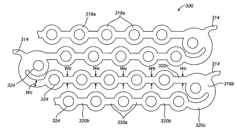
FIG. 14 illustrates an alternative embodiment of an expandable medical device
or
stent 300 in which the number of ductile hinges is increased and the length of
the
substantially non-deforming struts is decreased to form a stent with the
bending forces
distributed over a larger area. Such designs can be useful in stents
constructed of materials
which have limited ductility (elongation to failure). For example, materials
having
elongation to failures of less than about 35%, especially less than 25% can be
used to
manufacture a stent having ductile hinges when the number of ductile hinges or
the total
volume of material over which elastic and plastic strain energies are
distributed is increased.
FIG. 14, shows a portion of an expandable cylindrical tube which is formed by
a
repeating pattern of interconnected hole containing sections and ductile
hinges. A plurality of
the cylindrical tubes of FIG. 14 can be interconnected by bridging elements
314, such as the
S-shaped bridging elements shown in the embodiment of FIGS. 1 and 2. The
bridging
elements 314 allow the tissue supporting device to bend axially.
The stent 300 includes substantially rigid or non-deforming sections 318a and
318b
interconnected by ductile hinges 320a, 320b, and 320c. The substantially non-
deforming
sections 318a and 318b have a width greater than widths of the adjacent
ductile hinges 320a,
320b, and 320c which are reduced sections acting as stress/strain
concentration features. The
increased number of ductile hinges in each U-shaped section of the stent 300,
compared to
-22-
CA 02569607 2006-12-06
WO 2005/120397 PCT/US2005/019663
the previous embodiments, allows the distribution of the bending over a larger
area. Less
concentrated bending and less strain per area of the hinges allows the stent
300 to be designed
for use with materials which have stress-strain curves lower than those of
stainless steel and
cobalt chromium alloys.
In the embodiment of FIGS. 1 and 2, each alternating U-shaped segment of the
cylindrical tube 12 has two ductile hinges 20 which interconnect two rigid
struts 18 and one
rigid link 22. In contrast, the stent 300 of FIG. 14 includes in each
alternating U-shaped
segment at least three rigid sections 318a and 318b and at least three ductile
hinges 320a-c.
The structures of the ductile hinges 320 and in particular, the widths of the
hinges, are
selected to distribute deformation throughout all the hinges and prevent
concentrated bending
in particular hinges. To achieve uniform deformation of the hinges, the hinge
widths vary
with the ductile hinges 320a farthest from an apex of the U-shaped segment
having the
smallest width Wa. The hinges 320b have a width Wb which is larger than Wa,
and the
hinges 320c have a width We which is larger than Wb. This change in the widths
of the
hinges is a function of the axial position of the hinge on the cylindrical
element. Although
uniform hinges have been shown, some or all of the hinges can also be tapered
to further
achieve uniform distribution of bending through the hinges of the structure.
As shown in FIG. 14, each of the substantially non-deforming sections 318a are
circular in shape with circular openings 324 therein containing the beneficial
agent. The
substantially non-deforming sections 318a are staggered in adjacent legs of
the U-shaped
structures to achieve a compact structure. However, other shapes of the non-
deforming
sections 318a and the holes, such as rectangular, oval, or polygonal can also
be used. The
non-deforming sections 318b positioned near the apex of the U-shaped
structures are formed
as a part of the U-shapes. These non-deforming sections 318b can also take on
different
shapes. As shown the non-deforming sections 318b project to an interior of the
U-shapes,
however, one or more non-deforming sections can also project to an exterior of
the U-shapes.
The number of openings 324 per non-deforming section 318a and 318b can be one,
as
shown, or two or more. In addition, non-deforming sections with different
numbers and
shapes of openings can be combined in one structure. In addition to the non-
deforming
-23-
CA 02569607 2006-12-06
WO 2005/120397 PCT/US2005/019663
sections containing agent within the cylindrical tubes, the bridging elements
314 can also
contain one or more substantially non-deforming sections with openings therein
to provide
agent distributed substantially uniformly along the length of the stent.
FIG. 15 illustrates and alternative embodiment of a stent 400 having a
structure
formed of a plurality of interconnected U-shaped structures formed from non-
deforming
sections 418 and ductile hinges 420 similar to those shown in FIG. 14. The non-
deforming
sections 418 include one or more beneficial agent containing opening 424.
In FIG. 15, the stent 400 is shown laid flat for ease of illustration.
However, when the
stent is formed, such as by laser cutting from a tube, the structure
illustrated in FIG. 14 is
curved about a longitudinal axis X. The adjacent U-shaped structures of the
stent 400 are
each offset in the longitudinal direction from the adjacent U-shaped
structures to form a
continuous spiral ribbon or helical band. The structure of the stent 400 can
eliminate or
reduce the use of bridging elements by forming a continuous band of
alternating U-shaped
structures where the band can be formed in a cylindrical helical structure.
THERAPEUTIC AGENTS
The present invention can be used for delivery of anti-restenotic agents
including
taxol, rapamycin, other limus drugs, cladribine, colchicines, vinca alkaloids,
heparin,
hinrudin and their derivatives, as well as other cytotoxic or cytostatic
agents and microtubule
stabilizing and microtubule inhibiting agents. Although anti-restenotic agents
have been
primarily described herein, the present invention may also be used to deliver
other agents
alone or in combination with anti-restenotic agents.
Other therapeutic agents for use with the present invention may, for example,
take the
form of small molecules, peptides, lipoproteins, polypeptides, polynucleotides
encoding
polypeptides, lipids, protein-drugs, protein conjugate drugs, enzymes,
oligonucleotides and
their derivatives, ribozymes, other genetic material, cells, antisense
oligonucleotides,
monoclonal antibodies, platelets, prions, viruses, bacteria, eukaryotic cells
such as endothelial
cells, stem cells, ACE inhibitors, monocyte/macrophages and vascular smooth
muscle cells.
Such agents can be used alone or in various combinations with one another. For
instance,
-24-
CA 02569607 2006-12-06
WO 2005/120397 PCT/US2005/019663
anti-inflammatories may be used in combination with antiproliferatives to
mitigate the
reaction of tissue to the antiproliferative. The therapeutic agent may also be
a pro-drug,
which metabolizes into the desired drug when administered to a host. In
addition, therapeutic
agents may be pre-formulated as microcapsules, microspheres, microbubbles,
liposomes,
niosomes, emulsions, dispersions or the like before they are incorporated into
the matrix.
Therapeutic agents may also be radioactive isotopes or agents activated by
some other form
of energy such as light or ultrasonic energy, or by other circulating
molecules that can be
systemically administered.
Exemplary classes of therapeutic agents include antiproliferatives,
antithrombins (i.e.,
thrombolytics), immunosuppressants, antilipid agents, anti-inflammatory
agents,
antineoplastics including antimetabolites, antiplatelets, angiogenic agents,
anti-angiogenic
agents, vitamins, antimitotics, metalloproteinase inhibitors, NO donors,
nitric oxide release
stimulators, anti-sclerosing agents, vasoactive agents, endothelial growth
factors, beta
blockers, hormones, statins, insulin growth factors, antioxidants, membrane
stabilizing
agents, calcium antagonists (i.e., calcium channel antagonists), retinoids,
anti-macrophage
substances, antilymphocytes, cyclooxygenase inhibitors, immunomodulatory
agents,
angiotensin converting enzyme (ACE) inhibitors, anti-leukocytes, high-density
lipoproteins
(HDL) and derivatives, cell sensitizers to insulin, prostaglandins and
derivatives, anti-TNF
compounds, hypertension drugs, protein kinases, antisense oligonucleotides,
cardio
protectants, petidose inhibitors (increase blycolitic metabolism), endothelin
receptor agonists,
interleukin-6 antagonists, anti-restenotics, and other miscellaneous
compounds.
Antiproliferatives include, without limitation, sirolimus, paclitaxel,
actinomycin D,
rapamycin, and cyclosporin.
Antithrombins include, without limitation, heparin, plasminogen, a2-
antiplasmin,
streptokinase, bivalirudin, and tissue plasminogen activator (t-PA).
Immunosuppressants include, without limitation, cyclosporine, rapamycin and
tacrolimus (FK-506), sirolumus, everolimus, etoposide, and mitoxantrone.
-25-
CA 02569607 2006-12-06
WO 2005/120397 PCT/US2005/019663
Antilipid agents include, without limitation, HMG CoA reductase inhibitors,
nicotinic
acid, probucol, and fibric acid derivatives (e.g., clofibrate, gemfibrozil,
gemfibrozil,
fenofibrate, ciprofibrate, and bezafibrate).
Anti-inflammatory agents include, without limitation, salicylic acid
derivatives (e.g.,
aspirin, insulin, sodium salicylate, choline magnesium trisalicylate,
salsalate, dflunisal,
salicylsalicylic acid, sulfasalazine, and olsalazine), para-amino phenol
derivatives (e.g.,
acetaminophen), indole and indene acetic acids (e.g., indomethacin, sulindac,
and etodolac),
heteroaryl acetic acids (e.g., tolmetin, diclofenac, and ketorolac),
arylpropionic acids (e.g.,
ibuprofen, naproxen, flurbiprofen, ketoprofen, fenoprofen, and oxaprozin),
anthranilic acids
(e.g., mefenamic acid and meclofenamic acid), enolic acids (e.g., piroxicam,
tenoxicam,
phenylbutazone and oxyphenthatrazone), alkanones (e.g., nabumetone),
glucocorticoids (e.g.,
dexamethaxone, prednisolone, and triamcinolone), pirfenidone, and tranilast.
Antineoplastics include, without limitation, nitrogen mustards (e.g.,
mechlorethamine,
cyclophosphamide, ifosfamide, melphalan, and chlorambucil), methylnitrosoureas
(e.g.,
streptozocin), 2-chloroethylnitrosoureas (e.g., carmustine, lomustine,
semustine, and
chlorozotocin), alkanesulfonic acids (e.g., busulfan), ethylenimines and
methylmelamines
(e.g., triethylenemelamine, thiotepa and altretamine), triazines (e.g.,
dacarbazine), folic acid
analogs (e.g., methotrexate), pyrimidine analogs (5-fluorouracil, 5-
fluorodeoxyuridine, 5-
fluorodeoxyuridine monophosphate, cytosine arabinoside, 5-azacytidine, and
2',2'-
difluorodeoxycytidine), purine analogs (e.g., mercaptopurine, thioguanine,
azathioprine,
adenosine, pentostatin, cladribine, and erythrohydroxynonyladenine),
antimitotic drugs (e.g.,
vinblastine, vincristine, vindesine, vinorelbine, paclitaxel, docetaxel,
epipodophyllotoxins,
dactinomycin, daunorubicin, doxorubicin, idarubicin, epirubicin, mitoxantrone,
bleomycins,
plicamycin and mitomycin), phenoxodiol, etoposide, and platinum coordination
complexes
(e.g., cisplatin and carboplatin).
Antiplatelets include, without limitation, insulin, dipyridamole, tirofiban,
eptifibatide,
abciximab, and ticlopidine.
Angiogenic agents include, without limitation, phospholipids, ceramides,
cerebrosides, neutral lipids, triglycerides, diglycerides, monoglycerides
lecithin, sphingosides,
-26-
CA 02569607 2006-12-06
WO 2005/120397 PCT/US2005/019663
angiotensin fragments, nicotine, pyruvate thiolesters, glycerol-pyruvate
esters,
dihydoxyacetone-pyruvate esters and monobutyrin.
Anti-angiogenic agents include, without limitation, endostatin, angiostatin,
fumagillin
and ovalicin.
Vitamins include, without limitation, water-soluble vitamins (e.g., thiamin,
nicotinic
acid, pyridoxine, and ascorbic acid) and fat-soluble vitamins (e.g., retinal,
retinoic acid,
retinaldehyde, phytonadione, menaqinone, menadione, and alpha tocopherol).
Antimitotics include, without limitation, vinblastine, vincristine, vindesine,
vinorelbine, paclitaxel, docetaxel, epipodophyllotoxins, dactinomycin,
daunorubicin,
doxorubicin, idarubicin, epirubicin, mitoxantrone, bleomycins, plicamycin and
mitomycin.
Metalloproteinase inhibitors include, without limitation, TIMP-1, TIMP-2, TIMP-
3,
and SmaPI.
NO donors include, without limitation, L-arginine, amyl nitrite, glyceryl
trinitrate,
sodium nitroprusside, molsidomine, diazeniumdiolates, S-nitrosothiols, and
mesoionic
oxatriazole derivatives.
NO release stimulators include, without limitation, adenosine.
Anti-sclerosing agents include, without limitation, collagenases.and
halofuginone.
Vasoactive agents include, without limitation, nitric oxide, adenosine,
nitroglycerine,
sodium nitroprusside, hydralazine, phentolamine, methoxamine, metaraminol,
ephedrine,
trapadil, dipyridamole, vasoactive intestinal polypeptides (VIP), arginine,
and vasopressin.
Endothelial growth factors include, without limitation, VEGF (Vascular
Endothelial
Growth Factor) including VEGF-121 and VEG-165, FGF (Fibroblast Growth Factor)
including FGF-1 and FGF-2, HGF (Hepatocyte Growth Factor), and Angl
(Angiopoietin 1).
Beta blockers include, without limitation, propranolol, nadolol, timolol,
pindolol,
labetalol, metoprolol, atenolol, esmolol, and acebutolol.
Hormones include, without limitation, progestin, insulin, the estrogens and
estradiols
(e.g., estradiol, estradiol valerate, estradiol cypionate, ethinyl estradiol,
mestranol, quinestrol,
estrond, estrone sulfate, and equilin).
-27-
CA 02569607 2006-12-06
WO 2005/120397 PCT/US2005/019663
Statins include, without limitation, mevastatin, lovastatin, simvastatin,
pravastatin,
atorvastatin, and fluvastatin.
Insulin growth factors include, without limitation, IGF-1 and IGF-2.
Antioxidants include, without limitation, vitamin A, carotenoids and
vitamin E.
Membrane stabilizing agents include, without limitation, certain beta blockers
such as
propranolol, acebutolol, labetalol, oxprenolol, pindolol and alprenolol.
Calcium antagonists include, without limitation, amlodipine, bepridil,
diltiazem,
felodipine, isradipine, nicardipine, nifedipine, nimodipine and verapamil.
Retinoids include, without limitation, all-trans-retinol, all-trans-14-
hydroxyretroretinol, all-trans-retinaldehyde, all-trans-retinoic acid, all-
trans-3,4-
didehydroretinoic acid, 9-cis-retinoic acid, 11-cis-retinal, 13-cis-retinal,
and 13-cis-retinoic
acid.
Anti-macrophage substances include, without limitation, NO donors.
Anti-leukocytes include, without limitation, 2-CdA, IL-1 inhibitors, anti-
CD116/CD18 monoclonal antibodies, monoclonal antibodies to VCAM, monoclonal
antibodies to ICAM, and zinc protoporphyrin.
Cyclooxygenase inhibitors include, without limitation, Cox-1 inhibitors and
Cox-2
inhibitors (e.g., CELEBREX and VIOXX ).
Immunomodulatory agents include, without limitation, immunosuppressants (see
above) and immunostimulants (e.g., levamisole, isoprinosine, Interferon alpha,
and
Interleukin-2).
ACE inhibitors include, without limitation, benazepril, captopril, enalapril,
fosinopril
sodium, lisinopril, quinapril, ramipril, and spirapril.
Cell sensitizers to insulin include, without limitation, glitazones, P par
agonists and
metformin.
Antisense oligonucleotides include, without limitation, resten-NG.
-28-
CA 02569607 2006-12-06
WO 2005/120397 PCT/US2005/019663
Cardio protectants include, without limitation, VIP, pituitary adenylate
cyclase-
activating peptide (PACAP), apoA-I milano, amlodipine, nicorandil,
cilostaxone, and
thienopyridine.
Petidose inhibitors include, without limitation, omnipatrilat.
Anti-restenotics include, without limitation, include vincristine,
vinblastine,
actinomycin, epothilone, paclitaxel, and paclitaxel derivatives (e.g.,
docetaxel).
Miscellaneous compounds include, without limitation, Adiponectin.
While the invention has been described in detail with reference to the
preferred
embodiments thereof, it will be apparent to one skilled in the art that
various changes and
modifications can be made and equivalents employed, without departing from the
present
invention.
-29-