Note: Descriptions are shown in the official language in which they were submitted.
DEMANDE OU BREVET VOLUMINEUX
LA PRESENTE PARTIE DE CETTE DEMANDE OU CE BREVET COMPREND
PLUS D'UN TOME.
CECI EST LE TOME 1 DE 2
CONTENANT LES PAGES 1 A 23
NOTE : Pour les tomes additionels, veuillez contacter 1e Bureau canadien des
brevets
JUMBO APPLICATIONS/PATENTS
THIS SECTION OF THE APPLICATION/PATENT CONTAINS MORE THAN ONE
VOLUME
THIS IS VOLUME 1 OF 2
CONTAINING PAGES 1 TO 23
NOTE: For additional volumes, please contact the Canadian Patent Office
NOM DU FICHIER / FILE NAME
NOTE POUR LE TOME / VOLUME NOTE:
CA 02570108 2006-12-11
WO 2006/085921 PCT/US2005/020383
POLYGONAL NANOSTRUCTURES OF POLYNUCLEIC ACID MULTI-CROSSOVER
MOLECULES AND ASSEMBLY OF LATTICES BASED ON DOUBLE CROSSOVER
COHESION
GOVERNMENT LICENSE RIGHTS
[0001] The experiments reported in this application were
supported in part by: the National institute of General Medical
Sciences, grant no. GM-29554; the Office of Naval Research, grant
no. N00014-98-1-0093; the National Science Foundation, grant nos.
DMI-0210844, EIA-0086015, DMR-01138790 and CTS-0103002; and
DARPA/AFSOR, grant no. F30602-01-2-0561. The U.S. Government has
a paid-up license in this invention and the right in limited
circumstances to require the patent owner to license others on
reasonable terms as provided for by the terms of the above
grants.
BACKGROUND OF THE INVENTION
Field of the Invention
[0002] The present invention relates to polynuCleiC acid
nanostructures and lattices.
Description of the Related Art
[0003] The control of the structure of matter on the finest
possible scale requires the successful design of both stiff
intramolecular motifs and robust intermolecular interactions.
Previous motifs used to design 2D crystalline arrays have
included the double crossover (DX) (Fu et al., 1993; Winfree et
al., 1998), triple crossover (TX) (LaBean et al., 2000), the DNA
parallelogram (Mao et al., 1999), and the four-by-four structure
(Yan et al., 2003). These motifs have been used to produce 2D
crystalline arrays lacking symmetry or with twofold symmetry
(Seeman, 2003). By contrast, all previous attempts to produce
trigonal or hexagonal arrays have met with failure or produced
CA 02570108 2006-12-11
WO 2006/085921 PCT/US2005/020383
only very tiny structures. Given the inherent rigidity of
triangles, the importance of trigonal motifs in nature (Kappraff
et al., 1990), it is key to solve this problem. The flexibility
of 3-arm junctions was discovered in the first attempt to
assemble a hexagonal lattice (Ma et al., 1986). Triangles built
from bulged 3-arm junctions (Liu et al., 1994) demonstrated
cyclic closure with trimers and above, not just from the hexamers
one would have expected (Qi et al., 1996). Triangles whose edges
were flanked by coplanar helices derived from DX molecules
behaved in a similar fashion (Yang et al., 1998).
[0004] Brun et al. (2004), reported experimental evidence of
two new complexes, quadruple crossovers and triangles, where
atomic force microscopy images (AFM) show that the triangles are
capable of hexagonally tiling the plane. However, the triangular
units used by Brun et al. to form a hexagonal lattice have single
nucleic acid helices for its edge and are not robust, as the AFM
image of the lattice formed appears to show that some pentagons
and squares are present in the lattice.
[0005] Citation of any document herein is not intended as an
admission that such document is pertinent prior art, or
considered material to the patentability of any claim of the
present application. Any statement as to content or a date of
any document is based on the information available to applicant
at the time of filing and does not constitute an admission as to
the correctness of such a statement.
SUMMARY OF THE INVENTION
[0006] The present invention provides a polynucleic acid
structure which is composed of one or more polygonal units. Each
polygonal unit has, as its edges, connected nucleic acid multi-
crossover domains. Each edge of a polygonal unit has at least
2
CA 02570108 2006-12-11
WO 2006/085921 PCT/US2005/020383
one free end (extension of the edge) with two parallel nucleic
acid helices terminating in a double cohesive (sticky) end.
[0007] The invention also provides a method for producing the
polynucleic acid structure according to the present invention
which involves mixing single stranded polynucleotides, each being
designed to be self-complementary and/or complementary to another
single stranded polynucleotide so as to be capable of self-
annealing into a polygonal unit, and annealing the mixture after
heat denaturation to form the polygonal unit. The method may
further involve the self-assembly of an array of polygonal units
by annealing complementary exposed cohesive ends on the polygonal
units.
BRIEF DESCRIPTION OF THE DRAWINGS
[0008] Figures 1A-lE illustrate the following motifs: the DX
motif (Fig. 1A); the bulged junction triangle (Fig. 1B); the DX
triangle (Fig. 1C); a trigonal arrangement of six DX triangles of
two different species (Fig. 1D); a schematic trigonal lattice of
the two triangles shown in Fig. 1D (Fig. 1E).
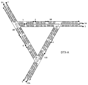
[0009] Figures 2A and 2B schematically show the arrangement
and nucleotide sequences of two DNA DX triangles, DTX-A (Fig. 2A;
SEQ ID NOs:1-13) and DTX-B (Fig. 2B; SEQ ID NOs:1-2, 4, 6, 9, 11,
and 13-19).
[0010] Figures 3A-3F present Atomic Force Microscopy (AFM)
images of pseudo-hexagonal trigonal arrays. Field sizes are
indicated in the upper right corners. Fig. 3A shows a pair of 2D
arrays. The honey-comb nature of the arrays are evident. Fig. 3B
is a zoom (enlargement) of the array shown on the right in Fig.
3A. Fig. 3C is a zoom (enlargement) of another array. Fig. 3D
shows an image containing two stacked arrays, virtually complete
on the lower right, partial on the upper left. Fig. 3E is a
zoomed (enlarged) image containing 15 DX triangles. Fig. 3F is a
3
CA 02570108 2006-12-11
WO 2006/085921 PCT/US2005/020383
further zoom (enlargement) of Fig. 3E showing six complete
triangles, similar to the arrangement in Fig. 1D, and with a
center-center hexagon outline superimposed.
[0011] Figure 4 is an illustration showing the 3D character of
a DX triangle. Each edge consists of a DX molecule (two fused
DNA double helices). Each edge is below one DX and above
another; for example, the horizontal edge at the top lies above
the diagonal DX on the left and below the diagonal DX on the
right. The central axes of the three DX edges span 3-space.
[0012] Figure 5 schematically shows the arrangement and
nucleotide sequences of 3D DX triangle (SEQ ID NOs: 97-118).
[0013] Figures 6A-6C show three different sections of 2D AFM
images corresponding to eliminating cohesive ends from each
different direction. Note the well-formed arrays in each
section, with the best array from the middle section (Fig. 5B).
Dimensions flanking the images are in microns.
[0014] Figures 7A and 8B show illustrations of a 6-helix
bundle down its central axis (Fig. 7A), and along its side (Fig.
7B). It can be seen that it is just a fused set of DX molecules,
at 120° to each other.
[0015] Figure 8 schematically shows the arrangement and
nucleotide sequences of the 6-helix bundle (SEQ ID NOs:20-31)
presented in Figs. 7A and 7B.
[0016] Figures 9A-9C are AFM images of three sets of 2D
sections for the 6-helix bundle.
[0017] Figures 10A-10C show illustrations of skewed TX
triangles. One side of the skewed TX triangle is shown in Fig.
10A. It is clearly made of a pair of DX ends fused by the TX
motif at the center. Fig. lOB has one side (closest to the
reader) in a similar orientation as in Fig. 10A, but the other
two sides have been added, including one side viewed edge on. It
4
CA 02570108 2006-12-11
WO 2006/085921 PCT/US2005/020383
is evident that this motif spans 3-space. Fig. 10C is a top view
of the trigonal motif.
[0018] Figure 11 schematically shows the arrangement and
nucleotide sequences of the skewed TX triangle (SEQ ID NOs:32-64)
presented in Figs. 10A-10C.
[0019] Figures 12A-12C are AFM images of three 2D sections of
the skewed TX triangle shown in Figs. 12A-12C. The 2D patterning
is shown most clearly in Fig. 12B, whereas the other two (Figs.
12A and 12C) are not well-formed arrays.
[0020] Figure 13 shows an illustration of a DX parallelogram
(PDX-E-E) with two turns beyond the vertices and 8 between them
in both directions.
[0021] Figure 14 schematically shows the arrangement and
nucleotide sequences (SEQ ID NOs:65-96) of the DX parallelogram
(PDX-E-E) presented in Fig. 13.
[0022] Figures 15A and 15B are AFM images of a view (Fig. 15A)
and a zoom (Fig. 15B) of the 2D lattice formed from the motif
shown in Figs. 13 and 14.
[0023] Figure 16 shows an illustration of a DX parallelogram
(PDX-E-O) with a repeating pattern of alternating even and odd
numbers of half helical turns between junctions.
[0024] Figure 17 schematically shows the arrangement shows the
arrangement and nucleotide sequences (SEQ ID NOs:ll9 to 152) of a
DX parallelogram (PDX-E-O) presented in Fig. 16.
[0025] Figures 18A-18D are AFM images of the 2D lattice formed
from the PDX-E-O parallelogram motif shown in Figs. 16 and 17.
DETAILED DESCRIPTION OF THE INVENTION
[0026] The polynucleic acid structures of the present
invention are polynucleic acids that are assembled to form
branched multimers of repeating units composed at least partially
CA 02570108 2006-12-11
WO 2006/085921 PCT/US2005/020383
of mufti-crossover molecules in accordance with the method of the
present invention.
[0027] A plurality of mufti-crossover molecules, which form a
basic unit of a robust nucleic acid motif, such as a nucleic acid
triangle, are assembled from single stranded oligonucleotides or
polynucleotides to produce the polynucleic acid unit molecules of
the present invention. Similarly, more complex polynucleic acid
structures of the present invention having two dimensional or
three dimensional periodic lattices with symmetrical
intermolecular contacts (translational symmetry) are assembled
from basic units of linked mufti-crossover molecules.
[0028] The term "robust" as used herein is meant to refer to
producing the designed structure exclusively, and no others.
This applies not only to motifs but also to structures such as
arrays and lattices. For instance, if a DX triangle is designed,
then its component strands will only self-assembled into the
designed DX triangle motif/structure.
[0029] DNA molecules containing two crossover sites between
helical domains have been widely suggested as intermediates in
recombination processes involving double stranded breaks.
Accordingly, "double crossover molecules" are those nucleic acid
molecules containing two branched junctions (Holliday junctions
corresponding to the crossover sites) linked together by ligating
two of their double helical arms. By branched junction is meant
a point from which three or more helices (arms) radiate.
[0030] There are five isomers of double crossover molecules
(Fu et al., 1993), which fall into two broad classes of molecules
differentiated by the relative orientations, parallel (DP) or
antiparallel (DA), of their helix axes. As parallel double
helical molecules are usually not well behaved, antiparallel
isomers of double crossover molecules are the preferred building
block components intended to be used in the present invention.
6
CA 02570108 2006-12-11
WO 2006/085921 PCT/US2005/020383
However, parallel double helical molecules may be suitable as
well.
[0031] The present inventors have now developed a new motif,
the DX triangle, which is capable of forming a trigonal array.
This motif is derived by combining the DX motif (Fig. 1A) with
the bulged triangle motif (Fig. 1B). The resulting motif is
illustrated in Fig. 1C. The DX molecule has been shown to be
about twice as stiff as conventional linear duplex DNA (Li et
al., 2002; Sa-Ardyen et al., 2003). Thus, one might expect that
this doubly-thick triangle would be more rigid than the simple
bulged junction triangle. In addition, the DX triangle is
capable of a double intermolecular interaction that may be more
robust than the single helical interactions used previously,
because it is less sensitive to errors in twist. The self-
assembly of a trigonal .array from this motif is shown in Example
1 hereinbelow. Example 1 demonstrates that improving or
stabilizing the intermolecular contacts is the key feature of the
DX triangle motif that enables formation of trigonal arrays.
[0032] The DX triangle and the trigonal arrays or lattices
formed from this motif as mentioned above and disclosed in
Example 1 hereinbelow are preferred embodiments of the
polynucleic acid structure of present invention. It is intended
that the polynucleic acid structure of the present invention
encompass not only DX triangle motifs and trigonal
arrays/lattices formed therefrom but also other multi-crossover
motifs, such as but not limited to, a skewed TX-DX triangle and a
DX parallelogram disclosed in Example 2 hereinbelow, and
arrays/lattices formed therefrom.
[0033] The polynucleic acid structure of the present invention
is composed of one or more polygonal units. When only a single
polygon is present, the polygonal polynucleic acid structure is a
unit building block for forming arrays and lattices, whereas
7
CA 02570108 2006-12-11
WO 2006/085921 PCT/US2005/020383
plural linked polygonal units can be the array or lattice or can
be used to further extend the array or lattice in two or three
dimensions.
[0034] Each polygonal unit has, as its edges, connected
nucleic acid multi-crossover domains. The terms "edge" or
"edges" are used synonymously with the terms "side" or sides"
when referring to geometrical structures such as a polygon. A
polygon as used herein is a closed geometrical structure having
three or more edges or sides. While a polygon is generally
thought to be confined to a plane, it is intended for the
purposes of the present invention to include motifs such as the
three dimensional DX triangle and skewed TX-DX triangle shown in
Figs. 5, 10C and 11 (polygonal when viewed from above).
[0035] As would be recognized and appreciated by those of
skill in the art, although the edges of each polygonal unit may
be described as being formed by one or more nucleic acid multi-
crossover molecule, it may not be possible to identify the
discrete limits of individual nucleic acid mufti-crossover
molecules; rather, it may be more appropriate to think of
connected nucleic acid mufti-crossover domains forming the edges
of a polygonal unit. This is more consistent with the manner in
which polynucleic acid structures are produced according to the
present invention, where individual nucleic acid strands self-
assemble to form a polygonal unit based on sequence
complementarity. Accordingly the edges are not formed as
individual molecules to be linked together but rather are self-
assembled as a whole into a polygonal unit.
[0036] Each edge or side of the polygonal unit has at least
one free end with two parallel helices. A "free end" is intended
to mean an extension of an edge beyond a vertex where one edge is
connected to another edge of the polygonal unit. Each free end
has at least two parallel nucleic acid double helices where at
8
CA 02570108 2006-12-11
WO 2006/085921 PCT/US2005/020383
least two of the parallel helices each terminate in a cohesive or
sticky end. When a free end has only two parallel helices, then
the free end has a double cohesive end which can cohere with
another double cohesive end that is complementary. The double
cohesive ends can be the same or different cohesive ends. Each
edge can alternatively have both of its ends as free ends. As
another embodiment, a polygonal unit can have edges with one free
end, edges with two free ends, edges with no free ends, or a
combination thereof.
[0037] The nucleic acid multi-crossover domains preferably can
be double or triple crossover domains or a combination thereof,
such as exemplified by the skewed TX-DX triangle presented in
Example 2 hereinbelow.
[0038] The polygonal unit can be any polygon that can be
suitably extended from two or more of its edges to join other
polygonal units.and form an array or lattice. Preferably, the
polygonal unit is a triangle or a parallelogram, although it is
not limited to such.
[0039] A preferred embodiment of the polynucleic acid
structure of the present invention is an array of triangular
units linked together by complementary double cohesive ends to
form a trigonal array. More preferably, the array is a trigonal
array of two different triangular units. Another preferred
embodiment is an array of parallelogram units linked by
complementary double cohesive ends.
[0040] The present invention further provides a method for
producing a polynucleic acid structure according to the present
invention. This method involves synthesizing single stranded
polynucleotides, each being designed to be self-complementary
and/or complementary to another single stranded polynucleotide so
as to be able to self anneal into a polygonal unit; mixing the
single stranded polynucleotides to form a mixture of
9
CA 02570108 2006-12-11
WO 2006/085921 PCT/US2005/020383
polynucleotides; heat denaturing the mixture; and annealing the
heat denatured mixture of single stranded polynucleotides to form
the polygonal unit.
[0041] Single stranded polypeptides are mixed together and
heated at a temperature above the melting temperature or
denaturation temperature of the complementary strands, e.g.,
90°C, to eliminate any initial secondary structures present in
the mixture, and then cooled slowly to allow the strands to
anneal based on sequence complementarity.
[0042] Once the polygonal units are self-assembled, the
assembled polygonal units can form arrays and lattices based on
joining of double cohesive ends on polygonal units. The self-
assembled, polygonal units are first heated to ensure that the
double cohesive ends are exposed, and then the exposed double
cohesive ends that are complementary are annealed to form an
array of polygonal units. More than one polygonal unit, such as
different polygonal units, can be mixed to form an array of
different polygonal units.
[0043] It should also be understood that when synthesizing the
single stranded oligonucleotides or polynucleotides for forming
the topologically closed nucleic acid structure, the choice of
sequence is substantially arbitrary, provided that strands
intended to form a hairpin or to be opposite one another are
complementary. It is preferable to use previously described
symmetry minimization algorithms (Seeman, 1990; Seeman, 1981 and
1982) in order to optimize the sequences and incorporate the
desired features while avoiding unwanted cross-hybridization or
branch migration.
[0044] It should also be appreciated that the term "nucleic
acid" refers to both DNA and RNA and hybrids of the two. The
structure need not resemble anything which can theoretically be
made from nature. A particular oligonucleotide or polynucleotide
to
CA 02570108 2006-12-11
WO 2006/085921 PCT/US2005/020383
strand may employ bases other than the standard five, adenine,
cytosine, guanine, thymine and uracil. Derivatized (e. g.,
methylated) and other unusual bases such as iso-guanine, iso-
cytosine, amino-adenine, K, X, ~r, (Piccirilli et al., 1990),
inosine and other derivatives of purine and pyrimidine may be
used. A preferable feature in the selection of the bases is that
they be capable of interacting with a base opposing them to form
a specifically paired attraction. In natural DNA and RNA,
hydrogen bonding forms this interaction. However, opposite ion
charges, hydrophobic interactions and van der Waals forces may
also be acceptable forms of interaction. These interactions
expand the choices over naturally occurring bases to give a wider
assortment of physical properties.
[0045] Within a particular strand, the heterocyclic base may
be entirely missing from the sugar moiety. This may be
particularly desirable where the strands bend, form a junction,
or where one desires fewer forces holding the strands together.
[0046] A particular strand need not have a single contiguous
ribose-phosphate or deoxyribose-phosphate backbone. It could be
a peptide nucleic acid with a peptide backbone. One may employ a
simple inorganic or organic moiety or polymeric spacer between
segments of polynucleotide. Spacers such as polyethylene,
polyvinyl polymers, polypropylene, polyethylene glycol,
polystyrene, polypeptides (enzymes, antibodies, etc.) peptide
nucleic acids (PNA), polysaccharides (starches, cellulose, etc.)
silicones, silanes and copolymers, etc., may be employed. An
example of such a hybrid structure is dodecadiol having
phophoramidite at one end. This structure has been inserted
covalently instead of four T nucleotides to form a hairpin loop
in a fashion similar to the nucleotides it replaces. See Mitchel
J. Doktycz, Ph.D. Thesis (1991), University of Illinois, Chicago.
11
CA 02570108 2006-12-11
WO 2006/085921 PCT/US2005/020383
The term "oligonucleotide", "polynucleotide" and "nucleic acid"
are intended to cover all of these structures.
[0047] In nature and the field of molecular biology, double
stranded DNA generally occurs in the B form. However, for the
purposes of this invention it may be desirable for DNA or other
double stranded polynucleotide to exist in the A, C, D or Z form.
Various bases, derivations and modifications may be used to
stabilize the structure in the A, C, D or Z form as well.
[0048] Three dimensional polynucleic acid structures are
particularly well suited for use as a scaffolding medium since
they are stiff molecules unlikely to be perturbed markedly by
tethering smaller non-interactive molecules to it. Another
application for this structure is in the formation of
polycatenated polymers.
[0049] The structure also makes a suitable material for
immobilizing enzymes and other catalysts. By employing an open
design for the structure, one or more enzymes may be bound to the
structure and still permit free mobility of substrates and
products to and from the enzyme. Instead of binding the enzyme
directly to the structure, the structure may form a cage to
entrap the enzyme(s). This technique has additional advantages
of not modifying the enzyme.
[0050] Conventional enzyme immobilization techniques depend on
random attachment and thus the solid phase particles formed are
not uniform in either activity or structure. By contrast, one
can attach a predetermined number of enzymes to the
polynucleotide strands being added to form a structure with a
fixed number and orientation of enzymes.
[0051] The structure may be so formed to create a mesh or
screen-like material. This material can be used as a filter of
very precise porosity. For added strength, plural layers of mesh
12
CA 02570108 2006-12-11
WO 2006/085921 PCT/US2005/020383
may be linked together or a layer may be bound to any other
conventional substrate.
[0052] The structures of and produced by the present invention
have numerous two dimensional and three dimensional structural
uses. Because of the minute size of the structures, they have
application in the field of nanotechnology.
[0053] More current uses include use as a solubilizer or
stabilizer for chemicals, particularly pharmaceuticals. For
example, a drug may be bound to the interior of a three
dimensional polynucleic acid structure. Since DNA degrades in
acidic conditions and RNA degrades in alkaline conditions, one
can direct the drug to be released in whatever part of the
digestive system desired.
[0054] Having now generally described the invention, the same
will be more readily understood through reference to the
following examples which are provided by way of illustration and
are not intended to be limiting of the present invention.
EXAMPLE 1
Trigonal 2D DNA Crystals Based on Double Crossover Cohesion
[0055] Two-dimensional pseudo-hexagonal trigonal arrays have
been constructed by self-assembly from DNA. The motif used is a
bulged-junction DNA triangle whose edges and extensions are DNA
double crossover (DX) molecules, rather than conventional DNA
double helices. The experiments described below in this example
were performed to establish whether the success of this system
results from the added stiffness of DX molecules or the presence
of two sticky ends at the terminus of each edge. Removal of one
sticky end precludes lattice formation, suggesting that it is the
double sticky end that is the primary factor enabling lattice
formation.
13
CA 02570108 2006-12-11
WO 2006/085921 PCT/US2005/020383
MATERIALS AND METHODS
[0056] The strands were synthesized by conventional
phosphoramidite procedures (Caruthers, 1985), and were purified
by denaturing polyacrylamide gel electrophoresis. Stoichiometric
mixtures of the strands (estimated by OD~6o) for each triangle
were prepared separately to a concentration of 0.5 ~M in a
solution containing 40 mM Tris-HCl, pH 8.0, 20 mM acetic acid,
2mM EDTA, and 12.5 mM magnesium acetate. Each mixture was cooled
from 90°C to room temperature in a 500 ml water bath over the
course of 48 hrs. To form the array, the two complexes DTX-A
(Fig. 2A) and DTX-B (Fig. 2B) were mixed in stoichiometric
quantities, warmed to 45°C, and cooled slowly to room temperature
in a thermos containing a 500 ml water bath over 24 hours;
sometimes the sample was cooled another 24 hours to 16°C. Atomic
Force Microscopy (AFM) imaging was performed by spotting a 5-7 ~,L
sample drop on freshly cleaved mica, which was left to adsorb to
the surface for 3 min. To remove buffer salts, 5-10 drops of
double distilled water were placed on the mica, the drop was
shaken off, and the sample was dried with compressed air.
Imaging was performed in contact mode under 2-propanol in a fluid
cell on a NanoScope IV (Digital Instruments) instrument, using
commercial cantilevers with Si3N4 tips (DI) .
RESULTS
[0057] Two triangles were designed to produce a trigonal
lattice arrangement when combined. The sequences (SEQ ID NOs:1-
19) of the triangles are presented in Figs. 2A and 2B. For
purposes of economy, some strands were used in both triangles.
The edges of the triangles contain 65 nucleotide pairs in each of
their DX helices, and they terminate in 5' sticky ends six
nucleotides in length. There are four turns per edge within each
14
CA 02570108 2006-12-11
WO 2006/085921 PCT/US2005/020383
triangle. The triangles are designed to cohere with each other
to produce a continuous DX structure 13 double helical turns (~46
nm) in length. Fig. 1D illustrates a group of six triangles,
three of each species, flanking a hexagon. The edge of the
hexagon, lacking one triangle is 9 turns (~30 nm) in length; the
center-to-center distance should be ~34 nm. Figure 1E shows the
way that the two DX triangles are designed to associate into
pseudo-hexagonal trigonal 2D arrays. The trigonal lattice shown
in Fig. 1E show an elaboration of the 6-triangle complex
illustrated in Fig. 1D.
[0058] The triangles migrate as single bands on non-denaturing
gels (data not shown). Figures 3A and 3F show atomic force
micrographs of arrays produced by the self-assembly of the
triangles.
[0059] The honeycomb structure of arrangements is evident from
the images shown in Figs. 3A-3F. The quality of the lattice is
evident in the images shown in Figs. 3A-3C. The lattices have a
certain tendency to stack on each other, as shown in Fig. 3D; the
array in the upper left illustrates this point clearly, because
the array on top is only about half the size of the array below'
it. Note that the arrays seem to stack over each other so that
the cavities appear to be continuous between layers. The zoomed
images shown in Figs. 3E-3F demonstrate clearly the hexagonal
nature of the array; the center-to-center hexagon in Fig. 3F has
an edge of ~38 nm, in good agreement with the expected length.
[0060] Given the previous failures to form uniform hexagonal
arrays or even hexagonal arrays at all, it is of central
importance to establish which of the differences between the
current system and previous systems has proved to be the key
change, the greater stiffness of the DX, or the cohesion of the
double sticky ends. To resolve this issue, the laboratory of the
present inventors have repeated these experiments by removing the
CA 02570108 2006-12-11
WO 2006/085921 PCT/US2005/020383
sticky (cohesive) ends from one of the helices on each of the
triangles. When these modified molecules were put through the
same protocols that was done with the doubly sticky-ended
triangles, the lattices of the sort shown in Figs. 3A-3F were
unable to be produced. Thus, the difference is the use of double
sticky (cohensive) ends.
[0061] The present inventors suspect that the previous
failures were due to differences between ideal and actual twists
along a single helix; two helices apparently are able to bind
successfully while maintaining the orientation of the plane
defined by the two helix axes of the DX edges. Nevertheless, the
possibility that the flexibility of the single-helical connection
contributes to the failure of those molecules to form honeycomb
arrays cannot be excluded.
[0062] Thus, the substitution of DX arms for double helical
arms leads to robust self-assembly in 2D. If this conclusion is
correct, one ought to be able to use this approach in other
motifs that have proved ineffective or difficult when used as
components of 2D arrays connected by single helical sticky ends.
The present inventors have tested this notion in a number of
systems, and found that it is correct. The present inventors
have successfully built robust 2D arrays using DX versions of a
small 3D triangle (Liu et al., 2004), a 6-helix bundle (Mathieu
et al., 2001), a large and unwieldy DNA parallelogram (Mao et
al., 1999), and a previously unreported 3D TX motif, as described
below in Example 2. The present inventors expect that the use of
this form of cohesion with double sticky (cohesive) ends will
prove of value both in two dimensional applications, and in three
dimensional assemblies as well.
16
CA 02570108 2006-12-11
WO 2006/085921 PCT/US2005/020383
EXAMPLE 2
New Systems From DX Molecules
[0063] The first three of these new systems are 3-space
spanning motifs. If one combines them along the three vectors
defined by their complementary sticky end pair directions (all
are connecting DX units in essence), a 3D solid will result. All
three motifs behave well on non-denaturing gels, migrating as a
single band.
3D DX Triangle
[0064] A DX triangle, different from the DX triangle of Fig.
1C and Figs. 2A-2B, is illustrated in Fig. 4. A schematic
illustration of a 3D DX triangle with double cohesive ends at the
free ends (extensions) of its edges is presented in Fig. 5. A
good screen for the geometrical viability of a 3D system is to
eliminate one pair of cohesive ends from that system and then to
see if it forms a good 2D array, as assayed by the AFM. If all
three 2D sections of the system are good, it is an indication
that geometrical design problems have been solved. The present
inventors have been markedly successful in this regard for the 3D
DX triangle, as shown in Figs. 6A-6C.
[0065] Some tube-formation is visible in these images, likely
because the DX motif selected (DAE--that has an even number of
half-turns between crossovers; Fu and Seeman, 1993) tends to have
internal bends; another motif (DAO--with an odd number of half-
turns; Fu and Seeman, 1993) lacking this problem has also been
developed. Note that the 2D arrays are rhombic, not trigonal,
because one direction of propagation has been eliminated.
A Six-Helix Bundle
[0066] The 10.5-fold helicity of DNA (Wang, 1979; and Rhodes
and Klug, 1980) means that 7- and 14-nucleotide separations
17
CA 02570108 2006-12-11
WO 2006/085921 PCT/US2005/020383
between features such as crossovers rotate them by 120°. This
feature was utilized to produce a 6-helix bundle of DNA (Mathieu
et al., 2001), illustrated in Figs. 7A and 7B, by combining the
designed strand sequences SEQ ID N0:20-31 as shown in Fig. 8.
[0067] The laboratory of the present inventors has made arrays
in each of the three directions with this motif, similar to the
3D DX triangle. The top two helices in front connect to the
bottom two helices in the rear, and similarly for the other two
sets. These are shown in Figs. 9A-9C. Well defined patterns are
visible, but it is clear that the overall structure of the arrays
contains many faults. The faults visible in these lattices,
particularly the middle one, are suspected to be the result of
too few crossovers between the helices near their ends.
Skewed TX Triangle
[0068] The skewed TX triangle motif is made up of TX molecules
whose helices are extended pair-wise, as shown in Fig. 10A.
Three of these molecules are put together in a skewed trigonal
fashion, spanning 3-space by combining the designed strand
sequences SEQ ID NOs: 32-64 (Fig. 11). The three 2D sections for
this motif are shown in Figs. 12A-12C.
DX Parallelogram
[0069] A 2D system based on DNA parallelograms (Mao et al.,
1999) has also proved to be intractable when single helices
(single sticky/cohesive ends) were used, but has led to visible
arrays when DX molecules with double sticky/cohesive ends are
used. The initial parallelogram system was based on systems
where there was one helical turn beyond each crossover point, and
four helical turns between them (Mao et al., 1999). Two versions
of the DX parallelogram with double sticky/cohesive ends were
designed. DX molecules are characterized by the relative
18
CA 02570108 2006-12-11
WO 2006/085921 PCT/US2005/020383
orientations of their helices and the number of half helical
turns between junction points. The orientations of the helices
were antiparallel in both designs, but the number of half helical
turns between junctions differed. The first version was designed
to have all even number of half helical turns between junctions
and therefore this molecule is called the PDX-E-E. The
periodicity of this molecule was 40nm. SEQ ID NOs: 65-96 were
designed as the strand sequences of this PDX-E-E DNA
parallelogram (Fig. 14). When the system was doubled to two
helical turns beyond the vertices and eight helical turns between
them, lattices were not obtained. This motif is shown in Figure
13. It is clear from Figs. 15A and 15B that it is possible to
form parallelogram arrays from the motif in Figs. 13 and 14,
which was previously impossible. This design did not yield an
extensive, well-ordered array, and the angle could not be
accurately measured for this motif. The second version was
designed to have a repeating pattern of every other number of
half helical turns between junctions being even and odd and
therefore this molecule is called the PDX-E-O (Figs. 16 and 17).
The overall periodicity of this molecule was also measured to be
41 nm and the torsion angles between the arms of branched
junctions were measured to be 52°, as illustrated in the AFM
images (Fig. 18A-18D). The arrays have small cavities of 14 nm
and large cavities of 27 nm. These new designs provide a larger
size parallelogram that has utility in patterning.
[0070] Having now fully described this invention, it will be
appreciated by those skilled in the art that the same can be
performed within a wide range of equivalent parameters,
concentrations, and conditions°without departing from the spirit
and scope of the invention and without undue experimentation.
[0071] While this invention has been described in connection
with specific embodiments thereof, it will be understood that it
19
CA 02570108 2006-12-11
WO 2006/085921 PCT/US2005/020383
is capable of further modifications. This application is
intended to cover any variations, uses, or adaptations of the
inventions following, in general, the principles of the invention
and including such departures from the present disclosure as come
within known or customary practice within the art to which the
invention pertains and as may be applied to the essential
features hereinbefore set forth as follows in the scope of the
appended claims.
[0072] All references cited herein, including journal articles
or abstracts, published or corresponding U.S. or foreign patent
applications, issued U.S. or foreign patents, or any other
references, are entirely incorporated by reference herein,
including all data, tables, figures, and text presented in the
cited references. Additionally, the entire contents of the
references cited within the references cited herein are also
entirely incorporated by reference.
[0073] Reference to known method steps, conventional methods
steps, known methods or conventional methods is not in any way an
admission that any aspect, description or embodiment of the
present invention is disclosed, taught or suggested in the
relevant art.
[0074] The foregoing description of the specific embodiments
will so fully reveal the general nature of the invention that
others can, by applying knowledge within the skill of the art
(including the contents of the references cited herein), readily
modify and/or adapt for various applications such specific
embodiments, without undue experimentation, without departing
from the general concept of the present invention. Therefore,
such adaptations and modifications are intended to be within the
meaning and range of equivalents of the disclosed embodiments,
based on the teaching and guidance presented herein. It is to be
understood that the phraseology or terminology herein is for the
CA 02570108 2006-12-11
WO 2006/085921 PCT/US2005/020383
purpose of description and not of limitation, such that the
terminology or phraseology of the present specification is to be
interpreted by the skilled artisan in light of the teachings and
guidance presented herein, in combination with the knowledge of
one of ordinary skill in the art.
[0075] Thus the expressions "means to..." and "means for...",
or any method step language, as may be found in the specification
above and/or in the claims below, followed by a functional
statement, are intended to define and cover whatever structural,
physical, chemical or electrical element or structure, or
whatever method step, which may now or in the future exist which
carries out the recited function, whether or not precisely
equivalent to the embodiment or embodiments disclosed in the
specification above, i.e., other means or steps for carrying out
the same functions can be used; and it is intended that such
expressions be given their broadest interpretation.
21
CA 02570108 2006-12-11
WO 2006/085921 PCT/US2005/020383
REFERENCES
Brun, Y., Gopalkrishnan, M., Reishus, D., Shaw, B., Chelyapov, N.
and Adleman, L., Building Blocks for DNA Self-Assembly, In:
Foundations of Nanoscience: Self-Assembled Architectures
ana Devices, ea. by u. xei~, a Symposium aL ~nowbira, uzan,
April 21-23, pp. 2-15, Science Technica, Inc. (2004)
Caruthers, M.H., Gene synthesis machines:DNA chemistry and its
uses, Science, 230:281-285 (1985)
Fu, T.-J.; Seeman, DNA Double Crossover Structures, Biochemistry,
32:3211-3220 (1993)
Kappraff, J., Connections, McGraw-Hill, New York, 209-253 (1990)
LaBean, T.; Yan, H.; Kopatsch, J.; Liu, F.; Winfree, E.; Reif,
J.H.; Seeman, The Construction, Analysis, Ligation and Self-
Assembly of DNA Triple Crossover Complexes, N.C. J. Am.
Chem. Soc., 122:1848-1860 (2000)
Li, X.; Zhan, Z.-Y. J.; Knipe, R.; Lynn, D. G., J. Am. Chem.
Soc., 124:746 (2002)
Liu, B.; Leontis, N.B.; Seeman, N.C. Nanobiol., 3:177-188 (1994)
Liu, D.; Wang, M.; Deng, 2.; Walulu, R.; Mao, Tensegrity:
Construction of Rigid DNA Triangles from Flexible Four-Arm
DNA Junctions, C. J. Am. Chem. Soc., 126:2324-2325 (2004)
Ma, R.-I.; Kallenbach, N.R.; Sheardy, R.D.; Petrillo, M.L.;
Seeman, N.C., 3-Arm Nucleic Acid Junctions Are Flexible,
Nucl. Acids Res., 14:9745-9753 (1986)
Mao, C.; Sun, W.; Seeman, N.C., Designed Two-Dimensional DNA
Holliday Junction Arrays Visualized by Atomic Force
Microscopy, J. Am. Chem. Soc., 121:5437-5443 (1999)
Mathieu, F.; Mao, C.; Seeman, N.C., A DNA Nanotube Based on a
Six-helix Bundle Motif, J. Biomol. Struct & Dyns., 18:907-
908 (2001)
Piccirilli, J.A.; Krauch, T.; Moroney, S.E.; Brenner, S.A.,
Nature, 343:33-37 (1990)
Qi, J.; Li, X.; Yang, X.; Seeman, N.C., J. Am. Chem. Soc.,
118:6121-6130 (1996)
22
CA 02570108 2006-12-11
WO 2006/085921 PCT/US2005/020383
Rhodes, D.; Klug, A., Helical Periodicity of DNA Determined by
Enzyme Digestion, Nature 286:573-578 (1980)
Sa-Ardyen, P.; Vologodskii, A.V.; Seeman, N.C., The Flexibility
of DNA Double Crossover Molecules, Biophys. J. 84:3829-3837
(2003)
Seeman, N.C., DNA in a material world, Nature, 421:427-431 (2003)
Seeman, N.C., J. Biomol. Str. & Dyns. 8: 573-581 (1990)
Seeman, N.C., In: Biomolecular Stereodynamics, ed. R.H. Sarma,
Academic Press, pp. 269-277 (1981)
Seeman, N.C., J. Theor. Biol. 99:237-247 (1982)
Wang, J.C., Helical Repeat of DNA in Solution, Proc. Nat. Acad.
Sci. (USA) 76:200-203 (1979)
Winfree, E.; Liu, F.; Wenzler, L. A.; Seeman, N. C., Design and
Self-Assembly of Two-Dimensional DNA Crystals, Nature,
394:539 (1998)
Yan. H.; Park, S.H.; Finklestein, G.; Reif, J.H.; LaBean, T.H.,
DNA-Templated Assembly of Protein Arrays and Highly
Conductive Nanowires, Science, 301:1882-1884 (2003)
Yang, X., Wenzler, J. Qi, X. Li and N.C. Seeman, Ligation of DNA
Triangles Containing Double Crossover Molecules, Journal of
the American Chemical Society 120:9779-9786 (1998)
23
DEMANDE OU BREVET VOLUMINEUX
LA PRESENTE PARTIE DE CETTE DEMANDE OU CE BREVET COMPREND
PLUS D'UN TOME.
CECI EST LE TOME 1 DE 2
CONTENANT LES PAGES 1 A 23
NOTE : Pour les tomes additionels, veuillez contacter 1e Bureau canadien des
brevets
JUMBO APPLICATIONS/PATENTS
THIS SECTION OF THE APPLICATION/PATENT CONTAINS MORE THAN ONE
VOLUME
THIS IS VOLUME 1 OF 2
CONTAINING PAGES 1 TO 23
NOTE: For additional volumes, please contact the Canadian Patent Office
NOM DU FICHIER / FILE NAME
NOTE POUR LE TOME / VOLUME NOTE: