Note: Descriptions are shown in the official language in which they were submitted.
CA 02570738 2006-12-11
WO 2006/000514 PCT/EP2005/052527
Fluidtight puncturing and occlusion device for an
anatomical structure
The invention relates to an occlusion and puncturing
device for both direct cannulation and puncturing in a
hollow organ, and especially a vascular organ, but also in
tracheas, intestines, etc. This device dispenses with the
need for a surgical closure procedure when withdrawing the
puncturing or cannulation system.
Technological background of the invention
It is necessary, during an intervention on an artery,
even as benign as a puncture or cannulation, to suture the
wounded membrane, or even to affix a patch, as described
in US-3 988 782.
A still increasing use is made in medicine and in
surgery of instruments which are introduced via the blood
system. Several devices were thus developed for closing a
blood vessel with a clip after a percutaneous puncturing.
US-2002/0082641 describes a method and a device for
fitting a flat-shaped vascular clip corresponding roughly
to the preamble of claim 1. However, this clip causes
accentuated deformation (swelling) of the wall to be
sutured and is relatively aggressive to install. Similar
devices are known by US 2002/002386 and US 2002/082641.
They describe in every case quite bulky percutaneous
devices, which are put in place after the puncturing
proper, the latter being performed with a cutting
instrument like a lancet. These devices are generally used
(like US 2002/002386) for introducing a catheter. Using
such devices implies applying non-neglectible promptings
on the surrounding tissues. After the removal of the
device introduced in the blood stream, the fitting of the
CA 02570738 2006-12-11
WO 2006/000S14 PCT/EP2005/052527
2
clip itself, even when made carefully, will shake the
vessel as well as the surrounding tissues.
Heart surgery often requires the heart to be shut
down so as to obtain a stationary and bloodless operating
site allowing precise and delicate surgical handwork. This
requires the use of extracorporeal circulation (ECC) so as
to perfuse the systemic organs (brain, liver, kidneys,
etc.) with oxygenated blood during the period when the
heart is shut down.
To do this, the aorta has to be clamped, this
operation usually consisting in closing off the vessel by
an external clamp that is interposed between the arterial
cannula allowing extracorporeal circulation and the
orifice of the coronary arteries. This procedure isolates
the coronary circulation from the blood flow provided by
the ECC and therefore allows the heart to be shut down.
Injection of a solution into the network of coronary
arteries (cardioplegia) protects the heart itself during
the shutdown period.
Installing the extracorporeal circulation (ECC)
system, the clamping and the cardioplegia conventionally
require the sternum to be cut open and splayed
(sternotomy). Sternotomy is firstly a destructive surgical
procedure that frequently poses postoperative
complications for the patient.
. In recent years, alternative techniques have been
developed for heart surgery with the aim of being less
aggressive for the patient. The exclusion of sternotomy is
one of these approaches. In this case, the invention is
carried out by mini-incisions that allow endoscopic
instruments to be introduced. The work is performed here
on "free" (unbridled) organs, unlike the prior art devices
as described above.
CA 02570738 2006-12-11
WO 2006/000514 PCT/EP2005/052527
3
The object of the invention is to allow a
connection to be made to an anatomical structure under
pressure, making it possible to carry out a puncturing or
cannulation step without spilling fluid, without having to
close up the connection hole by suturing and while exerting
as few mechanical constrains as possible on the organ.
Another object of the invention is to be able to
close up a tear in a hollow organ, such as an artery,
rapidly and lastingly.
Another object of the invention is the
manufacturing of a device of such reduced dimensions that
it can be used in minimally invasive surgery, i.a. with
restricted operation access, via incisions measuring about
10 to 30 mm.
Suae-ary of the invention
The subject of the invention is combined device for
plugging and fluidthight puncturing of a wall of a hollow
organ, comprising a proximal and a distal part, which
comprises at its.distal side a head bearing:
- a surgical staple for the wall of the hollow organ,
placed towards the distal part, said staple comprising a
substantially flat back that can deform between a closed
position of the staple and an open position of the staple,
and at least two spaced-apart pins extending each along an
axis, a free end of the pins converging when the back is in
the closed position, the axes of the pins of the two rows
tending to align parallel to each other when the staple is
in open position the back of said staple being provided
with a central opening
- a fluidthight puncturing system comprising a
puncturing device and a hollow channel, the diameter of
which corresponds to the central opening of the staple,
CA 02570738 2006-12-11
WO 2006/000524 PCT/EP2005/052527
4
said puncturing system being mounted on a support and
connected to a fluid carrying conduit
- a keeping means for keeping the said staple in
place, the said means being capable of bringing the said
staple from its open position to its closed position;
- a support connected to a traction member extending
towards the distal part of the device, forming the head of
the device; and
- an introducer capable of introducing the head
of the device inside an organism.
The advantage of the invention is that it allows
connection, especially by puncturing or cannulation, to an
artery under pressure, such as the aorta, without having to
manipulate it in order to close up the puncture holes with
a suture, as the latter procedure is potentially
deleterious, having a propensity to trigger embolisms or
tearing of the arterial tissue and haemorrhaging. In
contrast, in the present case the various layers of the
arterial wall are kept in place virtually in their initial
relative positions, which results in rapid cicatrization
without any degradation in the properties of the wall.
According to a first advantageous embodiment, the
staple is elastically deformable. In this case, the means
for keeping the said staple in place is a clamp provided
with two jaws and with locking means, the free ends of the
jaws being provided with grasping means capable of
cooperating with the gripping points of the staple, locking
means keeping the said staple in place on the jaws in an
open position.
According to a second advantageous embodiment, the staple
is plastically deformable. In this case the means for
keeping the said staple in place is a clamp provided with
two jaws and with locking means, the free ends of the jaws
CA 02570738 2006-12-11
WO 2006/000514 PCT/EP2005/052527
being provided with grasping means capable of cooperating
with the gripping points of the staple, second locking
means keeping the said staple in place on the jaws in the
open position, the jaws of the said clamp being able to be
5 actuated by second clenching means causing plastic
deformation of the back of the staple.
The clamp has preferably two jaws.
The tip of the puncturing device is advantageously
manufactured and placed in the puncturing system in such a
manner that the pins of the staple are placed on opposite
sides of an incision made in the wall of a hollow organ and
on a substantially symmetrical way with respect to said
incision.
Advantageously, the distal head of the device is
detachable from the introducer. The use of a detachable
introducer allows the use of flexible conduits, thereby
clearing the operating area.
The closing of the staple is advantageously
actuated by the removal of...the support. It is thus
impossible to actuate the device in error during an
operation. At the worst, this removal "automatically"
closes the wound.
The introducer includes preferably, at its
proximal side, a member for unlocking the clamp, so
releasing the staple. This arrangement has the advantage of
requiring the operator to keep the device in place against
the hollow organ when closing the staple, thus preventing
the latter from being incorrectly positioned. This
unlocking member can be chosen to be actuated manually,
electrically or pneumatically.
In a preferred embodiment, the support for the
hollow needle furthermore supports a retention device,
advantageously a sucker, which can be applied against the
CA 02570738 2006-12-11
WO 2006/000514 PCT/EP2005/052527
6
wall of the hollow organ and can keep the head of the
device in place thereon, the said sucker being connected to
a duct that can be connected to a source of negative
pressure. The presence of such a retention means allows the
operator, once the device is in place, to devote himself
completely to handling the other surgical instruments.
According to an advantageous embodiment, the puncturing
device, the staple and the staple retaining means are
placed inside the sucker. No torsion couple is thus applied
to the device, which enhances the holding in place of the
cannula.
The introducer is advantageously plastically deformable so
as to be able to present the puncturing device at right
angles to a wall lying at any angle.
According to a preferred embodiment, the puncturing device
includes a trocar that can be inserted through a hollow
conduit.
Another subject of the invention is a surgical staple for a
surgical ..device as described above, comprising., a
substantially flat back that can deform between a closed
position of the staple and an open position of the staple,
and at least two spaced-apart pins wherein:
- the back comprises two parts that are
hinged with respect to each other;
- the at least two pins are arranged in
two rows on these two parts, substantially lying along axes
perpendicular to a plane of each of the two parts
- in the closed position of the back a
free end of the pins of the two rows converge;
- when the back is in the open
position, the axes of the pins of the two rows tend to
align parallel to each other and/or the tips of the pins
are separated by a gap larger than in the closed position;
CA 02570738 2006-12-11
WO 2006/000514 PCT/EP2005/052527
7
said staple being provided with a central opening and
with gripping points for keeping the staple in one
position.
When the staple is in place, its back does not protrude
from the wall of the hollow organ. An advantage of such a
design is that this staple is less cumbersome, does not
perturbate the various layers of the stapled tissues and
may possibly be removed without damaging the tissues.
According to a first advantageous embodiment, the back is
elastically deformable. In this case, the two parts are
preferably joined together by springy joins.
According to a second advantageous embodiment, the back is
plastically deformable.
Brief description of the drawings
Other features and advantages of the invention
will become apparent from the following description of
particular embodiments of the invention, reference being
made to the appended drawings in which:
x.= - Fig. 1 is perspective vipw of a surgical staple
used in the combined device according to the invention,
highly enlarged, in closed position;
- Fig.. 2 is a perspective view of the staple of
Fig. 1 in open position;
- Figs. 3 to 9 relate more particularly to a
device of the invention in the case of an elastically
deformable staple;
- Fig. 3 is a perspective view on another scale
of the clamp of the combined puncturing device according to
the invention;
- Fig. 4 is a perspective view of the clamp of
Fig. 3 in the closed position;
CA 02570738 2006-12-11
WO 2006/000514 PCT/EP2005/052527
8
- Fig. 5 is an exploded view of the various
constituents of the combined puncturing device according to
the invention;
- Fig. 6 is a perspective view of the head of the
combined device at the moment of its application to an
artery;
- Fig. 7 is a more detailed perspective view of
the head of the device after it has been applied to an
artery;
- Fig. 8 is a detailed perspective view of the
release of the staple according to the invention;
- Fig. 9 is a perspective view of the removal of
the head of the device after the staple has been fitted;
- Fig. 10 is a perspective view of another
embodiment of the device according to the invention;
- Fig. 11 is a schematic sectional view of the
staple in operation;
- Figs. 12 to 14 relate more particularly to an
embodiment of the device according to the invention
provided with a plastically deformable staple;
- Fig. 12 is an exploded view of the head of a
device provided with a plastically deformable staple;
- Figs. 13 to 14 are schematic perspective views
of the two steps of fitting the staple using the device of
Fig. 12;
- Figs. 15a to 15c are plan and elevation views
of another embodiment of a plastically deformable staple;
- Figs. 16 to 19 are perspective views of another
embodiment of the introducer and of the placement device of
the staple;
- Figs. 20 and 21 are perspective views showing
the removal of a staple;
CA 02570738 2006-12-11
WO 2006/000514 PCT/EP2005/052527
9
- Figs. 22 to 28 are perspective views of still
another embodiment of the introducer and of the placement
device of the staple;
- Fig. 22 is an exploded view of this embodiment;
- Fig. 23 is a more detailed view of the
embodiment of Fig. 22;
- Figs. 24 and 25 are perspective views of the
embodiment of Fig. 22;
-Fig. 26 is a sketch of the putting in place of
the embodiment of Fig. 22;
- Fig. 27a is a sectional view of the putting in
place of the embodiment of Fig. 22;
- Fig. 27b is a sectional view of another
embodiment of the device of Fig. 22;
- Fig. 28 is a detailed perspective view of the
putting in place of staple with the embodiment of Fig. 22;
- Fig. 29 is a general perspective view of a
step following the one described at Fig. 22;
Detailed description of the dravings
Figs. 1 and 2 show, highly enlarged, a staple 2
according to the invention. To give a rough idea, the
thickness of the back 4 of the staple 2 shown is about one
millimetre and the length of the pins around 4 mm. The back
4 comprises two parts 6, 8 that can move relative to each
other, each supporting a row of pins 10. These pins lie
substantially along an axis perpendicular to the plane of
each of these parts 6, 8 of the back 4. They are preferably
arranged in a staggered fashion so as to ensure uniform
tissue clamping.
CA 02570738 2006-12-11
WO 2006/000514 PCT/EP2005/052527
The staple may be made either of a springy
(generally metallic) material, as for example Nitinol, or
of a plastically deformable material.
In the case of a springy material, in the absence
5 of external stresses, shown in Fig. 1, the axes of the pins
10 of the two rows converge, making an angle of about 30 to
40 . This is an important point since, as will be seen
later, it allows the lips of a wound to be closed up
without disturbing the relative positions of the various
10 layers of a wall of an organ.
When a torsion 11 is applied to the back 4 (see
Fig. 2), the axes of the two rows of pins 10 become aligned
parallel to an axis substantially perpendicular to the
general plane of the back 4. In this configuration, the
staple offers minimal resistance to penetration into the
the tissues. The two parts 6, 8 of the back 4 of the staple
are joined together by zig-zagged springy joins 12, thereby
making it possible, apart from aligning the pins, to move
the two parts 6, 8 away from each other. The two parts 6, 8
of the back 4 are separated by a central opening 9 that
allows a puncturing needle 14, which will be described in
detail later, to be inserted through the back 4 and through
the two rows of pins 10. Two orifices 16 made in the two
parts 6, 8 serve as means for grasping the staple 2. Their
usefulness will become apparent with reference to Figs. 3
and 4.
Fig. 3 shows, again in the case of a staple with
an elastic back, a clamp 18 provided with two jaws 20 that
can pivot relative to a common axis 22. Each of the jaws 20
is provided with a gripping stud 24 for cooperating with
the orifices 16 in order to grasp the staple 2. When the
clamp is in the open position, the studs 24 are aligned so
as to allow easy engagement and disengagement of staple 2.
CA 02570738 2006-12-11
WO 2006/000514 PCT/EP2005/052527
11
By bringing the two handles 26 of the clamp 18 together
(which can be performed manually just before a surgical
operation), a pulling force is exerted on the jaws 20
which, via the studs 24, brings the staple 2 into its
stressed position, in which the two rows of pins 10 have
parallel axes and in which the central opening 9 is
distended. When the clamp 18 is in the closed position (as
shown in Fig. 4), the distended zigzag joins 12 of the
staple 2 exert a force on the jaws 20 tending to re-open
them. Their retention in the closed position, and
consequently the securing of the staple 2 to the clamp 18,
is provided by a locking mechanism. It is the puncturing
needle 14 itself, inserted through two holes 28 passing
through the handles 26 of the jaws, which in this case
provides this locking mechanism.
If a staple with a plastically deformable back is
used, a clamp 18 of slightly different configuration will
be used, as shown later.
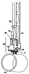
.~. Figs. 5 and 6 show, in exploded view and after
assembly respectively, the various elements of the combined
puncturing device of the invention.
The puncturing needle 14 is inserted into a
support 30 which is connected to a conduit 32, the proximal
end of which is provided with a connector 34 for connection
to= a source of fluid such as, for example, a cardioplegia
solution. It may be seen that the needle support 30
supports a particular member laterally, namely a sucker 36
connected via a second conduit 38 (running parallel to the
conduit 32) to a vacuum pump.
The clamp 18, the needle 14, its support 30 and
the sucker 36 together form the "head" 39 of the combined
puncturing device of the invention. This head 39 is
supported by an introducer 40, which is terminated here by
CA 02570738 2006-12-11
WO 2006/000514 PCT/EP2005/052527
12
a "fork", i.e. two tips 42 that project from its distal end
and that, when inserted on either side of the clamp 18 into
grooves 44 provided for this purpose, allow the head 39 to
be manipulated without the pivoting of the jaws of the
clamp 18 being impeded.
The succession of Figs. 6, 7, 8 and 9 fully
explain both the operating method and the advantages of the
device of the invention.
In Fig. 6, the elastically deformable staple 2
has been mounted on the clamp 18 and the latter is kept
closed by the needle 14. The head 39 of the device
supporting the staple 2 is then inserted into the
introducer 40. The device is introduced into the thoracic
cage of a patient and the head 39 is applied to the wall 46
of a blood vessel or of a hollow organ to be punctured (for
example, the aorta). Since the material of the introducer
40 is ductile, the operator has the ability to bend it if
the wall 46 of the vessel is not at the right angle. He
firmly sinks the pins 10 -of the staple 2 into the wall 46,
their axes lying at that moment parallel to the axis of the
needle 14 and/or the needle 14 itself (as will be seen
later, it is not essential for the needle 14 to be inserted
at the same time as the pins 10 of the staple 2). The
dimensions of the pins 10 have been designed according to
the thickness of the wall to be penetrated, namely a length
long enough to ensure firm anchoring and an optimum
diameter in order to prevent tearing of the tissues when
the staple 2 is closed up.
The operator then fixes the head of the device
against the wall. In this case, he applies vacuum to the
sucker 36, which consequently presses the head 39 firmly
against the wall 46 of the vessel and keeps it in place
thereon. The operator can then disengage the head 39 of the
CA 02570738 2006-12-11
WO 2006/000514 PCT/EP2005/052527
13
introducer 40, by moving it translationally sideways,
releasing the tips 42 of the "fork". Since the conduits 32
and 38 are flexible, the operator can move them away from
the operating area and continue the operation in progress
without further worrying about them.
It should be noted that the sucker 36 is.not the
only device that can be used to immobilize the device
against the wall of the artery - it is also possible to use
a clamp or a lasso loop surrounding the perimeter of the
artery.
The situation is now at the stage shown in Fig.
7. Either blood can be removed from the aorta or, on the
contrary, a product of some kind can be injected thereinto,
via the conduit 32 and the hollow needle 14.
In Fig. 8, the operation is in the terminal
phase: it is no longer necessary, at this moment, to keep
the perfusion needle 14 in place. The operator refits the
introducer 40, by inserting the tips 42 of the "fork" into
the grooves.A4 of the clamp 18. He grasps the proximal en:d
34 of the conduits 32, 38 using a device (not shown),
possibly attached to the introducer 40, releases the vacuum
on the sucker 36 and pulls on the support 30.
As the needle 14 comes out, it unlocks the clamp
18 and releases the staple 2. If this is a springy staple,
the two rows of pins 10 of the staple 2 come together
and/or converge through the action of the zigzag joins 12,
causing the lips of the incision caused by the needle 14 to
be firmly pressed against one another and preventing any
blood spilling into the operating area. This closing-up
movement is supplemented, if necessary, by the axes of the
pins 10 converging, this having the effect of retaining the
staple 2 in the implanted position.
CA 02570738 2006-12-11
WO 2006/000514 PCT/EP2005/052527
14
If a staple 2 with a plastically deformable back
4 is used, it obviously does not exert a spring effect on
the clamp 18. Instead the staple 2 is fitted by pushing on
the branches of the clamp, either manually or through the
agency of a mechanical, pneumatic or electrical stressing
means (not shown).
Next, the clamp 18 is removed, as shown in Fig.
9. The staple 2, made of a biocompatible material well
tolerated by the body, remains in place. Given its
geometry, if necessary it can even be removed without any
problem during a subsequent operation.
Fig. 10 shows another embodiment of the device of
the invention. In this embodiment, the needle 14 is
replaced with a hollow cannula 48 through which the
operator introduces a trocar 50, so as to make a neater
(straight) incision and one that is better centred with
respect to the rows of pins 10 of the staple 2 into the
wall 46. The cannula has the advantage of allowing an
optimum flow rate of the fluid that passes through it. Of
course, it is possible to use the trocar 50 only if the
introducer is straight or substantially straight. Moreover,
if a conventional puncturing needly is used, the relative
position of the stud 24 or of the jaws 20 of the clamp 18
may be offset with respect to the axis of the needle 14 in
.25 order to centre the incision better.
Fig. 11 shows in greater detail the action of the
staple 2 on the tissues. As may be seen, the various layers
(the outer layer 51, the tunica media vasorum 52 and the
outer and inner layers of the tunica intima vasorum 53,
etc.) of the wall are held in place practically without any
deformation relative to their initial position, which
promotes problem-free cicatrization. As may be seen, the
pins 10 have a length, relative to the thickness of the
CA 02570738 2006-12-11
WO 2006/000514 PCTIEP2005/052527
tissues penetrated, that is sufficient to allow deep
anchoring. It is not compulsory that, as represented, they
go right through the wall. The pins may if required be
provided with barbs (not shown). In a general way, one
5 ensures that the pins 10 are not too long and that they
will thus not penetrate the opposite side of the punctured
organ; their actual length (equal, shorter or longer than
the thickness of the wall to penetrate) is determinated
according to the nature of the organ itself and the
10 physiological properties of the wall.
It should be understood that the staple 2,
although shown here with two rows of a limited number of
pins 10 (two and three, in this case), may comprise a
different number of pins (from 2 to N pins) depending on
15 the characteristics of the wound to be closed. It is
advantageous for the pins 10 of the two rows to be arranged
in a staggered fashion, as shown, so as to close the wound
over its entire length. Although the present device has
been shown here within the context of a mini-invasive
surgical operation, it may also be used in standard
surgery.
Moreover, if it is desired to use the device only
in its wound-closing function, the needle 14 is replaced
with a simple end-fitting that does not extend beyond the
jaws of the clamp.
Fig. 12 shows another embodiment of the head 39
of the device, suitable for using a staple with a
plastically deformable back. In this case, the clamp 18 is
provided with a bracelet acting as a locking means, which,
in the absence of any stress, keeps the studs 24 of the
clamp in a position such that the staple 2 can be fitted
thereonto in the open state (i.e. with the pins 10 in a
substantially parallel position). The support of the needle
CA 02570738 2006-12-11
WO 2006/000514 PCT/EP2005/052527
16
14 comprises a core 56 forming a cam, while the internal
faces of the handles 26 of the clamp 18 each comprise a
ramp 58 capable of cooperating with the external faces of
the cam-forming core 56.
The device in the process of puncturing is shown
in Fig. 13. When the puncturing operation has been
completed, the operator exerts a relative pulling force on
the needle 14, the head of the device also being held
firmly against the wall (see Fig. 14). As in the case of
the other version of the device, this pulling force is
exerted either manually (via a lever system for example) or
pneumatically or even electrically.
During the transition, the core 56, acting as a
cam, forces the handles 26 of the clamp 18 to move apart,
thus tightening the jaws 20 and consequently closing the
staple 2, which constricts the punctured tissue.
Figs 15a to 15c display another embodiment of the
staple 102, in this case a plastically deformable staple.
=~ As in staple 2, staple 102 comprises joins.112. When being
bent by clamp 18, the joints 112 urge the axes of the pins
10 fitted on the parts 106, 108 of the back to converge.
Hooks 113, 115 interlock, forbidding the staple to open
once it has been closed on a wound.
Figs. 16 to 19 display another embodiment of
introducer 140. This introducer is fitted, besides the
already described "fork" 142, with a movable "fork" 141.
The role of this fork is to make easier the removal of the
puncturing needle 14.
As needle 14 locks the jaws of the clamp 18, it
is submitted, at the moment of the removal, to a non-
neglectible friction resistance. To prevent that the
operator would have to exert a too high traction force on
the needle at the moment of the removal- implying a risk to
CA 02570738 2006-12-11
WO 2006/000514 PCT/EP2005/052527
17
make a mistake- a means to slightly close the handles 26 of
the clamp 14 is provided, which nullifies the friction on
the needle 14. At Fig. 16, the introducer 140 (that was
removed during the operation to free the operating field)
is put in place again on the head of the device by the
operator. At Fig. 17, the tips 142 of the fork are sled
into the grooves 44 of the clamp 18, the movable fork 141
being maintained close to the fixed fork 142 via control
rods 143. The movable fork 141 is then sled towards the
proximal side of the device by the operator, causing the
handles of the clamp 18 to be temporarily held in closed
position. The needle may then be removed without effort
(see Fig.18). The operator then pushes the movable fork
towards the distal end of the device, freeing the clamp 18
and provoking the closing of the staple 2 (Fig. 19).
One advantage of introducer 140 is that it allows
a possible removal of the staple 2, either in the case of
mistake during the operation,.or during a possible further
operation. As disclosed at Figs. 20 and 21, to remove an
already placed staple, it suffices to use a clamp 19
inserted on an introducer 140 to be able to grasp this
staple again: the gripping studs 24 having been inserted
into the orifices 16 of staple 2, the movable fork is
pulled backwards; clamp 19 closes, bringing the pins 10
back in parallel plans. Staple 2 is then pulled out without
problem.
Figs. 22 to 29 display another embodiment of the
device of the invention.
Though the whole elements are similar to what was
described above, this embodiment is more complete and still
reduces possible mistakes.
Fig. 22 displays an exploded view of the various
elements of this embodiment. The most distinctive element
CA 02570738 2006-12-11
WO 2006/000514 PCT/EP2005/052527
18
is the presence of a sucker 236 made integral with support
230 and which encloses the clamp and the puncturing system.
Another distinctive element is the fact that the
role of the introducer (40, 140) is ensured partially by
the shaft 260 of a trocar 250 and partially by a pusher
262, as explained hereinbelow.
The head 239 of the device is shown in a more
detailed manner at Fig. 23. As in the former embodiments,
this head 239 is connected to the proximal part of the
device by a first conduit 232 for the passage of fluid, the
latter opening onto the cannula 48, and by a second conduit
238 for ensuring vacuum in the sucker 236.
When introducing the head 239 into the body of
the patient, it suffices (as shown at Fig.26) to slide the
trocar 250 and its rigid shaft 260 into the first conduit
232 to obtain a device which is perfectly rigid in its
medium part, which allows to neglect the use an introducer
40, 140 as described above. During the removal of the
device, the pusher 262 will play a similar rctle, as shown
hereinbelow.
Fig. 23 to 27 show the various steps for
introducing the puncturing device: the clamp being in open
position, the gripping studs 24 of the clamp are moved
towards each other, bringing the staple in insertion
position, i.e. with the axes of the two rows. of pins 10
essentially parallel to each other (see Fig. 24); the shaft
260 of the trocar is sled into the conduit 232 and the
clamp holding the staple is brought into the bell-shaped
sucker 236 (see Fig. 25). The locking of the clamp is
assured here by the internal wall of the bell 236, which
maintains the clamp via a contact with the tip of the
handles 26. The operator, handling the rigidified device by
its proximal end, introduces the head into the body of the
CA 02570738 2006-12-11
WO 2006/000514 PCT/EP2005/052527
19
patient up to the wall 46 of the organ to be treated,
pierces this wall with the trocar 250, while the pins 10
run trough the wall 46. Vacuum is established in the bell-
shaped sucker 236, thus urging the head of the device
against the wall 46. The trocar 250 and its rigid shaft 260
are then removed.
Fig. 17a displays a sectional view of the various
elements of the head 239 of the placed device.
The mouth 264 of the sucker, substantially
saddle-form, fits perfectly to the curve of the wall 46.
The hollow cannula 48 goes through the wall 46. A possible
loss of fluid provoked by the incision is balanced by the
sucking through the vacuum conduit 238. The profile of the
inner wall of the sucker is designed so as to maintain the
clamp in closed position, and, accordingly, the staple
remains in open position.
One advantage of this embodiment is that the
traction exerted on the wall 46 is not off-centred with
respect to the solicitationszexerted on the cannula 48, as
in the case shown at Fig.10, which reduces the
possibilities of unexpectedly tearing the device away
during the operation.
Fig. 27b shows another possible embodiment: the
mouth 264 of the sucker 236 is here somewhat longer than
what is displayed at Fig. 27a, so that the wall 46 is
pierced only by the trocar 250 and the cannula 48. It is
merely at the very moment of the removal that the staple 2
comes in contact with the wall 46 and that the pins pierce
said wall. To prevent an untimely bending of the wall
provoked by the sucking, a stopping ring 270 is fitted on
the cannula 48.
Figs. 28 and 29 show the removal step of the
puncturing device and the occlusion of the punctured
CA 02570738 2006-12-11
WO 2006/000514 PCT/EP2005/052527
orifice: the operator introduces, from the proximal side of
the device, le pusher 262 into the vacuum conduit 268 of
the sucker 236. The vacuum is then interrupted and he pulls
the proximal side of the conduit via a handle 263. The
5 sucker goes back, while the clamp 18 is firmly maintained
in place by the pusher 262. As soon as the sucker has
passed the free tips of the handles 26 of the clamp 18, the
latters are freed and, in the case of a resilient staple,
said staple takes its closed form, the tips of the pins
10 come close to each other closing the lips of the incision.
As explained with respect to the introducer 40, the shaft
of the trocar 260 and the pusher 262 are bendable so that
the head of the device may be brought normal to the wall
46, whatsoever the position of the organ to puncture.
15 At Fig. 29, the operator proceeds with the
removal of the device. An end knob 268 of the pusher,
cooperating with a (non visible) groove placed on the clamp
18, drawn the clamp out of the body.
The device may be delivered in a "kit" fashlon,
20 said kit comprising an applicator 140, which allows the
possible removal of an ill-placed staple, or of a staple
that has become unnecessary due to healing of the wound.
CA 02570738 2006-12-11
WO 2006/000514 PCT/EP2005/052527
21
CIaIMs
1.- A combined device for plugging and fluidthight
puncturing of a wall of a hollow organ, comprising a
proximal and a distal part, which comprises:
- a surgical staple for the wall of the hollow organ,
placed towards the distal part, said staple comprising a
substantially flat back that can deform between a closed
position of the staple and an open position of the staple,
and at least two spaced-apart pins extending each along an
axis, a free end of the pins converging when the back is in
the closed position, the axes of the pins of the two rows
tending to align parallel to each other when the staple is
in open position the back of said staple being provided
with a central opening
- a fluidthight puncturing system comprising a
puncturing device and a hollow channel, the diameter of
which corresponds to the central opening of the staple,
said puncturing system being mounted on a support and
connected to a fluid carrying conduit
- a keeping means for keeping the said staple in
place, the said means being capable of bringing the said
staple from its open position to its closed position;
- a support connected to a traction member extending
towards the distal part of the device, forming the head of
the device; and
- an introducer capable of introducing the head of the
device inside an organism.
2.- A surgical device according to Claim 1, wherein the
staple is elastically deformable, the means for keeping the
said staple in place being a clamp provided with two jaws
and with locking means, the free ends of the jaws being
provided with grasping means capable of cooperating with
CA 02570738 2006-12-11
WO 2006/000514 PCT/EP2005/052527
22
the gripping points of the staple, locking means keeping
the said staple in place on the jaws in a open position.
3.- A surgical device according to Claim 1, wherein the
staple is plastically deformable, the means for keeping the
said staple in place being a clamp provided with two jaws
and with locking means, the free ends of the jaws being
provided with grasping means capable of cooperating with
the gripping points of the staple, second locking means
keeping the said staple in place on the jaws in the open
position, the jaws of the said clamp being able to be
actuated by second clenching means causing plastic
deformation of the back of the staple.
4.- A surgical device according to any one of the preceding
claims wherein the clamp has two jaws.
5.- A surgical device according to Claim 4 wherein a tip of
the puncturing device is manufactured and placed in the
puncturing system in such a manner that the pins of the
staple are placed on opposite sides of an incision made in
the- wall of a hollow organ and on a substantially
symmetrical way with respect to said incision.
6.- A surgical device according to any one of the preceding
claims wherein a distal head of the device is detachable
from the introducer.
7.- A surgical device according to any one of the preceding
claims wherein the closing of the staple is actuated by the
removal of the support.
8.- A surgical device according to any one of the preceding
claims wherein the introducer includes, at its proximal
side, a member for unlocking the clamp, so releasing the
staple.
9.- A surgical device according to any one of the preceding
claims wherein the support includes retention means that
can be applied against the wall of a hollow organ and can
CA 02570738 2006-12-11
WO 2006/000514 PCT/EP2005/052527
23
keep the head of the device in place during a puncturing
operation.
10.- A surgical device according to Claim 9 wherein the
retention means comprises a sucker, said sucker being
connected to a conduit that can be connected to a source of
negative pressure.
11.- A surgical device according to Claim 10 wherein the
puncturing device and the staple retaining means are placed
inside the sucker.
12.- A surgical device according to any one of the
preceding claims wherein the introducer is plastically
deformable so as to be able to present the puncturing
device at right angles to a wall lying at any angle.
13.- A surgical device according to any one of the
preceding claims wherein the puncturing device includes a
trocar that can be inserted through a hollow conduit.
14.- A surgical staple for a surgical device according to
claim 4, comprising a substantially flat back that can
deform between a closed pos:i.tion of the staple and an open
position of the staple, and at least two spaced-apart pins
wherein:
- the back comprises two parts that are
hinged with respect to each other;
- the at least two pins are arranged in
two rows on these two partsi substantially lying along axes
perpendicular to a plane of each of the two parts
- in the closed position of the back a
free end of the pins of the two rows converge;
- when the back is in the open
position, the axes of the pins of the two rows tend to
align parallel to each other and/or the tips of the pins
are separated by a gap larger than in the closed position;
CA 02570738 2006-12-11
WO 2006/000514 PCT/EP2005/052527
24
said staple being provided with a central opening and
with gripping points for keeping the staple in one
position,
When the staple is in place, its back does not protrude
from the wall of the hollow organ.
15.- A surgical staple according to Claim 14 wherein the
back is elastically deformable.
16.- A surgical staple according to Claim 15, wherein the
two parts are joined together by springy joins.
17.- A surgical staple according to Claim 14 wherein the
back is plastically deformable.