Note: Descriptions are shown in the official language in which they were submitted.
CA 02570868 2006-12-15
WO 2006/009665 PCT/US2005/020801
Device and Method for Insertion of a Cannula of an Infusion Device
Technical Field
The present invention relates to a device for assisting in the
introduction of a cannula of an infusion device into the skin of a patient for
delivery
of a substance to the patient.
Background
Infusion devices are used to deliver substances such as medications
into the subcutaneous layer of skin of a patient. Devices for assisting in
insertion of
the cannula of an infusion device into the skin of the patient are known. For
example, some devices utilize springs to automatically drive a needle into the
skin of
a patient to introduce the cannula of the infusion device into the
subcutaneous layer.
Because a needle is used to introduce the cannula of the infusion
device into the subcutaneous layer of skin, there is a risk associated with
inadvertent
exposure to the needle. Further, patients may react adversely to viewing the
needle
prior to insertion and may, for example, be reluctant to place the needle into
the
skin. Prior devices may not adequately shroud this needle prior to and/or
after
introduction of the infusion device.
Other issues of concern in the design and use of insertion devices
include ease of use by the patient and sterilization. For example, some
patients may
have difficulty loading the infusion device into the insertion device.
It is therefore desirable to provide new designs for devices used to
assist in the introduction of an infusion device into the skin of a patient.
Summary
Embodiments made in accordance with the present invention include
devices that can be used to assist in the introduction of the cannula of an
infusion
device into the skin of a patient for delivery of a substance to the patient.
For example, one embodiment of a device includes a needle used to
insert the cannula of an infusion device into the skin of a patient. Once the
cannula
of the infusion device is inserted into the skin, the device moves the needle
to a
retracted state within the device.
CA 02570868 2006-12-15
WO 2006/009665 PCT/US2005/020801
In another embodiment, a device is configured to move a needle and
associated cannula of an infusion device from a delivery state to a trigger
state at
which the cannula of the infusion device is inserted into the skin of a
patient. Upon
full insertion of the cannula at the trigger state, the device is then
configured to
move the needle to a retracted state within the device.
In another embodiment, a device includes a needle that can be used to
insert a cannula of a site into the skin of a patient. Upon insertion of the
cannula, the
needle can be removed from the skin. In one embodiment, a cap is provided that
can
be placed onto the device prior to and after use of the device to provide a
sterile
environment and/or to reduce exposure to the needle.
In another embodiment, a device includes features that retain
components of the device and site contained therein in desired positions while
the
device is in a ship state. In an example embodiment, the device can include
tabs on
a sleeve that engage beads on a cap to retain the sleeve in a desired position
with
respect to a housing while in the device is in a ship state. In an example
embodiment, the device can also include a boss formed by the cap to retain the
site
at a desired position with respect to a needle of the device while the device
is in the
ship state.
The above summary of the present invention is not intended to
describe each disclosed embodiment or every implementation of the present
invention. Figures in the detailed description that follow more particularly
exemplify embodiments of the invention. While certain embodiments will be
illustrated and described, the invention is not limited to use in such
embodiments.
Description of the Drawings
Figure 1 is a side view of an example embodiment of a device used to
introduce a cannula of an infusion device into a patient made in accordance
with the
present invention.
Figure 2 is an exploded side view of the device of Figure 1.
Figure 3 is a perspective view of a housing of the device of Figure 1.
Figure 4 is a side view of the housing of Figure 3.
Figure 5 is an end view of the housing of Figure 3.
2
CA 02570868 2006-12-15
WO 2006/009665 PCT/US2005/020801
Figure 6 is a perspective view of a cylinder hub of the device of
Figure 1.
Figure 7 is side view of the cylinder hub of Figure 6.
Figure 8 is another side view of the cylinder hub of Figure 6.
Figure 9 is an end view of the cylinder hub of Figure 6.
Figure 10 is a perspective view of a needle hub of the device of
Figure 1.
Figure 11 is a side view of the needle hub of Figure 10.
Figure 12 is another side view of the needle hub of Figure 10.
Figure 13 is an end view of the needle hub of Figure 10.
Figure 14 is a perspective view of a sleeve of the device of Figure 1.
Figure 15 is a side view of the sleeve of Figure 14.
Figure 16 is another side view of the sleeve of Figure 14.
Figure 17 is an end view of the sleeve of Figure 14.
Figure 18 is a top view of an adhesive portion of the device of Figure
1.
Figure 19 is a cross-sectional view taken along line 19-19 of the
adhesive portion of Figure 18.
Figure 20 is an exploded view of the adhesive portion of Figure 18.
Figure 21 is a perspective view of a cap of the device of Figure 1.
Figure 22 is a side view of the cap of Figure 21.
Figure 23 is an end view of the cap of Figure 21.
Figure 24 is a side view of the device of Figure 1 with the cap
removed.
Figure 25 is a side view of the device of Figure 24 in a trigger state.
Figure 26A is a cross-sectional view taken along line 26A-26A of the
device of Figure 1 in a ship state.
Figure 26B is a cross-sectional view taken along line 26B-26B of the
device of Figure 1 in the ship state.
Figure 27A is a cross-sectional view taken along line 27A-27A of the
device of Figure 24 in a delivery state.
Figure 27B is a cross-sectional view taken along line 27B-27B of the
device of Figure 24 in the delivery state.
3
CA 02570868 2006-12-15
WO 2006/009665 PCT/US2005/020801
Figure 28A is a cross-sectional view taken along line 28A-28A of the
device of Figure 25 in a trigger state.
Figure 28B is a cross-sectional view taken along line 28B-28B of the
device of Figure 25 in the trigger state.
Figure 28C is a cross-sectional view of the device of Figure 28B
illustrating the adhesive portion being sheared from a surface of the sleeve.
Figure 29A is a cross-sectional view of the device of Figure 28A with
the needle hub retracted.
Figure 29B is a cross-sectional view of the device of Figure 28B with
the needle hub retracted.
Figure 30A is a cross-sectional view taken along line 30A-30A of the
device of Figure 24 in a retracted state.
Figure 30B is a cross-sectional view taken along line 30B-30B of the
device of Figure 24 in the retracted state.
Figure 31 is a cross-sectional view of another example embodiment
of a device used to introduce an infusion device into a patient made in
accordance
with the present invention.
Figure 32 is a perspective cross-sectional view of the device of Figure
31.
Figure 33 is a perspective cross-sectional view of a portion of the
device of Figure 32 in enlarged form.
Figure 34 is a perspective view of a sleeve of the device of Figure 31.
Figure 35 is a side view of the sleeve of Figure 34.
Figure 36 is an end view of the sleeve of Figure 34.
Figure 37 is a cross-sectional view taken along line 37-37 of the
sleeve of Figure 36.
Figure 38 is an end view of a cap of the device of Figure 31.
Figure 39 is a cross-sectional view taken along line 39-39 of the cap
of Figure 38.
Figure 40 is a cross-sectional view taken along line 40-40 of a portion
of the cap of Figure 38 in enlarged form.
4
CA 02570868 2006-12-15
WO 2006/009665 PCT/US2005/020801
Figure 41 is a cross-sectional view of another example embodiment
of a device used to introduce an infusion device into a patient made in
accordance
with the present invention.
Figure 42 is a perspective view of a clip of the device of Figure 41.
Figure 43 is a top view of the clip of Figure 42.
Figure 44 is a side view of the clip of Figure 42.
Figure 45 is a side view of a sleeve of the device of Figure 41.
Detailed Description
Embodiments of the present invention relate to devices for assisting
in the introduction of an infusion device, specifically a cannula of the
infusion
device, into the subcutaneous layer of skin of a patient.
Referring to Figures 1 and 2, one example embodiment of a device
100 is shown. The device 100 is used to introduce a cannula of an infusion
device,
such as a set, site, or other access device, into the skin of the patient. The
set, site, or
other access device can then be used to deliver drugs or other fluid to the
patient,
such as from an infusion pump.
The device 100 generally includes a housing 110, a cylinder hub 120,
a needle hub 130, a sleeve 140, a spring 150, an adhesive portion 160, and a
cap
170. Each of the components of the device 100, described further below, is
configured to assist in the introduction of a cannula of an infusion device
into the
skin of a patient.
Referring now to Figures 3-5, the housing 110 is shown. The
housing 110 is preferably, cylindrical in shape and includes a closed upper
end 111
and an open lower end 112. The housing 110 further preferably includes a
portion
118 with a knurled surface to enhance a patient's grip on the housing 110, as
well as
a threaded portion 113 positioned adjacent the open lower end 112.
Referring now to Figures 6-9, the cylinder hub 120 is shown in
greater detail. The cylinder hub 120 includes first and second ends 221 and
222 and
an interior passage 223. In addition, two opposing slots 225 are formed on
opposite
sides of the cylinder hub 120 and generally extend from a mid-portion 224 of
the
hub 120 to the first end 221. Further, the cylinder hub 120 includes opposing
apertures 226 formed in the cylinder hub 120 adjacent the second end 222.
5
CA 02570868 2006-12-15
WO 2006/009665 PCT/US2005/020801
The first end 221 of the cylinder hub 120 is coupled to the upper end
111 of the housing 110 by tabs 119 on the housing 110 engaging shoulders 228
formed by the cylinder hub 120. See, for example, Figures 6-8, 26A, and 26B.
In
addition, members 121 of the housing 110 are received in slots 229 of the
cylinder
hub 120. In alternative designs, the housing 110 and cylinder hub 120 can be
formed as a single unit.
Referring now to Figures 10-13, the needle hub 130 includes a main
body 331 with first and second ends 332 and 333, and a needle 336 (hollow or
solid)
coupled to the main body 331. The main body 331 includes opposing wings 334
formed at the first end 332 and opposing barbs 335 at the second end 333.
The needle hub 130 is positioned in the interior passage 223 of the
cylinder hub 120 such that the opposing wings 334 of the needle hub 130 extend
through the opposing slots 225 of the cylinder hub 120. See Figures 6, 8, 26B,
27B,
28B, 29B, and 30B. In addition, the opposing barbs 335 of the needle hub 130
extend through the opposing apertures 226 of the cylinder hub 120 and engage
shoulders 227 formed by the apertures 226 so that the needle hub 130 is held
in a
fixed position relative to the cylinder hub 120 and the housing 110. See, for
example, Figures 6, 8, 26A, 27A, and 28A.
Referring now to Figures 14-17, the sleeve 140 is shown. The sleeve
140 is preferably cylindrical in shape and includes first and second ends 441
and 442
and interior passage 443. Opposing projections 444 extend into the passage 443
adjacent to a shoulder 445. On the exterior of the sleeve 140 channels 446 are
formed, as well as railways 447 with barbs 448 formed on ends thereof.
The sleeve 140 is coupled to the housing 110 such that the housing
110 can be moved longitudinally with respect to the sleeve 140. Specifically,
the
railways 114 of the housing are received in the channels 446 of the sleeve
140.
Likewise, the railways 447 of the sleeve 140 are received in the channels 115
of the
housing 110. Barbs 448 on the railways 447 of the sleeve 140 engage
projections
116 in the channels 115 of the housing 110 so that the housing 110 remains
slideably coupled to the sleeve 140 in opposition to the force exerted by the
spring
150 (described further below).
The spring 150 includes first and second ends 152 and 154. See, for
example, Figure 26B. The spring 150 surrounds a portion of the cylinder hub
120
6
CA 02570868 2006-12-15
WO 2006/009665 PCT/US2005/020801
and extends within the passage 443 of the sleeve 140. The first end 152 of the
spring 150 is seated on the shoulder 445 of the sleeve 140, and the second end
154
of the spring 150 engages the opposing wings 334 of the needle hub 130
extending
through the opposing slots 225 of the cylinder hub 120.
The spring 150 is in a compressed state as shown in Figures 26A,
26B, 27A, 27B, 28A, and 28B and therefore applies force against the wings 334
of
the needle hub 130, biasing the needle hub 130 in an upward direction.
However,
barbs 335 of the main body 331 of the needle liub 130 are engaged against
shoulders
227 of the apertures 226 of the cylinder hub 120 to retain the needle hub 130
in
place with respect to the cylinder hub 120. See, for example, Figure 26A.
Likewise,
the spring 150 forces the housing 110 and the sleeve 140 apart until barbs 448
of the
sleeve 140 engage projections 115 of the housing 110 to maintain coupling
between
the housing 110 and the sleeve 140.
Referring now to Figures 18-20, an adhesive portion 160 is
positioned on a surface 449 at the second end 442 of the sleeve 140 (see
Figures 14
and 17). The surface 449 preferably acts as a framework that stabilizes the
adhesive
portion 160 prior to placement on the patient. In a preferred embodiment
shown, the
adhesive portion 160 includes layers 662, 663, and 664, as well as liners 661
and
665. Liners 661 and 665 also preferably include tabs 666 and 667 that allow
for
removal of the liners 661 and 665 as described below.
The adhesive portion 160 can be coupled to the surface 449 of sleeve
140 in a variety of manners. In a preferred embodiment, the liner 661 is
removed,
and layer 662 is coupled to the surface 449 using an adhesive. In addition, as
described further below, in a preferred embodiment a top surface 669 of layer
664
and/or a lower end of the infusion device includes an adhesive to couple the
infusion
device to the adhesive portion 160 as the infusion device is moved into
contact with
the adhesive portion. See Figures 28A, 28B, and 28C.
In addition, the liner 665 is preferably removed, and a lower surface
668 of the layer 664 includes an adhesive to couple the adhesive portion 160
to the
skin of the patient.
Preferably, the site is loaded into the device 100 prior to application
of the adhesive portion 160 onto the device 100, and preferably both liners
661 and
665 are removed as described above prior to attachment of the adhesive portion
to
7
CA 02570868 2006-12-15
WO 2006/009665 PCT/US2005/020801
the sleeve 140 and coupling of the cap 170 to the housing 110. In this manner,
the
patient preferably does not need to remove any liners prior to application of
the
adhesive portion 160 to the skin and introduction of the site into the skin.
Preferably, the layer 664 does not include any holes, but instead is
pierced by the needle 336 as the needle 336 is advanced towards the skin, as
described further below. This configuration can enhance the fit between the
adhesive portion 160 and the skin of the patient.
In a preferred embodiment, the adhesive portion 160 includes
adhesive on one or more of surfaces 668 and 669 to allow the adhesive portion
160
to be coupled to the sleeve 140, site, and/or to the skin of the patient.
Typical
adhesives that can be used on the adhesive portion 160 include, without
limitation,
acrylic adhesive, synthetic rubber-based adhesive, acrylate adhesive, and
silicone-
based adhesive.
In example embodiments, the adhesive portion 160 includes films
with adhesives thereon, such as a TegadermTM film manufactured by 3MTM or an
IV3000TM film manufactured by Smith & Nephew. For example, in the preferred
embodiment shown, the tape layer 662 is 3MTM 9731 tape, and layers 663 and 664
are 3MTM TegadermTM p/n 9842.
Referring now to Figures 21-23, the cap 170 is illustrated. The cap
170 includes a closed first end 772 and an open second end 774. The cap 170
preferably includes an exterior with a knurled surface 778 to enhance the
patient's
grip on the cap 170. In addition, the interior of the cap 170 includes a
threaded
portion 776 positioned adjacent the open second end 774 so that the threaded
portion
776 can be threaded onto the threaded portion 113 of the housing 110 to seal
the
device 100. See Figures 1, 26A, and 26B.
In a preferred embodiment, a gasket 122 is provided on the threaded
portion 113 of the housing 110 to create a seal between the cap 170 and the
housing
110 as the cap 170 is threaded onto the housing 110. See Figures 26A and 26B.
In
this manner, the internal components of the device 100 (e.g., needle 336 and
site
800) can be maintained in a substantially sterile state prior to removal of
the cap
170. Further, the cap 170 can function to maintain the device 100 in a ship
state
(i.e., the housing 110 can not be moved relative to the sleeve 140) prior to
removal
of the cap 170 from the housing 110.
8
CA 02570868 2006-12-15
WO 2006/009665 PCT/US2005/020801
In alternative embodiments, the cap 170 and/or housing 110 can be
formed to provide a tamper-evident seal so that the patient can determine when
the
cap 170 has been previously uncoupled from the housing 110. For example, in an
alternative embodiment of the device 100' shown in Figures 50-52, a tamper-
evident
band 178 is shown. The band 178 includes tabs 179 that are coupled to the cap
170
as shown in Figure 50. As the cap 170 is removed from the housing 110 (i.e.,
threads 514 on cap 170 are unthreaded from threads 512 on housing 110), the
tabs
179 break away from the cap 170, and the seal 178 remains coupled to the
housing
110, as shown in Figure 51. If the cap 170 is later threaded back onto the
device
100', the breaks between the tabs 179 and the cap 170 are evident, allowing
the
patient to identify that the cap 170 of the device 100' has been previously
removed.
The cap 170 and band 178 can be placed on the device 100' during
manufacturing as a single unit. For example, as shown in Figure 52, the cap
170 and
band 178 can be pushed onto the device 100' (note that threads 512 and 514 can
be
rounded to allow the cap 170 to be pressed onto the device 100') so that
portion 520
of the band 178 passes over and engages shoulder 522 of the housing 110 to
retain
the band 178 on the housing 110 when the cap 170 is unthreaded and tabs 179
are
broken. In addition, notches 524 formed periodically along the band 178
prevent the
cap 170 from bottoming out against the band 178 as the cap 170 and band 178
are
pushed onto the device 100' so that the tabs 179 remain intact. A portion 502
extending along an interior circumference of the cap 170 can also be formed to
engage the outer surface of the housing 110 to create a seal between the
housing 110
and the cap 170.
It can be desirable to provide a tamper-evident seal, for example, so
that the patient can assure that the device 100' is has not been previously
opened and
is sterile prior to use. Other methods of indicating tampering can also be
used.
As previously noted, the device 100 can be used to introduce a
cannula of an infusion device into the subcutaneous layer of skin of the
patient. In a
preferred embodiment, the infusion device includes a site 800, the site 800
including
a cannula for delivery of a substance into the subcutaneous layer of skin of
the
patient. Site 800 is linked by tubing (not shown) with a fluid source, such as
an
infusion pump (not shown) to deliver fluid to the patient through the cannula.
In a
preferred embodiment, the site 800 can be made in accordance with that
disclosed in
9
CA 02570868 2006-12-15
WO 2006/009665 PCT/US2005/020801
U.S. Patent Application Serial No. 10/705,736, filed November 10, 2003 and
entitled "Subcutaneous Infusion Device and Method," the entirety of which is
hereby incorporated by reference. However, sites of other configurations can
also
be used.
Referring now to Figures 1 and 24-3 0, the device 100 is illustrated in
various states of use. As shown in Figures 1, 26A, and 26B, the device 100 is
in a
ship state prior to use. As shown in Figures 24, 27A, and 27B, the device 100
is in a
delivery state ready to deliver the cannula of an infusion device into the
skin of the
patient. As shown in Figures 25, 28A, 28B, and 28C the device 100 is in a
trigger
state, or the state at which the needle 336 and the cannula of the site 800
have been
fully inserted into the subcutaneous layer of skin of the patient, and the
needle hub
130 and associated needle 336 are about to be retracted. As shown in Figures
29A
and 29B, the device 100 is in a retracted state with the needle hub 130 and
associated needle 336 having been retracted into the device 100. As shown in
Figures 30A and 30B, the device 100 is in a fully retracted state with the
housing
110 and sleeve 140 returned to an uncompressed position relative to one
another.
An example method of use of the device 100 is as follows. The
device 100 is provided to a patient with the cap 170 coupled to the housing
110, as
shown in Figures 1, 26A, and 26B. Preferably, the site 800 has been previously
loaded (i.e., preloaded) into the device 100 during, for example, the
manufacturing
process for the device 100.
The cap 170 is then unthreaded from the housing 110, and the sleeve
140 of the device 100 is positioned so that the adhesive portion 160 (i.e.,
surface
668) contacts the skin 900 of the patient. See Figures 24, 27A, and 27B.
Next, in the illustrated preferred embodiment, the patient applies
pressure to the upper end 111 of the housing 110 to move the housing 110 and
associated structures including the cylinder hub 120 and needle hub 130
(including
needle 336 and site 800) in a direction A with respect to the sleeve 140 and
toward
the skin 900 of the patient. As the needle 336 of the needle hub 130 and
associated
site 800 are moved in the direction A, the needle 336 and the cannula 806 of
the site
800 are introduced into the skin 900 of the patient. In addition, as the
needle hub
130 is moved toward the sleeve 140, the spring 150 is further compressed.
CA 02570868 2006-12-15
WO 2006/009665 PCT/US2005/020801
Once the needle 336 and cannula 806 of the site 800 have been fully
inserted into the skin 900, the device 100 is in a trigger state, as
illustrated in Figures
25, 28A, 28B, and 28C. In this state, the barbs 335 that couple the needle hub
130
to the cylinder hub 120 are biased inwardly through contact with the
projections 444
formed by the sleeve 140.
As the housing 110, cylinder hub 120, and needle hub 130 are
displaced further in the direction A, it is preferable that the needle hub 130
is
positioned so that a lower portion of the site 800 travels slightly beyond the
second
end 442 of the sleeve 140 as shown in Figure 28C. This "over-travel" assures
that
the adhesive portion 160 is properly sheared away from the surface 449 of the
sleeve
140 and allows for the coupling of the site 800 to the adhesive portion 160.
For
example, in preferred embodiments, the lower portion of the site 800 travels
beyond
the second end 442 of the sleeve 140 by between 50 to 100 thousandths of an
inch,
more preferably approximately 70 thousandths of an inch.
In addition, as the housing 110, cylinder hub 120, and needle hub 130
are displaced further in the direction A as described above, barbs 335 of the
needle
hub 130 are forced inwardly by the projections 444 of the sleeve 140, and the
barbs
335 are thereby uncoupled from engagement with the cylinder hub 120. Once the
barbs 335 of the needle hub 130 are released from the cylinder hub 120, the
needle
hub 130 is free to move longitudinally within the passage 223 of the cylinder
hub
120 in a direction B opposite to that of the direction A. The spring 150,
which has
been compressed through the movement of the housing 110 in the direction A,
propels the needle hub 130 and associated needle 336 in the direction B up
through
the cylinder hub 120 into the upper end 111 of the housing 110, while leaving
the
site 800 and associated cannula 806 positioned in the skin 900 of the patient,
as
shown in Figures 29A and 29B.
Once the patient removes pressure from the upper end 111 of the
housing 110, the spring 150 causes the housing 110 and cylinder hub 120 to
move in
the direction B as shown in Figures 30A and 30B to a fully retracted state.
Finally, the sleeve 140 is removed from contact with the skin 900,
and the cap 170 can be replaced onto the threaded portion 113 of the housing
110 of
the device 100. Subsequently, the device 100 can be discarded.
11
CA 02570868 2006-12-15
WO 2006/009665 PCT/US2005/020801
Many alternative designs for the device can be provided. For
example, in Figure 31 a portion of an alternative device is shown including
cylinder
hub 120' and needle hub 130'. The cylinder hub 120' and needle hub 130' are
similar
to cylinder hub 120 and needle hub 130 described above, except that the
cylinder
hub 120' includes projections 129 formed near the first end 221 of the
cylinder hub
120, and the needle hub 130' includes barbs 139 formed on the first end 332.
The
barbs 139 are configured to ride inside the interior passage 223 of the
cylinder hub
120' during retraction of the needle 336 in the direction B until the barbs
139 extend
beyond the projections 129 of the cylinder hub 120'. Once this occurs, the
barbs 139
expand outward slightly. In this configuration as shown in Figure 31, the
barbs 139
prevent the needle hub 130' and associated needle 336 from being moved back in
the
direction A. In this manner, the barbs 1291ock the needle hub 130' in the
retracted
position. This configuration can be beneficial, used separately or in
conjunction
with the force of the spring 150 forcing the needle hub 130' in the direction
B, to
further reduce the possibility of inadvertent exposure to the needle 336 after
retraction.
Additional details regarding alternative designs for device 100 can be
found in U.S. Patent Application Serial No. 10/705,725, filed November 10,
2003
and entitled "Device and Method for Insertion of a Cannula of an Infusion
Device,"
the entirety of which is hereby incorporated by reference.
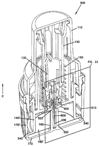
Referring now to Figures 31-40, another embodiment of a device 500
is shown. Device 500 is similar to device 100 described above. However, device
500 includes features that retain components of device 500 and/or site 800
contained
therein in desired positions while device 500 is in a ship state (i.e., prior
to removal
of the cap 170' from device 500).
More specifically, in the illustrated embodiment, sleeve 140' and cap
170' of device 500 include features that function to: (i) retain the sleeve
140' in a
desired position with respect to the housing 110 while device 500 is in the
ship state;
and/or (ii) retain the site 800 of an infusion device at a desired position
with respect
to the needle 336 while the device 500 is in the ship state.
Referring now to Figures 32-37, the sleeve 140' is illustrated.
Generally, sleeve 140' is similar to sleeve 140 described above, except that
sleeve
140' includes tabs 540 spaced about surface 449 at second end 442 of sleeve
140'. In
12
CA 02570868 2006-12-15
WO 2006/009665 PCT/US2005/020801
the example shown, four tabs 540 are spaced circumferentially ninety degrees
apart
about the surface 449. The tabs 540 extend outward radially from the surface
449.
More or fewer tabs (e.g., two tabs or one tab) can also be used.
Referring now to Figures 32, 33, and 38-40, tabs 540 on sleeve 140'
are configured to engage beads 510 formed adjacent closed first end 772 of cap
170'
when the device 500 is in the ship state (i.e., with cap 170' threaded onto
device
500). In the illustrated embodiment, cap 170' includes four beads 510 formed
on an
internal surface of the cap 170'. The beads 510 are pitched at an angle and
spaced
circumferentially ninety degrees apart about the cap 170'. In alternative
designs,
more or fewer beads (e.g., two beads or one bead) can also be used.
In the illustrated embodiment, beads 510 are formed to correspond to
tabs 540 on the sleeve 140'. Spaces 512 (see Figure 40) are formed between
adjacent beads 510. Spaces 512 allow the tabs 540 to pass between and clear
the
beads 510 when the cap 170' is placed onto the device 500 and threaded onto
the
housing 110 during manufacture.
Once the device 500 is in the ship state shown in Figures 31-33, the
tabs 540 and beads 510 function to maintain the device 500 in the ship state.
If the
sleeve 140' moves in a direction C while the device 500 is in the ship state
(due to,
for example, a sudden shock caused by dropping or otherwise jarring the device
500), tabs 540 of the sleeve 140' contact beads 510 of the cap 170', thereby
limiting
further movement of the sleeve 140' in direction C. In this manner, device 500
is
maintained in the ship state, and inadvertent movement of the sleeve 140' to
the
trigger state and resulting retraction of the needle hub 130 can be avoided.
When device 500 is ready for use and cap 170' is unthreaded from the
housing 110, tabs 540 ride along the pitch of beads 510 until tabs 540 reach
spaces
512 between adjacent beads 510, which allow tabs 540 to clear beads 510 and
cap
170' to be removed from housing 110.
Referring back to Figures 32, 33, and 38-40, cap 170' also includes a
boss 520 extending from closed first end 772 of cap 170'. In the illustrated
embodiment, boss 520 is cylindrical and forms a central cavity sized to
receive
needle 336 and cannula 806 of the site 800. A free end 522 of the boss 520
extends
to a point adjacent to a bottom surface of the site 800 when the site 800 is
in the
preloaded position (i.e., the site 800 having been loaded into the device 500
during,
13
CA 02570868 2006-12-15
WO 2006/009665 PCT/US2005/020801
for example, the manufacturing process). If the site 800 travels slightly
downward
on the needle 336, the bottom surface of the site 800 contacts the free end
522 of the
boss 520, thereby limiting further travel of the site 800. Therefore, if the
device 500
receives a shock while in the ship state, which can potentially cause the site
800 to
slide downward relative to the needle 336, the boss 520 contacts and maintains
the
site 800 at a desired position with respect to the needle 336.
In the illustrated embodiment shown in Figures 32 and 33, adhesive
portion 160' forms an aperture 165 sized to allow the boss 520 to extend
therethrough.
Referring now to Figures 41-45, an alternative embodiment of a
device 600 is shown. Device 600 is similar to device 500 described above,
except
that device 600 includes a clip 650. Clip 650 includes a main body 625 and
projections 610 that extend through apertures 620 formed in sleeve 640.
With the clip 650 positioned on the sleeve 640 as shown in Figure 41,
the projections 610 extend partially below a bottom surface of the site 800
and
thereby function to engage the bottom surface of the site 800 if the site 800
travels
downward on needle 336. In addition, if the sleeve 640 is moved relative to
the
housing 110 while the clip 650 is in place on the device 600, the projections
610
contact the site 800 to thereby limit further movement of sleeve 640 relative
to the
housing 110.
When the device 600 is ready for use, the cap 170 is removed. The
user can then remove the clip 650 from the device 600 by grasping tabs 622
(see
Figure 42-44) and pulling the projections 610 out of apertures 620. Once the
clip
650 is removed from device 600, the sleeve 640 can be moved relative to the
housing 110 to introduce the cannula of the site 800 into the skin.
In alternative embodiments, the clip 650 can be replaced by a pin that
is extended through apertures in the sleeve and/or housing to retain the
sleeve and/or
site in place prior to removal of the pin. In other embodiments, tape can be
used
instead of the clip. For example, tape can be positioned to extend across
channels
446 formed in the sleeve 140. See Figures 14-17. In this configuration, the
tape can
limit travel of the railways 114 of the housing in the channels 446 of the
sleeve 140,
thereby fixing the sleeve relative to the housing.
14
CA 02570868 2006-12-15
WO 2006/009665 PCT/US2005/020801
Further, in the illustrated embodiments, the boss is formed as an
integral part of the cap. However, in other embodiments, the boss can be
formed
separate from the cap. For example, the boss can be formed as a cylindrical
piece
that is positioned between the site and the cap. In other embodiments, the
boss can
replace by one or more projections or pillars that extend from the base of the
cap up
to a point adjacent to the bottom side of the site.
Devices made in accordance with the principles described herein can
be advantageous for various reasons. For example, each device can provide ease
in
placement of the site on the skin, preferably allowing the user to place the
site with
the device where desired on the body using a single hand to operate the
device.
Further, several embodiments disclosed herein include structures that
cover or hide the needle prior to insertion of the site, and also cause the
needle to be
retracted into the device after insertion to protect against inadvertent
contact with the
needle.
In addition, several embodiments of the devices disclosed herein can
automatically retract the needle while leaving the site placed on the skin,
thereby
reducing the patient's contact with the exposed needle. Preferably, this
retraction is
automatic in that once the device reaches the trigger state there is no
further action
required by the patient to cause the needle to be retracted. The automatic
retraction
of the needle also limits the dwell time of the needle in the patient,
increasing
comfort for the patient.
In addition, the action of inserting the needle into position on the skin
using the devices disclosed herein can function to hold the site on the
surface of the
skin during needle retraction. This can assist in adherence of the adhesive
portion to
the skin and reduce the chances of separation between the adhesive portion and
site
and the skin during needle retraction.
In addition, the housing and cap of several of embodiments of the
devices disclosed herein allow the various components of the devices including
the
needle and infusion device to be delivered to the patient in a self-contained,
sterile
environment prior to use. The configuration further minimizes the need for
packaging surrounding the devices, reducing manufacturing cost and increasing
ease
in use of the devices. The configuration also allows the housing and cap to
protect
and maintain the infusion device on the needle of the device. The
configuration and
CA 02570868 2006-12-15
WO 2006/009665 PCT/US2005/020801
disposable nature of the devices further allow ease in discarding of the
devices after
use.
Also, the configuration of several embodiments of the devices
disclosed herein can allow the site to be preloaded into the device, thereby
providing
ease of use for the patient and reducing the patient's exposure to the needle.
For
example, single-use embodiments disclosed herein preferably do not require
that the
patient load the site into the device prior to insertion, but instead provide
the device
with the site preloaded.
Some embodiments of the devices allow for both automatic delivery
of the site and withdrawal of the needle, thereby automating the entire
introduction
process for the patient.
While single use devices are preferred, reusable devices wherein the
needle retracts but can be reloaded are also anticipated.
The above specification, examples and data provide a complete
description of the manufacture and of the invention. Since many embodiments of
the invention can be made without departing from the spirit and scope of the
invention, the invention resides in the claims hereinafter appended.
16