Note: Descriptions are shown in the official language in which they were submitted.
CA 02570914 2006-12-12
WO 2006/009867 PCT/US2005/021521
MEDICAL STENTS
TECHNICAL FIELD
This invention relates to medical devices, such as, for example,
endoprostheses.
BACKGROUND
The body includes various passageways such as arteries, other blood vessels,
and other
body lumens. For various treatments and diagnostic techniques, it is often
desirable to deliver a
medical device into these lumens. For example, these passageways sometimes
become occluded
or weakened. The passageways can be occluded by, e.g. a tumor, restricted by
plaque, or
weakened by an aneurysm. When this occurs, the passageway can be reopened or
reinforced, or
even replaced, with a medical endoprosthesis.
An endoprosthesis is typically a tubular member that is placed in a lumen in
the body.
Examples of endoprostheses include stents and covered stents, sometimes called
"stent-grafts".
An endoprosthesis can be delivered inside the body by a catheter that supports
the endoprosthesis
in a compacted or reduced-size form as the endoprosthesis is transported to a
desired site. Upon
reaching the site, the endoprosthesis is expanded, for example, so that it can
contact the walls of
the lumen. The expansion mechanism may include forcing the endoprosthesis to
expand radially.
For example, the expansion mechanism can include the catheter carrying a
balloon, which carries
the endoprosthesis. The balloon can be inflated to deform and to fix the
expanded
endoprosthesis at a predetermined position in contact with the lumen wall. The
balloon can then
be deflated, and the catheter removed.
In another delivery technique, the endoprosthesis is self-expanding. For
example, the
endoprosthesis can be formed of an elastic material that can be reversibly
compacted and
expanded. During introduction into the body, the endoprosthesis is restrained
in a compacted
condition. Upon reaching the desired implantation site, the restraint is
removed, for example, by
retracting a restraining device such as an outer sheath, enabling the
endoprosthesis to self-expand
by its own internal elastic restoring force. Another self-expansion technique
uses shape memory
metals which can "remember" a particular geometric configuration, e.g. an
expanded condition,
upon exposure to a trigger, such as an increase in temperature.
CA 02570914 2006-12-12
WO 2006/009867 PCT/US2005/021521
SUMMARY
In one aspect, the invention features a medical stent with a stent body
including a
generally tubular member, the generally tubular member including a wall that
defines at least one
void, and a radiopaque material bonded to the stent body by a polymer.
In another aspect, the invention features a medical stent with a stent body
including a
generally tubular member, the generally tubular member having a wall that
defines at least one
void. The medical stent also includes a radiopaque material that is bonded to
the stent body by a
polymer. The polymer spans the void, and the radiopaque material is suspended
within the void.
In another aspect, the invention features a medical stent with a stent body
that defines a
generally tubular member and that includes a pattern of voids defined through
a tubular stent
wall. The geometry and/or location of the voids are selected to facilitate
expansion and/or
contraction of the stent. The medical stent also includes a radiopaque marker
suspended within
one of the voids. The radiopaque marker renders the medical stent radiopaque
independently of
the stent body.
In another aspect, the invention features a method of making a stent, the
method
including combining a radiopaque material with a first polymer, and attaching
the first polymer
to an end of a stent body defining a generally tubular member. The generally
tubular member
has a wall that defines at least one void. The first polymer spans the void,
and the radiopaque
material is suspended within the void.
In other aspects, the invention features a medical device including a void,
and a polymer
that e.g. spans the void, and a radiopaque material suspended within the void.
The medical
device may include, for example, a plurality of voids. Examples include mesh-
forms, such as
filters, embolic protection devices, and valves.
Embodiments can include one or more of the following features.
The generally tubular member can include a pattern of voids defined through a
tubular
stent wall, and radiopaque material can be suspended within a plurality of the
voids. The
radiopaque material (e.g., the radiopaque marker) can be proximate an end or
both ends of the
stent body. The medical stent can include a plurality of radiopaque markers,
and each
radiopaque marker can be suspended within a void and located proximate an end
of the stent
body. The polymer can include a continuous element that extends over about 50
percent or more
of the circumference of the stent body. The polymer can be in the shape of a
ring. The ring can
2
CA 02570914 2006-12-12
WO 2006/009867 PCT/US2005/021521
have a thickness of about 125 percent of the thickness of the stent body or
less, and/or a width of
about 25 percent of the length of the stent body or less. The ring can include
at least two layers
of polymeric material. The polymer can be shaped to complement an edge of the
stent body.
The polymer can be a fluoropolymer (e.g., expanded-polytetrafluoroethylene).
The polymer can
encapsulate the radiopaque material. The radiopaque material can be dispersed
in the polymer.
The radiopaque material can include a body of radiopaque metal. The body of
radiopaque metal
(e.g., the radiopaque marker) can have a thickness of about 110 percent of the
thickness of the
stent body or less, and about 75 percent of the thickness of the stent body or
more. The body of
radiopaque metal can have a thickness of from about 0.001 inch to about 0.01
inch (e.g., from
about 0.005 inch to about 0.008 inch). The radiopaque material can be a metal
(e.g., tungsten,
tantalum, platinum, palladium, lead, gold, titanium, silver), a metal alloy, a
metal oxide, bismuth
subcarbonate, or barium sulfate. The radiopaque material can have a density of
about ten grams
per cubic centimeter or greater. The medical stent can further include a
therapeutic agent. The
generally tubular member and/or the polymer can include the therapeutic agent.
The method can include providing a first strip of the first polymer,
positioning a plurality
of radiopaque markers on the first strip of the first polymer, and attaching
the first strip to the
stent body. The method can include positioning the radiopaque markers on the
first strip at
locations that correspond to voids defined by the stent body. The attachment
of the first strip to
the stent body can include assembling the first strip in contact with the
stent body and bonding
the first strip to the stent body. The first strip can be attached to the
stent body by an adhesive,
by melting, and/or by sintering or partially sintering the first strip. The
method can include
attaching the first strip to a second strip. The second strip can include a
second polymer. The
method can include attaching the first strip to the second strip with an
adhesive. The method can
include melt-bonding the first strip to the second strip. The method can
include sintering or
partially sintering the first strip to the second strip. The first polymer and
the second polymer
can be different polymers. The method can further include applying the second
strip to at least
one radiopaque marker to encapsulate the radiopaque marker. Combining a
radiopaque material
with a first polymer can include dispersing the radiopaque material in the
first polymer.
Combining a radiopaque material with a first polymer can include attaching
(e.g., adhering) at
least one radiopaque marker to the first polymer. Adhering a radiopaque marker
to the first
polymer can include spraying the radiopaque marker with a dispersion and/or
dipping the
3
CA 02570914 2006-12-12
WO 2006/009867 PCT/US2005/021521
radiopaque marker in a dispersion, and placing the radiopaque marker on the
first polymer. The
dispersion can include tetrafluoroethylene or fluorinated ethylene propylene
(FEP). Attaching at
least one radiopaque marker to the first polymer can include heating the
radiopaque marker and
the first polymer. The method can include positioning at least one radiopaque
marker in a void
that is defined by the stent body. The first polymer can include a
fluoropolymer (e.g., expanded-
polytetrafluoroethylene). Attaching the first polymer to an end of a stent
body can include
sintering or partially sintering the first polymer to the end of the stent
body. The method can
further include contouring an edge of the first polymer.
Embodiments can include one or more of the following advantages.
In some embodiments, the location of an endoprosthesis with a polymer body
that
includes radiopaque material can be readily ascertained (e.g., by using x-ray
fluoroscopy). In
certain embodiments (e.g., embodiments in which both ends of an endoprosthesis
include
polymer rings with T-shaped radiopaque markers), both the location and the
orientation of an
endoprosthesis can be readily ascertained.
An endoprosthesis with a polymer body that includes radiopaque material can
have a low
profile. In some embodiments, a polymer body that includes radiopaque markers
can be attached
to an endoprosthesis without substantially increasing the profile (e.g., the
deployment diameter)
of the endoprosthesis. In certain embodiments, an endoprosthesis with a
polymer body that
includes radiopaque material (e.g., radiopaque markers) can provide more space
for the
radiopaque material than an endoprosthesis that lacks such a polymer body. As
a result, the
endoprosthesis with the polymer body may be adapted to incorporate more
radiopaque material
than the endoprosthesis that does not include the polymer body.
Radiopaque material that is incorporated into a polymer body in an
endoprosthesis may
be less likely to detach from the endoprosthesis than radiopaque material that
is not incorporated
into a polymer body. Thus, the endoprosthesis with the polymer body may have a
relatively low
likelihood of inflicting harm during use (e.g., by eliciting emboli
formation).
An endoprosthesis with a polymer body incorporating radiopaque material may
not
require an extra structure or structures within its endoprosthesis body to
hold the radiopaque
material.
An endoprosthesis with a polymer body (made of, e.g., expanded
polytetrafluoroethylene) at one or both of its ends can be less likely to
result in stent end effects
4
CA 02570914 2006-12-12
WO 2006/009867 PCT/US2005/021521
(harm to the body lumen, such as injury to body tissue, resulting from contact
with one or both
ends of the stent) than an endoprosthesis that does not have a polymer body at
one or both of its
ends. The polymer body can cover, e.g., pointed stent ends, making them less
likely to harm
surrounding tissue. In some embodiments, an endoprosthesis that includes a
polymer body can
withstand fatigue better than an endoprosthesis without such a polymer body.
An endoprosthesis with a polymer body at one or both of its ends that includes
radiopaque material can be quickly and/or inexpensively produced, relative to
an endoprosthesis
that includes radiopaque material but lacks such a polymer body. In some
embodiments, the
manufacturing throughput of an endoprosthesis with a polymer body at one or
both of its ends
that includes radiopaque material can be relatively high.
In embodiments, a polymer body that includes radiopaque material can be
relatively easy
to assemble. In some embodiments, an endoprosthesis that includes the polymer
body can be
easier to assemble than, for example, an endoprosthesis with radiopaque
markers that require
attachment at several locations on and/or within the endoprosthesis body.
Still further aspects, features, and advantages follow.
DESCRIPTION OF DRAWINGS
FIG 1A is a perspective view of a stent.
FIG. 1B is a side view of the stent of FIG. 1A.
FIG 1C is an enlarged view of region 1C in FIG 1B.
FIG 1D is a cross-sectional view of region 1C, taken along line 1D-1D.
FIGS. 2A-2H are schematic views of the assembly of a stent.
FIGS. 3A-3C are schematic views of the assembly of a stent.
FIGS. 4A-4C illustrate delivery of a self-expanding stent.
FIGS. 5A-5C illustrate delivery of a balloon-expandable stent.
FIGS. 6A and 6B illustrate a method of forming a stent.
DETAILED DESCRIPTION
Structure
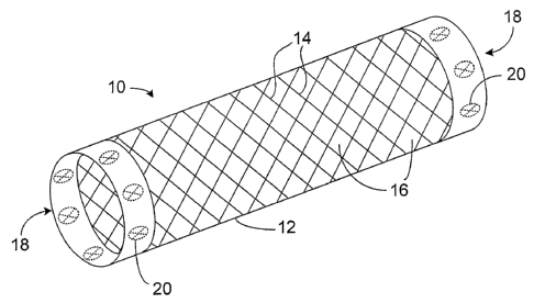
Referring to FIGS. 1A and 1B, a stent 10 includes a generally tubular stent
body 12
formed of strand materials 14. Strand materials 14 define a pattern of voids
16 in the wall of
stent body 12. Voids 16 facilitate the expansion and contraction of stent 10,
and enhance the
5
CA 02570914 2006-12-12
WO 2006/009867 PCT/US2005/021521
flexibility of stent 10. At each of its ends, stent 10 includes a polymer body
18 in the shape of a
ring that is attached to stent body 12. Radiopaque markers 20, in the form of
solid metal slugs,
are embedded in polymer body 18. A plurality of markers are spread
circumferentially around
the stent ends.
Referring as well to FIGS. 1C and 1D, markers 20 are positioned within voids
16 such
that markers 20 do not overlap with, or contact, strand materials 14.
Furthermore, markers 20
have approximately the same thickness as strand materials 14. As a result, a
relatively thick
body of radiopaque material can be provided without substantially increasing
the thickness
profile of stent 10.
The markers 20 include one or more radiopaque materials to enhance the
visibility of
stent 10 under x-ray fluoroscopy. A radiopaque material can be, for example, a
metal (e.g.,
tungsten, tantalum, platinum, palladium, lead, gold, titanium, silver); a
metal alloy (e.g., stainless
steel, an alloy of tungsten, an alloy of tantalum, an alloy of platinum, an
alloy of palladium, an
alloy of lead, an alloy of gold, an alloy of titanium, an alloy of silver); a
metal oxide (e.g.,
titanium dioxide, zirconium oxide, aluminum oxide); bismuth subcarbonate; or
barium sulfate.
In some embodiments, a radiopaque material can be a metal with a density of
about ten grams
per cubic centimeter or greater (e.g., about 25 grams per cubic centimeter or
greater, about 50
grams per cubic centimeter or greater). The radiopaque material is provided as
a solid metal slug
and/or a radiopaque powder distributed in the polymer body. Suitable
radiopaque materials are
discussed in Heath, U.S. Patent No. 5,725,570, the entire contents of which
are hereby
incorporated by reference.
The thickness and width of the markers provide a desirable radiographic image.
In
embodiments, the thickness of one or more of the markers is comparable to the
thickness of the
stent body. For example, the thickness of the marker is about +/-25 percent,
about +/- ten
percent, about +/- five percent, or less than the thickness of the stent body.
In embodiments, the
thickness is from about 0.001 inch to about 0.01 inch (e.g., from about 0.005
inch to about 0.008
inch). In embodiments, the width of the markers is such that the markers can
be positioned
within the voids of the stent body without contacting or overlapping the stent
body when the
stent is in an expanded, implanted condition. In embodiments, the markers are
sized to be
positioned within the voids without contacting or overlapping the stent body
when the stent is in
a collapsed, delivery condition and an expanded, implanted condition. In
particular
6
CA 02570914 2006-12-12
WO 2006/009867 PCT/US2005/021521
embodiments, the width of the markers is 90 percent or less, e.g., 50 percent
or less or ten
percent or less than the width of the voids in the expanded and/or contracted
condition. In
particular embodiments, the maximum width of the markers is about two
millimeters or less,
e.g., one millimeter or less or one millimeter to 0.1 millimeter. Preferably,
markers located at the
ends of the stent do not extend substantially beyond the periphery of the
stent body, so that the
length of the stent is not increased. In embodiments, the markers extend less
than about two
millimeters beyond the length of the stent body (e.g., less than about 1.5
millimeters, less than
about one millimeter, less than about 0.5 millimeter). In embodiments, the
markers are discrete
elements (e.g., metal slugs) that provide sufficient radiopacity independently
of the stent body
(without requiring the presence of the stent body) to provide a desirable
radiopaque image.
The location, shape, and number of markers provide a particular radiographic
image. To
indicate one or both ends of the stent, markers are provided at the ends of
the stent. In
embodiments, markers are provided along the body of the stent at predetermined
distances from
the end of the stent. A single marker or multiple markers can be provided
along the stent axis
and/or circumferentially about the axis. A pattern of markers can provide an
indication of stent
orientation about the axis. The markers can be shaped to indicate orientation,
e.g. cylindrical,
disk-shaped or T-shaped markers can be provided. In some embodiments, the
markers can be in
the form of radiopaque wires (e.g., individual radiopaque wires or bundles of
radiopaque wires).
In certain embodiments, the radiopaque wire markers can have a diameter of
from about 0.001
inch to about 0.015 inch (e.g., about 0.01 inch), and/or a length of from
about 0.5 millimeter to
about two millimeters, and/or an aspect ratio (the ratio of the length of the
radiopaque wire
markers to the diameter of the radiopaque wire markers) of from about 1/1 to
about 20/1. In
certain embodiments, the radiopaque wire markers can have rounded or tumbled
edges. In
embodiments, one or more of the radiopaque wire markers can be in the form of
a coil. Markers
of different shapes can be used on the same stent.
The polymer body is biocompatible, compatible with the radiopaque material
incorporated in the polymer body, of sufficient strength to retain the
markers, and of sufficient
flexibility to accommodate stent expansion and flexing during delivery or
after implantation.
The polymer body is formed of one or more layers of a polymer such as a
fluoropolymer (e.g.,
expanded-polytetrafluoroethylene), Corethane rt, a polyisobutylene-polystyrene
block copolymer
such as SIBS (see, e.g., U.S. Patent No. 6,545,097), fluorinated ethylene
propylene (FEP),
7
CA 02570914 2006-12-12
WO 2006/009867 PCT/US2005/021521
tetrafluoroethylene (TFE), and silicone (e.g., in embodiments of stent 10 that
are used for non-
vascular applications). The thickness of the polymer body is sufficient to
securely retain and
bond the marker to the stent body. The polymer body bonds to portions of the
stent body
adjacent a void in which a marker is positioned. In embodiments, the polymer
overlaps the
adjacent regions. The thickness of the overlap region is selected to reduce
the overall thickness
profile of the stent. In embodiments, the thickness of the overlap region on
an exterior wall
surface of the stent is 25 percent or less, e.g., ten percent or one percent
or less than the thickness
of the stent wall. In particular embodiments, the thickness of the overlap
region is about 200
microns or less. In embodiments, the thickness of the portions of the polymer
body overlapping
the marker similarly does not greatly increase the thickness profile of the
stent. The polymer
body extends in particular embodiments into the void between the marker and
the stent body to
prevent direct contact between the marker and the stent body. The polymer body
can include a
drug, e.g. an antiproliferative, that elutes from the polymer body into
adjacent tissue to, e.g.,
inhibit restenosis.
In embodiments, the polymer body can extend over from about ten percent to
about 100
percent of the circumference of stent body 12, e.g. more than 50 percent. The
width of the
polymer body along the stent axis extends over about one percent to 100
percent of the length of
the stent. In particular embodiments, the width of the polymer body is about
ten millimeters or
less, e.g., about two millimeters.
The polymer body can be formed and bonded to the stent by solvent casting, or
dipping a
suitable polymer directly onto the stent. Alternatively, a preformed polymer
body can be bonded
to the stent. In particular embodiments, the polymer body is formed from one
or more preformed
polymer strips. In particular embodiments, the markers are sandwiched between
the strips,
which are bonded together by an adhesive or co-melted, and/or which are
sintered or partially
sintered together.
In certain embodiments, a stent body can be formed of strands. The strands can
be, e.g.,
woven, knitted, or crocheted. In embodiments, a stent body can be in the form
of a sheet-form
body with apertures (formed by, e.g., cutting or etching). The stent body can
be defined by a
metal or a polymer. The stent can be self-expanding or balloon expandable.
Stents are further
described in Heath, incorporated supra, and Wang, U.S. Patent No. 6,379,379,
the entire contents
of which are hereby incorporated by reference.
8
CA 02570914 2006-12-12
WO 2006/009867 PCT/US2005/021521
Manufacture
Referring to FIGS. 2A-2G, the manufacture of a stent with radiopaque markers
is
illustrated. Referring to FIG. 2A, radiopaque markers 20 are attached to one
side 50 of a
preformed polymer (e.g., expanded-polytetrafluoroethylene) strip 52. The
markers 20 are
adhered to polymer strip 52, for example, by spraying and/or dipping markers
20 in a low-
viscosity dispersion (e.g., TFE, FEP), and then placing markers 20 on polymer
strip 52. The
strip 52 is heated, e.g., in an oven, such that the dispersion will cure and
sinter or partially sinter
with polymer strip 52. In embodiments, the temperature during heating is below
the melting
point of polymer strip 52. Thus, the heat can cause polymer strip 52 to soften
and adhere to
markers 20, without causing polymer strip 52 to melt. In embodiments, the
polymer in the low-
viscosity dispersion can be cross-linked and/or sintered or partially sintered
to polymer strip 52,
thereby securing markers 20 to polymer strip 52. For efficient manufacturing,
the polymer strip
to which markers 20 are attached can be longer than the circumference of the
stent. The strip is
then cut to a desired length to accommodate a stent of a desired size.
Referring now to FIG. 2B, the polymer strip 52 is arranged into a ring 54
(shown in FIG.
2C) after markers 20 have been adhered to polymer strip 52. While outer
surface 56 of ring 54
includes markers 20, inner surface 58 of ring 54 does not include any markers
20. The diameter
of the ring corresponds to the inner diameter of the stent when the stent is
in a desired expanded
configuration.
Referring to FIG. 2C, ring 54 is inserted onto a mandre160, such that inner
surface 58
contacts mandre160. In some embodiments, mandre160 is a coated mandrel (e.g.,
coated with
zirconium-nickel or titanium nitrate). In certain embodiments, a coating can
help mandre160 to
retain ring 54.
Referring now to FIGS. 2D and 2E, after ring 54 is inserted onto mandre160, a
stent
body 12 is positioned on mandre160, such that end 62 of stent body 121ies on
top of ring 54.
Strand materials 14 are positioned between markers 20, and markers 20 are
contained within
voids 16. The assembly is heated to attach the ring 54 (e.g., by partial
sintering) to the stent
body.
Referring to FIGS. 2F and 2G, a securement layer 64 is positioned over the
outer surface
of the stent body and attached to ring 54. Securement layer 64 covers markers
20. Securement
9
CA 02570914 2006-12-12
WO 2006/009867 PCT/US2005/021521
layer 64 can be made of, e.g., a polymer in the form of a preformed strip. The
strip is formed of,
e.g., the same polymer as the strip 52.
The securement layer 64 can be attached to ring 54 by adhesive-bonding (e.g.,
using
TFE) and/or by sintering or partially sintering securement layer 64. The
attachment of
securement layer 64 to ring 54 forms polymer body 66, in which markers 20 are
embedded. The
portion of the stent body covered by the polymer body is likewise sandwiched
between strip 52
and layer 64 to securely fix the markers and the polymer body 66 to the stent.
(The polymer strip
and the securement layer are attached to minimize gaps between the layers.)
Referring to FIG. 2H, polymer body 66 can be cut or trimmed (e.g., laser-
trimmed) to
reduce flaps of excess polymer material. In embodiments, polymer body 66 can
be scalloped
(e.g., to decrease stent end effects) and/or contoured or shaped (e.g., to
smoothen polymer body
66, to enhance the biocompatibility of polymer body 66, to make polymer body
66 complement
the edge of stent body 12).
Referring now to FIGS. 3A-3C, in some embodiments a polymer ring 65 formed of
markers 20 sandwiched between polymer strip 52 and securement layer 64 is
inserted onto
mandrel 60. Thereafter, stent body 12 is inserted onto mandrel 60, such that
end 62 of stent
body 121ies on top of ring 65. Strand materials 14 of stent body 12 are
positioned between the
locations of markers 20 within ring 65. A second securement layer 67 is then
added over ring 65
and end 62 of stent body 12, such that end 62 is sandwiched between securement
layer 64 and
securement layer 67.
Stent Delivery
FIGS. 4A-4C show the delivery of a self-expanding stent 200. Stent 200 is
deployed on a
catheter 202 and covered by a sheath 204. When the target site is reached,
sheath 204 is
retracted and stent 200 self-expands into contact with the body lumen.
Radiopaque markers 206
embedded within polymer bodies 208 at each end of stent 200 allow for
determination of the
location of stent 200 (e.g., by x-ray radiography).
Referring now to FIGS. 5A-5C, the delivery of a balloon-expandable stent 300
is
illustrated. Stent 300 is carried on a catheter 302 over a balloon 304. When
the treatment site is
reached, balloon 304 is expanded to expand stent 300 into contact with the
lumen wall.
CA 02570914 2006-12-12
WO 2006/009867 PCT/US2005/021521
Radiopaque markers 306 embedded within polymer bodies 308 at each end of stent
300 allow for
determination of the location of stent 300.
Stent 200 and/or stent 300 can be used in vascular and/or non-vascular
applications.
Stent 200 and/or stent 300 can be used, for example, to treat stenoses,
aneurysms, or emboli. In
some embodiments, stent 200 and/or stent 300 can be used in the coronary
and/or peripheral
vascular system, e.g., for iliac, carotid, superior femoral artery (SFA),
renal, and/or popliteal
applications. In certain embodiments, stent 200 and/or stent 300 can be used
in non-vascular
applications. For example, stent 200 and/or stent 300 can be used in
tracheal/bronchial, biliary,
and/or esophageal applications.
Other Embodiments
Referring to FIGS. 6A and 6B, an end 102 of the stent body of a stent 100 is
modified to
form a larger void volume for accommodating radiopaque markers. In FIG. 6A,
forces
(indicated by arrows F) are applied against points 104 to deform the stent to
increase the void
area to accommodate larger radiopaque markers 106 (shown in FIG. 6B).
Alternatively or
additionally, strand materials used to form a stent can be manipulated during
the stent formation
process (e.g., during weaving, knitting, crocheting) to include extra room at
the edges of the stent
for, e.g., radiopaque markers.
In embodiments, a stent can include a polymer body at only one of its ends,
rather than at
both of its ends. In certain embodiments, a stent can include a polymer body
that is not located
at either end of the stent. For example, a polymer body can be located at the
middle of the stent
body. In such embodiments, the stent can further include a polymer body at one
or both of its
ends, or can lack polymer bodies at either of its ends.
The polymer body can include more than one form of radiopaque material. For
example,
a polymer body can include embedded radiopaque markers and can have a
radiopaque powder
dispersed throughout it.
As a further example, a polymer body that includes radiopaque material can be
incorporated into other types of medical devices. For example, the polymer
body can be
incorporated into various types of endoprostheses, such as a covered stent, an
AAA (abdominal
aortic aneurysm) stent-graft, an endograft, or a surgical vascular bypass
graft, or other devices,
including prosthetic venous valves and embolic protection devices and filters.
11
CA 02570914 2006-12-12
WO 2006/009867 PCT/US2005/021521
Other embodiments are within the scope of the following claims.
12