Note: Descriptions are shown in the official language in which they were submitted.
CA 02571035 2006-12-20
WO 2006/004749 PCT/US2005/022922
COMPOSITIONS AND METHODS FOR TREATING
NEUROLOGICAL DISORDERS
STATEMENT AS TO RIGHTS TO INVENTIONS MADE UNDER
FEDERALLY SPONSORED RESEARCH AND DEVELOPMENT
This invention was made with government support under a grant awarded by the
Department of Health and Human Services. The government has certain rights in
this
invention.
CROSS-REFERENCES TO RELATED APPLICATIONS
This application claims benefit of priority from U.S. Provisional Patent
Application
60/582,999, filed June 25, 2004, which is hereby incorporated in its entirety
as if fully set
forth.
FIELD OF THE INVENTION
Compositions useful for treating neurological disorders including
neurodegenerative
disorders associated with deleterious protein aggregation, aberrant protein
folding and/or
neurodegenerative autoimmune disorders such as brain amylogenic diseases are
described.
Methods of using said compositions also are described.
BACKGROUND OF THE INVENTION
Neurological diseases are generally characterized by the loss of neurons from
one or
more regions of the central nervous system. Examples of neurological diseases
include
Alzheimer's disease, neurofibromatosis, Huntington's disease, depression,
amyotrophic
lateral sclerosis, Multiple Sclerosis, stroke, Parlcinson's disease, and multi-
infarct dementia.
They are complex in both origin and progression, and have proved to be some of
the most
difficult types of disease to treat. In fact, for some neurological diseases,
there are no drugs
available that provide significant therapeutic benefit. The difficulty in
providing therapy is
all the more tragic given the devastating effects these diseases have on their
victims.
Alzheimer's disease (AD) is a degenerative brain disorder characterized
clinically by
progressive loss of memory, cognition, reasoning, judgment and emotional
stability that
gradually leads to profound mental deterioration and ultimately death. AD is a
very
common cause of progressive mental failure (dementia) in aged humans and is
believed to
CA 02571035 2006-12-20
WO 2006/004749 PCT/US2005/022922
represent the fourth most common medical cause of death in the United States.
AD has
been observed in all races and ethnic groups worldwide and presents a major
present and
future public health problem. The disease is currently estimated to affect
about four million
individuals in the United States alone. AD is at present incurable. The
administration of
certain therapies has been used to treat symptoms of AD in humans. However, no
treatment
that effectively prevents AD or reverses its symptoms or course in humans is
currently
known.
The brains of individuals with AD exhibit characteristic lesions, termed
senile
plaques, and neurofibrillary tangles. Senile plaques characteristic of AD are
most
frequently localized extracellularly while neurofibrillary tangles are most
frequently
localized intracellularly. Large numbers of these lesions are generally found
in patients
with AD in several areas of the human brain important for memory and cognitive
function.
Smaller numbers of these lesions in a more restricted anatomical distribution
are sometimes
found in the brains of aged humans who do not have clinical AD. The principal
chemical
constituent of the senile plaques and vascular amyloid deposits (amyloid
angiopathy)
characteristic of AD is a protein designated aniyloid-(3 peptide (A(3), which
may also be
referred to as (3AP, A,6P or (3/A4. Extracellular plaques containing A,6 may
be dense or
diffuse. Dense plaques are often referred to as fibrillar plaques. A(3 was
first purified and a
partial amino acid sequence reported in Glenner and Wong (1984) Biochem.
Biophys. Res.
Commun. 120:885-890. The isolation procedure and the sequence data for the
first 28
amino acids are described in U.S. Pat. No. 4,666,829. Forms of Ao having amino
acids
beyond number 40 were first reported by Kang et al. (1987) Nature 325:733-736.
Neuropathologically, AD is characterized, to varying degrees, by four major
lesions:
a) intraneuronal, cytoplasmic deposits of neurofibrillary tangles (NFT), b)
parenchymal
amyloid deposits called neuritic plaques, c) cerebrovascular Ao amyloidosis
(e.g., amyloid
angiopathy), and d) synaptic and neuronal loss. One of the key events in AD is
the
deposition of amyloid (e.g., AB peptide) as insoluble fibrous masses
(amyloidogenesis)
resulting in extracellular neuritic plaques and deposits around the walls of
cerebral blood
vessels. The major constituent of the neuritic plaques and cerebral amyloid
angiopathy is
A(3, although these deposits also may contain other proteins such as
glycosaminoglycans
and apolipoproteins.
2
CA 02571035 2006-12-20
WO 2006/004749 PCT/US2005/022922
Solomon, B. et al. (1997) PNAS 94(8):4109-1.2 showed that monoclonal antibody
against the N-termini of A,13 can bind to and disaggregate preexisting
assemblies of Ao-
peptide and/or prevent fibril aggregation in vitro and prevent toxicity to
neuronal cell
cultures. Schenk, D. et al., Nature 400(6740):173-177 (1999) demonstrated that
immunization witli amyloid-(3 attenuated Alzheimer's disease-like pathology in
PDAPP
transgenic mice serving as an animal model for amyloid-,li deposition and
Alzheimer's
disease-like neuropathologies. They reported that immunization of young
animals prior to
the onset of Alzheimer's disease-type neuropathologies essentially prevented
the
development of 0-amyloid plaque formation, neuritic dystrophy and
astrogliosis, whereas
treatment in older animals after the onset of Alzheimer's disease-type
neuropathologies was
observed to reduce the extent and progression of these neuropathologies. This
effect is
mediated by antibodies, since peripherally administered antibodies against Ao
have been
shown to reduce brain parenchymal amyloid burden (Bard F. et al., (2000) Nat.
Med.
6(8):916-9). In addition, intranasal immunization with freshly solubilized
A(.i 1-40 reduces
cerebral amyloid burden (Weiner, H.L. et al., (2000) Ann. Neuro. 48(4):567-
79). Two
studies, performed by Morgan, D. et al., (2000) Nature 408(6815):982-5; and
Janus, C. et
al., (2000) Nature 408(6815):979-82, using animal model systems demonstrated
that a
vaccination-induced reduction in brain amyloid deposits resulted in cognitive
improvements. Additional studies have addressed various aspects of the same
topic,
including Dodart et al., (2002) Nat. Neuroscience 5(5):452-7, and Kotilinek,
L.A. et al.,
(2002) J. Neuroscience 22(15):6331-5. Although AB vaccination has shown some
success
in various studies using animal models of AD, human clinical studies
immunizing with AB
1-40/42 peptides formulated in an adjuvant (QS21) were terminated because of
deleterious
and/or an unacceptably high occurrence of side effects such as
meningoencephalitis. Thus,
there is a need for therapeutically acceptable modalities for the treatment
and/or prevention
of AD and related neurodegenerative disorders associated with protein
aggregation.
Autoimmune diseases are characterized by an abnormal immune response directed
to self or autologous tissues. Based on the type of immune response (or immune
reaction)
involved, autoiinmune diseases in mammals can generally be classified into one
of two
different types: cell-mediated (i.e., T-cell-inediated) or antibody-mediated
disorders.
Multiple Sclerosis (MS) is a T-cell mediated autoimmune disease (Trapp et al.
New Eng. J.
Med. 338(5):278 (1998)). More than 1,000,000 young adults worldwide between
the ages
of thirty and forty have MS. MS is the most common disease of the central
nervous system
3
CA 02571035 2006-12-20
WO 2006/004749 PCT/US2005/022922
and is the most common cause of neurological disability in young adults.
Pathophysiologically, circulating autoreactive T cells mediate much of the
central nervous
system destruction seen in MS patients (Rudick et al. New Eng. J. Med.
337:1604(1997)).
In MS, T-cells react with myclin basic protein (MBP) which is a coinponent of
myelin in the central nervous system. The demonstration that activated T-cells
specific for
MBP can be isolated from MS patients supports the proposition that MS is an
autoimmune
disease wherein T-cells destroy the self or autologous neural tissue
(Allegretta et al.
S cience: 247: 778 (1990)).
MS is currently treated with certain anti-inflammatory and immunosuppressive
agents, such agents include: (i) corticosteroids, which have both
immunomodulatory and
immunosuppressive effects; (ii) interferon-0; (iii) glatiramer acetate (GA);
(iv) azathioprine,
a purine analog which depresses both cell-mediated and humoral immunity; (v)
intravenous
immune globulin; (vi) methotrexate, which inhibits dihydrofolate reductase and
depresses
cell-mediated and humoral immunity; (vii) cyclophosphamide, an alkylating
agent which
has cytotoxic and immunosuppressive effects; and (viii) cyclosporine, which
has potent
immunosuppressive effects by inhibiting T cell activation. Despite treatment
with such
anti-inflammatory or immunosuppressive drugs, more than 50% of the patients
with MS
steadily deteriorate as a result of focal destruction of the spinal cord,
cerebellum, and
cerebral cortex.
Many of the drugs currently used to treat MS have limited long-term efficacy,
in
part, because they have significant cytotoxic effects. For example, prolonged
treatinent
with cyclophosphamide can lead to alopecia, nausea, vomiting, hemorrhagic
cystitis,
leukopenia, myocarditis, infertility, and pulmonary interstitial fibrosis.
Treatment with
iinmunosuppressive agents can eventually induce "global" immunosuppression in
the
treated patient, which greatly increase the risk of infection. Patients
subjected to prolonged
global immunosuppression have an increased risk of developing severe medical
complications from treatment, such as malignancies, kidney failure and
diabetes.
An alternative approach to the treatment of MS is the use of intravenous or
oral
administration of MBP to modulate the T-cell immune response that may be
associated
therewith. Intravenous administration of MBP or fragments thereof containing
immunodominant epitopes of MBP suppresses the immune system by causing clonal
anergy, or T-cell unresponsiveness, which deactivates T-cells specific for
MBP. The end-
result is that MBP-specific T cells no longer proliferate in response to MBP.
The inability
of the T-cell to proliferate results in a decrease in T-cell mediated
destruction of neural
4
CA 02571035 2006-12-20
WO 2006/004749 PCT/US2005/022922
tissues.
An immunochemical analog of MBP used in treating MS is glatiramer acetate
(GA),
or copolymer-1 (COP-1) (iJ.S. Pat. No. 3,849,550; PCT Application
WO/95/31990). GA,
in its commercially available form, is a mixture of random synthetic
polypeptides composed
of L-alanine, L-glutamic acid, L-lysine and L-tyrosine in a molar ratio of
6.0:1.9:4.7:1Ø It
was first synthesized as an immunochemical mimic of MBP. For example, certain
monoclonal antibodies to GA cross-react with MBP (Teitelbaum et al. Proc.
Natl. Acad.
Sci. USA 88:9258 (1991)). Also, GA has been found to induce T suppressor cells
specific
for MBP (Lando et al. J. Immunol. 123:2156 (1979)). Experiments in mice
indicate that
GA also specifically inhibits MBP-specific T cells that are involved in the
destruction of
central nervous systein tissue in Experimental Allergic Encephalomyelitis
(EAE)
(Teitelbaum et al. Proc. Natl. Acad. USA 85:9724 (1995)).
Administration of GA may: (i) increase the percentage of NK cells; (ii) reduce
serum IL-2 receptors; (iii) suppress TNF-a; and (iv) increase TGF-0 and IL-4
(Ariel et al.
Multiple Sclerosis 3(5), S053 (1997)).
Although patients with MS have been relatively successfully treated with
parenterally administered GA (Bornstein et al. Transactions American
Neurological
Association, 348 (1987)), the current treatment regime and overall effects
could be
improved.
Citation of the above documents is not intended as an admission that any of
the
foregoing is pertinent prior art. All statements as to the date or
representation as to the
contents of these documents is based on the information available to the
applicant and does
not constitute any admission as to the correctness of the dates or contents of
these
documents.
SUMMARY OF THE INVENTION
The present invention provides methods and compositions for treating
neurological
diseases or disorders in mammals in need of such treatment. Said neurological
diseases or
disorders can be associated with a systemic or localized deposition of protein
or
proteinaceous material (e.g., amyloidosis), deleterious protein aggregation
(protein mis-
folding) and/or neurodegenerative autoimmunity. Particular interest is in the
amyloid
forming diseases such as Alzheimer's disease and/or other brain amylogenic
diseases
including prion-related diseases, Huntington disease, Parkinson's disease and
cerebral
amyloid angiopathy (CAA) (Revesz, T. et al. (2003) J. Neuropathol. Exp.
Neurol.
5
CA 02571035 2006-12-20
WO 2006/004749 PCT/US2005/022922
62(9):885-98). The treatment of said amyloid related diseases can include
preventing new
ainyloid plaque (deposition) formation, maintaining current ainyloid plaque
levels, and/or
decreasing the amount of existing amyloid plaque or total brain amyloid
protein (including
A,6 that may not be deposited into plaques) as measured by determining total
amyloid load
(soluble and non-soluble AO) or the ainount of fibrillar Ao-amyloid load. Said
neurological
diseases or disorders can be associated with a cell-mediated autoimmune
disease such as
Multiple Sclerosis. The treatment of said autoimmune disorders can include
preventing the
fonnation of autoreactive T cells, maintaining current autoreactive T cell
concentrations,
and/or decreasing the concentration of autoreactive T cells.
The present invention claims and utilizes various formulations of a proteosome
based composition, and/or a GA composition, optionally in a submicron
emulsion, or a
nanoemulsion, as therapeutics for treating neurological diseases or disorders
in mammals
including AB plaque related diseases or disorders, and cell-mediated
autoimmune diseases
or disorders.
BRIEF DESCRIPTION OF THE DRAWINGS
Figure 1: Effect of subcutaneous immunization on total Afl levels in the
brain.
To quantify ainyloid burden, the right hemisphere was extracted in 5.0 M
guanidinium-
chloride (pH 8) for 3 hours at room teinperature. Dilutions were used to
measure levels of
A040 and A,fl42 by sandwich enzyme-linked immunosorbent assays (ELISA).
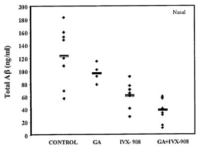
Figure 2: Effect of nasal immunization on total A(3 total levels in the brain.
Total A(3 concentration levels of A040 and A042 from individual mice following
nasal
treatment measured by sandwich enzyme-linked immunosorbent assays (ELISA).
Figure 3: Activation of CD11b+ cells lead to clearance of Afl fibril in
parenterally and nasally treated mice. (A) Staining of A(.3 fibril in
hippocampal region
with thioflavin-S (Magnificationxl0) or co-staining for total Ao with anti Ao
antibody
(R1288) and anti-CD11b (microglia/macrophage) Magnification x40) following
subcutaneous immunization. (B) Co-staining anti Ao antibody (R1288) and anti-
CD11b.
(microglia/macrophage) (in hippocampal region Magnification x40) following
nasal
immunization.
Figure 4: Immunohistology of brain sections following MOG subcutaneous
immunization and nasal glatiramer acetate vaccination. Serial sections of the
6
CA 02571035 2006-12-20
WO 2006/004749 PCT/US2005/022922
hippocampus region from untreated, or immunized mice 50 days post immunization
were
labeled using anti CD11b, CD3, IFN--y..and TGF-(3 antibodies (magnification
x20, insert
figure magnification x60).
Figure 5: Reduction in astrocytosis following nasal administration of GA+IVX-
908. Well-defined hippocampal regions (Bregma -1.44mm), were selected for
quantification of activated astrocytes using GFAP staining. The level of
astrocyte
activation was expressed as a percentage per mm2 hippocampal region; p=0.039
GA+IVX-
908 vs. control; p=0.02 vs. EAE (MOG).
Figure 6: Neuropathology in brain sections following MOG subcutaneous
immunization and nasal glatiramer acetate vaccination. Serial sections of the
cortex
from untreated, or treated mice 50 days post immunization were labeled using
markers of
neurotoxicty: SM132, TiJNEL, and iNOS (original magnification x20). Arrows
identify
labeling for markers studied. Labeling for markers of neurotoxicity was
observed in EAE
animals, but not in GA-IVX-908 treated animals.
Figure 7: Blood brain barrier integrity in hippocampus section following MOG
subcutaneous immunization and nasal glatiramer acetate vaccination. Serial
sections
of the cortex :froin untreated, or treated mice 50 days post immunization were
labeled using
marker of plasma staining -fibrinogen. Labeling for markers of fibrinogen was
observed in
EAE animals, but not in GA-IVX-908 treated animals. (Magnification x20, small
figure
magnification x40).
Figure 8: Neuropathology in olfactory sections following MOG subcutaneous
immunization and nasal glatiramer acetate vaccination. Serial sections of the
cortex
from untreated, or treated mice 50 days post immunication were labeled using
markers of
fibril amyloid: ThS, microglia activation CD11b, BBB integrity, Fibrinogen.
(Magnification x20)
Figure 9: Staining for CD68+ cells in the CNS in untreated, MOG immunized
and GA+IVX-908 treated mice. Arrows identify CD68+cells which infiltrate the
CNS in
EAE, but remain localized to choroids plexus in GA+IVX-908 treated mice. No
staining
was observed in untreated mice. Sections are taken from the cerebellum
(Magnification
x20).
7
CA 02571035 2006-12-20
WO 2006/004749 PCT/US2005/022922
MODES OF PRACTICING THE INVENTION
The term "neurological disease" refers to a disease or disorder, which
involves the
neuronal cells of the nervous system. Specifically included are: prion
diseases (e.g,
Creutzfeldt-Jakob disease); pathologies of the developing brain (e.g.,
congenital defects in
amino acid metabolism, such as argininosuccinicaciduria, cystathioninuria,
histidinemia,
homocystinuria, hyperammonemia, phenylketonuria, tyrosinemia, and fragile X
syndrome);
pathologies of the mature brain (e.g., neurofibromatosis, Huntington's
disease, depression,
amyotrophic lateral sclerosis, Multiple Sclerosis); conditions that strike in
adulthood (e.g.
Alzheimer's disease, Creutzfeldt-Jakob disease, Lewy body disease, Parkinson's
disease,
Pick's disease); and other pathologies of the brain (e.g., brain mishaps,
brain injury, coma,
infections by various agents, dietary deficiencies, stroke, multiple infarct
dementia, and
cardiovascular accidents).
The preferred diseases or disorders of the current invention are those
diseases
affecting the mature brain, such as Multiple Sclerosis, and those which
typically strike in
adulthood, such as Alzheimer's disease.
The term "Alzheimer's disease", abbreviated herein as "AD" refers to a
neurodegenerative disease of the central nervous system. Broadly speaking, the
disease
falls into two categories: late onset, which occurs in old age (typically
above 65 years) and
early onset, which develops well before the senile period, e.g., between 35
and 60 years. In
both types of the disease, the pathology is similar, but the abnormalities
tend to be more
severe and widespread in cases beginning at an earlier age. AD is
characterized by the
accumulation of extracellularly localized brain amyloid (e.g., A,13 peptide),
amyloid plaques
(which may be further distinguished as dense or diffuse) and intracellularly
localized
neurofibrillary tangles concentrated in certain vulnerable regions of the
brain such as the
hippocampus and cortex. AD is a progressive disease resulting in senile
dementia.
Amyloid plaques are areas of disorganized neurofibrillary fibers which may be
associated
with neutrophils up to 150 mm across with extracellular amyloid-beta (AB)
deposits at the
center, visible by microscopic analysis of sections of brain tissue.
Neurofibrillary tangles
are intracellular deposits of tau protein (often hyperphosphorylated)
consisting of two
filaments twisted about each other in pairs. AD is associated with the
abnormal
acculnulation of A13 peptide resulting from altered proteolytic processing of
amyloid
8
CA 02571035 2006-12-20
WO 2006/004749 PCT/US2005/022922
precursor protein (APP). Abnormal accumulation of AB has been correlated with
a variety
of mutations, such as autosomal dominant APP mutations and mutations in genes
encoding
proteins referred to as presenilin 1 (PS 1) and presenilin 2 (PS2), which
subsequently
influence the proteolytic activity of -y (gamma), or B (beta) secretase,
causing increased
levels of, for exainple, A131-42; wllereas, the proteolytic activity of a
(alpha) secretase is
presumably associated with the normal processing of APP. Various types of
plaques are
found in AD, including, but not limited to neuritic plaques associated with
abnormal
dystrophic neurites. Also characteristic of the disease is the presence of an
inflammatory
response in the CNS, including activated microglia and astrocytes. The
accumulation of
aggregates of dysfunctional protein (e.g., A13 and prion protein) associated
with neurological
disorders are tliought to cause or contribute to or otherwise influence the
development of
certain neurological disorders (Ingelsson, M. and Hyman B.T. (2002) Aimals of
Med.
34:259-271). Neurodegenerative disorders associated with deleterious protein
aggregation
include Alzheimer's disease, Pick's disease, Parkinson's disease, prion
disease, Huntington
and motor neuron disorders. (Shastry, Neurochemistry International, 2002, 43:1-
7).
The tenn "amyloid" refers to the extracellular (e.g., AB, prion diseases, and
multiple
myeloma light chain disease) or intracellular (e.g., neurofibrillary tangles
of tau protein in
AD and alpha-synuclein in Parkinson's disease) deposition of protein
aggregates
(Trojanowski J.Q. and Mathson M.P. (2003) Neuromolecular Medicine 4:1-5).
Amyloid
deposition can be found in the brain of AD and Down's Syndrome patients as
well as in
arteries, arterioles, capillaries and veins of the central nervous system.
Amyloid deposits
can be recognized by the ability to bind dyes such as Congo red and thioflavin
S, and form
fibrils, including a cross f3-pleated sheet confirmation.
The term "amyloidosis" refers to a large heterogeneous group of disorders
characterized by aberrant insoluble deposits of nonnally soluble proteins,
which may be
misfolded proteins, including protein aggregates.
In addition to Alzheimer's disease (AD), early onset Alzheimer's disease, late
onset
Alzheimer's disease, and presymptomatic Alzheimer's disease, other diseases
characterized
by amyloid deposits are, for example, Serum Amyloid A (SAA) amyloidosis,
hereditary
Icelandic syndrome, multiple myeloma, prion diseases and the like, or other
brain
amylogenic diseases (Revesz, T. et al. (2003) J. Neuropathol. Exp. Neurol.
62(9):885-98),
may be treated according to compositions and methods set forth herein. The
most common
9
CA 02571035 2006-12-20
WO 2006/004749 PCT/US2005/022922
prion diseases in animals are scrapie of sheep and goats and bovine spongiform
encephalopathy (BSE) of cattle (Wilesmith and Wells(1991) Curr. Top.
Microbiol.
Immunol. 172:22-38). Four prion diseases have been identified in humans: (i)
kuru, (ii)
Creutzfeldt-Jakob disease (CJD), (iii) Gerstmann-Streussler-Sheinker disease
(GSS), and
(iv) fatal familial insomnia (FFI) (Gajdusek, D.C. (1977) Science
197(4307):943-60 and
Medori, R. et al., (1992) N. Engl. J. Med. 326(7):444-9).
The principal constituent of the senile plaques is the A(3 peptide. The AO
peptide is
an internal fragment of 39-43 amino acids of precursor protein APP. Several
mutations
within the APP protein have been correlated with the presence of Alzheimer's
disease (See,
e.g., Goate, A. et al., (1991) Nature 349(6311):704-6, Murrell, M. et al.,
(1991) Science
254(5028):97-9, Mullan, M. et al., (1992) Nat. Genet. 1(5):345-7).
The term "O-ainyloid precursor protein" (APP) as used herein is defined as a
polypeptide that is encoded by a gene of the same name localized in humans on
the long
arm of chromosome 21 and that includes Ao within its carboxyl third. APP is a
glycosylated, single-membrane-spanning protein expressed in a wide variety of
cells in
many maminalian tissues.
APP mutations are thought to influence the development of Alzheiiner's disease
by
increased or altered proteolytic processing of APP to A,6, particularly
processing of APP to
increased amounts of the long fonn of A,13 (i.e., A# 1-42 and A,131-43).
Mutations in other
genes, such as the presenilin genes, PS 1 and PS2, are thought indirectly to
affect proteolytic
processing of APP to generate increased amounts of long form A,13 (see Hardy,
J. (1997)
Trends Neurosci. 20(4):154-9). These observations indicate that A,li, and
particularly its
long form, is a causative element in Alzheimer's disease.
The term "APP fragments" as used herein refers to fragments of APP other than
those which consist solely of Ao or A(3 fragments. That is, APP fragments will
include
amino acid sequences of APP in addition to those which form intact A,6 or a
fragment of
AO.
The terms "beta-amyloid peptide" is synonymous with ",6-amyloid peptide",
",6AP",
"OA", and "Ao". All of these terms refer to a plaque forming peptide derived
from
fragments of amyloid precursor protein.
CA 02571035 2006-12-20
WO 2006/004749 PCT/US2005/022922
As used herein, the definition of the terms fibrillar A,li and total Ag are as
follows.
"Fibrillar" A,6 is A,6 peptide contained in extracellular amyloid deposits
which may also be
referred to as A(3 plaques or plagues; in some cases, Ao plaques may be
further
distinguished as diffuse or dense. "Total" amyloid load or total A(31oad is
the sum of
soluble and non-soluble (e.g., fibrillar) AO peptide, most of wliich is
presumed to be
extracellular. It is appreciated that there is a dynamic relationship between
soluble and non-
soluble A(.3, where, extracellular non-fibrillar A,(3 may represent a source
of A,6 which may
become fibrillar amyloid.
As used herein, the term Experimental Allergic Encephalomyelitis (EAE) is the
primary animal model for MS. EAE can readily be induced in small mammals by
immunization with MBP in an appropriate adjuvant or by passive transfer of
CD4+, MBP-
reactive T-cells (Alvord Jr, E. C., et al. eds. in Experimental Allergic
Encephalomyelitis a
Useful Model for Multiple Sclerosis, A. R. Liss, N.Y., 1984; Makhtarian et al.
Nature 309:
356 (1984); Ben-Nun et al. J. Immunol. 129:303 (1982)). The T-cells that
induce EAE in
both mice and rats recognize peptides corresponding to immunodominant regions
of MBP
presented by antigen-presenting cells on class II Major Histocompatibility
Complex (MHC)
molecules.
According to one aspect of the present invention there is provided a method of
treating a neurological disease or disorder in a mammal. The method of this
aspect of the
present invention may be made effective by administering a therapeutically
effective
amount of GA and a proteosome based composition to a subject in need thereof.
A further
aspect of the present invention is wherein said neurological disease or
disorder is a amyloid
plaque-forming disease or disorder. The most prominent neurological diseases
or disorders
treated with GA and a proteosome based composition according to one aspect of
the present
invention is Alzheimer's disease and Multiple Sclerosis. In addition to a
therapeutic
composition of the GA combined with a proteosome for treatment of the
aforementioned
neurological disease or disorders, further embodiments include, but are not
limited to, using
the following therapeutic compositions for treatment: a proteosome based
coinposition
without a GA composition, GA in a submicron emulsion composition, or GA in a
nanoemulsion composition. The previous embodiments would also include any
pharmaceutically acceptable diluent, excipient, stabilizer or carrier.
11
CA 02571035 2006-12-20
WO 2006/004749 PCT/US2005/022922
A yet further aspect of the present invention is wherein the treatment of said
neurological disease or disorder in a mammal coinprises administering a
therapeutically
effective amount of GA and a proteosome based composition which elicits an
antibody
independent response in said mammal.
A further embodiment of the present invention consists of a method for
treating an
amyloidal disease, which may be Alzheimer's disease. The treatment of
amyloidal disease
may be carried out by, for example, preventing an increase in fibrillar
amyloid load,
preventing an increase in total amyloid load, maintaining the current
fibrillar and/or total
amyloid load, or decreasing the fibrillar and/or total amyloid load in the
brain.. As it is
known that amyloidal proteins may reside throughout the body, embodiments of
the present
invention are not limited to brain amyloid. Furthermore, although the present
invention
specifically discusses 0-amyloid, other amyloid classes such as serum amyloid
A (SAA),
prion disease, hereditary Icelandic syndrome, Huntington disease,
Parkinsonism, Down's
Syndrome and cerebral amyloid angiopathy are considered within the current
invention.
According to the aforementioned amyloidal diseases, treatment may be
accomplished by
administering one of the following therapeutic compositions: a therapeutically
effective
amount of GA and a proteosome based coinposition, a proteosome based
composition
without a GA composition, GA in a submicron emulsion, and/or GA in a
nanoemulsion.
The previous embodiments would also include any pharmaceutically acceptable
diluent,
excipient, stabilizer or carrier.
GLATIRAMER ACETATE
An immunochemical analog of MBP that is effective in treating multiple
sclerosis
(MS) is glatiramer acetate (GA), or copolymer-1 (Cop-1) (U.S. Pat. No.
3,849,550; PCT
Application WO/95/31990). GA, in its commercially available form, is a mixture
of
random synthetic polypeptides composed of L-alanine, L-glutamic acid, L-lysine
and L-
tyrosine in a molar ratio of 6.0:1.9:4.7:1Ø It was first synthesized as an
immunochemical
mimic of MBP. For example, certain monoclonal antibodies to GA cross-react
with MBP
(Teitelbaum et al. Proc. Natl. Acad. Sci. USA 88:9258 (1991)). Also, GA has
been found to
induce T suppressor cells specific for MBP (Lando et al. J. Immunol. 123:2156
(1979)).
Experiments in mice indicate that GA also specifically inhibits MBP-specific T
cells that
are involved in the destruction of central nervous system tissue in EAE
(Teitelbaum et al.
Proc. Natl. Acad. USA 85:9724 (1995); and Angelov, D.N. et al. PNAS
100(8):4790-4795
12
CA 02571035 2006-12-20
WO 2006/004749 PCT/US2005/022922
(2003)), indicate that GA can be used to treat amyotrophic lateral sclerosis,
where induction
of a well-regulated autoimmune response appears to influence survival in the
presence of an
anti-self T-cell response, which may be enhanced by administration of GA.
GA, according to the present invention, may be prepared by methods known in
the
art. For example, GA may be prepared by the process disclosed in U.S. Pat. No.
3,849,550,
wherein the N-carboxyanhydrides of tyrosine, alanine, 7-benzyl glutamate and E-
N-
trifluoro-acetyllysine are polymerized at ambient temperature in anhydrous
dioxane with
diethylamine as an inhibitor. The deblocking of the -y -carboxyl group of the
glutamic acids
is carried out with hydrogen bromide in glacial acetic acid and is followed by
the removal
of the trifluoracetyl groups from the lysine residues by 1M piperidine. The
resulting
mixture of polypeptides consists essentially of polymers of alanine, glutainic
acid, lysine,
and tyrosine, in a molar ratio of about 6:2:5:1.
GA is also available commercially from Teva Pharmaceuticals, Kfar-Saba,
Israel.
GA may be prepared for use according to the instant invention in any of the
forms
which maintain its therapeutic utility. These include mixtures of peptides
having various
molecular weight ranges. GA having a desired molecular weight range can be
obtained by
methods known in the art. Such methods include gel filtration high pressure
liquid
chromatography of GA to remove high molecular weight species as disclosed in
WO
95/31990. In one embodiment, the GA has about 75% of its polymer species
within the
molecular weight range of about 2 KDa to about 20 KDa. In another embodiment,
GA has
an average molecular weight from about 4 KDa to 9 KDa. It is understood that
GA may be
subjected to enzymatic or other degradation in order to comprise polymer
species of a
length different from, or otherwise modified, from conventional GA according
to the known
methods.
GA AND PROTEOSOMES
GA formulated with proteosomes (e.g., IVX-908 or Protollin) may be
administered
by, for example, injection or intranasally. When delivered by injection, such
delivery may
be as one injection (combined simultaneous administration). Alternatively,
delivery of a
GA composition and a proteosome based composition may be delivered separately,
accomplished by injection at a plurality of sites which may occur
simultaneously or at
temporally distinct times and where one site is presented only with a GA
composition (i.e.,
13
CA 02571035 2006-12-20
WO 2006/004749 PCT/US2005/022922
no proteosomes) and, at a second site only a proteosome based composition is
administered
(i. e., no GA). It is also contemplated that GA may be administered by
injection while at the
same or different time a proteosome based composition is administered, for
example,
intranasally. Thus, in one embodiment of the instant invention, a GA
composition is
delivered by injection and separately, a proteosome based composition is
delivered
intranasally.
GA peptides of the instant invention may also be prepared to contain a
hydrophobic
anchor sequence moiety which would be expected to enhance non-covalent
association with
proteosomes. The production and manufacture of proteosome-amphiphilic
determinant
vaccines designed for either parenteral or especially for mucosal
administration including,
gastro-intestinal administration to induce both systemic and mucosal antibody
responses is
discussed in U.S. Pat No. 6,476,201. An amphiphilic determinant is a molecule
having
hydrophobic and hydrophilic regions which, when appropriately formulated with
proteosomes, align with the proteosomes to form a complex which elicits an
immunologic
response in a subject. Typical amphiphilic determinants include glycolipids,
liposaccharides (including detoxified lipopolysaccharides), lipopeptides,
transmembrane
domains, envelope or toxoided proteins, or proteins or peptides with intrinsic
hydrophobic
amino acid anchors.
EMULSION COMPOSITIONS
GA can be administered in or formulated with a submicron emulsion or
nanoemulsion as described in US Patent 5,961,970 or 5,716,637, respectively.
The
composition comprises an oil-in -water submicron emulsion with about 0.5 to
about 50% of
an oil, about 0.1 to about 10% of an emulsifier, about 0.05 to about 5% of a
nonionic
surfactant, and about 0.00001 to about 1.0% of GA as an aqueous continuous
phase. The
submicron emulsion has a mean droplet size in the range of between about 0.03
and about
0.5 m, and preferably 0.05 and 0.2 m.
The nanoemulsion approach provides vaccine compositions containing
nanoemulsions of particles having a lipid core, surrounded by at least one
phospholipid
bilayer. The particles have a mean diameter in the range of about 10 to about
250 nm, as
determined on a weight basis, and the GA is incorporated therein, either
intrinsically prior
to the homogenization process or extrinsically thereafter. The particles are
typically
suspended in an aqueous continuous phase and each lipid particle comprises a
lipid core,
14
CA 02571035 2006-12-20
WO 2006/004749 PCT/US2005/022922
wherein the lipid is a solid or liquid crystal in bulk at a temperature of
about 25 C or higher.
Usually the amount of GA entrapped is about 0.001 to about 5%. Alternatively,
the GA can
be formulated with a proteosome and/or the nanoemulsion can further comprise a
bioadhesive or mucoadhesive macromolecule.
PROPOSED MECHANISM
The invention also includes, for example, a method for inhibiting or reducing
the
level of an amyloid-O peptide (soluble and/or non-soluble), fragment or
derivative thereof,
which comprises immunizing a mammal with GA formulated with a proteosome based
composition, or a proteosome based composition without GA, wherein the
reduction or
inhibition of the peptide, fragment or derivative thereof occurs without the
generation of
antibodies, as demonstrated herein using B-cell deficient ( MT) mice, which
are incapable
of eliciting an antibody response. Altl7ough not wishing to be bound by
theory, preferably,
the inhibition or reduction of amyloid (e.g., A(3 peptide) is via an
activation of immune cells
such as brain localized microglia, or neutrophils and/or macrophages which may
be found
in the brain or peripherally, wherein the activation of these cells is
independent of any
antibody or antigen-specific mechanism. It is most preferred that the
reduction or inhibition
occurs without causing Experimental Allergic Encephalomyelitis (EAE) in the
mammal
(including meningoencephalitis).
The reduction of ainyloid load (e.g., Ao comprising fibrillar plaques plus non-
fibrillar A(3 that may not be deposited into plaques) relates to the reduction
of amyloid
deposition and/or Afl plaque formation and thereby the treatment of AD and
related diseases
or amyloidogenic diseases or disorders. Although the various aspects of the
invention
should not be limited to any particular theory or mechanism, it is believed
that activated
microglia co-localize with amyloid plaques and the activation of microglial
cells is
dependent upon the presence of amyloid deposition, which deposition primes the
endogenous microglial cells for activation. Thus, activated microglial cells
participate in
clearing amyloid (e.g., A,6 plaque) deposits. Additional information on the
possible
mechanisms for AD is found in Schenk, D. (2002) Nature 3:824-828. Deposition
of Af3
and formation of amyloid plaques appears to be accompanied by a complex
inflammatory
a.nd neurotoxic cascade. Therefore, it is thought that an anti-inflammatory
mechanism of
treatment may be beneficial. Such anti-inflammatory processes are often
consistent with
expression of anti-inflammatory IL-4/IL-10 (Th2) and TGF-B (Th3) immune
responses.
CA 02571035 2006-12-20
WO 2006/004749 PCT/US2005/022922
Consequently it is a surprising and unexpected finding that the instant
formulations
comprising proteosome based compositions and GA stimulate a non-antibody
mediated
immune response that causes the reduction of AB-amyloid containing plaques, as
it has
previously been proposed that proteosomes are more often associated with the
stimulation
of a Thl type immune (cytokine) response associated with a pro-inflammatory
immune
response and would be appositive to and distinct from a Th2 immune response.
Nevertheless, auginentation of microglial phagocytosis by the Thl-type
cytokine INF-
gainma (75% above untreated), might also suggest a feedback mechanism for
accelerated
clearance of the inflammatory infiltrate in the CNS (Cha, A. et. al. (2001)
GLIA 33(l):87-
1,0 95). Furthermore, INF-gamma by itself can also lead to the transcriptional
inhibition of the
beta-amyloid precursor protein (Ringheeim G. E. et al. Biochem Biophys Res
Commun
(1996) 224(1):246-51).
PROTEOSOME BASED COMPOSITIONS
The subject of US Patents 6,476,201 and 5,961,970 describes how in order for
multivalent subunit vaccines to stimulate optimal immune responses to each of
the
components, the proper components should be appropriately associated and each
be
available to the immune system so that they may be efficiently recognized and
processed by
cells of the immune system. Such recognition and processing may include uptake
by
membranous cells (M-cells) located within, for example, the nasal epithelia
and subsequent
delivery to underlying cells of the host immune system. Prime examples of such
non-
covalently complexed vaccines include proteosome based vaccines which can
consist of
Neisserial outer membrane proteins non-covalently complexed to a wide variety
of antigens
including peptides, lipopeptides, transmembrane or toxoided proteins,
polysaccharides or
lipopolysaccharides (LPS) (for further review see the following references; US
Patent
5,726,292, Immunogenicity and Efficacy of Oral or Intranasal Shigellaflexneri
2a and
Shigella sonnei Proteosome-Lipopolysaccharide Vaccines in Animal Models;
Infect.
Immun. 61:2390; Mallett, C. P., T. L. Hale, R. Kaminski, T. Larsen, N. Orr, D.
Cohen, and
G. H. Lowell. (1995)); Intranasal or intragastric immunization with proteosome-
Shigella
lipopolysaccharide vaccines protect against lethal pneumonia in a murine model
of
shigellosis (Infect. Immun. 63:2382-2386; Lowell G H, Kaminski R W, Grate S et
al.
(1996)); Intranasal and intramuscular proteosome-staphylococcal enterotoxin B
(SEB)
toxoid vaccines: immunogenicity and efficacy against lethal SEB intoxication
in mice
(Infec. Immun. 64:1706-1713; Lowell, G. H. (1990)); Proteosomes, Hydrophobic
Anchors,
16
CA 02571035 2006-12-20
WO 2006/004749 PCT/US2005/022922
Iscoms and Liposomes for Improved Presentation of Peptide and Protein
Vaccines, (in New
Generation Vaccines: G. C. Woodrow and M. M. Levine, eds. (Marcel Dekker, NY)
Chapter 12 (pp. 141-160)); and Proteosome-lipopeptide vaccines: enhancement of
immunogenicity for malaria CS peptides; (Lowell, G. H., W. R. Ballou, L. F.
Smith, R. A.
Wirtz, W. D. Zollinger and W. T. Hockmeyer (1988) Science 240:800)).
Proteosome based compositions as used herein refers to preparations of outer
membrane proteins (OMPs, also known as porins) from Gram-negative bacteria,
such as
Neisseria species (see, e.g., Lowell et al., J. Exp. Med. 167:658, 1988;
Lowell et al., Science
240:800, 1988; Lynch et al., Biophys. J. 45:104, 1984; Lowell, in "New
Generation
Vaccines" 2nd ed., Marcel Dekker, Inc., New York, Basil, Hong Kong, pages 193,
1997;
U.S. Patent No. 5,726,292; U.S. Patent No. 4,707,543), which are useful as a
carrier or an
adjuvant for immunogens, such as bacterial or viral antigens. Proteosomes
prepared as
described above also contain an endogenous lipopolysaccharide (LPS)
originating from the
bacteria used to produce the OMP porins (e.g., Neisseria species). Any
preparation method
that results in the outer membrane protein component in vesicular or vesicle-
like form,
including multi-molecular membranous structures or molten globular-like OMP
compositions of one or more OMPs, is included within the definition of
proteosome.
"Liposaccharide" as used herein refers to native or modified
lipopolysaccharide or
lipooligosaccharide (collectively, also referred to as "LPS") derived from
Gram-negative
bacteria such as Shigella flexneri or Plesiomonas shigelloides, or other Gram-
negative
bacteria (including Alcaligenes, Bacteroides, Bordetella, Brucella,
Cainpylobacter,
Chlamydia, Citrobacter, Edwardsiella, Elzrlich.a, Enterobacter, Escherichia,
Francisella,
Fusobacterium, Gardnerella, Henaophillus, Helicobacter, Klebsiella,
Legionella,
Moraxella, Morganella, Neiserria, Pasteurella, Proteus, Providencia, other
Plesiomonas,
Porplz.yronaonas, Prevotella, Pseudonaonas, Rickettsia, Salmonella, Serratia,
other Sliigella,
Spirilluna, Veillonella, Vibrio, or Yersinia species). It should be noted that
LPS as used
herein may be non-detoxified or detoxified.
In other embodiments exogenous LPS isolated from the same or different
bacteria
from which a proteosome was prepared may be mixed therewith; one such
proteosome
based composition is referred to herein as IVX-908 (may also be referred to as
Protollin).
In other words, proteosomes of the IVX-908 type are preparations of OMPs
admixed with at
least one kind of liposaccharide to provide an OMP-LPS composition (which can
finiction
17
CA 02571035 2006-12-20
WO 2006/004749 PCT/US2005/022922
as an immunostimulatory composition). Thus, the OMP-LPS (IVX-908) adjuvant can
be
comprised of two of the basic components (1) an outer membrane protein
preparation of
proteosomes prepared from Gram-negative bacteria, s'uch as Neisseria
meningitides, and (2)
a preparation of one or more liposaccharides. It is also contemplated that
coinponents of
IVX-908 may be or include lipids, glycolipids, glycoproteins, small molecules,
or the like.
Proteosome based compositions as disclosed herein may include one or more
components which, at least in part, function as an adjuvant possessing the
capacity to
stimulate a host immune system. It is appreciated that such proteosome
components may
include an outer membrane protein (OMP) component (fusion protein or fragment
thereof)
of gram negative bacteria as well as a lipopolysaccharide (LPS) component of a
same or
different gram negative bacteria. Such components may function as, for
example, ligands
which stimulate a host immune response by interacting with certain receptors
(e.g., Toll-like
receptors) produced by one or more host cells of a vaccine recipient.
Without wishing to be bound by theory, one or more components of vaccine
formulations disclosed herein may interact with Toll-like receptors (TLRs)
associated with
an innate and/or adaptive immune response of a vaccine recipient. There are at
least 10
TLRs (see Takeda et. al., Annu Rev Immunology (2003) 21:335-76). One or more
ligands
which interact with and subsequently activate certain TLRs have been
identified, with the
exception of TLR8 and TLR10. Outer membrane proteins of Neisseria
meningitides, for
example OMP 2 (also referred to as Por B), interacts with TLR2, while LPS of
most but not
all gram negative bacteria interacts with TLR4. Accordingly, one mechanism by
which
vaccine formulations described herein may contribute to a biological affect
includes
activation of one or both of TLR2 and TLR4. However, in another aspect of the
instant
invention, it is equally possible that activation of other TLRs (other than
TLR2 and TLR4)
may serve a similar function or further enhance the qualitative and/or
quantitative profile of
cytokines expressed, and which may be associated with a host Thl/Th2 immune
response.
The qualitative and/or quantitative activation of one or more TLRs is expected
to
elicit or influence a relative stimulation (balanced or imbalanced) of a Th1
and/or Th2
immune response, with or without concomitant humoral antibody response.
Ligands interacting with TLRs other than TLR2 and TLR4 may also be used in
vaccine compositions described herein. Such vaccine components may, alone or
in
combination, be used to influence the development of a host immune response
sufficient to
18
CA 02571035 2006-12-20
WO 2006/004749 PCT/US2005/022922
treat or protect from amylogenic disease, as set forth herein. Such TLRs and
associated
ligands include, but are not limited to, those presented in List 1.
List I
TLR family Ligands
(example of possible source)
TLRI Soluble factors (Neisseria meningitides)
Tri-acyl lipopeptides (bacteria,
mycobacteria)
TLR2 Lipoproteins and lipopeptides
Porins (Neisseria)
Atypical LPS (Leptospira interrogans)
Atypical LPS (Porphyromonas gingivalis)
Peptidoglycan (Gram-positive bacteria)
Lipoteichoic acid (Gram-positive
bacteria)
HSP70 (host)
Glycolipids (Treponema maltophilum)
TLR3 Double-stranded RNA (e.g., viral)
TLR4 LPS (Gram-negative bacteria)
Taxol (plant)
HSP60 (host)
HSP70 (host)
HSP60 (Chlamydia pnemoniae)
Fibrinogen (host)
TLR5 Flagellin (bacteria)
19
CA 02571035 2006-12-20
WO 2006/004749 PCT/US2005/022922
TLR6 Di-acyl lipopeptides (mycoplasma)
TLR7 Imidazoquinoline (synthetic compounds)
Loxoribine (synthetic compounds)
Bropirimine (synthetic compounds)
TLR8 Ligand yet to be identified
TLR9 CpG DNA (bacteria)
TLR10 Ligand yet to be identified
Any one or combination of the identified TLRs (List 1) may be activated by any
one
or combination of TLR ligand components of a vaccine formulation contemplated
herein. It
is further appreciated that stimulation of any one or multiplicity of TLRs may
be
accoinplished using any one or a multiplicity of TLR ligands at concentrations
suitable with
the route of administration (e.g., intranasal, injection etc.).
Therefore, according to the instant invention, it is understood that a vaccine
formulation may include any one or more TLR ligand(s), including recombinant
ligands
(fusion proteins or fragments thereof) combined with an antigenic vaccine
component, or
optionally including CD 14 receptor, with or without exogenous addition of a
lipopolysaccharide component.
In one aspect of the instant invention only the TLR binding portion of any one
or
more TLR ligands may be isolated by, for example, recombinant DNA technologies
and
formulated witli or without GA as a therapeutic treatment and/or prophylactic
prevention of
Alzheimer's Disease or similar disease or disorder. Such a polypeptide may
also be
prepared by one or another synthetic procedures well known to those of skill
in the art. Not
wishing to be bound by theory, one such isolated binding domain may be
isolated from a
portion of Neisseria rneningitidis outer membrane protein referred to as Porin
B which is
suspected of binding to TLR2. Other such polypeptide ligand binding domains of
TLRs
may also be used in similar fashion alone or in one or another combination.
Such a
formulation may be used with or without the necessity of administering a
Proteosome-GA
CA 02571035 2006-12-20
WO 2006/004749 PCT/US2005/022922
fonnulation, or may be used subsequent to administration of a Proteosome-GA
formulation.
In addition, it is appreciated that a variant (e.g., conservative amino acid
substitution) of
such a TLR binding portion of a TLR ligand maybe used to activate a TLR as
long as the
variant maintains the ability to bind (and activate) the TLR. In still a
further aspect of the
instant invention, such a TLR binding portion of a TLR ligand may be
reiterated one or
more times using recombinant DNA technologies to prepare a polypeptide
containing
multiple copies of such binding portion, or even multivalent (i.e., hybrid)
polypeptides
comprising multiple binding domains of the same or different TLR ligands.
In certain proteosome based compositions, one or more of the component parts
of
the vaccine fonnulation need not be non-covalently complexed but rather may be
mixed
with the proteosome composition (e.g., IVX-908, Protollin). Proteosome based
compositions tenned Projuvant contain only small amounts of endogenous LPS (or
lipooligosaccharide (LOS)) while IVX-908/protollin proteosome based
compositions
contain additional exogenous LPS which may be from the same or different grain
negative
bacterial species as the OMP components or may be a mixture of LPS derived
from more
than one gram negative bacteria.
In one embodiment, the final liposaccharide content by weight as a percentage
of the
total proteosome protein can range from about 1% to about 500%, more
preferably in a
range from about 20% to about 200%, or in a range from about 30% to about 150%
or in a
range of about 10% to about 100%. A preferred embodiment of the instant
invention is the
immunostimulatory composition wherein the proteosome based component is
prepared from
Neisseria naeningitides and the final liposaccharide content is between 50% to
150% of the
total proteosome protein by weight. The final LPS content may represent the
combination
of endogenous LPS (e.g., LOS) plus exogenously added LPS (or LOS). In another
embodiment, proteosome based compositions (e.g., Projuvant) are prepared with
endogenous lipooligosaccharide (LOS) content from NeiseYria ranging from about
0.5% up
to about 5% of total OMP. Anotlier embodiment of the instant invention
provides
proteosomes with endogenous liposaccharide in a range from about 12% to about
25%, and
in a preferred embodiment between about 15% and about 20% of total OMP. The
instant
invention also provides a composition containing liposaccharide derived from
any Gram-
negative bacterial species, which may be from the same Gram-negative bacterial
species
that is the source of proteosomes or from a different bacterial species.
21
CA 02571035 2006-12-20
WO 2006/004749 PCT/US2005/022922
U.S. Patent 6,476,201 relates to the production and manufacture of proteosome-
amphiphilic determinant vaccines designed for either parenteral or mucosal
administration
to induce both systemic (serum) and mucosal (including respiratory and
intestinal) antibody
responses. Mucosal administration is preferred, and includes, but is not
limited to,
respiratory (e.g. including intranasal, intrapharyngeal and intrapuhnonary),
gastro-intestinal
(e.g. including oral or rectal) or topical (e.g. conjunctival or otic)
administration. An
amphiphilic determinant is a molecule having hydrophobic and hydrophilic
regions which,
when appropriately formulated with proteosoines, align with the proteosomes to
form a
complex which elicits an immunologic response in a subject. Typical
amphiphilic
determinants include glycolipids, liposaccharides (including detoxified
lipopolysaccharides), lipopeptides, transmeinbrane, envelope or toxoided
proteins, or
proteins or peptides with intrinsic hydrophobic amino acid anchors. These
determinant
materials can be obtained from grain negative bacteria including Escherichia,
Klebsiella,
Pseudonaonas, Hemophilus, Brucella, Shigella and Neisseria. More specifically,
the
proteosome vaccines in which meningococcal outer membrane protein proteosoine
preparations (prepared from any strain of N. ineningitides or N. gonorrhea or
other
Neisserial species) are non-covalently coinplexed to native or detoxified
Shigella or
Neisserial lipopolysaccharides or lipooligosaccharides to form vaccines are
designed to
protect against diseases caused by gram negative organisms that contain any of
the
component parts of the complex including Meningococci or Shigellae. More
specifically,
the proteosome vaccines contain LPS that induce antibody responses that
recognize type-
specific somatic polysaccharide O-antigens of Shigella lipopolysaccharides and
thereby
confer homologous protection against shigellosis. The lipopolysaccharides,
when
complexed to proteosomes, induce anti-shigella protective immune responses.
The
proteosome vaccines are prepared and purified from either Shigella sonnei or
Plesiornonas
shigelloides for immunity against Slaigella sonnei disease, from
Shigellaflexneri 2a for
immunity to Shigellaflexneri 2a disease, and so forth, using LPS derived from
homologous
or antigenically cross-reacting organisms to confer homologous immunity
against
shigellosis caused by S. flexnef i 2a (or 3a etc.), S. boydii, S. sonnei etc.
Further, US Patent
6,476,201 describes the administration of proteosome-Shigella vaccines that
are multivalent
in that two independently made proteosome vaccines using shigella LPS antigen
derived
from S. flexneri 2a (for S. flexneri 2a disease) and from P. shigelloides or
S. sonnei (for S.
sonnei disease) are administered together thereby inducing antibodies that
recognize the two
organisms and thereby conferring protection against the two types of diseases.
22
CA 02571035 2006-12-20
WO 2006/004749 PCT/US2005/022922
Furthermore, a proteosome-Shigella LPS vaccine, in which proteosomes from
group B type
2b meningococci are complexed to P. shigelloides LPS using hollow fiber
diafiltration
technology to produce a vaccine administered by mucosal respiratory and/or
gastro-
intestinal routes and to induce antibodies that recognize the somatic 0-
antigen LPS of S.
sonnei, are thereby used to protect against shigellosis.
An exemplary but non-limiting proteosome based composition of the present
invention is proteosome based mucosal adjuvant IVX-908 (Protollin) which is a
non-
covalent formulation of Neisseria meningitides outer membrane proteins
(proteosomes) and
exogenously added LPS prepared from Shigellaflexneri.
FORMULATION AND ADMINISTRATION
Hereinafter, the phrases "physiologically acceptable carrier" and
"phannaceutically
acceptable carrier" which may be interchangeably used refer to a carrier or a
diluent that
does not cause significant irritation to an organism and does not abrogate the
biological
activity and properties of the administered coinpound.
Herein the tenn "excipient" refers to an inert substance added to a
phannaceutical
coinposition to further facilitate administration of an active ingredient.
Examples, without
limitation, of excipients include calcium carbonate, calcium phosphate,
various sugars and
types of starch, cellulose derivatives, gelatin, vegetable oils and
polyethylene glycols.
Techniques for formulation and administration of drugs may be found in
"Reinington's Pharmaceutical Sciences," Mack Publishing Co., Easton, Pa.,
latest edition.
Suitable routes of administration may, for example, include oral, rectal,
transmucosal, transnasal, intestinal or parenteral delivery, including
intramuscular,
subcutaneous and intramedullary injections as well as intrathecal, direct
intraventricular,
intravenous, intraperitoneal, intranasal, or intraocular injections, but the
preferred route of
administration for proteosomes is intranasally.
GA formulations comprising proteosome based compositions, or in submicron
emulsions or nanoemulsions can be administered parenterally, orally,
intranasally, or
topically, or, as indicated herein, in any combination thereof. Although in
certain
embodiments it is preferable to administer them parenterally or mucosally.
23
CA 02571035 2006-12-20
WO 2006/004749 PCT/US2005/022922
Dual or multiple routes of administration are also included herein. Dual
routes of
administration may include, for example, proteosome based preparations
prepared and
administered intranasally but separately from GA which may be administered by
injection
at the same time or at a time different from intranasal administration of
proteosomes.
Proteosome (Projuvant) or proteosome:LPS (i.e., IVX-908) compositions (in the
absence of
GA) may also be administered by injection simultaneously with GA or at a
different time.
For injection, the active ingredients of the invention may be formulated in a
physiologically
acceptable carrier, preferably in physiologically compatible buffers such as
Hank's solution,
Ringer's solution, or physiological salt buffer. For transdermal and possibly
transmucosal
adininistration, penetrants appropriate to the barrier to be permeated may be
used in the
formulation. Such penetrants are generally known in the art.
For oral administration, the compounds can be formulated readily by combining
the
active compounds with pharmaceutically acceptable carriers well known in the
art. Such
carriers enable the compounds of the invention to be formulated as tablets,
pills, dragees,
capsules, liquids, gels, syrups, slurries, suspensions, and the like, for oral
ingestion by a
patient. Pharmacological preparations for oral use can be made using a solid
excipient,
optionally grinding the resulting mixture, and processing the mixture of
granules, after
adding suitable auxiliaries if desired, to obtain tablets or dragee cores.
Suitable excipients
are, in particular, fillers such as sugars, including lactose, sucrose,
mannitol, or sorbitol;
cellulose preparations such as, for example, maize starch, wheat starch, rice
starch, potato
starch, gelatin, gum tragacanth, methyl cellulose, hydroxypropylmethyl-
cellulose, sodium
carboinethylcellulose; and/or physiologically acceptable polymers such as
polyvinylpyrrolidone (PVP). If desired, disintegrating agents may be added,
such as cross-
linked polyvinyl pyrrolidone, agar, or alginic acid or a salt thereof such as
sodium alginate.
For nasal administration, the active ingredients for use according to the
present
invention may be conveniently delivered in the form of, for example, an
aerosol spray
presentation from a pressurized pack or a nebulizer with the use of a suitable
propellant,
e.g., dichlorodifluoromethane, trichlorofluoromethane, dichloro-
tetrafluoroethane or carbon
dioxide. In the case of a pressurized aerosol, the dosage unit may be
determined by
providing a valve to deliver a metered amount. Capsules and cartridges, made
of gelatin for
example, for use in a dispenser may be formulated to contain a powder mix of
the
compound and a suitable powder base such as lactose or starch.
24
CA 02571035 2006-12-20
WO 2006/004749 PCT/US2005/022922
The preparations described herein may be formulated for parenteral
administration,
e.g., by bolus injection or continuous infusion. Formulations for injection
maybe presented
in unit dosage form, e.g., in ampoules or in multidose containers with
optionally, an added
preservative. The compositions may be suspensions, solutions or emulsions in
oily or
aqueous vehicles, and may contain formulatory agents such as suspending,
stabilizing
and/or dispersing agents.
The pharmaceutical compositions of the present invention can be administered
for
prophylactic and/or therapeutic treatment of diseases related to the
production of and/or
deposits of amyloid (e.g., A(.3) anywhere in the subject's body, but
especially in the brain. In
therapeutic applications, the pharmaceutical compositions are administered to
a mammal
already suffering from the disease and in need of treatment. The
pharmaceutical
compositions will be administered in an amount sufficient to inhibit or reduce
further
deposition of Ao into plaque and/or clear already formed plaque and/or to
stimulate removal
of already existing A(3 aggregates, and/or stimulate a reduction in A(3 that
may not be
contained in plaques. An amount adequate to accomplish this is defmed as a
"therapeutically effective dose or amount."
For prophylactic applications, the pharmaceutical compositions of the present
invention are administered to a maminalian subject susceptible to an amyloid
related disease
(e.g., A(3 related Alzheimer's disease), but not already suffering from such
disease. Such
mammalian subjects inay be identified by genetic screening and clinical
analysis, as
described in the medical literature (e.g. Goate (1991) Nature 349:704-706).
The
pharmaceutical compositions will be able to inhibit, prevent or reduce amyloid
deposition
of, for example, A(3 into plaque at a symptomatically early stage, preferably
preventing even
the initial stages of the 0-amyloid related disease. The amount of the
compound required
for such prophylactic treatment, referred to as a prophylactically-effective
dosage, may, but
not necessarily, be generally the same as described above for therapeutic
treatment.
For purposes of this specification and the accompanying claims the terms
"patient",
"subject" and "recipient" are used interchangeably. They include humans and
other
maminals (e.g., cow and other bovine) which are the object of either
prophylactic,
experimental, or therapeutic treatment.
As used herein the term "treating" includes substantially inhibiting, slowing
or
reversing the progression of a disease, substantially ameliorating clinical
symptoms of a
CA 02571035 2006-12-20
WO 2006/004749 PCT/US2005/022922
disease or substantially preventing the appearance of clinical symptoms of a
disease, in a
statistically significant manner.
Depending on the severity and responsiveness of the condition to be treated,
dosing
can be of a single or a plurality of administrations at one or a plurality of
sites or delivery
means, with the course of treatment lasting from several days to several weeks
or until cure
is effected or a statistically significant diminution of the disease state is
achieved. The
amount of a treatment to be administered will, of course, be dependent on the
subject being
treated, the severity of the affliction, the manner of administration, the
judgment of the
prescribing physician, etc. Methods for calculating statistical significance
are lcnown in the
relevant art.
"Treatment" of MS is intended to include both treatment to prevent or delay
the
onset of any manifestation, clinical or subclinical, e.g., histological,
symptoms thereof of
Multiple Sclerosis, as well as the therapeutic suppression or alleviation of
symptoins after
their manifestation by abating autoimmune attack and preventing or slowing
down
autoimmune tissue destruction. "Abatement", "suppression" or "reduction" of
autoimmune
attack or reaction encompasses partial reduction or amelioration of one or
more symptoms
of the attack or reaction. A "substantially" increased suppressive effect (or
abatement or
reduction) of the "autoimmune reaction" means a significant decrease in one or
more
markers or histological or clinical indicators of MS. Non-limiting examples
are a reduction
by at least 1 unit in limb paralysis score.
GA is generally administered to treat MS in a dose of 0.01 mg to 1000 mg/day.
In
one embodiment a dosage in the range of 0.5-50 mg is employed. However,
according to
one aspect of the instant invention it is anticipated that the use of one or
more adjuvants set
forth herein (or combination thereof) may be formulated with a composition
comprising GA
wllereby lower or higher doses or frequency of administration of GA may be
permitted and
that it is not necessary that the dose of GA be effective by itself.
Establishing the effective dosage range as well as the optimum amount is well
within the skill in the art in light of the information given in this section.
For example,
dosages for mammals, and huinan dosages in particular are optimized by
beginning with a
relatively low dose of GA (e.g., 1 mg/day), progressively increasing it (e.g.,
logarithmically) and measuring a biological reaction to the treatment; for
example, (i)
measuring induction of regulatory cells (CD4+ and/or CD8+) (Chen, Y. et al.,
Science, 255:
1237 (1994)); (ii) measuring reduction in class II surface markers on
circulating T-cells; (iii)
26
CA 02571035 2006-12-20
WO 2006/004749 PCT/US2005/022922
measuring the number of TGF-0 expressing cells or the relative amount of
detectable TGF-
0; (iv) assessing the number and activation of immune attack T-cells in the
blood (e.g., by
limiting dilution analysis and ability to proliferate); or, (v) by scoring the
disease severity,
according to well-known scoring methods (e.g., by measuring the number of
attacks, joint
swelling, grip strength, stiffness, visual acuity, ability to reduce or
discontinue medication).
An effective dosage is any dose that causes at least a statistically or
clinically significant
attenuation in one of these markers and preferably one that attenuates at
least one symptom
characteristic of MS during the dosing study.
Assessment of the disease severity in MS can be accomplished according to well-
kn.own methods depending on the type of disease. Such methods include without
limitation:
severity and number of attacks over a period of time; progressive accumulation
of disability
(which can be measured, e.g., on the Expanded Disability Status Scale); number
and extent
of lesions in the brain (as revealed, e.g., by magnetic resonance imaging);
and frequency of
autoreactive T-cells.
Having now generally described the invention, the same will be more readily
understood through reference to the following examples which are provided by
way of
illustration, and are not intended to be limiting of the present invention,
unless specified.
EXAMPLES
Amyloid precursor,protein (APP) transgenic mice were immunized with myelin
oligodendrocyte glycoprotein (MOG) peptide (amino acids 35-55) in complete
Freund's
adjuvant (CFA) with subsequent administration (injection) of pertussis toxin
(PT) to
determine their susceptibility to Experimental Allergic Encephalomyelitis
(EAE) coinpared
to their non-transgenic littermates. As controls, animals were immunized with
bovine
serum albumin (BSA) or human (3-amyloid peptide (A(3 amino acids 1-40). EAE
developed
to an identical degree in APP transgenic animals (Mucke et al., Ann NY Acad
Sci (777) 82-
88 (1996)) and their non-transgenic littermates, and no EAE was observed in
animals
immunized with A(3 1-40 or with BSA. However, when the brains were examined
neuropathologically, there was less staining for A(3 in animals that developed
EAE. By
quantifying the amount of staining for A(3 fibril in the hippocampus using
thioflavin S, a
92% reduction in the MOG-immunized mice was found vs. controls (p=0.001) and a
73%
reduction compared to mice immunized with Ap 1-40 (p=0.03) (See Table 1, Fig
1). By
quantifying total brain A(3 content by ELISA, a 94% reduction in A(3 in MOG-
immunized
27
CA 02571035 2006-12-20
WO 2006/004749 PCT/US2005/022922
animal was determined as compared to control groups (p<0.001) and an 86%
reduction
when compared to A(3 1-40 immunized xnice (p=0.03) (See Table 1, Fig 1).
In order to determine whether the effect was unique to MOG-induced EAE, EAE
was induced with proteolipid protein (PLP) peptide (amino acids 139-15 1) in
CFA. A 76%
reduction of staining for A(3 fibrils was found and a 70% reduction in A(3
levels in animals
with PLP induced EAE (p<0.02) (See Table 2, Fig 3A). In order to induce EAE
with PLP,
Tg2576 mice as described by Hsiao, K. et al., Science 274 (5284):99-102 (1996)
were
utilized, which are on a B6/SJL background. Similar results were found with
Tg2576 mice
and J20 immunized mice described by Mucke et al., Ann NY Acad Sci (777) 82-88
(1996),
to induce MOG-EAE (See Table 1). These results demonstrate that the
observation was not
related to the antigen used for EAE induced or the animal model of AD studied,
nor the
genetic background of the animal, nor the gender (50% male/female). No changes
were
observed in animals immunized with BSA in CFA. Of note, there was no
difference in the
total Ao (amyloid) load in J20 mice greater than 13 months in age when
compared to
Tg2576 mice greater than 16 months of age.
In previous studies of immune approaches for the treatment of the mouse model
of
AD in which animals were immunized with A(3 peptide forinulated in CFA, anti-
aggregating 0 amyloid antibodies have been shown to have a role both in vitro
by Solomon,
B. et al, Proc Natl Acad Sci 94:4109-12 (1997), and in vivo by Weiner, H. L.
et al., Ann
Neurol 48, 567-79 (2000), and Schenk, D. et al., Nature 400, 173-7 (1999), in
reducing
amyloid load, and their activity has been linked to a specific epitope in the
N-terminal
region of A(3 (Frenkel, D. et al., Neuroimmunol 88, 85-90 (1998), and Frenkel,
D. et al.,
Proc Natl Acad Sci U S A 97, 11455-9 (2000)). Antibody levels were measured
against A(3
in animals immunized with MOG or PLP to determine if there was cross
reactivity with A(3
and A(3 antibody titers as compared to those from animals immunized with AP.
As shown
in Tables 1 and 2, anti-A(3 antibodies in animals immunized with MOG or PLP
were not
detected. To definitively establish that antibodies were not playing a role,
16-month-old J20
mice bred to MT B-cell deficient mice were immunized with MOG 35-55 in CFA
followed by administration of Pertussis toxin. As shown in Table 1 and Figure
1, MT B-
cell deficient APP+ mice, had a 90% reduction in amyloid compared to control
as measured
by either ThS staining (p<0.001) or by ELISA for total brain amyloid
(p<0.001). These
28
CA 02571035 2006-12-20
WO 2006/004749 PCT/US2005/022922
results indicate that the reduction of A(3 following MOG immunization occurred
by an
antibody independent mechanism.
Previous studies have suggested that activated microglia may play an important
role
in clearing A(3 in vivo (Schenk, D. et al., Nature 400, 173-7 (1999), Rogers,
J. et al., Glia
40, 260-9 (2002), Mitrasinovic, O.M. et al., Neurobiol Aging 24, 807-15
(2003), Webster,
S.D. et al., Exp Neuro1161, 127-38 (2000), Bacskai, B.J. et al., J Neurosci
22, 7873-8
(2002), Nicoll, J.A. et al., Nat Med 9, 448-52 (2003), Akiyama, H. & McGeer,
P.L., Nat
Med 10, 117-8; author reply 118-9 (2004)). Activated microglia (or microglia-
like cells)
may be further distinguished based upon being brain derived or originating
from outside the
brain, i.e., peripherally, such as neutrophils and macrophages. Procedures to
distinguish
brain derived microglia from peripheral neutrophils and macrophages are known
in the art.
To investigate the potential role of microglia activation in A(3 clearance,
the brains of APP
tg mice immunized (by foot pad injection or intranasally) with MOG/CFA were
stained
with CDl lb, a marker of activated microglia. As shown in Figures 3A and 4 and
Table 4
immunostaining of the hippocampus of APP tg MOG-immunized mice revealed
increased
numbers of activated microglia (349 34.3 cells/hippocampus region) which co-
localized
with amyloid plaques (Table 4). Only minimal staining was observed in control
animals
immunized with BSA/CFA (76 17 cells/hippocampus region) (Table 4).
Intermediate
levels of inicroglia activation were observed in animals immunized with
A(3/CFA (106 27
cells/hippocampus region). Furthermore, increased levels of microglia
activation were also
observed in MT B-cell deficient mice (p<0.001), and animals iminunized with
PLP
(p<0.001) (Fig. 3A, Table 4).
According to the first series of experiments described just above,
administration of
MOG/PLP plus CFA (followed by administration of pertussis toxin) was
associated with a
reduction of AB but was coincident with unwanted EAE. In these experiments
pertussis
toxin is used to open the blood-brain barrier for delivery of CFA formulated
compounds
into the brain. Therefore, in order to evaluate whether the reduction in AB
was directly
related to the induction in EAE and since MOG, PLP and GA have been studied in
relation
to MS related EAE, the effects of immunization of APP mice with glatiramer
acetate (GA),
which is a random amino acid copolymer of alanine, lysine, glutamic acid, and
tyrosine that
is effective in suppression of EAE and is an approved and widely used
treatment for
29
CA 02571035 2006-12-20
WO 2006/004749 PCT/US2005/022922
relapsing forms of MS (Teitelbaum, D. et al., J. Neural Transm Supp149, 85-91
(1997))
were evaluated.
Although as initially performed, administration of certain antigen/adjuvant
mixtures
provided herein is followed by an administration (injection) of pertussis
toxin; it is fully
appreciated that certain other compositions described herein may be
administered without
administration of pertussis toxin. Indeed, proteosome based compositions such
as IVX-908
with or without GA are administered without the administration of pertussis
toxin. In fact,
pertussis toxin was not used at any time for the nasal delivery of any
proteosome based
composition described herein.
Mice were thus immunized (by foot pad injection) with 100 g GA in CFA and
immediately thereafter and at 48 hours received an i.p. injection of 150 ng of
pertussis
toxin. Fifty days post immunization, mice were sacrificed. Glatiramer acetate
immunization led to a 92% reduction in amyloid fibril in the hippocampus
region vs.
untreated controls (p<0.01) and a 70% reduction of total amyloid load (p<0.01)
(See Table
1, Fig 1). There was no clinical EAE in animals immunized with GA/CFA plus
perhtssis
toxin.
As CFA cannot be administered to human subjects, the effect of nasal
vaccination
with glatirainer acetate in a mouse model of AD with adjuvants other than CFA
was
investigated; animals were treated with nasally administered glatiramer
acetate alone or
together with a mucosal adjuvant. A proteosome based mucosal adjuvant IVX-908
(Protollin) comprised of a non-covalent formulation of Neisseria menirigitides
outer
membrane proteins (proteosomes) and LPS from Slzigella flexneri, which has
been used
both in humans (Fries, L.F., et al., Infect Immun 69:4545-53 (2001)) and mice
(Plante, M. et
al., Vaccine 20, 218-25 (2001)), was prepared. Mice received multiple
treatments of the
proteosome adjuvant the first week and then were boosted on a weekly basis for
the next
five weeks after which neuropathologic analysis was performed. As controls,
animals were
nasally treated with IVX-908+BSA, IVX-908 alone or GA alone. Unexpectedly,
nasal
administration of GA formulated with IVX-908 resulted in an 84% reduction of
thioflavin-
positive fibrillar amyloid in the hippocampus (p<0.001 vs. control) and
reduction of 70%
compared to IVX-908 alone (p<0.01) (Table 3). In terms of total brain A(3
levels, a 73%
reduction was observed following nasal administration of glatiramer acetate in
NX-908
(p<0.001 vs. control) and a 45% reduction using IVX-908 alone (p<0.002) (Table
3, Figure
CA 02571035 2006-12-20
WO 2006/004749 PCT/US2005/022922
2). In addition, activated microglia were detected surrounding the amyloid
plaques in
treated animals. Nasal administration of GA alone did not affect A(3 fibrils
though there
was a slight reduction of total A(3 levels in the brain (p=0.044 vs. control).
Nasal
administration of BSA+IVX-908 or IVX-908 alone resulted in a 50% reduction of
total
amyloid load (p=0.02 vs. control) although there was no effect on fibrillar
A(3 staining. As
opposed to injection with CFA, nasal administration of IVX-908 alone or as
formulations
with GA or BSA did not induce EAE in any animal in these experiments.
No microglial activation in APP non-transgenic mice was found in any of the
immunization protocols: parenterally with CFA/PT plus GA, nasally with IVX-908
plus
GA (See Fig 3B), or nasally with IVX-908 alone. These results suggest that the
activation
of microglia following administration (immunization) with GA formulated with
IVX-908 or
IVX-908 alone may be dependent on the presence of amyloid deposition which may
serve
to prime endogenous microglia for activation.
Although there is no known cross-reactivity between GA and A(3 and we did not
observe anti-A(3 antibodies in either GA treated or EAE animals, it is
possible that
immunization with GA+IVX-908 could have resulted in priming of A(3 reactive T
cells. We
measured T cell proliferative responses and cytokine production (IL-2, IFN-7,
IL-6) after 7
weeks of weekly treatment with GA+IVX-908 (at which time the experiment was
terminated) by stimulating splenic T cells with A(3(1-40). We found no priming
of Ap
reactive T cells as measured by proliferation: Counts per minute to A(3 for
untreated = 3315
1682 cpm; for GA+IVX-908 = 4516 1412 cpm (background counts were 100-300).
Stimulation index (GA+IVX-908/untreated) =1.37; a minimal stinulation index of
greater
than 2.5 is considered positive. Furthermore, we did not find secretion of IL-
2, IFN--y, IL-6
above background in these cultures. This lack of T cell response to A(3 is
consistent with
our not detecting anti-A(3 antibodies as T cell help is required for
production of antibodies.
Similarly, we did not find priming to A(3 in EAE animals.
To examine the effect of GA+IVX-908 treatment on other brain sites besides the
hippocampus, we investigated the olfactory bulb and the cerebellum. We stained
the
olfactory bulb for A(3, CDl lb and fibrinogen and obtained similar results as
those observed
in the hippocampus (Figure 8). Following nasal administration of GA+IVX-908 we
found
an increased number of activated microglia as compared to control. The
activation also
31
CA 02571035 2006-12-20
WO 2006/004749 PCT/US2005/022922
occurred in animals with EAE, but was associated with leakage in the blood
brain barrier
(BBB) as measured by staining for fibrinogen.
When we examined the cerebellum, we did not find increased activation of
microglial staining in GA+IVX-908 treated animals suggesting that the
increased activation
is restricted to areas with A(3 deposition. Furthermore, no activation of
microglia was
observed anywhere in the brains of non-transgenic littermates following GA+IVX-
908
which further demonstrates that the increased activation is restricted to
areas with Ap
deposition (see Figure 3b).
To better understand the mechanism of the clearance we observed, we stained
for
CD68 expression, which is highly expressed on activated macrophages from the
periphery
as opposed to brain microglia. As shown in Figure 9, we obtained higher
staining for CD68
in animals with EAE as compared to those treated with GA+IVX-908. This pattern
of
staining shows the migration of macrophages from the choroids plexus to the
surrounding
brain parenchyma including the cerebellum and cortex. In GA+IVX-908 treatment,
there is
increased expression of CD68 primarily in the choroids plexus space. This
suggests that the
CD1 lb+ cells responsible for clearance of Ap in animals with EAE migrate to
the CNS
from the periphery and are associated with neuronal toxicity whereas the
CD11b+ cells in
GA+IVX-908 treated animals are primarily endogenous microglial cells and are
associated
with clearance of Ap without evidence of direct toxicity. As further support
for this
interpretation, we found that there is increased expression of CD68+ cells in
the cerebellum
of animals with EAE but not in GA+IVX-908 or untreated animals (not shown).
Moreover,
activated CD 11b+ cells following GA+IVX-908 treatment were only found in
regions
where there was accumulation of amyloid.
As shown in Table 4 and Figure 4, the reduction of Ap fibrils in the
hippocampus
was strongly correlated with increased numbers of both activated microglial
cells, as shown
by CD 11b staining, (r=-0.7 CD 11b vs. A(3 fibrils), and IFN--y vs. AP
fibrils). There was a
strong correlation between CD11b cells and IFN--y cells we observed increased
numbers of
microglia immunoreactive for macrophage colony-stimulating factor receptor (M-
CSFR) in
treated animals as compared to control (p<0.02) (Table 4). We observed a
reduction in
TGF-0 and the percentage of A(3 fibril in the hippocampus region (r=0.91). No
significant
changes were observed in IL-10 immunoreactive cells between control and GA+IVX-
908
treated animals though animals with EAE had less IL-10 than controls.
32
CA 02571035 2006-12-20
WO 2006/004749 PCT/US2005/022922
The following experiments were performed in order to evaluate whether
treatment
with GA plus IVX-908 induces neuronal cell toxicity or otller potential
negative effects: i)
determine the level of GFAP, a measure of astrocytosis (toxicity) consistent
with neuronal
cell damage; ii) measure the expression level of SM132, a marker for the
phosphorylation of
neurofilaments and known to increase with neuronal cell damage; iii) conduct a
TUNEL
assay as a means of measuring apoptotic cell death; iv) and determine the
level of iNOS, an
enzyme shown to be up-regulated under conditions of neuronal cell stress.
Results from the GFAP assay show that astrocytosis occurred in control
untreated
animals (area of activated astrocytes in hippocampus as measured by GFAP+
cells) (Figure
5). Astrocytosis was reduced in GA+IVX-908 given nasally (3.1%; p=0.039 vs.
control).
In contrast, there was no reduction in astrocytosis in EAE animals. These
results indicate
that clearance of A(3 as a consequence of treatment with GA+IVX-908 is less
toxic
(associated with less astrocytosis) than that observed in animals with EAE
even though
EAE is also associated with a reduction in A,6 deposits.
Results from experiments measuring SM132 (Figure 6) show that SM132 positive
cells having an abnormal ovoid morphology are associated with neuritic plaques
in
untreated control animals. Animals with EAE show an increase in the number of
SM132
positive cells with an abnormal ovoid morphology throughout the brain (in
association with
inflammation) including the cerebellum (although no neuritic plaques were
observed). In
contrast, animals treated with GA+IVX-908 show a reduction in the number of
SM132
positive cells (with an abnormal ovoid morphology) associated with neuritic
plaques. These
experiments suggest that GA+IVX-908 treatment is not associated with toxicity
as
measured by SM132.
Results from experiments measuring apoptotic cell death using a standard TUNEL
assay (Figure 6) show no TUNEL staining in control animals and increased TUNEL
staining in the cortex of EAE animals. No TUNEL staining was observed in
GA+IVX-908
treated animals. In addition, assays measuring iNOS indicate that iNOS is up-
regulated in
mice with EAE but not in animals treated with GA+IVX-908. No damage to
vascular
structures was detected in either GA-IVX-908 treated or EAE animals.
Collectively, these data indicate that even though there was clearance of A#
in
animals with EAE as well as in animals treated with GA +IVX-908, the latter
was not
associated with neuronal cell toxicity (Table 4 and Figure 6).
33
CA 02571035 2006-12-20
WO 2006/004749 PCT/US2005/022922
However, although activation of microglia following immunization with IVX-908
alone appears to require the presence of A/3 deposits, such immunization did
not result in
removal of such A(3 deposits (by activated microglia), but rather there was a
preferential
reduction in the amount of total amyloid burden. Such results may suggest the
possibility
that there are two populations of microglia which may be activated, or, as an
alternative
possibility, that microglia may be activated to different degrees, partially
or fully, where
fully activated microglia are capable of removing pre-existing AB plaques, and
partially
activated microglia participate in sequestration of soluble A,6 peptide. In
consideration of
the notion that soluble A(3 aggregates into insoluble A,6 plaques, such
treatment with IVX-
908 alone may slow or prevent continued formation of AO plaques, having
benefit to a
disease bearing subject.
Table 4 and Figure 3B, demonstrate that the reduction of A(3 fibrils in the
hippocampus was correlated with an increased number both of activated
microglial cells (as
shown by CD1 lb+ immunohistochemical staining) and of IFN-y secreting cells.
In
addition, there were increased numbers of macrophage colony-stimulating factor
receptor
(M-CSFR) positive microglia. It has been reported that increased expression of
M-CSFR
on mouse and human microglia accelerate phagocytosis of aggregated amyloid
both through
macrophage scavenger receptors and by increasing microglial expression of FcR
gamma
receptors (Mitrasinovic, O.M. & Murphy, G.M., Jr., Neurobiol Aging 24, 807-15
(2003)).
However, clearance of amyloid in the MT B-cell deficient mice also had
increased staining
for M-CSFR, therefore the observed effects are via a non-Fc-inediated
mechanism (Bacskai,
B.J. et al., J Neurosci 22, 7873-8 (2002)). In association with increases in
IFN-y, a
reduction in TGF-(3 expression was observed, suggesting that TGF-0 somehow
modulates
the ability of soluble Ao to aggregate into plaques, and it is further
believed that TGF-(3 is
associated with increased amyloid deposition (Wyss-Coray, T. et al., Nature
389, 603-6
(1997)). The decrease in TGF-(3 also may facilitate amyloid clearance. No
significant
changes were observed in IL-10 expression. Microglial activation might be
associated with
increased numbers of T cells, wliich may have played a role in promoting
microglial
activation as there was a correlation between numbers of T cells and numbers
of IFN-y
secreting cells.
In order to confirm that the CD11b+ cells detected in these experiments were
activated macrophages or microglia and not neutrophils, samples were incubated
with a
34
CA 02571035 2006-12-20
WO 2006/004749 PCT/US2005/022922
monoclonal antibody recognizing F4/80, a cell surface structure that can be
detected on the
surface of activated macrophages and microglia but not on neutrophils
(polymorphonuclear
leukocytes). The expression of F4/80 increases with maturation of macrophages
and
microglia. The results of these experiments indicate that all CDl lb+ cells
detected in these
experiments also stained positive for F4/80 (not shown), thereby indicating
that these
CD11b+ cells were activated macrophages or microglia, not neutrophils. In
addition, the
CDl lb+ cells that co-localized with A(3 plaques by H&E staining (Figure 4)
have a
mononuclear morphology and not the polymorphonuclear morphology characteristic
of
neutrophils.
In yet another series of experiments it was shown that microglia cell
activation
following nasal administration of GA + IVX-908 correlated with an increase in
the number
of T cells, as determined by CD3 staiining. These results suggest a possible
role for T cells
in promoting microglia cell activation, as there was a correlation between the
number of T
cells and the number of IFN-,y secreting cells detected r= 0.88 (Table 4).
Additionally, it
has been reported that TGF-0 may either increase or reduce A(3 fibril
formation in APP tg-
mouse Wyss-Coray, T. et al Nature 389, 603-6 (1997); Wyss-Coray et al. Nat.
Med. 7:612-
8(2001). In the experiments reported here, a reduction in TGF- 0 expression
was observed
in MOG (p<0.001) and GA+IVX-908 (p<0.001) treated mice compared to control
(Table
4); there was a strong correlation in the reduction TGF- 0 and the percentage
of A,6 fibrils in
the hippocampus region (r=0.95). No significant changes were observed in IL-10-
immunoreactive cells (not shown).
A decrease in the level of total brain A(3 was found when IVX-908 alone was
given
nasally (Table 3) (p=0.02 vs. untreated), but unlike IVX-908 formulated with
GA, there was
no effect of IVX-908 alone on clearance of thioflavin positive Al3 fibrils. NX-
908 is a
proteosome based adjuvant composed of N. ineningitides outer membrane proteins
(OMPs)
and exogenously added lipopolysaccharide (LPS). Neisseria ineningitidis OMP 2
(porin B)
and LPS/LOS are known to interact with TLRs displayed on the surface of
certain cell types
concerning the innate and/or adoptive immune system. It is possible that such
interactions
are required, at least in part, for the observed activity of IVX-908 and/or GA
formulated
with IVX-908, as set forth herein. The effect observed with IVX-908 may be
related to
reports that direct injection of LPS into the hippocampus can cause a
reduction of non-fibril
A(3 load but not fibril A,6 deposits (DiCarlo, G. et al., Neurobiol Aging 22,
1007-12
CA 02571035 2006-12-20
WO 2006/004749 PCT/US2005/022922
(2001)). However, it must be noted that the route of administration in these
experiments,
direct injection into the brain, is dramatically distinct from the nasal route
of administration
for delivery of proteosome based formulations described herein. Contrastingly,
other
reports indicate that although LPS (inflammation) given intraperitonealy may
stimulate the
activation of microglial cells, an increase in total amyloid was also
observed, LPS-induced
neuroinflammation increases the intracellular accumulation of amyloid
precursor protein
and A,6 peptide (Sheng et al., Neurobiology of Disease 14:133-145 (2003), and
references
cited therein). Furthermore, it is appreciated that direct injection of LPS is
expected to be
toxic.
In experiments discussed above, CD 1 lb+ cells were shown to be activated
microglia
or macrophages but not neutrophils by the absence of F4/80 signal. However, in
order to
determine if these CDl lb+ cells could be further distinguished as an
activated microglia as
opposed to an activated macrophage, samples were evaluated for the presence of
CD68, a
cell marker that is highly expressed on activated macrophages from the
periphery but is not
highly expressed on the surface of microglia cells originating from the brain.
As shown in
figures 8 and 9, we obtained higher staining for CD68 (macrophage are CDl lb+
and
CD68+) in EAE animals compared to samples derived from animals treated with
GA+IVX-
908; indicating that these CDl lb+ CD68+ cells are macrophages which have
migrated into
the brain parenchyma, including the cerebellum and cortex, from the periphery
(subarachnoid space). In contrast, following GA-IVX-908 treatment, there is an
increase in
CD68 expressing macrophage, but these cells remain primarily localized to the
subarachnoid space. The results from these experiments suggest that the CDl
lb+ cells
implicated in the clearance of A(3 in EAE animals migrate into the CNS from
the periphery
and are associated with neuronal toxicity; whereas, the CD11b+ cells detected
following
GA-IVX-908 treatment are primarily endogenous microglia cells and are
associated with
clearance of A(3 without evidence of direct toxicity. To further support this
interpretation,
we found that there is increased expression of CD68+ cells in the cerebellum
of EAE
animals but not in GA-IVX-908 treated or untreated animals (data not shown).
Moreover,
the activated CDl lb+ cells following GA-IVX-908 treatment were only found in
regions
where there was an accumulation of amyloid.
We also found increased levels of A(3 in the serum of animals administered
GA+IVX-908 as compared to untreated animals, suggesting that GA+IVX-9081eads
to
clearance of A(3 from the brain regions and that this A(3 may then be found in
the periphery.
36
CA 02571035 2006-12-20
WO 2006/004749 PCT/US2005/022922
However, such a redistribution of A(3 was not observed in animals having EAE,
which, as
described above, may relate to the source of activated CD11b+ cells. In
addition, the
number of CD1 lb+ CD68+ activated macrophage lining brain capillaries and the
choroid
plexus was increased in animals administered GA-IVX-908 compared to untreated
animals
(Figure 9). These findings are consistent with the elevated levels of A(3
detected in serum
samples obtained from GA-IVX-908 treated animals, and a concomitant decrease
in
amyloid angiopathy in GA-IVX-908 treated animals in association with CDl lb+
cells
(Figure 8).
Unexpectedly, it appears the final common pathway of amyloid (e.g., AJ3
plaque)
clearance may be via activated microglia. In EAE, IFN-y Thl type myelin
reactive T cells
are apparently activated in the periphery by immunization with MOG or PLP plus
CFA and
these T cells migrate to the brain where they release IFN-y (a Thl cytokine)
and activate
microglia. As a consequence, encephalomyelitis and paralysis of animals is
caused by the
damage to myelin and underlying axons. Immunization with BSA/CFA in the
periphery
does not lead to A(3 clearance, as BSA specific Thl type cells do not
accumulate in the
brain. Peripheral immunization with glatirainer acetate in CFA induces GA
specific T cells
that accumulate in the brain due to the cross reactivity of GA with MBP. The
cells are able
to secrete IFN-y and thus activate microglia, but are unable to cause EAE
because of altered
affinity for MBP and the concomitant secretion of anti-inflaminatory
cytokines.
We have demonstrated clearance of A(3 in association with microglia
activation. It
should be pointed out that microglial activation can have both positive and
negative effects
(Monsonego, A., and Weiner, H.L., Immunotherapeutic approaches to Alzheimer's
disease,
Science 302:834-838 (2003)). Microglia represent a natural mechanism by which
protein
aggregates and debris can be removed from the brain and there are reports that
microglial
activation following A(3 immunization or stroke may lead to A(3 clearance (see
Nicoll, J.A.,
et al., Neuropathology of human Alzheimer disease after immunization with
amyloid-beta
peptide: a case report, Nat Med., 9:448-452 (2003); Akiyama, H., and McGeer,
P.L.,
Specificity of mechanisms for plaque removal after A beta immunotherapy for
Alzheimer
Disease, Nat Med., 10:117-118; author reply 118-119 (2004); and Wyss-Coray,
T., et al.,
Prominent neurodegeneration and increased plaque formation in complement-
inhibited
Alzheimer's mice., Proc Natl Acad Sci., 99:10837-10842 (2002)). In animal
studies, Wyss-
Coray and colleagues demonstrated that there is prominent neurodegeneration
and increased
37
CA 02571035 2006-12-20
WO 2006/004749 PCT/US2005/022922
plaque formation in the complement-inhibited AD mouse model, in which
microglia were
significantly less activated than in wild type AD mice at the same age. This
supports the
concept that activated microglia have a beneficial role in decreasing amyloid
load without
major neurotoxicity in the AD mouse model.
Nasal administration of IVX-908 alone leads to reduction of amyloid as it is
able to
activate microglia, thougli not as efficiently as IVX-908 compositions
formulated with GA
which, in addition, may activate T cells. No microglia activation was observed
with GA
given peripherally with CFA or intranasally with IVX-908 in non-transgenic
animals. It has
been reported that A(3 deposition leads to slight activation of microglia
surrounding
AD plaque and it appears that this activation is required in order for
microglia to be further
activated by IVX-908 plus GA. It is possible that slight activation of
microglia is associated
with the expression of IFN-y or toll-like receptors which prime microglia for
further
activation (Sasaki, A. et al., Virchows Arch., 441(4):358-67 (2002)).
These findings have relevance to potential mechanisms for plaque removal
observed
in humans following immunization with Ap 1-42 in a Thl type adjuvant (QS21),
Nicoll,
J.A. et al., Nat Med 9, 448-52 (2003) reported autopsy findings from an AD
patient
vaccinated with A(31-42 given parenterally with adjuvant QS21 wliich resulted
in
widespread meningoencephalitis, infiltration of the brain by macrophages, and
a reduction
of amyloid deposits in the neocortex. Akiyama and McGeer reported a similar
reduction of
senile plaques in a cortical area affected by incomplete ischemia in a case of
AD and
suggest that their findings and those reported by Nicoll, et al may be related
to phagocytosis
of amyloid by highly reactive microglia in an antibody dependent manner.
Furthermore, by
using TUNEL staining (a marker for apoptotic cells) or NeUN immunostaining (a
marker
for viability of neurons) no evidence of toxicity was observed in GA plus IVX-
908 or IVX-
908 alone immunization compared to untreated mice.
A novel immune therapeutic approach for the treatment of Alzheimer's disease
is
provided herein that is antibody independent and is mediated by activated
microglia. By
combining a drug used to treat multiple sclerosis with a nasal adjuvant (IVX-
908);
microglia appear to be activated to clear AB-fibril plaques and reduce total
amyloid burden
using two compositions (GA and proteosome based adjuvants) that previously
have been
used in huinans for other indications with no toxicity. Given that studies in
animals have
demonstrated that reduction of A(3 plaque is associated with cognitive
improvement, nasal
38
CA 02571035 2006-12-20
WO 2006/004749 PCT/US2005/022922
vaccination with IVX-908 formulated with GA is an effective therapy for
patients with
Alzheimer's disease.
Table 1. Effect of subcutaneous immunization on total and fibrillar A(3 in the
brains
of J20 APP transgenic mice.
Mice EAE score Anti-A(3 Total brain A(3 % Area of
per Antibody* (ng/ml) thioflavin positive
group A(3 plaques
(hippocampus)
Control** 8 0 0 125.6 +_ 15.9 2.6 + 0.4
A(3 8 0 0.75 60.8 + 16 a 0.83 + 0.3 a
MOG 10 2.5 0.8 0 8.4+2.1 b 0.22+0.1 b
MOG B-deficient 6 2.7 + 0.4 0 11.4 + 2.7 b 0.27 + 0.04 b
GA 5 0 0 39.1+12.8c 0.26+0.1d
* Results are presented as level of OD at titer of 1:500 IgG.
** Control combines untreated (n=5) and BSA/CFA treated (n=3) animals as there
was no difference between these groups. For total brain Afl: untreated=126.7
+_ 19.5;
BSA/CFA 123.7 + 33.2. For % area of thioflavin positive Afl plaques:
untreated: 2.8 +
0.5; BSA/CFA =2.2 +_ 0.9.
ap<0.05 vs. control.
bp<0.001 vs. control; p<0.05 vs. A(3.
c p<0.01 vs. control.
dp<0.01 vs. control; p=0.05 vs. A,6.
39
CA 02571035 2006-12-20
WO 2006/004749 PCT/US2005/022922
Table 2. Effect of PLP immunization on total and fibrillar Aa in the brains of
Tg2576
APP transgenic mice.
Mice per EAE score Anti-A(3 Total brain A(3 % Area of
group Antibody* (ng/ml) thioflavin positive
A(3 plaques
(hippocainpus)
Control 5 0 0 133.1+ 32.7 2.67 + 0.4
PLP 5 1.7+0.7 0 47,7+18.4a 0.68+0.2
*The results are presented as level of OD 450 at titer of 1:500 IgG.
ap<0.02 vs. control (untreated mice).
bp<0.002 vs. control.
Table 3. Effect of nasal immunization on a total and fibrillar Afl in the
brains of J20
APP transgenic mice.
Mice EAE Anti-A(3 Total brain A(3 % Area of
per score Antibody* (ng/ml) thioflavin S
group positive A(3 plaques
(hippocampus)
Control* 8 0 0 125.6 15.9 2.6 + 0.4
GA 4 0 0 97.6+15.5 2.06+0.8
IVX-908 7 0 0 63.5 +_ 7.7 a 1.5 + 0.2
GA+IVX-908 8 0 0 38_7 + 6.8 b 0.48 + 0.09 c
Footnote
* Results are presented as level of OD 450 at titer of 1:50 IgG.
ap<0.02 vs. control, p<0.02 vs. GA.
bp<0.001 vs. control and GA, p<0.04 vs. IVX-908.
c p<0.001 vs. control, p<0.002 vs. GA, p<0.002 vs. IVX-908.
CA 02571035 2006-12-20
WO 2006/004749 PCT/US2005/022922
Table 4. Immunohistochemistry of hippocampus in immunized animals*
Number of cells per hippocampal region*
CD11b a CD3 b M-CSFR c IFN-y d TGF-(3 e
Control 76 + 17 5+ 3 11 + 4 7+ 3 73 + 12
Subcutaneous
(CFA/P.T.)
Ap 106+27 10+2 34+11 9 + 2 41+5
MOG 349+34.31V 142+191v 79+51 109+71v 20+5iii
MOGB- 462+231V 195+121v 77+61 119+_181v 22+_8ii
deficient
GA 227+611 61+121 57+8 85+1llv 31 7
NASAL
GA 136+12 35+19 30+9 53+3 77+10
IVX-908 406 + 161V 55 + 91 81 + 161 92 + 41v 40 11
GA+ 451+481V 67+911 119+301V 81+10iii 14+2iv
IVX-908 g
Table 4 footnote:
* Data represents quantification of three sections for each treatment and six
sections for the
control (3 untreated + 3 BSA/CFA treated as table 1).
a r = - 0.7 CDl lb vs. %AreaofAp fibril.
b r = - 0.65 CD3 vs. % Area of A(3 fibril; r= 0.74 CD3 vs. CD11b.
c r=- 0.7 M-CSFR vs. % Area of Ap fibril; r= 0.92 M-CSFR vs. CD11b.
d r= - 0.8 IFN- y vs. % Area of Ap fibril; r= 0.9 IFN- y vs. CD11b; r= 0.85
IFN- y vs. CD3.
@ r = 0.91 TGF-(3 vs. % Area of A(3 fibril; r= - 0.77 TGF-(3 vs. CDl lb; r= -
0.6 TGF-(3 vs.
CD3.
f r=0.67 IL-10 vs. % Area of A,6 fibril; r=-0.4 IL-10 vs. CD11b.
g p-- 0=0007 CD11b vs. GA; p<0.05 IFN-'Y vs. GA; p=0=0011 TGF-0 vs. GA.
I p<0.05 vs. control.
41
CA 02571035 2006-12-20
WO 2006/004749 PCT/US2005/022922
ii p<0.02 vs. control.
iiip<0.001 vs. control.
ivp<0.001 vs. control.
42
CA 02571035 2006-12-20
WO 2006/004749 PCT/US2005/022922
MATERIALS AND METHODS
Mice. (B6XD2)F1 (average age 14 months) or (B6XSJL)F1 APP+ (WT or MT)
(average 16 months) APP transgenic micer were housed and used in a pathogen-
free facility
at the Brigham and Woman's Hospital in accordance with all applicable
guidelines.
Materials. IVX-908 (Protollin) is a non-covalent formulation of Neisseria
naen.ingitides outer meinbrane proteins (proteosomes) and LPS from Shigella
flexneri that
has been safely tested in humans, and was obtained from ID Biomedical,
Montreal, Canada.
Glatiramer acetate (Copaxone ) is a random amino acid copolymer of alanine,
lysine,
glutamic acid and tyrosine that is an approved and widely used treatment for
relapsing
forms of MS, and was obtained from the Brigham and Women's Hospital pharmacy.
MOG
(35-55) and PLP (139-151) were synthesized at the Center for Neurologic
Diseases,
Brigham and Women's Hospital.
Induction and clinical evaluation of EAE in APP+ mice. (B6D2)F1 or
(B6XSJL)F1 APP+ (WT or B-cell deficient MT) and non-tg littermates were
immunized
in the hind footpads with 100 gg MOG(35-55), PLP 139-151 or 100 g 0-amyloid
peptide
(1-40) in CFA. Immediately thereafter and again 48 hours later mice received
an i.p.
injection containing 150 ng of pertussis toxin in 0.2 ml PBS. Animals were
monitored for
symptoms of EAE beginning 7 days after immunization and scored as follows: 0,
no
disease; 1, tail paralysis; 2, hind limb weakness; 3, hind limb paralysis; 4,
hind limb plus
forelimb paralysis; and 5, moribund.
Nasal vaccination. Glatiramer acetate: 25 g was given on days 1, 2, 4 and 5
the
first week followed by a weekly boost for six weeks. IVX-908: 1 g/mouse was
given on
days 1 and 5 the first week followed by a weekly boost for six weeks. BSA+IVX-
908: 25
g of BSA plus 1 ug IVX-908 were given on days 1 and 5 the first week, and 25
g of
BSA alone was given on days 2 and 4, followed by six weekly boosts of the
combination of
BSA+IVX-908. GA+IVX-908: 25 g of GA plus 1 g IVX-908 were given on days 1
and
5 the first weelc, and 25 g of GA alone was given on days 2 and 4, followed
by six weekly
boosts of the combination of GA+IVX-908.
Amyloid quantification. To quantify amyloid burden, the right hemisphere was
extracted in 5.0 M guanidinium-chloride (pH 8) for 3 hours at room
temperature. Dilutions
were used to measure levels of insoluble (amyloid-associated) A040 and A042 by
sandwich
enzyme-linked immunosorbent assays (ELISA). To measure A(3 fibrils, the left
hemisphere
43
CA 02571035 2006-12-20
WO 2006/004749 PCT/US2005/022922
was fixed in 4% Brain O/N followed by 4.5% sucrose for 4 hours then 20%
Sucrose for
O/N at 4 C. Brains were frozen in the presence of OCT paraformaldehyde, cut to
14- m
longitudinal sections used for immunohistological staining and amyloid fibril
quantification.
Well-defined hippocampal regions (Bregma -1.34), were selected for
quantification of the
amount of amyloid fibril in plaques using thioflavin-S staining. Images
(magnification x20)
from these sections were collected from a 3CCD color video camera and analyzed
with
appropriate software (NIH; Iinaging Research). The amount of amyloid fibril
was
expressed as a percentage per mm2 hippocampal region as measured by the
software.
Immunohistology. The staining was performed utilizing the following markers: T-
cells (CD3; BD Biosciences:553057), microglia/macrophages (CD11b;
Serotec:MCA74G),
(C-MFR; Cymbus Biotech:21080096), IFN-y (Pharmingen: 559065), IL-10
(Pharmingen:559063) and TGF-0 (RD:AB-20-PB). Anti-amyloid antibodies (R1282)
were
a gift of Deimis Seloke. Brain sections were further subject to Haematoxylin
staining.
Sections were evaluated in a blinded manner, and controls included use of
isotype-matched
mAbs as previously described. For each treatment the quantification was done
from the
hippocampal region of three different brain sections (the same region, Bregma -
1.34, that
were used for ThS staining). The results are expressed as the mean of the
labeled cells for
each marker.
Neuropathology. To examine for neurotoxicity the left hemisphere was fixed in
4%
paraformaldehyde overnight followed by 4.5% sucrose for 4 hours then 20%
sucrose for
overnight at 4 C. Brains were frozed in the presence of OCT paraformaldehyde,
cut to 14-
m longitudinal sections and used for immunohistological staining. We stained
for four
markers used for neuronal stress and blood brain barrier integrity: GFAP
(Sigma;G9269),
SMI32 (Serotec), TUNEL (Roche 1 684 817), iNOS (CHEMICON:AB5382), and
Fibrinogen (Dako: A0080). Astrocytosis is expressed as a percentage per mm2 of
the
hippocampal region covered by astrocytes. Staining for iNOS, SM132 and
Fibrinogen was
done as previously described (29). Staining for Terminal deoxynucleotidyl
transferase-
mediated dUTP nick-end labeling (TUNEL) was carried out according to
mantifacturer's
(Roche 1 684 817) recommendations. H&E staining was carried out to identify
the
morphology of cells counted. The staining was performed on two consecutive
sections per
animal and four animals per group in a blinded fashion using Imaging Research
software
from the NIH in an unbiased stereological approach. Staining per group from
the primary
motor cortex (Bregma lateral 1.44 min) is show in Figure 6.
44
CA 02571035 2006-12-20
WO 2006/004749 PCT/US2005/022922
Data analysis. All continuous and ordinal data are expressed as mean + sem.
Data
comparisons were carried out using Student's t-test when two groups were
compared, or
one-ANOVA analysis when three or more groups were analyzed.. Values of p less
than
0.05 were considered statistically significant; r values were calculated using
an Excel
statistical program.
All references cited herein, including patents, patent applications, and
publications,
are hereby incorporated by reference in their entireties, whether previously
specifically
incorporated or not.
Having now fully described this invention, it will be appreciated by those
skilled in
the art that the same can be performed within a wide range of equivalent
parameters,
concentrations, and conditions without departing from the spirit and scope of
the invention
and without undue experimentation.
While this invention has been described in comiection with specific
embodiments
thereof, it will be understood that it is capable of further modifications.
This application is
intended to cover any variations, uses, or adaptations of the invention
following, in general,
the principles of the invention and including such departures from the present
disclosure as
come within known or customary practice within the art to which the invention
pertains and
as may be applied to the essential features hereinbefore set forth.