Note: Descriptions are shown in the official language in which they were submitted.
CA 02571317 2006-12-20
WO 2006/002196 PCT/US2005/021981
OPTICAL NONINVASIVE VITAL SIGN MONITOR
CROSS-REFERENCE TO RELATED APPLICATIONS
This application claims the benefit of U.S. Provisional Patent Application
Serial
No. 60/581,584, filed June 21, 2004, which application is hereby incorporated
by reference
along with the application designated by Attorney Doclcet No. 13054-247,
entitled Optical
Noninvasive Vital Sign Monitor, filed in the names of Leslie A. Geddes,
Rebecca A. Roeder,
Kirk S. Foster and George P. Graber on June 20, 2005.
BACKGROUND OF THE INVENTION
This invention relates to the noninvasive measurement of parameters such as
blood
pressure, heart and respiratory rate and oxygen saturation in man and animals,
and more
particularly to the optical noninvasive measurement of blood parameters.
A number of noninvasive methods of measuring blood parameters are known. For
example, blood pressure has been measured by the auscultatory method which
uses a cuff and
a stethoscope or microphone, and by the oscillometric method which only
requires a cuff
applied to a body member. The conventional oscillometric method relies on the
small-
amplitude pulsatile pressure oscillations communicated to the cuff by the
underlying artery in
the body member during cuff deflation from above systolic pressure to zero
pressure. Such
arterial pressure oscillations cause corresponding oscillations in cuff
pressure which can be
amplified and used to identify systolic, mean and diastolic pressure. For
example, it has been
established by Posey et al. that the cuff pressure for maximal amplitude
oscillations
corresponds to mean arterial pressure. See Posey et al., "The Meaning of the
Point of
Maximum Oscillations in Cuff Pressure in the Direct Measurement of Blood
Pressure," Part
1, Cardiovascular Res. Ctr. Bull. 8(1):15-25, 1969. See also Ramsey,
"Noninvasive
Automatic Determination of Mean Arterial Pressure," Med. Biol. Eng. Conaput.
17:17-18,
1979; and Geddes et al., "Characterization of the Oscillometric Method for
Measuring
Indirect Blood Pressure," Annals of Biomedical Engineering, Vol. 10, pp. 271-
280, 1982. All
such references are incorporated herein by reference.
Commercially available oscillometric devices are useful for some applications
but are
not particularly suited for use on a subject's forehead, for example. A need
exists for
CA 02571317 2006-12-20
WO 2006/002196 PCT/US2005/021981
improvements in vital sign monitors to enable reliable monitoring with
noninvasive sensor
units which can be quickly applied to a subject during and after
cardiopulmonary
resuscitation (CPR), during transport, or during surgery or other procedures
in conscious and
anesthetized subjects.
2
CA 02571317 2006-12-20
WO 2006/002196 PCT/US2005/021981
SUMMARY OF THE INVENTION
The present invention meets the above need and others and provides significant
advantages with an optical noninvasive vital sign monitor comprising a
reflectance-type
optical sensor within a pressurizable capsule retained by a band or other
restraint, the capsule
having an optically transparent or translucent inner wall adapted for
placement against a
subject's skin. The optical sensor is mounted on the inside surface of the
pressurizable
capsule's inner wall, that is, the wall which contacts the subject's skin
during use, and
includes a light source and a photodetector aimed toward the inside surface of
the inner
capsule wall. Such internal mounting of the sensor provides a smooth contact
with the skin
surface and facilitates an even pressure distribution by the capsule.
According to one aspect of the present invention, the vital sign monitor
includes
optical oscillometric circuit means responsive to an output signal from the
optical sensor for
determining systolic pressure, mean pressure and diastolic pressure during a
transition in
capsule pressure between a pressure greater than normal systolic pressure and
a pressure less
than normal diastolic pressure, i.e., a transition through a range exceeding
the range that
spans the systolic and diastolic pressures that would be considered normal in
a subject for
which the monitor is designed to be used
The objects and advantages of the present invention will be more apparent upon
reading the following detailed description in conjunction with the
accompanying drawings.
3
CA 02571317 2006-12-20
WO 2006/002196 PCT/US2005/021981
BRIEF DESCRIPTION OF THE DRAWINGS
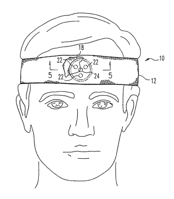
FIG. 1 is a diagram of one embodiment of a forehead-mounted optical
noninvasive
sensor unit according to the present invention, shown in position on a human
subject, with a
headband partially cut away to reveal a portion of a pressurizable capsule.
FIG. 2 illustrates the sensor unit of FIG. 1 with the headband unrolled, shown
from
the side facing the subject's forehead during use.
FIG. 3 is a front view of the pressurizable capsule of FIGS. 1 and 2.
FIG. 4 is a cross-section of the pressurizable capsule taken along line 4-4 of
FIG. 3,
showing LEDs and a photodetector mounted witliin the capsule on the inside
surface of the
capsule wall which contacts the subject's forehead.
FIG. 5 is a cross-section of the sensor unit taken along line 5-5 of FIG. 1.
FIG. 6 is a block diagram of one einbodiment of an optical noninvasive vital
sign
monitor according to the present invention.
FIG. 7 is a set of sample waveforms illustrating optical oscillometric blood
pressure
measurement witli a vital sign monitor according to the present invention.
FIG. 8 is a front view of one embodiment of an optical sensor for oxygen
saturation
measurement with a vital sign monitor according to the present invention.
FIG. 9 is an example of a calibration curve for use in oxygen saturation
measurement.
4
CA 02571317 2006-12-20
WO 2006/002196 PCT/US2005/021981
DETAILED DESCRIPTION OF PREFERRED EMBODIMENTS
For the purpose of promoting an understanding of the principles of the
invention,
reference will now be made to the embodiments illustrated in the drawings and
specific
language will be used to describe the same. It will nevertheless be understood
that no
limitation of the scope of the invention is thereby intended, such alterations
and further
modifications in the illustrated device and such further applications of the
principles of the
invention as illustrated therein being contemplated as would norinally occur
to one of
ordinary skill in the art to which the invention relates.
An optical sensor in accordance with the present invention is useful in
certain
applications on various body sites, such as the chest, leg or arm, e.g., the
wrist, but one
preferred embodiment is a forehead-mounted unit.
FIGS. 1-5 illustrate one embodiment of a forehead-mounted optical noninvasive
multifunction sensor unit 10 according to the present invention. A headband 12
made of a
relatively inelastic flexible fabric and having a Velcro tab or other
suitable fastener 14 on
each end has a pocket 16 formed therein to accommodate a pressurizable capsule
18 having
one or more reflectance-type optical sensors 20 mounted therein. In one
preferred
embodiment, the optical sensor includes a plurality of LEDs 22 symmetrically
spaced around
photodetector 24, while other embodiments have a plurality of photodetectors
surrounding
one or more LEDs. Significantly, the optical sensor is mounted on the inside
surface 26 of
the pressurizable capsule's inner wall 28, that is, the wall which contacts
the subject's
forehead during use. Such mounting provides a smooth contact surface with the
forehead and
facilitates an even pressure distribution.
Capsule 18 or at least its inner wall 28 is made of a smooth optically
transparent
material. The transmittance of the material is preferably greater than 50% at
the
wavelength(s) of light emitted by the LEDs. The capsule may have a wall
thickness of
approximately 0.010 inches. PVC or silicone is presently preferred, although
latex and
polyurethane or other materials are also suitable to varying degrees. The
headband may have
elongated inner and outer layers 30 and 32, respectively, or may have multiple
layers only
where desired for the pocket, which may have a circular or rectangular hole or
window 34
through its inner layer 30 as shown in FIGS. 2 and 5. LEDs 22 and
photodetector 24 are all
affixed to the inside surface of capsule wall 28 so as to be optically aligned
with the opening
34 and thereby with the forehead of a subject when the headband is in position
on the
subject's head.
CA 02571317 2006-12-20
WO 2006/002196 PCT/US2005/021981
Preferably, the LED(s) and photodetector(s) directly contact the inside
surface of wall
28 and are affixed thereto with an optically clear adhesive, e.g., Superglue
or other adhesive
suitable for the particular material used for the capsule. The LEDs and
photodetector may be
affixed to wall 28 before the capsule is completely formed or sealed, and the
capsule may
then be sealed so as to enclose pneumatically the LEDs and photodetector. The
LEDs and
photodetector may be affixed to the capsule wall individually, or as a sub-
assembly in which
they are held together in desired relative positions by a flexible carrier or
substrate, which is
preferably spaced from the device surfaces which contact the capsule wall so
as to facilitate
flush mounting of those surfaces to the capsule wall. Mounting of the LED(s)
and
photodetector(s) inside the sealed pressurizable capsule facilitates the use
of the device on
wet or diaphoretic subjects because the transducers are protected from water
and moisture.
Headband 12, not drawn to scale in FIG. 2, is sized to extend around the head
of the
subject and, for an adult subject, may have a height (vertical dimension in
the plane of FIG.
2) of approximately 2 inches on the subject's forehead. In one preferred
embodiment, outer
layer 32 is an inelastic band or strap extending around the subject's head,
while inner layer 30
extends only the length of the pocket and may be somewhat elastic or loosely
fitted to outer
layer 32 so as to allow for expansion of the pocket upon inflation of the
pressurizable capsule
contained therein. The capsule may have a circular cross-section as shown in
the drawings,
with a diameter of approximately 1 to 2 inches, preferably approximately 1.75
inches, for an
adult, and is provided with a pressure port (P) as depicted in FIGS. 3 and 4.
In one . -
alternative embodiment the capsule is a substantially rectangular bladder,
approximately 1.75
inches high and 4 inches long for an adult. The capsule size is
proportionately smaller for
smaller subjects, e.g., pediatric patients. The diameter of the window in
layer 30 is smaller
than the capsule height, as shown in the drawings. The window may
alternatively be in the
shape of a horizontal slot or other shape suitable for the arrangement of
optical sensor
components. The window may be a transparent part of inner layer 30, or the
entire inner
layer may be transparent.
Blood pressure, including systolic, mean and diastolic pressures, can be
obtained with
optical sensor unit 10 from the amplitude spectrum of the pulses obtained
during deflation of
capsule 18 from a suprasystolic pressure to zero pressure, as described below.
Monochromatic LEDs are suitable for monitoring blood pressure. For example,
the
transducer may employ infrared LEDs such as PDI-E801 or PDI-E804, 880 nm LEDs
available from Photonic Detectors, Inc. The LEDs and photodetector are
preferably matched
to operate at the same desired wavelength. One example of a suitable
photodetector is a
6
CA 02571317 2006-12-20
WO 2006/002196 PCT/US2005/021981
Fairchild Semiconductor QSD723 phototransistor, with a peak sensitivity at 880
nm.
Another suitable operating wavelength for the LEDs and photodetector is 805
nm, at which
wavelength the blood pressure pickup has no oxygen-saturation error, as will
be appreciated
from the discussion of oximetry below. An advantage of either of the example
wavelengths
is that there are virtually no environmental light sources in this infrared
region.
Referring to FIG. 6, pressurizable capsule 18 is connected by an inflation
tube 56 to a
pump 58 which is controlled by a microprocessor 60. Pressure in the line to
the pressurizable
capsule is measured by means of a pressure transducer 62 having a signal
output connected to
the microprocessor. Suitable transducers are available from Cobe Labs,
Littleton, Colorado.
A/D conversion may be provided in the microprocessor or in the transducer or
with a second
A/D converter provided between the two. The microprocessor controls the LEDs
and, during
blood pressure measurement, may be programmed to energize the LEDs
simultaneously. The
photodetector produces an output signal which is supplied to the
microprocessor through an
amplifier 64. The amplified photodetector output signal is converted to
digital form in the
microprocessor itself if the microprocessor has an internal A/D converter, or
in a separate
A/D converter provided between the amplifier and the microprocessor. The LEDs
may be
energized continually, or digital pulsing with synchronous detection can be
used to minimize
detection artifacts and maximize battery life by pulsing the LEDs.
The microprocessor is suitably programmed to identify, based on the digitized
output
signal of the photodetector, the points in the capsule pressure signal which
correspond to
systolic, mean and diastolic pressure, and displays the corresponding values
on a display 65
which may comprise separate indicators as shown in FIG. 6, or may provide an
output for
distant recording. The blood pressure readings correspond to blood pressure in
the forehead,
which is different than blood pressure in the arm, for example. Thus, the
monitor may be
said to perform site blood pressure measurement.
Blood pressure is measured during a transition in capsule pressure between a
selected
suprasystolic pressure and zero pressure. The transition may be an upward or
downward
transition but is described below in terms of a gradual downward transition
such as shown in
FIG. 7, which shows a sample optical pulse waveform 69 obtained during a
capsule pressure
cycle represented by curve 70, which is marked to indicate the points
corresponding to
systolic (S), mean (M), and diastolic (D) pressure. When capsule pressure is
raised above
systolic pressure, all oscillations are extremely small. As pressure in the
capsule falls below
systolic pressure, the pulses increase, and as the pressure is reduced
further, the optical pulse
amplitude increases further and reaches a maximum, labeled A. in FIG. 7, at
which point the
7
CA 02571317 2006-12-20
WO 2006/002196 PCT/US2005/021981
capsule pressure is equal to mean arterial pressure, labeled M in FIG. 7. With
a continued
decrease in capsule pressure, the oscillation amplitude decreases and returns
to a uniform
level.
The peak-to-peak amplitudes of the optical pulse waveform at the points
coinciding
with the occurrence of systolic and diastolic pressure are designated
respectively as As and Ad
in FIG. 7. Those amplitudes are calculated as predetermined percentages of
A,,,, and the
corresponding points in time are identified on the optical pulse waveform, by
interpolation if
necessary between adjacent pulses, after which the values of capsule pressure
at those points
in time are identified as systolic (S) and diastolic (D) pressure,
respectively. Appropriate
percentages or ratios are determined experimentally. For optical sensor unit
10 as described
above applied to the forehead of an adult human subject of average height and
weight,
systolic pressure occurs when the amplitude ratio As/Am is approximately 0.5;
diastolic
pressure occurs with a ratio of Ad/Am of approximately 0.8; these algorithms
depend on
capsule height.
Systolic pressure may alternatively be calculated as a function of both Am and
P,,,, the
mean capsule pressure, rather than on the basis of a fixed percentage of A,,,.
That is, the
microprocessor may calculate AS, the optical pulse amplitude corresponding in
time with
systolic pressure, according to an algorithm which includes mean capsule
pressure as a factor.
The following equation represents one form of such an algorithm:
AS = A. (a - b Pm)
where a and b are experimentally determined constants.
Heart rate can be obtained by counting the optical pulses when the capsule is
not
pressurized. Respiratory rate can also be obtained when the capsule is not
pressurized, from
the rhytlimic changes in the amplitude of the optical pulses, as described in
co-pending patent
application Serial No. 10/176,186, entitled Body-Member-Illuminating Pressure
Cuff For Use
In Optical Noninvasive Measurement Of Blood Parameters, filed June 20, 2002,
which patent
application is hereby incorporated by reference.
Referring again to FIG. 6, the vital sign monitor may have LEDs 22 which
operate at
different wavelengths for oxygen saturation measurement. Blood oxygen
saturation is
defined as the ratio of oxygenated hemoglobin (Hb02) to the total hemoglobin
(Hb + Hb02),
and is typically expressed as a percentage. The oximeter determines oxygen
saturation
(Sa02) by measuring the optical transmission at two wavelengths of light
passing through a
8
CA 02571317 2006-12-20
WO 2006/002196 PCT/US2005/021981
tissue bed. Although other wavelengths are contemplated, it is presently
preferred to operate
at wavelengths of approximately 650 nm and 805 nm for oxygen saturation
measurement. As
shown in the above-referenced co-pending patent application Serial No.
10/176,186,
hemoglobin (Hb) has negligible transmission at 650 nm, and hemoglobin (Hb) and
oxygenated hemoglobin (Hb02) transmit equally well at 805 nm; the latter
wavelength is
known as the isobestic point. That is, the transmission at 805 nm is
independent of oxygen
saturation. As adapted for oximetry, the optical sensor may have two pairs of
diametrically
opposed LEDs: a pair of red-emitting LEDs, preferably emitting at
approximately 650 nm,
and a pair of infrared-emitting LEDs, preferably emitting at approximately 805
nm. Red (R)
and infrared (IR) LED pairs 122-R and 122-IR may be arranged on perpendicular
axes as
shown in FIG. 8, with a photodetector 124 in the center of the LEDs as in the
first
embodiment. As an alternative to separate narrowband LEDs, a red LED and
infrared LED
may be combined in one multi-wavelength LED such as type Epitex L660/805/975-
40D00,
available from Epitex, Kyoto, Japan. Three such multi-wavelength LEDs may be
arranged as
in FIG. 1 if desired.
The red LEDs are switched on while the infrared LEDs are switched off, and
vice
versa, and the photodetector output signal is supplied to the microprocessor
for processing as
described above. The photodetector may be a broadband detector, such as that
identified
above, that detects reflected light from the red-emitting LED when that LED is
energized and
then detects reflected infrared radiation from the infrared LED when that LED
is energized.
The red and infrared LEDs are preferably energized alternately in rapid
succession,
e.g., at a rate of 200 pulses per second or more. This technique permits the
use of high-
intensity short-duration pulses. Synchronous detection is used to achieve the
highest signal-
to-noise ratio. Two benefits result: 1) a low average power and minimum
heating, and 2) the
system is less sensitive to stray ambient illumination. The red and infrared
signals are
sampled and processed to obtain Sa02, which may then be displayed on display
65 of the
monitor shown in FIG. 6. A short display response time and analog and digital
outputs may
be provided for connection to auxiliary equipment.
A baseline for measurement may be established by first inflating the capsule
to a high
pressure sufficient to squeeze all of the blood out of the blood vessels under
the capsule and
thus out of the optical path. For example, the capsule pressure may be held at
a maximum
pressure for a desired time to obtain the bloodless transmission reading,
which can be
assigned a value of 100% transmission. When the capsule pressure is released,
blood enters
the optical path and the red and infrared transmissions are measured. The
optical density is
9
CA 02571317 2006-12-20
WO 2006/002196 PCT/US2005/021981
computed for each of the transmitted signals, and the ratio of red to infrared
optical density is
calculated and scaled to provide an output value corresponding to the
percentage of oxygen
saturation.
Beer's law relates the optical density (D) to the concentration of a dissolved
substance. Optical density (D) is equal to In 1/T, where T is the
transmittance. Therefore the
oxygen saturation (Sa02) is given by:
Sa02 = ADR + B
Din
where A and B are constants. This equation predicts a linear relationship
based on Beer's
law. However, Beer's law applies to solutions in which the absorbing substance
is dissolved.
Blood is a suspension, and, consequently, the relationship between Sa02 and
the ratio of the
optical density for red and infrared radiation is nonlinear, as shown in FIG
9. Between 30%
and 60% saturation, the relationship is almost linear; above this range the
relationship is
nonlinear. The curve in FIG. 9 is an example of a suitable calibration curve
which may be
programmed into the microprocessor, e.g., in the form of a lookup table, for
calculation of
Sa02. Further information regarding methods of measuring blood oxygen
saturation may be
found in the following references which are hereby incorporated by reference:
Geddes,
"Heritage of the Tissue-Bed Oximeter," IEEE Engineering in Medicine
andBiology, 87-91,
March/April 1997; Geddes and Baker, Principles ofApplied Biomedical
Instruriaentation, 3'a
ed., Wiley, New York, 1989.
Calibration of the oximeter also involves balancing the outputs for the red
and
infrared channels to obtain the same optical sensitivity for both, and
ensuring that both
channels have a linear response to the red and infrared radiation. Optical
filters can be used
as calibration standards.
In another embodiment, an optical sensor within a pressurizable capsule as
described
above is placed on the subject's chest and restrained by a band, preferably a
relatively
inflexible band, around the subject's torso at chest height. For example, the
optical sensor
may be positioned over the manubrium (top of the sternum), or along the
sternum to the
xiphoid (bottom end of the sternum), where a substantially flat bone underlies
the tissue bed
and reflects incident radiation from the light source. The pressurizable
capsule may be
contained within a pocket in the band such as described above with respect to
FIGS. 1-5, and
the pocket may be sealed or may have an opening to allow for removal and
replacement of
the capsule.
CA 02571317 2006-12-20
WO 2006/002196 PCT/US2005/021981
The restraint may be a band extending completely around the body member, or
for
certain applications the pressurizable capsule may be restrained by an
adhesive strip or pad
adapted to adhere to the skin adjacent to the desired sensor location; the
restraint is designed
to hold the capsule against the skin sufficiently to allow the capsule, when
inflated, to apply
pressure to the skin and compress the arterial blood supply in the tissue bed.
While not
preferred, it may be suitable in certain applications to restrain the capsule
by other means,
such as by placing it under the weight of a subject, e.g., in contact with the
back of a patient
in a supine position. As noted above, an optical sensor in accordance with the
present
invention is useful in certain applications on various body sites, such as the
chest, leg or arm.
While the invention has been illustrated and described in detail in the
drawings and
foregoing description, the same is to be considered as illustrative and not
restrictive in
character, it being understood that only the preferred embodiments have been
shown and
described and that all changes and modifications that come within the spirit
of the invention
are desired to be protected.
11