Note: Descriptions are shown in the official language in which they were submitted.
CA 02571481 2006-12-20
WO 2006/007195 PCT/US2005/018457
_1-
CERAMIC DISC PROSTHESIS
CROSS-REFERENCE TO RELATED APPLICATIONS
The present invention claims priority to U.S. Provisional Application Serial
No.
60/584,054, filed on June 30, 2004 and entitled "Ceramic Artificial Disc,"
which is
hereby incorporated by reference in its entirety.
BACKGROUND OF THE INVENTION
Disease, advancing age, and trauma can lead to changes in various bones,
discs,
joints, and ligaments of the body. Some changes manifest themselves in the
form of
damage or degeneration to a spinal disc. It is known that an intervertebral
disc can be
subject to damage, such as compression, deformation, displacement, or wear,
and more
generally degeneration associated with the mechanical stresses that are
applied thereto
and that lead to anatomical and functional destruction of the disc and of the
vertebral
segment. This damage to the disc alters its mechanical behavior and causes
instability
which can, in turn, give rise to a painful osteoarthritic reaction.
This pain can sometimes be eliminated by spinal fusion in which two adjacent
vertebral bodies are joined together after removing the intervening
intervertebral disc. A
prosthetic device is usually placed between the two adjacent vertebral bodies,
in place of
the removed disc, to fill the space left by the removed disc and to allow bone
to grow
between the two vertebral bodies.
Alternatively, proposals have been made to replace the defective disc with an
artificial disc that preserves the natural mobility between adjacent vertebral
bodies. For
example, such prostheses can include first and second plates for fixing to
adjacent
vertebral bodies and an articulating mechanism, such as a ball joint,
interposed between
the two plates. Although such prostheses makes it possible to restore an
appropriate
height to the intersomatic gap, the articulating mechanism of the prosthesis
can be
subject to high levels of friction, thus making it sensitive to wear.
Other artificial discs include ceramic bearing surfaces positioned within
titanium
endplates. While the ceramic materials reduce wear, these implants are limited
by small
bearing surfaces. In addition, mounting stiff ceramic inserts into titanium
endplates can
CA 02571481 2006-12-20
WO 2006/007195 PCT/US2005/018457
-2-
generate high stresses in the cerarnic material and cause disc failure. Thick
titanium
endplates can also interfere with post-operative imaging, an important
diagnostic tool.
Despite known prostheses for the replacement of discs, there remains a need
for
additional prostheses that are sufficiently wear resistant, compatible with
post-operative
imaging, and suitable for secure and effective implantation within a patient's
spine.
SUMMARY OF THE INVENTION
The present invention provides a disc prosthesis that features superior wear
properties. The disc prosthesis is formed entirely or predominately of
cerainic material,
thus optimizing post-operative imaging. In one embodiment, the present
invention
provides an implantable prosthetic device for replacing spinal discs. The
device
includes first and second endplates each having an articulating contact
surface and a
bone facing surface. At least one of the first and second endplates is formed
of a
ceramic material. The contact surface of the first endplate has a shape
complementary to
the contact surface of the second end plate, such that when the endplates are
in contact
with each other, the contact surface of the first endplate can articulate with
respect to the
contact surface of the second endplate.
In another aspect, at least one non-ceramic bone attachment element is formed
on
or mated with at least one of the bone facing surfaces, such that the bone
attachment
element covers less than a full area of the bone facing surface. The bone
attachment
element can include bone-penetrating teeth formed from a material selected
from the
group consisting of a metal, a plastic, and combinations thereof. When
positioned
within a vertebral column, the bone-penetrating teeth are effective to resist
retropulsion.
In another aspect, the bone attachment element includes bone penetrating teeth
formed from a shape memory material. The teeth can be positioned substantially
parallel to the bone facing surface before activation and orientated to
penetrate bone
after activation.
The disc prosthesis device can further include a mating element for mating
with
an insertion tool. In one aspect, the device includes a male mating element
adapted to
mate with an insertion tool. In another aspect, the mating element is
positioned on the
bone attachment element and is adapted to mate with a male or female insertion
tool.
CA 02571481 2006-12-20
-3-
In another embodiment of the disc prosthesis device disclosed herein, the
first and
second endplates are formed of a ceramic material and each have an
articulating surface and
an opposed bone contacting surface. The articulating surfaces are
complementary to each
other such that the first and second endplates can articulate while in contact
with one another
and are unconstrained in their relative movement by any portion of the
prosthesis. The device
may also include bone penetrating features positioned on at least one of the
bone facing
surfaces.
In a further aspect, the device disclosed herein includes a coating that
encourages
bone in-growth and/or bone adhesion. For example, at least one of the bone
contacting
surfaces can includes a coating of a particulate ceramic or another
biocompatible material that
provides a porous surface for bone in-growth.
In another embodiment, a method of implanting a disc prosthesis is disclosed,
in
which there is provided a disc prosthesis having first and second endplates,
each including an
articulating surface and an opposed bone facing surface. At least one of the
bone facing
surfaces can have a bone attachment element formed from a heat-activated shape
memory
material. The method further includes the step of inserting the disc
prosthesis between two
vertebral bodies wherein the bone attachment element is in a first, non-bone
penetrating
orientation, and subjecting the bone attachment element to an activating force
effective to
orient the bone attachment element in a second, bone penetrating orientation.
In one aspect, the activating force is body heat. When the device is at room
temperature, teeth on the bone attachment element are oriented to be
positioned to lie against
the surface of the device. Once the bone attachment element is heated to a
sufficient degree,
the teeth become reoriented into a bone penetrating position and resist
retropulsion.
In another aspect of the invention, there is provided a disc prosthesis device
having
first and second endplates each having an articulating surface and an opposed
bone facing
surface, at least one of the bone facing surfaces including a bone attachment
element with at
least one bone penetrating feature formed from a heat-activated shape memory
material.
Another aspect of the present invention is a use of the device described above
for
implantation between two vertebral bodies.
CA 02571481 2006-12-20
-3a-
BRIEF DESCRIPTION OF THE DRAWINGS
The invention will be more fully understood from the following detailed
description
taken in conjunction with the accompanying drawings, in which:
FIG. 1 is a perspective view of one embodiment of a prosthetic disc device
positioned
within a vertebral column;
CA 02571481 2006-12-20
WO 2006/007195 PCT/US2005/018457
-4-
FIG. 2A is a perspective view of the prosthetic disc device according to one
embodiment of the present invention;
FIG. 2B is a side view of the prosthetic disc device of FIG. 2A;
FIG. 2C is an exploded view of the prosthetic disc device of FIG. 2A;
FIG. 3 is a perspective view of another embodiment of the prosthetic disc
device;
FIG. 4A is a perspective view of still another embodiment of the prosthetic
disc
device including a bone attachment element;
FIG. 4B is a side view of the prosthetic disc device of FIG. 4A;
FIG. 5A is a perspective view of a prosthetic disc device with another
embodiment of the bone attachment element;
FIG. 5B is a side view of the prosthetic disc device of FIG. 5A;
FIG. 6A is a perspective view of another embodiment of the prosthetic disc
device;
FIG. 6B is a side view of the prosthetic disc device of FIG. 6A;
FIG. 7A is a perspective view of yet another embodiment of the prosthetic disc
device;
FIG. 7B is a side view of the prosthetic disc device of FIG. 7A;
FIG. 8A is a view of the prosthetic disc device with a snap-on bone attachment
element;
FIG. 8B is a top view of the prosthetic disc device of FIG. 8A;
FIG. 9A is a perspective view of the prosthetic disc device with a bone
attachment element seated witliin a recess in a bone facing surface;
CA 02571481 2006-12-20
WO 2006/007195 PCT/US2005/018457
-5-
FIG. 9B is a top view of the prosthetic disc device of FIG. 9A;
FIG. 10A is a perspective view of another embodiment of the prosthetic disc
device having a bone attachment element seated within a recess of the bone
facing
surface;
FIG. l OB is a top view of the prosthetic disc device of FIG. I OA;
FIG. 11A is a perspective view of another embodiment of a prosthetic disc
device;
FIG. 11B is a top view of the prosthetic disc device of FIG. 1 1A;
FIG. 11C is a side view of the prosthetic disc device of FIG. 11A;
FIG. 12 is a side view of a prosthetic disc device in the process of being
inserted
between vertebral bodies of a vertebral column;
FIG. 13A is a side view of the prosthetic disc device with the bone attachment
element in the non-bone penetrating position;
FIG. 13B is a perspective view of the prosthetic disc device of FIG. 13A; and
FIG. 14 is a side view of the prosthetic disc device of FIG. 13A with the bone
attachment element in the bone penetrating position.
DETAILED DESCRIPTION OF THE INVENTION
Certain exemplary embodiments will now be described to provide an overall
understanding of the principles of the structure, function, manufacture, and
use of the
devices and methods disclosed herein. One or more examples of these
embodiments are
illustrated in the accompanying drawings. Those skilled in the art will
understand that
the devices and methods specifically described herein and illustrated in the
accompanying drawings are non-limiting exemplary embodiments and that the
scope of
the present invention is defined solely by the claims. The features
illustrated or
described in connection with one exemplary embodiment may be combined with the
CA 02571481 2006-12-20
WO 2006/007195 PCT/US2005/018457
-6-
features of other embodinnents. Such modifications and variations are intended
to be
included within the scope of the present invention.
The present invention provides a prosthesis that conserves vertebral function
and
provides wear resistant articulation surfaces. In one aspect, superior
tribological wear
properties are provided by forming at least a portion, and preferably all or
substantially
all, of the prosthesis from a ceramic material having sufficient strength to
withstand in-
vivo and insertion loading. Ceramic bearing surfaces provide excellent wear
resistant
properties that allow the device to articulate when positioned within a
patient's spine
such that at least some of the natural mobility between adjacent vertebral
bodies is
conserved. Ceramic materials are also compatible with post-operative imaging
such that
a surgeon can determine if the prosthesis is properly positioned and/or can
asses the
health of surrounding tissue and adjacent levels.
FIG. 1 illustrates a perspective view of a portion of a vertebral column
including
vertebral bodies la, lb, lc, ld, and natural discs 2a and 2b. A damaged disc
between
vertebral bodies lb and lc has been resected and replaced with a prosthetic
disc 10 that
has been inserted in the space created between the adjacent vertebral bodies
(lb, lc). As
explained below, ceramic bearing surfaces of prosthesis 10 allow vertebral
bodies Ib, lc
to maintain at least some degree of mobility. As further explained below, disc
10 can
include bone mating surfaces, coatings, and/or bone penetrating elements,
which
together or separately, facilitate bone in-growth and help to fix the
prosthesis in position.
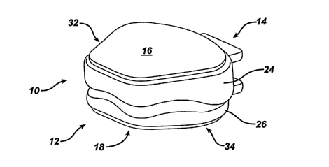
Referring to FIGS. 2A through 2C, disc 10 in one aspect, includes first and
second endplates 24, 26 each having an articulating contact surface 20, 22 and
a bone
facing surface 16, 18 where at least one of the first and second endplates is
formed of a
ceramic material. In one aspect, both endplates are formed entirely or
substantially from
ceramic materials.
As shown in FIGS. 2A through 2C, the prosthetic disc is generally shaped and
sized for positioning between adjacent vertebral bodies and contacting the
vertebral
bodies. As shown, disc 10 can have a somewhat rectangular or trapezoidal shape
with
an anterior surface 14 and a posterior surface 12, and a superior surface 16
and an
inferior surface 18. The superior 16 and inferior 18 surfaces are bone facing
surfaces
while interior surfaces 20, 22 (FIG. 2C) are articulating surfaces.
CA 02571481 2006-12-20
WO 2006/007195 PCT/US2005/018457
-7-
The sides of disc 10, including anterior surface 14 and posterior surface 12
can
be shaped to match the anatomical configuration of the inter-vertebral space
into which
disc 10 is inserted. For example, posterior surface 12 can have a width
shorter than that
of the anterior surface 14, causing the width to taper from the posterior to
the anterior.
In addition, the corners of the prosthesis can be rounded to reduce the chance
of the
prosthesis causing trauma to surrounding tissue. The height of disc 10 is also
adapted to
be compatible with the anatomical structure of a vertebral column, and in
particular, the
height should be such that it provides the desired spacing between adjacent
vertebrae.
Disc 10, in one aspect, is formed from a superior end plate 24 and an inferior
endplate 26, each having'a bone facing surface 16, 18 and an articulating
contact surface
20, 22 that allow the endplates to move relative to one another. FIG. 2C shows
an
exploded view of disc 10 with articulating surface 22 of inferior endplate 26
adapted to
articulate with articulating surface 20 of superior endplate 24. Articulating
surfaces 20;
22 can have a variety of shapes that allow movement of the adjacent vertebra
to which
they are attached. In one aspect, articulating surfaces 20, 22 are
complementary, with
articulating surface 22 having a convex configuration that is received within
a concave
articulating surface 20. Articulating surfaces 20, 22 can be formed such that
the
movement of endplates 24 and 26 (and their respective vertebral bodies) is
similar to the
action of a natural vertebral disc. For example, the shape of articulating
surfaces 20, 22
allows for flexion, extension, lateral bending, and/or rotational movement.
One skilled
in the art will appreciate that a variety of alternative articulating surface
shapes that
allow relative movement of the endplates can be used. In an alternative
embodiment,
articulation of endplates 24, 26 could be provided by a three-piece (or more)
design. For
example, a motile core could be positioned between endplates 24, 26. Other
exemplary
designs could include a ball-in-trough configuration.
In one aspect, endplates 24, 26 are configured such that relative movement is
not
limited or constrained by the structure of prosthetic disc 10. As the
endplates move
relative to one another they do not encounter another portion of disc 10 that
limits the
amount of flexion, extension, lateral bending, or rotational movement of the
endplates.
Once implanted, oilly the anatomy of the vertebral column and surrounding
tissue, or
other implants, limit movement of the endplates relative to one another.
CA 02571481 2006-12-20
WO 2006/007195 PCT/US2005/018457
-8-
Endplates 24, 26 can further include features for mating with an inserter tool
(not
shown). For example, a mating element 30 can extend from anterior surface 14
of at
least one the endplates and be adapted for gripping by an inserter tool. As
shown in
FIGS. 2A through 2C, mating element 30, in one aspect, includes a male tab,
having
substantially flat upper and lower surfaces, that extends from the surface of
each
endplate 24, 26. Alternatively, an inserter tool could mate with a female
mating element
30, including for example, a recess in the anterior surface 14 of disc 10. In
another
aspect, mating element 30 could be positioned on a side surface of prosthetic
disc 10.
For example, one or both of side surfaces 32, 34 of prosthetic disc 10 could
include a
slot within which an inserter tool could be seated.
In one embodiment, prosthetic disc 10 does not include an element particularly
adapted for mating with an inserter tool. Instead, an inserter tool adapted to
grip the
surfaces of disc 10, such as, bone facing surfaces 16, 18; sides 32, 34;
and/or anterior
surface 14. In one aspect, an insertion tool can instead have a shape
corresponding to
the anterior end of disc 10 and include features for gripping surfaces 16, 18.
FIG. 3
illustrates one embodiment of prosthetic disc 10 that includes area 35 on bone
facing
surfaces 16, 18 that can be engaged by an inserter tool. One skilled in the
art will
appreciate that a variety of inserter tools can be adapted to mate with disc
10.
The superior and inferior surfaces 16, 18 of prosthetic disc 10 are positioned
to
face the adjacent vertebral bodies between which they will be inserted. When
positioned
witllin the vertebral column these surfaces face the exposed bone of the
adjacent
vertebral bodies. In one aspect, these bone facing surfaces 16, 18 are bone
contacting
surfaces and are adapted to mate wit11 bone. Once inserted within the
vertebral column,
the surfaces 16, 18 contact the adjacent vertebral bodies and, over time, the
disc is
secured through osteointegration as bone grows into the ceramic material that
forms the
disc. Surfaces 16, 18 can also include features, such as porous regions and/or
recesses to
enhance and facilitate bone in-growth.
In one embodiment, porous regions on the bone facing surfaces are provided by
a
coating 36 that fosters bone growth and/or bone adhesion. In one aspect,
coating 36,
shown in FIG. 3, could be positioned on surfaces 16, 18 to provide a rough
and/or
porous structure into which bone can grow. For example, coating 36 could be
formed
from particles of ceramic material. Coating 36 could include a variety of
other materials
CA 02571481 2006-12-20
WO 2006/007195 PCT/US2005/018457
-9-
such as binders (e.g., an adhesive) and/or biologically active materials that
encourage
bone in-growth. Exemplary materials include calcium phosphate, hydroxy
apatite,
plasma sprayed titanium beads, plasma sprayed CCM and combinations thereof. As
explained below in more detail, surfaces 16, 18 can also, or alternatively,
include one or
more bone attachment elements to help secure disc 10 to bone.
Endplates 24, 26 can be formed from a variety of ceramic materials. Exemplary
materials include alumina, zirconia, yttria, and blends thereof. One skilled
in the art will
appreciate that a variety of ceramic materials having low friction, wear
resistant
properties can be used to form endplates 24, 26. For example, ceramics such as
Ceramtec's, Biolox Delta material or Amedica's, MC2 can be used.
In another embodiment of prosthetic disc 10, a non-ceramic bone attachment
element 38, 38', 38", 38"', 38"" is positioned on one or both of the bone
facing
surfaces 16, 18. While ceramic materials are very hard and have low wear
characteristics, forming bone penetrating members (e.g., spikes or teeth) from
ceramic
can be difficult because cerainic is brittle. To avoid this problem, bone
attachment
elements 38, 38', 38", 38"', 38 ' ' can provide bone penetrating spikes or
teeth 40, 40',
40" formed of a non-ceramic material. The spikes or teeth 40, 40', 40"
penetrate
and/or engage bone to prevent disc 10 from moving out of position once
implanted. In
addition, non-ceramic bone attachment elements can help to evenly distribute
forces
across the bone facing surfaces of the endplates.
Bone attachment element 38, 38', 38", 38"', 38"" can be formed from a variety
of non-ceramic materials such as metals and plastics. For example, the bone
attachment
element could be formed from a strip of metal, such as stainless steel,
titanium, or
another biocompatible metal that is stamped or machined. Alternatively, the
attachment
element could be a rigid plastic such as, for example, carbon fiber reinforced
plastics,
polyetlleretherlceton, and/or other biocompatible plastics.
In one embodiment, bone attachment element 38, 38', 38", 38"', 38" ' is
formed from a material that is at least partially compatible with an imaging
technique,
such as magnetic resonance imaging (MRI). This allows a surgeon to use post-
operative
imaging techniques to view the prosthesis and surrounding tissue, including
soft tissue.
Where the materials used to form the bone attachment element are partially
compatible
CA 02571481 2006-12-20
WO 2006/007195 PCT/US2005/018457
-10-
with post-operative imaging techniques, the bone attachment element is
preferably small
enough that it produces minimal interference with post-operative imaging.
FIGS. 4A through 5B illustrate exemplary embodiments of bone attachment
elements 3 8 positioned on bone facing surfaces 16, 18 and having teeth
adapted to
penetrate bone. In one aspect, bone attachment elements 38 comprise two
parallel strips
of material with raised portions defining teeth 40. The bone attachment
elements can be
positioned, for example, with their axes lt-l; and 12-12 oriented in the
anterior/posterior
direction. While two bone attachment elements are positioned on each surface
16, 18 in
FIGS. 4A through 5B, one skilled in the art will appreciate that disc 10 can
include more
than one bone attachment element per surface (e.g., three or more) or fewer
(e.g., one).
FIGS. 6A through 14 illustrate a variety of other exemplary configurations of
the bone
attachment elements. For example, the bone attachment element could be
positioned
around the perimeter of the bone facing surfaces (FIGS. 6A and 6B), across the
entire
surface of the bone facing surfaces (FIGS. 7A and 7B), in a cross
configuration (FIGS.
9A and 9B), around the perimeter and across the center of the bone facing
surfaces
(FIGS. IOA and 10B), and as connected parallel strips (FIGS. 11A and 11B).
As noted above, attachment elements 38, 38', 38", 38' ', 38"" can cover less
than the full area of bone facing surfaces 16, 18. A portion of the ceramic
bone facing
surfaces 16, 18 thus remain uncovered by bone attachment element 38, 38', 38",
38"",
38' ' and will accomodate bone in-growth. For example, FIGS. 5A through 6B
and 8A
through 14, illustrate disc 10 with at least a portion of bone facing surfaces
16, 18
uncovered by the bone attachment elements. In addition to facilitating bone in-
growth,
the small profile of the bone attachment elements can facilitate post-
operative imaging.
For example, where the bone attachment element is partially compatible with
imaging
techniques, covering only a portion of bone facing surfaces 16, 18 with the
bone
attachment element can minimize interference with post-operative imaging.
Bone attachment element 38, 38', 38", 38"', 38"" can be fixed to prosthetic
device 10 in a variety of ways, including, for example, brazing, adhesion,
mechanical
attachment, and combinations thereof. Endplates 24 and 26 can also include
features,
such as a recess to assist with attachment of the bone attachment element. In
one
embodiment, bone attachment element 38' is mated with disc prosthesis 10 by
way of a
mechanical interlock, such as, for example snap-fit, friction fit, tongue-and-
groove,
CA 02571481 2006-12-20
WO 2006/007195 PCT/US2005/018457
-11-
overmolding, overcasting, thermal interference fit, and combination thereof.
In one
aspect, shown in FIGS. 8A and 8B, the bone attachment element can mate or
interlock
with recesses 42 on the anterior and posterior surfaces 14, 12 of disc 10. The
ends of
bone attachment element 38' can be flexible and include a protrusion that
snaps into
recess 42.
In an alternative embodiment, shown in FIGS. 9A through 10B, bone attachment
element 38" is seated within a recess in the bone facing surfaces (16, 18).
For example,
the bone attachment element could be flush with the bone facing surfaces 16,
18, such
that surfaces 16, 18 are easily accessed by bone. FIGS. 9A through l0B also
illustrate
embodiments in which bone attachment element 38" is seated with a recess in
the
surfaces 16, 18 of the disc prosthesis. The bone attachinent element can be
mated within
the recess in a variety of ways including, for example, mechanical interlock
and
adhesion. In one exemplary embodiment, the bone attachment element is formed
within
a recess. For example, polymer can be poured into a recess (e.g., an
overhanging recess)
in the bone facing surfaces 16, 18 of disc prosthesis 10 and then hardened to
form bone
attachment element 38". Alternatively, attachment eleinent 38" could be held
on the
bone facing surface(s) with an adhesive, such as, for example brazing, bone
cement,
and/or an epoxy.
In another embodiment of the disc 10, bone attacliment element 38"', rather
than
endplates 24, 26, includes features for mating with an inserter tool. FIGS. 1
lA through
11C illustrate disc prosthesis 10 including mating element 30' defined by
extension
portion 50 formed integrally witll bone attachment element 38"'. Instead of an
inserter
tool mating witli the ceramic of the endplates 24, 26, the inserter tool can
mate with the
non-ceramic bone attachment element 38"'. As shown in FIGS. 11A through I IC,
extension portion 50 can have a "T" shape and extend from the anterior portion
of the
prosthetic disc. In one aspect, extension portions 50 are positioned on bone
mating
elements 38"'.
If damage occurs to one of the endplates, it is usually during the insertion
process
because of the force required to implant the prosthetic disc between adjacent
vertebral
bodies. Accordingly, the bone attachment elements 38"' can be designed to
break away
if the strain during insertion approaches a force which can break the ceramic
endplates.
For example, extension portion 50 can have a built in fault that will fracture
prior to the
CA 02571481 2006-12-20
WO 2006/007195 PCT/US2005/018457
-12-
cerainic endplates breaking. Alternatively, extension portion 50 can be
constructed of
materials that will give way prior to reaching a threshold force.
Teeth 40, positioned on bone attachment element 38, 38', 38", 38"', 38can
be positioned at an angle with respect to the bone facing surfaces 16, 18 of
disc 10, such
that the teeth are driven into bone by retropulsive forces acting on the disc.
The angle of
teeth 40 with respect to the bone facing surface of the endplate can be in the
range of
about 5 and 75 , and preferably in the range of about 30 and 70 . In one
embodiment,
teeth 40 are formed from a ridged material and are fixedly positioned. Teeth
40 can
alternatively be flexible to facilitate insertion such that they are pushed
down (i.e.,
retracted or flattened) during inser-tion. Once the disc is inserted, the
teeth will press
against the bone to resist retropulsion.
One skilled in the art will appreciate that teeth 40 can have a size, shape,
and
orientation that will be effective to resist removal or migration of disc 10.
In FIGS. 4A
and 4B the teeth are unidirectional and are inclined in the posterior-anterior
direction.
Teeth 40 will not grab bone as disc 10 is inserted, but will resist
retropulsion once the
disc is inserted. Alternatively, the teeth can be positioned at an angle with
respect to the
direction of insertion. For example, teeth 40 can be angled with respect to
the axis L-L
shown in FIG. 5A. This orientation helps to prevent lateral movement of the
prosthetic
disc.
In another embodiment, disc 10 includes rigid teeth 40' which can extend in
the
superior/inferior direction. Teeth 40' can penetrate bone to hold disc 10 in
place after
insertion. For example, FIGS 6A through 7B illustrate rigid teeth 40'
positioned on a
single bone attachment element. Movement of the disc in any direction will be
resisted
by the engagement of the teeth into bone.
In yet another embodiment of the disc prosthesis described herein, bone
attachment element 38"' is forined froin a shape memory material. When
activated,
the bone attachment element can move from a non-bone penetrating configuration
to a
bone penetrating configuration. In one aspect, the shape memory material is
heat
activated and once inserted into a vertebral column, heat (e.g., body heat
and/or a
supplemental heat source) activates the shape memory material. When activated,
bone
penetrating teeth 40" on bone attachment element 38"" move from a lowered
position
to an active, raised position.
CA 02571481 2006-12-20
WO 2006/007195 PCT/US2005/018457
- 13-
FIG. 12 illustrates a vertebral column with disc tissue removed and disc
prosthesis 10 ready for insertion. As shown, teeth 40" are oriented in a low
profile non-
bone penetrating position. FIGS. 13A and 13B further illustrate disc 10 with
teeth 40",
formed of a shape memory material, in the non-bone engaging position. Disc 10
can be
inserted with teeth 40" in their non-bone engaging position to facilitate
insertion by
reducing interference between teeth 40" and bone. Once disc 10 is positioned
between
vertebral bodies, bone attachment element 3 8 "' is activated by an activating
force, such
as body heat and/or a supplemental heat source, and teeth 40" become oriented
in a
bone engaging position as shown in FIG. 14A.
Bone attachment element 38"" can be formed from the variety of shape memory
materials. Exemplary materials include, for example, nickel-titanium
intermetallic
compounds (e.g., Nitinol) that exhibit thermal shape memory. Preferred
materials
include those that are activated at a temperature above room temperature, such
as at, or
slightly below, body temperature. In one embodiment, spike profiles are cut
into a sheet
of Nitinol and the sheet is heat treated so that the spikes will deploy (i.e.,
assume a bone
penetrating position) at body temperature. Spikes 40" can lie substantially in
the same
plane as bone attachment elements 38 " during implantations, then deploy into
a raised,
bone penetrating position once the disc is inserted.
One skilled in the art will appreciate that shape memory material based bone
penetrating features can assume numerous configurations. In an alterative
embodiment,
the bone attachment elements can be in the form of bone engaging hooks (not
shown),
which can also be fornzed from a shape memory material. By way of example, the
bone
engaging elements can include wires when in the non-activated, non-bone
penetrating
condition. In the activated condition, for example, the wires may lie
substantially in the
same plane as surfaces 16, 18. Upon activation by an activating force, the
wires are
transformed into hooks that penetrate and/or securely engage bone.
One of ordinary skill in the art will appreciate further features and
advantages of
the invention based on the above-described embodiments. Accordingly, the
invention is
not to be limited by what has been particularly shown and described, except as
indicated
by the appended claims. All publications and references cited herein are
expressly
incorporated herein by reference in their entirety.
What is claimed is