Note: Descriptions are shown in the official language in which they were submitted.
CA 02571695 2006-12-20
WO 2006/003423 PCT/GB2005/002603
GEL FORMULATIONS AND USES THEREOF
Field of the Invention
The invention relates to thermoreversible gel compositions and their
applications
generally in research, diagnostic and screening assays and methodology in the
use of a
cell- or particle- or reagent- support matrix based applications.
Background to the invention
There exists a need to make biological assays faster and simpler to perform
with an
overriding drive to make the processes cheaper yet maintain accuracy and
reproducibility. This is due to a rapid increase in the number of research and
diagnostic
molecular probes available (e.g. new fluorescent reporter molecules) and the
advantages
in terms of information content of multiplexing such assays across a range of
instruments. Increasingly there is a need for cell-, particle- and bead-based
(and a
cotnbination of these units) assays in which the presence, for example, of
cells with
certain features indicates disease processes. Similarly, the demand and
evolution of
rational approaches, in the search for bioactive molecules for new medicines
has resulted
in a need for low cost high-through-put screening (HTS) and the development of
cell-
and molecule-based assays, tools and arrays within the field of functional
analysis.
Such assays require or are enhanced by the availability of methodologies for:
i) the manipulation of cells/particles, analytes and reagents in liquid and
gel phases
for processing purposes,
CA 02571695 2006-12-20
WO 2006/003423 PCT/GB2005/002603
ii) the controlled delivery of fluorescent/bioluminescent molecules to
cells/particles
or the retention of the fluorescent/bioluminescent-associated properties of
said
particle/cells.
iii) the controllable immobilisation of said cells/particles for the purpose
of analysis
involving light collection
iv) the retention of cell viability and cell function for periods of time
sufficient for the
purposes of an analysis.
The present invention seeks to provide means for use in such methodologies.
A variety of hydrogels based upon thermoreversible polymers (in liquid form at
elevated
temperatures but in gel form at lower temperatures) are known, including
natural gel-
forming materials such as agarose, agar, furcellaran, beta-carrageenan, beta-
l,3-glucans
such as curdlan, gelatin, or polyoxyalkylene containiing compounds.
The present invention exploits to the distinct advantages and properties of
block
20. polymer-based gels, such as polyoxypropylene-polyoxyethylene block polymer
gels
(PBP), which unusually undergo transition to liquid form upon tenlperature
reduction.
This property can be described as 'reversed thermosetting'.
Selected properties of the PBP -preparations, such as detergent properties and
the
mechanical and thermoreversible properties of hydrogels in general, have been
documented and exploited in the art.
For example, reported uses and properties of PBP preparations include the
following:
2
CA 02571695 2006-12-20
WO 2006/003423 PCT/GB2005/002603
Su7factant properties
Polyoxypropylene-polyoxyethylene block polymer (PBP) has been used as a non-
ionic
surfactant for detergents, dispersants, binders, stabilisers, defoaming
agents, emulsiffing
agents to name but a few. At high aqueous PBP concentrations, beyond a
transition
temperature, a gel can form comprising amphiphilic block copolymer micelles. A
commercial PluronicOO preparation F-127 (PE099-PP069-PE099, with E and P being
polyoxyethylene and polyoxypropylene, respectively) has been used in gel form
for
pharmaceutical preparations.
Reverse agar and biofrlms
Gel-forming formulations of PBP have been described as 'reverse agar' in their
use. The
low temperature gelling formulation has been used to support the limited
growth of
micro-organisms in conventional microbiology applications. Such Pluronic -
based
hydrogels have been used extensively to assay biocide treatments. Gel-trapped
micro-
organism populations mimic the localized high cell densities observed in
biofilms and
are subject to the similar nutrient and chemical gradients found within
natural biofilms.
Such prior art uses with respect to micro-organisms has revealed that PBP
should have
low toxicity in the stable gel form.
Molecular sieve properties
PBP gel form has the potential to form ordered micellar structures and has
been used as
a separation media for nucleic acids, indicating the coherent movement of
molecules
through the gel upon the passage of a continuous or pulsed electrical current.
Microdevices have also been designed with sieving gels within the same device
to
perform separations involving both single- and double-stranded DNA over
distances on
the order of 1 em. Extensive comparisons have been made to compare different
gel
matrices on the basis of gel casting ease, reusability, and overall separation
performance
using for example a 100 base pair double-stranded DNA ladder as a standard
sample.
3
CA 02571695 2006-12-20
WO 2006/003423 PCT/GB2005/002603
Hydropads
Miniature size and high sensitivity of biochips is sought in diagnostics,
testing, and
research in medicine, veterinary science and applications, agriculture,
toxicology,
environmental monitoring, forensics etc. Three dimensional biochips
coinprising non-
thermoreversible gels (hydropads) have been developed consisting of an array
of three-
dimensional gel elements on the hydrophobic surface of a microscope slide. For
example, a gel-based biochip project was initiated in Engelhardt Institute of
Molecular
Biology of the Russian Academy of Science (EIMB) in 1989 resulting in the
development of the 'Immobilized Micro Array of Gel Elements' on chip (or IMAGE
chip), which can bear oligonucleotides, DNA, proteins, small compounds or
cells fixed
within semi spherical hydrogel pads. A simple two-step procedure has been
developed
for the large-scale manufacture of such chips. The gel pads can serve both as
support for
immobilisation and as individual nanolitre test tubes to carry out various
specific
interactions, chemical or enzymatic reactions. Chips have been produced that
contain
immobilized antibodies, antigens, enzymes, receptors, and different ligands.
Cell encapsulation
Polymeric gels have also been explored as cell encapsulation materials for
tissue
engineering. Isolated mammalian cells and tissues have countless applications
in
medicine and biotechnology, yet protecting and nourishing cells either in
vitro or in vivo
while harvesting the desired products has proven difficult.
Drug and dye delivery
Previous clinical applications have revealed the use of hydrogels for the
purpose of local
delivery of pharmacologically active agents to tissues. In contrast, previous
work on the
"liquid less" cell staining by dye diffusion from gels (polyacrylamide or
gelatin) has
been restricted to the use of gel systems lacking the unique thermoreversible
properties
4
CA 02571695 2006-12-20
WO 2006/003423 PCT/GB2005/002603
of PBP-based gels. Such studies have been described by Smolweski et al., 2001,
Cytoniet7y 44(4):355-60. Dye delivery to cells was observed but required a 2-
to 4-fold
increase in normal staining concentration of DNA dyes.
Summary of the invention
The present invention provides for a general means of combining the four
methodologies stated above by exploiting the features of block polymer (for
example,
based on polyoxypropylene-polyoxyethylene block polymer [PBP]) gels namely the
thermoreversible (gel-sol traiisition enabling manipulation; as defined below)
properties,
micelle formation under gelling conditions (enabling the gel to act as a
support/immobilizing matrix), optical properties (low absorbance and non-
fluorescent
enabling light-based optical analyses), controllable surfactant properties
(enabling
modified reporter molecule delivery to cells and a means of cell
solubilisation),
molecular sieving properties (providing controlled, i.e. modified and
regulated, delivery
of exogenous reporter molecules) aild low toxicity (enabling live cell
processing). The
invention also provides for a general means of making assays modular by
enabling the
control and manipulation of cells/particles and regulating reactant and
reporter molecule
access for use in such cell-based assays. Specific formulations of PBP gels
would
provide optimal performance for a given application or excipient property.
By 'thermoreversible' we ' refer to the property of gel formation upon raising
the
temperature of a PBP composition above a critical transition point while a
liquid or sol
form of the composition exists at temperatures below that transition point.
The current invention provides means for the recovery of test inoculums
witliout further
trauma and the use of gel in eukaryotic systems (e.g. human and animal cells).
The molecular sieving property of block polymer gels provides a means of the
coherent
delivery and behaviour of analytes (such as molecular reporter molecules) for
the
purpose of sequential or controlled delivery to a system under analysis.
5
CA 02571695 2006-12-20
WO 2006/003423 PCT/GB2005/002603
Tliermoreversible gelling properties of block polymer gels also avoids thermal
damage
thus providing an advantageous route for cell harvesting and recovery. One
strategy is to
encapsulate cells in a matrix that allows for the diffusion of small molecules
to and from
cells. Encapsulation offers promising results, since PBP polymers are often
biocompatible and provide a three-dimensional scaffolding for the simulation
of support
conditions in multicellular systems. To some extent the success of cellular
encapsulation
depends on the cell type. The purpose of the gel is to provide a matrix-gel
environment,
which allows cells, isolated from different tissue to maintain their original
cellular
phenotype. In an ideal situation the gel should provide an inert environment
(i.e. it does
not act to stimulate or activate cells to do something abnormal). The cells
placed in such
gels are able to function normally and perhaps overtime organize the gel with
the
synthesis of secreted macromolecules to form pseudo-tissue explants. The
current
invention incorporates all of the required features of a cell encapsulation
matrix. It
allows cells to be incorporated whilst the PBP gel is in liquid form. Upon gel
formation
the cells become trapped. It is possible to administer the necessary reagents
whilst the
gel is in either phase tllus ensuring the cells are able to absorb the
necessary agents
whilst in a supported environment. The gel with the trapped cells can be
placed at a
lower temperature thus allowing the cells to be extracted by transition to the
liquid phase
even from selected regions of the gel thereby permitting micro-selection of
cell
populations with given characteristics without any adverse effects. The same
applies to
any particle fixed in this way.
Potential advantages of block polymer gels for dye delivery include their
utility in
microgravity conditions and conditions where spillage is not desirable.
Thermoreversible PBP preparations containing potentially hazardous excipients
(e.g.
mutagenic dyes) would have the additional ' safety feature of forming a gel at
skin
temperatures and above thereby, reducing diffusion-limited transdermal
delivery. The
safe delivery of dye molecules can provide access to a wider range of
applications and
excipients. The present invention also allows for the solid-phasing of dye
delivery
6
CA 02571695 2006-12-20
WO 2006/003423 PCT/GB2005/002603
systems with significant safety advantages and for the cold formulation of
preparations
with thermolabile excipients.
Thus, a first aspect of the invention provides the use of a composition
comprising a
block polymer as a support matrix in the manipulation, processing or analysis
of
particles. In particular, the invention provides the use of a composition
comprising a
block polymer as a support matrix in the optical analysis of particles.
The block polymer composition is not used merely as a substrate or medium for
cell
culture.
Preferably, the support matrix exhibits the following properties:
1. gel-sol thermoreversibility;
2. micelle formation under gelling conditions;
3. optical compatibility (i.e. compatible with light-based optical assays;
electromagnetic spectrum 350 to 1300 nm);
4. controllable surfactant properties;
5. molecular sieving properties; and
6: biocompatibility.
It will be appreciated by persons skilled in the art that the block polymer
composition
may be used as a support matrix for any particulate matter.
In a preferred embodiment, the particles are derived from or constitute a
biological
sample. Preferably, the particles are cells, for example fixed or live
prokaryotic or
eukaryotic cells. The cells may be adherent or non-adherent.
Advantageously, the cells are selected from the group consisting of the
following cell
types:
7
CA 02571695 2006-12-20
WO 2006/003423 PCT/GB2005/002603
1. Animal cells including human and mammalian cells derived as biopsy
specimens
(e.g. by fine needle aspirates), as tissue explants, as primary cultures (e.g.
human
skin fibroblasts), as transformed cell lines (e.g. Epstein Barr virus
transformed
lymphoblasts), as immortalized cell lines (e.g. cell lines immortalized with
human
telomerase reverse transcriptase [hTERT]), and as established tumour cell
lines.
2. Human tumour cell lines including those representing specific sites and
diseases
of therapeutic, diagnostic and analytical interest, for example: Brain Cancer,
Bladder Cancer, Breast Cancer, Colon and Rectal Cancer, Endometrial Cancer,
Kidney Cancer (Renal Cell), Leukaemia, Lung Cancer, Melanoma, Non-
Hodgkin's Lymphoma, Pancreatic Cancer, Prostate Cancer, Skin Cancer (Non-
melanoma), Thyroid Cancer.
3. Cell lines with adherent (e.g. breast cancer cell lines MCF-7) or non-
adherent (e.g.
the leukaemia cell line CCRF-CEM or the classical small cell lung carcinoma
cell
line NCI-H69) properties.
4. Mammalian cell lines used in functional genomics studies (e.g. NIH 3T3
murine
cell line)
5. Human tumour cells cell lines available for the purpose of drug screening
methodologies such as those indicated in the US National Cancer Institute
tumour
cell line panel
(ref: http://dtp.nci.nih.gov/docs/misc/common files/cell list.html),
comprising
but not limited to: CCRF-CEM, HL-60(TB), K-562, MOLT-4, RPMI-8226, SR,
A549/ATCC, EKVX, HOP-62, HOP-92, NCI-H226, NCI-H23, NCI-H322M,
NCI-H460, NCI-H522, COLO 205, HCC-2998, HCT-116, HCT-15, HT29,
KM12, SW-620, SF-268, SF-295, SF-539, SNB-19, SNB-75, U251, LOX IMVI,
MALME-3M, M14, SK-MEL-2, SK-MEL-28, SK-MEL-5, UACC-25.7, UACC-
62, IGR-OV1, OVCAR-3, OVCAR-4, OVCAR-5, OVCAR-8, SK-OV-3, 786-0,
A498, ACHN, CAKI-1, RXF 393, SN12C, TK-10, UO-31, PC-3, DU-145, MCF7,
NCI/ADR-RES, MDA-MB-231/ATCC, HS 578T, MDA-MB-435, MDA-N, BT-
549, T-47D, LXFL 529, DMS 114, SHP-77, DLD-1, KM20L2, SNB-78, XF 498,
RPMI-7951, M19-MEL, RXF-631, SN12K1, MDA-MB-468, P388, P388/ADR.
8
CA 02571695 2006-12-20
WO 2006/003423 PCT/GB2005/002603
6. Human tumour cell lines selected for their functional expression of
specific
molecular entities such as transporters of xenobiotic molecules (e.g. the
ABCA3
drug transporter expressing in lung cancer lines H522M, A549, and EKVx).
7. Human tumour cells select for their convenient performance in gene transfer
studies (e.g. U2-OS human osteosarcoma cells).
8. Single- and multi-cellular forms of vertebrates (e.g. embryos, larval forms
or
derived dissociated.cell preparations of zebrafish Danio [Prachydanio] re74o).
9. Cell lines used in ADME/Tox (Absorption, Distribution, Metabolism,
Elimination/Toxicity) screening protocols (e.g. hepatocyte derived cell lines
such
as HepG2).
10. Embryonic stem cells derived from human or murine sources.
11. Neurones and/or supporting cells of the central nervous system (e.g.
astrocytes,
oligodendrocytes, microglia and Schwann cells).
12. Immortal somatic cell hybrids including hybrids that secrete antibodies
(e.g.
hybridomas)
13. Yeasts (e.g. Saccharomyces ce7 evisiae and Schizosacclzaromyces pombe)
14. Cells derived from plants (e.g. for the analysis of in vitro propagation
methodologies for new cultivars, rare species, and difficult-to-propagate
plants).
15. Immune response cells (e.g. antigen presenting dendritic cells)
16. Extra- and intra-cellular forms of animal parasites (e.g. Pl.asrnodium
falciparum).
17. Micro-organisms including pathogenic bacteria of diagnostic interest (e.g.
Methicillin-resistant Staphylococcus aureus).
18. Fungi including those used in pest-control (e.g. Entomopathogenic fungi
including
the genera Beauveria, Metarhizium and Tolypocladium).
19. Single ' and multicellular forms of free-living animals such the nematode
CaenoHiabditis elegans.
20. Cells derived from organotypic cultures (e.g. cell clumps, spheroids and
brain
slices).
21. Cells derived from explant material (e.g. cartilage, skin and invertebral
disc).
9
CA 02571695 2006-12-20
WO 2006/003423 PCT/GB2005/002603
Conveniently, the cells are capable of expressing a fluorescent molecule. For
example,
the cells may be engineered by recombinant DNA techniques to express green
fluorescent protein (GFP) and/or spectral variants and/or stability variants
thereof.
Preferably, the composition provides an inert environment for cells. More
preferably,
the composition is sterile prior to use.
In an alternative preferred embodiment, the particles are fluorescent beads.
Such beads
may provide a particle for calibration that has a specific size (large up to
30 m and sub-
resolution, e.g. below 200 nm), fixed amount of fluorophore, unique
fluorophore spectra
and mixtures thereof. Suitable beads are available from Molecular Probes
(Invitrogen),
Carlsbad, US (e.g. FluoSpheresTM).
Advantageously, the composition enables particles to be immobilised therein.
It will be appreciated by persons skilled in the art that the block polymer
composition for
use in the present invention must exhibit gel-sol thermoreversibility.
Preferably, the
composition comprises a block copolymer of polyoxyethylene and
polyoxypropylene,
such as a poloxamer.
Poloxamers are polyethylene-polypropylene glycol block polymers containing
ethylene
oxide (PEO) and propylene oxide (PPO) moles according to the formula (See
Table 1):
(PEO)a -(PPO)b -(PEO)c.
CA 02571695 2006-12-20
WO 2006/003423 PCT/GB2005/002603
Table I
Molecular Weights of Poloxamers
Av. fI'alues
P'oloxamerNo. .Pluronic A v..142ro1. ff,t a b c
401 4,400 6 67 6
402 5,000 13 67 13
403 5,750 21 67 21
407 F127 12,000 98 67 98
331 3,800 7 54 7
333 4,950 20 54 20
334 5,850 31 '54 31
335 6,000 38 54 38
338 F108 15,000 128 54 128
282 3,650 10 47 10
284 4,600 21 47 21
288 F98 13,500 122 47 122
231 2,750 6 39 6
234 4,200 22 39 22
235 4,600 27 39 27
237 F87 7,700 62 39 62
238 F88 10,800 97 39 97
212 2,750 8 35 8
215 4,150 24 35 24
217 F77 6,600 52 35 52
181 2,000 3 30 3
182 2,500 8 30 8
183 2,650 10 30 10
184 2,900 13 30 13
185 3,400 19 30 19
188 F68 8,350 75 30 75
122 1,630 5 21 5
123 1,850 7 21 7
124 2,200 11 21 11
101 1,100 2 16 2
105 1,900 11 16 11
108 F38 5,000 46 16 46
Certain number of the above poloxamers are also known as Pluronic , which is a
brand
name of BASF Corporation.
11
CA 02571695 2006-12-20
WO 2006/003423 PCT/GB2005/002603
Preferred are poloxamers wherein;
a is 46 to 128;
b is 16 to 67; and
cis46to 128.
More preferred are poloxamers wherein;
a is 46, 52, 62, 75, 97, 98, 122 and 128;
b is 16, 30, 35, 39, 47, 54 and 67; and
c is 46, 52, 62, 75, 97, 98, 122 and 128.
Most preferably, the block polymer is selected from the following poloxamers
with
recognized capacity to form gels (see
http://www.basf.com/static/OpenMarket/,Xcelerate/Preview_cid-
982931200587_pubid-
974236729499 c-Article.litml):
Generic name Pt oprietarja nanae
Poloxamer 407 Pluronic F127
Poloxamer 338 Pluronic F108
Poloxamer 288 Pluronicg F98
Poloxamer 237 Pluronic F87
Poloxamer 238 Pluronic F88
Poloxamer 217 Pluronic F77
Poloxamer 188 PluronicOO F68
Poloxamer 108 Pluronic F38
In a particularly preferred embodiment, the block polymer is poloxamer 407
(Pluronic
F127, BASF).
12
CA 02571695 2006-12-20
WO 2006/003423 PCT/GB2005/002603
The block polymer may be prepared in any suitable aqueous medium. For example,
the
block polymer is prepared in distilled water or a physiological buffer, such
as phosphate-
buffered saline (PBS).
Preferably, the composition has a pH of 7.2 to 7.4.
It will be appreciated that the block polymer should be present in the support
matrix
composition at a gelling concentration. In a preferred embodiment, the block
polymer is
present in the composition at a concentration of 24% (w/v).
Advantageously, the composition is in a liquid (sol) form under chilled
conditions (for
example, 0 to 5 C) and yet in a semi-solid gel form at room temperatures and
above. For
example, the composition may achieve a gel form at a transition temperature
between
room temperature and 37 C.
It will be appreciated that the transition teinperature of the composition may
be modified
by altering the formulation of the composition, for example by changing the
concentration of the block polymer in the composition. Aternatively, the
transition
temperature of the block polymer composition may be modified by the addition
of one
or more excipients, examples of which are given in Table 2.
13
CA 02571695 2006-12-20
WO 2006/003423 PCT/GB2005/002603
TabYe 2
Effect of specific additives on the sol-gel transition temperature for a 24%
w/v
preparation of Polyoxypropylene-Polyoxyethylene Block Polymer (PBP) 407.
Additive Effect on transition
tem erature ( C)
1% w/v sorbitol -1.4
5% w/v sorbitol -3.0
1% w/v hydroxyethylcellulose -0.3
5% w/v hydroxyethylcellulose -2.2
1 % w/v glycerol -0.2
5% w/v glycerol -2.4
1% w/v sodium chloride -4.2
5% w/v sodium chloride -10.3
1% w/v propylene glycol -0.8
5% w/v propylene glycol -3.4
1% w/v polyethylene glycol 400 0.0
5% w/v polyethylene glycol 400 +0.2
1% w/v polyethylene glycol 2000 +1.1
5% w/v polyethylene glycol 2000 +2.8
In a preferred embodiment, the composition is applied to a surface of a
microscope slide,
a coverslip or a multichamber plate (e.g. a multiwell plate).
In a further preferred embodiment, the composition serves a support matrix
for, the
analysis of particles involving light collection, including transmission,
phase-contrast,
fluorescence, fluorescence-lifetime, bioluminescence, chemo-luminescence,
anisotropy,
light scattering, and refractive index. For example, the composition may serve
as a
support matrix for the analysis of particles by imaging, microscopy or non-
imaging plate
based detection platforms.
Preferably, the particles are analysed by standard fluorescence microscopy.
14
CA 02571695 2006-12-20
WO 2006/003423 PCT/GB2005/002603
More preferably, the particles are analysed by confocal laser scanning
microscopy,
multi-photon excitation laser scanning microscopy or fluorescence microscopy
in vvllich
the image data collected are subjected to mathematical processing (including
deconvolution) to provide depth-specific information.
Conveniently, the light originates the light originates from a genetically
encoded
construct in a cell to express a fluorescent molecule such as cells
manipulated to express
a fluorescent molecule, for example green fluorescent protein and/or spectral
variants
and/or stability variants tliereof..
In a further preferred embodiment, the composition serves as a support matrix
for the
multi-dimensional analysis of particles, for example by 3D(x,y,z) imaging,
time (kinetic)
analysis and lamda (spectral) analysis.
Alternatively, the composition may serve as a support matrix for the kinetic
analysis of
particles.
In a particularly preferred embodiment of the first aspect of the invention,
analysis of the
particles is performed by high throughput screening.
In another preferred embodiment, the support matrix is for use in calibration,
optical
alignment or orientation in methodologies requiring the collection of light.
For example,
the analysis may be for calibration purposes, point-spread function
determination and
event orientation within optical slices of two or more dimensions.
In an alternative preferred embodiment, the composition serves as a particle
mountant.
In a further preferred embodiment, the composition provides a means of
coritrolling
and/or modifying access of reactants and reporter molecules to particles.
CA 02571695 2006-12-20
WO 2006/003423 PCT/GB2005/002603
Preferably, the composition further comprises fluorescent beads. For example,
beads
may be deposited on a surface or layer within said composition.
Advantageously, the composition coinprises fluorescent beads of different
sizes and/or
different colours (such beads are available commercially from Molecular Probes
[Invitrogen Corporation], Carlsbad, US).
Alternatively, or inaddition, the composition further comprises a dye, such as
a DNA
fluorochrome. Suitable dyes are available commercially (for example, from
Molecular
Probes [Invitrogen Corporation], Carlsbad, US). Preferably, the dye exhibits
cell
permeant properties with excitation and emission wavelengths in the visible
range
spectrum, including the near infrared. Example of suitable dyes include
calcein,
propidium iodide and the SYTO series of dyes.
Most preferably, the composition comprises 1,5-bis {[2-
(methylamino)ethyl]amino}-
4,8-dihydroxy anthracene-9,10-dione (DRAQ5TM; available from BioStatus
Limited,
Shepshed, UK) or a derivative thereof.
The composition may also further coinprise one or more of the following
additives:
1. a cell-fixing chemical, such as paraformaldehyde (PFA);
2. a chemo-attractant, i.e. a chemical agent, exogenously present, eliciting
directional
motility in a responsive cell;
3. an excipient for the purpose of cell protection or biological modification
(such as a'
growth factor or signalling molecule); and/or
'25 4. an excipient for the purpose of modifying the photophysical and/or
photochemical
effects of light illumination on cells or reporter molecules (for example, the
excipient may reduce photobleaching of fluorescent reporter molecules or
enhance
photobleaching of extracellular fluorescent reporter molecules).
16
CA 02571695 2006-12-20
WO 2006/003423 PCT/GB2005/002603
A second aspect of the invention provides a support matrix composition for the
manipulation, processing or analysis of particles comprising a block polymer
together
with fluorescent beads and/or a dye.
Preferably, the composition exhibits the following properties:
1. gel-sol thermoreversibility;
2. micelle formation under gelling conditions;
3. optical compatibility;
4. controllable surfactant properties;
5. molecular sieving properties; and
6. biocompatibility.
Further preferred embodiments of the second aspect of the invention are as
defined in
relation to the first aspect of the invention.
For example, the particles may be any particles as defined above in relation
to the first
aspect of the invention, for example live, non-adherent cells.
Similarly, the block polymer may be any block polymer as defined above in
relation to
the first aspect of the invention, for example a block copolymer of
polyoxyethylene and
polyoxypropylene. Preferably, the block polymer (such as' a block copolymer of
polyoxyethylene and polyoxypropylene) may be present in the composition at a
concentration of 24% (w/v).
Conveniently, the composition is applied to a surface of a microscope slide, a
coverslip
or a multichamber plate (for example, a 96-well, 384-well or 1536-well plate).
In a further preferred embodiment, the composition is suitable for the
analysis of
particles involving light collection, for example by imaging (e.g. 3D
imaging),
microscopy (e.g. fluorescence microscopy) or non-imaging plate based assays.
17
CA 02571695 2006-12-20
WO 2006/003423 PCT/GB2005/002603
Preferably, the light bcing analysed is fluorescence, bioluminescence or
chemoluminescence emissions. Most preferably, the composition is suitable for
high
throughput screening.
The composition according to the second aspect of the invention may be
suitable for
calibration, optical alignment or orientation in methodologies requiring the
collection of
light. For example, the composition may be used for calibration, point-spread
furiction
determination and event orientation within optical slices of two or more
dimensions.
Alternatively, the composition may serve as a particle mountant and/or may
provide a
means of controlling access of reactants and reporter molecules to particles.
Iin a furtlier preferred embodiment of the second aspect of the invention, the
composition
comprises fluorescent beads. For example, fluorescent beads may be deposited
on
surface within the composition. Preferably, the composition comprises
fluorescent
beads of different sizes and/or different colours (i.e. fluorescent spectral
properties).
In an alternative preferred embodiment of the second aspect of the invention,
the
composition further comprises a dye, such as a DNA fluorochrome (for example,
DRAQ5TM or a derivative. thereof; available from Biostatus Limited, UK).
A third aspect of the invention provides a method of making a support matrix
composition according to the second aspect of the invention comprising
incorporating
fluorescent beads and/or dye into a block polymer formulation. Preferably, the
method
comprises dissolving a block polymer in distilled water or phosphate-buffered
saline,
sterilising the solution formed thereby, and storing the solution at 4 C.
A fourth aspect of the invention provides a kit for making a support matrix
composition
according to the second aspect of'the invention comprising a block polymer,
fluorescent
beads and/or a dye.
18
CA 02571695 2006-12-20
WO 2006/003423 PCT/GB2005/002603
A fourth aspect of the invention provides a microscope slide, coverslip or
multichamber
plate comprising a support matrix composition as defined in any one of the
preceding
claims applied to a surface thereof. For example, the multichamber plate may
be a 96-
well, 384--well or 1536-well plate. Advantageously, the support matrix
composition
forms an addressable array for the purpose of mechanical delivery of analytes
and
subsequent optical analyses requiring the collection of light including
transmission,
phase-contrast, fluorescence, fluorescence-lifetime, bioluminescence,
chemoluminescence, anisotropy, light scattering, and refractive index.
In a preferred embodiment of the fourth aspect of the invention, the support
matrix
composition is provided on the microscope slide, coverslip or multichamber
plate in a
dried form which requires rehydration prior to use.
A fifth aspect of the invention provides a method of making a microscope
slide,
coverslip or inultichamber plate according to the fourth aspect of the
invention
comprising applying a support matrix composition as defined above in relation
to the
first or second aspects of the invention to a surface of the microscope slide,
coverslip or
multichamber plate.
Preferably, the method further comprises dehydrating the support matrix
composition
after it has been applied to the surface of the microscope slide, coverslip or
multichamber plate.
A sixth aspect of the invention provides a kit for making a microscope slide,
coverslip or
multichamber plate according to a fifth aspect of the invention coinprising a
microscope
slide, coverslip or multichamber plate and a support matrix composition as
defined
above in relation to the first or second aspects of the invention. Preferably,
a
multichamber plate comprising 96 wells, 384 wells or 1536 wells.
A seventh aspect of the invention provides a method of staining cells.
comprising
covering or mixing cells to be stained with a support matrix composition
according any
19
CA 02571695 2006-12-20
WO 2006/003423 PCT/GB2005/002603
to the second aspect of the invention. Preferably, the staining method permits
live cells
to be differentiated from dead (apoptotic) cells.
Additional aspects of the invention include
1. Use of polyoxypropylene-polyoxyethylene block polymer (PBP) at gelling
concentrations (and at gelling temperatures) as an optically compatible means
of
trapping and immobilising particles for the purpose of calibration, optical
alignment
and/or orientation in methodologies requiring the collection of light
(including
fluorescence, fluorescence-lifetime, bioluminescence, chemiluminescence,
anisotropy and light scattering).
2. The use of polyoxypropylene-polyoxyethylene block polymer (PBP) at gelling
concentrations as an optically coinpatible means of trapping and immobilising
live
and/or fixed cells for the purpose of analysis in methodologies requiring the
collection of liglit (including fluorescence, fluorescence-lifetime,
bioluminescence,
chemiluminescence, anisotropy and light scattering).
3. Use of a polyoxypropylene-polyoxyethylene block polymer (PBP) at gelling
concentrations as an over-layering mountant for adherent cultures or planar
preparations of live or fixed cells, for example to provide protection and/or
a
controlled environment by temperature change and gel concentration.
4. A method for the preparation of particles, beads or cells comprising
centrifugation
of the particles, beads or cells from an aqueous suspension into a
polyoxypropylene-polyoxyethylene block polymer (PBP) gel phase within the same
container.
5. A method for sequential live cell - lysed cell analysis in situ comprising
iminobilising live cells in polyoxypropylene-polyoxyethylene block polymer
(PBP)
CA 02571695 2006-12-20
WO 2006/003423 PCT/GB2005/002603
at gelling concentrations and then diluting to impart surfactant properties to
the
PBP in order to lyse the cells.
6. A composition for in situ fixing, immobilisation/structure support and cell
staining
comprising polyoxypropylene-polyoxyethylene block polymer (PBP) at gelling
concentrations and a cell fixing chemical and/or a dye.
7. Use of a polyoxypropylene-polyoxyethylene block polymer (PBP) at gelling
concentrations for the preparation and immobilisation of encapsulated cells on
porous or non-porous surfaces for the purpose of short term cultivation and or
a
sequential analysis in which the location of the sample is recognized for data
linkage purposes.
8. Use of a polyoxypropylene-polyoxyethylene block polymer (PBP) at gelling
concentrations for the preparation and immobilisation of encapsulated cells on
porous or non-porous surfaces for the purpose of short-term cultivation aiid
or a
sequential analysis in which the location of the sample is recognized for data
linkage purposes.
9. Use of a polyoxypropylene-polyoxyethylene block polymer (PBP) at gelling
concentrations for the preparation of encapsulated cells or particles for the
purposes
of sanlple protection, manipulation or analysis.
10. Use of a'polyoxypropylene-polyoxyethylene bloclc polymer (PBP) at gelling
concentrations for the controlled carrier and delivery of molecules to cells
or
particles by passive diffusion or electrophoresis for the purpose of a
controlled
analysis methodologies.
11. Use of a polyoxypropylene-polyoxyethylene - block polymer (PBP) at gelling
concentrations for the thermally controlled presentation of cells or particles
to a
surface.
21
CA 02571695 2006-12-20
WO 2006/003423 PCT/GB2005/002603
12. A method of preparation of Polyoxypropylene-Polyoxyethylene Block Polymer
(PBP) as defined above in which the gel form is prepared, deposited at known
volumes and by mechanical means into the wells of multi-well plates and
subsequently de-hydrated to provide for storage or transport. In a preferred
embodiment the multi-well plates will comprise 96-well, 384-well, or 1536-well
formats.
13. A method of preparation of Polyoxypropylene-Polyoxyethylene Block Polymer
(PBP) as defined above in which the gel form is prepared, deposited at known
volumes and by mechanical means onto a surface such as glass and subsequently
de-hydrated to provide for storage or transport. In a preferred embodiment the
pattern of PBP deposits would form an addressable array for the purpose of
mechanical delivery of analytes and subsequent optical analyses requiring the
collection of light including transmission, phase-contrast, fluorescence,
fluorescence-lifetime, bioluminescence, chemoluminescence, anisotropy, light
scattering, and refractive index.
14. A method of preparation of Polyoxypropylene-Polyoxyethylene Block Polymer
(PBP) as defined above in which the gel form is prepared as in claim 20 and
subsequently re-hydrated by the introduction of appropriate volumes water or
aqueous solutions of user-specified solutes. In a preferred embodiment the
multiwell plates will comprise 96-well, 384-well, or 1536-well formats.
15. A method of preparation of Polyoxypropylene-Polyoxyethylene Block Polymer
(PBP) as defined above in which the gel form is prepared as in claim 20 and
subsequently re-hydrated by the introduction of appropriate volumes of aqueous
suspensions of particles or live cells or fixed cells for the purpose of
optical
analyses requiring the collection of light including transmission, phase-
contrast,
fluorescence, fluorescence-lifetime, bioluminescence, chemoluminescence,
22
CA 02571695 2006-12-20
WO 2006/003423 PCT/GB2005/002603
anisotropy, light scattering, and refractive index. In a preferred embodiment
the
multiwell plates will comprise 96-well, 384-well, or 1536-well formats.
16. Method of sample preparation using Polyoxypropylene-Polyoxyethylene Block
Polymer (PBP) as defined above for the purpose of controlled re-hydration of
particles or live cells or fixed cells in with the process of re-hydration
results in a
stratification of the particles or live cells or fixed cells aiding the
process of optical
analysis by increasing their frequency within a given optical plane
Properties and general claims for PBP in thermoreversible aels for cell and
particle/bead
preparation, manipulation, processing and analysis using fluorescence-based
technologies
The current claims arise from the unique combinations of block polymer
properties for
novel applications. These properties are thermoreversible gel-sol formation
where sol
formation is favoured at low temperatures, particle/cell immobilisation, low
toxicity,
optical compatibility, molecular sieving and surfactant. The rapid formation
of a gel at
the transition temperature reduces the surfactant properties of aqueous PBP
providing an
immobilising and support matrix for the manipulation, analysis or processing
of live
cells. In a preferred embodiment research, diagnostic and screening assays
using
biological samples which need to be immobilised during continuous or periodic
analyses
by microscopy or imaging methods thereby reducing the compromising effects of:
cell
motility, cell detachment from a substrate, the effects of Brownian motion,
physical
disturbance of cell locations or the loss of inter-relationships during sample
manipulation. Live cell compatible immobilisation methodologies are vital for
the
sequential imaging of different optical planes for 3D re-construction or
acquisition of
images with time for kinetic analyses using laser scanning and camera-based
microscopy
approaches. PBP gel properties can be modified by formulation providing block
polymers with different transition temperatures suitable for different
applications.
23
CA 02571695 2006-12-20
WO 2006/003423 PCT/GB2005/002603
Protocol overview and aeneral considerations
The invention relates to the use of a cell- or particle- or reagent-
support/embedding
matrix based upon aqueous formulations of a thermo-reversible gel comprising
in a
preferred embodiment Polyoxypropylene-Polyoxyethylene Block Polymer (PBP)
providing advantageous properties for manipulation, processing aiid analysis.
The
following protocols describe typical methods for the preparation of
thermoreversible
gels exemplified with Pluronic F-127. F-127 (F-127 polyol manufactured by
BASF
Corp., NJ) powder can be dissolved in 1X buffer (e.g. exemplified here using
phosphate
buffered saline) in deionised water at around 1-4 C. The low temperature of 4
C is
necessary because both block copolymers can be readily dissolved in aqueous
media at
that temperature. This results in a homogenous solution, mainly consisting of
unimeric
molecules. For example a 21.2% (w/v) solution of F-127 in 1X buffer has a low
viscosity at temperatures approximately 4 C, at which the fluid can be
manipulated for
example by pressure, centrifugation etc. In this fluid form cells or particles
can be
introduced at low temperature preserving cell function or particle integrity
without the
thermal shock potential of using gels which only become liquid at elevated
temperatures
(>37 C). In the fluid form cell and particle mixing can be achieved simply. In
the fluid
form other excipients, such as dyes (fluorescent probes) and reporter
molecules, can be
introduced to generate homogeneous preparations. Recovery of cells or
particles from
the liquid phase, or from dilutions of the gel in chilled buffers, can be
achieved by
conventional methods including centrifugation methods, filtration or magnetic
separation.
At room temperature (above 15 C selected by formulation to achieve a gel form
at room
temperature to 37 C), the middle P block of F-127 copolymer becomes
hydrophobic.
The viscosity suddenly increases and the system becomes gel-like and provides
an
immobilising phase. Pluronic F-127 in a low concentration (non-gelling)
solution has
both non-ionic detergent propertie5 and dispersive properties ("Intracellular
ion activities
and membrane transport in parietal cells measured with fluorescent dyes."
Negulescu
PA, Machen TE. Methods Enzymol 192, 38-81 (1990)) which may not always be
24
CA 02571695 2006-12-20
WO 2006/003423 PCT/GB2005/002603
acceptable for cell handling, especially in studies in which detergent-lipid
interactions
may influence cellular membrane parameters.
The PBP gel form can act as a support matrix to immobilise cells or particles
for
example to reduce movement for the purposes of imaging. Immobilisation is
important
to allow for the inspection of high density and information-rich fields (e.g.
counting of
cell/particle subsets marked by different fluorescent dyes or features), the
imaging of
changes in cell/particle features with time, the multiplex analysis of
cells/particles that
require sequential acquisition, the imaging of asynchronous events in fields
of
cells/particles, and the high resolution imaging of sub-cellular events which
could be
compromised if the cell itself was mobile. Gel formation also reduces the
delivery rate of
dye molecules, for example to embedded cells, and therefore provides an
element of
control which can be exploited to enhance or extend the form or dynamic range
of an
assay (e.g. separating fast and slow staining populations with a convenient
analysis
timescale within a diffusion-limited system) and to permit the manipulation of
dye
preparations in higher viscosity and hence safer fomzulations (e.g. reducing
the rate of
aerosol formation or of transdermal delivery in laboratory accidents).
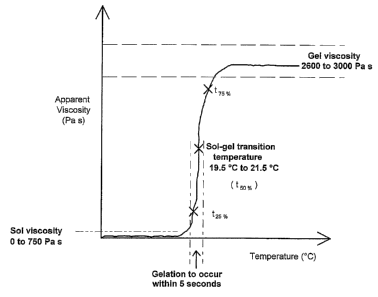
Figure 1 shows a typical PBP viscosity-temperature profile indicating the
parameters and
the range of values that are pertinent to the utilisation of a thermo-
reversible formulation
for cell/particle manipulation and dye delivery. The sol-gel transition
temperature
(t50.%) is a measure of the temperature at which to half the maximum gel
viscosity is
attained. The values t25% and t75% refer to the temperature at 25 and 75% of
the
maximum viscosity respectively. In addition to the measurement of these
values, the pH
of the formulation (e.g. as adjusted to pH 7.2-7.4 through the use of
phosphate buffered
saline as the buffer) is important for cell viability.
The handling concept behind the gel is that it would be stored routinely in
the cold
(providing a liquid reagent form). The liquid form is manipulated cold but
forms a gel
within seconds as the temperature is increased. Controlling the warming
process will
provide for different micellar qualities in terms of the degree of ordering.
The rapidity
CA 02571695 2006-12-20
WO 2006/003423 PCT/GB2005/002603
permits incorporation into rapid assays. The instantaneous conversion from sol-
to-gel
phase provides a route for incorporation into assays. The liquid form can be
used to trap,
support, over-layer, or suspend particles, beads, cells etc prior to or during
manipulation
(e.g. resuspend a cell pellet). Upon temperature shift (e.g. positive or
passive warming)
the gel stiffens providing a cell/particle mountant.
General methodologies can be described which provide for applications in which
live or
fixed cells, particles or beads can be incorporated into the gel with
formulations which
may include informative dyes or other reporter molecules. These basic
protocols can be
adapted for specific applications in candidate product screening in drug
discovery, cell-
and particle/bead-based biotechnologies and numerous applications in imaging
and
microscopy and non-imaging plate based assays.
Preferred aspects of the invention are described in the following non-limiting
examples,
with reference to the following figures:
Figure 1 shows a typical PBP viscosity-temperature profile indicating the
parameters
and the range of values that are pertinent to the utilisation of a thermo-
reversible
formulation for cell/particle manipulation and dye delivery.
Figure 2 shows carnera images of EGFP-associated fluorescence in U2-OS human
tumour cells held in gel, demonstrating the maintenance of cellular integrity
and EGFP
expression in the cytoplasm (arrow). (Panels: a, cell in full culture medium;
b-d, cells
overlayered with gel and imaged at 0 [b], 10 [c] and 60 [d] min at 37 C.
Figure 3 shows camera images of EGFP expressing U2-OS human tumour cells.
mounted in gel following exposure to the nuclear locating fluorescent dye
DRAQ5
(a: b)
Figure 4 shows camera images of DRAQ5 in gel stained SU-DHL-4 cells held in
gel.
26
CA 02571695 2006-12-20
WO 2006/003423 PCT/GB2005/002603
Figure 5 shows laser scanning microscopy detection of cell nuclei and
immunostaining
of a cell surface antigen for fixed cells supported in gel (bar = 10 m). Left
panel show
transmission image, middle panel shows green Alexa 488-NCAM
immunofluorescence,
and the right paiiel shows far-red DRAQ5 nuclear fluorescence.
Figure 6 shows the steps in a simple protocol to mount a sample pre-mixed in
gel onto a
standard microscope slide.
Figure 7 shows the steps in modified protocols for the preparation of samples
in gel on
slides and multi-well systems.
Figure 8 shows time-lapse imaging of beads in PBS or gel reveals efficient
trapping of
fluorescent objects for sequential image collection.
Figure 9 shows the effects of a magnetic field on dispersed magnetic beads in
a 24% PF-
127 gel at room temperature.
Figure 10 shows typical results for calcein loaded cells (MCF-7 human breast
carcinoma
cells cultured using routine methodologies in glass-bottomed chambers and
using a
camera-based system.
Figure 11 shows a wide-field (CCD-camera) focus series through a 170 nm bead
mounted in PF-127 24% w/v in water (inverted contrast). The slide was mounted
onto a
Nikon fixed stage upright microscope, and imaged using a x40 ELWD NA 0.6 air
objective lens (pixel resolution of 0.23 m). In fluorescence mode (470/40
excitation
and 525/50 emission) a focus series was collected using z-steps of 0.15 m a
total of 51
planes were captured which is an equivalent of 7.5 m total distance. A single
bead was
cut out of the total stack and was montaged to show the diffraction rings. An
image of a.-
sub-resolution fluorescent bead (i.e. smaller than about 200 nm) showed an
airy disk
consisting of a central spot surrounded by faint light and dark rings.
27
CA 02571695 2006-12-20
WO 2006/003423 PCT/GB2005/002603
Figure 12 shows the maximum projection of a focus series through a 170 nm bead
mounted in PF-127 24% w/v in water (inverted contrast). The conditions were
identical
to those described above. The beads remain stationary throughout the entire
series
which took approximately 3 minutes to collect. Each bead consists of a bright
(black)
centre and rings around the centre; showing that each of the beads is
stationary.
Figure 13 shows two typical wide field point spread functions (PSF) obtained
by
resampling the data in xz. The asymmetric image arises due to spherical
aberrations (i.e.
an air lens (refractive index looking into a 24% PF-127 gel sample refractive
index
1.357). This is a typical situation in high content screening instruments
screens where air
lenses are used routinely, while the live sample sits in gel within a
multiwell plate. The
PSF sits at a slight slant due to the fact that the alignment of the
instrument is slightly
out and off axis. Taken together the bead images provide a quantitative
evaluation of
the instrument performance in conditions identical to those used for a typical
live cell
multi-well imaging setup. Immobilising beads in media or physiological buffer
only for
this kind of evaluation would not be possible.
Figure 14 shows a comparison of the kinetics of uptake of DRAQ5 dye into U2-OS
human tumour cells held in PBS or gel
Figure 15 shows differential staining of live and dead (arrowed) human B cell
lymphoma cells viewed by transmission (panel a) or fluorescence of nuclei of
cells
stained with propidium iodide.
28
CA 02571695 2006-12-20
WO 2006/003423 PCT/GB2005/002603
EXAMPLES
Example I- Methodological aspects
A Typical protocol for the preparation of aqueous sterile PF-127 poloxamer
solutions
i) Aqueous poloxamer solutions were prepared on a percentage weight in
volume basis, by the cold process similar to that described by Schmolka in
1972
(Schmolka, I.R. (1972) Artificial skin I. Preparation and properties of
Pluronic F-127
gels for treatment of burns. J. Bionxed. Mater. Res. 6, 571-582.). PBP is
added slowly to
distilled water and stirred constantly. The sol is thoroughly mixed and stored
at 4 C until
required.
ii) PF-127 (e.g. batch number WPDL-510B) was obtained from BASF
Corporation (Preston, Lancashire, UK). PF-127 solutions used in cell mountant
protocols are prepared using, for example, phosphate buffered saline (PBS).
Different
formulations of PBS can be used. Typical formulations for Phosphate-Buffered
Saline
are:
a. PBS as a 1X liquid, pH: 7.4 ~: 0.05 (Potassium Phosphate monobasic (KHZP04)
1.06 mM, Sodium Chloride (NaCI) 155.17 mM, Sodium Phosphate dibasic
(Na2HPO4-7H20) 2.97 mM)
b. Dulbecco's Phosphate-Buffered Saline (D-PBS) (1X) liquid containing calcium
and magnesium (Calcium. Chloride (CaC12) (anhyd.) 0.901 mM, Magnesium
Chloride (MgC12-6H20) 0.493 mM, Potassium Chloride (KCI) 2.67 mM,
Potassium Phosphate monobasic (KH2PO4) 1.47 mM, Sodium Chloride (NaCI)
137.93 mM, Sodium Phosphate dibasic (NaZHPO4-7H20) 8.06 mM).
[REFERENCE: Dulbecco, R. and Vogt, M., (1954) Plaque formation and isolation
of pure lines with Poliomyelitis viruses. J. Exp. Med., 98:167].
29
CA 02571695 2006-12-20
WO 2006/003423 PCT/GB2005/002603
iii) PF- 127 solutions requiring steain sterilisation are transferred to 100
mL glass
bottles, autoclaved at 120 C for 20 minutes (USP, AXl1.nTF.XTj11) and
subsequently
stored at 4 C until required. Solid PF-127 requiring dissolution in D-PBS or a
buffer of
choice such as RPMI culture medium (either alone, full), supplemented or
supplemented
with glutamine and antibiotics) is weighed under aseptic conditions and added
to the
sterile medium without mixing and stored at 4 C for 12 hours. After this
period, any
clumps of PF-127 remaining are dispersed under aseptic conditions using a
sterile
spatula and the mixture stored for a further 24 hours at 4 C until PF-127
hydration was
complete as judged by the presence of a transparent solution (as defined by
reference to
refractive index).
iv) The presence of heat labile components in the buffer used in any cell
culture
experiments may prevent steam sterilisation of PF-127 hydrated in such media.
Instead,
immediately prior to use the PF-127 solutions can be filter sterilised (0.2 m
pore size
filters). This approach also permits the preparation of thermolabile
excipients, a
procedure not possible with dissolution in gels requiring heating to achieve
liquid form.
v) Over-strength PF-127 solutions are used to dissolve excipients, for example
drug stock solutions, such that upon mixing the required concentration of a
dye (e.g. 20
M DRAQ5TM, or 1 g/mL propidium iodide) and PF-127 gel was obtained.
B Typical step-wise protocol for the physical handling of PBP gel (exemplified
here as a 24% w/v preparation of PF-127 in PBS) for its use as a cell/particle
mountant
i) Cell preparations are made by a standard cell culture method of choice,
including: using attached cells growing on a microscope slide surface (e.g. a
chamber
slide or multichamber plate) or on a coverslip (e.g. coverslip culture), or
deposited upon
a microscope slide (e.g. by smear formation or droplet delivery or cyto-
centrifugation).
CA 02571695 2006-12-20
WO 2006/003423 PCT/GB2005/002603
ii) The PBP gel is prepared in a convenient container. Here a dropper-bottle
preparation is described for cells physically mixed into the PBP gel or for
cells deposited
on the surface of a microscope slide or growing on a coverslip).
iii) Remove the PBP gel dropper bottle from the 4 C refrigerator (store
upright
overnight at 4 C prior to use, and try not to introduce bubbles into the
liquid form when
using the dropper) and place it on crushed ice to maintain the PBP gel as
liquid form and
to further chill the glass dropper inside the bottle.
iv) Take a glass microscope slide (room temperature), place it on a flat
surface
and quickly use the dropper to deposit one drop of PBP gel into the centre of
the slide.
Return the dropper to the chilled bottle immediately. The gel will rapidly
stiffen on the
surface of the microscope slide. Do not touch.
v) Take a standard coverslip (room temp) and gently/evenly place it on top of
the central mound of gel without pressing or trapping air at the point of
contact. The
coverslip will appear as a "hat" balancing on the gel.
vi) Place the microscope slide on a bed of ice (or preferably onto a flat
metal
plate on a bed of ice or a Peltier device to provide. a convenient chilling
surface).
vii) Watch the gel carefully and within seconds the gel will undergo reverse
transition and become a liquid, spreading as a mountant under the coverslip.
viii) When gel spreading has occurred, remove the slide from the chilling
plate
and place the underside of the slide in contact with a warming surface, for
example a
palm of the hand. The gel will stiffen quickly, and retain the coverslip in
place even at
room temperature. The slide can be inverted without movement of the coverslip.
The
gel can be removed from the surface by irrigation using chilled water or
buffer.
31
CA 02571695 2006-12-20
WO 2006/003423 PCT/GB2005/002603
ix) Witli practice the deposition of the correct amount of PBP gel onto the
slide,
the application of the coverslip and the sequence of temperature shifts can
produce a
mounted sample in 30 secs with perfect filling of the coverslip and no trapped
bubbles.
x) The preparation is then analysed by standard microscopy methods.
C Typical protocol for the in situ staining of live cells at room temperature
using an
aqueous sterile PF-127 poloxamer solution prepared in phosphate buffered
saline at 24%
w/v for the purpose of staining nuclear DNA
i) An over strength aqueous poloxamer solution were prepared on a percentage
weight in volume basis as described and mixed with a concentrated stock
solution of the
DNA dye DRAQ5TM to yield a final concentration of 20 M DRAQ5TM in 24% PF-127.
ii) Using -an ice-chilled pipette a 4 C solution of DRAQ5TM/PF-127 is over
layered quicldy onto a cell monolayer culture (e.g. -human osteosarcoma cell
line U2-OS
growing in a chamber slide), obtained using standard cell culture methods.
Prior to over
layering the gel the culture medium is removed and the monolayer washed using
chilled
phosphate buffered saline and the chamber slide placed on a chilled surface.
iii) A coverslip is then placed onto the over layered gel and the mounting
procedure completed as described above.
iv) The preparation is then analysed by standard fluorescence microscopy
methods to examine nuclear morphology of the cells as they in situ stain with
the
DRAQ5TM/PF-127 preparation.
32
CA 02571695 2006-12-20
WO 2006/003423 PCT/GB2005/002603
D Typical protocol for the in situ staininLy of live cells at room temperature
usina an
aqueous sterile PBP solution prepared in PBS at 24% w/v for the purpose of
distinauishing live and dead (gpoptotic cells) using differential staining by
propidium
iodide
i) An over strength aqueous PBP solution was prepared on a percentage weight
in
volume basis as described and mixed with a concentrated stock solution of the
viability
dye propidium iodide to yield a final concentration of 1 g/mL in propidium
iodide in
24% PF-127).
ii) Using an ice-chilled pipette a 4 C solution of PI/PBP solution is mixed
with a
high-density suspension of cells for analysis (e.g. human B cell lymphoma cell
line
growing as a suspension culture), obtained using standard cell culture
methods. The
chilled, mixed sample is pipetted onto a chilled microscope slide and a
coverslip added
as described above.
iii) The preparation is then analysed by standard fluorescence microscopy
methods
to examine the presence of rapidly stained cells showing abnormal nuclear
morphology
(apoptotic or necrotic) or cells resisting staining representing those with
intact plasma
membranes. Here trapping in the cell permits the kinetics of staining to be
observed and
permits repeated analysis of a field of immobilised cells, which would
normally be lost
in an image/microscopy, based assay.
iv) Cell samples may be pre-stained with propidium iodide in aqueous
suspensions
prior to transfer to an aqueous PBP solution for example the transfer of
samples initially
prepared for flow cytometry and subsequently analysed by imaging in gel.
33
CA 02571695 2006-12-20
WO 2006/003423 PCT/GB2005/002603
E Typical protocol for the preparation of fluorescent cells (e.g. expressina
green
fluorescent protein) in PBP gel for live cell imagina
i) Cells carrying a fluorescent reporter are prepared using standard cell
culture
methods either as attached cultures or resuspended cells at high density in a
medium of
clioice.
ii) For attached cell cultures, PBP gel in liquid phase is over-layered as.
described
above.
iii) For cell suspensions, aliquots are mixed directly into the PBP gel in
liquid phase
and pipetted directly onto a microscope slide with a coverslip added as
described above.
iv) The live cell preparations are then analysed by standard fluorescence
microscopy
methods to examine features of interest.
Additional applications of the invention include the following:
Polyoxypropylene-Polyoxyethylene Block Polymer (PBP) at gelling concentrations
can
be used to act as an optically compatible means of trapping and immobilising
particles
for the purpose of calibration, optical alignment and orientation in
methodologies
requiring the collection of light including fluorescence of bioluminescence
emissions. In
a preferred embodiment fluorescent beads deposited on a surface within a PBP
gel
would be used in fluorescence microscopy systems (e.g. confocal laser scanning
microscopy system or multi-photon excitation laser scanning microscopy) to
provide a
means of calibration, point-spread function determination and event
orientation within
optical slices two or more dimensions.
Calibration samples include the co-mixing of beads with cells within the PBP
gel to
provide a depth versus fluorescence correction versus scattering for the
determination of
point spread function in the same live sample conditions. Such samples may
also be used
34
CA 02571695 2006-12-20
WO 2006/003423 PCT/GB2005/002603
to provide an indication of perfomiance of optical elements or instrument set-
up. Such a
method would be appropriate for any type of multi-dimension imaging which
requires
calibration of x, y or z-axis resolution. Calibration is required in order to
measure and
consequently correct for sample derived aberations. Embedded beads co-mixed
with the
cellular sample are therefore appropriate for multi-dimensional resolution
measurement
particularly x,y,z axis resolution, including the point spread function
obtained from sub-
resolution beads. Other aberations require depth dependent correction of
fluorescence,
fluorescence spectral overlap and cross talk measurement.
Polyoxypropylene-Polyoxyethylene Block Polymer (PBP) at gelling concentrations
can
be used to act as an optically compatible means of trapping and immobilising
live and
fixed cells for the purpose of analysis in methodologies requiring the
collection of light
including fluorescence or bioluminescence emissions. The cells may be non-
adherent or
processed cell suspensions. In a preferred embodiment the fluorescence would
originate
from a fluorescent molecule manipulated to be expressed by the cell such a
green
fluorescent protein (GFP).
Polyoxypropylene-Polyoxyethylene Block Polymer (PBP) at gelling concentrations
as
an over-layering mountant for adherent cultures or planar preparations of live
or fixed
cells providing a convenient mountant for protection of cells and in situ
staining or
labelling of cells. Here the sol-gel transition as a function of temperature
provides a
novel means of spreading the mountant at lower temperature and controlling the
gel
depth by halting spreading through gel formation by raising local temperature
of the
preparation. The adherent properties would allow for inversion of a mounted
specimen
so that inverted microscopy formats can be used. Here the gel provides an
aqueous-gel
phase between the specimen and another optical interface for imaging. In a
preferred
embodiment the fluorescence would originate from a fluorescent molecule
manipulated
to be expressed by the cell such a green fluorescent protein (GFP).
Polyoxypropylene-Polyoxyethylene Block Polymer (PBP) at gelling concentrations
can
be used in a method of preparation of particles, beads or cells ('analytes')
by the
CA 02571695 2006-12-20
WO 2006/003423 PCT/GB2005/002603
centrifugation from aqueous suspension into a PBP gel phase within the same
container.
In a preferred embodiment the PBP gel is present below an over-layering
aqueous phase
comprising a suspension of said analytes and maintains a gel-aqueous interface
by
temperature control. Centrifugation forces entry of analytes into the gel.
Analytes
deposited into the gel phase can be recovered by temperature-controlled
transition to a
sol following removal the aqueous over layer.
Analytes can be pre-labelled with fluorescent or bioluminescent probes.
Additionally
analytes which are fluorescent or bioluminescent molecular probes may be
present either
in the aqueous phase or in the gel phase to enable an optical analysis of the
suspended
particles, beads or cells. In a preferred embodiment the fluorescent molecular
probe is
the anthraquinone DRAQ5TM.
Polyoxypropylene-Polyoxyethylene Block Polymer (PBP) at low non-gelling
concentrations has surfactant properties which can provide cell disrupting or
lytic
properties for the release of molecules for primary and/or secondary analyses.
Modulation of properties would require a shift in concentration cf PBP by in
situ
dilution and or a shift in temperature. In a preferred embodiment PBP gels
solubilised in
situ would impart surfactant properties and provide for a sequential live cell-
lysed cell
analysis methodology.
Polyoxypropylene-Polyoxyethylene Block Polymer (PBP) at gelling concentrations
can
be combined with cell fixing chemicals (e.g. paraformaldehyde) and or dyes
(e.g. a DNA
fluorochrome) to provide unique multi-functional agents for in situ fixing,
immobilisation/structure support and cell staining. In a preferred embodiment
such
multi-function agents would reduce processing time, minimise cell loss through
a
reduction in the number of processing steps (e.g. in fixation schedule that
require
washing and fluid removal steps) and provide a means for maintaining osmotic
environments, metabolic gradients and structural/mechanical integrity.
36
CA 02571695 2006-12-20
WO 2006/003423 PCT/GB2005/002603
The formation of Polyoxypropylene-Polyoxyethylene Block Polymer (PBP) gels to
enable the preparation and immobilisation of encapsulated prokaryotic cells on
porous or
non-porous surfaces for the purpose of short term cultivation and or a
sequential analysis
in which the location of the sample is recognised for data linkage purposes.
In a
preferred embodiment temperature-shifting the low temperature liquid phase
encapsulation of a prokaryotic cell(s) could be used to trap cells at a
specific location at
which a drug can be delivered for the purpose of chemosensitivity testing.
The formation of Polyoxypropylene-Polyoxyethylene Block Polymer (PBP) gels to
enable the preparation and immobilisation of encapsulated eukaryotic cells on
porous or
non-porous surfaces for the purpose of short term cultivation and or a
sequential analysis
in which the location of the sample is recognised for data linkage purposes.
In a
preferred embodiment temperature-shifting the low temperature liquid phase
encapsulation of a eukaryotic cell(s) is used to trap cells at a specific
location at which a
subsequent analysis of a gene sequence(s) and or protein(s) or other cell-
originating
molecules.
The formation of Polyoxypropylene-Polyoxyethylene Block Polymer (PBP) gels to
enable the preparation and immobilisation of encapsulated cells on porous or
non-porous
surfaces for the purpose of short term cultivation and or a sequential
analysis in which
the location of the sample is recognised for data linkage purposes. In a
preferred
embodiment. temperature-shifting the low temperature liquid phase
encapsulation of a
eukaryotic cell(s) is used to trap cells at specific locations for the purpose
of detecting
and analysing the presence or absence of parasites including the intracellular
forms of
Plasmodium species in the diagnosis of malaria and for the purpose of species
atid
variant identification.
The formation of Polyoxypropylene-Polyoxyethylene Block Polymer (PBP) gels to
enable the preparation of encapsulated cells or particles for the purposes of
sample
protection, manipulation or analysis. In a preferred embodiment the low
temperature
liquid phase encapsulation of a cell or particle permits the generation of
droplets for the
37
CA 02571695 2006-12-20
WO 2006/003423 PCT/GB2005/002603
purpose of preparing arrays or replicates through the delivery of such
droplets to a
receiving surface or container prior to or following analysis of informative
features of
the encapsulated sample.
A methodology to provide a means of the pre-building of modular assay
systems/devices
for sequential processing regulated by the properties of the thermoreversible
gels. In
passing through the transition temperature, for example at the point of
droplet formation
or delivery, encapsulated samples would suffer reduced evaporation stress for
live cell
preparations but have increased surface adhesion properties. In a preferred
embodiment
encapsulated cells offer a physical protection for cells from mechanical
stress imparted
by sorting and arraying instrumentation.
The rapid forniation of the Polyoxypropylene-Polyoxyethylene Block Polymer
(PBP) gel
provides initially an immobilising layer on the cells. With the addition of
potential
chemo-attractants within the gel or in a layer above the gel, this gradient
becomes an
active layer for stimulating cells or attracting/sorting cells away from
unstimulated
counterparts. The thermoreversibility allows these cells to be selectively
removed and
further processed.
The micelle environment of the Polyoxypropylene-Polyoxyethylene Block Polymer
(PBP) provides for the controlled carrier and delivery of molecules (e.g.
reactants,
reporter fluorochromes or conjugates thereof) to cells or particles by passive
diffusion or
electrophoresis for the purpose of a controlled analysis methodologies. In a
preferred
embodiment the molecular sieve effects of the PBP gel would effect a
sequential
delivery of reactants and fluorescent or bioluminescent reporter molecules
within sample
preparations.
The addition of excipients for the purpose of cell protection or biological
modification
would impart additional functionalities to Polyoxypropylene-Polyoxyethylene
Block
Polymer (PBP) gels. For example, the inclusion of growth factors or signalling
molecules to maintain or modify specific cellular phenotypes.
38
CA 02571695 2006-12-20
WO 2006/003423 PCT/GB2005/002603
The addition of excipients for the purpose of modifying the photophysical and
photochemical effects of light illumination on cells or reporter molecules
would impart
additional functionalities to Polyoxypropylene-Polyoxyethylene Block Polymer
(PBP)
gels. For example, excipients may be included to reduce the photobleaching of
fluorescent reporter molecules.
The formation of Polyoxypropylene-Polyoxyethylene Block Polymer (PBP) gels to
enable the tliermally controlled presentation of cells or particles to
surfaces, which
enhance or enable assay performance. In a preferred embodiment the assay would
exploit surface plasmon resonance effects or light collection from highly
restricted
depths at optical interfaces.
The block copolymer relevant to this invention may comprise polyoxyethylene
and
polyoxypropylene. Accordingly, gel-forming preparations include those
described as
Pluronics F127, F108, F98, F87 and F88 (Pluronic is a registered trademark
of BASF
Corporation).
39
CA 02571695 2006-12-20
WO 2006/003423 PCT/GB2005/002603
Example Id -~Ise of block pol>>mer compositions of the invention in tlae
anal>>sis of
fluorescent or dved cells
l. General methods for the preparation of a cell line expressing EGFP and its
optical
analysis
Preparation of construct. The cell cycle phase marker DNA construct (GE
Healthcare;
Cardiff UK) was prepared from three DNA fragments that were fused in frame and
cloned into a pCI-Neo (Promega) vector that had been cut with Bg1II and Nhel
to
remove the CMV promoter. The three fragments used were the cyclinB 1 promoter,
the
N-terminal 171 amino acids of the human cyclin BI coding region and EGFP. The
cyclin B 1 promoter was amplified from a construct described previously22
using PCR
and the primers 5'-CGCGGCAGCTGCCCGAGAGCGCAGGCGC-3' and 5'-
CGCAAGCTTCCTCTTCACCAGGCAGCAGCTC-3'. The N-terminal region of
cyclin B 1 mRNA, encoding the cyclin B 1 destruction box and the CRS but
excluding
the CDK binding site was amplified with HindIII and BamHI ends using PCR and
the
primers
5'-GGGAAGCTTAGGATGGCGCTCCGAGTCACCAGGAAC-3' [SEQ ID NO:l] and
5'-GCCGGATCCCACATATTCACTACAAAGGTT-3' [SEQ ID NO:2] from a
cyclinBl cDNA described previously5. The gene for EGFP was amplified from
pEGFP-
N2 (Clontech) with primers
5'GGTACGGGCCGCCACCATGGGATCCAAGGGCGAGGAGCTGTTCAC [SEQ
ID NO:3] and
5'-GGTACGGGTTAACCGGTCTTGTACAGCTCGTCCATG 3' [SEQ ID NO:4].
All three fragments were fused and the integrity of the final clone confirmed
by
sequence analysis.
Cell reporter systeni. The parental cell line used in these studies was a
human
osteosarcoma cell line derived from a 15 year old Caucasian female U-2 OS
(American
Type Culture Collection [ATCC] HTB-96). U-2 OS cells was transfected with the
cell
cycle marker DNA construct using Fugene (Roche) according to the manufacturers
CA 02571695 2006-12-20
WO 2006/003423 PCT/GB2005/002603
instructions. Following selection with 1000 g/ml Geneticin (Sigma G7040) the
expressing cells were enriched using high-speed fluorescence activated cell
sorting
(MoFlow; DAKO-Cytomation) and sorted into 96 well plates (1 green fluorescent
cell/well). Colonies were expanded and clones whose green fluorescence varied
with the
cell cycle as predicted for a cyclin-based reporter, as determined by
conventional flow
cytometry, were expanded and a high expressing subline maintained.
Growing and 7aintenaface condition. The stably transfected cells were
maintained at
37 C and 5 % C02 usiilg standard tissue culture techniques. Media used was
McCoys
5A modified (Sigma) supplemented with 2mM glutamine, 100 units/ml penicillin,
100
mg/mi streptomycin, 10 % fetal calf serum and 1000 g/ml geneticin.
Time-Zapse%amera inzaging. High resolution fluorescence cell tracking was
performed
with cells seeded into 8 well Nunc coverglass chambers (Labtek Inc). Culture
dishes
were placed on to a time-lapse instrument designed to capture bright-field
phase images
and GFP fluorescence (480/25 nm excitation and 525/30 nm emission). An
Axiovert 100
microscope (Carl Zeiss, Welwyn Garden City, UK), was fitted with an incubator
for
370C/5% C02 maintenance (Solent Scientific, Portsmouth, UK), and an ORCA-ER 12-
bit, CCD camera (Hamamatsu, Reading, UK). Illumination was controlled by means
of a
shutter in front of the transmission lamp, and an x,y positioning stage with
separate z-
focus (Prior Scientific, Cambridge, UK) controlled multi-field acquisition.
Image
capture was controlled by AQM 2000 (Kinetic Imaging Ltd). All images were
collected
with a 40x, 0.75 NA air apochromat objective lens" providing a field size of
125x125
mm. Sequences were captured as required. When required analysis of the images
was
performed with the integrated AQM 2000 software package (Kinetic Imaging Ltd).
Each
cell in the field was tracked individually. Fluorescence tracking on a single
cell basis
was achieved in Lucida (KI Ltd). Fluorescence was recorded in a region of
interest.
41
CA 02571695 2006-12-20
WO 2006/003423 PCT/GB2005/002603
2. TAipical analysis of fluorescent cells in Qel.
Fig 2 shows typical results for EGFP-associated fluorescence in expressing
cells imaged
in culture medium (panel a), in which the marked region shows the presence of
a group
of 3 cells expressing high levels of EGFP in the cytoplasm (arrow). Following
capture of
the image cells, culture medium was aspirated and the cell monolayer
overlayered with a
chilled (4 C) 24% w/v gel (PF-127; in sol form) prepared in PBS, returned to
the
imaging platform (at 37 C; the overlayer forming a supporting gel at this
temperature)
and the location of the field re-found. Further fluorescence images were
captured of the
same cells at 0, 10 and 60 min incubation in gel. The images clearly show the
maintenance of cellular integrity (flattened cells) and GFP expression in the
cytoplasm.
3. Typical analyses of more than one fluor in live cells in gel: EGFP-
expressing cells in
Qel (24% w/v PF-127 prepared in PBS) co-stained with a DNA dye (DRAQ5).
Fig 3 shows typical results for EGFP-associated fluorescence in 3 samples
stained for
nuclear DNA using the fluorescent agent DRAQ5. Paired images show DRAQ5-
associated far-red fluorescence (Fig 3 left panels a, c and e) or EGFP green
fluorescence
(Fig 3 right panels b,d and f). Panels a and b show results for the same cells
stained in
PBS with DRAQ5 (20 M x 10 min) and imaged in PBS permitting the
identification of
cells (presence of a nucleus; arrowed) with high- (hgfp) or low- (lgfp) EGFP
expression
within the cytoplasm (arrowed). Panels c and d show cells also pre-stained
using
DRAQ5 in PBS but overlayered with gel (see above) after aspiration of the
DRAQ5
solution. The images in panels c and d show the continued ability to
distinguish hgfp and
lgfp expressing cells. Panels e and f show cells stained with DRAQ5 in the gel
overlayer
for lh (DRAQ5 20 M in 24% w/v PF-127prepared in PBS; at 37 C), demonstrating
the
ability to distinguish hgfp. and lgfp cells using an in-gel staining
methodology. The
images clearly show the maintenance of cellular integrity (flattened cells)
and GFP
expression in the cytoplasm.
42
CA 02571695 2006-12-20
WO 2006/003423 PCT/GB2005/002603
4. Typical light transmission and fluorescence analysis of cells stained in
gel using a cell
permeant dye (DRAQ5).
Human B cell lymphoma cells (line SU-DHL-4) were cultured in suspension using
routine methodologies. Cell culture typically contain live cells and a
background of
dying cells, debris and occasionally non-cellular particles. In a typical
analysis to
distinguish objects a comparison can be made of transmission and fluorescence
images.
A typical methodology would comprise cell samples pre-mixed with cooled gel
(24%
w/v PF-127 prepared in PBS and containing 20 M DRAQ5) and mounted under a
coverslip on a cooled microscope slide. The slide was then raised to room
temperature
for 30 min to permit the continued in-gel staining of nuclear DNA by DR.AQ5.
Fig 4
shows a typical field imaged for transmission (panel a) or far-red
fluorescence of a DNA
dye (DRAQ5; blue light excitation panel b). The images (see arrows) reveal the
positive
in-gel staining of intact cells, permitting the distinction of bi-nucleate
objects (i.e.
dividing cell), debris (indistinct nuclear signal) or non-cellular (non-DNA-
containing)
inclusions. The analysis exemplifies the imaging of non-adherent
cells/objects, held in
gel, enabling the sequential examination of cell/object features without loss
of location
in 3-dimensions.
5. Examples of the use of block polymer compositions of the invention for
immobilizing non-adherent cells for the use high resolution ima-ging to
determine
immunofluorescence localization.
An iinportant feature of the PF-127 gel formulations is that they provide an
easy method
for immobilizing suspension cells such as those prepared for flow cytometry.
This
enables high resolution imaging to be performed on cells that are not
originally tethered
to an optical surface.
Therefore PF-127 formulations provide a route for interfacing different
cytometry
platforms (e.g. a flow cytometry sample analysed by imaging) particularly
those that
require the sequential analysis of cells in suspension. Of particular interest
is the
43
CA 02571695 2006-12-20
WO 2006/003423 PCT/GB2005/002603
localization of a given fluorescence signal to a cellular compartment (e.g.
the expression
of the neural cell adhesion molecule [NCAM] on the cell surface of small cell
lung
carcinoma cells [SCLC cells]) or the expression of a signal in relationship to
neighbouring cells v,~here a support matrix is required to maintain a cellular
cluster
during, for example, multiple optical scans of a confocal or multiphoton
microscope.
Here the use of gel as a support matrix for fixed cells probed with an
appropriate
fluorescently-tagged antibody and a DNA stain is described. NCI-H69 cells were
cultured as suspension cells in RPMI-1460 culture media with 10% FCS using
standard
cell culture methodologies. Cells were harvested and fixed in ice-cold
methanol for 20
minutes. After washing in phosphate buffered saline the samples were processed
for
standard immunofluorescence as used for flow cytometric analysis and
fluore,scence
microscopy. These suspensions were prepared as flow analysis for NCAM (CD-56)
detection, using mouse anti-human (CD-56; BD Pharmingen, UK) monoclonal
antibody,
followed by a secondary staining using an anti-mouse Alexa 488 (Molecular
Probes,
InVitrogen, USA). Finally the preparations were labelled with DRAQ5 to
distinguish
the nucleus.
A small sample of cells (50 l at 1 x 10⁶ cells per ml) was placed in a
chamber coverslip
(Nunc) and PF-127 sol at 24% w/v in PBS was placed over the cell layer, and
left at
room temperature to form a gel layer (see part A for chamber slide
preparation). The
cells and cell clusters became immobilized under the gel matrix.
High resolution confocal laser scanning microscopy (BioRad 1024MP; BioRad
Microscience Ltd) was performed to obtain a three channel image of the cell
clump (Fig
5). The transmission image showed optical compatibility with 488/647 nrri
light. The
sample stability enabled imaging of the tightly coupled cells and provided
distinct edges
between cells depicting NCAM localization. Nuclear localization depicted the
cellular
localization and clearly shows the number of cells within the clump. There
were no
detectable background or optical scattering problems associated with the gel
mountant.
The example demonstrates the use of the gel with fixed cell preparations,
within a
44
CA 02571695 2006-12-20
WO 2006/003423 PCT/GB2005/002603
protocol compatible with flow cytometric analyses and the ability high
resolution
immunofluorescence signals in gel.
CA 02571695 2006-12-20
WO 2006/003423 PCT/GB2005/002603
Example HI - Exanzples of tiae production of a microscope slide or multiwell
plate
coated in a block polymer conzposition
1. Simple protocol to mount a sample pre-mixed in eel onto a standard
microscope slide
(Fig 6 panels a-e)
STEP a: A sample for analysis is mixed into gel (in sol form; held in a sample
tube on
ice) for example by the addition of a concentrated suspension of cells (e.g. 4
x 10' cells in a 10 l volume of PBS prepared using standard centrifugation
methodology) to a 250 l volume of 24% w/v F-127 prepared in PBS). Over-
strength preparations of gel can be used to provide a final concentration of
24% w/v F-127 prepared in PBS if required.
STEP b: The sample is quickly streaked across the surface of a standard
microscope
slide at room temperature and the gel stiffens within seconds.
STEP c: A coverslip is placed onto the gel.
STEP d: The slide is placed on an ice-pack and the gel transformed to a liquid
state
and spreads under the coverslip within seconds.
STEP e: Removing the slide from the ice-pack results in air-warming of the
slide to
room-temperature and the setting of the gel within seconds.
2. Modified protocol to mount a sample(s in gel at given locations on a
standard
microscope slide (Fig 7 panels a-k)
Pre-prepare stained or unstained cells, beads or particles in an aqueous
suspension,
aspirate supernatant and hold pellet on ice. Fig 2 (panels a-k) shows the
subsequent steps
for preparation of a single sample on a microscope slide, the procedure being
repeated
for multiple sainples as required.
STEP a: Press a silicon isolator (type shown in panel a is a S2560 silicon
isolator with
8 holes [each 2mm deep, 9mm diameter]; obtained from Sigma-Aldrich UK)
onto a microscope slide on an ice-pack.
46
CA 02571695 2006-12-20
WO 2006/003423 PCT/GB2005/002603
STEP b: Add 90 L of cold 35% PF-127 gel into a well. This can be achieved,
for
example, using a pre-chilled 1 ml micropipette tip.
STEPS c and d: Inject the cell/bead or particle sample through the gel at the
bottom of
the well in a volume of 10 uL and the suspension plumes to the central area
of the surface of the gel.
STEP e: Transfer slide to a warm heating block (held at 37 C) and the gel
stiffens.
STEP f: Carefully peel of the isolator.
STEP g: The gel disk revealed is self-supporting.
STEP h: Place slide onto an ice-pack until the bottom surface of the gel
starts to
liquefy.
STEP i: Place a coverslip onto the surface of the gel disk while the slide
remains on
the ice-pack. The disk continues to liquefy and starts to spread.
STEP j: Overlayer the slides with an absorbent paper and gently press to
complete
spreading of the sample and to remove excess liquid.
STEP k: Return to warm heating block to stiffen gel and complete preparation.
Return
to room temperature for storage (e.g. up to 24h).
3. Modified protocol to mount a sainple in a chamber/well (e.g, standard glass
bottom 8
well chamber slide) (Fig 7 panels 1& m)
Pre-prepare stained or unstained cells, beads or particles in an aqueous
suspension,
aspirate supernatant and hold pellet on ice. The procedure for the addition of
the gel and
sample is reversed from that described above. Fig 2 shows the main steps of
introducing
the cell/bead or particle sample in a volume of 10 L into an empty
well/chamber of a
multi-chamber slide held on an ice-pack (panel 1). Then add 90 uL of cold 35%
PF-127
gel into the well (panel m). This can be achieved, for example, using a pre-
chilled 1 ml
micropipette tip. The liquid gel overlayers the sample suspension. The slide
is then
warmed (e.g. on a heating block at 37 C) to stiffen the sample-gel interface,
as described
above, prior to analysis.
47
CA 02571695 2006-12-20
WO 2006/003423 PCT/GB2005/002603
4. Simple exemplar protocol for the preparation of dried films of block
polymer and
their reconstitution in a multi-well plate (Table 3)
An example of a methodology is described for the preparation of dried films of
PF-127
and their reconstitution by the addition of differing volumes of water (or a
given
solution) to provide a range of potential gel/liquid concentrations for
cell/bead or
particle immobilisation or manipulation. The steps are outlined below.
STEP a: A 19.3% w/v PF-127 solution in water was prepared. This concentration
permits a liquid state to be easily formed when chilled (e.g. at 4 C) but
still
retain some degree of loose gel/liquid state at room temperature (20 C).
STEP b: Volumes of cold gel were dispensed into a matrix of 48 wells within a
standard 96-well (flat bottomed) transparent plastic dish as indicated in the
Table.
STEP c: The plate was held on a heating block at 37 C for 24h to allow for the
desiccation of the gel into dried films covering the base of each well. Here
the process may be accelerated for example by vacuum drying.
STEP d: At this stage the dried films can be stored before commitment to
rehydration.
STEP e: Volumes of ice-cold PBS are dispensed into each well as indicated in
the
Table and the plate rotated briefly to aid the wetting of the dried. Here the
process may be accelerated by mechanical vibration.
STEP f. The plate is then held with the lid sealed for 24h at 4 C. Here the
rehydration
conditions may be varied (e.g. incubating at 37 C in a humidified
atmosphere).
STEP g: After rehydration the plate is returned to room temperature for the
assessment
of quality gel formation in the wells by direct and microscopic examination
of the transparency and the mechanical properties by agitating the well
contents with a pipette tip.
Results: Table 3 shows the ability to prepare liquid and gel-like phases in
all
combinations when assessed at room temperature. Here reconstitution was
achieved
48
CA 02571695 2006-12-20
WO 2006/003423 PCT/GB2005/002603
using PBS demonstrating the in situ preparation of gels with a buffer of
choice. Some
wells showing liquid phase (i.e. 'liquid) at room temperature would be capable
of
forming gels if the temperature was raised. Further, only some combinations
resulted in
the formation of a transparent and optically acceptable gel (i.e. 'transp.
gel') with in
other case a turbid-opaque gel/paste formed (i.e. 'gel'). The combination of a
75 L
19.3% PF-127 dried gel film reconstituted with a 50 L PBS volume provided a
transparent gel (reversible to a sol by chilling) with a nominal poloxamer
concentration
of 29%. This preferred combination would allowing for the retention of room
temperature (and 37 C) immobilisation properties and permit the further
addition of
sample volumes upon (for example reducing the final concentratioii of gel to
24%).
Table 3
Reconstitution of dried films of PF-127 (19.3% w/v gel in water)
and the effects of reconstitution with differing volumes of PBS
,.._..._.. :. _._ u...... .~_.
Dried gel reconstituted with PBS (pL)
_...r 10 25 50' 100 150 200
;_.._ ...........~..._....._
;pL gel dried:
._.... .._ ,~....__.,_. ..
10 trans. gel tiqui.d liquid liquid liquid " I.iquid
_ .._.....__~_ .. . . _
trans. gel trans. gel liquid liquid liquid liquid'
trans. gel liquid liquid liquid liquid [iquid
50 trans. gel li'quid liquidiiquid liquid liquid
75 trans. gel trans. gel trans. gel liquid 'liquid 'liquid
0rgel trans. gel trans. gel Biquid: liquid liquid
10'
;...___ ..._,..__
150 ,::: gel ggel liquid liquid liquid
uid fi uid
200 gel gel gel trans. gel Fiq q
49
CA 02571695 2006-12-20
WO 2006/003423 PCT/GB2005/002603
Example I-V - Example of the production of a block polymer composition
comprising
fluorescent beads and/or a dye
1 Simple protocol to prepare fluorescent beads in aael for the purpose of
iinmobilisation and analysis
Analysis of beads may, for example, require the determination of bead location
and
optical properties such as fluorescence. An example of a typical protocol for
the analysis
of fluorescence characteristics and bead location is shoum in Fig 8 for red-
fluorescent
approx. 1 m diameter beads (e.g. Becton Dickinson Calbrite APC beads; BD
Biosciences, USA) using excitation and emission conditions described by the
manufacturer. The general methodologies for preparing beads in gel have been
described
above. A concentrated preparation of beads (e.g. 1 drop into 0.5 ml gel and
mixed on
ice using a pipette micro-tip. A gel sainple was prepared on a microscope
slide. A time-
lapse imaging systern described above was used to sequentially image the
fluorescence
of beads either in PBS (as a film trapped under a coverslip ) or in 24% w/v
gel (PF-127
prepared in PBS). Irnages a to d shows the same field of view for beads in
PBS, imaged
4 times with a 1 sec interval between each image capture. Image e shows the 4
merged
images of a-d. Similarly, images f-i show 1 sec interval images for beads in
gel at room
temperature with the corresponding merged image shown in panel j. The beads
clearly
move in the PBS preparation, due to fluid movement and Brownian motion,
resulting in
a confused merged image. Beads remain at fixed locations in the gel for the
scanning
period demonstrating the immobilization properties of the gel for beads.
2. Simple protocol for the preparation of magnetic beads in gel and their
manipulation
in a magnetic field
Magnetic bead tecllnology is in common use for separation methodologies.
Unlabelled
magnetic beads (obtained from The Reagent Mine Ltd., Melton Mowbray, UK;
approx
2 m diameter) were dispersed into gel (24% w/v PF-127 prepared in water) at 4
C in a
2mL polypropylene sample tube. The suspension was then prepared on a
microscope
CA 02571695 2006-12-20
WO 2006/003423 PCT/GB2005/002603
slide for imaging by transmitted light using a standard microscope equipped
with a
camera system. Fig 9 (panel a) shows the dispersed beads immobilized in gel at
room
temperature. A neodymium magnet (The Reagent Mine Ltd., Melton Mowbray, UK)
was then placed 2.5 cm from the centre of the field of view and the same
height as the
slide surface and after 30 seconds the field re-imaged. Fig 9 (panel b) shows
the effect
of the magnetic field resulting in bead alignment along the lines of force,
demonstrating
the ability to move beads in a supporting gel for the purposes of alignment
and re-
location within the gel.
3. Preparation of a dye in gel
Vital-labelling methodologies often require the uptake of a non-fluorescent
form of a
dye which becomes fluorescent upon intracellular processing. Here the
preparation of
the vital dye calcein-AM is described for the in-gel staining of live cells
overlayered
with the gel-dye preparation. Calcein dye (calcein AM; 0.1 g/ml; C3099 Cat.
No.,
Molecular Probes, InVitrogen) was mixed into a 24% w/v PF-127 prepared in PBS
and
overlayered onto a monolayer culture of human MCF-7 cells in a chamber slide
and
incubated at room temperature for 15 min. Images were collected using standard
confocal microscope methodologies (system: BioRad 1024MP, BioRad
Microsciences,
UK). Fig 10 shows an optical section through cells demonstrating a typical
compartmentalization of the dye in some cells with more diffuse staining in
others. The
results demonstrate the ability to prepare a dye in gel for live cell marking
and function.
51
CA 02571695 2006-12-20
WO 2006/003423 PCT/GB2005/002603
Example V - Examples of the use of block polymer compositions of the invention
in
tiae calibration of equipment for optical analysis (e.g. the determination of
point
spread function)
1. Gel preparations used for immobilization have advanta eg ous
Imaging instruments (microscopes and HCS instruments) produce a spatially
sampled
array of fluorescence. Images may be produced by the optical system directly
(camera-
based) or built up by scanning (laser scanning microscope). Fewer changes in
refractive
index at the different optical interfaces are advantageous. The availability
of aqueous-
based gels provides an advantageous medium in terms of refractive index when
compared, for example, with higher RI glycerol-based mountants. Standard
refractometry was used to measure the RI values for typical gel preparations
and the
values obtained are shown in Table 4.
Table 4
Typical values for refractive index obtained for gel preparations
Sample Refractive index (RI)
PF-127 24% w/v prepared in water; at 1.357
37C
PF-127 24% w/v prepared in PBS; at 1.359
37C
PBS 1.333
water 1.331
Spatial resolution in theoiy: Illumination wavelengths (from an arc lamp) are
selected
by an excitation filter or spectrometer and the light is spread onto a field
aperture by a
high Numerical Aperture condenser lens. It then reflects from a 45 degrees
dichroic
mirror and an image of the field aperture is demagnified into the sample by an
objective
lens. In this way, the entire sample is evenly bathed in light. Fluorescence
is collected by
52
CA 02571695 2006-12-20
WO 2006/003423 PCT/GB2005/002603
the objective and fornns an image in the microscope that is either inspected
visually,
using a magnifying eyepiece, or passed to an appropriate photo-detector such
as a CCD
camera. All parts of the illuminated sample contribute to the image that
contains sharp
(in focus) features as well as out-of focus features. It is important to
consider the
performance characteristics of any fluorescence imaging instruments. An image
of a
sub-resolution fluorescent bead (i.e. smaller than about 200 nm) will show an
airy disk
consisting of a central spot surrounded by faint light and dark rings.
Measurement of the
airy disk gives parameters describing the microscope performance. The distance
from
the centre to the first dark ring describes horizontal (x, y) resolution and
is given by:
da.y = 0.61?, /NA
If a focus series of images of the bead is collected, the corresponding axial
(z) resolution
is:
dz =3.7d.zyU /NA
1~ = 7 efi active index of sa aple medium
), = wavelength
NA = objective lens Numerical Aperture
Total intensity in any horizontal plane is proportional to NA2/(magnification)
and is
constant near the focus, so there is no optical sectioning in a conventional
microscope.
Spatial resolution in practice: The rigor in which an assay can be implemented
on any
imaging system is dependent on reproducibility and calibration of the
instrument. It is
essential to understand the spatial performance of the imaging system in order
to extract
quantitative information or indeed undertake deconvolution processing to
extract 3D
information. Since the refractive index of the sample medium linearly
influences axial
resolution and the axial performance changes is depth due to spherical
aberration it is
important to calibrate the axial resolution 'in situ'. The accepted method for
obtaining
the x,y,z performance of a microscope is to acquire image from a sub-
resolution bead in
53
CA 02571695 2006-12-20
WO 2006/003423 PCT/GB2005/002603
the exact same conditions used for imaging the sample. However in water-based
samples (physiological buffers and media) it is essential to keep the cells
living and
acquire xyz calibration information from a bead. By placing the sample (cells)
and
beads in block polymer compositions high resolution images of immobilized
beads can
be obtained enabling axial performance to be extracted.
To exemplify the use of block polymer compositions with integrated sub-
resolution
beads we are able to obtain information on the optical performance of the
instrument at
different depths through the sample.
Materials and sample preparation:
(i) Sub-resolution beads can be obtained from many different manufacturers in
this case
Molecular Probes. PS-speck Microscope point Source Kit: 5051515 nm fluospheres
carboxylate modified microspheres 0.17 m yellow-green fluorescent
(concentration
107 per ml).
(ii) Block polymer composition (PF-127) at a formulation of 24% w/v in water
was
prepared as previously described above.
Step 1: Take 0.5 mis of 24% Pluronic F127 maintained at 4oC and mix with 5 l
bead solution.
Step 2: Maintain at 4oC on ice until ready to use
Step 3: Place 50 l on to a microscope slide (the droplet becomes gel)
Step 4: Place a coverslip (22mm x 22 mm) onto the droplet
Step 5: Cool the slide on an ice block and the droplet spreads and the
coverslip
becomes level.
Obtaining a focus series of images through the bead along the optical axis
(see figures)
Step 1: Firmly secure the slide to the microscope (vibrations will disturb
image
collection)
54
CA 02571695 2006-12-20
WO 2006/003423 PCT/GB2005/002603
Step 2: Choose the appropriate imaging conditions to obtain the focus series.
Results are shown in Figures 11 to 13.
Reference
White NS, Ei-rington RI. Fluorescence techniques for drug delive7y research:
theoty and
practice. Adv Drug Deliv Rev. 2005 Jan 2; 57(1):17-42.
CA 02571695 2006-12-20
WO 2006/003423 PCT/GB2005/002603
Example VI - Exanzples of tlae use of block- polymer compositions of the
invention in
tlze controlled deliverl, of reagents to cells
1. Delivery of a cell-permeant DNA dye to cells in gel
The delivery of reagents to cell, beads or particles immobilized in gel
permits the
analysis of modified interactioil kinetics over extended periods. Described
herein is an
example of the impact of gel-based delivery of a reagent, cell permeant DNA
dye
DRAQ5, in comparison with the lcinetics obtained by the staining in PBS alone.
Here
attached U-2 OS (American Type Culture Collection [ATCC] HTB-96) cells were
grown in glass-bottomed chamber slides using standard cell culture
methodologies. The
use of attached cultures allowed for their immobilization for staining in PBS
and a direct
comparison with the staining in gel. The culture medium was aspirated and
replaced
with either PBS supplemented with DRAQ5 (20 M) or overlayered with gel (24%
w/v
PF-127 prepared in PBS) also containing DRAQ5 (20 M). Samples were then
imaged
using a time-lapse microscope and the changes in nuclear associated far-red
fluorescence
monitored in individual cells analysed. Fig 14 shows the uptake lcinetics in
PBS versus
gel for individual cells. The well recognised asynchronous nature of cell
cultures under
normal growth conditions results in a range (2-fold) of cellular DNA contents
representing the cell cycle age distribution of the population. In PBS there
is a rapid
staining of cells with the expected spread in near-equilibrium values for
nuclear
fluorescence intensity. In gel staining also re-iterates the spread in values
but with
slower kinetics (>10-fold) as expected from a gel-diffusion limited staining
of cells.
2. Differential staining of live and dead cells in gel using a fluorescent dye
In Fig 15, propidium iodide (PI) enters into damaged cells (undergoing cell
death) due to
the inability of damaged plasma membranes to exclude the cationic dye. Intact
healthy
cells do not stain if membrane integrity is preserved. A typical analysis for
live/dead
cell discrimination in gel is described here. DoHH2 (human B cell lymphoma
cells) cell
line has a normal background of apoptotic (dying) cells that are normally
distinguishable
56
CA 02571695 2006-12-20
WO 2006/003423 PCT/GB2005/002603
by there positive staining using PI. Fig 2 shows a comparison of the
transmission and
red-fluorescence images, upon blue light excitation, of cells held and stained
in gel (24%
w/v PF-127 prepared in PBS; containing 1 g/ml propidium iodide) at room
temperature
for 15 min. There is the clear ability to distinguish positive and negative
staining cells
showing that gel delivery of a reagent can be used for the purpose of event
discrimination.
57