Note: Descriptions are shown in the official language in which they were submitted.
CA 02571736 2006-12-19
MATERIALS AND METHODS FOR LIGAMENT RECONSTRUCTION
FIELD OF THE INVENTION
[0001 ] The present invention relates to methods and devices for affixing
ligament grafts in bone
tunnels.
BACKGROUND OF THE INVENTION
[0002] Joint injuries may commonly result in the complete or partial
detachment of ligaments,
tendons and soft tissues from bone. Tissue detachment may occur in many ways,
e.g., as the
result of an accident such as a fall, overexertion during a work-related
activity, during the course
of an athletic event, or in any one of many other situations and/or
activities. These types of
injuries are generally the result of excess stress or extraordinary forces
being placed upon the
tissues.
[0003] In the case of a partial detachment, commonly referred to under the
general term
"sprain," the injury frequently heals without medical intervention, the
patient rests, and care is
taken not to expose the injury to undue strenuous activities during the
healing process. If,
however, the ligament or tendon is completely detached from its attachment
site on an associated
bone or bones, or if it is severed as the result of a traumatic injury,
surgical intervention may be
necessary to restore full function to the injured joint. A number of
conventional surgical
procedures exist for re-attaching such tendons and ligaments to bone.
[0004] One such procedure involves the re-attachment of the detached tissue
using "traditional"
attachment devices such as staples, sutures, and bone screws. Such traditional
attachment
devices have also been used to attach tendon or ligament grafts (often formed
from autologous
tissue harvested from elsewhere in the body) to the desired bone or bones. In
one procedure, a
damaged anterior cruciate ligament ("ACL") is replaced in a human knee.
Initially bone tunnels
are formed through the tibia and femur at the points of normal attachment of
the anterior cruciate
ligament. Next, a ligament graft with a bone graft on one of its ends is sized
so as to fit within
the bone tunnels. Suture is then attached to the bone graft and thereafter
passed through the tibia
and femoral bone tunnels. The bone graft is then pulled through the tibial
tunnel and up into the
femoral tunnel using the suture. As this is done, the ligament graft ligament
extends back out of
CA 02571736 2009-06-08
the femoral tunnel, across the interior of the knee joint, and then through
the tibial tunnel. The
free end of the ligament graft ligament resides outside the tibia, at the
anterior side of the tibia.
Next, a bone screw is inserted between the bone graft and the wall of femoral
bone tunnel so as
to securely lock the bone graft in position by a tight interference fit.
Finally, the free end of the
ligament graft ligament is securely attached to the tibia.
[0005] In another ACL reconstruction procedure, aligned femoral and tibial
tunnels are initially
formed in a human knee. A bone graft with a ligament graft attached thereto is
passed through
the tunnels to a blind end of the femoral tunnel where the block is fixed in
place by an anchor.
The ligament extends out of the tibial tunnel, and the end is attached to the
tibia cortex by staples
or the like. Alternatively, the end of the ligament may be fixed in the tibial
tunnel by an anchor
or by an interference screw. Various types of ligament and/or suture anchors
for attaching soft
tissue to bone are also well known in the art. A number of these devices are
described in detail
in U.S. Pat. Nos. 4,898,156, 4,899,743, 4,968,315, 5,356,413, and 5,372,599.
[0006] One known method for anchoring bone grafts in bone tunnels is through a
"cross-
pinning" technique, in which a pin, screw, or rod is driven into the bone
transversely to the bone
tunnel so as to intersect the bone graft and thereby cross-pin the bone graft
in the bone tunnel. In
order to provide for proper cross-pinning of the bone graft in the bone
tunnel, a drill guide is
generally used. The drill guide serves to ensure that the transverse passage
is positioned in the
bone so that it will intersect the appropriate tunnel section and the bone
graft.
[0007] While cross-pinning is effective, there is a continuing need for
improved methods and
devices for fixing a ligament graft in a bone tunnel.
SUMMARY OF THE INVENTION
[0008] The present invention generally provides methods and devices for fixing
a ligament graft
in a bone tunnel. In one exemplary embodiment, the method can include drilling
a first tunnel in
bone and inserting a ligament graft at least partially into the first tunnel.
An adhesive is
introduced into the first tunnel, and a fixation device is inserted through
the ligament graft in a
-2-
CA 02571736 2006-12-19
direction substantially transverse to an axis of the tunnel to maintain the
ligament graft in contact
with the adhesive. The cross-pin can be removed when the adhesive is cured
such that the
ligament graft is fixed in the first tunnel.
[0009] While the method can be used in a variety of surgical procedures, in
one exemplary
embodiment the method is used to repair an anterior cruciate ligament. Thus,
the ligament graft
can be at least partially inserted into a femoral or tibial tunnel formed in a
femur or tibia, and the
adhesive is introduced into the femoral or tibial tunnel. A fixation device,
such as a cross-pin,
can be inserted through a transverse tunnel formed in the femur or tibia and
intersecting the
femoral or tibial tunnel. In certain exemplary embodiments, the transverse
tunnel can include a
sleeve disposed therein and defining a pathway to the femoral or tibial
tunnel. The fixation
device is effective to maintain a first end of the ligament graft in the
femoral or tibial tunnel in
contact with the adhesive. A second end of the ligament graft can then be
tensioned and fixed in
the other one of the femoral or tibial tunnel, while the adhesive is drying.
Once the adhesive is
cured, the fixation device can be removed.
[0010] The first tunnel can have a variety of configurations, and it can
extend only partially
through the femur or tibial, or fully through the femur or tibia. In one
embodiment, the first
tunnel includes an end wall formed therein. The transverse tunnel can
intersect the first tunnel
adjacent the end wall. The adhesive can thus be introduced through the
transverse tunnel such
that the adhesive is disposed adjacent to the end wall of the first tunnel. In
one exemplary
embodiment, the ligament graft is positioned a distance apart from the end
wall of the first tunnel
prior to introducing the adhesive, and it is pulled toward the end wall and
into contact with the
adhesive after the adhesive is introduced into the first tunnel. The ligament
graft can optionally
be pulled through the first tunnel using a suture attached thereto.
[0011 ] In other embodiments, the ligament graft can be directly adhered to
the first tunnel, or
alternatively it can be coupled to an anchor, such as a bone graft. Where an
anchor is used, the
fixation device can be inserted through the anchor and the adhesive can be
disposed around and
optionally within the anchor. In yet another embodiment, the adhesive can be
formed from a
bioabsorbable material, such that it is eventually absorbed by the body.
Alternatively, the
adhesive can be a non-absorbable adhesive. In yet another embodiment, the
fixation device can
-3-
CA 02571736 2006-12-19
be adapted to prevent adhesion between the adhesive and the fixation device.
For example, the
fixation device can include a protective coating disposed thereon, or it can
be formed from a
protective material, such as a fluoropolymer plastic resin. The sleeve
disposed within the
transverse tunnel can also include a protecting coating, or it can be formed
from a protective
material.
[0012] In other embodiments, a method for fixing a ligament graft in a bone
tunnel is provided
and includes coupling an anchor to the bone graft, introducing the anchor and
bone graft into a
bone tunnel, and introducing an adhesive into the bone tunnel such that the
adhesive surrounds at
least a portion of the anchor, thereby affixing the anchor within the bone
tunnel. The anchor can
have a variety of configurations, but in one embodiment it has a first portion
that is adhered to
the bone tunnel, and a second portion that mates to the bone graft. The first
portion can include a
post with an eyelet formed on a terminal end thereof. In order to introduce
the anchor and bone
graft into a bone tunnel, a suture can be attached to the eyelet and tensioned
the suture to pull the
bone graft into the bone tunnel. The second portion of the anchor can also
have a variety of
configurations. For example, it can include threads formed thereon for
threading the second
portion into the bone graft. In another embodiment, the second portion can be
mated to the bone
graft using sutures. In particular, the second portion can include opposed
arms, and the bone
graft can be mated to the opposed arms by passing at least one suture through
the opposed arms
and through the bone graft.
[0013] In yet another embodiment, a ligament graft is provided and includes a
bone graft having
a ligament extending therefrom, and an anchor having a first portion mated to
the bone graft, and
a second portion having at least one surface feature adapted to receive an
adhesive for adhesively
mating the second portion to a bone tunnel. The surface feature on the second
portion of the
anchor can be, for example, an eyelet having an opening extending
therethrough, at least one cut-
out portion, or other surface features.
[0014] In other aspects, a method for fixing a ligament graft in a bone tunnel
is provided and
includes modifying the bone graft with a ligament attached thereto to include
at least one cut-out
portion, introducing the bone graft into a bone tunnel, and introducing an
adhesive into the bone
tunnel such that the adhesive surrounds at least a portion of the bone graft
and extends into the at
-4-
CA 02571736 2011-02-16
least one cut-out portion, thereby affixing the bone graft within the bone
tunnel. The bone graft
can be modified by, for example, forming a plurality of grooves around or on
the bone graft,
forming at least one thru-bore in the bone graft, etc.
[0015] In yet another embodiment, a method for fixing a ligament graft in a
bone tunnel is
provided and includes coupling a soft tissue ligament to an anchor,
introducing the anchor into a
bone tunnel, and introducing an adhesive into the bone tunnel such that the
adhesive surrounds at
least a portion of the anchor, thereby affixing the anchor with the soft
tissue ligament extending
therefrom within the bone tunnel. The anchor can include a first portion
having surface features
formed thereon that receive the adhesive therebetween to mate the first
portion within the bone
tunnel, and a second portion having a mating element that mates to a soft
tissue graft. The mating
element can be, for example, an eyelet.
In yet another aspect, there is provided a system for securing a ligament
graft in a bone
tunnel, the ligament graft comprising a bone graft having a ligament extending
therefrom, the
system comprising:
an anchor having a first portion matable to the bone graft, and a second
portion having at
least one surface feature adapted to receive an adhesive for adhesively mating
the second portion
to the bone tunnel; and
a cross-pin configured to maintain the anchor in a bone tunnel to allow an
adhesive
surrounding the second portion to cure.
In yet another aspect, there is provided use of the system described herein
for repairing a
ligament.
In yet another aspect, there is provided use of the system described herein
for fixing said
ligament graft in a bone tunnel.
BRIEF DESCRIPTION OF THE DRAWINGS
[0016] The invention will be more fully understood from the following detailed
description taken
in conjunction with the accompanying drawings, in which:
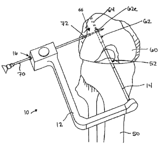
[0017] FIG. 1 illustrates a human knee and a guide device having a cannulated
guide rod
-5-
CA 02571736 2009-06-08
extending through bone tunnels formed in the tibia and femur;
[0018] FIG. 2 illustrates the human knee and guide device of FIG. 1, showing a
sleeve and trocar
assembly being inserted through the femur to form a transverse tunnel that
intersects the femoral
tunnel;
[0019] FIG. 3 illustrates the human knee of FIG. 2 with the guide device and
the trocar removed,
leaving the sleeve extending into the transverse tunnel, and a ligament graft
extending through
the tibial tunnel and into the femoral tunnel;
[0020] FIG. 4 illustrates the human knee of FIG. 3, showing a syringe inserted
through the sleeve
in the transverse tunnel and injecting an adhesive into the femoral tunnel;
[0021 ] FIG. 5 illustrates the human knee of FIG. 4, showing the ligament
graft pulled further into
the femoral tunnel and into contact with the adhesive such that the adhesive
surrounds an end of
the ligament graft;
-5a-
CA 02571736 2006-12-19
[0022] FIG. 6 illustrates the human knee of FIG. 5, showing a cross-pin being
inserted through
the sleeve, into the transverse tunnel, and through the ligament graft to
maintain the ligament
graft in a fixed position within the femoral tunnel;
[0023] FIG. 7 illustrates the human knee of FIG. 6 showing the sleeve removed
leaving the
cross-pin in place;
[0024] FIG. 8 illustrates the human knee of FIG. 7 showing the cross-pin
removed after the
adhesive has cured; and
[0025] FIG. 9A is a side perspective view of one exemplary embodiment of an
anchor
configured to mate to a bone graft and to be affixed within a bone tunnel;
[0026] FIG. 9B is a side perspective view of the anchor shown in FIG. 9A mated
to a bone graft
with a ligament graft extending therefrom;
[0027] FIG. 9C is an illustration showing the anchor, bone graft, and ligament
graft of FIG. 9B
implanted within a bone tunnel in bone, showing space in the bone tunnel for
receiving an
adhesive;
[0028] FIG. 10 is a side perspective view of another embodiment of an anchor
configured to
mate to a bone graft and to be affixed within a bone tunnel to anchor the bone
graft within the
bone tunnel, the anchor having annual grooves for receiving an adhesive;
[0029] FIG. 11 A is a side view of another embodiment of an anchor and a bone
graft, each
having suture bores formed therein for mating the bone graft to the anchor;
[0030] FIG. 11 B is another side view of the anchor and bone graft shown in
FIG. 11 A;
[0031 ] FIG. 11 C is a side view of the anchor and bone graft shown in FIGS.
11 A and 11 B mated
to one another using sutures;
[0032] FIG. 12A is a perspective view of one exemplary embodiment of a bone
graft modified to
have a cross-shaped cut-out for receiving adhesive therein;
[0033] FIG. 12B is a perspective view of another exemplary embodiment of a
bone graft
-6-
CA 02571736 2006-12-19
modified to have annular grooves for receiving adhesive therein;
[0034] FIG. 12C is a perspective view of yet another exemplary embodiment of a
bone graft
modified to have thru-bores features for receiving adhesive therethrough;
[0035] FIG. 12D is a perspective view of another exemplary embodiment of a
bone graft
modified to have a post formed thereon for receiving adhesive therearound; and
[0036] FIG. 13 is a perspective view of yet another embodiment of an anchor
having surface
features for receiving adhesive therearound, and having a mating element for
mating to a
ligament graft.
DETAILED DESCRIPTION OF THE INVENTION
[0037] The present invention provides methods and devices for fixing a
ligament graft in a bone
tunnel. The various methods and devices disclosed herein can be used in a
variety of surgical
procedures, however the methods and devices are particularly useful for
repairing an anterior
cruciate ligament (ACL) in a human knee. In an ACL repair, the torn ACL is
replaced with a
ligament graft which is anchored to the tibia and femur. The term "ligament
graft," as used
herein, is intended to include natural materials, such as autografts,
allografts, and xenografts,
including harvested ligaments and tendons, as well as synthetic materials. The
ligament graft
can also include an anchoring element attached thereto for anchoring the graft
to the tibia and
femur. For example, the ligament graft can include a bone graft, plug, or
other member, attached
to one or both terminal ends thereof. The term "bone graft," as used herein,
in intended to
include natural materials, such as autografts, allografts, and xenografts, as
well as synthetic
materials.
[0038] A person skilled in the art will appreciate that the various methods
and devices disclosed
herein can be used in a variety of surgical procedures, and that the
particular configuration of the
ligament graft can vary depending on the intended use, and virtually any
ligament graft known in
the art can be used with the methods disclosed herein.
[0039] FIGS. 1-8 illustrate one exemplary method for anchoring a ligament
graft in a bone
tunnel, and in particular in femoral and tibial tunnels of a human knee. In
general, the method
-7-
CA 02571736 2006-12-19
includes drilling a tunnel in bone and inserting a ligament graft at least
partially into the tunnel.
An adhesive is introduced into the tunnel, and a fixation device is inserted
through the ligament
graft in a direction transverse to an axis of the tunnel to maintain the
ligament graft in contact
with the adhesive. The fixation device can be removed when the adhesive is
cured such that the
ligament graft is fixed in the tunnel. The use of a fixation device to
maintain the ligament graft
in the tunnel while the adhesive cures allows an opposed end of the ligament
graft to be
tensioned and fixed using the same or a different anchoring technique. The
method is described
in connection with certain exemplary procedures for preparing bone tunnels and
inserting a
ligament graft into the bone tunnels. For example, in certain exemplary
methods, the ligament
graft is affixed in the femoral tunnel prior to affixing it in the tibial
tunnel. However, a person
skilled in the art will appreciate that the ligament graft can be affixed in
the tibial tunnel first or
at the same time. A person skilled in the art will also appreciate that a
variety of other
procedures known in the art can be used to prepare the bone tunnels and to the
insert a ligament
graft into the bone tunnels.
[0040] Referring first to FIG. 1, a bone tunnel is drilled through the tibia
50 and femur 60, using
conventional surgical equipment and techniques, to form a tibial tunnel 52 and
femoral tunnel
62. The tibial and femoral tunnels 52, 62 can extend completely through the
tibia and femur 50,
60, however in an exemplary embodiment the femoral tunnel 62 terminates part
way through the
femur 60 such that a femoral socket is formed. A suture tunnel 64 can
optionally extend through
the remainder of the femur 60 in longitudinal alignment with the femoral
tunnel 62 to allow a
suture to pass therethrough and pull and graft into the tunnels 52, 62, as
will be discussed in
more detail below. The suture tunnel 64 preferably has an inner diameter that
is less than an
inner diameter of the femoral tunnel, such that a terminal end of the femoral
tunnel defines an
end wall 62e.
[0041 ] Once the tibial and femoral tunnels 52, 62 are prepared, a transverse
tunnel can be
formed in the femur. The transverse tunnel preferably intersects the femoral
tunnel 62 such that
a pathway is formed through the transverse tunnel to the femoral tunnel 62.
This pathway can be
used to inject an adhesive into the femoral tunnel 62, as will be discussed in
more detail below.
As shown in FIG. 1, a guide device 10 can optionally be used to locate and
align the transverse
tunnel. As shown, the guide device 10 generally includes an L-shaped frame 12
having a
-8-
CA 02571736 2006-12-19
cannulated guide rod 14 extending from one end thereof, and a housing with an
opening 16
formed therethrough on an opposed end thereof. The cannulated guide rod 14 can
be inserted
through the tibial tunnel 52 and the femoral tunnel 62 to maintain the guide
frame 12 in a fixed
position. As shown in FIG. 2, a sleeve and trocar assembly 70, 72 can then be
inserted through
the opening 16 in the housing on the guide frame 12, and drilled into the
lateral side of the femur
until the sleeve and trocar abut the cannulated guide rod 14. The transverse
tunnel 66 will thus
extend through the femur and into the femoral tunnel 62, preferably at a
location substantially
adjacent to the distal end wall 62e of the femoral tunnel. A person skilled in
the art will
appreciate that the transverse tunnel 66 can extend at any angle relative to
the femoral tunnel 62.
After the transverse tunnel 66 is completely formed, the trocar 72 can be
removed leaving the
sleeve 70 in place, as shown in FIG. 3. The sleeve 70 can be inserted
partially into the transverse
tunnel 66, such that it does not extend into the femoral tunnel 62, or
alternatively the sleeve 70
can at least partially extend into the femoral tunnel 62. This will allow the
sleeve 70 to function
as a stop for the ligament graft, as will be discussed in more detail below.
The procedure can,
however, be performed without the use of the sleeve 70.
[0042] After the tibial, femoral, and transverse tunnels 52, 62, 66 are
formed, a ligament graft 80
can be introduced into the tibial and femoral tunnels 52, 62. While various
procedures known in
the art can be used to introduce the ligament graft 80 into the tibial and
femoral tunnels 52, 62, in
one exemplary embodiment a suture 82 can be attached to a leading end of the
ligament graft 80
and it can be threaded through the tibial and femoral tunnels 52, 62 using,
for example, a guide
pin or other device. Tension can then be applied to the suture to pull the
ligament graft 80 up
through the tibial tunnel 52 and at least partially into the femoral tunnel
62. As shown in FIG. 3,
the ligament graft 80 can be pulled to a location that is spaced a distance
apart from the end wall
62e of the femoral tunnel, such that the ligament graft 80 stops short of the
transverse tunnel 66.
Where the sleeve 70 extends into the femoral tunnel 62, the sleeve 70 will
prevent the ligament
graft 80 from being fully advanced into contact with the end surface 62e of
the femoral tunnel
62.
[0043] As shown in FIG. 4, once the ligament graft 80 is positioned within the
femoral tunnel 62
the surgeon can inject an adhesive 98 into the femoral tunnel 62 to secure the
ligament graft 80
therein. While the adhesive 98 can be injected through any of the tunnels
using a variety of
-9-
CA 02571736 2006-12-19
techniques, in the illustrated exemplary embodiment the adhesive 98 is
injected through the
transverse tunnel 66 using a syringe 90. In particular, the syringe 90 is
placed through the sleeve
70 in the transverse tunnel 66 and into the femoral tunnel 62, and a plunger
92 on the syringe 90
is then moved into a barrel 94 to eject the adhesive 98 from the barrel 94,
through the needle 96,
and into the femoral tunnel 62. The amount of adhesive 98 can vary, but in an
exemplary
embodiment, the adhesive 98 fills a terminal end portion of the femoral tunnel
62 adjacent to the
end surface 62e.
[0044] Once the terminal end portion of the femoral tunnel 62 is filled with
the adhesive 98, the
suture 82 is further tensioned to pull the ligament graft 80 into the terminal
end portion such that
the ligament graft 80 abuts the end wall 62e of the femoral tunnel 62, as
shown in FIG. 5. As a
result, the ligament graft 80 is brought into contact with the adhesive 98,
which is spread in and
around the ligament graft 80. The adhesive 98 thus extends between the end
surface 62e and the
ligament graft 80, and forms a cap that surrounds the ligament graft 80, and
optionally that
extends into portions of the ligament graft 80. In other embodiments, the
ligament graft 80 can
be fully pulled into the femoral tunnel 62 and into contact with the end
surface 62e, and then the
adhesive 98 can be injected into the tunnel 62 until it completely surrounds
and encapsulates an
end portion of the ligament graft 80.
[0045] Since the adhesive 98 can take time to cure, the first end of the
ligament graft 80 can be
temporarily secured in the femoral tunnel 62 using a fixation device. FIG. 6
illustrates one
exemplary embodiment of a fixation device in the form of a cross-pin 100 that
is used to secure
the ligament graft 80 in the femoral tunnel 62. As shown, the cross-pin 100 is
in the form of an
elongate member that is inserted through the sleeve 70 in the transverse
tunnel and through a
loop formed in the terminal end of the ligament graft 80. The cross-pin 100
can also extend
across the femoral tunnel 62 and into a further portion of the transverse
tunnel. In an exemplary
embodiment, the cross-pin 100 has a length that allows a portion of the cross-
pin 100 to remain
outside of the sleeve 70 while the remainder of the cross-pin 100 extends
through the transverse
tunnel 66, through the graft 80, and completely across the femoral tunnel 62.
Once the ligament
graft 80 is temporarily secured within the femoral tunnel 62 using the cross-
pin 100, the sleeve
70 can be removed leaving the cross-pin 100 in place, as shown in FIG. 7. The
second end of the
ligament graft 80 can also be tensioned and secured within the tibial tunnel
52 using the same
-10-
CA 02571736 2006-12-19
anchoring technique, or using other anchoring techniques known in the art.
When the adhesive
98 is finally cured, the cross-pin 100 can be removed leaving the ligament
graft fixed within the
femoral tunnel 62, as shown in FIG. 8.
[0046] A person skilled in the art will appreciate that the cross-pin 100 can
have a variety of
configurations, and that various other fixation devices known in the art can
be used. For
example, while an elongate rod is shown, in other embodiments the fixation
device can be in the
form of a screw or other anchoring element. The fixation device can also be
formed from a
variety of materials, but in certain exemplary embodiments it is preferably
formed from or
coated with a material that prevents adhesion to the adhesive. For example,
the fixation device
can be formed from or coated with a fluoropolymer plastic resin, such as
Teflon . A person
skilled in the art will appreciate that a variety of other materials can be
used to prevent adhesive
between the cross-pin and the adhesive. The sleeve 70 can also optionally be
formed from or
coated with a protective material that prevents adhesive to the adhesive.
[0047] The materials used to form the adhesive can also vary, and virtually
any bone glue or
cement known in the art can be used. Since a cross-pin is temporarily used to
maintain the
ligament graft within the femoral tunnel while the adhesive dries, the drying
time for the
adhesive can significantly vary and the surgeon has more freedom to select a
desired adhesive.
The adhesive can also be bioabsorbable or non-absorbable. In certain exemplary
embodiments,
the adhesive preferably has a viscosity that allows it to be injected into the
femoral tunnel
without the adhesive dripping, but that allows it to spread around the
ligament graft as the graft is
pulled fully into the femoral tunnel. By way of non-limiting example,
exemplary adhesives
include bone glues, such as biocompatible bone glues including 2-octyl
cyanoacrylate and the
like and equivalent thereof, bone cements, such as conventional biocompatible
bone cements
including polymethylmethacrylate and the like, as well as dental implant
cements such as
Premiere Dental Implant Cement .
[0048] As previously indicated, the ligament graft can also have a variety of
configurations, and
it can be directly affixed within the bone tunnel, or an anchoring element can
be used to affix the
ligament graft within the bone tunnel. FIGS. 9A-13 illustrates various
exemplary embodiments
of ligaments grafts. In the embodiments shown in FIGS. 9A-11C, each ligament
graft is in the
-11-
CA 02571736 2006-12-19
form of a bone-to-bone graft that is mated to an anchoring element having
surface features that
facilitate adhesion thereof within a bone tunnel. In the embodiments shown in
FIGS. 12A-12D,
each ligament graft is also in the form of a bone-to-bone graft, however,
rather than using an
anchoring element, the bone graft is modified to include surface features to
facilitate adhesive
thereof within a bone tunnel. FIG. 13 illustrates yet another embodiment of a
ligament graft in
the form of a soft tissue graft that is attached to an anchoring element
having surface features
adapted to facilitate adhesive thereof within a bone tunnel. A person skilled
in the art will
appreciate that the ligament graft can have a variety of other configurations,
and that any
combination of these features or other features known in the art can be used.
Moreover, the
various ligament grafts can be implanted using the techniques previously
described herein, or
they can be implanted using various other techniques known in the art.
[0049] As indicated above, in the embodiment shown in FIGS. 9A-9C, an
anchoring element
110 can be used to affix a bone-to-bone graft 120 within a bone tunnel. While
the anchoring
element 110 can have a variety of configurations, in one exemplary embodiment
it preferably
includes a first portion 112 that is adapted to be adhesively mated to a bone
tunnel, and a second
portion 114 that extends from the first portion 112 and that is adapted to
mate to a bone graft 122
of a bone-to-bone graft 120.
[0050] The first portion 112 can have a variety of configurations, and various
techniques can be
used to facilitate adhesion between the first portion 112 and a bone tunnel.
In the illustrated
embodiment, the first portion 112 is in the form of a post having an eyelet
formed on a terminal
end thereof. The eyelet includes an opening 113 formed therein for receiving a
suture which can
be used to pull the anchor 110 and ligament graft 120 attached thereto into a
bone tunnel. The
eyelet, as well as the post, can also facilitate adhesion of the first portion
112 to a bone tunnel, as
an adhesive will surround the post and extend into the eyelet, as shown in
FIG. 9C. A person
skilled in the art will appreciate that the first portion can have a variety
of other shapes, and it
can include a variety of other features formed thereon, such as grooves,
bores, protrusions, etc.
[0051 ] The second portion 114 of the anchor 110 can also have a variety of
configurations, and
various techniques can be used to mate the second portion 114 to a bone graft
122. As shown in
FIGS. 9A-9C, the second portion 114 includes threads 114a formed thereon for
allowing the
-12-
CA 02571736 2009-06-08
second portion 114 to be threaded into the bone graft 120, as shown in FIG.
9B. Other mating
techniques, including adhesives, can be used to mate the second portion 114 to
the bone graft
122.
[0052] FIG. 10 illustrates another embodiment of an anchor 130 that is adapted
to mate to a bone
graft, and that is adapted to be affixed within a bone tunnel using an
adhesive. The anchor 130 is
similar to the anchor 110 shown in FIG. 9A as it includes a first portion 132
having a post and an
eyelet, and a second portion 134 adapted to mate to a bone graft. In this
embodiment, however,
the second portion 134 includes annular cut-out regions 116 formed along a
length thereof for
receiving an adhesive. The adhesive can be injected into and around the cut-
out regions 116 and
the second portion 134 can be inserted into a bore drilled in a bone graft to
adhere the second
portion 134 to the bone graft.
[0053] FIGS. 11A-11C illustrate yet another embodiment of an anchor 140
adapted to mate to a
bone graft 152 of a bone-to-bone ligament graft, and to be adhered within a
bone tunnel. Similar
to the embodiments shown in FIGS. 9A-10, the anchor 140 includes a first
portion 142 having a
post and an eyelet formed thereon, and a second portion 144 that is adapted to
mate to a bone
graft. In this embodiment, the second portion 144 includes opposed arms 145a,
145b that are
adapted to receive a bone graft 152 therebetween. While the arms 145a, 145b
can optionally be
adhesively mated to the bone graft 152, the arms 145a, 145b can also or
alternatively be sutured
to the bone graft 152. Thus, the opposed arms 145a, 145b can each include one
or more thru-
bores formed therein. FIGS. 11A-11C illustrate two thru-bores 144a, 144b
formed in each arm
145a, 145b. Two corresponding thru-bores 152a, 152b can also be drilled
through the bone graft
152 to allow suture 146a, 146b to extend through the bone graft 152 to attach
the bone graft 152
to the arms 145a, 145b, as shown in FIG. 11C. A person skilled in the art will
appreciate that
various other techniques can be used to mate the bone graft 152 to the arms
145a, 145b, and that
the arms 145a, 145b can have a variety of other configurations. For example,
the arms 145a,
145b can threadably engage the bone graft 152.
[0054] In use, the anchoring elements 110, 130, 140 of FIGS. 9A-11C can be
implanted within a
bone tunnel using various methods known in the art. In an exemplary
embodiment, however, the
anchors 110, 130, 140 are affixed within a bone tunnel using the exemplary
methods previously
-13-
CA 02571736 2006-12-19
described herein. In particular, after the anchoring element 110, 130, 140 is
attached to a bone
graft, the anchoring element 110, 130, 140 can be pulled through the tibial
tunnel and at least
partially into the femoral tunnel. This can be achieved using various
techniques, for example, by
tensioning a suture that is attached to the eyelet of the anchor. An adhesive,
e.g., adhesive 98 as
shown in FIG. 9C, can then be introduced into the femoral tunnel, preferably
via the transverse
tunnel, to fill a terminal end portion of the femoral tunnel. The anchor 110,
130, 140 can be
further pulled into the femoral tunnel, causing the adhesive to spread around
the first portion of
the anchor 110, 130, 140. Alternatively, the anchor 110, 130, 140 can be fully
pulled up into the
femoral tunnel prior to introducing the adhesive. While not necessary, a cross-
pin can optionally
be inserted through the hole in the eyelet to maintain the anchor 110, 130,
140, and thus the
ligament graft, within the femoral tunnel until the adhesive cures. Where a
cross-pin is not used,
a fast drying adhesive is preferably used to allow the other end of the
ligament to be anchored in
the tibial tunnel without having to wait an extended period of time for the
adhesive to cure.
[0055] In other embodiments, as previously discussed, the bone graft itself
can be modified to be
adhesively affixed within a bone tunnel. FIGS. 12A-12D illustrate various
exemplary
embodiments of such bone grafts. In general, each bone graft, which includes a
ligament
attached thereto, is modified to include surface features formed thereon for
receiving an adhesive
therein. Such a configuration allows the adhesive to completely surround and
extend into the
bone graft, thereby providing a secure connection between the bone graft and
the bone tunnel.
The shape, quantity, size, and configuration of the surface features can vary.
FIG. 12A illustrates
one embodiment of a bone graft 160 having a cross-shaped groove 162 formed in
a terminal end
thereof. The grooves can also extend along at least a portion of the sidewall
of the bone graft
160, as shown. In the embodiment shown in FIG. 12B, the bone graft 170
includes annular cut-
out portions 172 formed therearound and spaced apart from one another along at
least a portion
of a length of the bone graft 170. FIG. 12C illustrates yet another embodiment
of a bone graft
180 having thru-bores 182 extending therethrough for receiving the adhesive
therein. In yet
another embodiment, shown in FIG. 12D, the bone graft 190 is modified to
include a reduced
diameter region 192 such that the bone graft includes a small post formed
thereon. In each of the
embodiments, the cut-out portions can be formed using instruments and
techniques known in the
art. In use, the cut-out portions allow the adhesive to extend therein and
surround the bone graft,
thereby mating the bone graft to the bone tunnel. The bone grafts can be
implanted using
-14-
CA 02571736 2009-06-08
techniques previously described, or using other techniques known in the art.
[0056] FIG. 13 illustrates another embodiment of a ligament graft. In this
embodiment, the
ligament graft is in the form of a soft tissue graft 210 that does not include
a bone graft. The soft
tissue graft is mated directly to an anchor 200. The configuration of the
anchor 200 can vary,
and various techniques can be used to mate the soft tissue graft 210 to the
anchor 200. In the
illustrated embodiment, the anchor 200 generally includes a first portion 202
that is adapted to
adhesively mate to a bone tunnel, and a second portion 204 that is adapted to
mate to the soft
tissue graft 210. The configuration of the first portion 202 can vary, but it
preferably includes
features that facilitate mating thereof to a bone tunnel. Any of the features
previously described
can be used herein, including annular cut-out portions, as shown. In this
embodiment, the cut-
out portions define flanges 206 which can help maintain the anchor 200 within
the bone tunnel,
at least temporarily until an adhesive is disposed therearound and cured to
lock the anchor 200 in
place. The second portion 204 of the anchor can also vary, and it can have
virtually any shape
and size that facilitates attachment of a soft tissue graft 210 thereto. As
shown in FIG. 13, the
second portion 204 includes an eyelet 207 formed thereon for receiving the
soft tissue graft 210.
The second portion 204 also includes a wedge-shaped body 208 extending
distally therefrom.
The wedge-shaped body 208 can help prevent back-out of the anchor 200 once the
anchor is
inserted in a bone tunnel. In particular, when tension is applied to the soft
tissue graft 210, the
soft tissue graft 210 will cause the anchor 200 to pivot. As a result, the
wedge-shaped body 208
will extend into and engage the wall of the bone tunnel, thereby preventing
movement of the
anchor 200 within the bone tunnel. A person skilled in the art will appreciate
that the anchor 200
can have a variety of other configurations, and that it does not need to
include any features to
facilitate back-out or to maintain the anchor 200 in a fixed position, as an
adhesive can be used
to lock the anchor 200 within the bone tunnel. In use, the anchor 200 can be
implanted using
techniques previously described, or using other techniques known in the art.
[0057] One of ordinary skill in the art will appreciate further features and
advantages of the
invention based on the above-described embodiments. Accordingly, the invention
is not to be
limited by what has been particularly shown and described, except as indicated
by the appended
claims.
-15-