Note: Descriptions are shown in the official language in which they were submitted.
CA 02571784 2006-12-21
WO 2006/007211 PCT/US2005/018660
MULTI-JOINT IMPLANT
CROSS-REFERENCE TO RELATED APPLICATIONS
The present invention claims priority to U.S. Provisional Application Serial
No.
60/584,022, filed on June 30, 2004 and entitled "Multi-Joint (Disc & Facet)
Implant,"
which is hereby incorporated by reference in its entirety.
FIELD OF THE INVENTION
The present invention relates to spinal implants and methods.
BACKGROUND OF THE INVENTION
The vertebrae in a patient's spinal column are linked to one another by the
disc
and the facet joints, which control movement of the vertebrae. Each vertebra
has a pair
of articulating surfaces located on the left side, and a pair of articulating
surfaces located
on the right side, and each pair includes a superior articular surface, which
faces upward,
and an inferior articular surface, which faces downward. Together the superior
and
inferior articular surfaces of adjacent vertebra form a facet joint. Facet
joints are
synovial joints, which means that each joint is surrounded by a capsule of
connective
tissue and produces a fluid to nourish and lubricate the joint. The joint
surfaces are
coated with cartilage allowing the joints to move or articulate relative to
one another. In
combination with the intervertebral disc, the two facet joints form the spinal
three-joint
complex.
Diseased, degenerated, impaired, or otherwise painful facet joints and/or
discs
can require surgery to relieve pain or restore function to the three-joint
complex.
Subsequent surgery may also be required after a laminectomy, as a laminectomy
predisposes the patient to instability and may lead to post-laminectomy
kyphosis
(abnormal forward curvature of the spine), pain, and neurological dysfunction.
Current
clinical data have suggested that degeneration of one member of the three
joint complex,
that is either the discs or the facet joints, contributes to the degeneration
of the other.
While implants are available for replacing either a diseased disc or the facet
joints, there
are no implants that can be used to replace the entire spinal three-joint
complex.
CA 02571784 2006-12-21
WO 2006/007211 PCT/US2005/018660
-2-
Accordingly, there remains a need for improved systems and methods for
repairing and/or replacing the spinal three-joint complex.
SUMMARY OF THE INVENTION
The present invention provides various methods and devices for repairing or
replacing damaged, injured, diseased, or otherwise unhealthy posterior
elements, such as
the facet joints, the laniina, the posterior ligaments, and/or other features
of a patient's
spinal column. In one exemplary embodiment, a spinal implant is provided
having an
anterior portion that is adapted to be positioned between adjacent vertebrae,
and a
posterior portion that is adapted to be positioned around a spinal cord and to
couple to at
least one adjacent vertebra.
The implant can have a variety of shapes and sizes, but in one exemplary
embodiment the implant is substantially C-shaped such that the posterior
portion is
curved or semi-circular with an opening formed therein and the anterior
portion includes
opposed arms that extend from the posterior portion. The posterior and
anterior portions
of the implant can have a variety of configurations, but in one exemplary
embodiment
the anterior portion of the implant has a shape that is adapted to allow
articulation of
adjacent vertebrae relative to one another, and the posterior portion of the
implant
includes at least one extension member extending therefrom and adapted to mate
to a
posterior surface of a vertebra. The extension member(s) can have a variety of
configurations, and it can be rigidly or movably coupled to the implant. In
one
exemplary embodiment, the extension member(s) is an elongate member having at
least
one thru-bore formed therein for receiving a fastening element adapted to mate
the
extension member(s) to a vertebra. The implant can also include any number of
extension members, but in one exemplary embodiment the implant includes first
and
second extension members extending from opposed lateral sides of the posterior
portion
of the implant. The first and second extension members can extend in a
superior
direction from a superior surface of the posterior portion of the C-shaped
member, or
alternatively they can extend in an inferior direction from an inferior
surface of the
posterior portion of the implant. In another exemplary embodiment, the implant
can
include first and second superior extension members extending in a superior
direction
from opposed lateral sides of the posterior portion of the implant, and first
and second
WO 2006/007211 PCT/US2005/018660
CA 02571784 2006-12-21
-3-
inferior extension members extending in-an-inferior direction from opposed
lateral sides
of the posterior portion of the implant.
In another exemplary embodiment, the implant can include a substantially C-
shaped superior member, a substantially C-shaped inferior niember, and at
least one
vi central member disposed therebetween. In certain embodiments, the implant
can include
first and second central members disposed between the anterior portion of the
superior
and inferior members, and third and fourth central members disposed between
the
posterior portion of the superior and inferior members. In use, the central
meinber(s)
can be adapted to articulate between the superior and inferior members. In
other
embodiments, the central member(s) can be compressible to allow movement of
the
superior and inferior members relative to one another.
The present invention also provides exemplary methods for stabilizing adjacent
vei-tebrae. In one embodiment, the method can include positioning a posterior
portion of
an itnplant around a spinal cord and between resected facets of adjacent
vertebrae. The
posterior portion of the implant can be adapted to couple to at least one of
the adjacent
vertebrae. The method can further include positioning an anterior portion of
the implant
between the adjacent vertebrae such that the adjacent vertebrae are adapted to
move
relative to one another.
In another exemplary enlbodiment, a method for stabilizing adjacent vertebrae
is
provided and includes accessing a patient's spinal column using a posterior
surgical
approach, removing a disc disposed between adjacent vertebrae, posteriorly
positioning
an implant around a spinal cord, between facet joints of adjacent vertebrae,
and between
the adjacent vertebrae, and coupling a posterior portion of the implant to at
least one of
the adjacent vertebrae. In an exemplary embodiment, the posterior portion of
the
implant is coupled to at least one of the adjacent vertebrae by inserting at
least one bone
screw through at least one extension formed on the posterior portion of the
implant and
into the vertebra.
Another aspect of the present invention is a use of the implant described
above for stabilizing adjacent vertebrae.
BRIEF DESCRIPTION OF THE DRAWINGS
The invention will be more fully understood from the following detailed
description taken in conjunction with the accompanying drawings, in which:
CA 02571784 2006-12-21
WO 2006/007211 PCT/US2005/018660
-4-
FIG. 1A is a perspective view of one exemplary embodiment of a spinal implant
having a unitary disc replacement member with first and second facet
replacement
members movably coupled thereto;
FIG. 1B is a cross-sectional view of the spinal implant shown in FIG. lA taken
across line B-B;
FIG. 1C is a superior perspective view of the spinal implant shown in FIG. lA
implanted between adjacent vertebrae, showing only the inferior vertebra;
FIG. 1D is a posterior perspective view of the spinal implant shown in FIG. lA
implanted between adjacent superior and inferior vertebrae;
FIG. 2A is an anterior perspective view of another exemplary embodiment of a
spinal implant having a multi-piece construction;
FIG. 2B is a superior cross-sectional view, taken in the axial plane, of the
spinal
implant shown in FIG. 2A implanted between adjacent vertebrae, showing only
the
inferior vertebra;
FIG. 2C is a superior perspective view of the spinal implant shown in FIG. 2A
implanted between adjacent vertebrae, showing only the inferior vertebra;
FIG. 2D is a posterior perspective view of the spinal implant shown in FIG. 2A
implanted between adjacent superior and inferior vertebrae;
FIG. 3A is an anterior perspective view of yet another exemplary embodiment of
a spinal implant having a multi-piece construction with extensions members
formed
thereon; and
FIG. 3B is a posterior perspective view of the spinal implant shown in FIG. 3A
implanted between and coupled to adjacent superior and inferior vertebrae.
CA 02571784 2006-12-21
WO 2006/007211 PCT/US2005/018660
-5-
DETAILED DESCRIPTION OF THE INVENTION
Certain exemplary embodiments will now be described to provide an overall
understanding of the principles of the structure, function, manufacture, and
use of the
devices and methods disclosed herein. One or more examples of these
embodiments are
illustrated in the accompanying drawings. Those of ordinary skill in the art
will
understand that the devices and methods specifically described herein and
illustrated in
the accompanying drawings are non-limiting exemplary embodiments and that the
scope
of the present invention is defined solely by the claims. The features
illustrated or
described in connection with one exemplary embodiment may be combined with the
features of other embodiments. Such modifications and variations are intended
to be
included within the scope of the present invention.
The present invention provides various methods and devices for repairing or
replacing damaged, injured, diseased, or otherwise unhealthy posterior
elements, such as
the facet joints, the lamina, the posterior ligaments, and/or other features
of a patient's
spinal column. In one exemplary embodiment, an implant is provided having an
anterior
portion that is adapted to be positioned between adjacent vertebrae and a
posterior
portion that is adapted to be positioned around a spinal cord and to couple to
at least one
adjacent vertebra. In use, the implant can allow the adjacent vertebrae to
move relative
to one another, thereby restoring normal function to the vertebrae.
FIGS. lA-1B illustrate one exemplary embodiment of a spinal implant 10 for
replacing or repairing a damaged spinal disc, the facet joints, and optionally
other
posterior elements of the spine. In general, the implant 10 includes a
posterior portion
12a that is adapted to be positioned around the spinal cord, and an anterior
portion 12b
that is adapted to be positioned between adjacent vertebrae. The anterior and
posterior
portions 12a, 12b are collectively referred to herein as a disc replacement
component 12.
The implant 10 can also include a facet replacement component 20 that is
adapted to
couple the disc replacement component 12 to at least one adjacent vertebrae.
In use, the
facet replacement component 20 can move relative to the disc replacement
component
12 to allow the adjacent vertebrae to move relative to one another, thereby
restoring
normal function to the vertebrae.
CA 02571784 2006-12-21
WO 2006/007211 PCT/US2005/018660
-6-
The disc replacement component 12 can have a variety of configurations, but in
the illustrated exemplary embodiment it is substantially U-shaped or C-shaped.
A
person skilled in the art will appreciate that the terms "U-shaped" or "C-
shaped" are
intended to include any implant having a generally or partially curved
structure with an
opening in one side thereof. Further, these terms are intended to include any
implant
that has an open anterior portion and a posterior portion that can be disposed
around a
spinal cord, and an anterior portion that can be disposed between adjacent
vertebrae.
The shape and configuration of the implant is not intended to be limited to
only a U- or
C-shaped configuration.
The posterior portion 12a of the disc replacement component 12 can have a
variety of shapes and sizes, but in the illustrated exemplary embodiment the
posterior
portion 12a is in the form of a substantially semi-circular member having a
relatively
large central opening 17a formed therein. Such a shape allows the posterior
portion 12a
to be positioned around the spinal cord in a patient's spinal column. The
posterior
portion 12a can also have a relatively low profile, so as to allow the
posterior portion
12a to be positioned between the spinous processes of adjacent vertebrae.
The anterior portion l2b of the disc replacement component 12 can also have a
variety of shapes and sizes, but in the illustrated exemplary embodiment the
anterior
portion 12b of the disc replacement component 12 includes opposed arms 14, 16
that
extend from the posterior portion 12a in a substantially parallel arrangement
and that
define an opening 17b therebetween. The opening 17b between the arms 14, 16
can be
smaller than the opening 17a at the posterior portion 12a of the disc
replacement
component 12, but it is preferably large enough to allow the spinal cord to
pass
therethrough when the implant 10 is being implanted. While the shape and size
of each
arm 14, 16 can vary, in one exemplary embodiment each arm 14, 16 is in the
form of a
lobe that extends from the posterior portion 12a, and that has a height hl, h2
that is
greater than a height h3 of the posterior portion 12a, and a width w1, wz that
is greater
than a width w3 of the posterior portion 12a of the implant 10, as shown in
FIGS. 1A and
1B. Such a configuration allows the arms 14, 16 to occupy additional space
between the
adjacent vertebrae, thereby providing sufficient support for the vertebrae.
CA 02571784 2006-12-21
WO 2006/007211 PCT/US2005/018660
-7-
The opposed arms 14, 16 can also include a variety of other features that can
vary depending on the intended use and desired result once implanted. For
example, in
one exemplary embodiment each arm 14, 16 can have a shape that is adapted to
allow
the adjacent vertebrae to articulate relative thereto. For example, as shown
in FIGS. 1A
and 1B, each arm 14, 16 includes curved or domed superior and inferior
surfaces 14s,
16s, 14i, 16i. The domed surfaces 14s, 16s, 14i, 16i can be formed on any
portion of
each arm 14, 16, but in one exemplary embodiment the domed surfaces 14s, 16s,
14i,
16i are formed along the anterior portion 12b of the implant 10 adjacent to
the terininal
end of each arm 14, 16. As a result, when the arms 14, 16 are positioned
between
adjacent vertebrae, the domed surfaces 14s, 16s, 14i, 16i will be
substantially centrally
located relative to the adjacent vertebrae, thereby allowing the vertebrae to
articulate
relative thereto. While domed surfaces 14s, 16s, 14i, 16i are shown, the arms
14, 16 can
have a variety of other configurations to allow articulation of adjacent
vertebrae. For
example, each arm 14, 16 can include a ball or other member movably disposed
therein
or coupled thereto.
In another exemplary embodiment, the opposed arms 14, 16 can be adapted to
engage the adjacent vertebrae. Techniques for mating the arms 14, 16 to
adjacent
vertebrae include, by way of non-limiting example, surface features, such as
teeth, that
engage the endplates of the vertebrae, surface coatings or materials that
allow bone
growth into the implant 10 to occur, or other materials or features that will
engage the
adjacent vertebrae.
In an exemplary embodiment, where engagement features are included, at least a
portion of the implant 10 is preferably compressible to allow movement between
the
adjacent vertebrae. For example, the arms 14, 16, or a portion of the arms 14,
16, can be
compressible by forming the arms 14, 16 from a compressible material,
embedding a
compressible material in the arms 14, 16, or by coupling a compressible
material to a
portion of the arms 14, 16. Suitable compressible materials include, by way of
non-
limiting example, biocompatible polymers and metals.
As previously indicated above, the implant 10 can also include a facet
replacement component 20 that is adapted to couple the disc replacement
component 12
to at least one adjacent vertebrae. While the facet replacement component 20
can have a
variety of configurations, and it can be formed integrally with, coupled to,
or be separate
CA 02571784 2006-12-21
WO 2006/007211 PCT/US2005/018660
-8-
from the disc replacement component 12, in one exemplary embodiment, as shown
in
FIGS. 1A and 1B, the facet replacement component 20 is in the form of first
and second
lateral extensions 20a, 20b that extend from the disc replacement component
12. The
lateral extensions 20a, 20b are coupled to opposed sides of the posterior
portion 12a of
the disc replacement component 12, and they can extend in either a superior
direction, as
shown, to couple to a superior vertebra, or they can extend in an inferior
direction. A
person skilled in the art will appreciate that the implant 10 can include any
number of
lateral extensions extending in any direction to facilitate attachment thereof
to one or
more vertebrae.
Each extension 20a, 20b can be coupled to the disc replacement member 12
using a variety of techniques. In one exemplary embodiment, as shown, the
extensions
20a, 20b are movably coupled to the disc replacement member 12 to allow the
extensions 20a, 20b to pivot with respect to the disc replacement member 12 as
the
adjacent vertebrae move. A movable connection can be forined using, for
example, a
ball and socket joint, a polyaxial joint, flexible extensions 20a, 20b, etc.
Alternatively,
the extensions 20a, 20b do not need to be connected to the disc replacement
member 12,
but rather they can merely be disposed within corresponding recesses or
sockets formed
within the disc replacement member 12, as shown in FIG. lA which illustrates
first and
second recesses 18a, 18b formed in the posterior portion 12a of the disc
replacement
member 12. While not necessary, an interference fit, a compression fit, or a
mechanical
interlock can optionally be used to retain the extensions 20a, 20b within the
recesses
18a, 18b. A person skilled in the art will appreciate that a variety of
techniques can be
used to provide a movable connection between the facet replacement member 20
and the
disc replacement member 12.
Each extension 20a, 20b can also have a variety of shapes and sizes, and the
particular shape and size can vary depending on the intended implant location.
In the
illustrated embodiment, each extension 20a, 20b has a substantially elongate
cylindrical
shape with rounded or spherical ends. One of the spherical ends on each
extension 20a,
20b allows the extension 20a, 20b to pivot within the recesses 18a, 18b formed
in the
disc replacement member 12. The other end of each extension 20a, 20b is
preferably
spherical to avoid potential damage to surrounding tissue. The size of each
extension
20a, 20b can also vary, but in an exemplary embodiment each extension 20a, 20b
CA 02571784 2006-12-21
WO 2006/007211 PCT/US2005/018660
-9-
preferably has a length that allows the extensions 20a, 20b to mate to the
pedicles or
lamina of a vertebra.
The extensions 20a, 20b can also include features to facilitate attachment to
a
vertebra. While virtually any technique can be used, including both rigid and
dynainic
comiections, in one exemplary embodiment each extension 20a, 20b can be
adapted to
receive a fastening element for mating the extensions 20a, 20b to a vertebra.
As shown
in FIGS. lA and 1B, each extension 20a, 20b includes a thru-bore 22a, 22b
formed
therein with a bone screw 30a, 30b disposed therethrough. The bone screws 30a,
30b
can be polyaxially movable relative to the thru-bores 22a, 22b of the
extensions 20a,
20b, but in an exemplary embodiment the bone screws 30a, 30b are monoaxial to
rigidly
connect the extensions 20a, 20b to a vertebra.
FIGS. 1C and 1D illustrate the implant 10 in use positioned between adjacent
superior and inferior vertebrae Vs, Vi (FIG. 1C only illustrates the inferior
vertebra Vi).
In an exemplary embodiment, the spinal column is accessed using a posterior
surgical
approach (which can include posterio-lateral approaches). Minimally invasive
techniques can be used to access the spinal column. Once the spinal column is
accessed,
and prior to positioning the implant 10 between adjacent superior and inferior
vertebrae
VS, Vi, standard surgical techniques can be used to remove the natural disc
disposed
between the adjacent vertebrae VS, Vi, and also the facet joints extending
between the
adjacent vertebrae V, V;.
Once the disc and/or facets are prepared, the implant 10 can be guided between
the adjacent vertebrae VS, Vi by passing the spinal cord through the opening
17b
between the opposed arms 14, 16 of the disc replacement member 12, and into
the
opening 17c in the posterior portion 12a of the disc replacement member 12. A
spinal
distractor or other devices known in the art can be used to distract the
adjacent vertebrae
VS, Vi and guide the disc replacement member 12 therebetween. Alternatively,
the disc
replacement member 12 can have a shape that is adapted to distract the
vertebrae V, Vi
as the disc replacement member 12 is inserted therebetween. Once implanted, as
shown,
the opposed arms 14, 16 are positioned between the adjacent vertebrae Vs, Vi,
and the
posterior portion 12a of the implant is positioned around the spinal cord and
between the
spinous processes SS, Si of the adjacent vertebrae Vs, Vi. The adjacent
vertebrae VS, Vi
can articulate relative to the disc replacement member 12, or alternatively
the disc
CA 02571784 2006-12-21
WO 2006/007211 PCT/US2005/018660
-10-
replacement member 12 can be adapted to engage the adjacent vertebrae Vs, Vi,
as
previously discussed.
Once the disc replacement member 12 is properly positioned, the extension
members 20a, 20b can be positioned within the recesses 18a, 18b in the disc
replacement
member 12, or can otherwise be coupled to the disc replacement member 12. A
fastening element can be inserted through each extension member 20a, 20b and
into the
pedicles or lamina of the vertebra to couple the extension member 20a, 20b to
the
vertebra. As shown in FIG. 1D, first and second bone screws 30a, 30b are
inserted
through the extension members 20a, 20b and into the pedicles of the superior
vei-tebra VS
(or the pedicles of an inferior vertebra Vi, if desired). While the extension
members 20a,
20b are rigidly connected to the superior vertebra Vs, the extension members
20a, 20b
can move with respect to the disc replacement member 12, thereby allowing the
adjacent
vertebrae Vs, Vi to articulate relative to one another.
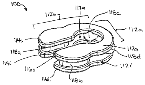
FIGS. 2A and 2B illustrate another exemplary embodiment of a spinal implant
100. The implant 100 is similar to implant 10 and generally includes a
posterior portion
112a that is adapted to be positioned around a spinal cord and that can be
coupled to at
least one adjacent vertebrae, and an anterior portion 112b that is adapted to
be positioned
between adjacent vertebrae. In this embodiment, however, the implant 100 does
not
have a unitary configuration, but rather it has a multi-piece construction. In
particular,
the implant 100 includes a superior member 112s, an inferior member 112i, and
at least
one central member disposed therebetween and adapted to allow movement of the
superior and inferior members 1 12s, 1 12i relative to one another.
The superior and inferior members 11 2s, 112i can vary in shape and size, but
in
one exemplary embodiment the superior and inferior members 112s, 112i are
substantially U- or C-shaped such that that posterior portion 112a of the
implant 100 is
substantially curved or semi-circular with a central opening 117a formed
therein to
allow the implant 100 to be positioned around the spinal cord using a
posterior surgical
approach, and the anterior portion 112b of the implant 100 includes opposed
superior
and inferior arms 114s, 116s, 114i, 116i that can be positioned between
adjacent
vertebrae and that define an opening 1 17b therebetween for allowing the
spinal cord to
pass therethrough. The superior and inferior members 112s, 112i can also have
substantially planar configurations, or they can have a shape that is adapted
to match the
CA 02571784 2006-12-21
WO 2006/007211 PCT/US2005/018660
-11-
contour of a vertebral endplate.
As indicated above, the implant 100 can also include at least one central
member
disposed between and adapted to allow movement of the superior and inferior
members
112s, 112i relative to one another. While the implant 100 can include any
number of
central members, in the illustrated exemplary embodiment the implant 100
includes a
four central members 118a, 118b, 118c, 11 8d disposed between the superior and
inferior
members 1 12s, 112i. In particular, a first central member 11 8a is positioned
between the
first superior arm 114s and the first inferior arm 114i, a second central
member 118b is
positioned between the second superior arm 114s and the second inferior arm
114i, and
third and fourth central members 118c, 118d are positioned on opposed lateral
sides of
the posterior portion 112a of the implant 100 between the superior and
inferior members
112s, 112i.
The central members 118a, 118b, 118c, 118d can be coupled to the superior and
inferior members 112s, 112i, or they can be removably disposed between the
superior
and inferior members 112s, 112i. For example, in one embodiment the central
members
11 8a, 11 8b, 118c, 118d can be fixedly mated to one or both of the superior
and inferior
members 112s, 112i. The central members 118a, 118b, 118c, 118d can, however,
be
adapted to pivot, rotate, or otherwise move relative to the superior and
inferior members
11 2s, 112i. In another embodiment, the central members 118a, 118b, 118c, 118d
can
merely be disposed between the superior and inferior members 112s, 112i. The
superior
and inferior members 112s, 112i can optionally includes recesses formed
therein and
adapted to seat the central members 11 8a, 118b, 11 8c, 11 8d.
The shape and size of each central member 11 8a, 11 8b, 11 8c, 11 8d can also
vary, but in one exemplary embodiment the central members 118a, 11 8b, 11 8c,
118d are
adapted to allow movement between the superior and inferior members 112s,
112i. This
can be achieved by, for example, forming the central members 11 8a, 118b, 11
8c, 118d,
or at least a portion of the central members 118a, 11 8b, 11 8c, 118d, from a
compressible
or resilient material. Alternatively, the central members 118a, 118b, 118c,
118d can be
inflatable to allow the superior and inferior members 112s, 112i to move
relative to one
another. In other exemplary embodiments, the central members 118a, 11 8b, 11
8c, 11 8d
can have a shape that allows the superior and inferior members 112s, 112i to
articulate
relative thereto. While the shape can vary, in the illustrated exemplary
embodiment the
CA 02571784 2006-12-21
WO 2006/007211 PCT/US2005/018660
-12-
first and second central members 118a, 11 8b disposed between the arms 114s, 1
14i,
116s, 1 16i of the anterior portion 1 12b of the implant 100 each have a
generally oblong
shape, and the third and fourth central members 118c, 118d disposed between
the
superior and inferior members 112s, 1 12i of the posterior portion 112a of the
implant
100 each have a generally spherical shape. A person skilled in the art will
appreciate
that a variety of other techniques can be used to movably couple the central
members
118a, 118b, 118c, 11 8d to the superior and inferior members 112s, 112i, or to
allow
movement between the superior and inferior members 11 2s, 11 2i.
FIGS. 2C and 2D illustrate the implant 100 positioned between adjacent
superior
and inferior vertebrae VS, V; (FIG. 2C only illustrates the inferior vertebra
Vi). The
implant 100 can be implanted using a variety of surgical techniques, and the
particular
technique can vary depending on the particular configuration of the implant
100. In one
exemplary embodiment, a posterior surgical approach is used, as previously
described
with respect to FIGS. 1C-1D. Once the vertebrae Vs, V; are prepared, the
implant 100
can be inserted between the adjacent vertebrae Vs, V; with the central members
11 8a,
118b, 118c, 11 8d pre-disposed between the superior and inferior members 112s,
112i.
Where the central members 118a, 11 8b, 118c, 11 8d are inflatable, the implant
100 is
preferably inserted between the adjacent vertebrae Vs, Vi with the central
members 118a,
11 8b, 11 8e, 11 8d in a deflated state. Once properly positioned between the
adjacent
vertebrae, an inflation medium, such as air or fluid, e.g., saline or gel, can
be introduced
into the central members 118a, 11 8b, 118c, 11 8d to inflate the central
members 118a,
11 8b, 11 8c, 11 8d. The inflation medium can optionally be adapted to harden
to form
rigid central members 11 8a, 118b, 11 8e, 11 8d. While the central members 11
8a, 118b,
118c, 118d are preferably pre-disposed between the superior and inferior
members 112s,
112i when the implant 100 is inserted between the adjacent vertebrae V5, V;, a
person
skilled in the art will appreciate that techniques could be used to allow the
central
members 11 8a, 118b, 11 8e, 11 8d to be implanted prior to implanting the
superior and
inferior members 112s, 112i.
While not shown, the implant 100 can also include features to facilitate
engagement of the adjacent vertebrae at a location between the vertebrae
and/or on the
posterior surface of the adjacent vertebrae. As previously discussed, suitable
techniques
for mating the implant 100 to adjacent vertebrae include, by way of non-
limiting
CA 02571784 2006-12-21
WO 2006/007211 PCT/US2005/018660
-13-
example, surface features, such as teeth, that engage the endplates of the
vertebrae
and/or a posterior portion of the vertebrae, surface coatings or materials
that allow bone
growth into the implant 100 to occur, or other materials or features that will
engage the
adjacent vertebrae. By way of non-limiting example, in one exemplary
embodiment the
posterior portion 112a and/or the anterior portion 11 2b of the implant 100
can include
teeth formed on the superior and inferior members 1 12s, 1 12i and adapted to
engage the
endplates of the adjacent vertebrae and/or a posterior portion of the
vertebrae, such as
the pedicles.
In another exemplary embodiment, the implant 100 can include one or more
extension members formed thereon or mated thereto and adapted to couple to at
least
one vertebrae. The extensions members can be similar to extension members 20a
and
20b shown in FIGS. lA-1D, or they can have a variety of other configurations
that allow
the implant 100 to couple to at least one adjacent vertebrae. By way of non-
limiting
exainple, FIGS. 3A and 3B illustrate one exemplary embodiment of an implant
100' that
is similar to implant 100 but that has flange-like extension members 120a',
120b', 120c',
120d' formed thereon for mating the superior and inferior members 112s', 112i'
to
adjacent vertebrae. In particular, the implant 100' includes first and second
extension
members 120a', 120b' formed on the superior member 112s' and extending in a
superior
direction, and first and second extension members 120c', 120d' formed on the
inferior
member 112i' and extending in an inferior direction. The extension members
120a',
120b', 120c', 120d' can be formed at any location on the implant 100, but in
an
exemplary embodiment as shown the extension members 120a', 120b', 120c', 120d'
are
positioned on opposed lateral sides of the posterior portion 112a' of the
implant 100' to
allow the extension members 120a', 120b', 120c', 120d' to be coupled to the
posterior
surface of the adjacent superior and inferior vertebrae VS, Vi, as shown in
FIG. 3B.
Each extension member 120a', 120b', 120c', 120d' can have a variety of shapes
and sizes. In the embodiment shown in FIGS. 3A and 3B, the extension members
120a',
120b', 120c', 120d' are integrally formed with the superior and inferior
members 112s',
112i', and thus they have a substantially planar configuration. Each extension
member
120a', 120b', 120c', 120d' also includes a thru-bore formed therein and
adapted to receive
a fastening element, such as a bone screw 130a', 130b', 130c', 130d', for
mating the
extension members 120a', 120b', 120c', 120d' to the vertebrae. A person
skilled in the
CA 02571784 2006-12-21
WO 2006/007211 PCT/US2005/018660
-14-
art will appreciate that a variety of other mating techniques can be used.
FIG. 3B illustrates the implant 100' in use, and as shown the first and second
extension members 120a', 120b' are mated to a superior vertebra Vs, and the
third and
fourth extension members 120c', 120d' are mated to an adjacent inferior
vertebrae Vi.
As a result, the superior member 112s' is maintained in a substantially fixed
position
relative to the superior vertebra Vs, and the inferior member 112i' is
maintained in a
substantially fixed position relative to the inferior vertebra V;. The central
members
118a', 118b', 118', only three of which are shown in FIGS. 3A and 3B, allow
the
vertebrae Vs, V; to move relative to one another, thereby restoring normal
function to the
vertebrae Vs, V;.
One skilled in the art will appreciate further features and advantages of the
invention based on the above-described embodiments. Accordingly, the invention
is not
to be limited by what has been particularly shown and described, except as
indicated by
the appended claims. All publications and references cited herein are
expressly
incorporated herein by reference in their entirety.
What is claimed is: