Note: Descriptions are shown in the official language in which they were submitted.
CA 02571904 2006-12-19
SYSTEM AND METHOD OF D.TE TIN PATHOGENS
Field of the Invention
[00011 The present invention relates to the field of detecting pathogens. In
particular,
it relates to a system and method for detecting, identifying, characterizing
and surveilling
pathogen and host markers, collecting and disseminating information concerning
those
pathogens and their hosts in real time to and from an instant location,
providing
instantaneous treatment recommendations and educational information.
Background of the Invention
[00021 Detection and characterization of an infectious disease is a complex
process
that ideally begins with the identification of the causative agent (pathogen).
This has
traditionally been accomplished by direct examination and culture of an
appropriate
clinical specimen. However, direct examination is limited by the number of
organisms
present and by the observer's ability to successfully recognize the pathogen.
Similarly, in
vitro culture of the etiologic agent depends on selection of appropriate
culture media as
well as on the microbe's fastidiousness. The utility of pathogen culture is
further
restricted by lengthy incubation periods and limited sensitivity, accuracy and
specificity.
[0003] When in vitro culture remains a feasible option, the identification and
differentiation of microorganisms has principally relied on microbial
morphology and
growth variables which, in some cases, are sufficient for strain
characterization (i.e.
isoenzyme profiles, antibiotic susceptibility profiles, and chematographic
analysis of fatty
acids).
[00041 If culture is difficult, or specimens are not collected at the
appropriate time,
the detection of infection is often made retrospectively, if at all, by
demonstrating a
serum antibody response in the infected host. Antigen and antibody detection
methods
have relied on developments in direct (DFA) and indirect (IFA)
immunofluorescence
analysis and enzyme immunoassay (EIA)-based techniques, but these methods can
also
possess limited sensitivity.
-1-
CA 02571904 2006-12-19
[00051 These existing methods have several drawbacks. First, the process can
take
several days to return results. In the case of highly communicable and/or
dangerous
pathogens, confirmation of pathogen type may not be received until the host
has already
exposed others or has passed beyond treatment. Second, the transportation of
samples to
:laboratories for culture growth increases the risk of errors, such as
misidentifying the
sample, or exposure of unprotected personnel to a sample containing a highly
communicable pathogen. Thirdly, the pathogen tests are limited based on the
suspected
pathogen list provided by the observer (i.e. doctor), meaning that additional
unsuspected
pathogens are not tested for but may be present.
[00061 Related to this method of diagnosis is the response to an outbreak of
infectious disease. If an outbreak is suspected or detected, the existing
response is the
hundreds of years old method of quarantine. In cases of infectious disease
outbreaks for
which appropriate treatments andlor sensitive, specific, and rapid
screening/diagnostic
tests are lacking, quarantine remains the only means of preventing the
uncontrolled
spread of disease. When infection is suspected simply based on epidemiological
grounds,
or even based on comparable disease presentation, healthy or unexposed
individuals may
be quarantined along with infected individuals, elevating their likelihood of
contracting
the disease as a consequence of quarantine. Availability of a rapid
confirmatory test for
the pathogen in question would greatly reduce the time spent in quarantine,
and would
therefore reduce the likelihood of contacting the disease from truly infected
persons.
[00071 Although quarantine remains a method of last resort for protecting
public
health, delays in providing a correct diagnosis, and subsequently appropriate
treatment,
occur on a daily basis in hospitals and physician's offices alike. The problem
stems from
the fact that many diseases have very similar clinical presentations in the
early stages of
infection, and in the absence of a thorough patient/travel history, malaria or
SARS for
example, can be misdiagnosed as the common flu (i.e. fever, chills), albeit
with
potentially fatal consequences. Had a multi-pathogen test which differentiates
diseases
with similar presentations been available, a tragedy may have been averted.
[00081 In contrast to reliance on morphological characteristics, pathogen
genotypic
and proteomic traits generally provide reliable and quantifiable information
for the
detection and characterization of infectious agents. Moreover, microbial
DNA/RNA can
-2-
CA 02571904 2006-12-19
be extracted directly from clinical specimens without the need for
purification or isolation
of the agent.
[0009] On a global scale, molecular techniques can be applied in a high
throughput
manner in screening and surveillance studies monitoring disease prevalence and
distribution, evaluation of control measures, and identification of outbreaks.
[00101 Point-of-care diagnostic devices (PDDs) have been developed for a
number of
individual infectious diseases. In most cases these assays are
immunochromatographic
single colorimetric strip tests designed to detect a single infectious agent
(either a
pathogen-specific antigen or an antibody response to one) in a small volume of
blood or
serum.
[00111 None of these current assays has the capability to detect multiple
pathogens or
simultaneously detect genomic and proteomic markers of multiple pathogens.
Similar
limitations exist for other rapid diagnostic assays. Since almost all these
tests rely on a
single visual colorimetric change for their readout, the opportunities to
detect multiple
pathogens are severely impeded and the majority of current PDDs are restricted
to the
detection of a single pathogen. Consequently, evaluating patients for
potential infectious
agents or testing a unit of blood for common transmissible agents requires
multiple
consecutive point-of-care tests to be performed, complicating clinical
management,
slowing results and significantly escalating costs.
[0012] Many PDDs do not meet what are considered essential requirements
including: ease of performance, a requirement for minimal training, the
generation of
unambiguous results, high sensitivity and specificity, the generation of same
day results
(preferably within minutes), relative low cost, and no requirement for
refrigeration or
specialized additional equipment.
[00131 In summary, despite current availability of excellent diagnostic
reagents (e.g.
antibody and nucleic acid probes) that recognize specific targets for many
microbial
pathogens, the current strategies have inadequate performance characteristics.
Contributing to this is the fact that these reagents are conjugated to organic
dyes, gold-
labelled particles or enzymes that lack sufficient sensitivity to be detected
at the single
molecule level. Furthermore, the current PDD platforms and detection schemes
typically
-3-
CA 02571904 2006-12-19
rely on single macroscopic colorimetric changes and are not well suited to the
simultaneous detection of multiple pathogens.
[00141 More recent advances in molecular diagnostics, including real-time PCR
combined with automated specimen processing, have addressed a number of the
limitations of earlier "in-house" and non-standardized gene amplification
assays. These
assays represent a significant advance in detecting, quantifying, and
characterizing many
microbes and currently represent the "gold" or reference standard for
infectious disease
diagnostics for a number of pathogens. However, these assays are still
complex,
expensive, and require specialized equipment, creating a number of barriers to
their
potential application at point-of-care.
[0015] Finally, current genomic or proteomic detection strategies require a
sample
processing and technical commitment to one strategy or the other. There is no
current
capacity to simultaneously detect both antigenic targets for some pathogens
and genetic
targets for others. This limits the simultaneous detection of preferred
pathogen-specific
targets and presents a barrier to fully exploiting the complementary power of
both
strategies.
100161 A system is needed which enables pathogen detection, identification and
characterization, as well as host characterization in a much more timely
manner than
existing methods. Preferably, such a system would support a modular pathogen
selection
platform, based on the specific needs of the caring physician or clinic in the
context in
which the device is used (i.e. for screening or diagnosis). Further, the
system would also
enable simultaneous detection, identification and characterization of multiple
pathogens
in a single sample whereby the pathogens are differentiated by optical
pathogen-specific
profiles stored in a pre-existing database.
Summary of the Invention
[00171 According to an aspect of the invention there is provided a method of
performing one or more of: detecting, identifying and characterizing pathogens
and
characterizing pathogen hosts using markers for pathogens and hosts,
comprising the
steps of: a) preparing a marker-detection medium containing signatures of the
identity
and characteristics of pathogens and optionally of hosts; b) collecting a
sample from a
-4-
CA 02571904 2006-12-19
host; c) combining the sample with the marker-detection medium and d)
analyzing the
signatures to detect, identify and characterize the pathogens, and optionally,
characterize
the host.
100181 Preferably, the sample collected is a blood sample, although plasma,
serum,
cerebral spinal fluid (CSF), bronchioalveolar lavage (BAL), nasopharyngeal
(NP) swab,
NP aspirate, sputum and other types of samples can also be used, and the
marker
detection system is a pathogen-detection medium preferably comprising
microbeads
conjugated to biorecognition molecules (BRMs) and the microbeads are injected
with
quantum dots or a similar fluorescent particle or compound. Also preferably,
each of the
microbeads contains a unique combination of quantum dots to provide a unique
optical
barcode associated with each microbead for detecting unique pathogen-specific
and / or
host-specific signatures.
[0019] Preferably, the analysis step comprises illuminating the microbead-
pathogen
sample with a laser as it flows through a microfluidic channel and collecting
the resulting
spectra with a spectrophotometer/CCD camera, photomultiplier tube and/or a
collection
of avalanche photodetectors (APDs). Each spectrum correlates with a previously
assigned pathogen.
[0020] Optionally, the method may include producing a list of host
characterization
markers associated with said host sample as part of analysis step d).
[0021] Optionally, the method may include an additional step e) of providing a
list of
treatment options based on the list of pathogens generated in analysis step
d).
[0022] Optionally, the method may also include step f) of correlating
geographic
location information data with the list of pathogen and host markers generated
in analysis
step d) via a GPS locator.
[00231 Preferably, the method further includes an additional step g) of
transmitting,
preferably wirelessly, said list of pathogen markers and said list of host
identifier markers
and said geographic location data to a remote database as well as transmitting
treatment
and educational information from the database to the filed device. It will be
appreciated
that the steps of the process are not necessarily conducted in the specified
order.
[0024] The method further includes detection of pathogen-conjugated microbeads
in
a flow stream propelled by electrokinetic or hydrodynamic flow through a
microfluidic
-5-
CA 02571904 2006-12-19
channel. As the barcoded beads pass a laser beam at one end of the channel,
the spectra
emitted by the quantum dots within the beads, (as part of the barcode), or
outside the
beads (as part of a bead-pathogen complex detection mechanism, which may
include
fluorophores as described below) are collected by a spectrometer/CCD camera
system,
photomultiplier tube and/or a collection of APDs and analyzed by appropriate
software.
[0025] According to another aspect of the invention a system of components is
provided which is capable of executing any of the above methods.
[00261 The advantages of the present invention include a vast reduction in the
amount
of time necessary to identify pathogens in a patient sample, compared with
most methods
currently in use, as well as the ability to provide rapid on-site information
concerning
treatment and quarantine measures for any identified pathogens. Another
advantage is
the ability to collect patient and pathogen data in a global database and mine
the
information contained in this database to produce trends and tracking measures
for
various pathogens and their hosts, which information may be used for
surveillance,
research, therapeutic design, and other purposes.
[0027] Other and further advantages and features of the invention will be
apparent to
those skilled in the art from the following detailed description thereof,
taken in
conjunction with the accompanying drawings.
Brief Description of the Drawings
[00281 The invention will now be described in more detail, by way of example
only,
with reference to the accompanying drawings, in which like numbers refer to
like
elements, wherein:
Figure 1 is a flow chart detailing the series of steps in the inventive method
disclosed herein;
Figure 2 is a block diagram for a pathogen detection device; and
Figure 3 is a block diagram of multiple devices communicating with a central
database.
-6-
CA 02571904 2006-12-19
Detailed Description of the Preferred Embodiments
100291 Referring now to Figure 1, the present inventive method is described by
a
series of steps set out in a flowchart.
100301 The first step 12 is to collect a sample from a host (e.g. a human,
animal or
environmental sample), preferably a blood sample, although plasma samples,
serum
samples, CSF, BAL, NP aspirates, NP swabs, sputum and other types of physical
samples
can be used, as appropriate. This sample is then analyzed 14 and a list of
pathogens
identified in the sample is generated 16. A GPS receiver 22 determines the
location of
the sample reader and thus, the sample. The list of identified pathogens and
the location
information are both sent 20 to a central database for storage and processing.
Meanwhile,
a list of treatment options is displayed at 18, based on the identified
pathogens, for the
operator's consideration.
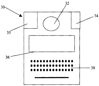
[0031] The analysis 14 is performed by a pathogen detection device 30 as shown
in
Figure 2. This device 30 is portable, preferably hand-held, and has an outlet
32 for
receiving a sample and a display 36 to show the list of detected pathogens
within the
sample. An input device 38, such as a keyboard, is also provided to enable
scrolling and
viewing of the display and input of additional information (field notes,
etc.). Pathogens
in a sample are identified based on matching of spectra to previously stored
data
corresponding to each pathogen supported by the device. The spectra database
may be an
internal database on the device 30 (kept in flash memory or similar storage to
allow for
updating) or retrieved by communicating with an external database. A GPS
receiver 35 is
also preferably located in the device 30, along with a display showing the GPS
co-
ordinates. Ideally, all communication is conducted wirelessly for maximum
range and
portability. The pathogen detection device 30 is ideally capable of detecting
multiple
pathogen, multiple BRMs from the same pathogen as well as host markers within
a single
sample, and preferably markers of different types, such as protein-based
markers and
gene-based markers.
[00321 The method of detection used can be varied among suitable available
methods, however, a preferred method is the use of biorecognition molecules
(BRMs)
conjugated to quantum dot-doped microbeads or nanobeads/nanoparticles.
Alternatives
include single quantum dots or fluorophores conjugated to BRMs. Quantum dots,
also
-7-
CA 02571904 2006-12-19
known as semiconductor nanocrystals, are electromagnetically active
nanotechnology-
based particles, ranging in size from 2 nanometers (nm) to 8 nm. A
particularly useful
property of quantum dots is that they are fluorescent, that is they emit light
after brief
illumination by a laser. In addition, quantum dots of different sizes will
fluoresce in
different colors and the fluorescing color can be modified by the particle's
shape, size
and composition. BRMs are biological molecules that bind only to a single
other
biological molecule and are pathogen specific. For example, "antibodies" are
BRMs that
bind to proteins and "oligonucleotide probes" are BRMs that bind to
complementary gene
sequences (e.g. DNA or RNA). Pathogens and hosts have both unique and shared
genetic
and protein markers, and each marker can be bonded to by a specific BRM.
[0033] A microbead, which is a polystyrene (or similar polymer) bead that can
be 100
nanometers-10 micrometers in diameter and doped with a collection of quantum
dots, is
physically conjugated to a BRM. By introducing unique combinations of quantum
dots
of different sizes (i.e., colors) and at different concentrations into the
microbeads,
microbeads with thousands of distinctive combinations of quantum dot colors
and
intensities can be created. When a laser illuminates the microbeads, the
quantum dots
fluoresce to produce a distinctive combination of colors. These color
combinations are
an example of a barcode, in this case an optical bar code, analogous to a UPC
syrnbol,
and similar known types of imprinted barcodes. Since each BRM recognizes a
distinct
pathogen or host marker and each microbead has a unique barcode, each BRM-
conjugated microbead provides a barcode for the specific pathogen or host
marker
recognized by its BRM. These BRM-conjugated microbeads, as well as BRM-
conjugated quantum dots, may be lyophilized into a powder and provided in the
sample
analysis kit.
[0034] To differentiate between BRM-conjugated beads bound to pathogens and
those that are not, an additional confirmatory detection signal in the form of
anti-human
IgG, and/or an anti-human IgM molecule, or a pathogen-specific antibody (i.e.
anti-X
antibody), or an oligonucleotide (complementary to a pathogen gene of
interest)
conjugated to a fluorophore, is included. The readout of a successful pathogen
detection
test comprises the bead barcode signal and a second signal generated by the
fluorophore,
-8-
CA 02571904 2006-12-19
[00351 One example of pathogen detection is an antigen capture system. The
antigen
capture system includes a capture antibody (i.e. a BRM) which is bound to the
barcoded
microbead which is responsible for capturing the antigen from the sample. A
second
antibody (detection antibody) which recognizes the pathogen antigen/protein
then binds
to the complex. This detection antibody is conjugated to a fluorophore. When
the
sample is analyzed, if the signal for the detection antibody is not detected,
the pathogen
does not register as detected, either because it is not present in the sample
or because of
assay failure. The latter case is eliminated if the correct signals from the
positive control
sample, i.e. detection of the appropriate bar code of the BRM-quantum dot-
containing
microbead run in parallel with all clinical tests are detected.
[00361 Another example of pathogen detection is an antibody capture system. In
the
antibody capture system the BRM which is bound to the barcoded microbead is a
pathogen-specific antigen or protein (natural, recombinant, or synthetic). The
complementary antibody to the antigen, if present in the clinical sample would
bind the
antigen attached to the bead. This complex is recognized by the addition of a
secondary
(detection) anti-human antibody (Anti-Human IgM or Anti-Human IgG). This
detection
antibody is conjugated to a fluorophore. Again, when the sample is analyzed,
if the signal
for the detection antibody is not detected alongside the signal from the bead
barcode the
pathogen does not register as detected, either because it is not present in
the sample, or
due to assay failure. The latter case is eliminated if the expected signals
from positive
control sample, as mentioned above, register correctly.
[00371 Still another example of pathogen detection is a genomic analysis
system. In
the genomic analysis system the BRM which is bound to the barcoded microbead
is a
pathogen-specific oligonucleotide (RNA or DNA) (1-25 bases in length). Upon
addition
to the sample, the oligonucleotide will hybridize to its complementary
sequence on the
pathogen gene. A second oligonucleotide sequence complimentary to a downstream
portion of the gene of interest is subsequently added and will hybridize to
the gene, if
present. This second sequence is conjugated to a fluorophore. Again, when the
sample is
analyzed, if the signal for the second sequence is not detected, the pathogen
does not
register as detected, either because it is not present in the sample or
because of assay
-9-
CA 02571904 2006-12-19
failure. A correctly detected positive control sample as referred to above
eliminates the
latter scenario.
[0038] The biological (e.g. blood) sample is added to a vial, and different
pathogen
markers bind the various microbeads carrying specific pathogen BRMs. The
combined
sample is then washed or otherwise treated to remove extraneous matter and
unattached
microbeads. The detection antibodies conjugated to the fluorophores are then
added to
produce a bead-sample-detector complex.
[0039] The bead-sample-secondary detector complex is flowed through a
microfluidic channel via hydrodynamically or electrokinetically-driven flow
and passed
through a laser beam located at one end of the channel. The laser beam
illuminates the
quantum dots in the complex and the emitted wavelengths are guided to either a
spectrometer/CCD system, photomultiplier tube and/or a series of APDs. Signal
deconvolution software translates the signal and the corresponding optical
code is
compared to pathogen-specific spectra stored in the database of pathogens or
host
characteristics supported by the detection device. Then, a list of detected
pathogens and
pathogen and host characteristics is produced. The response time from the
taking of the
original biological sample to the production of the pathogen list can be
measured in
minutes.
[0040] Ideally, the pathogen detection device 30 is a portable, hand-held
device with
an integrated laser and spectrophotometer, photomultiplier tube and/or series
of APD
units, specifically designed PDMS microfluidic channel chips, a supply of BRM
conjugated barcoded beads for identification of various pathogens as well as
appropriate
bead-pathogen complex detection markers (quantum dot, fluorophore, small bead
labeled
IgG/IgM/anti-pathogen antibodies or oligonucleotides). The device 30 may store
a
pathogen identity database on board, or access a remote database, preferably
via the
Internet, preferably wirelessly, and identify the pathogen from a remote,
central database.
If an on-board database is used, a communications system 34 for contacting and
receiving
updates from a larger, central database is provided.
[0041] The pathogen detection device 30 may include a GPS tracking device
which
transmits specific geographic information, preferably wirelessly to the same
central
database.
-10-
CA 02571904 2006-12-19
[0042] Once the pathogen list is produced, the pathogen detection device 30
may
additionally provide further information of value to the diagnosing doctor.
Ideally, a
treatment protocol is provided (step 18), including any special measures
necessary to
avoid communication of the pathogen. Other information, such as
pathophysiology,
disease history and bibliographic references can be provided, enabling the
pathogen
detection device 30 also to be used as an educational tool in the appropriate
scenarios.
100431 An outbreak scenario for use of the device in a standard pathogen
detection
setting follows. An airport is a point of entry representing a major pathogen
travel
vector, as well as presenting problems with implementing traditional detection
and
quarantine methods. By equipping medical staff with a number of pathogen
detection
devices as described herein, and a supply of microbead sample vials able to
detect
pathogens typically transmitted by travelers, incoming passengers can be
processed on-
site by taking a blood sample and injecting it into a sample vial. The
analysis is
performed by the pathogen detection device within minutes and the sampled
passenger
can be quickly released or redirected for treatment and observation, as
necessary. While
a single device is limited in processing capability, the ability to provide
multiples of
identical devices can enable processing of passengers in a matter of hours,
not days.
Faster processing allows appropriate treatment and quarantine measures to be
taken
earlier, and be more effective, reducing the probability of the pathogen
spreading
unchecked.
[00441 As an example, a pathogen detection device may contain BRM-conjugated
barcoded microbeads for detection of three different pathogens, say, HIV,
Hepatitis B
and Hepatitis C. The microbeads associated with each pathogen have a
separately
identifiable barcode, for example, HIV may have red beads (e.g. detecting the
antibody
gp41 as indicator of HIV infection), Hepatitis B yellow beads (e.g. detecting
the antibody
NSP4 as indicator of Hepatitis B infection), and Hepatitis C red-yellow beads
(e.g.
detecting the antibody HBSAg as indicator of Hepatitis C infection), and
preferably all
using orange probes-pathogen complex detection markers or any color-probe that
is
spectrally different than the color of the barcodes. Thus, the detection
system can readily
identify any detected pathogen merely by the wavelength (which identifies
color) or
intensity of the bead spectra.
-11-
CA 02571904 2006-12-19
[0045] From this model, the system can readily be expanded, for example, to
five
pathogens, adding, for example, pathogen detection microbeads for malaria and
dengue
virus. From there, extrapolation to more pathogens (10, 20, 100) is mostly
limited by the
ability to create a sufficient number of barcodes, which is based primarily on
the doping
of the microbeads and limits of the detection mechanism. As the number
increases,
barcodes may be based on intensity levels, as well as wavelength.
[0046] Detecting and providing a treatment protocol for a pathogen represents
merely
the first step in a potentially much larger process for tracking and
controlling the spread
of pathogens as shown in Figure 3. Tailoring the device to be modular and be
able to
detect either an array of pathogens (i.e. BRMs for multiple pathogens) with
similar
clinical presentations, act as a screening tool (e.g. for identifying
individuals vaccinated
for selected diseases) or allowing physicians or clinics to select the
pathogens of interest
in their particular communities, allows for unprecedented diagnostic
flexibility at the
bedside. Incorporation of multiple BRMs for the same pathogen enhances
detection
accuracy and overcomes the limitations associated with use of single BRMs for
pathogen
detection (i.e. mutations and strain differences which may result in false
negative or false
positive results). The test results data along with the geographic location
data (but no
other information about the patient e.g. name, address and other privacy-
protected data)
provided by the GPS unit, are transmitted to a central database 40. The
information is
preferably sent wirelessly, and immediately upon generation of the pathogen
list (step
20). The central database 40 is in contact with a substantial number of
pathogen
detection devices 30 at any given time.
[0047] The central database 40 can be local, national or global, or a
combination of
different databases of these types. Ideally, one top-level central database 40
is provided
which receives information constantly from all devices 30 worldwide. Over
time, the
database becomes a repository of information on every pathogen supported by
the
detection platform lending itself to mining for, among others, frequency and
global
patterns of detection of pathogens, long-term pathogen trends (i.e.
colonization of new
territories), and correlations between pathogens and host markers which may
indicate
enhanced susceptibility or resistance to the disease.
-12-