Note: Descriptions are shown in the official language in which they were submitted.
= CA 02572061 2006-12-22
METHOD AND APPARATUS FOR DETECTING THE PRESENCE OF
DERMAL MELANIN IN EPITHELIAL TISSUE
Over the last four decades there has been a world-wide
increase in the incidence of melanoma. Although adequate
sun protection has been identified as the first step
toward preventing the occurrence of melanoma, early
diagnosis and excision is the key to the survival of the
many individuals who will still develop the disease.
A range of techniques have been developed to assess
pigmented lesions. With the most common of these,
dermatoscopy, a hand held microscope is used to visualise
morphological characteristics at the dermo-epidermal
junction. Clinicians then attempt to diagnose the
presence of melanoma by analysing the lesion by colour,
pattern and specific morphological features.
In addition to conventional dermatoscopy, a number of new
techniques recently been developed by Astron Clinica
Limited based on research undertaken at the University of
Birmingham. These techniques are described in WO 00/75637
and WO 98/22023. The techniques use a quantitative
understanding of the way light is absorbed and scattered
within skin to produce maps of melanin, blood and
collagen.
= CA 02572061 2006-12-22
The original research undertaken at the University of
Birmingham argued that the Kubelka-Munk theory is
sufficient to model light transport within skin. If exact
scattering and absorption coefficients can be specified,
then the Kubelka-Munk theory can be applied at each
wavelength in the visible range and corresponding
remittance spectrum obtained. This predicted spectrum,
which will determine the colour of the skin, will be
dependent on the histological characteristics of the
tissue. Three parameters capture most of the variation in
remitted spectra from healthy skin. These three
parameters are concentration of epidermal melanin,
concentration of blood and thickness of the papillary
dermal layer (collagen thickness).
Using the RGB response curves for a digital camera
together with a model of the scattering and absorption
characteristics of the skin, it is possible to calculate
the set of image values which would be measured by a
digital camera when skin with a known remittance spectrum
S(A) is illuminated with light of known spectral
characteristics I(A). This is done by calculating the
convolution integral for each channel, given as,
Ired = JI (A)s(A)R(A)dA i lgreen = jI (A)S(A)G(A)dA , i. = f
I(.Z,)S(A)B(A)d.Z,
2
CA 02572061 2006-12-22
where R(A), G(X) and B(A) are the response curves for the
red, green and blue channels and lredr ibi,,e and lgreen are
the corresponding values recorded by the camera at a
given pixel
By ranging through all potential combination of melanin,
blood and collagen, it is possible to generate all
possible spectra and therefore all possible sets of image
values which would be measured by a digital camera. Once
this information has been obtained a link can be
established between image values and histological
parameter values. This link can be expressed as a
mathematical function.
An image, acquired using a digital camera, consists of a
large number of very small pixels, each of which have a
set of image values, (1red, lgreen and iblue) . By applying
the mathematical function, linking these image values to
histological parameter values, it is possible to obtain
values for melanin, blood and collagen at every pixel
within an image of skin. This information can then be
displayed in the form of histological parametric map. The
SIAscope (RTM), developed by Astron Clinica Limited,
relies on a specially adapted camera which is able to
capture 4 channels of image data. As well the normal RGB
channels, it also acquires an image in the infra-red
3
CA 02572061 2006-12-22
region of the spectrum. With this additional information,
it is possible to produce an additional parametric map of
dermal melanin.
Determining measurements of epidermal melanin, blood,
collagen and dermal melanin directly from measurements of
remitted light S(A) requires that a suspect lesion is
illuminated with light of known spectral characteristics
I(A). Using such an approach it is therefore necessary to
follow a strict calibration procedure where lighting
levels are strictly controlled. This limits the use of
such an approach to analyzing small areas of skin as once
larger areas of tissue are imaged, over which the surface
geometry of the imaged tissue varies, calibration is no
longer possible and analysis becomes inaccurate. The maps
produced by such a technique have been shown to be of
great value to clinicians in their diagnosis of melanoma.
However, due to the required calibration procedures, it
is typically only possible to produce a map of dermal
melanin over a small area of skin, currently 15mm
diameter.
Although effective, prior art techniques thus currently
require detailed individual analysis of every suspect
lesion on a given patient and thus rely on the clinician
being able to quickly identify all potentially dangerous
4
CA 02572061 2006-12-22
lesions. While this is straight-forward for the majority
of patients, some individuals present with large numbers
of skin lesions. In this situation it would be useful to
have some tool which would be able to automatically
identify all lesions requiring a detailed inspection.
In order to overcome the problems arising due to strict
calibration requirements an alternative technique has
been developed. This is described in detail in Astron
Clinica's prior patent application WO 04/010862. The
technique relies on a mathematical function linking
histological parameters with ratios of image values,
rather than the actual image values. Determining
measurements from ratios of image values removes the need
for calibration. This can be demonstrated mathematically
by considering the case where illumination which can be
described by
I(~)=a,I(~),
where al is a wavelength independent scaling factor which
captures changes in illumination intensity and i(A)
captures the wavelength dependence of the incident light.
The amount of light remitted from a tissue will depend on
both the histological characteristics of the tissue and
the angle of the tissue to the camera. The remitted
spectrum can therefore be expressed as
5
CA 02572061 2006-12-22
S(A) = a2 S(A)
where a2 is a wavelength independent scale factor which
depends on the angle of the tissue to the camera and S(A)
is the remitted spectrum which depends on the histology
of the imaged tissue. Ratios of image values are now
given as,
a f I(~,)S(~.)G(~.)d~, a jI(~,)S(~.)B(~,)d~,
rgeenOverRed a jI(~,)S(~.)R(~,)dA / rblueOverRed a f I(A)S(A)R(A)dA where
a=alaz. The factor a, which captures all variation
due to illumination changes and changes in surface
geometry of the images tissue, will cancel out in each of
the equations above leaving only wavelength dependent
terms. Thus the image ratios can be seen to be
independent of both illumination and surface geometry.
Variation in skin histology can then be thought of in
terms of a parameter space and spectra are computed,
using the Kubelka-Munk model, which correspond to each
point with parameter space. By applying the above
equations it is then possible to calculate the two image
ratios rqreenOverRed and rblueoverRed which correspond to a given
spectra. Using the above technique, measurements of blood
and melanin concentrations can be made without having to
control for surface geometry and lighting conditions.
6
CA 02572061 2006-12-22
Although very effective for characterising normal skin,
the described technique in WO 04/010862 is however
limited to obtaining measurements of melanin and blood
concentrations. The technique is not suitable for
obtaining measurements of collagen as changes in collagen
have an equal effect at every wavelength and therefore no
effect on a ratio of two spectral measures. Further
disclosed techniques are unable to determine whether
melanin is present only within the epidermis of the skin
or whether melanin has penetrated into the dermis. To be
of use as a screening tool it must be possible to measure
dermal melanin as if information showing the presence of
dermal melanin could be displayed this would alert the
clinician to any suspicious lesions
An alternative system which assists with the
identification of suspect lesions is therefore desirable.
In accordance with one aspect of the present invention
there is provided an apparatus for detecting the presence
of melanin in the dermis of an epithelial tissue having a
dermis and an epidermis, said apparatus comprising:
a camera operable to obtain image data
representative of light remitted by an epithelial tissue
having a dermis and an epidermis illuminated by polarised
light;
7
CA 02572061 2006-12-22
a chromophore determination module operable to
process image data obtained by said camera to determine a
measurement of the concentration of blood and melanin at
points in an epithelial tissue in an obtained image; and
a dermal melanin detection module operable to
utilise measurements of the concentration of blood and
melanin determined by said chromophore distribution
module and image data obtained by said camera to
determine the difference between detected remitted light
from points in an epithelial tissue in an obtained image
and expected levels of remitted light from epithelial
tissue having said identified concentrations of blood and
melanin in which said melanin is solely present in the
epidermis of said epithelial tissue to identify points in
said epithelial tissue where melanin is present in the
dermis of said epithelial tissue.
In accordance with another aspect of the present
invention there is provided a method of detecting the
presence of melanin in the dermis of an epithelial tissue
having a dermis and an epidermis, comprising:
obtaining image data representative of light
remitted by an epithelial tissue having a dermis and an
epidermis illuminated by polarised light;
8
CA 02572061 2006-12-22
processing obtained image data to determine a
measurement of the concentration of blood and melanin at
points in an epithelial tissue in an obtained image; and
utilising said determined measurements of the
concentration of blood and melanin and said obtained
image data to determine the difference between detected
remitted light from points in an epithelial tissue in an
obtained image and expected levels of remitted light from
epithelial tissue having said identified concentrations
of blood and melanin in which said melanin is solely
present in the epidermis of said epithelial tissue to
identify points in said epithelial tissue where melanin
is present in the dermis of said epithelial tissue.
Further aspects and embodiments of the present invention
will become apparent with reference to the accompanying
drawings in which:
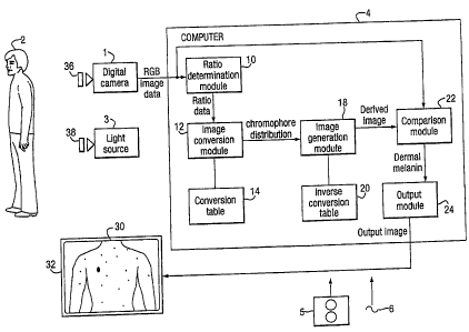
Figure 1 is a schematic block diagram of a dermal
melanin detection system in accordance with a first
embodiment of the present invention;
Figure 2 is a schematic cross sectional view through
a layer of skin illustrating the structure of the skin
and the interaction of that structure with incident
light;
9
CA 02572061 2006-12-22
Figure 3 is a flow diagram of the processing
performed by the dermal melanin detection system of
Figure 1;
Figure 4A is a plot of ratios of green/red and
blue/red remitted light for a sample of epithelial tissue
illustrating the effect of increasing epidermal melanin
and blood in the absence of dermal melanin; and
Figure 4B is a plot of ratios of green/red and
blue/red remitted light for a sample of epithelial tissue
illustrating the effect of increasing dermal melanin
concentration for two separate combinations of epidermal
melanin and blood concentrations.
Specific Embodiment
Figure 1 is a schematic block diagram of an embodiment of
the present invention. In accordance with this
embodiment, a digital camera 1 comprising a conventional
digital camera is provided which is arranged to obtain an
image of an individual 2 illuminated by a light source 3.
The images obtained by the digital camera 1 are then
transmitted to a computer 4 which is configured by
software either provided on a disk 5 or by receiving an
electrical signal 6 by via a communications network to be
configured into a number of functional modules 10-24
which cause the computer 4 to process the image data
CA 02572061 2006-12-22
received from the digital camera 1 to generate an output
image 30 which is shown on a display 32.
In the present embodiment the functional modules 10-24
comprise: a ratio determination module 10 for converting
RGB image data into ratio data, an image conversion
module 12 and a conversion table 14 for processing ratio
data to generate data indicative of concentrations of
blood and melanin; an image generation module 18 and an
inverse conversion table 20 operable to generate derived
image data utilizing chromophores distribution data; a
comparison module 22 for comparing received RGB image
data and derived image data generated by the image
generation module 22; and a output module 24 for
outputting an output image on basis of the comparison
between received and derived image data performed by the
comparison module 22.
The functional modules 10-24 illustrated in Figure 1 are
purely notional in order to assist with the understanding
of the working of the claimed invention and may not in
certain embodiments directly correspond with blocks of
code in the source code for the software. In other
embodiments the function performed by the illustrated
functional modules 10-24 may be divided between different
11
CA 02572061 2006-12-22
modules or may be performed by the re use of the same
modules for different functions.
As will be described in detail later, the processing
undertaken by the ratio determination module 10 and the
image conversion module 12 is similar to that described
in Astron Clinica's prior patent application WO
04/010862. This enables the computer to process image
data obtained by the digital camera 1 and determine
measurements of blood and melanin present in the skin of
the individual 2 being analysed. The image generation
module 18 and inverse conversion table 20 are then
utilised to process the determined measurements to
generate image data representative of the expected
appearance of skin including such chromophore
concentrations, where all of the identified melanin is
assumed to be present in the epidermis. The comparison of
this derived image with the actual image data from the
digital camera 1 by the comparison module 22 then enables
areas where melanin is present in the dermis of the
individual's skin to be identified. As is explained this
identification is achieved without having to control the
lighting illuminating the individual 2 being analysed and
hence enables the imaging an analysis of large areas of
an individual's skin in a single image.
12
CA 02572061 2006-12-22
The accuracy of the system of the present embodiment has
been compared with the detection of dermal melanin using
conventional techniques. In tests involving data
collected from 25 lesions 9 melanomas and 16 benign
lesions, assessment of all lesions showed a perfect match
between identification of dermal melanin utilising the
above described system and conventional techniques for
all the melanomas. For the benign lesions all results
agreed apart from one which showed a low presence of
dermal melanin using conventional techniques where none
was identified using the above technique.
Interaction of Light with the Skin
Prior to describing the detailed processing of the
various functional modules 10-24 of the computer 4, the
physical structure of skin and the interaction of skin
with light will be briefly explained with reference to
Figure 2.
As shown in Figure 2, skin has a layered structure
comprising an outer cornified layer 50, the epidermis 52,
and the dermis which itself can be divided into the
papillary dermis 54 which contains the blood supply 55
for the skin and the reticular dermis 56.
When light is incident on the skin, much of the light is
immediately reflected when coming into contact with the
13
CA 02572061 2006-12-22
outer cornified layer 50. A proportion of incident light
does, however, pass through the cornified layer 50 and
proceeds to interact with the constituents of the
epidermis 52 and the papillary dermis 54.
As light passes through the epidermis 52 and the
papillary dermis 54 the light is absorbed by various
chromophores present in the skin, most notably
chromophores such as haemoglobin present in the blood in
blood vessels 55 in the papillary dermis, melanin, a
pigment produced by melanocytes 57 in the epidermis 52
and collagen a fibrous material present throughout the
skin. By the time the incident light reaches the
reticular dermis 56 the scattering of light is highly
forward and therefore for that reason the reticular
dermis 56 can for all intents and purposes be considered
returning no light.
In addition to chromophores present in the epidermis 52
and papillary dermis 54 absorbing various wavelengths,
certain structures in the skin most notably collagen
cause incident light to be reflected. The outward
appearance of the skin can therefore be considered to be
a mixture of the light immediately reflected by the
cornified layer 50 and the remitted light which has
interacted with the chromophores present in the epidermis
14
CA 02572061 2006-12-22
52 and the papillary dermis 54. As has been demonstrated
in the applicant's prior US patent US6324417 and co-
pending US patent applications US09/760387, US10/240071,
US10/521639 and US10/532158 it is possible to process
light remitted from the skin to obtain measurements of
various chromophores present in the skin.
In order to obtain measurements of the concentrations and
distribution of chromophores in the papillary dermis 54
and epidermis 52, the effect of reflection of light
directly by the cornified layer 50 is required to be
removed so that a measurement of the remitted light which
has interacted with the chromophores present in the
epidermis 52 and papillary dermis 54 can be made.
Returning to Figure 1, in this embodiment a first
polarising filter 36 is provided in front of the lens of
the digital camera 1 and a second polarising filter 38
cross polarised with the first is provided in front of
the light source 3. As the interaction of light with
collagen in the skin is such to cause the light to lose
its polarisation, by providing these filters. Light from
the light source 3 passing through the second polarising
filter 38 which is reflected directly by the cornified
layer 50 without interacting with the other layers of the
skin is caused to be filtered by the first polarising
CA 02572061 2006-12-22
filter 36. The image data obtained by the digital camera
1 is thereby caused to be solely representative of the
light remitted which has interacted with the structures
of the epidermis 52 and papillary dermis 54 of an
individual's skin.
Processing of Obtained Image Data
Referring to Figure 3 which is a flow diagram of the
processing performed by the computer 4 of Figure 1,
initially (S3-1) an image is obtained by the digital
camera 1 of the individual 2 illuminated by the light
source 3. In this embodiment the digital camera 1
comprises a conventional digital camera. The image data
generated by the digital camera 1 therefore comprises RGB
values ranging from 0 to 255 for a large array of pixels
where the RGB values are indicative of the extent light
received by a photo receptor within the camera 1 for each
pixel in an image appears to be red, green and blue where
a completely black pixel has RGB values of 0, 0, 0 and a
completely bright white pixel has RGB values of 255, 255,
255.
When an image of an individual 2 has been obtained by the
camera 1, the image is then passed to the ratio
determination module 10 which converts (S3-2) the
conventional RGB data for each pixel in an image into
16
CA 02572061 2006-12-22
ratio data. This is achieved by the ratio determination
module processing the received RGB image data and
determining for each pixel in a received image the ratio
of the value for the green channel for a pixel relative
to the value for the red channel for that pixel and the
ratio of the value for the blue channel for the pixel
relative to the value for the red channel for the pixel.
By processing the image data for each pixel in a received
image in this way a pair of ratio values rgreenoverRed and
rblueOverRed is derived for each of the pixels. Ratio data
comprising an array of the determined ratio values is
then passed by the ratio determination module 10 to the
image conversion module 12.
After the ratio determination module 10 has converted the
RGB values for an image into ratio data, the image
conversion module 12 then processes (s3-3) the generated
array of ratio values to obtain values indicative of the
concentration of blood and melanin at individual points
on the surface of the skin of the individual.
In this embodiment this is achieved by processing each
pair of ratio values for each pixel rgreenoverRed and
rblueoverRed in an array in turn by scaling the ratio values
so the scaled ratio values comprise integer values
ranging 0 and 1023. These scaled ratio values are then
17
CA 02572061 2006-12-22
utilised to access the conversion table 14 which in this
embodiment is a 1024 by 1024 a lookup table associating
pairs of scaled ratio co-ordinates with pairs of
concentrations of blood and melanin liable to give rise
to such ratio values. In this embodiment, the conversion
table 14 comprises a table associating blood and melanin
concentrations with various ratio values, where the ratio
values fall within and slightly beyond the expected range
of the colour space for skin. In the event that the
combination of rgreenOverRed and rblueOverRed values for a
particular pixel falls outside the range of values for
which chromophores concentration data is stored within
the conversion table 14, in this embodiment the
conversion module 12 returns a null value for the
concentration of blood and melanin for the pixel with
such rgreenOverRed and rblueoverRed values .
After chromophore distribution values for blood and
melanin for each of the pixels in an image have been
calculated by the conversion module 12, this chromophore
distribution data is then passed by the conversion module
12 to the image generation module .18 which together with
the inverse conversion table 20 proceeds to determine
(S3-4) derived image data representative of the
appearance of skin having containing the determined
distribution of blood and melanin concentrations as
18
CA 02572061 2006-12-22
determined by the image conversion module 12 under fixed
lighting conditions where all the melanin in the skin is
present only in the epidermis 52.
In this embodiment, this conversion is achieved by the
image generation module 18 accessing the inverse
conversion table 20 which is a lookup table which
associates each possible pair of determined blood and
melanin concentrations for a pixel with a set of expected
RGB values, where the expected RGB values are derived
from a model of the interaction of skin with light. In
the case of pixels which are associated with values of
within the chromophore distribution data no RGB values
are determined. This derived image data is then passed to
the comparison module 22 which proceeds (s3-5) to utilize
the differences between this derived image data and the
actual image data obtained by the digital camera 1 to
identify portions of an image corresponding to
concentrations of dermal melanin.
To appreciate the processing undertaken by the comparison
module 22 to derive information relating to dermal
melanin, it is first necessary to understand the effect
of introducing dermal melanin on the ratios of the image
values. Figure 4A shows a plot of the ratios rqreenOverRed
against rblueOverRed for varying concentrations of epidermal
19
CA 02572061 2006-12-22
melanin and blood. Each vertex of the grid represents a
different combination of epidermal melanin and blood,
with the arrows showing how the ratios change as each of
the parameters is increased. It can be seen that as
either epidermal melanin or blood concentration
increases, both ratios decrease.
Figure 4B shows a plot of rgreenoverRed against rblueoverRed for
varying dermal melanin concentration for two fixed
combinations of epidermal melanin and blood. It can be
seen that increasing dermal melanin has a different
effect on the ratios to increasing epidermal melanin. For
the two curves the ratios rgreenOverRed and rblueOverRed are seen
to decrease initially followed by a rapid increase as
dermal melanin concentration increases. Ten circles have
been shown on the curves at equally incremented values of
dermal melanin concentration and illustrate how the
behaviour changes from a decrease to an increase. It can
be seen that, after a very small increase in dermal
melanin concentration the trajectory of the ratios
rapidly changes.
This change arising due to variations in concentration in
dermal melanin has been verified experimentally through
comparing light remitted by normal skin and skin known to
contain dermal melanin. Analysing remitted light from a
CA 02572061 2006-12-22
normal mole where the epidermal melanin concentration is
known to increase as predicted by the theory both the
ratios rgreenOverRed and rblueoverRed can be observed to
decrease. In contrast analysing remitted light from a
blue naevus, which is a skin lesion known to have
increasing dermal melanin concentrations, the ratios are
observed to increase initially and then to decrease as is
predicted by the model.
In this embodiment, when the comparison module 22
receives derived image data from the image generation
module 18, initially the comparison module 22 determines
a ratio a(x,y) for the red channel value for each pixel
in a derived image relative to the actual value for the
red channel for each corresponding pixel in the image
obtained by the camera 1.
If the illumination and surface geometry of the imaged
tissue are identical at every point then a(x,y) will have
same value at every point. As illumination or surface
geometry of the tissue varies there is a corresponding
change in the values of a(x, y) . Additionally, as well as
capturing changes in illumination and tissue geometry,
a(x,y) also gives a good indication of how far points
deviate from the model of normal skin illustrated in
Figure 4A. Ratios of image values, obtained from an image
21
CA 02572061 2006-12-22
of normal skin, will decrease as epidermal melanin
increases as demonstrated earlier. In contrast dermal
melanin causes an increase in image ratios. Increasing
image values correspond to less epidermal melanin and
therefore lighter skin.
Thus if a (x,y) is calculated at a point (x,y) where there
is a high concentration of dermal melanin it will be much
lower than the surrounding tissue. This is because high
dermal melanin concentration will result in high image
ratios and therefore a low prediction of epidermal
melanin. Low epidermal melanin will give rise to a large
predicted value for iTed but the actual value will be low
due to the presence of dermal melanin. This will then
produce a low value for a(x,y).
In practice, although variations in a(x,y) for pixels in
an image also capture variation in illumination intensity
and changes in surface geometry, these factors only
result in a change in a(x,y) of about 20-30%. In contrast
variations in dermal melanin concentration typically
result in a change in a of about 3-4 times.
Image data identifying portions of an image corresponding
to concentrations of dermal melanin can therefore be
determined by applying a suitable thresholding function
22
CA 02572061 2006-12-22
to the a values. In this embodiment, this is achieved by
the comparison module 22 using the following function:
ab
amap(x, Y) = a(xgY) -1.5
where abg is the mean value of a across normal skin, a
(x,y) is the a ratio derived for pixel (x,y) and where all
negative value of amap are set to zero. In this
embodiment, the value abg for an image is derived by
thresholding the original image to find areas which are
significantly darker than the surrounding skin and then
determining the average a value determined for portions
of the image excluding those areas darker than a
threshold. These dark areas are excluded as they would
normal constitute areas of abnormally high melanin
concentration, such as within a mole.
The above function results in a normalised quantity which
increases dramatically as the value of a(x,y) decreases.
By subtracting the factor 1.5, variations due to
illumination change and tissue geometry are effectively
removed leaving only variation due to the presence of
dermal melanin.
After values of amaP(x,y) have been derived for all pixels
for which blood and melanin concentrations were derived
by the image conversion module 12, this measure
23
CA 02572061 2006-12-22
indicative of the presence of dermal melanin is passed to
the output module 24. The output module 24 then causes
the calculated amaP(x,y), which typically range between 0
and 4 to be scaled so as to range between 0 and 255 and
then (s3-6) causes an image 30 representing the
calculated amap(x,y) values to be displayed on the screen
of the display 32.
Although in the above embodiment, a specific method for
obtaining measurements of blood and melanin
concentrations based on ratios of RGB colour values has
been described, it will be appreciated that the above
system could be adapted to utilise measurements of blood
and melanin concentrations obtained by different means.
Thus for example instead of obtaining image data for red,
green and blue colour channels, image data representative
of other colour channels could be utilised. In such
alternative embodiments ratio data could be calculated
using data from any channel captured using a digital
camera, where a channel is defined to be some linear
combination of wavelengths in the UV, visible or IR
regions of the electromagnetic spectrum, having an
intensity can be measured using an appropriately designed
optical filter.
24
CA 02572061 2006-12-22
Although in the above embodiment ratio data is described
as being obtained by dividing image data of other colour
channels by image data for a red colour channels, it will
be appreciated that other ratios could be utilised. In
general, however, it is preferable that ratio data is
obtained by dividing image data for a high frequency
colour channel by image data for a lower colour channel.
This is because data obtained for lower frequency colour
channels tends to be more stable and hence reduces the
variability in the obtained ratios.
In other alternative embodiments, processing image data
to obtain ratio data could be replaced by a conversion of
RGB image data into appropriate polar co-ordinates. In
such embodiments, colour angles defining an apparent hue
could be utilised in a similar way to the described ratio
data to obtain estimated measurements of blood and
melanin concentrations.
In the above embodiment , a ratio a(x,y) for the red
channel value for each pixel in a derived image relative
to the actual value for the red channel for each
corresponding pixel in the image obtained by the camera 1
is determined which is a measure of the illumination and
surface geometry of imaged tissue. In alternative
embodiments, a measurement of the actual 3D geometry of
CA 02572061 2006-12-22
the tissue being imaged could be obtained and the image
data representative of the expected appearance of imaged
tissue having the measured geometry could then be
generated. By providing a system in which the actual
geometry of an imaged tissue was measured, expected
variations in appearance due to surface geometry could
then be modelled. Areas of difference between the actual
appearance of tissue and the appearance of tissue
modelled assuming that melanin is present only in the
dermis could then be identified.
Although in the above embodiment, the difference between
the derived image and actual image is characterised using
a method of based on a ratio calculation, this
differences could also be parameterised with some other
mathematical formula, for example, some form of
normalised difference.
Although the embodiment of the invention described with
reference to the drawings comprises computer apparatus
and processes performed in computer apparatus, the
invention also extends to computer programs, particularly
computer programs on or in a carrier, adapted for putting
the invention into practice. The program may be in the
form of source or object code or in any other form
suitable for use in the implementation of the processes
26
CA 02572061 2006-12-22
according to the invention. The carrier can be any entity
or device capable of carrying the program.
For example, the carrier may comprise a storage medium,
such as a ROM, for example a CD ROM or a semiconductor
ROM, or a magnetic recording medium, for example a floppy
disc or hard disk. Further, the carrier may be a
transmissible carrier such as an electrical or optical
signal which may be conveyed via electrical or optical
cable or by radio or other means.
When a program is embodied in a signal which may be
conveyed directly by a cable or other device or means,
the carrier may be constituted by such cable or other
device or means.
Alternatively, the carrier may be an integrated circuit
in which the program is embedded, the integrated circuit
being adapted for performing, or for use in the
performance of, the relevant processes.
27