Note: Descriptions are shown in the official language in which they were submitted.
CA 02572217 2006-12-22
WO 2006/007475 PCT/US2005/022274
-1-
OPHTHALMIC CLIP AND ASSOCIATED SURGICAL METHOD
BACKGROUND OF THE INVENTION
[0001] The present invention is directed to an ophthalmic
clip for treating vision disorders, such as presbyopia and/or
glaucoma and an associated surgical method for application of
the clip.
[0002] Presbyopia is a vision disorder associated with aging
resulting from the failure of the accommodation mechanism of the
eye. The accommodative mechanism is driven principally by
parasympathetic inervation of the ciliary smooth muscle. In the
non-presbyopic eye, this causes the muscle to slide forward in a
unified manner and produces an inward movement of the muscle.
The result is a reduction in the diameter of the ciliary muscle
collar that instigates a series of events leading to an ability
to see near obj ectso clearlyy.
[0003] Presbyopia is most frequently treated by the use of
reading glasses, bifocals, and progressive multi-focal contact
lenses. However, the inconveniences associated with eyeglasses
and contact lenses have prompted investigation into,'and the
development of, surgical techniques aimed at correcting
presbyopia.
[0004] Glaucoma, specifically primary open angle glaucoma, is
an eye disease that progressively damages the optic nerve, thus
producing certain characteristic defects in the afflicted
individual's peripheral vision. Primary open angle glaucoma
occurs when the eye's drainage canals become clogged over time,
causing a gradual and irreversible loss of vision. It is most
commonly treated with eye drops, such as PILOCARPINE, PROPINE,
TIMOLOL and XALATAN, which may have side effects. Oral
medications are also used.
[0005] A method for treating presbyopia and glaucoma and a
scleral clip for use in the method are disclosed in my U.S.
Patent No. 6,517,555 and U.S. Published Application No. US
2004/0092968, both of which are incorporated herein by
reference. The method involves applying a plurality of clips to
the sclera underneath the conjunctiva. In the treatment of
CA 02572217 2006-12-22
WO 2006/007475 PCT/US2005/022274
-2-
presbyopia, the clips serve to support or reinforce the ciliary
muscles so that they may work to alter the lens diameter for
focusing on close objects. In the treatment of glaucoma, the
tensioning of the sclera with the clips stretches the tissues of
the eye that provide for drainage, thus reducing blockage of the
drainage canals and facilitating drainage of fluid from the eye.
[0006] While the clips disclosed in my above-referenced
patent and application are designed for use in the methods
described therein, the development process has indicated a need
for improved clips that (a) are easier to apply, (b) more
securely grip the sclera, and (c) have a lower profile, thus
making them more comfortable to the wearer.
[0007] Thus, it the object of the invention to provide an
improved clip uniquely suited for use in the treatment of
presbyopia and/or glaucoma and a method for applying the clip to
the eye.
SUMMARY OF THE INVENTION
[0008] These objects, as well as others which will become
apparent upon reference to the following detailed description
and accompanying drawings, are accomplished by a clip for
attachment to the sclera that includes a pair of opposed teeth
or feet that are adapted to be received in shallow,
complementarily-shaped pockets made in the sclera, thus securing
the clip thereto. The clip comprises a body portion having a
working length of from approximately 3.5 to 6.0 mm, a width of
from approximately 1.0 to 2.5 mm, and a thickness of from 600 m
to 2.00 mm. Depending from the opposite ends of the body are
feet for securing the clip to the sclera and which have a
working length of approximately 200 m. The middle portion of
the body of the clip is either curved downwardly (i.e., toward a
plane defined by the opposed feet) or enlarged (in thickness)
with respect to the ends so that the clip, when secured to the
sclera, pushes downwardly thereon to compress the sclera. In a
second embodiment, the working length of the feet is between
approximately 1.5 mm and 2.5 mm. In a third embodiment, an
additional foot extends from each end of the body so as to
CA 02572217 2006-12-22
WO 2006/007475 PCT/US2005/022274
-3-
overlie the feet referred . to above and define a space
therebetween for capturing the portion of the sclera defined by
the incision for receiving the first-mentioned feet and the
surface of the sclera. Three additional embodiments also
include features that help maintain the clip in place after it
is applied to the sclera.
[0009] The clip is formed of a resilient, biocompatible
material. Preferably, the clip is made entirely of PMMA.
Alternatively, the body of the clip may be made from PMMA, while
the feet are made from titanium.
[00010] In a further aspect of the invention, a method for
applying the clip is also provided. Pursuant to the method, the
location of the ciliary muscles in the eye are determined, and
an incision is made in the conjunctiva to gain access to the
sclera overlying the ciliary muscles. The incision is opened to
expose the sclera and opposed pockets are made in the surface of
the sclera for receiving the feet of a clip, as described above.
The clip is attached to the eye by introducing the feet of the
clip into the pockets made in the sclera, with the downward
curve of the body of the clip compressing the surface of the
sclera inwardly. The conjunctiva is then closed over the clip.
Optionally, a fibrin adhesive may be applied to the conjunctiva
after it is closed over the clip in order to expedite the
healing process.
BRIEF DESCRIPTION OF THE DRAWINGS
[00011] Fig. 1 is a horizontal sectional view of an eyeball.
[00012] Fig. 2 is an anterior view of the eye showing the
extrinsic eye muscles.
[00013] Fig. 3 is a perspective view of an improved clip in
accordance with the present invention.
[00014] Fig. 4 is a front elevation of the clip of Fig. 3.
[00015] Fig. 5 is an end view of the clip of Fig. 3.
[00016] Fig. 6 is a top view of the clip of Fig. 3.
[00017] Figs. 7-9 are similar views to Figs. 3, 4 and 6, and
illustrate a second embodiment of an ophthalmic clip according
to the present invention.
[00018] Figs. 10-12 are similar to Figs. 3, 4 and 6, and
CA 02572217 2006-12-22
WO 2006/007475 PCT/US2005/022274
-4-
illustrate a third embodiment of an ophthalmic clip according to
the present invention.
[00019] Figs. 13-15 are perspective views of three additional
clip embodiments according to the present invention.
DETAILED DESCRIPTION
[00020] The method that utilizes the clip of the present
invention is based upon the theory that the cause of presbyopia
is the failure of the ciliary body to adjust the lens diameter
in order to focus images onto the retina for close objects. The
ciliary muscles change the lens diameter by using the sclera as
a support or fixation structure. As the sclera of the eye
weakens due to age, the ciliary muscles lack the support needed
to alter the lens diameter for focusing on close objects. Thus,
to allow the ciliary muscle to alter the lens diameter to see
close objects, the sclera must be supported or reinforced.
Accordingly, an improved clip for reinforcing the sclera is
provided, so as to form a stronger and more stable support for
the ciliary muscles. The clip of the present invention
accomplishes this by compressing or depressing the sclera. In
effect, the sclera is strengthened, and the ciliary muscles are
then able to again function properly to provide near vision.
[00021] It is believed that the method and its associated clip
may also be advantageously used for the treatment of open angle
glaucoma. Glaucoma, like presbyopia, is an age-related disease
and is caused by a buildup of fluid pressure in the eye which
damages the optic nerve. Over time, glaucoma destroys
peripheral vision, thus shrinking the field of vision. In a
healthy eye, the fluid produced by the ciliary tissues
surrounding the lens drains out of the eye through a series of
drainage canals around the outer edge of the iris. With age,
because the ciliary muscles lack support, they are less capable
of maintaining these drainage canals in an open condition to
allow free drainage of fluid. By supporting the sclera with the
clip disclosed herein, and according to the present method,
support is provided for the ciliary muscles, and the tissues of
the eye that provide for drainage are stretched, thus reducing
blockage of the fluid drainage canals and facilitating the
CA 02572217 2006-12-22
WO 2006/007475 PCT/US2005/022274
-5-
drainage of fluid from the eye.
[00022] With reference to Fig. 1, there is seen a simplified
sectional view of a human eye 10 having a lens 12 contained
within a lens capsule 14. The ciliary body and ciliary muscle
16 are connected to the lens capsule 14 and also to the choroid
18. The sclera 20 overlies the choroid 18 and, at the front of
the eye, the ciliary muscles 16, and terminates in the sclera
spur 22 at the cornea 24 of the eye. The conjunctiva 26
surrounds the cornea 24 and overlies the bulbar sheath (or
Tenon's capsule) 28 which, in turn, overlies the sclera 20 on
the front of the eye 10. Blood is supplied to the sclera by
arteries in the superior, inferior, medial and lateral rectus
muscles 30, 32, 34, and 36 respectively, best seen in Fig. 2.
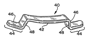
[00023] An improved clip, generally designated 40, for
application to the sclera is shown in Figs. 3-6. The clip,
generally designated 40 includes a body portion 42, with two
opposed feet 44 extending from the opposite ends of the body.
As can be appreciated, the clip 40 should present no sharp edges
that would irritate or damage tissue that comes into contact
therewith.
[00024] In practice, the body 42 has a length that may vary
from approximately 3.5 mm to 6.0 mm, depending on the desired
degree of compression of the sclera. Where less compression is
indicated, most likely in younger patients, a shorter clip is
used. Conversely, where more compression is indicated, most
likely in older patients, a longer clip is used. As can be
appreciated, the length of the body 42 also generally defines
the working length of the clip. The body 42 has a width of from
approximately 1.0 mm to 2.5 mm, and a thickness of from between
approximately 600 gm to 2.00 mm.
[00025] The opposed feet 44 are generally L-shaped (as seen in
Fig. 4), with the free end of the long leg 46 of the L having a
curved or semi-circular configuration (best seen in Figs. 3 and
6) so as to reduce the likelihood of damage to tissue contacted
by the feet. These curved ends are adapted to be received in
pockets made in the surface of the sclera, as will be discussed
in greater detail below.
CA 02572217 2006-12-22
WO 2006/007475 PCT/US2005/022274
-6-
[00026] The feet 44 have a working length, as defined by the
long leg 46 of the L, of approximately 200 m. The short leg 48
of the L measures between approximately 100 m in length and 200
m in length. Thus, the overall length of the clip 40 is the
sum of the length of the two feet 44 and the length of the body
42, and consequently ranges from approximately 4.0 mm to 6.5 mm.
[00027] In keeping with an aspect of the invention, the body
42 of the clip 40 is formed with a reverse bend (i.e., the body
curves downwardly) so that, when the clip 40 is applied to the
eye, the clip 40 pushes down on or compresses the sclera, thus
causing additional deformation of the sclera. Alternatively,
the central portion of the body 42 of the clip 40 may be greater
in thickness than the ends to achieve the same effect. The
amount of the reverse bend is generally the same as the length
of the leg 48 of the feet 44, i.e., from 100 m to 200 m, but
may be more or less depending upon the amount of scleral
compression needed.
[00028] The reverse bend exerts an inward force to assist the
failing contraction of an aging ciliary body, thus providing
what is known as the "Balkoff wedge effect," named after George
Balkoff, M.D. More specifically, the pressure created by the
reverse bend pushes the ciliary muscle inward and forward,
modifying the position of the ciliary processes and the location
of the zonular plexus, and thus releasing the tension of the
zonule and provoking the deformation of the crystalline lens by
allowing the lens to move forward and increase its anterior
curvature. This allows for an increase in the lenticular power,
thus causing the lens to accommodate. Alternatively, the clip
body could be enlarged in the central area between the points to
provide the same effect.
[00029] The clip 40 may be made of a variety of suitable
biocompatible materials, including titanium and polymethyl
methacrylate (PMMA). Preferably, the entire clip is molded from
PMMA. Alternatively, the body 42 of the clip 40 may be molded
from PMMA, while the feet 44 are made from titanium. The
titanium feet 44 are secured to the body 42 by overmolding the
1
CA 02572217 2006-12-22
WO 2006/007475 PCT/US2005/022274
-7-
body with the feet 44 in situ, so that the molten PMMA flows
around securement legs 50 that extend from the short leg 48 of
the feet 44. The securement legs 50 may be as much as 500 m in
length, to insure that a sufficient length is received in the
body 42 to maintain structural integrity. The clip may also be
coated with appropriate bioactive materials, such as sytostatic
drugs which have anti-inflammatory characteristics.
[00030] Turning to Figs. 7-9, a further embodiment of an
ophthalmic clip 40 according to the present invention is shown.
The clip is similar, except dimensionally, to that shown in
Figs. 3-6, and identical reference numerals are used. With
reference to Figs. 7-9, the body 42 has a length that may vary
from approximately 2.0 mm to 5.0 mm (preferably approximately
2.5 mm), depending on the desired degree of compression of the
sclera. The clip 40 has a width of from approximately 1.0 mm
to 2.5 mm (preferably approximately 1.0 mm), and a thickness of
from between approximately 200 m to 1.00 mm (preferably
approximately 250 m).
[00031] The opposed feet 44 are generally L-shaped (as seen in
Fig. 8), with the free end of the long leg 46 of the L having a
curved or semi-circular configuration (best seen in Figs. 7 and
9) so as to reduce the likelihood of damage to tissue contacted
by the feet. These curved ends are adapted to be received in
pockets made in the surface of the sclera, as will be discussed
in greater detail below.
[00032] The feet 44 have a working length, as defined by the
long leg 46 of the L, of approximately 1.5 mm to 2.5 mm
(preferably approximately 2.5 mm), the longer length helping to
maintain the clip in the pockets in the sclera. The short leg
48 of the L measures between approximately 800 m in length and
1.5 mm in length and is preferably approximately 800 Am in
length. The overall length of the clip 40 ranges from
approximately 5.0 mm to 7.5 mm.
[00033] The body 42 of the clip 40 is formed with a reverse
bend (i.e., the body curves downwardly) so that, when the clip
40 is applied to the eye, the clip 40 pushes down on or
CA 02572217 2006-12-22
WO 2006/007475 PCT/US2005/022274
-8-
compresses the sclera, thus causing additional deformation of
the sclera. In practice the reverse bend has a radius of
curvature of between 6.0 mm and about 9.0 mm and is preferably
approximately 7.5 mm.
[0,0034] Turning to Figs. 10-12, a third embodiment of a clip
54 according to the present invention is shown. The clip 54 is
similar to, that shown in Figs. 7-9, so that identical reference
numerals are used for corresponding structure. As seen in Figs.
10-12, the clip 54 includes an additional foot 56 extending from
each end of the body and overlying the foot 46 so as to define a
space 58 therebetween. The space 58 is adapted to receive
therein the portion of the sclera defined by the incision for
making the pocket 52 for receiving the foot 46 and the surface
of the sclera, and measures approximately 300-400 m. When
applied to the sclera, the feet 46, 56 capture the sclera
therebetween to help maintain the clip in position.
[00035] The clip may include other features that help ensure.
that the feet are retained in the pockets made in the sclera.
With reference to Fig. 13, a fourth clip embodiment is shown,
generally designated 60. The clip 60 comprises two parts 62,64.
The first part 62 is similar to the clip 40 shown in Figs. 7-9.
The second part 64 overlays the first part 62, and includes a
series of retaining pegs (3 shown) 66a,b,c that are received in
mating apertures 68a,b,c in the feet and body of the first part
62. When the feet of the first clip portion are received in the
pockets made in the sclera, a pin hole" is made in the pocket
through which the retaining pegs 66a, 66c are placed in order to
be inserted into the apertures 64a, 64c, thus positively
securing the clip 60 to the sclera. The central peg 66b on the
second part 64 and aperture 68b on the first part 62 help to
properly locate and align the two parts of the clip 60 during
their assembly and application to the eye.
[00036] A further alternative of the clip is shown in Fig. 14,
and is generally designated 70. The clip 70 is generally
similar to that shown in Figs: 7-9, but includes a central
aperture 72 in the body portion of the clip through which is
received a "screw" or other fastener 74. The screw 74 has a
CA 02572217 2006-12-22
WO 2006/007475 PCT/US2005/022274
-9-
point that, upon insertion into the central aperture 72, bites
into the sclera to a depth of, e.g., approximately 100 m to
secure the clip 70 thereto. For example, the shank of the screw
74 may include, e.g., a spiral thread or hook to positively
secure the clip to the sclera.
[00037] Turning to Fig. 15, an additional embodiment of the
clip, generally designated 80 is shown that also has means for
ensuring that the clip stays in place on the sclera. The clip
80 is similar to that shown in Figs. 7-9, except that each foot
has at least one aperture 82 therein through which scleral
tissue will grow after application of the clip 80 to the eye as
part of the healing response to making the incision for the
pockets that receive the feet of the clip. The tissue that
grows through apertures 82 thus serves to hold the clip in place
on the eye.
[00038] A method of applying the clip of the present invention
to the eye will now be set forth. First, the eyelid is held
open with a lid speculum and a topical anesthetic, such as a
sub-conjunctival lidocaine, is appl"ied to the eye. Then, the
location of the ciliary body is determined, for example, by
using commercially-available ultrasound equipment. With
reference to Fig. 2, an incision 38 is then made in the
conjunctiva parallel to the scleral-limbal junction so as to
dissect the conjunctiva bypassing the Tenon's capsule 28. The
incision is then deepened into the episclera. The incision is
opened and, if necessary, the Tenon's capsule is laterally moved
to expose the sclera 20. Opposed pockets 52 are made in the
surface of the sclera for receiving the opposed feet of the clip
using a preset marker. The openings of the pockets are spaced
approximately 3.5 to 6.0 mm apart, depending on the length of
the clip body, and have a depth (in a direction generally
parallel to or concentric with the surface of the sclera) that
corresponds to the length of the foot, i.e., from between
approximately 200 m to 2.5 mm. The pockets extend no deeper
into the sclera from the surface thereof than approximately 50
percent of its thickness, i.e., no deeper than about 350 m, and
CA 02572217 2006-12-22
WO 2006/007475 PCT/US2005/022274
-10-
preferably extend no deeper than approximately 200 m.
[00039] The clip is then loaded onto an application tool,
which may simply comprise a grasping forceps, which grips the
short legs of the feet to apply an axially compressive force to
the clip along its body, thus bending the body and moving the
feet toward each other. The feet are then introduced into the
pockets 52 made in the sclera. If the clip 54 according to
Figs. 10-12 is used, the portion of the sclera between the
incision for the pocket 52 and the surface of the sclera is
received in the space 58 between in the feet 46 and 56. If the
clip 60 according to Fig. 13 is used, additional pin holes are
made in the pockets for receipt of the pegs 66a, 66c. Once
these holes in the pocket are made, the second part of the clip
62 is assembled onto the first part of the clip 62. If the clip
70 according to Fig. 14 is used, the screw 74 is inserted into
the aperture 72 and manipulated so that the point thereof bites
into the sclera.
[00040] The applied clips have a generally low profile,
closely adhering to the curvature of the eye, thus providing
reinforcement to the sclera. The Tenon's capsule 28 is then
reapposed over the clip and the conjunctiva closed. No suturing
is needed as the conjunctiva self seals. Preferably, a fibrin
adhesive, such as Tisseel VH fibrin sealant available from
Baxter Healthcare Corporation, may be applied over the closed
conjunction to accelerate healing. The procedure is then
repeated for each of the four quadrants, as deemed necessary by
the surgeon, so that the clips are applied to the eye equally
spaced about the cornea 24 between the adjacent rectus muscles.
An ointment is applied to the eye, which is then patched for 24
hours.
[00041] As can be readily appreciated, the procedure can be
simply reversed by merely again gaining access to the sclera by
making an incision in the conjunctiva over the clip, moving the
Tenon's capsule to expose the clip, and then removing the clip.
[00042] The application of each clip should deform the uvea
and move the sclera inwardly approximately 0.5 mm, for a total
of 2 mm if four clips are applied. This will increase the
CA 02572217 2006-12-22
WO 2006/007475 PCT/US2005/022274
-11-
amplitude of accommodation, thus reversing the effects of
presbyopia. This inward movement of the sclera should also
increase the angle of the canals of Schlemn, thus increasing the
aqueous flow and decreasing the intra-ocular pressure, to
ameliorate the effects of glaucoma.
[00043] Thus, a method and a clip for performing the method
have been provided that fully meet the objects of the present
invention. While the invention has been described in terms of a
preferred ophthalmic clip and method, there is no intent to
limit the invention to the same. Indeed, the clip may have
application to medical procedures in addition to that described
above. Instead, the invention is defined by the scope of the
following claims.