Note: Descriptions are shown in the official language in which they were submitted.
, 74613-14D
CA 02572470 2007-O1-16
CONFOCAL-REFLECTION STREAK LIDAR APPARATUS
WITH STRIP-SHAPED PHOTOCATHODE, FOR
APPLICATION AT A WIDE RANGE OF SCALES
This is a divisional of Canadian Patent
Application Serial No. 2237894 filed November 14, 1995.
BACKGROUND
1. FIELD OF THE INVENTION
This invention relates generally to imaging the
volume of a turbid medium, together with objects embedded or
suspended in such a medium; and more particularly to use of
streak-lidar apparatus to monitor phenomena at an extremely
broad range of scales - including detection of a tumor less
than a millimeter across, in living tissue; or an underwater
object in the ocean, or vehicles in fog, or a variety of
other objects in turbid media.
2. PRIOR ART
The present invention has applications spanning a
range of sizes, and is believed to integrate diverse,
heretofore nonanalogous fields. For reasons to be explained
in this document, these fields have not previously been
linked.
These application fields include imaging of
volumes of the atmosphere with aircraft moving through such
volumes - over a range (and atmospheric volume) on the
scale of kilometers. It also includes imaging of ocean
volumes - together with submarines, sunken ships, submerged
fuel drums and the like, over a field of examination that is
some one to two kilometers wide and perhaps many kilometers
long.
- 1 -
74613-14D
CA 02572470 2007-O1-16
In addition these applications include medical
imaging of human or animal tissue, with tumors in the
tissue. The tumors may be a small fraction of a millimeter
in diameter, either suspended within the living tissue or
growing on human or animal organs at a remote interior
surface of the tissue. Here the volumes of tissue that can
be imaged range from perhaps two to twenty centimeters
across.
- la -
,74613-14D
CA 02572470 2007-O1-16
Intermediate-scale applications include imaging of a fogged-in airport
and its environs, together with the land and air vehicles and other structures
in the
area, or imaging of a riot zone (or battlefield) filled with tear gas or other
nebulized
material together with people, vehicles and the like in that zone.
These many types of imaging have not heretofore been linked.
Probably the reason for this is that prior artisans have not fully appreciated
how to
use lidar to obtain a direct distance-to-depth mapping in a simple natural
real-time
display, capable of direct volume implications.
At a medical or laboratory scale, most previous users have instead
become entangled in fiber-optic encoders and other counterproductive
digressions.
Furthermore, most or all previous workers in lidar have failed to appreciate
the
critical importance of the confocal condition though that condition is
recognized in other fields. (By "confocal condition" we refer to
configurations that
cause emitted and reflected probe beams to lie very nearly coincident upon one
another.)
An example of failure to appreciate the importance of that condition
appears in U. S. Patent 4,704,634 of Kato who actually uses a pulsed,
unconstrained spherical wave (or "flood beam") as his emitted beam.
Accordingly
bench-scale lidar configurations have not been reasonably optimized.
At ocean-volume scale, lidar systems heretofore have not been made
effective at all. In this case, in addition to the failures of recognition
outlined in the
preceding paragraph, previous workers have evidently overlooked the potential
use of streak lidar.
U. S. Patent 3,719,775 (predating the invention of the streak tube) to
-2-
74613-14D
CA 02572470 2007-O1-16
Takaoka, addressing terrain-imaging applications, mentions in passing the use
of
a vertical fan-shaped beam, carried by an aircraft with the wide dimension of
the
beam at right angles to the direction of motion. That configuration is not
Takaoka's invention, and he teaches nothing about its effective use.
Heretofore neither Takaoka nor any other artisan has proposed use
of such a fan-shaped beam, projected from aircraft either with a streak tube,
or with any other efFective means of reading terrain-generated reflection.
The point of commonality in all the applications, at different scales,
mentioned earlier is the magnitude of the effective turbidity on a per-unit-
distance
(or -volume) basis. This is the consideration that controls ability to probe
and
resolve turbid media with a pulsed laser and a streak tube. Thus ocean volumes
while vastly greater in extent than living tissue are correspondingly lesser
in
turbidity.
Several techniques have evolved over the years for overcoming the
problems associated with detecting targets in a light-scattering medium.
Ocean-volume scale One technique uses a narrow beam from a
pulsed laser, such as a doubled YAG, to scan the ocean. Generally, the beam
transmitter and the receiver aperture, which must be quite large to collect
sufficient
energy, are scanned together, using scanning,mirrors or other devices such as
prisms.
The energy received from each pulse is detected with a
photomultiplier, or similar quantum-limited device, and the resulting signal
is
amplified with a logarithmic-response amplifier, digitized and then processed.
Because the pulses are short, typically 10 nanoseconds, the detection
electronics
-3-
74613-14D
CA 02572470 2007-O1-16
must be very fast, digitizing at 200 MHz or faster.
Since the pulse rate is low, the processing rates required to analyze
the data from each pulse are within the state of the art. Such methods require
the
use of mechanical scanners that are slow and difficult to build, particularly
if they
are to be mounted on aircraft.
In accordance with a primary advantage of the present invention, the
need for fast digitizing electronics and mechanical scanners is eliminated.
(As will
be seen, however, in certain of the applications outlined above, at least in
principle
fast electronics can be substituted for a streak tube.)
Another technique is range gating, which uses a pulsed flood beam
and a number of fated image intensifiers with charge-coupled devices (CCDs).
The intensifiers are gated on when the beam pulse reaches a specific depth.
Typically one gate is applied just as the pulse beam that encounters
the object returns to the receiver, so that the full reflected return is
obtained. A
second intensifier is gated on a little later to detect the shadow of the
object. The
image of the target is obtained by taking the difference of the two images,
which
then eliminates the seawater backscatter and enhances the target signature.
Several drawbacks are associated with the range-gating technique.
Range gating does not allow utilization of all, or substantially all, of the
information
returned from each pulse to create three-dimensional data sets.
Rather in such prior-art systems, although a volume of the medium is
illuminated, by range gating only one specified layer (depth increment) of the
illuminated medium is selected. Thus the signal above and below the range gate
is rejected discarded.
-4-
74613-14D
CA 02572470 2007-O1-16
As will be clear, of the energy transmitted into the volume of the
medium and returned toward the transceiver, only a small fraction is used.
This
operating arrangement constitutes a monumental waste of optical energy.
Additionally, a full-depth data set cannot be created from a single
pulse. Rather, full-depth information can be obtained only by collecting many
pulses, during which process the platform, aircraft or other vehicle must
remain.
stationary. (To create a full-depth image, the number of shots required is a
large
multiplicity. Consideration of this fact is another way of appreciating the
amount of
energy wasted.)
Despite the availability of such techniques, existing lidar systems are
limited by the size of the receiver optics that can be used in a scanner.
Generally
the light reflected from targets that are deeply positioned, or suspended in a
very
turbid medium, is weak.
Although large-diameter optics can aid in maximizing the amount of
light collected from weak returns, the size of the optics that can be used in
a
scanner is restricted by the size of the moving prisms or mirrors. Such
cumbersome mechanisms sometimes can be eliminated, as in selected
applications of the present invention, by utilizing the motion of a vehicle
e~a.,
boat or aircraft carrying the system so that the dimensions for scanning can
be reduced to one.
The scanning problem, however, is still formidable and restricts the
size of the apertures that can be used. Moreover, volume scanning systems are
very expensive, and require considerable power and weight. Consequently, for
large-scale applications the ability to install such systems in aircraft or
other
-5-
74613-14D
CA 02572470 2007-O1-16
vehicles is restricted.
Furthermore, those systems that utilize range gating, instead of
volume scanning, suffer from poor range resolution and area coverage. When a
target object is at a different depth from the expected, the optical return is
subtracted as well as the background, and poor performance results.
Additionally,
very large pulse energies are required to obtain signal-to-noise ratios
sufficient for
detecting objects at even moderate depths.
What has been needed heretofore is an imaging system that
provides an accurate and reliable image of a suspended object, eliminates the
problems associated with mirror scanning for large-scale systems, and utilizes
all,
or substantially all, of the information returned from each pulse to eliminate
laser-
energy waste.
Medical scale Streak tubes have been demonstrated in
transillumination geometries to detect the presence of small tumors in tissues
(see, e~a., U. S. Patents 5,278,403 and 5,142,372 to Alfano; and 5,140,463 to
Yoo). The transillumination technique, however, yields only two-dimensional
images and cannot determine the depth of a tumor.
Furthermore, transillumination yields only a shadow signature. Such
data are subject to relatively poor detection range.
As can now be seen, in the field of the invention the prior art has
failed to provide solutions to important difficulties of observing the
operating
environment and receiving communications.
SUMMARY OF THE DISCLOSURE
The present invention corrects the failings of the prior art. The
-6-
74613-14D
CA 02572470 2007-O1-16
invention provides an imaging system for detecting an object in a turbid
medium
such as living tissue, or water or air. The invention is useful in probing the
contents of any turbid medium through which light can pass, even if absorbed
and
scattered, as long as some return can be obtained.
The system includes a means for generating a periodic series of
discrete pulse beams in the shape of fan beams, each of which is substantially
uniform in intensity or with greater amounts of energy at the ends of the fan
to compensate for losses due to the greater distance to illuminate sections of
the medium.
In operation, a single pulse beam is emitted to illuminate a section of
the medium. A large-aperture optic collects the back-reflected portions of the
pulse beam and focuses the reflected portions on a field-limiting slit. That
slit,
located in front of the photocathode, rejects multiply reflected light.
For best measurement performance it is very important that the
successive depths illuminated by the pulsed beam i. e., the incremental
volumes, transverse-needle-shaped probe volumes through which the beam
successively passes all be imaged in common back to the slit. This condition
is most straightforwardly met by arranging the collecting optics to receive
light
through a second fan-shaped volume that at least nearly coincides with the
volume of the transmitted fan-shaped pulse beam.
A lens, positioned between the field-limiting slit and photocathode,
reimages the image on that onto the photocathode. Coupled to the streak tube
is
an imaging detector, typically a CCD, which detects signals generated by the
streak tube in response to the reflected portions of the pulse beam impinging
on
-7-
74613-14D
CA 02572470 2007-O1-16
the photocathode.
Other imaging detectors, such as a TV camera or photodiode array,
may be used instead. To obtain a volume display of the medium, the pulsed
beam can be repeated while its physical location and that of the reflection
are
shifted together for example by moving the generating means and receiver
normal to the longitudinal axis of the pulse beam so that each pulse
illuminates
adjacent sections of the turbid medium.
A volume display is thus generated by combining the returns from
adjacent sections of the medium. All, or substantially all, of the light
returned from
each pulse is used unlike the situation previously described for range-gating
systems.
The streak-tube photocathode is substantially a thin strip behind a
field-limiting slit on which the illuminated strip of the ocean, or other
scattering
medium, is imaged by the receiver optics. That strip is essentially fixed,
unlike for
example the system of the Kato patent discussed earlier which requires a
rectangular photocathode to accommodate the migrating, electronically shifted
region, on the cathode, from which the downstream streak-tube components will
draw their signal.
In the present system, since the strip is fixed we say that the cathode
is "substantially a thin strip". It is to be understood that this language
encompasses use of a rectan uc~lar cathode if only a thin-strip section is
used.
The thin-strip, in either case, is fixed in location on the cathode
surface but should be wide enough to accept the entire image when the slit is
opened to its maximum width. A variable-width slit is very desirable,
providing
_g_
74613-14D
CA 02572470 2007-O1-16
easy adjustment for optimal viewing over a wide variety of turbidity
conditions and
detection ranges. This condition, closely related to the confocal geometry
mentioned earlier, has been ignored in many prior-art systems.
When the laser beam pulse, typically a few nanoseconds in duration
for ocean scanning and one or a few picoseconds for medical and laboratory
applications, returns to the receiver from the near surface of the medium, the
electronic sweep of the deflecting system is initiated.
The following time history of the returning signal spread across the
lateral surface of the tube anode is then a record of the reflection from the
medium itself. The image includes any bodies embedded in the medium, such as
mines or submarines in the ocean or tumors in living tissue. The image also
includes the reflection from the near surface of each such object, and the
shadow
below the object.
Because the slit-shaped cathode is long and covers the width of the
ocean illuminated by the fan-shaped beam from the laser, the image on the
anode
phosphor or area detector is a wide vertical section of the ocean or other
medium.
in addition to imaging objects fuily embedded e~a., immersed and floating
in the medium, the invention also applies to imaging objects on the bottom
(for the ocean) or at a far interior surface of the medium, and to obtaining a
profile
of bottom or far-surface topography.
This may be the only way to distinguish silt-covered objects such as
archaeological remains lying on the bottom of the ocean, or tumors growing on
a
living organ in a human or animal body, from the bottom or the organ itself.
Even
the gross relief of the sea bottom or of an organ can be imaged, often quite
_g_
74613-14D
CA 02572470 2007-O1-16
plainly, by this process.
For ocean or air scanning the invention described herein can be
employed, for example, from a fixed-wing aircraft or helicopter, from boats on
the
water surface, or from submerged vehicles for search at great depths or from
a fixed tower, as appropriate. A.tower may be best for imaging, as an example,
aircraft in fog at an airport.
The invention is equally applicable to the analysis of very small
volumes using very short laser pulses, on the order of a picosecond duration
for
example, since the streak tube can capture such time intervals. These volumes,
and the objects in them, may be for example submillimeter tumors in a human
breast that is only, say, 2 to 20 cm thick. In the case of relatively thick
tissue,
imaging inward from two or three difFerent surfaces may be necessary.
As previously mentioned, the linking of these divergent applications
at several different ranges of scale is believed to be novel. With respect to
the
prior art, we are not aware of suggestion of any single technology for use in
these
several volume-size ranges, which thus represent nonanalogous arts.
The image on the anode can be photographed by a CCD camera or
similar device, particularly by logarithmic-response area-array CCD-like
detectors.
Bitwise, the image is read out slowly, but all in parallel, compared with the
rapid
progress of the returning signal which is serial with respect to the sweep
direction
of the streak tube. The anode can also be replaced by a thinned backside-
illuminated CCD.
Either technique for acquiring pixel-based images facilitates viewing
of the phenomena on a cathode-ray screen directly or, after encoding the
signal,
-10-
74613-14D
CA 02572470 2007-O1-16
processing such images to enhance them. Those versed in the
art are aware of various enhancement techniques, such as
subtracting the mean return from all the return values for a
recorded section of the medium.
Subsequent display of such sections can be
manipulated by adding many sections together to provide a
three-dimensional view of the breast, or airport environs,
or underwater scene. Such three-dimensional data sets are
obtained by moving the sensor system normal to the fan beam
between each exposure, so that each sectional image is from
an adjacent section of the medium.
Besides giving an overall picture of the
situation, this technique also enhances detection (and
reduces false alarms) by enabling operators or programmed
computers to notice small or fragmentary images, near the
electronic detection limit, that might not be apparent in
any single section image.
All of the light recaptured is utilized in
creating three-dimensional data sets. This characteristic
of the system avoids wasting energy from the laser.
According to one aspect of the present invention,
there is provided a system for imaging a volume of a turbid
medium, with objects therein, said system being for use with
means for bodily transporting at least part of the system
with respect to said turbid volume; said system comprising:
means for projecting a pulsed thin-fan-shaped beam to
selectively illuminate a thin section of such turbid volume;
a streak-tube cathode for receiving reflected light back,
approximately along the illumination-propagation direction,
from the thin section of turbid volume; means for focusing
the reflected light onto the streak-tube cathode
- 11 -
74613-14D
CA 02572470 2007-O1-16
substantially directly; said focusing means comprising:
(1) no "glass plate stack" image slicer for optically
mapping portions of said reflected light onto portions of a
light-receiving surface, and (2) no other type of image
slicer for optically mapping portions of said reflected
light onto portions of a light-receiving surface, and (3) no
pixel-encoding fiber bundle for optically mapping a two-
dimensional reflected image into a line image, and (4) no
other pixel-encoding fiber bundle for optical mapping of a
reflected image, and (5) no other optical image-mapping
device other than basic optical elements such as a lens or
mirror; streak-tube means, responsive to the focused
reflected light, for forming therefrom a corresponding
composite electronic image of the turbid-ocean-volume thin
section as a function of propagation depth; means for
restricting the light received by the streak-tube cathode,
from the focusing means, to substantially only reflection
directly from said selectively illuminated thin section;
means for sequentially operating the beam-projecting means,
during operation of such bodily-transporting means, to
project a sequence of beam pulses to illuminate successive
thin sections, and generate a corresponding sequence of
composite electronic images.
According to another aspect of the present
invention, there is provided a streak-lidar imaging system
for medical applications and comprising: a pulse laser for
multispectral transmission to a biological sample; and
streak-lidar depth-resolved imaging means selectively
responsive to different spectral components of a return
beam.
- lla -
74613-14D
CA 02572470 2007-O1-16
BRIEF DESCRIPTION OF THE DRA4dINGS
Fig. 1 is a generalized schematic elevational
showing of a preferred embodiment that employs a moving
platform to translate the apparatus of the invention, to
scan objects in a turbid medium;
Fig, 1A is similar but more specifically employing
an aircraft as the moving platform to view objects
underwater;
Fig. 1B is a like showing of a medical scanner
using a rotating mirror;
- llb -
74613-14D
CA 02572470 2007-O1-16
Fig. 1 C is a showing in plan of the same device;
Fig. 1 D is a showing like Fig. 1 B of a similar scanner but with a
translating mirror;
Fig. 1 E is a highly schematic showing, with some portions generally
in perspective or isometric projection, and other portions merely
diagrammatic, of
a handheld medical probe with associated equipment;
Fig. 1 F is a highly schematic plan of an airport scanner;
Fig. 2 is a block diagram of a preferred embodiment of the invention;
Figs. 3(a) through (c) are timing diagrams showing signals obtained
through using the systems of Figs. 1 through 2;
and CCD;
Fig. 4 is a diagram of the beam distribution on the MCP, phosphor
Fig. 5 is a schematic diagram of the laser and the projection optics of
the Fig. 2 preferred embodiment; and
system.
Fig. 6 is a schematic diagram of the detection system of the Fig. 2
DETAILED DESCRIPTION OF THE PREFERRED EMBODIMENTS
The present invention provides a system for detecting targets located
in a light-reflecting medium, such as dirty, hazy or foggy air; and such as
water or
living tissue. The system can be used to observe a water interface, the
structure
of the medium including the distribution of particulate matter and suspended
or otherwise embedded bodies and a bottom or far-interior-surface profile.
More particularly the invention can be used to detect objects in any medium
through which light can pass, even if absorbed and scattered, provided that
some
-12-
74613-14D
CA 02572470 2007-O1-16
substantially directly reflected light can be obtained.
The system includes a light source for producing a series of discrete
narrow, fan-shaped pulse beams which have a modified nonuniform intensity
distribution to produce uniform signal-return. The reflected portions of the
pulse
beam are received by a detection system comprising receiving optics, a streak
tube and an imaging area detector.
In operation, the invention by some means physically shifts the
emitted or received beams together. To say this more precisely, the invention
shifts (e~g., translates) the positions of at least portions of both the
emitted and
reflected beams together. For example the apparatus of the invention may be
mounted on a platform (such as a vehicle) adapted for movement along the
turbid
medium. A light source emits periodic pulse beams to illuminate a succession
of
thin slices of the turbid medium.
The detection system includes a light-collecting optical element, a
field-limiting slit, a streak tube and an imaging area detector. The light-
collecting
optic receives reflected light and images it onto a field-limiting slit, which
rejects
multiply scattered light.
A lens or other focal element, disposed between the field-limiting slit
and the photocathode of the streak tube, is preferably used to focus the image
at
the slit onto the photocathode. Because of the narrow fan-shaped illumination
and
the field-limiting slit at the cathode, the light collected is substantially
directly
reflected light, and not light multiply reflected by the medium thus providing
improved image contrast.
To collect the maximum amount of light from weak returns, the
-13-
74613-14D
CA 02572470 2007-O1-16
aperture of the optic should be as large as possible. The streak-tube
photocathode, however, should be big enough to encompass the image of the fan-
beam-illuminated volume.
For this purpose the cathode itself may be slit-shaped, with a very
large aspect ratio such as 200:1 or 300:1 so as to avoid wasting expensive
sensitive surface area, and thus to economically do its job. If the cathode
happens to have a much lower aspect ratio, even 1:1, the system uses only a
slit-
shaped portion generally fixed in position, but of variable width as will be
explained.
Inside the streak tube, a cross-sectionally slit-shaped stream of
photoelectrons emitted from the cathode is accelerated and then
electrostatically
focused on the phosphor layer or anode of the streak tube. On passage from the
cathode to the anode, the photoelectrons pass through a deflecting electric
field,
whose strength is tamped to sweep the photoelectrons across the anode.
The tamping deflection field is created by a varying voltage applied
to the deflecting plates in the tube.
The result at the anode is a two-dimensional signal, the resultant of
(1 ) the temporal variation of the detected light reflected from progressively
deeper
regions of the turbid medium, in one dimension, and (2) the lateral variation
in
intensity of the reflected light along the narrow, fan-shaped pulse beam in
the
perpendicular dimension.
The focused electrons can be sensed directly by an area detector,
such as a thinned backside-illuminated CCD. Alternatively the electron energy
can
be converted to light by a phosphor layer on the anode, and the light emitted
from
-14-
74613-14D
CA 02572470 2007-O1-16
the phosphor then passed to a detector array.
A volume display of the medium is generated by coordinating the
return signals for successive transmitted/reflected beams with the beam
positions.
As mentioned earlier, the two beams are shifted together while the laser is
repetitively pulsed.
This shifting can be accomplished in either of two basic ways: by
translating (or rotating) the transmitter and receiver together, or (2) by
translating
or rotating an optical element, most commonly a mirror, that controls the beam
positions. Motion preferably is normal to the long dimension of the fan-shaped
pulse beam, so as to illuminate adjacent sections of the medium with best
efficiency (and compactness of the imaged volume).
In the first case, the motion of a vehicle is used to provide the scan
or motion of the fan-shaped pulse beam and the likewise fan-shaped volume
through which the return beam is collected.
In either case, all or substantially all of the light returned from each
pulse is used to create three-dimensional data sets. The coordination of
signals
with beam positions to provide a volume display can be accomplished by simply
displaying (or analyzing) the resulting successive two-dimensional signals
sequentially, with a comparable time base.
The result is to show (or automatically evaluate without showing) a
kind of movie that emulates a virtual visual experience (or data-collection
process)
of travel through the medium. The movie can be run and watched in real time
while data are collected, or later at actual speed, or faster or slower, or in
stop
frames, just as an ordinary video is shown.
-15-
74613-14D
CA 02572470 2007-O1-16
Alternatively the data can be instead processed to produce a two-
dimensional picture of the three-dimensional volume of the medium in
perspective or isometric, or any other viewing mode preferred using any of
the myriad available computer programs for visualizing three-dimensional
bodies.
If desired, through holographic projection an actual three-dimensional
image can be formed and viewed. Many other uses of the incrementally collected
volumetric data will now be clear to those skilled in the relevant arts.
By using very short pulses, on the order of one to a few picoseconds
in duration for example, the present invention can be used to resolve
correspondingly very small objects. The streak tube collects the rapid return
of
the backscattered light, distributing the return in space and then reading the
return
out slowly.
The return is in the low-nanoseconds and medium-to-high-
picosecond range, and the system of this invention allows a readout in
millisec-
onds, thus obviating the necessity for faster electronic readouts.
At relatively long ranges, on the other hand, such as the ranges
suitable for airborne surveillance of ocean volumes, modern electronics
actually is
fast enough to allow dispensing with the streak tube entirely, and simply
using a
very fast frame cache to collect the data serially with respect to the narrow
dimension of the slit. This system is within the scope of certain of the
appended
claims. The cache can be read out entirely in parallel, just as is done with
the
streak tube in other embodiments of the invention.
With a streak tube, all of the signal from each pulse of the fan-
shaped pulse beam width and depth that is back-reflected is observed at once,
-16-
74613-14D
CA 02572470 2007-O1-16
avoiding the need to use a multiplicity of pulses to obtain three-dimensional
information.
Normally, laser beams are nonuniform in intensity, with maximum
intensity at the center of the beam and minimum at the outermost edges. This
can be changed by applying fiapered coatings to the laser mirrors, or by the
use of
optical means external to the laser.
An optical inverter, comprising a series of lenses and a diamond-
shaped mirror arrangement, enhances the intensity at the outer portions of the
pulse beam by optically inverting in one dimension along the fan width the
intensity pattern of the pulse beam. The result is a pulse beam that
compensates
for the effect caused by longer paths at the ends of the fan to produce a
signal
return that is substantially uniform in intensity.
Fig. 1 shows a representative configuration for embodiments of the
invention in which the two beams are displaced together by actual bodily
physical
translation of the transmitting and receiving apparatus. A moving platform or
stage 10 carries the apparatus 14 of the invention i. e., mainly the laser,
streak tube, electronics, and associated optics.
A narrow, fan-shaped pulse beam 12 is projected from the transmitter
to the medium 13, with the long dimension of the beam normal to the direction
11
of platform motion. The beam 12 illuminates a thin section 15 in the medium.
The beam picks out reflections and shadows for objects 17 that are
fully immersed or embedded in the medium 13, as well as irregularities and
objects 19 at the far interior surface 13' of the medium 13.
Coverage of a volume of the medium is obtained by issuing a series
-17-
74613-14D
CA 02572470 2007-O1-16
of discrete pulse beams 16-18 to illuminate adjacent sections of the medium.
During (or after) processing of the successive section images, the sections
can be
displayed to show a scan through a volume of the medium.
Thus the motion 11 of the platform 10 carrying the system 14 is used
to provide the scan of the pulse beam. The pulse rate to generate the series
of
discrete beams is set by the platform velocity.
In general, the rate may be high and the beam width 15 at the
surface of the medium narrow compared with the resolution determined by the
image-detector pixels. This is done to preserve temporal resolution, which can
be
reduced if the spatial width becomes large. In order to reduce the number of
readouts of the CCD, the pulses can be accumulated on a chip.
Fig. 1A is a direct extensioh of Fig. 1 to the case in which the
platform is an aircraft and the medium is the ocean. Objects of particular
interest
in this case, as suggested in the drawing, may be submarine craft, bottom-
tethered submarine platforms, drums of waste or fuel, etc. The system may
also,
however, be used to locate and monitor whales, or large schools of fish or
even contaminants released in great quantity in the case of spills, if
sufficient
difference of reflectivity relative to the seawater is available.
Figs. 1 B and 1 C show a medical system in which scanning is
provided by rotation 211 of a mirror 210. The medium 213 here may be a human
breast, or other living tissue.
A window 206 compresses the tissue slightly, for better viewing from
within the apparatus housing 207. Mounted within the housing, in addition to
the
rotating mirror 210, are an optical bench 210, the laser 222, and a lens and a
-18-
74613-14D
CA 02572470 2007-O1-16
stationary deflecting mirror 209. After traversing that mirror, the pulse beam
212
bounces from the rotating mirror 210 through the window 206 and into the
tissue
213.
Objects of interest include tiny tumors 217 embedded in the breast
213, or growing on the surface of a nearby organ 213', which here provides the
previously mentioned "far interior surface" of the medium 213. An opaque organ
213', or one whose reflectivity is very different from that of the breast 213,
may
itself be imaged in relief i. e., in silhouette.
A rotating mirror 210 introduces variations in angle of incidence
which may be undesirable in certain sensitive work, and also introduces a
variation in the lateral resolution with depth. Fig. 1 D shows a similar
system in
which translation 311 of a mirror 310 is substituted for rotation to avoid
these
potentially adverse effects.
Here the optical path is folded, using three mirrors 310, 310', which
instead translate in synchronism to also avoid variation in focal distance. In
Fig.
1 D the scanning mirror moves 311 on one table 305, and the two compensating
mirrors 310' move 311' in tandem on a second table 304.
The compensating-mirror table 304 moves in the same direction as
the scanning-mirror table 305 but at half the speed. The output beam 312 scans
308 linearly, as does the return beam (not shown) which traverses the same
path
in reverse to hold focus in return as well.
Fig. 1 E shows a handheld scanner 580, connected to a picosecond-
pulse laser 522 by a flexible fiber-optic coupler 510. The laser pulse is
shaped by
lenses in the scanner 580 housing, to form outgoing beam 512 which as before
-19-
74613-14D
CA 02572470 2007-O1-16
exits through a window 506 to illuminate the tissue 513 of a breast, or other
living
tissue.
Confocal at 512/536 with the laser beam 512 is the return beam 530,
which is focused by a lens 536 onto a variable field-limiting slit 526. From
the slit
the beam traverses another flexible coupler 525 to reach the photocathode 532
of
the streak tube 534 with deflecting plates 542, CCD 548 etc. The CCD is
coupled to electronics which produces the images 570 of tumors 517 on a CRT
display 256.
Alternatively the entire streak tube can be packaged in the handheld
unit 580, with cabling from the CCD output to a remote display. In either
event,
the handheld probe 580 can be readily placed anywhere on the body to probe
tissue with a minimum of patient discomfort.
By tilting the probe at a fixed position, the volume of interest can be
swept out or in many situations the face of the probe can be slid along the
patient's skin to obtain a more nearly translational scanning. Since imaging
is in
real time, the clinician can immediately probe areas of interest, generating
(and
recording for later use) optimal images.
Such easy scanning offers a tremendous advantage over X-rays,
MRI, etc. and is generally comparable to current ultrasound technology in ease
and noninvasiveness of use. Streak lidar, however, provides orders of
magnitude
finer resolution than ultrasound.
In operation the only source of motion is an operator's hand-imparted
motion of the handheld scanner 580. Some idea of the position of the scanner
relative to the breast is desirable, though as will be recognized parts of the
body
-20-
,74613-14D
CA 02572470 2007-O1-16
are intrinsically malleable and not readily amenable to precise location.
One way to provide positioning is by inclusion of a three-axis
accelerometer 585a, 585b, 585c, with data cables to the electronics for
interpretation. Another is passive, using modulation of a magnetic field
imposed
on the region of the testing laboratory where the scanner 580 is being held.
In
these two cases, relatively straightforward software must be provided for
debriefing the electronics, and calculating and presenting positional data for
recording in synchronism with the lidar display 256.
Still another approach is to provide two or more video cameras 581,
for recording visual images indicating the scanner's position in synchronism
with
recording of the CRT display 256, 570. This system requires little or no data
processing for scanner-position determination.
Yet another way to provide positioning information is by disposing
three transmitters of microwave or like radiation at calibrated points near
the test
area, and making the previously mentioned units 585a, 585b, 585c microwave
triangulation receivers rather than accelerometers. Like the first two
positioning
systems discussed above, this one does require some data processing.
Fig. 1 F represents an airport with a runway, taxiways 402, and fog
413 throughout the area. The pulse beam from the laser 422 is redirected by a
rotating mirror 410 to form the probe beam 412, which pierces the fog to image
the aircraft 417 on the ground and in the air as well as buildings 419. The
rotation 411 of the mirror unavoidably introduces variations in focus and
incidence
angle, which in this context are probably immaterial.
Fig. 2 shows a block diagram of a preferred embodiment of the
-21 -
74613-14D
CA 02572470 2007-O1-16
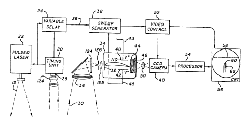
invention. A timing unit 20 initiates the probing sequence by causing the
laser 22
to emit a narrow, fan-shaped pulse beam 12 to illuminate a thin section of the
medium. After the Q-switch 84 (Fig. 5) in the laser 22 has closed, causing the
laser to fire, the timing unit 20 initiates operation of the variable delay
unit 24.
That unit issues a delay pulse 26 to initiate operation of the receiving
unit. To ensure that the delay is correct, a detector 28, such as a
photomultiplier,
is preferably used to sense reflected portions 30 of the pulse beam. The
timing
unit 20 measures this time and resets the variable delay unit 24 to ensure
that the
next delay pulse 26 is correct. Since the delay is variable, the invention can
be
operated at very different ranges i. e., from aircraft altitudes to medical-
scanner distances.
The reflected portions 30 of the pulse beam are collected and
focused on the photocathode 32 of a streak tube 34 by an optical element,
shown
here as a lens 36. The image, which includes a wide spread of scattered light,
is
chopped by the field-limiting slit 126 that is aligned with the returned image
of the
fan-beam, and serves to reject scattered light as well as limit the width of
the
electron image to a width smaller than the temporal sampling obtained by the
pixels in the imaging detector.
For best image quality, a lens 125 or other focal element preferably
is positioned as between the field-limiting slit 12 and the photocathode 32,
to
reimage the image at the field-limiting slit 126 onto the photocathode 32. The
photoelectrons 110 emitted from the photocathode 32 are accelerated by the
streak-tube anode voltage, and are focused into a line on the anode 44 by the
electrostatic or magnetic field distribution in the streak tube 34.
-22-
74613-14D
CA 02572470 2007-O1-16
The photoelectrons also are deflected by the electrostatic field set up
between the deflection plates 40 and 42 in the streak tube 34. In other words,
one field forms the image, and the other field set up between the deflection
plates
40 and 42 positions the image.
The delay pulse 26 initiates the action of a sweep generator 38,
which causes a linearly increasing voltage 43 and 45 to be applied to the
deflection plates 40 and 42 on the streak tube 34. The line-shaped electron
image is deflected by the plates 40 and 42 so that the line sweeps across the
streak-tube anode 44, thus converting a temporal variation in the input signal
into
a spatial distribution on the anode 44.
The temporal variation arises from different propagation times into
and out of the medium, from the apparatus to each successively illuminated
level
or depth within the medium, and then back to the apparatus. That time is of
course proportional to the distance or depth.
Hence the present invention provides in a very natural and elegant
fashion a direct mapping of each such depth, and thus in turn a direct mapping
of
each section of the turbid-medium volume being scanned, into distance along
the
anode (and any later display screen).
The anode 44 may be made of a phosphor, but since there are few
photoelectrons 110 from the return when the beam has penetrated many diffusion
lengths in the medium, additional photon gain is desired. Thus the anode 44 is
preferably made of a microchannel plate (MCP) intensifier, which provides the
gain
required to make photoelectrons 110 detectable.
The electron output of the MCP is reconverted to photons by a
-23-
74613-14D
CA 02572470 2007-O1-16
phosphor layer 46, so that the image of the temporal variation over the narrow
fan-shaped pulse beam 12, now converted to a two-dimensional image, can be
coupled to a detector array 48 by a coupling device, such as a lens 50.
Other coupling devices, such as a fiber-optic light pipe, may be used.
The detector array 48 shown is a CCD, but it could easily be a diode array,
and in
particular a photodiode n-channel MOSFET array or diode-limited CCD that
provides a logarithmic response to high light levels.
If the accelerating voltage is high, gain can be obtained through the
ionization created by the electrons directly in the detector. Thus the anode
44 can
be made of a backside thin CCD fabricated for this purpose, and an MCP and
phosphor are not required.
Before each new image arrives, the CCD detector array 48 is set to
read out the preceding frame, in preparation for receiving the new image. Once
the sweep generator has completed the voltage rise and resets, a command is
issued to the video control 52 to read the image on the CCD.
The data are then passed to a processor 54, or directly to a cathode
ray tube display 56, where a waterfall-like display of the section of the
medium
probed by the pulse beam 12 can be watched directly. Typical images are that
of
the surface 58 of the medium, a reflecting object 60 suspended in the medium,
and a shadow 62 from the reflecting object.
The subsequent display of such sections can be manipulated by
adding many sections together to provide as previously described a
volume display of the interior of the medium. Specifically, to collect such
sections
the emitter and sensor systems together can be moved normal to the
longitudinal
-24-
,74613-14D
CA 02572470 2007-O1-16
axis of the pulse beam 12 between (and, without any problem, during) each
exposure, so that the beams themselves are wholly shifted to illuminate and
reflect
from adjacent sections of the medium or portions of the beams can together
be shifted by motion of a mirror, tens, fiber-optic image relay, etc. to
accomplish a
like result.
As described above, the present invention if used outdoors to
probe deep depths would be limited by sunlight to operation at night only.
Daytime operation requires narrow-band interference filters 124, placed in
front of
the streak-tube cathode 32, to pass the laser wavelength and block all others.
The combination of the filters 124 and the short exposure time for
each element in the detector array 48 (typically 5 nanoseconds even for ocean
scans, thereby e~a. resolving 56 cm in depth) holds the background at each
pixel
to at most a few photoelectron counts.
Fig. 3 shows a timing diagram of signals obtained from the reflected
portions 30 of the pulse beam. The time history of the reflected portions 30
of the
beam is a record of the reflection from the medium itself, and from any bodies
suspended in the medium such as aircraft in fog, mines or submarines in
seawater, or tumors in body tissue including the reflection from the nearest
surface of such objects and of the shadow beyond them.
Because the part of the medium illuminated by the pulse beam 12 is
limited to a very thin section, the image on the phosphor layer 46 is a wide,
deep
section of the medium. The image can be photographed by means of a CCD
camera or similar device, particularly by logarithmic-response area-array CCD-
like
detectors, which read out slowly compared to the short duration of the
returning
-
74613-14D
signal.
CA 02572470 2007-O1-16
To obtain a higher power-aperture product, we now prefer to use a
fiber-optic coupler in place of the lens 48 that is between the streak tube
output
screen 46 and the CCD or other video camera. Besides increasing the optical
power, this substitution also reduces the overall length of the apparatus.
Consequently the phenomena on the cathode ray tube display 56
can be viewed directly, or the image can be processed as at 54 to obtain
enhanced imagery after the signal has been encoded. For the latter operation,
various common enhancement means, such as subtracting the mean return from
the recorded section, can be used.
In the regions of the pulse beam in which there are no objects, as
shown in Fig. 3(a), if air (or vacuum) intervenes between the equipment and
the
medium as in airborne ocean surveillance there is a sharp return from the air-
to-
medium interface 64 and then a smaller exponential return representing
backscatter from the medium itself. The signal ends with a second sharp return
68 from the bottom or far interior surface of the medium, assuming that the
system
can respond for such a depth.
The range capability of the system depends on the attenuation length
of light in the medium traversed. For example, in seawater the attenuation
length
of light varies from 40 meters, for Jerlov Type I clear ocean water, to a few
meters, for Jerlov Type C turbid bay water. Media of even much-denser
turbidity,
such as living tissue, can be probed equally well, but only to correspondingly
much shallower distances for example perhaps 10 to 30 cm for human flesh.
When the pulse beam encounters a wholly immersed object 17 (Fig.
-26-
,74613-14D
CA 02572470 2007-O1-16
1 ), as shown in Fig. 3(b), the reflected portions of the pulse beam are
typified by a
sharp leading edge 70 which varies over the width of the pulse beam due to the
roundness of the object. Following the return is a shadow 72. Thus the
combination of the sharp leading edge 70 and the shadow makes up the signature
of a suspended or embedded body.
By utilizing the streak tube in a lidar (backscatter) configuration, fully
three-dimensional images are obtained; these reveal tumor depth as well as
lateral
position. This characteristic is highly beneficial in comparison with
transillumination systems (heretofore commonly favored in medical work), which
as previously mentioned can provide only two-dimensional images and no depth
information.
In addition, the lidar signature includes both a reflection and a
shadow signature, which in combination may be exploited using matched-filter
processing to significantly increase detection range as compared with
transillumination images.
One exciting new potential use of optical probing of human tissue, in
addition to tumor monitoring, is the exploitation of differential absorption
techniques to image vascular structure, measure total blood volume in tissues,
and determine blood oxygenation.
Combination of such spectral techniques with the high-resolution
three-dimensional capability of the streak-lidar approach allows precise
localization, in three dimensions, and imaging of these features. This is
within the
scope of certain of the appended claims.
Spectroscopic imaging is based on the fact that, although the
-27-
74613-14D
CA 02572470 2007-O1-16
wavelength dependence of tissue scattering is small, the wavelength dependence
of blood absorption (hemoglobin) is large. Furthermore the optical absorption
and
reflection spectra of hemoglobin are quite sensitive to blood oxygenation.
For example, the differential absorption between Hb02 and Hb at 760
nm is 0.25/cm m M, resulting in a difference in extinction coefficient of
0.38/cm at
typical brain-oxygenation levels. By using two wavelengths, one sensitive to
such
differential absorption and a reference wavelength relatively insensitive to
differential absorption (the isobestic wavelength for hemoglobin is 800 nm),
blood
oxygenation may be determined independently of total hemoglobin concentration
or blood volume.
By also, for example, overlaying the results in different colors,
relative blood oxygenation can be displayed in any of various volumetric
(e~a.,
movie-like) fashions similar to those described earlier.
Such spectral dependencies of optical properties can be exploited for
multiple applications, such as the following.
(1 ) Imaging of vascular structure by exploiting the differences in
absorption coefficient between blood vessels (hemoglobin) and
surrounding tissues:
The three-dimensional imaging capability of streak-tube lidar allows
determination of depth as well as lateral position of the structures.
(2) Determination of tissue blood volume;
Due to the high absorption of hemoglobin, tissues with elevated blood
volume due to internal bleeding or tumors will exhibit significant optical
contrast compared with normal tissues. Such optical contrast can be further
-28-
74613-14D
CA 02572470 2007-O1-16
enhanced by using contract agents such as indocyanine green for targeting
of small, rapidly growing tumors which are often characterized by the
"leakiness" of their blood vessels.
(3) Determination of blood oxygenation in tissues using differential
absorption
between HbOz and Hb:
Such techniques have been utilized to noninvasively monitor cerebral
oxygenation during cardiopulmonary bypass surgery.
Each of these techniques has been demonstrated in transillumination
geometries,
and resolution of millimeter-scale structures and smaller have been reported.
To
our knowledge, time resolved backscatter imaging has not been performed. As
described above, such a capability as afforded by streak lidar allows depth-
resolved imaging to localize structures in three dimensions. This is a unique
capability not available from alternative diagnostic techniques.
Streak lidar is entirely capable of multiple-wavelength measurements.
A broadband picosecond laser available commercially from Optical Sciences is
tuneable over the entire visible and near infrared band, and may be configured
to
successively fire at multiple different wavelengths on a per-shot basis.
The photocathode of the streak tube has a broad spectral response
extending to the near infrared. By operating the laser as just described, the
streak-lidar system can include spectral resolution with high-resolution three-
dimensional imaging.
Also, spectral filters or a dispersive element associated with the
streak-lidar receiver enable measurement of fluorescence or Raman-shifted
-29-
74613-14D
CA 02572470 2007-O1-16
returns. Fluorescent markers can be used for a very great variety of medical
observations.
In addition to detecting objects that are wholly embedded (for
example, immersed or floating) in the medium, the present invention also
detects
objects or irregularities 19 (Fig. 1 ) at the bottom or at a remote interior
surface 13'
of the medium 13. When the beam encounters such an object 19 as shown in
Fig. 3(c), the system detects a return 74 from the object or contour 13' at
the
bottom or far surface before it detects a return 68 from an adjacent region of
the
bottom or far surface where no object is present.
Thus, for example, with a profile of the ocean-bottom topography,
silt-covered objects such as archaeological remains or mines (Fig 1A) can be
distinguished from the bottom itself.
A diagram of the beam distribution on the MCP, phosphor and CCD
appears as Fig. 4. The task of identifying the various components in the
return
requires an analysis of the waveforms, such as those shown in Figs. 3(a) to
3(c),
over the width 15 (Fig. 1 ) of the fan.
This analysis is enabled on an intuitive visual basis by a principal
embodiment of the invention, which utilizes the streak tube to present a
spatial
display of all parts of the fan beam as a direct, real-time map of position
versus
time, or depth.
The laser and the output projectjon optics are depicted in detail in
Fig. 5. For ocean-scanning applications the laser required for the lidar of
this
invention is a typical Q-switched laser that can produce pulse widths of the
order
of 5 to 15 nanoseconds. For purposes of illuminating and penetrating the
ocean,
-30-
74613-14D
CA 02572470 2007-O1-16
of 5 to 15 nanoseconds. For purposes of illuminating and penetrating the
ocean,
wavelengths in the vicinity of 470 nanometers are optimum. In very turbid
water,
however, yellow matter reduces the penetration at this wavelength so that the
optimum wavelength can be as long as 532 nanometers. Applicable lasers are
doubled Nd-YAG or Nd-YOS, excimer lasers using the C-A transition in XeF, and
copper vapor. All of these can provide considerable power, on the order of
joules
per pulse at the reasonably high rates required for observations from
aircraft.
Diode-pumped Nd-YAG, for example, could provide 1 joule at 30 Hz.
Shown in Fig. 5 is a typical diode-pumped YAG laser, consisting of
the YAG rod 74, diode pumps 76 with a reflector 78, and an output-coupling
mirror
80 forming the resonant cavity of the laser. The diode pumps 76 are driven by
a
diode drive 82 triggered by the timing unit 20. When the rod 74 has been
exposed to the pump energy and is maximally excited, the Q-switch 84 is opened
and the lasing action sweeps through the excited states to produce an intense
short pulse. These lasers commonly emit in the infrared, 1.06 micrometers;
however, a nonlinear crystal in the path of the beam 86 can be arranged so
that
the frequency of the radiation is doubled to give the desired wavelength at
0.53
micrometers.
The output of the laser, for the energy levels required, will be a beam
with a half width of 4-6 mm. The beam will be expanded so that it can cover a
5-
by-1500-meter area on the ocean surface, from a typical altitude of 1500 m, by
means of an anamorphic optical element which has a focal length of -1.5 m
aligned with the flight direction. This would produce the 5-meter-wide slice,
and a
focal length of -7.5 mm focal length in the other direction would produce the
1000
-31 -
74613-14D
CA 02572470 2007-O1-16
If the pulse beam is gaussian 88, an optical inverter can be used to
enhance the intensity of the outer portions of the beam. After the beam is
directed downward by a mirror 90 and slightly diverged by lens 92, it arrives
at a
diamond-shaped mirror arrangement 94 which cuts it into two parts, as shown by
the dashed lines, and reflects it outward to a set of mirrors 96 which return
the beams to the central mirror arrangement 94. Because the beams reflect from
three mirrors, the parts of the beam that were outside 98, and were the least
intense, now fall at the inside of the beam 100. In the same respect, the
parts of
the beam that were in the inside 102, which were the most intense, now fall on
the
outside of the beam 104. This results in an inverted intensity pattern which
then
compensates for the increased path length to the ends of the pattern and for
the
cosine losses on illumination and on the return, to provide a more uniform
signal
over the illuminated region.
Fig. 6 is a schematic diagram of the detection system with the
preferred embodiment. The most important part of the detection system is the
streak tube. Any of the existing and commercially available designs are
applicable
to the invention, but there are characteristics which make some streak tubes
better
than others. The important specifications are cathode size, resolution and
speed.
The photocathode 32 should be as wide as possible to permit the
use of a large light-collecting optic. This is because the signal E that is
collected
by a detector element with an area A, in an optical system with a numerical
aperture n.a. is given by the equation,
E - ~ B(n.a.)zA (1)
-32-
.74613-14D
CA 02572470 2007-O1-16
where B - magnetic flux density, and
n.a. - 1/(2~f/#), f = focal length.
The brightness of the lidar return is given by the laser energy, and the
highly
attenuated scattering from the object, or the medium. The numerical aperture
of
the light-collecting optics is limited practically to 0.5 (f11 optics), since
the focal
length f is equal to the aperture diameter. The only way to obtain an
increased
signal is to increase the detected sample area on the photocathode. For
example,
if a 30-mm-long photocathode (which could be as narrow as the field-limiting
slit)
were used to cover 300 samples over 1500 m of surface, the focal length of the
optic could only be as large as 17 mm, and the aperture area to collect the
return
laser light would only be 2.2 cm2, which is very small. Large photocathodes,
however, are available in X-ray imaging tubes and scintillation detectors, and
electron optics are capable of imaging the photoelectrons. At present, there
are
intensifier tubes with S-20 300 mm photocathodes that would permit use of
light-
collecting optics with aperture areas as great as 220 cm2. These intensifier
tubes
have a signal strength a hundred times greater than that of smaller, more
readily
available tubes. Thus the possibility of building or obtaining a large streak
tube
what would use the electron optics of larger intensifiers is well within the
state of
the art.
Again referring to ocean-volume scanning, in order to usefully image
a 1500 m swath width, the resolution of the streak tube should be sufficient
to
permit observing three hundred samples in width and time. (For other
applications, depending on desired and feasible image quality, like resolution
parameters are appropriate.) Moreover, to view depths of 150 to 300 m in ocean
-33-
,74613-14D
CA 02572470 2007-O1-16
work, a streak tube should have 5-to-10-nanosecond resolution.
For medical applications, 1000 to 10,000 times finer resolution is
desired, calling for picosecond pulse widths. Propagation times, round-trip,
are
also much smaller on the order of a small number of nanoseconds at most.
Using the known speed of light as 3 108 m/sec, these pulse and
propagation-time values provide very fine spatial resolutions on the order of
1
psec ~ 3 ~1 OB m/sec = 3 10'4 m, or 0.3 mm; and volume dimensions (e~a.,
depth)
on the order of 1 nsec ~ 3 108 m/sec = 3 ~10-' m or 30 cm. In practice the
speed
of light is slower by a factor of roughly 4/3 in water and some other turbid
media,
leading to different resolutions (about 0.2 mm) and volume dimensions (about
20
to 25 cm).
By the phrase "on the order of we mean to refer to ranges of
variation that encompass roughly an order of magnitude, or a half order in
either
direction. Thus for example with our invention medical imaging systems may
produce resolutions ranging from around 0.07 mm to 0.6 mm (using the half
order-
in-either-direction convention) or 0.2 to 2 mm (using a full-order-upward
convention).
For medical applications the optimal wavelength is not 0.53 Nm as
before, but rather in the near infrared at 0.78 to 0.82 Nm. Wavelength
shifting is
feasible to obtain these values too.
Even with a photocathode 32 as large as 300x1 mm, as Fig. 6
shows, the final image can be placed on a CCD as small as 7.5x7.5 mm. (Stan-
dard CCD size is 6.6x8.8 mm.) the light 30 from a fast large-aperture light-
collecting optic 36 (f/1, 170 mm focal length), shown in Fig. 2, is focused on
the
-34-
74613-14D
CA 02572470 2007-O1-16
fiber-optic input window 106 and passes to the photocathode 32. The extraction
electrode grid 108 accelerates the emitted photoelectrons 110, which are
focused
on the phosphor layer 46 by the focus electrodes 112. A varying voltage on the
deflection plates 40 and 42 causes the position of the photoelectron beam 110
to
change rapidly, giving an output whose intensity versus distance is
proportional to
the input intensity versus time.
At the phosphor layer 46, the photoelectrons 110 are converted to
photons, with some gain due to the accelerating voltage. The photons are then
coupled to a second photocathode 114 at the input of an image intensifier
consisting of microchannel plates (MCPs) 116. This permits the event to spread
over the MCP structure, to reduce the poor noise factor caused by wide pulse
shapes and losses in pore structures that degrade typical MCP performance. At
the output of the MCPs 116, a second phosphor layer 118 converts the
photoelectrons to photons. The size of the second phosphor layer 118 and the
MCPs 116 is about 40 mm, thus permitting a 30x30 mm image area. Typical
dynamic electron-optic resolutions and MCP resolutions are on the order of 10
lineslmm.
The last part of the detection system is the coupling of the second
phosphor layer 118 to the detector array 122. Coupling to the CCD is often
done
by a lens 50, as shown in Fig. 2, or by a fiber-optic coupler. The
demagnification
required is about the same in both cases, as is the loss in gain of 16 that is
the
result of a 4x reduction to typical 6.6x8.8-mm CCDs containing 25 Nm
photodetec-
tors.
Commercially available streak tubes have photocathodes up to 30
-35-
.74613-14D
CA 02572470 2007-O1-16
mm in diameter and output phosphors up to 44 mm in diameter, and may have
built-in MCPs. Speed and resolution are compatible with the specifications
given
above.
For lidar imaging in turbid media there is an optimal choice of
receiver field of view to resolve an object at a given depth. Although
limiting the
field of view rejects scattered light and thus improves target contrast, the
resulting
lower light levels are accompanied by increased shot noise which hinders
detection.
Conversely, the net lidar return increases with a wider field of view
(and lower shot noise), but target contrast is reduced due to the so-called
"veiling
luminance" generated by multiply scattered light.
For imaging at a given depth in a given turbid medium, the optimum
field of view is a compromise between these two extremes. By providing for a
variable slit width in the streak-tube receiver, the field of view may be
easily
adjusted to provide optimal viewing over a wide variety of turbidity
conditions and
detection ranges.
It will be understood that the foregoing disclosure is intended to be
merely exemplary, and not to limit the scope of the invention which is to be
determined by reference to the appended claims.
-36-