Note: Descriptions are shown in the official language in which they were submitted.
CA 02572803 2007-01-02
DESCRIPTION
CORNEAL EPITHELIAL SHEET AND
PROCESS FOR PRODUCING THE SAME
Technical Field
[0001]
The present invention relates to a corneal epithelial sheet and a process for
producing
the same. The present invention can be used for treating diseases (ocular-
surface diseases)
that need transplantation of the corneal epithelium. Particularly, the present
invention
provides an effective means of treating corneal diseases occurring in
bilateral eyes.
Background Art
[0002]
In surgical treatment for ocular-surface diseases in which the cornea is
covered with
the conjunctival epithelium to cause haze, at the present time, corneal
epithelium
transplantation is carried out. However, in refractory keratoconjunctivitis
with severe
inflammation (Setevens-Johonson syndrome, ocular cicatricial pemphigoid,
corneal corrosion,
and the like), the prognosis is extremely poor. The biggest reason therefor is
thought to be
because the corneal epitheliuin from a different system (allo) having a strong
antigenicity is
recognized and immunologically rejected by the immune system of a host. In
addition,
complications caused by systemic and local administration of a large amount of
immunosuppressive drugs for preventing the postoperative rejection is one of
the largest
factors for poor prognosis. On the other hand, when an allogeanic corneal
epithelium is used,
there is a problem as to the shortage of donors. Therefore, it is thought that
transplantation
using autologous corneal epithelium tissue is ideal. As to the case of
unilateral eye disease
(such as corneal corrosion), there have been reports that the corneal
epithelium from the
normal eye could be successfully transplanted to the affected eye. However,
most of the
refractory corneal diseases are bilateral-ocular diseases, so that the above-
mentioned technique
actually cannot be used.
On the other hand, as a surgical treatment method for refractory
keratoconjunctive
diseases such as Setevens-Johonson syndrome, ocular cicatricial pemphigoid,
chemical injury,
recurrent pterygium, and the like, which had not been adaptive to cornea
transplantation, since
1999, the present inventors have carried out cultured corneal epithelium
transplantation using
amniotic membrane as a substrate, after having gained the approval of the
Ethics Committee of
Kyoto Prefectural University of Medicine, and have relatively excellent
treatment results (see
non-patent document I to 3). Furthermore, since 2002, the present inventors
have carried out
autologous transplantation of cultured oral mucosal epithelium after having
gained the
approval of the Institutional Review Board for Medical Research involving
human subjects of
Kyoto Prefectural University of Medicine and have improved the treatment
results (see patent
1
CA 02572803 2007-01-02
document I and non-patent document 4).
[0003]
[patent document 1] Pamphlet of International Publication W003/043542
[non-patent document 1] Noriko Koizumi et al., Ophthalmology 2001; 108: 1569-
74.
[non-patent document 2] Noriko Koizumi et al., Arch Ophthalmol 2001;119:298-
300.
[non-patent document 3] Nakamura T et al., Cornea. 22; 70-71: 2003.
[non-patent document 4] Nakamura T et al., Invest Ophthalmol Vis Sci. 2003;
44:
106-16.
SUMMARY OF THE INVENTION
[Problems to be Solved by the Invention]
[0004]
Since most of the refractory keratoconjunctive diseases are bilateral eyes
diseases,
most of treatments using the cultured corneal epithelium transplantation using
amniotic
membrane as a substrate must depend upon the use of tissue from another
individual (allo).
Such a transplantation has problems such as the rejection and bacterial
infection, which largely
effect on postoperative performances. On the other hand, since treatment
method using
autologous transplantation of cultured oral mucosal epithelium can use an
autologous (auto)
oral mucosal epithelium, it is possible to avoid the risk of the rejection,
and the like.
However, there are problems, for example, a problem of transparency of a
cultured epithelial
sheet, and a case in which angiogenesis occurs after operation. Further
development of the
treatment is expected in the future.
[Means to Solve the Problems]
[0005]
Under the above-mentioned circumstances, the present inventors have sought for
a
further progressive probability of reconstructing an ocular-surface using
autologous tissue.
As a result, the present inventors have reached a new idea of constructing a
corneal
epithelium-like mucosal epithelial layer by preparing an autologous cell and a
cell whose
origin is different from that of the autologous cell, and hybridizing both
cells. Then, the
present inventors chose an oral mucosal epithelial cell as the autologous cell
and chose a
corneal epithelial cell as the cell whose origin is different from that of the
oral mucosal
epithelial cell, and made an attempt to construct a corneal epithelial sheet.
As a result, it was
confirmed that a cell group, in which cells derived from the two kinds of
cells had been
hybridized, formed a differentiated and layered structure of mucosal
epithelial layers.
Furthermore, from the transplantation experiment using an animal, it was
confirmed that the
thus obtained mucosal epithelial layer kept homeostasis of the ocular-surface
and had excellent
survival.
Herein, a living organism includes various inucosal epithelium tissues. The
tissues
have many common features peculiar to the mucosal epithelium. When this fact
is taken into
consideration, for example, conjunctival epithelial cells, nasal mucosal
epithelial cells, and the
2
CA 02572803 2007-01-02
like, although they are different from the cells constituting the other
mucosal tissue, can be
used as the autologous cells for forming a corneal epithelium-like mucosal
epithelial layer as
mentioned above similar to the oral mucosal epithelial cells. In particular,
since conjunctival
epithelial cells are cells constituting neighboring tissue of the corneal
epithelium, they are
thought to be an effective source of cells.
Furthermore, in general, cells forming a tissue are generated from precursor
cells or
undifferentiated cells by receiving external signal, and the like. Taken this
fact into
consideration, as long as they have differentiation potency to the above-
mentioned cells (oral
mucosal epithelial cells, conjunctival epithelial cells, and the like), the
undifferentiated cells
can be used as the autologous cells for forming a mucosal epithelial layer,
similar to the
above-mentioned cells.
On the other hand, as shown in the below-mentioned Examples, in the case where
human oral mucosal epithelial cells and human amniotic membrane epithelial
cells are
hybridized, it was experimentally confirmed that a mucosal epithelial layer
similar to the
corneal epitlielium could be prepared. That is to say, it was determined that
a corneal
epithelium-like mucosal epithelial layer could be formed even in the case
where hybridization
is carried out by using cells other than the corneal epithelial cells as the
cells to be combined
with the oral mucosal epithelial cell that is an autologous cell. Herein, it
is assumed that the
promising candidate cells to be used similarly to the amniotic membrane
epithelial cells
include the conjunctival epithelial cells constituting the neighboring tissue
of the corneal
epithelium.
[0006]
As mentioned above, the present inventors have made keen examinations and
confirmed that a corneal epithelium-like mucosal epithelial layer can be
constructed by
culturing and hybridizing at least two kinds of cells including autologous
cells. The present
invention was made based on the above-mentioned results and provides the
following
configurations.
That is to say, as the first aspect, the present invention provides a corneal
epithelial
sheet comprising: a cell layer including a first cell that is an autologous
cell and a second cell
whose origin is different from the first cell, the first cell and the second
cell being layered.
In one embodiment of the present invention, the first cell is at least one
autologous
cell selected from the group consisting of a cell derived from oral mucosal
epithelium, a cell
derived from conjunctival epithelium, a cell derived from nasal mucosal
epithelium, and a cell
derived from other mucosal epithelium, as well as a cell derived from an
undifferentiated cell
capable of constructing any of the mucosal epitheliums; and the second cell is
at least one cell
selected from the group consisting of a cell derived from corneal epithelium,
a cell derived
from conjunctival epithelium, and a cell derived from amniotic membrane
epithelium.
In another embodiment of the present invention, the first cell is a cell
derived from
oral mucosal epithelium.
In a further embodiment of the present invention, the second cell is a cell
derived
3
CA 02572803 2007-01-02
from corneal epithelium or a cell derived from amniotic membrane epithelium.
As another aspect, the present invention provides a corneal epithelial sheet
further
including a collagen layer in addition to the cell layer. In this embodiment,
the cell layer is
formed on the collagen layer. It is preferable that the collagen layer is
derived from amniotic
membrane or amniotic membrane from which the epithelium has been removed.
It is preferable that cell layer in the corneal epithelium sheet of the
present invention
comprises at least one of the following properties or characteristics;
cells of the outermost layer are not cornified;
cells of the outermost layer are flat-shaped; and
a barrier function is provided.
[0007]
As the second aspect, the present invention provides a process for producing a
corneal epithelial sheet. The production process of the present invention
includes:
a) separately preparing a first cell being an autologous cell and a second
cell whose
origin is different from that of the first cell;
b) seeding the first cell and the second cell on a collagen layer and
culturing them;
and
c) after proliferation of the first cell and second cell resulting in
formation of a cell
layer, bringing the surface of the cell layer into contact with the air.
In one embodiment of the present invention, the first cell is at least one
autologous
cell selected from the group consisting of an oral mucosal epithelial cell, a
conjunctival
epithelial cell, a nasal mucosal epithelial cell, and other mucosal epithelial
cell, as well as an
undifferentiated cell capable of constructing any of the mucosal epitheliums;
and the second
cell is at least one cell selected from the group consisting of a corneal
epithelial cell, a
conjunctival epithelial cell, and an amniotic membrane epithelial cell.
In another embodiment of the present invention, the first cell is an oral
mucosal
epithelial cell.
In a further embodiment of the present invention, the second cell is a cell of
another
individual.
In a further embodiment of the present invention, the second cell is a corneal
epithelial cell or an amniotic membrane epithelial cell.
The step b is carried out under any of conditions,
1) in coexistence of supporting cells; and
2) in coexistence of supporting cells and in a state in which an isolation
membrane
with a pore size through which the supporting cell cannot pass exists between
the supporting
cells and the collagen layer.
Preferably, the collagen layer is derived from amniotic membrane or amniotic
membrane from which the epithelium has been removed.
BRIEF DESCRIPTION OF THE DRAWINGS
4
CA 02572803 2007-01-02
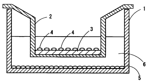
[0008]
Fig. I is a cross-sectional view schematically showing a state of an
instrument, etc.
when oral mucosal epithelial cells and corneal epithelial cells are cultured
on amniotic
membrane. In a culture dish 1, a culture insert 2 is disposed. On the bottom
surface of the
culture dish 1, a 3T3 cell layer 5 is formed. Furthermore, on the bottom
surface of the culture
insert 2, amniotic membrane 3 is placed, and oral mucosal epithelial cells and
corneal
epithelial cells 4 are cultured thereon. Reference numeral 6 denotes a culture
medium.
1: culture dish (first container), 2: culture insert (second container), 3:
amniotic
membrane, 4: oral mucosal epithelial cells and corneal epithelial cells, 5:
3T3 cell layer, 6:
culture medium.
Fig. 2 shows an optical microscope image (left) and a fluorescence stain image
(right) on day I of culture of oral mucosal epithelial cells and corneal
epithelial cells on the
amniotic membrane. RO represents a rabbit oral epithelium. In the fluorescence
stain
image, a Dil signal (orange color) showing the presence of cells derived from
the oral mucosal
epithelial cell was observed.
Fig. 3 shows an optical microscope image (left) and a fluorescence stain image
(right) on day 3 of culture of the oral mucosal epithelial cells and corneal
epithelial cells on the
amniotic membrane. RO represents a rabbit oral epithelium. In the fluorescence
stain
image, a Dil signal (orange color) showing the presence of cells derived from
the oral mucosal
epithelial cell was observed.
Fig. 4 shows an optical microscope image (left) and a fluorescence stain image
(right) on day 7 of culture of the oral mucosal epithelial cells and corneal
epithelial cells on the
amniotic membrane. RO represents a rabbit oral epithelium. In the fluorescence
stain
image, a Dil signal (orange color) showing the presence of cells derived from
the oral mucosal
epithelial cell was observed.
Fig. 5 shows results of immunostaining the cell layer formed on the amniotic
membrane. The left images show the staining properties of keratin 1(K1) and
keratin 10
(K10); the middle images show the staining properties of keratin 3(K3) and
keratin 12 (K 12);
and the right images show the staining properties of keratin 4 (K4) and
keratin 13 (K13).
Fig. 6 shows a state of the ocular surface (anterior ocular part) (left image)
and a
fluorescein stained image of the ocular surface (right image) on day 2 after
transplantation of
the corneal epithelial sheet according to Example.
Fig. 7 shows a state of the ocular-surface (anterior ocular part) (left image)
and a
fluorescein stained image of the ocular-surface (right image) on the first
week after
transplantation of the corneal epithelial sheet according to Example.
Fig. 8 shows a state of the ocular-surface (anterior ocular part) (left image)
and a
fluorescein stained image of the ocular-surface (right image) on the second
week after
transplantation of the corneal epithelial sheet according to Example.
Fig. 9 shows a HE (hematoxylin eosin) stained image of the corneal epithelial
sheet
on the second week after transplantation.
5
CA 02572803 2007-01-02
Fig. 10 shows results of immunostaining the corneal epithelial sheet on the
second
week after transplantation. The left images show the staining properties of
keratin 1(K1) and
keratin 10 (Kl0); the middle images show the staining properties of keratin
3(K3) and keratin
12 (K12); and the right images show the staining properties of keratin 4 (K4)
and keratin 13
(K13).
BEST MODE OF CARRYING OUT THE INVENTION
[0009]
The first aspect of the present invention relates to a corneal epithelial
sheet. Herein,
"corneal epithelial sheet" is a sheet-like structure including a
characteristic similar to the
corneal epithelium in at least a part of the structure.
The corneal epithelial sheet of the present invention includes a
characteristic cell
layer. The cell layer includes an autologous cell (herein, also referred to as
"a first cell") and
a cell whose origin is different from the autologous cell (herein, also
referred to as "a second
cell"), and a multi-layered structure is formed by these cells. In this
specification, the
formation from two kinds or more of cells whose origins are different from
each other in this
way is also referred to as "hybridization." On the other hand, "multi-layered"
means being
formed of a plurality of cell layers. The corneal epithelial sheet of the
present invention
includes typically about 4 to 8 layers of cells.
The form (state of hybridization) including cells in the cell layer is not
particularly
Iimited. For example, each kind of cells may be dispersed or any kind of cells
(or plural
kinds of cells) may be present in a group. Alternatively, the content of each
kind of cells may
not be constant over the cell layer.
[0010]
The first cell according to the present invention is an autologous cell. The
"autologous" herein denotes a subject using the corneal epithelia) sheet
according to the
present invention, that is, a person (recipient) undergoing transplantation.
On the other hand,
persons other than the "autologous" person denote "another individual."
The kinds of the first cells are not particularly limited as long as they are
capable of
forming a corneal epithelium-like mucosal epithelial layer when they are
hybridized with the
below mentioned second cells. An example of the first cell can include a cell
derived from
oral mucosal epithelium, a cell derived from conjunctival epithelium, a cell
derived from nasal
mucosal epithelium, or a cell derived from an undifferentiated cell (i.e. a
stem cell of mucosal
epithelium) capable of constructing any of the mucosal epitheliums. In this
specification,
"derived from or origin" is used to intend to specify a starting material.
Therefore, for
example, the cell derived from (or whose origin is) the oral mucosal
epithelium is a cell
constructed by using an oral mucosal epithelial cell as a starting material.
Furthermore, in the
present invention, "an undifferentiated cell capable of constructing the
mucosal epithelium"
denotes a cell having differentiation potency to the mucosal epithelium. For
example, an
undifferentiated cell capable of constructing the oral mucosal epithelium
refers to as a cell
6
CA 02572803 2007-01-02
capable of being differentiated to an oral mucosal epithelial cell. Specific
examples of the
undifferentiated cell can include a precursor cell or a stem cell forming a
specific tissue such
as oral mucosal epithelium, conjunctival epithelium, or the like, or an
epithelial stem cell
having lower degree of differentiation, and the like.
[0011]
The cell layer according to the present invention may include two different
kinds or
more of the first cells. For example, a cell layer may be constructed in a
state in which a cell
derived from the oral mucosal epithelium and a cell derived from the
conjunctival epithelium
are contained.
[0012]
The "oral mucosal epithelium" according to the present invention includes an
oral
inner marginal mucosa epithelium part, a labial part, a palate part, a buccal
part, and the like.
Whether such cells are derived from the oral mucosal epithelium can be
confirmed by
observing the expression of keratin 4 or keratin 13, which are specific to the
oral mucosal
epithelium as an index. Alternatively, it can be also confirmed by observing
that keratin 3 is
expressed as an index. This keratin 3 is known to be one of the keratins
specific to the cornea
but it is confirmed to be expressed also in the oral mucosal epithelium. Note
here that it can
be said that it is preferable to use oral mucosal epithelial cells as
materials for producing
compositions for corneal epithelium transplantation from the viewpoint in that
this keratin 3
specific to the cornea is expressed.
On the other hand, also by examining the expression of genes specific to an
oral
mucosal epithelial cell, it can be confirmed that the cells are derived from
the oral mucosal
epithelium.
Similarly, as to cells derived from a tissue other than the oral mucosal
epithelium, the
derivation thereof can be confirmed by examining a marker specific to the
tissue or gene
expression.
[0013]
A specific example of the second cell can include a cell derived from the
corneal
epitheliuin, the conjunctival epithelium or the amniotic membrane epithelium.
Among them,
it is preferable that the second cell is a cell derived from the corneal
epithelium or the
conjunctival epithelium. A cell layer constructed by using cells derived from
an ocular
surface tissue can have a property closer to that of the corneal epithelium.
It is particularly
preferable that the second cell is a cell derived from the corneal epithelium.
It is
advantageous because the cell layer having a further closer property to that
of the corneal
epithelium can be obtained.
The second cell may be an autologous cell or a cell of another individual.
When a
cell layer is constructed by an autologous cell, a cell layer free from a
problem of
immunological rejection can be obtained. When a cell layer is constructed by a
cell of
another individual, it becomes easy to obtain cells as a raw material, so that
the cell layer is
advantageous from the viewpoint of production. The cell layer of the present
invention may
7
CA 02572803 2007-01-02
include different two kinds or more of second cells. For example, a cell layer
may be
constructed in a state in which a cell derived from the corneal epithelium and
a cell derived
from the conjunctival epithelium are included.
[0014]
Whether a cell layer in the corneal epithelial sheet of the present invention
is derived
from the corneal epithelium can be confirmed by observing that keratin 3 or
keratin 12, which
are specific to the corneal epithelium, are expressed as an index.
Alternatively, it can be also
confirmed by observing that keratin 4 is expressed as an index.
Note here that in the case where cells derived from a tissue other than the
corneal
epithelium, the derivation thereof can be confirmed by examining a marker
specific to the
tissue or gene expression.
[0015]
Preferably, the cell layer in the corneal epithelial sheet of the present
invention
includes some of the following characteristics or properties. Particularly
preferably, the cell
layer includes all of the following characteristics or properties.
( l) The cells of the uppermost layer are not cornified. This is one of the
features of
corneal epithelium. When this feature is observed, the corneal epithelial
sheet of the present
invention is similar to the corneal epithelium and is expected to exhibit the
same function as
that of the corneal epithelium. Note here that "cornified" is also referred to
as "keratinized",
which represents the phenomenon in which keratin is generated in a cell and
the cell organelle
such as the nucleus is lost. Whether the cells are cornified can be confirmed
by observing,
for example, the presence or absence of flatness or nucleus in a cell as an
index.
(2) The cells of the uppermost layer are flat-shaped. That is to say, an oral
mucosal
epithelial cell layer is configured by forming a layer of cells having flat
shape on a layer of
cells having approximately cuboidal shape. It is thought that when the
uppermost layer is
covered with flat-shaped cells, the tightness between cells is increased and a
below-mentioned
barrier function is attained. Also in the corneal epithelium, cells in the
uppermost layer are
flat-shaped. When this feature is observed, the corneal epithelial sheet of
the present
invention is similar to corneal epithelium and is expected to exhibit the same
function as that
of corneal epithelium.
(3) A barrier function is provided. The barrier function means a function of
preventing liquid, gas, or the like, from infiltrating from the surface or a
function of preventing
liquid froin releasing through the surface layer. When such barrier function
is provided, it is
possible to maintain moisture (tear) on the surface after transplantation and
to prevent more
than necessary moisture from being released. The cornea can maintain moisture
on the
surface thereof as it has a barrier function, and thereby it resists blinking.
Therefore, the
barrier function is one of the most important features required for a material
for cornea
transplantation. When this feature is observed, the corneal epithelial sheet
of the present
invention is similar to the corneal epithelium and is expected to exhibit the
same function as
that of the corneal epithelium. Whether or not this barrier function is
provided can be
8
CA 02572803 2007-01-02
examined based on the extent of infiltration of solution including an
indicator such as
Horseradish peroxidase.
The corneal epithelial sheet of the present invention can be used as
transplantation
material (substitute for the corneal epithelium) to a patient with damaged
cornea or failure
cornea, etc. In transplantation, it is preferable that a graft is fixed to and
allowed to survive
by fixing it to the surrounding tissue with a surgical suture. Furthermore, it
is preferable that
after transplantation, the surface of the transplanted part is protected by
temporarily being
covered with a therapeutic contact lens.
[0016]
In the corneal epithelial sheet in accordance with one embodiment of the
present
invention, the cell layer is formed on a collagen layer. That is to say, in
this embodiment, a
collage layer is provided in addition to the cell layer. The collage layer
herein is preferably
derived from the amniotic membrane. It is advantageous because it is possible
to obtain a
corneal epithelial sheet with excellent biocompatibility and low
immunogenicity due to its
high biocompatibility and low immunogenicity of the amniotic membrane. It is
preferable to
use a collagen layer derived from the amniotic membrane from the viewpoint of
production of
a corneal epithelial sheet. That is to say, as mentioned below, a corneal
epithelial sheet
having a collagen layer can be obtained by seeding predetermined cells on the
collagen layer
as a substrate and culturing them, and the amniotic membrane has a property in
which cells are
attached and proliferate thereon. Therefore, the use of the collagen layer
derived from the
amniotic membrane enables excellent adhesion and proliferation of cells and
the formation of
the cell layer.
It is further preferable that the collage layer is derived from the amniotic
membrane
from which the epithelium has been removed by, for example, a scraping
procedure. It is
advantageous because no epithelial components are contained, a corneal
epithelial sheet with
further reduced immunogenicity can be obtained. Since cells can be adhered and
proliferated
on the amniotic membrane from which the epithelium has been removed, it is
advantageous in
terms of production that a high quality corneal epithelial sheet can be
constructed for shorter
time.
Whether the collagen layer is made of the amniotic membrane from which the
epithelium has been removed can be confirmed by examining that a cell of the
amniotic
membrane epithelial layer is not contained in the collagen layer. Note here
that it is
preferable that the human amniotic membrane is used as the amniotic membrane.
[0017]
This corneal epithelial sheet of the present invention can be used as a
transplantation
material (substitute for the corneal epithelium) for patients with injured or
defective cornea,
etc. In the case where the corneal epithelial sheet including a collagen layer
is used, it is
transplanted to the corneal epithelium defective part so that the collagen
layer is located to the
side of the eyeball. On the other hand, in the case of using a corneal
epithelial sheet obtained
by forming a cell layer on the collagen layer and then removing the collagen
layer, it is
9
CA 02572803 2007-01-02
transplanted to the corneal epithelium defective part so that the side in
which the collagen
layer has been present is located to the side of the eyeball.
In transplantation, it is preferable to promote survival of the graft by
fixing it to the
surrounding tissue with a surgical suture. Furthermore, after transplantation,
it is preferable
that the surface of the transplanted part is protected by temporarily being
covered with a
therapeutic contact lens.
[0018]
The corneal epithelial sheet of the present invention can be prepared by the
following
process (second aspect of the present invention). The second aspect of the
present invention
relates to a process for production a corneal epithelial sheet and the
following steps.
a) separately preparing a first cell being an autologous cell and a second
cell wliose
origin is different from that of the first cell;
b) seeding the first cell and the second cell on a collagen layer and
culturing them;
and
c) after proliferation of the first cell and second cell resulting in
formation of a cell
layer, bringing the surface of the cell layer into contact with the air.
[0019]
The production process of the present invention is characterized in that two
kinds or
more of cells are co-cultured. As shown in the below-mentioned Examples (co-
culturing the
oral mucosal epithelial cells and the corneal epithelial cells), according to
the process having
such a characteristic, excellent cell proliferation and rapid formation of
cell layer according to
this were observed. By co-culturing two kinds or more of cells in this way, a
cell layer can be
formed for a shorter time.
[0020]
An example of the first cell preferably includes an oral mucosal epithelial
cell, a
conjunctival epithelial cell, a nasal mucosal epithelial cell or other mucosal
epithelial cells, or
an undifferentiated cell capable of constructing any of the mucosal
epithelium. On the other
hand, as the second cell, a corneal epithelial cell, a conjunctival epithelial
cell, or an amniotic
membrane epithelial cell is preferably used. These cells are harvested from a
living tissue in
which these cells are present. Specifically, for example, after a part of the
tissue where a
target cell exists by using a surgical knife, and the like, followed by
procedures such as
removing the connective tissue and separating cells, and the like. Then, cells
are prepared in
a shape of a cell suspension (suspension). Note here that the first cell may
include two
different kinds of cells. Similarly, the second cell may include two different
kinds of cells.
[0021]
It is suggested that oral mucosal epithelium as a preferable harvesting source
of the
first cell has a stem cell and it is thought to easily induce differentiation
of them to cells
forming an epithelial cell layer. Furthermore, the use of the oral mucosal
epithelial cells has
the following advantages: they can be harvested easily; a large number of
cells can be
harvested; and when a patient with bilateral-eye disease is treated,
transplantation material
CA 02572803 2007-01-02
derived from the autologous cells can be prepared. In particular, with the
advantage that a
patient from which corneal epithelial cells cannot be harvested,
transplantation materials
derived from autologous cells can be used, it is expected that the clinically
important problem
about immunological rejection can be significantly solved.
As the oral mucosal epithelial cell, a cell existing in the dental root part
(a cell of the
oral inner marginal mucosa epithelium), a cell of labial part, a cell of
palate part, a cell of
buccal part, and the like, can be used. Among them, it is particularly
preferable to use a cell
of oral inner marginal mucosal epithelial cell because it has a high
proliferation ability and low
antigenicity. The oral mucosal epithelial cells can be harvested by ablating a
site where a
targeted cell exists by using a scalpel or by scraping it out. Oral inner
marginal mucosal
epithelial cell can be harvested from the oral inner marginal mucosal
epithelial cell that was
separated from enamel cement transition portion. Note here that in order to
remove
impurities such as connective tissue, preferably a treatment with enzyme such
as Dispase or
trypsin, etc., filtration treatment are carried out.
Oral mucosal epithelial cells harvested from an oral cavity of an individual
other than
a patient to whom a corneal epithelial sheet constructed according to the
present invention is to
be transplanted may be used. However, when taking the immunological rejection
into
consideration, preferably the oral mucosal epithelial cell from a patient
him/herself is
harvested and cultured.
Since mucous membrane of oral cavity has high proliferation ability and the
wound is
generally healed by oral administration of antibiotic, disinfection with, for
example, Isodine,
for several days after operation. Therefore, it is thought that invasion to a
patient
himself/herself due to the harvest of mucosa.
[0022]
On the other hand, as the second cell, another individual's (allo) corneal
epithelial
cell can be preferably used. As such a corneal epithelial cell, donor's
eyeball free from
infection is available from, for example, eye bank (Northwest eye bank, etc.).
The cells that
can be used as the second cell are not limited to the corneal epithelial cell.
The conjunctival
epithelial cell, an amniotic membrane epithelial cell, and the like, may be
used. However,
when the corneal epithelial cells constituting the corneal epithelium in a
living organism or the
conjunctival epithelial cells existing in the vicinity thereof are employed, a
corneal epithelial
sheet capable of reproducing the property of the corneal epithelium more
excellently. As
shown in the below-mentioned Examples, when the corneal epithelial cell is
used as the second
cell, it was confirmed that a cell layer similar to the corneal epithelium
could be constructed.
This fact supports the above-mentioned prediction and supports that the
corneal epithelial cell
is particularly preferable for the second cell. On the other hand, as
mentioned in the
below-mentioned Example, it was confirmed that when the amniotic membrane
epithelial cell
was used as the second cell, a cell layer capable of excellently reproducing
the properties
required for the cornea could be formed. This fact shows that the amniotic
membrane
epithelial cells can be preferably used as the second cell.
11
CA 02572803 2007-01-02
[0023]
Autologous cells can be used as the second cell. However, when another
individual's cells are used, the cells can be obtained more easily. For
example, when a
corneal epithelial sheet for the treatment of a patient with bilateral eye
disease is produced, the
corneal epithelial cells as the second cell are available.
[0024]
The respectively prepared first cell and the second cell (hereinafter, also
referred to as
"first cell, and the like") are seeded on a collagen layer and cultured (step
b). In general, the
first cell and the second cell, which are prepared in a form of a cell
suspension, are dripped on
a collage layer and cultured.
Typically, the seeding of the first cells and the seeding of the second cells
are carried
out simultaneously (herein, "simultaneously" includes not only a case where
the seedings are
carried out literally simultaneously but also a case where the first seeding
is carried out and
then the second seeding is carried out without substantial time interval).
However, the first
and second cells may be seeded at different timing. For example, the second
cells may be
seeded several minutes to several tens of minutes after the first cells are
seeded. Thus, by
stagerring the time of seeding cells, for example, a cell layer in which a
region rich in the cells
derived from the first cell is localized can be constructed. Thereby, a
structure of the cell
layer and the property thereof can be changed or adjusted.
[0025]
The ratio of the first cells and the second cells to be seeded is not
particularly limited.
Typically, the number of the first cells to be seeded is substantially the
same as that of the
second cells to be seeded. In an experiment in which the oral mucosal
epithelial cells were
used as the first cells and the corneal epithelial cells were used as the
second cells, the ratios of
the number of the first cells: second cells were changed to 3 : 7, 5: 5, and 7
: 3 and comparison
was made. As a result, no difference in terms of the cell proliferation and
layering were
clearly observed among them (data are not shown).
[0026]
Herein, the kinds of collagens as a material of the collagen layer are not
particularly
limited, and type I collagen, type III collagen, and type IV collagen, and the
like, can be used.
A plural kinds of collagens can be used in combination thereof. Such collagens
can be
extracted and purified from the connective tissue of the skin and cartilage,
etc. of animals such
as swine, bovine, sheep, etc., by an acid solubilization method, alkali
solubilization method,
oxygen solubilization method, and the like. Note here that for the purpose of
deteriorating
the antigenicity, it is preferable that a so-called atherocollagen obtained by
removing
telopeptide by a treatment with the use of catabolic enzyme such as pepsin,
trypsin, etc.
As the collagen layer, it is preferable to use a collagen derived from
amniotic
membrane, particularly derived from human amniotic membrane. Herein, the
collagen layer
is "derived from amniotic membrane" broadly means that the collagen gel is
obtained by using
amniotic membrane as a starting material. Human amniotic membrane is a
membrane
12
CA 02572803 2007-01-02
covering the outermost layer of the uterus and the placenta, and a basal
membrane and an
epithelium layer are formed on parenchymal tissue that is rich in collagen.
Human amniotic
membrane can be harvested by, for example, human embryonic membrane, placenta,
etc.
obtained at the time of afterbirth at delivery. Specifically, the human
amniotic membrane can
be prepared by treating and purifying the integrated material including human
embryonic
membrane, placenta, and umbilical cord obtained right after delivery. The
method of treating
and purifying can employ a method described in, for example, Japanese Patent
Unexamined
Publication No. 5-5689. That is to say, amniotic membrane is detached from the
embryonic
membrane obtained at delivery and remaining tissue is removed by a physical
treatment such
as ultrasonic cleansing and an enzyme treatment, and the like. Then,
appropriate cleaning
process is carried out and thus the human amniotic membrane can be prepared.
The thus prepared human amniotic membrane can be cryopreserved before use. The
human amniotic membrane can be frozen in a liquid mixing equal volume ratio of
DMEM
(Dulbecco's modified Eagle's medium) and glycerol at, for example, -80 C. By
the
cryopreservation, not only the improvement in operation but also reduction of
the antigenicity
can be expected.
Intact amniotic membrane may be used as a collagen layer but it is preferable
that
amniotic membrane from which the epithelium is removed by a scraping
treatment, etc. is used.
For example, after thawing, cryopreserved human amniotic membrane is subjected
to a
treatment with EDTA or proteolytic enzyme so as to loosen the adhesion between
cells and
then the epithelium is scraped by using a cell scraper, etc. Thus, the human
amniotic
membrane from which the epithelium has been removed can be prepared.
[0027]
When the human amniotic membrane from which the epithelium has been removed is
used as the collagen layer, the first cells, and the like, are preferably
seeded on the side of the
collagen layer with the side where the epithelium has been removed and exposed
(i.e., the side
of the basal membrane). It is advantageous because it can be thought that this
face side is
rich in type IV collagens and the seeded first cells, and the like, can be
proliferated and layered
well.
The first cells and second cells can be seeded on the collagen layer so that,
for
example, the cell density becomes about 1 x 10³ cells/cm2 or more, preferably
in the range from
about 1 x 10³ cells/em2 to about 1 x 10⁵ cells/em2, and further preferably in
the range from about
1 x 10⁴ cells/cm2 to about 1 x 10⁵ cells/cm2.
[0028]
It is preferable that the first cells and the like are cultured in the
presence of
supporting cells. The supporting cell is also referred to as a feeder cell and
supplies a culture
medium with a growth factor, etc. When the first cells and the like are
cultured in the
coexistence of the supporting cells, the proliferation efficiency of the cells
is improved. As
the supporting cells, for example, a 3T3 cell (Swiss mouse 3T3 cell, mouse
NIH3T3 cell,
3T3J2 cell, etc.) and the like may be used. Among them, it is preferable to
use a mouse
13
CA 02572803 2007-01-02
NIH3T3 cell as a supporting cell from the viewpoint of proliferation
efficiency, ease in
handling, etc.
It is preferable that the supporting cells are inactivated by using mitomycin
C, etc.
It is advantageous because the inhibition of the proliferation of the first
cells and the like due
to the proliferation of the supporting cells themselves is prevented, and the
proliferation
efficiency of the first cells and the like is enhanced. Such inactivation can
be carried out by a
radiation treatment, and the like.
[0029]
The cell density of the supporting cells may be, for example, about I x 102
cells/cm2 or
more, preferably in the range from about 1 x 10² cells/cm2 to about 1 x 10'
cells/cm2 , and further
preferably in the range from about 1 x 10³ cells/cmz to about I X 105
cells/cmZ. As to the ratio
with respect to the number of the first cells and the second cells, culture
may be carried out
under the conditions in which the supporting cells to be used may be, for
example, 1/103 times
to I x 102 times, and preferably 1/102 times to I time as the total number of
the first cells and
the second cells. When the number of the supporting cells is small, the
proliferation rate of
the first cells and the second cells is lowered; and when it is too small,
excellent layered
structure of the first cells and the like is cannot be obtained. On the other
hand, it is not
preferable that the number of the supporting cells is too large, because the
proliferation rate of
the oral inucosal epithelial cells is lowered.
[0030]
When the first cells are cultured in the coexistence of supporting cells, it
is preferable
that an isolation membrane having a pore size through which the supporting
cells cannot path
is provided between the supporting cells and the collagen layer. The use of
the isolation
membrane makes it possible to prevent the supporting cells from entering the
side of the
collagen layer (i.e. the side of oral mucosal epithelial cells) at the time of
culturing. As a
result, the supporting cells may not be mixed in the finally obtained corneal
epithelium-like
sheet. This means that a corneal epithelial sheet being free from problem of
immunological
rejection by the supporting cells can be constructed. This is clinically
significant so much.
As the isolation membrane, an isolation membrane having a pore size through
which
the supporting cells cannot path can be used by appropriately selecting the
known membrane.
For example, a membrane having a pore size of about 0.4 m to 3.0 m made of
polycarbonate
can be used. A material of the isolation membrane is not particularly limited.
In addition to
polycarbonate, polyester and the like may be used. Such isolation membranes
are on the
market and easily available.
An example of the culture method using an isolation membrane may include the
following method. Firstly, inactivated supporting cells are seeded and
cultured on a container
such as a dish (a first container), thereby forming a layer of supporting
cells on the surface of
the container. Next, a second container, which has a bottom face made of an
isolation
membrane, is set in the first container so that the bottom face of the second
container is located
in a culture medium. Then, the collagen layer is formed on the bottom face,
that is, on the
14
CA 02572803 2007-01-02
isolation membrane, a collagen layer is formed. Then, on the collagen layer,
the first cells
and the like are seeded and cultured.
On bottom surface of the second container, a collagen layer may be previously
formed (for example, on the bottom surface of the second container, the
amniotic membrane
from which an epithelium has been removed is placed. In this state, drying
process may be
carried out). This second container may be set in the first container in which
supporting cells
are seeded, and then on the collagen layer, the first cells and the like may
be seeded and
cultured.
[0031]
The culture medium used for culturing the first cells and the like is not
particularly
limited as long as the cells can be proliferated and a layered structure of
the cells can be
formed. For example, it is possible to use a medium, in which DMEM (Dulbecco's
modified
Eagle's medium) that is generally used for growing epithelial cells and Ham's
F12 medium are
mixed with each other at the predetermined ratio, and FBS, growth factor,
antibiotics, and the
like are added. Specific examples include a mixing medium of DMEM and Ham's
F12
medium (mixing volume ratio of 1: 1) to which FBS (10%), insulin (5 mg/ml),
cholera toxin
(0.1 nM), epithelial cell growth factor (EGF) (10 ng/ml) and penicillin-
streptomycin (50
IU/mI) are added. Furthermore, a mixing medium of DMEM and Ham's F12 medium to
which triiodothyronine (e.g. 2 nM), glutamine (e.g. 4 mM), transferrin (e.g. 5
mg/mI), adenine
(e.g. 0.18 mM), and/or hydrocortisone (e.g., 0.4 mg/ml) are further added, may
be used.
[0032]
When the first and second cells are cultured on a collagen layer, these cells
are
proliferated and a cell layer is formed (in this process, at least a part of
the cells are thought to
be differentiated). After the formation of a cell layer, a step (step (c)) of
bringing the surface
layer of the cell layer into contact with the air is carried out. Note here
that this step herein
also is referred to as Air lifting. This step (c) is carried out for
differentiation of cells forming
the cell layer and inducing the barrier function.
This step can be carried out by lowering the surface of the culture medium by
temporarily removing a part of the culture medium by using a dropper, a
pipette, and the like,
thereby temporarily exposing the surface of the oral mucosal epithelial cell
layer to the outside
of the culture medium. Alternatively, this step can be carried out by lifting
up the oral
mucosal epithelial cell layer together with the collagen layer, thereby
temporarily exposing the
surface from the culture medium surface. Furthermore, by using the tube etc.,
the air may be
fed into the culture medium so as to bring the surface of the cell layer into
contact with the air.
From the viewpoint of the ease in operation, it is preferable that by lowering
the surface of the
culture medium, thereby exposing the surface of the cell layer to the outside.
The period when this step (c), that is, the period of time when the uppermost
layer of
the layered structure of cells is brought into contact with the air differs
depending upon the
state of the cells, culture conditions, and the like, but the period may be,
for example, three
days to two weeks, preferably within a week, and further preferably within
three days.
CA 02572803 2007-01-02
According to the above-mentioned method of the present invention, on the
collagen
layer, a corneal epithelium-like cell layer, in which the first cells and the
like are layered, is
formed. The thus obtained corneal epithelial sheet together with the collagen
layer used as a
substrate of the first cell and the like can be used as a transplantation
material (substitute for
the corneal epithelium) for patients with injured or defective cornea. In this
case, the sheet is
transplanted to the corneal epithelium defective part so that the collagen
layer is located to the
side of the eyeball. In transplantation, it is preferable to promote survival
of the graft by
fixing it to the surrounding tissue with a surgical suture. Furthermore, it is
preferable that
after transplantation, the surface of the transplanted part is protected by
temporarily being
covered with a therapeutic contact lens.
Note here that a graft from which a part or all of the coliagen layer has been
removed
may be used. The collagen layer can be removed by appropriately combining a
chemical
treatment with EDTA, etc., an enzymatic treatment by proteolytic enzyme, etc.,
and a physical
treatment such as scraping by using forceps.
[0033]
Hereinafter, one example of specific transplantations is described. Firstly,
cicatrical
tissue is incised in the corneal limbus of a patient with keratoconjunctive.
Then, the
cicatrices conjunctiva tissue invading into the cornea is ablated so as to
expose the parenchyma
of cornea, followed by suturing corneal epithelial sheet at a portion slightly
inside the limbus.
The operative procedure is in principle the same as the operative technique
which the
institution the present inventors belong have carried out as clinical
applications to 70 cases or
more (operation procedure relating to a corneal epithelial sheet obtained by
culturing a corneal
epithelial cell or a corneal epithelial sheet obtained by culturing an oral
mucosal epithelial
cell). It is thought that the operative procedure can be carried out extremely
stably.
Hereinafter, Examples (including experimental examples) of the present
invention
will be described.
[Example 1]
[0034]
<Production and evaluation of hybridized corneal epithelial sheet>
1-1. Harvest of amniotic membrane
After giving a pregnant woman who does not have a systemic complication and
would undergo Caesarean section sufficient informed consent together with an
obstetrician in
advance, the amniotic membrane was obtained during the Caesarean section in
the operation
room. The operation was carried out cleanly. In accordance with the operation
work, the
operators washed hands, and then wore a special gown. Before delivery, a clean
vat for
obtaining the amniotic membrane and physiologic saline for washing were
prepared. After
delivery, the placenta tissue was transferred to the vat and the amniotic
membrane tissue was
manually removed from the placenta. A portion where the amniotic membrane and
the
placenta were strongly adhered to each other was separated with scissors.
[0035]
16
CA 02572803 2007-01-02
1-2. Treatment of amniotic membrane
Treatment process of amniotic membrane included: (1) washing, (2) trimming,
and
(3) storing sequentially in this order. Throughout all the processes,
operation is desired to be
carried out in a clean draft. For all containers and instruments for use,
those sterilized were
used, and for dishes, etc. sterilized disposable ones were used. The obtained
amniotic
membrane was washed for removing blood component attached thereto and further
washed in
a sufficient amount of physiological saline (0.005% ofloxacin was added).
Then, the
amniotic membrane was transferred to a phosphate buffer solution (PBS) in a
dish and cut and
divided into the size of about 4 x 3 cm with scissors. The divided pieces of
amniotic
ineinbrane were stored in several dishes filled with a stock solution, and
thereafter amniotic
membranes in good condition were selected among them.
[0036]
1-3. Storage of amniotic membrane
One cc each of stock solution was placed in 2 cc sterilized cryotube and one
sheet
each of the amniotic membrane, which had been obtained, washed and selected,
was placed
and labeled, then stored in a refrigerator at -80 C. For the stock solution,
50% sterilized
glycerol in DMEM (Dulbecco's Modified Eagle Medium: GIBCOBRL) was used. The
expiration date for use of stored the amniotic membrane was determined at
three months and
expired amniotic membrane was disposed of by incineration.
[0037]
1-4. Treatment of amniotic epithelium
The amniotic membrane was subjected to treatment for removing the epithelium
and
then used for culture. First of all, the amniotic membrane stored at -80 C was
thawed at
room temperature, and then well washed in sterilized a phosphate buffer
solution (PBS) in the
dish. After washing, the amniotic membrane was stored in a 0.02% EDTA solution
(Nacalai
tesque) at 37 C for 2 hours, and then the epithelium was mechanically scraped
off by using a
cell scraper (Nunc, USA) and used as a substrate for culture. Note here that,
it was confirmed
that one layer of the amniotic epithelium was completely scraped by this
procedure process by
the optical microscope and electron inicroscope (scanning electron microscope)
operations.
[0038]
1-5. Harvest of oral mucosal epithelial cells
In 6-week old Japanese white rabbit, tooth was pulled out. Then, the oral
mucosal
epithelium was carefully separated from the enamel cement transition portion.
Note here that
a series of operations were carried out by using sterilized instruments as
antiseptically as
possible.
The obtained oral mucosal epithelium was immersed twice in a phosphate buffer
solution (PBS) containing 50 IU/ml penicillin streptomycin and Gentacin for 30
minutes under
the condition of room temperature. Thereafter, the tissue was immersed in a
phosphate buffer
solution (PBS) containing 1.2U Dispase (Nacalai tesque) for one hour at 37 C
and immersed
and treated in 0.05% trypsin-EDTA solution (GBCOBRL) for 30 minutes so as to
separate
17
CA 02572803 2007-01-02
.
cells. An enzyme activity was stopped by immersing in DMEM containing 10%
fetal bovine
serum (FBS). Thereafter, excess tissues were removed by using a 60 m cell-
filter so as to
isolate the oral mucosal epithelial cell (oral inner margin epithelial cell)
(oral mucosal
epithelial cell suspension).
[0039]
1-6. Harvest of corneal epithelial cells
In 6-week old Japanese white rabbit (a different rabbit from the rabbit from
which the
oral mucosal epithelial cell had been harvested), the limbus strip with the
size of 5 mm x 10
mm was harvested from the corneal limbus by using a surgical knife.
The obtained tissue strip was immersed twice in a phosphate buffer solution
(PBS)
containing 50 IU/mI penicillin streptomycin and Gentacin for 30 minutes under
the condition
of room temperature. Thereafter, the tissue was immersed in a phosphate buffer
solution
(PBS) containing 1.2U Dispase (Nacalai tesque) for one hour at 37 C and
immersed and
treated in 0.05% trypsin-EDTA solution (GBCOBRL) for 15 minutes so as to
separate cells.
An enzyme activity was stopped by immersing in DMEM containing 10% fetal
bovine serum
(FBS). Thereafter, excess tissues were removed by using a 60 m cell-filter so
as to isolate
the corneal epithelial cell (corneal epithelial cell).
[0040]
1-7. Preparation of co-cultured cell
As the co-culture cells (support cells), NIH-3T3 cells (hereinafter, referred
to as "3T3
cell") were used. The 3T3 cell that had been cultured in advance and become
confluent in
75F flask (BD product of Falcon) was immersed in 0.05% mitomycin C solution
for two hours
so as to suppress the proliferation activity. Sequentially, they were was
washed with a
phosphate buffer solution (PBS) several times so as to remove mitomycin C,
followed by
treating with 0.05% trypsin-EDTA solution (PBS) so as to prepare a 3T3
suspension.
[0041]
1-8. Cell culture and induction of mucosal epithelium
By using human amniotic membrane from which the epithelium had been scraped as
a substrate, the oral mucosal epithelial cells and corneal epithelial cells
were co-cultured with
3T3 cells that were subjected to the above-mentioned treatment by the
following procedure.
For culturing instruments, a 6-well culture dish (Corning, NY) and a culture
insert (a container
for inserting culture) (polycarbonate, average pore size: 3.0 m, Corning NY)
were used.
First of all, 3T3 cell suspension was seeded on the culture dish so that the
cell density
was about 1 x 10⁴ cells/cm2 and cultured under conditions at 37 C and in
5%CO2.
Furthermore, the amniotic membrane substrate was allowed to stand still to be
attached on the
culture insert with the side of the scraped epithelium upward, and dried for
10 minutes at room
temperature. Thereafter, on the culture insert to which the amniotic membrane
was attached,
oral mucosal epitlielial cell suspension and corneal epithelial cell
suspension were seeded so
that the cell density was about 1 x 10⁴ cells/cm2.
After the above-mentioned operation, as shown in Fig. 1, the culture insert
was
18
CA 02572803 2007-01-02
disposed in the culture dish and 3T3 cells, oral mucosal epithelial cells and
corneal epithelial
cells were cultured in the same culture medium. Note here that Fig. I is a
schematic
cross-sectional view showing a state during culturing. In the culture dish 1,
the culture insert
2 is placed and on the bottom surface of the culture dish 1, the 3T3 cell
layer 5 is formed.
Furthermore, on the bottom surface of the culture insert 2, the amniotic
membrane 3 is placed,
and the oral mucosal epithelial cells and corneal epithelial cells 4 are
cultured thereon.
Reference numeral 6 denotes a culture medium.
As the culture medium, a DMEM / Ham's F12 mixture medium (mixing volume
ratio: 1:1) including 10% FBS, insulin (5 mg/ml), cholera toxin (0.1 nM),
penicillin-streptomycin (50 IU/ml) and human recombinant epithelial cell
growing factor
(BGF) (10 ng/ml) was used.
The culture was carried out in the above-mentioned medium for seven days
(Submerge). Thereafter, for inducing the mucosal epithelium, by a so-called
Air-lifting
method, culture was carried out for about three days. The Air-lifting method
is a method of
lifting the liquid surface of the culture medium to the surface of the oral
mucosal epithelial cell
layer formed on the amniotic membrane to bring the cell layer into contact
with the air.
During submerging, the culture medium was replaced with new one every other
day and after
carrying out the Air-lifting method, the culture medium was replaced with new
one every day.
A multi-layered cell layer including 5 to 6 layers was formed by about 10 days
culture(including three days of culture by the air-lifting method) according
to the
above-mentioned method.
[0042]
1-9. Identification of cells constituting cell layer
Whether the cell layer on amniotic membrane, which has been obtained by the
above-mentioned method, is formed as hybridization of the oral mucosal
epithelial cells and
the corneal epithelial cells was confirmed by the following procedure.
Firstly, a Dil coloring
agent was added to an oral mucosal epithelial cell suspension before seeding
on the amniotic
membrane and allowed to stand still for about 15 minutes at room temperature
(Dil label).
The thus obtained labeled oral mucosal epithelial cells together with the
corneal epithelial cells
were seeded on the amniotic membrane from which the epithelium had been
removed.
Thereafter, the culturing was carried out in the same conditions mentioned
above. On day 1,
day 3 and day 7 of culture, the formed cell layer was observed by using an
optical microscope
and a fluorescence microscope. The results are shown in Fig. 2 (Day 1), Fig. 3
(Day 3), and
Fig. 4 (Day 7). In each figure, left image is an optical microscope image and
right image is a
fluorescence microscope image. On day 1, a state in which cells are
excellently proliferated
is shown (see left image of Fig. 2). Furthermore, a state in which Dil signals
are scattered is
observed (see right image of Fig. 2), showing that the oral mucosal epithelia]
cells and the
corneal epithelial cells are present together. On day 3, a state in cells are
arranged regularly
(see left image of Fig. 3). Furthermore, a state in which Dil signals are
scattered is still
observed (see right image of Fig. 3), showing that two kinds of cells (the
oral mucosal
19
CA 02572803 2007-01-02
epithelial cells and the corneal epithelial cells) are proliferated in a form
of hybridization so as
to form a cell layer. On day 7, similar to the state on day 3, excellent cell
proliferation and
hybridization of cells are observed (see Fig. 4). Furthermore, in accordance
with the cell
proliferation and multi-layering, a region in which Dil signals are observed,
is increased (see
right image of Fig. 4).
[0043]
1-10. Evaluation of histological properties of cell layer
The cell layer, which had been finally obtained by the above-mentioned methods
(1-1
to 1-8), were observed by using an optical microscope. As a result, at the
basal side (the side
of the amniotic membrane) of the cell layer, a group of relatively cuboidal -
shaped cells similar
to the basal cell existed. Furthermore, it was confirmed that the cells of the
outermost layer
had a flat shape but included a nucleus and that the surface thereof was not
cornified unlike the
skin. As mentioned above, the optical microscope observation showed that the
epithelium
layer (corneal epithelial sheet) similar to the cornea was formed on the
amniotic membrane.
[0044]
Then, in order to examine the physiological property of the cell layer,
immunostaining was carried out. After the obtained cell layer was cut into an
appropriate
size and frozen and embedded in an OCT compound. Then, the resultant compound
was
sliced with a cryostat to prepare slide sections. In immunostaining, the
consideration on
keratins, that is, respective cytoskeleton proteins were carried out. That is
to say, keratin 1/10
specific to epidermis, keratin 3/12 specific to the cornea, and keratin 4/13
specific to mucosa
were considered. The method will be described below. A slide section was
washed with a
phosphate buffer solution (PBS) and then blocking with 1% fetal bovine serum
(FBS) was
carried out to suppress the non-specific antibody reaction. Thereafter, an
antibody against
each keratin (primary antibody) was reacted at room temperature for one hour.
After
reaction, the slide section was washed with PBS containing triton-X for 15
minutes three
times, followed by reacting with fluorescence labeling antibody (secondary
antibody) at room
temperature for one hour. After reaction, the slide section was washed with a
phosphate
buffer solution (PBS) for 15 minutes three times and sealed, followed by
observing the tissue
with a confocal microscope.
The antibody reactions of the respective keratins with respect to the cell
layer will be
described below. Firstly, for keratins I and 10 specific to epidermis, the
staining was not
observed (see left image of Fig. 5). On the other hand, the staining of
keratin 3 specific to
cornea was observed in a wide range (see middle image of Fig. 5). Keratin 3
was observed in
the corneal epithelial cells and the oral mucosal epithelium and it was
thought that the property
thereof was maintained under the culture conditions. The staining of keratin 3
was strong in
the upper part of the cell layer. The staining of keratin 12 was also observed
in a wide range
(see middle image of Fig. 5) and in particular, in the upper part of the cell
layer, strong staining
was observed. This staining property was caused by the corneal epithelial cell
to be used,
suggesting that the property thereof was maintained after culture.
CA 02572803 2007-01-02
For keratins 4 and 13 specific to mucosa, staining was observed in the almost
entire
region (see right image of Fig. 5).
From the above-mentioned results, as the histological property of the formed
cell
layer, in the aspect of the cytoskeletal, the keratin (keratins 3 and 12)
specific to the cornea are
maintained, showing the similarity to the corneal epithelium. Furthermore,
unlike the
epidermis, the cell layer has not been differentiated to cornification. It was
confirmed that the
cell layer had a feature of the not-cornified mucosal epithelium and
simultaneously maintained
the keratin specific to the cornea.
[0045]
1-11. Transplantation experiment
In accordance with the above-mentioned methods (1-1 to 1-8), a sheet having a
cell
layer on the amniotic membrane (hereinafter, referred to as "corneal
epithelial sheet") was
produced. Specifically, firstly, oral mucosal epithelial cells (autologous
cells) were prepared
from a 6-week old Japanese white rabbit and corneal epithelial cells (allo
corneal epithelial
cells) were prepared from a different individual (a 6-week old Japanese white
rabbit). Then,
both cells were seeded on the amniotic membrane from which the epithelium had
been
removed, followed by culturing. Thus, a corneal epithelial sheet was obtained.
Meanwhile, to the rabbit from which the oral mucosal epithelial cell had been
harvested, all the
conjunctival epithelium having a thickness of 100 gm were removed from 4-mm
outside of the
limbus by using a crescent knife. By this operation, since the epithelial
cells containing
corneal epithelial stem cells were lost, artificial exhaustion of the ocular
surface stem cells was
thought to be reappeared. Then, the above-mentioned corneal epithelium
transplantation
sheet was transplanted into the region slightly inner from the limbus. In
transplantation, by
using 10-0 nylon fiber was used to stitch the sheet to the peripheral tissue.
After
transplantation, on the graft, a therapeutic contact lens was placed. After
the operation,
antibiotics and steroid ophthalmic ointment were applied twice a day. At the
time of
transplantation, the ocular surface had a transparency the same as that of the
corneal
epithelium transplantation sheet before transplantation (the results are not
shown in the
drawings).
[0046]
The ocular surface which had undergone the transplantation was observed on day
2,
on the first week, and on the second week after transplantation. In addition,
a fluorescein
staining test was carried out. The fluorescein staining test was carried out
by directly
administering a test paper into which a moisture such as ophthalmic solution
including an
antimicrobial drug had been included and causing eyeblink twice or three
times, followed by
observing the fluorescein staining of the ocular surface. When the corneal
epithelium
remained, due to the tight intercellular adhesive structure, a fluorescein is
not infiltrated and no
staining by the fluorescein was observed.
On day 2 after the transplantation, the transplanted corneal epithelium
transplantation
sheet maintained transparency (see left image of Fig. 6). Furthermore, it was
confirmed by
21
CA 02572803 2007-01-02
fluoresceine staining that the corneal epithelial sheet remained on the ocular
surface without
being damaged (see right image of Fig. 6). Meanwhile, since the graft (corneal
epithelial
sheet) showed no staining of fluorescein, it was confirmed that the corneal
epithelial sheet had
a barrier function similar to the corneal epithelium. Furthermore, since by
the fluoresceine
staining, staining of fluoresceine was confirmed over the entire periphery of
the graft,
therefore it was confirmed that the tissue existing in the transplanted part
was not
contamination of the remaining conjunctival epithelium.
Note here that since cells of the corneal epithelium are tightly adhered to
each
other, the fluorescein staining agent does not invade from the surface and
staining of
fluorescein is not observed in fluorescein straining test. On the other hand,
when the
adhesion between cells becomes loosen or the barrier function is damaged by
exfoliation of the
cell itself, invasion of the fluorescein staining agent occur, and the tissues
are stained.
Therefore, by examining the staining property of fluorescein staining was
examined, it can be
confirmed whether or not the transplanted corneal epithelial sheet had the
barrier function
similar to the corneal epithelium.
[0047]
On the other hand, one week after the transplantation, the graft (corneal
epithelial
sheet) also remained on the ocular-surface. Moreover, it was confirmed that
the graft was
expanded to the surrounding as compared with the state on day 2 after the
transplantation (see
left image of Fig. 7). Furthermore, it was confirmed that the graft showed no
fluorescein
staining and maintained a barrier function necessary for the corneal
epithelium (see right
image of Fig. 7). The transparency was also not changed from that observed on
day 2 after
the transplantation and was highly maintained (see left image of Fig. 7).
The condition of the ocular surface two weeks after the transplantation was
not
particularly changed from the condition after one week after the
transplantation. That is to
say, the graft (corneal epithelial sheet) remained on the ocular surface and
the transparency
thereof was high (see left image of Fig. 8). Furthermore, it was confirmed
that the graft
showed no staining of fluorescein (see right image of Fig. 8) and the barrier
function was
maintained.
[0048]
From the above-mentioned results, it was demonstrated that the corneal
epithelial
sheet obtained by the method mentioned above had an excellent survival
property and the
survival property was maintained for a long time. Furthermore, it was
confirmed that the
corneal epithelial sheet extended to the surrounding after transplantation,
exhibited a barrier
function necessary to the corneal epithelium for a long time, and exhibited
high transparency.
That is to say, the corneal epithelial sheet obtained by the above-mentioned
method excellently
functioned as a substitute of the corneal epithelium and could be used as a
transplant material
for reconstructing the ocular surface in the case where the cornea was injured
and damaged.
[0049]
1-12. Evaluation of histological property of corneal epithelial sheet after
transplantation
22
CA 02572803 2007-01-02
Next, the corneal epithelial sheet two weeks after the transplantation was
extracted
and the histological property thereof was examined. Fig. 9 shows a HE staining
image of the
corneal epithelial sheet (right image was expanded view). On the upper part of
amniotic
membrane (see marks *), similar to the corneal epithelium, a cell layer in
which cells are
regularly arranged is confirmed. In this cell layer, to the upper layer, the
number of the
flat-shaped cells is increased and the structure extremely similar to the
corneal epithelium is
maintained.
Fig. 10 shows the results of staining test with respect to various keratins.
The
staining property almost similar to that of the corneal epithelial sheet
before transplantation
was shown. That is to say, staining of keratins 1 and 10 specific to epidermis
was not
observed (see left image of Fig. 10), staining of keratins 3 and 12 specific
to cornea was
observed (see middle image of Fig. 10), and staining of keratins 4 and 13
specific to mucosa
was also observed (see right image of Fig. 10). As mentioned above, it was
confirmed that
the corneal epithelial sheet maintained the keratins (keratins 3 and 12)
specific to the corneal
epithelium also after transplantation. These results strongly support that the
corneal epithelial
sheet will exhibit the same function as the corneal epithelium for a long time
from the
histological viewpoint.
[Example 2]
[0050]
<Production of hybrid-type corneal epithelial sheet using human cells and
evaluation
thereofl
A human oral mucosal epithelial cell suspension was prepared by harvesting
cells
from a healthy volunteer after obtaining his/her agreement by the same method
described in
the above-mentioned 1-5 (harvest of oral mucosal epithelial cells). Meanwhile,
a cornea
obtained from an eye bank (Northwest eye bank) was treated by the same method
described in
the above-mentioned 1-6 (Harvest of corneal epithelial cells) so as to obtain
a human corneal
epithelial cell suspension. The thus prepared human oral mucosal epithelial
cells and the
corneal epithelial cells were co-cultured with 3T3 cells by using human
amniotic membrane
from which the epithelium had been scraped as a substrate. For about 14 days
of culture
(including three days of culture by the air-lifting method), a corneal
epithelium-like cell layer
was formed. Note here that the method of preparing the human amniotic membrane
from
which the epithelium had been scraped and co-culturing condition with 3T3
cells were the
same as those described in the above-mentioned 1-1 (Harvest of amniotic
membrane) to 1-4
(Treatment of amniotic epithelium).
When the form, structure and immunostaining, and the like, of the obtained
cell layer
were examined, the results were the same as those of a rabbit model. That is
to say, the
produced hybrid-type cultured cell layer had a multi-layered structure
including 4 to 8 layers.
No staining of keratins I and 10 specific to epidermis was observed, staining
of keratins 3 and
12 specific to cornea was observed, and staining of keratins 4 and 13 specific
to mucosa was
also observed.
23
CA 02572803 2007-01-02
From the above-mentioned results, it was confirmed that in the case where
human
oral mucosal epithelial cells and human corneal epithelial cells were used, by
hybridizing these
cells, a corneal epithelium-like cell layer was formed.
[Example 3]
[0051]
<Production of hybrid corneal epithelial sheet using amniotic membrane
epithelial cells and
evaluation thereof>
A human oral mucosal epithelial cell suspension was prepared by harvesting
cells
from a healthy volunteer after obtaining his/her agreement by the same method
described in
the above-mentioned 1-5 (harvest of oral mucosal epithelial cells). Meanwhile,
a human
amniotic membrane was obtained by the method described in the above-mentioned
1-1
(harvest of amniotic membrane) and 1-2 (treatment of amniotic membrane).
Thereafter, the
epithelial cells were separated by a treatment with 0.05% trypsin-EDTA
solution
(GIBCOBRL) for 15 minutes (amniotic membrane epithelial cell suspension). The
thus
prepared human oral mucosal epithelial cells and the human amniotic membrane
epithelial
cells were co-cultured with 3T3 cells by using human amniotic membrane from
which the
epithelium had been scraped as a substrate. For about 14 days of culture
(including three
days of culture by the air-lifting method), a corneal epithelium-like cell
layer was formed.
Note here that the method of preparing the human amniotic membrane from which
the
epithelium had been scraped and co-culturing condition with 3T3 cells were the
same as those
described in the above-mentioned 1-1 (Harvest of amniotic membrane) to 1-4
(Treatment of
amniotic epithelium).
When the form, structure and immunostaining, and the like, of the obtained
cell layer
were examined, the produced hybrid-type cultured cell layer had a multi-
layered structure
including 4 to 8 layers. No staining of keratins 1 and 10 specific to
epidermis was observed,
staining of keratins 3 and 12 specific to cornea was observed, and staining of
keratins 4 and 13
specific to mucosa was also observed.
From the above-mentioned results, it was confirmed that in the case where
human
oral mucosal epithelial cells and human amniotic membrane epithelial cells
were used, when
these cells were hybridized, a corneal epithelium-like cell layer was formed.
Industrial Applicability
[0052]
A corneal epithelial sheet of the present invention has a structure that is
extremely
similar to the structure of the corneal epithelium and shows an excellent
survival property after
transplantation. Furthermore, the corneal epithelial sheet of the present
invention has a
barrier function necessary for the corneal epithelium to exhibit its function
and has high
transparency. Thus, the corneal epithelial sheet provided by the present
invention is
extremely excellent as a transplantation material for re-constructing the
corneal epithelium.
Recently, surgical reconstructive operations for refractory keratoconjunctive
diseases
24
CA 02572803 2007-01-02
have been progressed dramatically. With the appearance of amniotic membrane
transplantation and cultured cornea transplantation, the operation technique
has been almost
established. However, since most of the refractory keratoconjunctive diseases
are bilateral
eye diseases, most transplantations use another individual's (allo) tissue,
which requires a
long-time and strict postoperative care including suppression of the rejection
by administration
of immunosuppressive drugs or prevention of bacterial infection. Under present
circumstances, such things significantly deteriorate the quality of life (QOL)
of patients. A
transplantation using the hybrid-type corneal epithelial sheet of the present
invention is an
ocular-surface reconstructive operation using a tissue including an autologous
(auto) cell.
Therefore, the postoperative administration of immunosuppressive drugs is
limited. In
addition, since an autologous tissue is included, it is thought that the
survival rate is more
excellent as compared with a conventional allo transplantation, thus improving
the QOL of a
patient.
[0053]
The present invention is not limited to the description of the above
embodiments and
Examples of the present invention. A variety of modifications, which are
within the scopes of
the claims and which can be easily achieved by a person skilled in the art,
are included in the
present invention.
All of the articles, publication of unexamined patent application, and Patent
Gazette
cited herein are hereby incorporated by reference.