Note: Descriptions are shown in the official language in which they were submitted.
CA 02572804 2011-12-28
HYDROXYPHENYL CROSS-LINKED MACROMOLECULAR NETWORK AND
APPLICATIONS THEREOF
BACKGROUND OF THE INVENTION
[0002] Articular cartilage performs an essential function in healthy joints.
It is
responsible for absorbing and dissipating impact and frictional loads in order
to divert these
loads away from bones, to protect the bones from damage. , Cartilage performs
this function
by transferring the loading force to a fluid phase within a three-dimensional
network of
aggrecan molecules, themselves constrained (described in the next paragraph)
within the joint
space. Aggrecan molecules have up to 100 chondroitin sulfate (CS) chains
attached to a core
protein, with each chondroitin sulfate chain possessing multiple negatively
charged sulfate
groups along their length. The effect of all these sulfate groups is to cause
each of the
chondroitin sulfate chains in a single aggrecan molecule to repel one another,
(resulting in the
aggrecan molecule having the maximum possible volume at rest), and also to
cause adjacent
aggrecan molecules in a cartilage aggregate to repel one another.
[0003] In healthy cartilage, aggrecan molecules are attached to long
hyaluronan
chains, which are in turn constrained in large cartilage aggregates within the
joint space by an
extracellular collagen fibril matrix. Thus, even though adjacent chondroitin
sulfate chains in
each aggrecan molecule (and adjacent aggrecan molecules attached to the same
or a different
hyaluronan chain) repel one another, they are nonetheless constrained within
the collagen
matrix. See Fig. 1 depicting normal, healthy cartilage. Because the
chondroitin sulfate
chains are so repulsive, the hyaluronan-aggrecan network (or macromolecular
network)
expands as much as possible within the constraints of the collagen matrix to
achieve the
lowest possible energy state at rest; i.e. to allow the maximum possible
spacing between
adjacent negatively charged sulfate groups. As a result, network molecules are
highly
resistant to being shifted or displaced in order to avoid approaching an
adjacent network
molecule. These large cartilage aggregates are trapped at one fifth their free
solution volume
within a meshwork of collagen fibers, which resist any further swelling.
Cartilage aggregates
with their high negative charge density bind large solvent domains, and
contribute to
cartilage's ability to absorb loads and resist deformation. Upon compression,
the distance
CA 02572804 2007-01-03
WO 2006/010066 PCT/US2005/024391
between the fixed-negative charge groups on the proteoglycans decreases, which
increases
the charge-to-charge repulsive forces as well as the concentration of free-
floating positive
counterions (such as Ca2+ and Nat). Both effects contribute to the
viscoelastic nature of
cartilage and its ability to resist deformation and absorb compressive loads,
further described
below.
[0004] Within the macromolecular network are water molecules which provide a
substantially continuous fluid phase. The macromolecular network diverts
impact and
frictional loads away from bones by transferring them to the continuous fluid
(water) phase as
follows. As a joint undergoes a load, the force is absorbed first by the
macromolecular
network, where it acts on and tends to deform or compress the network. The
force sets up
pressure gradients in the fluid phase in order to induce fluid flow to
accommodate network
deformation or compression resulting from the load. But the fluid cannot
negotiate the tight
macromolecular network, packed with the repulsive chondroitin sulfate chains,
sufficiently to
accommodate a bulk flow of water without shifting or displacing the network
molecules..
Hence, individual water molecules may diffuse within the network, but the bulk
fluid phase is
substantially constrained from flowing through the network except at a much
slowed rate due
to the resistance to displacement of network molecules. Because the water
molecules cannot
flow readily despite the pressure gradients, the energy from the impact or
frictional load is
transferred to and absorbed by the fluid phase where it contributes to
compressing the liquid
water until the water can be sufficiently displaced to accommodate the network
conformation
and the pressure gradients have subsided. The overall result is that cartilage
absorbs the
potentially harmful load, thereby diverting it from bone.
[0005] Through this elegant mechanism, normal cartilage is capable of
absorbing
significant loads by transferring the bulk of the loading force to a fluid
phase constrained
within a macromolecular network. This arrangement has yet to be adequately
duplicated via
artificial or synthetic means in the prior art. Consequently, there is no
adequate remedy for
cartilage degenerative disorders, such as arthritic disorders, where the
aggrecan molecules
become separated from their hyaluronan chains and are digested or otherwise
carried out
from the cartilage aggregates.
[0006] Osteoarthritis and rheumatoid arthritis affect an estimated 20.7 and
2.1 million
Americans, respectively. Osteoarthritis alone is responsible for roughly 7
million physician
visits a year. For severe disabling arthritis, current treatment involves
total joint replacement
2
CA 02572804 2007-01-03
WO 2006/010066 PCT/US2005/024391
with on average 168,000 total hip replacements and 267,000 total knee
replacements
performed per year in the U.S. alone. Defects in articular cartilage present a
complicated
treatment problem because of the limited capacity of chondrocytes to repair
cartilage.
Treatment strategies to date have focused on the use of autologous
chondrocytes expanded in
culture or the recruitment of mesenchymal stem cells in vivo by chemotactic or
mitogenic
agents. The intent of these strategies is to increase and/or activate the
chondrocyte
population so as to resynthesize a normal, healthy articular cartilage
surface. One major
difficulty associated with these strategies is the inability to maintain these
agents at the site of
the defect. Hyaluronan has been proposed as a candidate for the development of
biomaterials for local delivery of chondrocytes or bioactive agents because of
its unique
properties, including excellent biocompatibility, degradability, and
rheological and
physiochemical properties. However, it has been unknown whether chondrocytes
suspended
in a tissue engineered hyaluronan matrix would be able to synthesize a new
cartilage matrix
with mechanical properties comparable to normal, healthy articular cartilage.
This is because
conventional biomaterials made from hyaluronan are formed through chemistries
that are
incompatible with maintaining cell viability. Chondrocytes must be introduced
to the
matrices after matrix formation with variable and normally poor results.
[0007] Accordingly, there is a need in the art for an artificial or synthetic
matrix that
can effectively divert a loading force from bones in an effective manner.
Preferably, such a
matrix can be provided in situ or in vivo to repair or replace articular
cartilage during an
orthopedic surgical procedure. Most preferably, the artificial or synthetic
matrix can be
provided to an in situ or in vivo target site as a liquid or a plurality of
liquids, and can set up
in place to provide a substantially seamless integration with existing
cartilaginous and/or
bony tissue in a patient.
[0008] It also is desirable to provide an artificial or synthetic matrix that
can be used
or adapted to synthesize a variety of replacement tissues.
3
CA 02572804 2007-01-03
WO 2006/010066 PCT/US2005/024391
SUMMARY OF THE INVENTION
[0009] A synthetic, implantable tissue matrix material is provided including a
macromolecular network that includes the following structure
OH HO
R, R2
wherein RI and R2 each comprises a structure selected from the group
consisting of
polycarboxylates, polyamines, polyhydroxyphenyl molecules, and copolymers
thereof, and
wherein R1 and R2 can be the same or different structures.
[0010] A variety of synthetic, implantable tissue materials also are provided
which
include or are composed of the tissue matrix material mentioned in the
preceding paragraph,
including a synthetic, implantable cartilage material; a synthetic,
implantable vocal cord
material; a synthetic, implantable vitreous material; a synthetic, implantable
soft tissue
material; and a synthetic, implantable mitral valve material.
BRIEF DESCRIPTION OF THE DRAWINGS
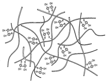
[0011 ] Fig. 1 is a schematic diagram of normal, healthy human cartilage.
[0012] Fig. 2 is a schematic diagram of a dihydroxyphenyl cross-linked
macromolecular network according to the invention.
[0013] Fig. 3 is a structural formula of a hyaluronan molecule.
[0014] Figs. 4a-4c are graphs showing comparative results for mechanical
testing in a
confined compression test (equilibrium stress versus applied strain) of T-HA
(Fig. 4a), T-
Aggrecan (Fig. 4b) and 50% T-HA/50% T-Aggrecan composite (Fig. 4c) hydrogels
according to the invention versus published results for articular cartilage
plugs (Example 3).
The relationship between glycosaminoglycan (GAG) concentration and material
compressive
strength is shown in Fig. 4d.
[0015] Fig. 5 is a graph showing comparative data of glucose utilization for
chondrocytes embedded in T-HA hydrogels (1.7% and 4.7% T-HA) compared to
cultured on
tissue culture plastic (control).
4
CA 02572804 2007-01-03
WO 2006/010066 PCT/US2005/024391
[0016] Fig. 6 is a series of four photographs illustrating a surgical
procedure to
implant a T-HA hydrogel into articular cartilage defects according to an
aspect of the
invention described in Example 6.
[0017] Fig. 7 is a series of two photographs showing the T-HA hydrogel
implants one
month after implantation into the medial trochlar facet of a Yucatan pig as
described in
Example 6, as well as the opposing (articulating) patella surface.
[0018] Fig. 8 is a series of photographs illustrating the histological results
of control
side (unfilled) and experimental side (TB-HA hydrogel filled) canine vocal
cords, 3 months
post-operatively, following a vocal cord repair procedure using a T-HA
hydrogel as a
synthetic vocal cord material as described in Example 7.
[0019] Fig. 9 is a series of photographs illustrating the histological results
of
surgically augmented vocal cords in a rabbit model using a T-HA hydrogel as a
synthetic
vocal cord material, also as described in Example 7.
[0020] Fig. 10 is a series of photographs of control (unoperated) and
experimental
(surgically replaced) eyes one month post-operative, following a vitreous
replacement
procedure using T-HA hydrogel as a synthetic vitreous material as described in
Example 8.
[0021 ] Fig. 11 shows comparative electroretinogram (ERG) results recorded for
both
control and vitreous replaced eyes in response to flashes of light in a rabbit
model as
described in Example 8.
[0022] Fig. 12 is a series of electron micrographs of the retina from four
quadrants of
control (unoperated) and experimental (surgically replaced) eyes one month
post-operative,
following a vitreous replacement procedure using T-HA hydrogel as a synthetic
vitreous
material as described in Example 8.
[0023] Fig. 13 is a series of photographs showing representative results of
histological results for a 100 mg/ml T-HA hydrogel plug implanted
subcutaneously into an
immunocompetent rat at one month post-operatively as described in Example 9.
[0024] Fig. 14 is a photograph of a cadaveric canine heart used to specify T-
HA
hydrogel materials for mitral valve repair as described in Example 10.
CA 02572804 2007-01-03
WO 2006/010066 PCT/US2005/024391
DETAILED DESCRIPTION OF PREFERRED EMBODIMENTS OF THE INVENTION
[0025] As used herein, the term polycarboxylate means a molecule, structure or
species having a chain length of at least two functional groups or units,
wherein at least two
such groups or units of the chain are or comprise carboxylic acid groups that
are sterically
accessible to a nucleophilic substitution reaction as described herein. Also
as used herein, the
term polyamine means a molecule, structure or species having a chain length of
at least two
functional groups or units, wherein at least two such groups or units of the
chain are or
comprise primary amine groups that are available for a nucleophilic
substitution reaction.
Also as used herein, a polyhydroxyphenyl molecule means a molecule having a
chain length
of at least two functional groups or units, wherein at least two such groups
or units of the
chain are or comprise hydroxyphenyl groups that can be linked to another
hydroxyphenyl
group via a C-C bond. Also as used herein, a hydrogel is a material that is
prepared
comprising a macromolecular network that is used or useful in tissue
replacement or
engineering applications, e.g. as artificial cartilage, as a material to coat
surgical instruments
to prevent tissue irritation, or to provide a semi-permeable membrane such as
for use in an
artificial kidney, etc.
[0026] The invention includes a novel structure of a macromolecular network
that has
been formed by linking hydroxyphenyl groups attached to adjacent long chain
macromolecules, resulting in effectively cross-linking the macromolecules to
provide a large
network. The basic cross-linking structure of the network is shown below
OH HO
R1 R2
where R1 and R2 are each long chain macromolecules. R1 and R2 can be the same
molecule
or different molecules, but it will be understood that to provide a suitable
network, Rl and R2
will be different molecules for at least a portion of the dihydroxyphenyl
linkages in a network
according to the invention. It is not necessary, though it is preferred, that
R1 and R2 are the
same species of molecule.
[0027] By providing a plurality of these dihydroxyphenyl linkages between
adjacent
macromolecules, a network of dihydroxyphenyl cross-linked macromolecules is
provided as
6
CA 02572804 2007-01-03
WO 2006/010066 PCT/US2005/024391
shown schematically in Fig. 2. In the figure, the macromolecules are
represented
schematically by cylindrical strands, each preferably having at least two
hydroxyphenyl
groups attached along its length. It is noted that not every hydroxyphenyl
group must be
linked to another hydroxyphenyl group.
[0028] Briefly, the disclosed invention involves covalent coupling of
hydroxyphenyl
containing compounds, including but not limited to tyramine, through their
primary amine (or
carboxyl) groups to carboxyl (or primary amine) groups on various polymeric
scaffold
materials, including but not limited to hyaluronan or chondroitin sulfate
(e.g. in the form of
aggrecan), via a carbodiimide-mediated reaction. After isolation and
purification of the
hydroxyphenyl-substituted polymeric scaffolds, the hydroxyphenyl residues are
selectively
cross-linked by horseradish peroxidase (HRP) in the presence of very dilute
hydrogen
peroxide to form hydrogels. As will become apparent, the hydrogels made as
described
herein are or can be used as a fully implantable, non-immunogenic synthetic
tissue matrix
material that can be implanted into the body for a variety of purposes as will
be described.
As used herein, 'implantable' refers both to surgical implantation of a
hydrogel as through a
surgical incision, and to provision of the hydrogel within the body via
injection, e.g. using a
syringe. Whether surgically implanted or injected, the implantable hydrogels
can be provided
within the body already cross-linked (ex vivo cross-linking) or otherwise it
can be cross-
linked in situ at the site of implantation within the body as will be further
described.
[0029] The first step in providing the macromolecular network is to prepare or
provide the long-chain macromolecules having periodic hydroxyphenyl groups
attached. In
one embodiment, the macromolecules are polyhydroxyphenyl molecules which
already have
multiple or periodic hydroxyphenyl groups, such as polyphenols. Suitable
polyphenols
include polyamino acids (e.g. polytyrosine), epigallocatechin (EGC), and
epigallocatechin
gallate (EGCG) isolated from green tea, less preferably other polyphenols.
[0030] In a further embodiment, the hydroxyphenyl groups can be added to the
macromolecules periodically or randomly along their length via a chemical
reaction. A
preferred method of adding hydroxyphenyl groups to the macromolecules is to
utilize a
carbodiimide-mediated substitution reaction pathway to provide an amide bond
between a
primary amine having a hydroxyphenyl group and a carboxylic acid group
attached to the
macromolecules. In this method, the long-chain macromolecule preferably is a
polycarboxylate molecule, having periodic carboxylic acid groups along its
length. The
7
CA 02572804 2007-01-03
WO 2006/010066 PCT/US2005/024391
hydroxyphenyl groups are provided as part of smaller molecules having primary
amine
groups that can be attached to the carboxyl carbon atoms of a carboxylic acid
group on the
long-chain macromolecules via the carbodiimide pathway. The reaction proceeds
as follows:
A R-N-C-N-R
'7)
O-, C Rexbon A
B
H
R-N-C=N-R
D HO ))-R-NH2 +~.
PeacUon B
O ) H-R-~-OH
E + O
11 F
R-NH-C-NH-R
where:
Structure A is a carbodiimide;
Structure B is a polycarboxylate (though only one CO2H group is shown);
Structure C is the product of Reaction A and is an activated O-acylisourea;
Structure D is a primary amine having a hydroxyphenyl group;
Structure E is a hydroxyphenyl-substituted polycarboxylate; and
Structure F is an acylurea byproduct;
wherein individual Rs can be individually selected, the same or different from
one
another, to be a straight chain or branched alkane or acyl group, or any other
structure that
does not interfere with the carbodiimide reaction pathway to provide the amide
bond between
the NH2 and CO2H groups as shown in Structure E above.
8
CA 02572804 2007-01-03
WO 2006/010066 PCT/US2005/024391
[0031] In the above-illustrated pathway, Reaction A represents a carbodiimide
activation of the carboxyl group to provide an activated 0-acylisourea
intermediate. The
electropositive carbon atom of this intermediate is receptive to nucleophilic
attack by the lone
pair of electrons on a nitrogen atom of an adjacent primary amine molecule
having an
attached hydroxyphenyl group. The products of this nucleophilic substitution
reaction
(Reaction B) are a hydroxyphenyl-substituted polycarboxylate and an acylurea
byproduct
which can be dialyzed out to provide a substantially pure hydroxyphenyl-
substituted
polycarboxylate product.
[0032] Certain side-reactions are possible in the above-described carbodiimide
reaction pathway chemistry and should be considered by the person having
ordinary skill in
the art. First, the carbodiimide can react with nucleophiles other than the
carboxylate oxygen
atom of the polycarboxylate molecule required to form the desired 0-
acylisourea (reaction
A). Such nucleophiles may include the amine and/or hydroxyphenyl groups of
Structure D
illustrated above. In particular, there are three potential side-reactions for
Reaction A which
can reduce the effective concentration of the carbodiimide and the primary
amine having the
hydroxyphenyl group (Structures A and D), and potentially lead to the creation
of undesired
adducts on the polycarboxylate (Structure B):
9
CA 02572804 2007-01-03
WO 2006/010066 PCT/US2005/024391
R-N=C=N-R R-NH-C-NH-R
N
Reaction C: NH,
I
HO-C- R
HO-{ ( ) )- R
R-N=C=N-R R-NH-C=N-R
OH I
Reaction D: I (0)
O 1
I H2N-R
HZN-R
O
Reaction E: R-N C=N-R R-NH-C-NH-R
Z
KO
[0033] The product of an amine reaction with the carbodiimide (Reaction C)
will not
have a free amine group effectively reducing the amount of tyramine available
for reaction
with the O-acylisourea. This reaction also reduces the amount of carbodiimide
available for
formation of the desired O-acylisourea. The products of the hydroxyphenyl
reaction
(Reaction D) are not UV absorbent, which will make their detection by UV-
spectroscopy in
the final hydroxyphenyl-substituted polycarboxylate product (explained below)
more
difficult. However, because these products still contain free amine groups,
they can form
amide bonds with the polycarboxylate molecule via Reaction B. This can give
rise to two
unproductive hyaluronan-substituted structures, neither of which can
participate in the
peroxidase cross-linking reaction in the second step (described below) of
preparing the cross-
linked network due to the absence of an extractable phenolic hydroxyl hydrogen
atom needed
to generate the free radical (also explained below). Finally, the carbodiimide
can react non-
CA 02572804 2007-01-03
WO 2006/010066 PCT/US2005/024391
productively with water (Reaction E) to produce the same acylurea shown above
as a
byproduct of Reaction B, but with none of Structure E, the desired product.
[0034] Once the desired O-acylisourea product has been formed in Reaction A,
there
is again the possibility for certain additional side-reactions:
0
11
R-NH-C=N-R R-NH-C-NH-R
Acylurea
0 + H2O (Structure F)
Reaction F: c~ +
o1~1, C
Activated
O-Acylisourea
(Structure C)
Polycarboxylate
(Structure B)
0
R-NH-C=N-R I I
R-NH-C-N-R
O I
Reaction G: O= C
N-Acylurea
(Structure G)
0
R-NH-C=N-R I I
I R-NH-C-NH-R
O~~O
Reaction H: P +
+
o~c o~_,, C10
[0035] The O-acylisourea (Structure C) can be hydrolyzed as shown in Reaction
F
releasing the original unmodified polycarboxylate (Structure B) and the
acylurea of the
11
CA 02572804 2007-01-03
WO 2006/010066 PCT/US2005/024391
carbodiimide (Structure F). This is an unproductive reaction similar to
reaction E, which
reduces the effective concentration of the carbodiimide. The O-acylisourea,
can also undergo
an intramolecular rearrangement (Reaction G) to form two unreactive N-
acylureas. These
structures form unproductive adducts on the carboxylate molecule which cannot
contribute to
the peroxidase catalyzed cross-linking reaction (step 2 discussed below) for
preparing a
network according to the invention. The O-acylisourea can also react (Reaction
H) with a
second carboxyl group on either the same or a different polycarboxylate
molecule to form an
acid anhydride. This molecule can then react with Structure D to form the
desired amide and
regenerate the second carboxyl group. Thus there are two potential side-
reactions for the 0-
acylisourea, which can reduce the effective concentration of the carbodiimide
(Reactions F
and G), and potentially lead to creation of undesired adducts on the
polycarboxylate
molecule.
[0036] Negative effects of these side reactions can be addressed through
conventional
techniques without undue experimentation.
. [0037] Alternatively to the pathway shown above where the macromolecule
(Structure B) is a polycarboxylate, the macromolecule can be a polyamine
having multiple or
periodic amine groups along its length, wherein the hydroxyphenyl groups then
are provided
as part of smaller carboxylic acid molecules. Suitable polyamines include:
polyhexosamines
such as chitosan (polyglucosamine); polyamino acids such as polylysine;
polydeoxyribonucleotides such as poly (dA) (polydeoxyadenylic acid), poly(dC)
(polydeoxycytidylic acid), and poly(dG) (polydeoxyguanylic acid); and
polyribonucleotides
such as poly(A) (polyadenylic acid), poly(C) (polycytidylic acid), and poly(G)
(polyguanylic
acid). The carbodiimide-mediated reaction pathway proceeds exactly as
explained above to
form the amide bond between the amine group and carboxylic acid group except
that, as will
be understood by a person having ordinary skill in the art, the resulting
product will be
hydroxyphenyl-substituted polyamine instead of a polycarboxylate. Other
peptides and/or
proteins also can be used as the macromolecules in the present invention,
either which have
hydroxyphenyl groups disposed along their length, or to which hydroxyphenyl
groups can be
provided via a substitution reaction as described herein. For example, in
addition to the
peptides already disclosed herein, polyarginine can be used as the
macromolecule.
[0038] When substituting onto a polycarboxylate molecule, suitable
hydroxyphenyl-
containing compounds for use in the present invention include those having a
free primary
12
CA 02572804 2007-01-03
WO 2006/010066 PCT/US2005/024391
amine that can be used to modify scaffold materials having multiple or
periodic CO2H
groups, including tyrosine (2-amino-3-(4-hydroxyphenyl) proprionic acid) and
tyramine
(tyrosamine or 2-(4-hydroxyphenyl) ethylamine). When substituting onto a
polyamine,
suitable hydroxyphenyl-containing compounds include those having a free CO2H
group that
can be used to modify scaffold materials having multiple or periodic primary
NH2 groups,
including tyrosine, 3-(4-hydroxyphenyl) propionic acid and 4-
hydroxyphenylacetic acid.
[0039] The second step in preparing a cross-linked macromolecular network
according to the invention is to link the resulting macromolecules, now having
one or more
hydroxyphenyl groups attached, via a dihydroxyphenyl linking structure. In
this step
hydroxyphenyl groups attached to different macromolecules are linked via the
reaction
mechanism shown below using a peroxide reagent in the presence of a
peroxidase:
13
CA 02572804 2007-01-03
WO 2006/010066 PCT/US2005/024391
OH HO
NH-R R-NHS j
Hydroxyphenyl-Substituted
Polycarboxylates
(Structure E)
Peroxidase
H2O2
O= .O
(Free R2dca15)/
C
NH- R R- NH o
Isomerizes
O O
H H
NH- R R- NH
Dimerizes
O O
H H
NH- R R- NH
Enolizes
OH HO
O~-" NH - R Dihydroxyphenyl R- NHS
Link C
14
CA 02572804 2007-01-03
WO 2006/010066 PCT/US2005/024391
[0040] (It is noted that some dihydroxyphenyl linking may occur between
different
hydroxyphenyl groups attached to the same molecule as well). Peroxidase in the
presence of
a dilute peroxide (preferably H202) is able to extract the phenolic hydroxyl
hydrogen atom
from hydroxyphenyl containing compounds (such as tyramine) leaving the
phenolic hydroxyl
oxygen with a single unshared electron, an extremely reactive free radical.
The free radical
isomerizes to one of the two equivalent ortho-position carbons and then two
such structures
dimerize to form a covalent bond effectively cross-linking the structures,
which after
enolizing generates a dihydroxyphenyl dimer (a dihydroxyphenyl linkage such as
dityramine
linkage as described below).
[00411 For clarity, only a single dihydroxyphenyl linking reaction is shown
above,
but it will be understood that several or multiple such linkages will be
produced when
macromolecules having attached hydroxyphenyl groups are subjected to the
reaction
conditions (peroxide and peroxidase). Hydrogen peroxide is indicated in the
above
mechanism, but other suitable peroxides can be used. Also, the peroxidase
preferably is
horseradish peroxidase (HRP). Alternatively, any other suitable enzyme (or
other agent) can
be used that is capable of generating free-radicals for cross-linking scaffold
materials
containing hydroxyphenyl groups, preferably under ordinary metabolic
conditions as
described below.
[0042] We have shown that the interaction of horseradish peroxidase (Type II)
and
hydrogen peroxide (H202) is suitable for the production of cross-linked
macromolecular
networks. The mechanism comprises four distinct steps: (a) binding of peroxide
to the heme-
Fe(III) complex of the peroxidase to form an unstable peroxide complex,
"Compound I"; (b)
oxidation of the iron to generate a ferryl species with a pi-cation radical in
the heme
porphyrin ring, "Compound II"; (c) reduction of Compound II by one substrate
(i.e.
hydroxyphenyl or water) molecule to produce a product (i.e. hydroxyphenyl or
superoxide)
radical and another ferryl species, "Compound III"; (d) reduction of Compound
III by a
second substrate (i.e. hydroxyphenyl or water) molecule to release a second
product (i.e.
hydroxyphenyl or superoxide) radical and regenerate the native enzyme. Thus
the peroxidase
enzyme can either form hydroxyphenyl radicals required for cross-linking
through interaction
of hydroxyphenyl groups at the enzyme active site to directly create the
desired radicals or
through first generation of superoxide radicals, which then diffuse from the
enzyme and
interact with hydroxyphenyl groups to generate the desired radicals. Other
compounds that
CA 02572804 2007-01-03
WO 2006/010066 PCT/US2005/024391
have the potential to produce the same effect include any porphyrin containing
compound
(i.e. Photofrin below), which includes the peroxidase family, hemoproteins, or
the structurally
related chlorin compounds.
[0043] A number of other free radical initiators can be used to crosslink the
hydroxyphenyl modified macromolecules described herein. A majority are based
on the
formation or inclusion of reactive oxygen species (ROS) such as, but not
limited to,
molecules of hydrogen peroxide, ions of hypochlorite, radicals like the
hydroxyl radical, and
the superoxide anion which is both ion and radical. Additional reactive
molecules such as
reactive nitrogen species or reactive sulfur species, or those free radical
species involved in
synthetic polymerization have the potential to be used for hydroxyphenyl cross-
linking.
[0044] ROS are commonly produced in nature through the use of enzymes, and
substrates. Additional enzymatic systems which have the potential to be used
in the cross-
linking process, as a result of production of superoxide radicals, include,
but are not limited
to xanthine-xanthine oxidase and NADPH-NADPH oxidase.
[0045] Another class of ROS free radical initiators that can be used involves
the use
of metallic cations. One example is based on the Fenton reaction, which takes
place between
hydrogen peroxide and a bivalent cation, such as Fez+. This process generates
powerful free
radicals when the catalyst reacts with hydrogen peroxide. The principal
chemical reaction
associated with Fenton's reaction is shown below:
H2O2 + Fe 2+ => OH= + Off + Fe 3+
where, Fe 2+ = ferrous ion, Fe3+ = ferric ion, OH= = hydroxyl radicals
[0046] In addition to the initiation reaction described above that produces
hydroxyl
radicals, the Fenton's process can also produce superoxide radicals and
hydroperoxide anions
by additional chain propagation reactions described below. The perhydroxyl
radical is known
to be a weaker reductant compared to superoxide radical and hydroperoxide
anions.
H202 + OH- => H02= + H2O
HO2= => H+ + O2="
H02= + O2=-=> HO2- + 02
where 02=" = superoxide radical anion, HO2- = hydroperoxide anion, H02= =
perhydroxyl
radical.
16
CA 02572804 2007-01-03
WO 2006/010066 PCT/US2005/024391
[0047] We have demonstrated the ability for this reaction to crosslink
tyramine
substituted hyaluronan in the laboratory using ferrous sulfate in conjunction
with hydrogen
peroxide. Compounds which include, but are not limited to, bivalent cations of
copper,
chromium, vanadium and cobalt can be used in a similar manner. It is to be
noted that while
the hydroxyl free radical can be used to form a dityramine crosslink, it has
also been shown
to cleave HA chains, and thus may ultimately be unsuitable for ideal hydrogel
formation.
Additional molecules or methods which can generate ROS include:
= rubidium or cesium ions in the presence of oxygen to form superoxide
radicals;
= trivalent cations, which with hydrogen peroxide form free radicals and
bivalent
cations as shown below, which can subsequently follow the reactions involved
in
the Fenton process.
Fe+3 + H202 = Fe+2 + -OOH + H+
= the cytotoxic and antitumor therapy Photofrin, which upon illumination with
laser
light at a wavelength of 630 nm causes propagation of a radical generating
reaction that produces superoxide and hydroxyl radicals. In the absence of
light,
but the presence of hydrogen peroxide, the porphorin ring in Photofrin should
operate by the same reaction as for the peroxidase enzyme above.
= UV light and hydrogen peroxide to form hydroxyl and superoxide free
radicals.
= the persulfate family in combination with TEMED.
[0048] As noted above, one alternative method for generating such free-
radicals is to
use Photofrin as an alternative, non-enzymatic, light-activated cross-linking
agent to cross-
link the macromolecular network described herein, e.g. tyramine-substituted
hyaluronan to
form tyramine cross-linked hyaluronan hydrogels. Photofrin , which is known in
the art,
generates free radicals which could initiate the cross-linking reaction as
described herein in a
manner similar to the peroxidase-H202 mechanism described above. Photofrin is
a
porfimer sodium manufactured in powder or cake form by Wyeth-Ayerst Lederle
Parenterals,
Inc.
[0049] The dihydroxyphenyl cross-linked macromolecular network is superior to
conventional cartilage or other tissue replacement or substitution methods and
products, at
least with respect to the ability to carry out an in situ cross-linking
procedure, because the
preferred cross-linking reaction is enzyme driven (peroxidase). This means the
cross-linking
17
CA 02572804 2007-01-03
WO 2006/010066 PCT/US2005/024391
reaction is carried out under ordinary in vivo or metabolic conditions of
temperature such as
35-39 C (e.g. about 37 C), pH range of 6-7 (e.g. about 6.5), reagents etc. (A
peroxide, such
as hydrogen peroxide, is the only required reagent for the cross-linking
reaction). In addition,
Photofrin already is used in in vivo applications, e.g. ablative treatment of
Barrett's
esophagus, and the iron-based cross-linking mechanism also can be optimized
for in vivo
performance. Thus, the cross-linking reaction can be performed in vivo, to
provide a cross-
linked hydrogel at a surgical situs, such as an orthopedic surgical situs, to
promote maximum
seamless integration between the hydrogel and native tissue such as bony and
cartilaginous
tissue. Integration of the new hydrogel scaffold with native cartilage matrix
may occur
immediately as the hydroxyphenyl-substituted macromolecular scaffold quickly
penetrates
into the existing cartilage matrix prior to cross-linking, and cross-links not
only with other
hydroxyphenyl-substituted macromolecular scaffold material but potentially
with tyrosine
residues of resident proteins in the existing cartilage matrix. This would
eliminate a typical
problem found with pre-formed matrix plugs, which is their poor integration
into the native
cartilage tissue. The ability to cross-link the hydrogel directly on the
articular surface
eliminates the need to surgically enlarge a defect to fit a pre-cast plug, as
is necessary for
hydrogels whose chemistries are toxic to or otherwise prohibit their formation
inside the
patient. It should be noted that most cartilage damage as a result of
arthritis presents as a
variable thinning of the articular surface, not holes of defined shape.
[0050] For the peroxidase mechanism, because the cross-linking reaction
requires
both the peroxide and a peroxidase (preferably horseradish peroxidase),
solutions containing
all but one of these components can be prepared for convenient application to
a surgical site.
For example, a solution comprising a tyramine - (or other hydroxyphenyl
containing species)
substituted polycarboxylate (such as tyramine-substituted hyaluronan, etc.)
and the
peroxidase can be prepared, with a second solution prepared containing the
peroxide.
Alternatively, the peroxide and the peroxidase can be swapped between the
first and second
solutions, the important thing being that the peroxide and peroxidase are kept
separate (i.e. in
separate solutions) until the cross-linking reaction is to be carried out.
Then, the first solution
is applied, (e.g. to an in vivo surgical situs), and the second solution is
applied or sprayed
over the first, in vivo, to cause in situ cross-linking of the tyramine
residues. The cross
linking reaction occurs in vivo. Other combinations will be evident from the
present
disclosure which are within the skill of a person of ordinary skill in the
art.
18
CA 02572804 2007-01-03
WO 2006/010066 PCT/US2005/024391
[0051] Furthermore, because the cross-linking reaction occurs under ordinary
metabolic conditions, additional living cells, such as chondrocytes,
progenitor cells, stem
cells, etc., can be provided directly to a medium containing the non-cross-
linked
hydroxyphenyl-substituted polycarboxylates or polyamines (or polyphenols),
i.e. to the first
or second solution from the preceding paragraph, wherein the cell-rich medium
is applied
with the macromolecules to the site in vivo, and the molecules are
subsequently cross-linked
via addition of peroxidase and peroxide. The result is a cross-linked
macromolecular
network containing the desired cells dispersed within it. Such a cell-enriched
network is not
possible in conventional tissue replacement matrices due to the harsh
conditions of
temperature and pH under which they are prepared. Further, as described below
in Example
5, it has been demonstrated that the cells provided to the network as
described above remain
viable even after cross-linking of tyramine-substituted hyaluronan (also
described below).
[0052] In a preferred embodiment particularly suitable for preparing synthetic
cartilage as well as other synthetic or artificial tissues, the macromolecule
used to produce the
network is hyaluronan or hyaluronic acid (HA), and the hydroxyphenyl group is
supplied in
the form of tyramine. Hyaluronan (HA) is a ubiquitous molecule, which is most
concentrated
in specialized tissues such as cartilage, vocal cords, vitreous, synovial
fluid, umbilical cord,
and dermis. In these tissues, its function is manifold, influencing tissue
viscosity, shock
absorption, wound healing, and space filling. HA has been shown to influence
many
processes within the extracellular matrix (ECM) in native tissues where it is
present including
matrix assembly, cell proliferation, cell migration and embryonic/tissue
development.
[0053] HA is composed of repeating pairs of glucuronic acid (glcA) and N-
acetylglucosamine (glcNAc) residues linked by a 131,3 glycosidic bond as shown
in Fig. 3.
The glucuronic acid residue is particularly pertinent to the production of a
macromolecular
network as described herein as this sugar provides an available carboxyl group
periodically
along the repeat disaccharide structure of HA that is useful for
hydroxyphenyl, i.e. tyramine,
substitution. For each hyaluronan chain, this simple disaccharide is repeated
up to 10,000
times or greater resulting in macromolecule that can have a molecular weight
on the order 10
million daltons (10 megadaltons). Adjacent disaccharide units of HA are linked
by a 131,4
glycosidic bond, also seen in Fig. 3. Each gicA residue has a carboxylic acid
group (CO2H)
attached to the number 5 carbon atom of the glucose ring. Under biological
conditions, HA is
a negatively charged, randomly coiled polymer filling a volume more than 1,000
times
19
CA 02572804 2007-01-03
WO 2006/010066 PCT/US2005/024391
greater than would be expected based on molecular weight and composition
alone. As noted
above, the strong negative charges attract cations and water, which allow HA
to assume the
form of a strongly hydrated gel in vivo, giving it a unique viscoelastic and
shock-absorbing
property. HA represents a readily available and desirable scaffolding material
for tissue
engineering applications as it is non-immunogenic, non-toxic and non-
inflammatory. Also as
a naturally occurring extracellular matrix (ECM) molecule it offers the
advantages of being
recognized by cell receptors, of interacting with other ECM molecules, and of
being
metabolized by normal physiological pathways.
[0054] Tyramine is a phenolic molecule having an ethyl amine group attached
para t.o
the OH group on the benzene ring. When these species are used, the mechanism
for tyramine
substitution onto the singly bound oxygen atom of a CO2H group on HA proceeds
via the
carbodiimide-mediated reaction mechanism described above as illustrated
immediately
below. The preferred carbodiimide species is 1-ethyl-3-(3-
dimethylaminopropyl)carbodiimide (EDC) as shown.
A CH7
CH,CH, - N=C= N-CH,CH,CH2NH+ CI
CH,
1~c/o /JJ
B RetialA
HA
H
CH3
CH,CH,-N-C= -CH,CH,CH,NHCI
\CH3 C
I +
D HO-O-CH,CH,NH, ~p
HA
Readian B
NHCH,CH,-O-OH
E + o
HA II ,CH, F
CH3CH2-NH-C-NH-CH2CH2CH,NH CI
CH,
CA 02572804 2007-01-03
WO 2006/010066 PCT/US2005/024391
where:
Structure A is EDC;
Structure B is hyaluronan (though only one CO2H group is shown);
Structure C is the product of Reaction A and is 1-ethyl-3-(3-
dimethylaminopropyl)
isourea;
Structure D is tyramine;
Structure E is tyramine-substituted hyaluronan; and
Structure F is 1-ethyl-3-(3-dimethylaminopropyl) urea (EDU).
[0055] In the above pathway, a negatively charged oxygen atom of the carboxyl
group of the hyaluronan molecule attacks, via a nucleophilic reaction
mechanism, the
electron-deficient diimide carbon atom on the carbodiimide molecule (EDC) to
form the
activated 0-acylisourea (Reaction A). The result is that the carbon atom of
the HA
carboxylate group becomes sufficiently electron deficient to be susceptible to
nucleophilic
attack by the unshared pair of electrons on the amine group of a tyramine
molecule (Reaction
B). Reaction A is preferably catalyzed by a suitable catalyst that will result
in the formation
of an active ester during Reaction A, thus permitting the reaction to be
carried out at
substantially neutral pH (e.g. pH=6.5). Suitable catalysts include N-
hydroxysuccinimide
(NHS), less preferably 1-hydroxybenzotriazole (HOBt) or N-
hydroxysulfosuccinimide
(NHSS), less preferably another suitable catalyst or combinations thereof
effective to enhance
the carbodiimide reaction by formation of an active ester in order to minimize
the
unproductive hydrolysis of carbodiimides at higher pHs. Less preferably other
carbodiimides
besides EDC can be used, including 1-cyclohexyl-3-[2-(4-
methylmorpholino)ethyl]carbodiimide (CMC), and dicyclohexylcarbodiimide (DCC).
[0056] The result of Reaction A above is O-acylisourea-substituted hyaluronan;
essentially the EDC molecule has been temporarily substituted onto the
carboxylic acid group
of a glcA residue from the HA molecule, making the carbon atom of the
carboxylic acid
group slightly positively charged. The electron pair from the terminal amine
group of a
tyramine molecule is then substituted onto the carbon atom via a nucleophilic
substitution
reaction as explained in the preceding paragraph (Reaction B). The result of
Reaction B is
the tyramine-substituted HA molecule (T-HA) and acylurea, a byproduct. It will
be
understood that Reactions A and B will result in a plurality of tyramine
substitutions on the
21
CA 02572804 2007-01-03
WO 2006/010066 PCT/US2005/024391
periodic glcA residues of HA molecules; a single substitution has been shown
here for
brevity and clarity.
[0057] After formation of T-HA, a plurality of T-HA molecules are reacted via
peroxide and peroxidase enzyme to cross-link T-HA molecules as previously
described and
illustrated above. That is, the hydroxyphenyl groups on the tyramine residues
now attached
to HA molecules react with peroxide (preferably H202) in the presence of a
peroxidase to
remove the phenolic hydrogen atom resulting in a tyramine free radical, with
the unpaired
electron associated with the phenolic oxygen atom. This free radical species
isomerizes or
resonates, resulting in a resonance structure (or free radical isomer) with
the unpaired
electron now associated with an ortho carbon atom on the phenolic ring. In
this position, the
unpaired electron quickly reacts with a similarly situated unpaired electron
on another
tyramine free radical to form a covalent bond therebetween. The result is a
free-radical
driven dimerization reaction between different tyramine free radical residues
attached to
different glcAs of the same or different HA molecules. This dimerized species
further
enolizes to restore the now-linked tyramine residues, resulting in a
dityramine linkage
structure. It will be understood that a plurality of reactions as herein
described will occur
between adjacent tyramine residues, resulting in a cross-linked macromolecular
network of
T-HA molecules having the following cross-linking structure:
OH HO
NHCH2CH2 CH2CH2NH
O\ C C~ O
lHA HA
[0058] The cross-linked T-HA network can be provided with aggrecan molecules
in a
conventional manner, e.g. via link proteins, to provide a cross-linked T-HA
network having
aggrecan molecules attached to the HA chains. Thus, a network similar to that
found in a
normal cartilage aggregate can be provided, with the dityramine bonds holding
the network
together thereby constraining the contained aggrecan network, instead of
collagen fibrils as in
normal cartilage.
22
CA 02572804 2007-01-03
WO 2006/010066 PCT/US2005/024391
[0059] It will be understood from the present invention that other
glycosaminoglycans
(GAGs), polysaccharides and polycarboxylic acids can be used as the
macromolecules for
producing the cross-linked network disclosed herein. For example, suitable
GAGs, other
than HA, include chondroitin, chondroitin sulfate, dermatan sulfate, heparan
sulfate and
heparin. Other suitable polycarboxylates include: proteoglycans such as
versican, aggrecan,
and cartilage aggregates composed of aggrecan, hyaluronan and link protein;
polyuronic
acids such as polypectate (polygalacturonic acid), polyglucuronic acid, pectin
(polygalacturonic acid methyl ester), colominic acid (poly[2,8-(N-
acetylneuraminic acid)]),
and alginate (poly[mannuronate-co-guluronate]); and amino acids (having at
least 2 amino
acid units) that meet the definition of polycarboxylate given above, such as
polyaspartic acid,
and polyglutamic acid. All of these can be substituted with one or a plurality
of
hydroxyphenyl groups using the carbodiimide-mediated reaction pathway
disclosed herein by
a person of ordinary skill in the art without undue experimentation.
[0060] As mentioned above, it is also to be understood that native polyphenol
compounds, which already contain two or more hydroxyphenyl groups that can be
cross-
linked using the described enzyme catalysis chemistry can be used in place of
the
polycarboxylates and polyamines described above which must have the
hydroxyphenyl
groups added by a chemical reaction.
[0061 ] In another preferred embodiment, a network of tyramine cross-linked
chondroitin sulfate molecules (either alone or provided as part of aggrecans)
is provided to
simulate or replace normal cartilage. Chondroitin sulfate is identical to
hyaluronan except:
1) the repeat disaccharide structure contains N-acetylgalactosamine (ga1NAc)
rather than
glcNAc, a difference in only the position of the hydroxyl group attached to
the 4- carbon
(circled in Fig. 3); 2) the presence of O-sulfation on the hydroxyl groups at
the 4- and/or 6-
position of the ga1NAc residue and/or the 2-position of the glcA residue (Fig.
3); and 3) the
size of the chondroitin sulfate chains, which are smaller than hyaluronan with
between 20 to
100 repeating disaccharide units. (An aggrecan molecule is made up of multiple
- roughly
100 - chondroitin sulfate chains linked to a core protein through a linkage
saccharide located
at each chain's reducing end). In this embodiment, the negatively charged S042-
groups of
adjacent (cross-linked) chondroitin sulfate molecules provide the principal
repulsive force
contributing to the compression resistance of the network aggregate while the
tyramine cross-
links constrain the chondroitin sulfate network from breaking or dissipating.
The result is a
23
CA 02572804 2007-01-03
WO 2006/010066 PCT/US2005/024391
similarly non-displaceable chondroitin sulfate network (and concomitant water-
impermeability) as in normal cartilage, but without the extracellular collagen
fibril matrix or
the HA chains found in normal cartilage. In fact, by directly cross-linking
chondroitin sulfate
molecules, (instead of their core HA molecules as in the previously described
embodiment),
the repulsive force between adjacent chondroitin sulfate molecules maybe
strengthened,
resulting in even stronger fluid flow resistance compared to normal cartilage.
This may result
in greater loading force absorption and dissipation capacity than normal
cartilage because the
interstitial fluid phase is even more constrained from flowing. In this
embodiment, where
chondroitin sulfate molecules are directly cross-linked, certain cartilage
degenerative
conditions are entirely circumvented; e.g. conditions where the core protein
to which
chondroitin sulfate molecules are ordinarily bonded in normal cartilage
becomes cleaved
between the HA binding domain (GI) and the second globular domain (G2) thus
allowing the
chondroitin sulfate rich region to diffuse out from the cartilage aggregate.
In this
embodiment, because the chondroitin sulfate molecules are directly cross-
linked to one
another, unassociated with an aggrecan or other proteoglycan molecule, they
cannot be
cleaved or carried away as in normal cartilage.
[0062] Nonetheless, a tyramine cross-linked T-HA network (having an HA
backbone
chain with attached aggrecan molecules, which in turn include chondroitin
sulfate chains)
may be preferred because of the high availability of HA. This may be
beneficial in the case
of cartilage replacement or repair using the present invention, because the
body's normal
metabolic pathway for generating cartilage may be able to build directly onto
an implanted
tyramine cross-linked T-HA network as will be described.
[0063] One further particular application where a cross-linked network
according to
the invention will have substantial utility is in the production of an
artificial kidney. The
kidney filters blood by two mechanisms: one is by size exclusion and the
second is by charge
exclusion. MEMS devices have been designed for use in artificial kidney
devices, which
contain precisely defined micropores that can effectively mimic only the size
exclusion
characteristics of the kidney. In a healthy kidney, the charge exclusion
related filtration is the
result of heparan sulfate proteoglycans present in a basement membrane, which
separates two
distinct cell types important for other kidney related functions. To mimic
this charge barrier
in the MEMS engineered artificial kidney, hydrogels can be prepared composed
of either
heparan sulfate or heparin that are cross-linked via dihydroxyphenyl
(dityramine) links as
24
CA 02572804 2007-01-03
WO 2006/010066 PCT/US2005/024391
described herein and provided within the pores of the MEMS device. This
heparin/heparan
sulfate hydrogel can then be sandwiched between two hyaluronan derived
hydrogels (e.g. T-
HA described above) as described herein, and containing one of each of the
cell types
normally found in a normally functioning kidney. The central heparin/heparan
sulfate
hydrogel provides the charge exclusion properties for the device. The outer
two hyaluronan
hydrogel layers provide protection from the immune system and fouling by
normal cellular
and molecular debris. Inclusion of the two cell types on opposite sides of the
filtration barrier
provides a cellular component in its normal physiologic orientation.
[0064] In another promising application, the hydrogels herein described can be
applied in developing an artificial pancreas. A problem in development of an
artificial
pancreas is the short half life of MEMS engineered glucose sensors due to
fouling of the
detector electrode in vivo. Coating of the surface of these detectors with a
hyaluronan
hydrogel (e.g. T-HA) as described herein would permit diffusion of the small
molecular
weight glucose molecules that they are designed to detect while providing
protection from the
immune system and fouling by normal cellular and molecular debris.
[0065] In summary, it will be evident from the foregoing that macromolecules
useful
as scaffold materials for formation of hydrogels include but are not limited
to
polycarboxylates (containing free carboxylate groups), polyamines (containing
free primary
amine groups), polyphenols (containing free hydroxyphenyl groups) and their
copolymers,
examples of which have been described above. When polyphenols are used, the
first step in
preparing the network described above can be omitted because polyphenols
already contain
multiple or periodic hydroxyphenyl groups. Otherwise, both polycarboxylates
and
polyamines must have hydroxyphenyl groups added or substituted along their
length,
preferably via the above-described carbodiimide reaction pathway. The second
step in
preparing the network is to carry out an enzyme driven dimerization reaction
between two
hydroxyphenyl groups attached to adjacent macromolecules (whether
polycarboxylates,
polyamines or polyphenols) in order to provide a cross-linked structure. This
step is carried
out using a peroxide reagent (preferably hydrogen peroxide) in the presence of
a suitable
enzyme (preferably HRP) under metabolic conditions of temperature and pH.
[0066] In the case of the preferred dityramine cross-linked T-HA network, in
the first
step the carboxyl groups on high molecular weight hyaluronan (HA) are
substituted with
tyramine which introduces reactive hydroxyphenyl groups into the HA molecule.
This
CA 02572804 2007-01-03
WO 2006/010066 PCT/US2005/024391
tyramine substitution reaction preferably is mediated by the carbodiimide, 1-
ethyl-3-(3-
dimethylaminopropyl)carbodiimide (EDC) with the degree of tyramine
substitution on HA
controlled by the molar ratios and absolute concentrations of tyramine, EDC
and HA used in
the reaction mix. Excess reagents such as unused tyramine and EDC are
subsequently
removed by dialysis, allowing isolation and recovery of high molecular weight
tyramine-
substituted HA (T-HA). The percent tyramine substitution within each T-HA
preparation is
easily calculated by measuring: 1) the concentration of tyramine present in
the preparation,
which is quantitated spectrophotometrically based on the unique UV-absorbance
properties of
tyramine at 275 nm (see Example 2 below); and 2) the concentration of total
carboxyl groups
in the HA preparation, which is quantitated spectrophotometrically by a
standard hexuronic
acid assay. By this technique, T-HA preparations which contain a percent
tyramine
substitution of only 4 - 6% have been routinely synthesized experimentally. At
this level of
tyramine substitution, the vast majority (preferably at least 60, 70, 80, 90,
or 95, percent) of
the HA molecule remains chemically unaltered, and therefore biologically
functional. From
this formulation of T-HA (i.e. 4 - 6% tyramine substitution) a wide range of
biomaterials with
a wide range of physical properties can be produced by simply varying the
concentration of
the T-HA used in the second step of the process.
[0067] In the cross-linking reaction, solutions of T-HA are cross-linked to
form
hydrogels through an enzyme (peroxidase) driven reaction, which catalyzes the
formation of
a covalent bond between two tyramine adducts on adjacent HA molecules,
producing a single
dityramine cross-link. The formation of multiple, e.g. hundreds, of these
dityramine cross-
links per HA molecule result in formation of a stable 3-dimentional scaffold
or hydrogel.
Addition of very dilute peroxide (preferably H202) is required to initiate the
cross-linking
reaction as it is the peroxide, not -HA, that is the actual substrate for the
peroxidase enzyme.
The products of the reaction of the peroxidase enzyme on peroxide are free
radicals that are
preferentially taken up by the hydroxyphenyl rings of tyramine resulting in
the formation of
the dityramine cross-links. The dityramine linked structures are fluorescent
blue (see
Example 2), a property which is used to both image the hydrogels and to
quantify the degree
of cross-linking within the hydrogels. Since the cross-linking reaction is
enzyme driven, the
hydrogels can be formed under physiologic conditions, and therefore can be
formed in the
presence of included cells or bioactive agents, or directly adjacent to living
tissue while
maintaining cell and tissue viability.
26
CA 02572804 2007-01-03
WO 2006/010066 PCT/US2005/024391
[0068] The resulting hydrogels are optically clear with a wide range of
physical
properties depending on the initial T-HA concentration. For example, hydrogels
formed from
T-HA solutions of 6.25, 12.5, 25, 50 and 100 mg/ml T-HA have been shown
experimentally
to have physical properties (rigidity, rheology and texture) of a jelly, a
gelatin, a dough, a
resilient rubber-like composition (similar to a rubber ball), and a cartilage-
like material
respectively - see Example 3. These materials have potential applications in a
wide range of
clinical settings including tissue engineering of both orthopedic (i.e.
cartilage, bone, tendon,
meniscus, intervertebral disk, etc.) and non-orthopaedic (kidney, liver,
pancreas, etc.) tissues,
gene and drug delivery, coating of non-biological devices for in vivo
implantation (i.e.
glucose sensors, artificial hearts, etc.), wound repair, biosensor design, and
vocal cord
reconstruction.
[0069] Advantageous properties of the hydrogels described herein include the
ability
to: 1) provide easy characterization and quality control; 2) integrate with
existing tissue
matrices; 3) directly incorporate into newly formed matrices; 4) directly
include cells and
bioactive factors; 5) maintain biocompatibility; 6) control bioresorption; 7)
cast easily into
complicated anatomical shapes (see Example 4 below); and 8) exhibit the
mechanical
properties of native tissues such as articular cartilage.
[0070] Current biologically-based surgical procedures for cartilage repair
include
autologous chondrocyte implantation, drilling, abrasion chondroplasty,
microfracture, and
mosaic arthroplasty. All these procedures treat only focal articular cartilage
injuries, and not
cartilage denuded joint surfaces such as seen in severe osteoarthritis and
rheumatoid arthritis.
Also, they use either cartilage tissue plugs or expanded chondrocytes
harvested from the
patient to fill cartilage defects. These tissues or chondrocytes are expected
to fill the defect
by synthesizing entirely de novo material, such as newly synthesized hyaline
cartilage, that
has integrated with existing cartilage matrices and has the biomechanical
properties of normal
cartilage. However, such procedures all promote the formation of a reparative
tissue
(fibrocartilage) rather than true hyaline cartilage with further mechanical
damage to
fibrocartilage thought to predispose the joint to osteoarthritis. Furthermore,
the availability
of endogenous cartilage as a repair material is quite limited with its
acquisition presenting its
own risks and morbidity to the patient. As evident from the foregoing
discussion and as will
become further apparent based on the following Examples, the synthetic
macromolecular
networks and resulting hydrogels disclosed herein present practical materials
for promising
27
CA 02572804 2007-01-03
WO 2006/010066 PCT/US2005/024391
new therapies in patients suffering from cartilage degenerative diseases. The
materials are
entirely synthesized from commercially available ex vivo reagents and so
involve no
morbidity to the patient which conventionally would be required to harvest
endogenous
material. In addition, the hydrogel (particularly T-HA) can be implanted as an
effective
cartilage substitute in cartilage denuded joints as a direct intervention for
patients suffering
from cartilage-degenerative diseases because they can be synthesized so as to
emulate the
behavior of normal, healthy cartilage.
[0071] Rather than relying on synthetic or natural materials or on
chondrocytes to
produce de novo an implantable, synthetic cartilage-like extracellular matrix
(ECM), the
present inventors initially focused on purifying the molecules that give
cartilage its form and
structural characteristics, and then minimally modifying these molecules to
make a material
resistant to biological degradation. While chondrocytes still may be relied on
for
maintenance of the synthetic ECM provided by the macromolecular (e.g. T-HA)
network
post-implantation (e.g. chondrocytes can be embedded into the hydrogel
materials as
described above), they are not relied on for de novo synthesis. Instead, the
basic structure of
the synthetic materials described here is modified by cross-linking via a
dihydroxyphenyl,
preferably dityramine linkage chemistry, to ensure its survival. On further
development and
experimentation, as will be seen in the following Examples it was discovered
that hydrogels
can be made from such materials having a wide array of viscoelastic and other
physical
properties that can be tuned by appropriate and judicious selection of reagent
concentrations
and cross-linking conditions to approximate or simulate the properties of
other native tissues
for which it is or may be desirable to provide a synthetic implantable
substitute.
[0072] As the Examples below demonstrate, the present hydrogels can be
prepared
having widely varying properties that are suitable for any number of synthetic
tissue
implantation or augmentation, as well as other clinical applications. As
already described,
the present materials can be used to repair cartilage defects produced as a
result of either
injury or disease. Defects due to injury that can be so repaired can be sports-
or accident-
related, and may involve only the superficial cartilage layer, or may include
the underlying
subchondral bone. Defects due to disease which can be repaired using the
compositions
described herein include those resulting from osteoarthritis and rheumatoid
arthritis. Whether
from injury or disease, such defects may be in either mature or growth plate
cartilage.
Formulations for hydrogels for synthetic growth plate cartilage may require
the inclusion of
28
CA 02572804 2007-01-03
WO 2006/010066 PCT/US2005/024391
unsubstituted scaffold material in order to allow for controlled bioresorption
of the
biomaterial during growth.
[0073] Another potential clinical application for treatment of damaged or
arthritic
joints is as a replacement for synovial fluid. Conventionally referred to as
viscosupplementation therapy, this currently involves injection of a solution
of uncross-
linked HA into a damaged or arthritic joint, which provides sustained pain
relief for weeks
even though the HA is cleared from the joint in 1-2 days. Use of the T-HA
hydrogels
described herein should provide an extended benefit due to their longer in
vivo persistence
compared to uncross-linked HA.
[0074] Another field where the hydrogels described herein will be useful is
the repair,
reconstruction or augmentation of cartilaginous as well as soft tissues of the
head and neck.
The availability of biomaterials for soft tissue augmentation and head and
neck reconstruction
has remained a fundamental challenge in the field of plastic and
reconstructive surgery.
Significant research and investment has been undertaken for the development of
a material
with appropriate biological compatibility and life span. The outcomes of this
research have
not been promising. When placed in immunocompetent animals the structural
integrity of
currently proposed materials has been shown to fail as the framework is
absorbed.
Furthermore, though conventional synthetic materials offer excellent lifespan,
they present
certain unavoidable pitfalls. For example, silicones have been fraught with
concerns of
safety and long-term immune related effects. Synthetic polymers PTFE (gortex)
and silastic
offer less tissue reactivity but do not offer tissue integration and can
represent long term risks
of foreign body infections and extrusion. The materials described in this
application will be
useful to prepare a synthetic soft-tissue scaffold material for the
augmentation or repair of
soft-tissue defects of the head and neck. In particular, a cross-linked
tyramine-substituted
hyaluronan (T-HA) hydrogel, which is non-inflammatory, non-immunogenic, and
which can
be prepared having the appropriate degree of viscoelasticity (see Examples
below), could be
used as an effective implantable scaffold material. In addition, the unique
ability of the
preferred enzyme-driven cross-linking chemistry to maintain cell viability
permits inclusion
of cells such as chondrocytes directly into the hydrogels during formation
which can be
performed in situ at a defect site. Thus, the need to sculpt or mold an
anatomically
compatible graft shape to fit a particular defect site is eliminated.
29
CA 02572804 2007-01-03
WO 2006/010066 PCT/US2005/024391
[0075] The dityramine cross-linked T-HA network described above has particular
utility for producing artificial or synthetic cartilage. The present hydrogel
materials can be
used, for example, as a novel, biocompatible and biocompliant material to
prepare cartilage
implants which are frequently used in reconstructive procedures of the head
and neck to
repair cartilaginous or bony defects secondary to trauma or congenital
abnormalities.
Applications specific to the ear include otoplasty and auricular
reconstruction, which are
often undertaken to repair cartilaginous defects due to trauma, neoplasm
(i.e., squamous cell
carcinoma, basal cell carcinoma, and melanoma), and congenital defects such as
microtia.
Applications specific to the nose include cosmetic and reconstructive
procedures of the nose
and nasal septum. Dorsal hump augmentation, tip, shield and spreader grafts
are frequently
used in cosmetic rhinoplasty. Nasal reconstruction following trauma, neoplasm,
autoimmune
diseases such as Wegeners granulomatosis, or congenital defects require
cartilage for repair.
Septal perforations are difficult to manage and often fail treatment.
Cartilage grafts would be
ideal for these applications, as autologous or donor cartilage is often
unavailable.
Applications specific to the throat include laryngotracheal reconstruction,
which in children
usually requires harvesting costal cartilage, which is not without morbidity.
Auricular and
septal cartilage is often inadequate for this application. Synthetic
cartilaginous materials
prepared from hydrogels disclosed herein can be synthesized to suit each of
the foregoing
applications, based on tuning parameters of hydrogel synthesis such as reagent
concentration,
substitution and cross-linking rates, etc., as evident from the below
Examples.
Laryngotracheal reconstruction is usually performed for airway narrowing due
to subglottic
or tracheal stenosis. The etiology may be traumatic (i.e., intubation trauma,
or tracheotomy)
or idiopathic. Other possibilities include chin and cheek augmentation, and
use in ectropion
repair of the lower eyelid, in addition to numerous craniofacial applications.
It should be
noted that these applications may not need cartilage with the exacting
mechanical properties
of articular cartilage. Inclusion of a cell population or bioactive agents may
also be desirable.
[0076] The hydrogel materials described herein also can be used for repair and
narrowing of the nasal cavity, normally following overly aggressive surgical
resection, to
prevent the chronic pooling of fluid in the nasal passages that leads to
infection and
encrustation. Another promising application is in laryngotracheal
reconstruction in both
children and adults, as a result of laryngotracheal injury due for example to
intubation during
a surgical procedure such as cardiovascular surgery. Damaged tracheal
cartilage at the
anterior and posterior portion of the tracheal ring can be replaced with pre-
cast hydrogel
CA 02572804 2007-01-03
WO 2006/010066 PCT/US2005/024391
formed in the shape of an elongated blocked "T" or an inside out canoe, e.g.
via methods
disclosed below in Example 4. Hydrogels as herein described also can be used:
^ to provide cricoid ring replacements.
^ to protect the carotid artery following neck resection for cancer -- the
hydrogel
can be placed between the carotid artery and the skin as a protective barrier
for the
carotid artery against loss of the skin barrier.
^ as a protective coating during neuronal repopulation of a resected nerve --
often
fibrous tissue forms faster than the neuronal repopulation preventing its
eventual
formation. Placement of the nerve ends within a hydrogel pre-cast tube could
exclude fibrous tissue formation from the site of repopulation.
^ for reconstruction of the mastoid cavity following ablative ear resection
normally
as a result of ear infection.
^ for inner ear reconstruction; specifically in place of prosthetic silastic
implants for
anvil/stapes replacements. The hydrogels can be used to replace natural
cartilage
used as the top portion of these graphs, or to completely replace these graphs
with
an entirely hydrogel graph construct.
^ for repair of soft tissue defects including chin and cheek augmentation, and
use in
ectropion repair of the lower eyelid, in addition to numerous craniofacial
applications.
^ for cosmetic and reconstructive purposes in sites other than the head and
neck, for
example use as breast implants for breast augmentation.
^ as a wound sealant, for example to fill the void left after removal of lymph
nodes
(i.e. due to cancer) in the breast or neck, to seal the lymphatics and abate
uncontrolled fluid drainage into the resection site that may lead to infection
and
other complications.
[0077] In addition to synthetic cartilaginous tissues as described above, the
macromolecular network materials described herein and the hydrogels made from
them also
can be used in other tissue engineering applications to produce other
synthetic orthopaedic
tissues, including, but not limited to, bone, tendon, ligament, meniscus and
intervertebral
disk, using similar strategies and methodologies as described above for the
synthesis of
artificial forms of cartilage. As evidenced in the Examples below, the
materials also can be
used to make synthetic non-orthopaedic tissues including but not limited to
vocal cord,
31
CA 02572804 2007-01-03
WO 2006/010066 PCT/US2005/024391
vitreous, heart valves, liver, pancreas and kidney, using similar strategies
and methodologies
as described above for the synthesis of artificial forms of cartilage.
[0078] Another field where the hydrogel materials disclosed herein present
promising
utility is in certain gastrointestinal applications where it is necessary to
treat or prevent the
formation of scar tissue or strictures in abdominal or gastrointestinal
organs. There already
are a number of products at various stages of clinical and FDA approval, which
generally are
termed 'hydrogels,' that are designed or intended to be useful in the
treatment and prevention
of scarring and/or stricture formation. The hydrogels of the present invention
are superior to
these other hydrogels in that the ones disclosed here can be made entirely
from non-
immunogenic materials as opposed to exogenous materials such as silicones or
other
synthetic polymers, and they can be cross-linked in situ within a patient. The
hydrogel
compositions disclosed herein can be used in similar applications as the
already known
hydrogels are used or intended to be used, including the following:
^ for treatment of strictures or scarring of the gastrointestinal tract. The
treatment
involves injection of the hydrogel material at the site of an anticipated
stricture to
prevent scarring, or at a site of existing stricture after therapy to enlarge
the
narrowed GI tract to prevent the stricture from reoccurring.
^ for treatment of esophageal strictures. Esophageal strictures are a common
complication of gastroesophageal reflux disease (GERD). GERD is caused by
acid, bile and other injurious gastric contents refluxing into the esophagus
and
injuring the esophageal lining cells. Approximately 7-23% of GERD patients
develop an esophageal stricture, or fibrous scarring of the esophagus.
Esophageal
scarring also can be caused by ablative therapies used to treat Barrett's
esophagus.
The major complication of such ablative therapies is that the ablative injury
extends too deeply into the esophageal wall and results in an esophageal scar
or
stricture. Esophageal strictures prevent normal swallowing and are a major
cause
of patient morbidity. The hydrogel materials described herein may be used to
treat or prevent esophageal strictures resulting from GERD, Barrett's
esophagus,
and esophageal ablative therapies.
^ for treatment of Crohn's disease. Crohn's disease causes strictures or scars
that
block off or narrow the lumen of the bowel, preventing normal bowel function.
The present hydrogels may be useful to treat or prevent such strictures.
^ for treatment of primary sclerosing cholangitis (PSC). PSC is a rare disease
of the
32
CA 02572804 2007-01-03
WO 2006/010066 PCT/US2005/024391
bile ducts of the liver. The bile ducts form a branching network within the
liver
and exit the liver via two main branches that are combined into the common
bile
duct which drains the liver and gallbladder of bile into the duodenum. The
bile
ducts are very narrow in diameter, measuring only up to 2 mm normally at their
largest most distal portions, and yet they must normally drain liters of bile
every
day from the liver into the duodenum. Any blockage of these ducts can result
in a
serious condition known as jaundice, which allows many toxins and especially
hemoglobin breakdown products to accumulate in the body. PSC is a scarring or
stricturing disease of the bile ducts within the liver and in the extrahepatic
bile
ducts described above that connect the liver to the small intestine. The bile
duct
strictures of PSC may be treated or prevented with the present hydrogels.
^ for treatment of chronic pancreatitis. Chronic pancreatitis is a chronic
inflammatory disease of the pancreas that may be complicated by scars or
strictures of the pancreatic ducts. These strictures block the drainage of
pancreatic
juice, which normally must exit the pancreas through a system of ducts or
drainage conduits into the small intestine. The pancreatic juice contains many
digestive enzymes and other elements important to normal digestion and
nutrient
absorption. Blockage or narrowing of the pancreatic ducts by chronic
pancreatitis
can results in severe complications in which the pancreas autodigests and
forms
life-threatening abdominal infections and or abscesses. The pancreatic
strictures
of chronic pancreatitis may be treated or prevented with the present
hydrogels.
^ for treatment of gallstone-induced bile duct and pancreatic duct strictures.
Gallstones are a very common disorder, a principal complication of which is
the
formation of bile duct and pancreatic duct strictures, which may be treated or
prevented with the hydrogels.
^ for treatment of ischemic bowel disease. The intestines are prone to the
formation
of scars or strictures when their blood supply is compromised. Compromised
blood flow is called ischemia, and can be caused by many pathologies,
including
cardiovascular disease, atherosclerosis, hypotension, hypovolemia, renal or
hepatic disease-induced hypoalbuminemia, vasculitis, drug-induced disease, and
many others. The end stage result of all of these etiologies can result in
intestinal
strictures that block off the bowel and prevent its normal function. The
present
hydrogels may be used to treat or prevent ischemic bowel strictures.
^ for treatment of radiation-induced intestinal strictures. Radiation therapy
for
33
CA 02572804 2007-01-03
WO 2006/010066 PCT/US2005/024391
cancer is associated with numerous morbidities, important among which is
intestinal stricture formation. The present hydrogels may be used to treat or
prevent radiation-induced intestinal strictures.
[0079] In addition to making synthetic tissues, the hydrogels disclosed here
also can
be used to provide a coating for non-biological structures or devices to be
used in surgery or
otherwise for in vivo implantation, such as surgical instruments, or ceramic
or metal
prostheses. Such a coating would provide a barrier between the non-biologic
device material
and living tissue. The role of hydrogels as a barrier for non-biologic devices
includes, but is
not limited to: 1) prevention of absorption of macromolecules and/or cells on
the surfaces of
non-biologic devices, which can lead to protein fouling or thrombosis at the
device surface;
2) presentation of a non-toxic, non-inflammatory, non-immunogenic,
biologically compatible
surface for devices made from otherwise non-biologically compatible materials;
3)
compatibility with device function such as diffusion of glucose for a glucose
sensor,
transmission of mechanical force for a pressure sensor, or endothelization of
a vascular graft
or stent; 4) enhancement of device function, such as providing a charge
barrier to an existing
size barrier in a MEMS based artificial nephron; 5) incorporation into non-
biologic devices of
a viable cell population entrapped within an aqueous, physiologically
compatible
environment; and 6) inclusion of drugs or bioactive factors such as growth
factors, anti-viral
agents, antibiotics, or adhesion molecules designed to encourage
vascularization,
epithelization or endothelization of the device.
[0080] Based on the foregoing, the hydrogels of the present invention may be
used to
provide a non-allergenic coating for a variety of implantable devices
including an implantable
glucose sensor for management of diabetes. In addition, the hydrogels may be
used to
provide: a charge barrier for the development of MEMS-based artificial
nephrons; an
aqueous, physiologically compatible environment in which embedded kidney cells
such as
podocytes can be incorporated into a MEMS-based artificial nephron design; and
a coating
for implantable MEMS devices designed for a variety of purposes including, but
not limited
to, drug delivery, mechanical sensing, and as a bio-detection system.
[0081] The disclosed hydrogels, and particularly a hyaluronan-based hydrogel,
also
may be covalently attached to silicon-based devices, e.g. through first
covalent attachment of
the primary amine of tyramine to the silicon surface to provide a
hydroxyphenyl coated
surface chemistry. This may use the same chemistry used to bind DNA that has
been
34
CA 02572804 2007-01-03
WO 2006/010066 PCT/US2005/024391
modified with a free amine to silicon surfaces. The HA-based hydrogel then is
covalently
coupled to the hydroxyphenyl coated surface by the same peroxidase driven
chemistry used
in its preferred cross-linking mode described above.
[0082] The hydrogels also can be used for coating non-biologic cardiovascular
devices such as catheters, stents and vascular grafts. These would include
devices made from
materials conventionally not used because of their biological incompatibility,
but which have
superior design characteristics to those devices currently in use. Bioactive
factors could be
incorporated into the hydrogels to promote endothelization or epithelization
of the hydrogel,
and thus of the implanted device.
[0083] A particularly promising application mentioned above is in the design
and
implementation of an implantable artificial glucose sensor for the treatment
and management
of diabetes. Effective glycemic control requires frequent measurement of blood
glucose
levels, which currently requires a pin prick (or "finger stick") to obtain a
blood sample.
There is tremendous clinical interest in a reliable, cost-effective method of
blood glucose
measurement and in preventing hypoglycemia, which is the cause of most severe
life-
threatening events. From a technological standpoint, microsensors have been
very successful
over the last decade in a wide variety of applications. The successful
development of a
biocompatible long term implantable glucose sensor would significantly impact
routine
monitoring of glucose levels by diabetic individuals and play a major
contributory role in the
further development of a bioartificial pancreas.
[0084] A design of a sensor for use during cardiovascular surgery has been
published,
Clark LC, Lyons C, "Electrode system for continuous monitoring in
cardiovascular surgery,"
Annals of New York Academy of Science, 102:29-45 (1962).. Subsequently,
efforts have
been directed toward developing and testing an implantable device that could
mimic the
native glucose/insulin control system. Besides the obvious advantage of
serving as part of a
bioartificial pancreas, such a system could be coupled with telemetry hardware
and thereby
give the patient advance warning of hypoglycemia.
[0085] Prior work on implantable glucose sensors generally follows one of two
approaches. The first involves placing sensors into blood vessels such as the
vena cava or the
carotid artery. The second involves placing sensors subcutaneously. These
sensors may
involve a microdialysis probe or more commonly, an amperometric enzymatic-
based
CA 02572804 2007-01-03
WO 2006/010066 PCT/US2005/024391
transducer. It is believed the risk of thrombosis and hematogenous spread of
infection
mitigate against the long term use of intravascular sensors. While the exact
relationship
between blood and subcutaneous glucose concentrations is still being
investigated, recent
work suggests that mass transfer modeling methods can significantly improve
the estimates
of blood glucose levels that are based on subcutaneous data. Furthermore,
there are
significant advantages associated with subcutaneous sensors: clinical safety,
ease of insertion
and removal, ease of coupling these sensors to a telemetry system and cost.
There is
substantial evidence that subcutaneous placement of a glucose sensor will work
and will lead
to much longer life of the sensor than if it were to contact blood directly.
[0086] However, a major problem in the design of any continuous glucose sensor
for
clinical use remains the long-term drift of the sensor, usually caused by
fouling of the
electrode when exposed to human tissue or the gradual loss of enzyme activity.
The
introduction of various membranes to act as a glucose or a hydrogen peroxide
barrier has, in
general, improved sensor performance but it has not resulted in long term
stability. The
much heralded membrane for this purpose, Nafion, rapidly deteriorates when
implanted in the
body. Introduction of an implant into subcutaneous tissue elicits both acute
and chronic
inflammatory responses. Together these result in a complexly orchestrated
growth of new
tissue which ultimately envelops the implant with a foreign body capsule
(FBC). In the short
term, it is likely that inflammatory cells metabolize glucose and thereby
cause artifacts in the
glucose readings. When discussing the problems with long-term use of
subcutaneous
sensors, experts maintain that the diminished response in vivo can be ascribed
to the protein
or cellular coating around the sensor which interferes with the mass transport
of glucose. If
suitable covering membranes for the sensor could be provided to exclude
interfering
substances or control coating or encapsulation with proteins and cells, the
excellent
performance in vitro may be matched in vivo. The use of the HA-based hydrogels
described
herein as a coating agent to both minimize the FBC and keep it away from the
sensor
membrane should prove a useful solution.
[0087] The purpose is to control the tissue response to an implantable glucose
sensor
using a HA-coating on the sensor membrane. A sheath of HA-based hydrogel will
give the
sensor membrane "breathing space" by preventing proteins and cells from
clogging the
diffusion of glucose and oxygen into the sensor. Prior experience has
indicated that HA and
its derivatives are extremely biocompatible and as a consequence are used in
situations where
36
CA 02572804 2007-01-03
WO 2006/010066 PCT/US2005/024391
the host tissue response needs to be minimized (e.g., in eye implantation
surgery). Thus,
sensor performance should be enhanced in the long term when HA-based hydrogels
are cast
around sensor membranes as it relates to the development of an implantable
glucose sensor
with the long term perspective that such a sensor should result in improved
blood glucose
monitoring and ultimately improved quality of life for the diabetic
population. In addition,
the novel cross-linking structure of the HA-based hydrogels herein disclosed
will ensure
long-term maintenance of such a coating which will provide significant
longevity to a
subcutaneously implanted glucose sensor.
[0088] Still another promising application is in the production of a
bioartificial kidney
for the treatment of end-stage renal disease (ESRD). The only current
treatment options for
ESRD patients are renal replacement therapy (all forms of dialysis) and
transplantation.
Transplantation is limited by the shortage of donor organs, and is complicated
by the
necessary and expensive life-long use of immunosuppressive drugs.
Alternatively, although
dialysis can prolong the life of ESRD patients, average life expectancy on
dialysis is reduced
by 50%, and the remaining quality of life is far from ideal. Repeated vascular
access and
handling of the patient's blood leads to frequent and sometimes life
threatening infections.
[0089] The functional unit of the kidney is the nephron. The nephron begins
with a
filtering structure, the glomerulus, which is of a tuft of capillaries
surrounded by epithelial
cells (podocytes) and supported by mesenchymal cells (the mesangium). The
glomerulus is
connected directly to the tubule of the nephron, a long tube lined with a
single layer
epithelium of polarized cells. The tubule cells function to salvage fluid,
electrolytes and
nutrients from the filtrate (by both intracellular transport and pericellular
movement)
concentrating the filtrate into urine. All nephrons connect into the
collecting system, a
network of epithelial-lined tubes, which has some additional reabsorptive
properties, but
primarily functions to direct the urine to the bladder. The filtration unit of
the nephron, the
glomerulus, consists of the endothelial cell of the capillary arteriolar wall,
the podocyte
surrounding the exterior of the capillary, and the glomerular basement
membrane (GBM)
sandwiched between the two cell types. The glomerular capillaries are some of
the smallest
vascular beds in the body, and the glomerular endothelial cells are
specialized for their
function by being fenestrated to allow direct contact of the blood plasma to
the filtration
barrier. Although these fenestrated endothelia do restrict the movement of
leukocytes and
very large molecules into the filtrate, the permselectivity of the filtration
barrier is defined by
37
CA 02572804 2007-01-03
WO 2006/010066 PCT/US2005/024391
the podocyte and the GBM.
[0090] The GBM is a classic basement membrane structure composed of the
prototypic molecules: type IV collagen (a3, a4, a5 heterotrimers), laminin
(Laminin-11, a5,
(32, yl heterotrimers), HS proteoglycans (perlecan and agrin) and nidogen
(nidogen-1 and -2);
as well as several additional ECM molecules including, collagen V,
fibronectin, a CS
proteoglycan (bamacan) and several small leucine-rich proteoglycans (biglycan,
decorin,
podocan). The GBM is synthesized by both the endothelial cell and the
podocyte. Each cell
produces a complete basement membrane which subsequently fuses during
development to
form one, double thickness basement membrane. The GBM has important functions
in
providing the appropriate microenvironment and substrata for the podocytes and
endothelial
cells. Without a normal GBM, both cell types lose their typical morphology and
cellular
differentiation characteristics, which subsequently destroys glomerulus
function. The GBM
also functions in filtration by restricting the movement of water and has some
contribution to
the size and charge selectivity, however, the majority of permselectivity is
dictated by the
podocyte.
[0091] The podocyte is a highly specialized epithelial cell and has unique
function in
the glomerulus. The podocyte extends lamellipodia that wrap around the
capillaries,
branching into very fine interdigitations with other podocytes. On cross
section, these
interdigitated cellular extensions are called foot processes (FP) and the
spaces between the
FPs, where filtration occurs, are called slits. The podocyte synthesizes a
macromolecular
structure that spans the slit, the slit diaphragm (SD), which forms a bridge
between two
adjacent FPs. The molecular composition and structure of the SD is not fully
understood.
The SD appears to be a modified adherens junction containing additional
podocyte-specific
proteins, the most notable being nephrin. Nephrin extends from the plasma
membrane of one
FP and forms a homodimeric interaction with another nephrin molecule extending
from the
adjacent FP, creating a zipper-like structure when viewed in cross section by
electron
microscopy. How the SD and nephrin function as a permselective barrier is not
known, but is
currently a very active area of research.
[0092] Biological microelectromechanical systems (bioMEMS) are a promising
area
of exploration for development of next generation bioartificial kidneys. Drug
delivery
systems, immunoisolators, and capillary networks, as well as precise control
of cell
differentiation and growth have been demonstrated for bioMEMS. The kidney is
the first
38
CA 02572804 2007-01-03
WO 2006/010066 PCT/US2005/024391
organ for which chronic substitutive therapy has been accepted, and
application of the
bioMEMS toolkit to treatment of ESRD is both evolutionary in the technology
and
revolutionary in the end product. Silicon micromachining technology has
evolved such that
structures with feature sizes on the order of 1 - 100 nanometers can be
reliably produced in
quantity. These dimensions are on the order of those for the glomerular slit
diaphragm. The
facility with which standard silicon bulk and surface micromachining
technology permits
microfluidic control, patterned deposition of cells and extracellular matrix
proteins, and
immunoisolation of cells, lends itself to tissue engineering of artificial
organs. The
engineering of nanoscale semiconductor filtration membranes could permit
independent
control and investigation of charge-size selectivity with the potential to
lead to the tissue
engineering of a bioartificial glomerulus and eventually a complete nephronal
unit.
[0093] One of the first components in the miniaturization of a bioartificial
kidney is
development of a nanofabricated hemofiltration membrane (NHM) from bioMEMS
components. The NHM is intended to serve the hemofiltration function of the
glomerulus in
the nephron-like devices of a bioartificial kidney. NHM arrays can be
fabricated using
standard silicon micromachining techniques containing slit pores of
approximately the
dimensions of the glomerular slit diaphragm, and conducted experiments to
demonstrate its
size barrier characteristics similar to those of the glomerular basement
membrane. The
chemistries and hydrogels described in this patent application can be used to
provide two
additional and necessary components to the filtration characteristics of the
NHM that are
required for glomerular function. The first is a charge barrier component
similar to those of
the glomerular basement membrane. This would be provided by application of a
layer of
heparan sulfate (HS) based hydrogel. HS is a type of GAG similar to HA and CS.
The
second addition is inclusion of the podocytes, which are responsible for the
majority of the
filtration function of the glomerulus through the slit diaphragm. The
podocytes would be
applied to the surface of the HS-based hydrogel layer in a HA-based hydrogel
layer, which
would also serve to provide a layer of biocompatibility. The presence of the
HS layer should
facilitate proper matrix-cell interactions and stimulate the deposition of an
appropriate
basement membrane.
[0094] The hydrogels described herein, including but not limited to tyramine-
based
hyaluronan hydrogels, also can be used as research and clinical reagents. One
promising
application is controlled or extended release drug delivery. In this
application, the drug can
39
CA 02572804 2007-01-03
WO 2006/010066 PCT/US2005/024391
be trapped within a sphere or other suitable shape of hydrogel material
composed of a central
spherical or other shaped core of hydrogel formulated at a relatively high
macromolecular
concentration (and thus lowest porosity), onto which concentric spherical
layers of hydrogel
are coated, each successively coated layer being formulated of a progressively
lower
macromolecular concentration (and thus higher porosity). Release of the drug
is then
controlled by the rate of hydrogel degradation if so engineered, binding of
the drug to the
hydrogel scaffold and diffusion of the drug through the scaffold pores. The
hydrogel sphere
then is implanted into a patient at an appropriate location to effect extended
release of the
drug.
[0095] Targeted drug deliver also can be achieved through an affinity-based
strategy
based on designed affinity of drug laden hydrogel particles to specific tissue
and cell types.
To this end, the hydrogels can be used as an affinity-based medium for the
selective binding
and thus purification of specific cell populations through incorporation of
targeted cell
binding molecules within the hydrogels during or prior to cross-linking. Once
a select cell
population is bound to the hydrogel affinity-based medium they could be
released for further
investigation, or directly entrapped while bound to the hydrogel affinity-
based medium into
other formulations of the hydrogels for other tissue engineering or clinical
applications.
[0096] Such an affinity-based medium also can be used for the selective
binding and
purification of hyaluronan binding proteins. As the entire medium can be made
solely of
hyaluronan with no other support material background binding should be quite
low. By using
other materials as the scaffold material (such as aggrecan) other affinity-
based media can be
prepared for purification of molecules that selectively bind to those scaffold
materials.
[0097] Such an affinity-based medium also can be used for selective binding
and
purification of specific macromolecules or cell populations through
incorporation of protein
A within the hydrogels during cross-linking. Antibodies specific to the
macromolecule or
cell population of interest can then be used to coat the protein A infused
hydrogels with the
antibodies optimally oriented with their antigen binding (Fab) arms directed
outward and their
constant (Fe) domain bound to the protein A. Once a select cell population or
macromolecule
is bound to the protein A hydrogel, it could be released for further
investigation, or directly
entrapped while bound to the protein A hydrogel into other formulations of the
hydrogel for
other tissue engineering or clinical applications. Alternatively antibody
could be directly
incorporated into the hydrogels.
CA 02572804 2007-01-03
WO 2006/010066 PCT/US2005/024391
[0098] The disclosed hyaluronan-based hydrogel materials also have utility as
a
diagnostic for the presence of hyaluronidases which can be predictive of the
metastatic
potential of certain cancers; e.g. by coating of a biopsy slide with
hyaluronan hydrogel and
measurement of the extent and localization of the loss of intrinsic
fluorescence of the
hydrogel material due to its dityramine cross-links as the hydrogel is
digested by endogenous
hyaluronidases. By using other materials as the scaffold material (such as
aggrecan) other
degradative enzymes could be detected such as metaloproteinases.
[0099] Further aspects of the invention will be understood in conjunction with
one or
more of the following examples, which are provided by way of illustration.
EXAMPLES
EXAMPLE 1
[0100] Experimental quantities of tyramine-substituted hyaluronan hydrogels
having
dityramine cross-links according to the invention have been prepared as
follows. HA is
dissolved at 1 mg/ml based on hexuronic acid in 250 mM 2-(N-
morpholino)ethanesulfonic
acid (MES), 150 mM NaCl, 75 mM NaOH, pH 6.5 containing a 10-fold molar excess
of
tyramine relative to the molar concentration of HA carboxyl groups. Tyramine
substitution
onto the carboxyl groups is then initiated by the addition of a 10-fold molar
excess of EDC
relative to the molar concentration of the HA carboxyl groups. A 1/10th molar
ratio of N-
hydroxysuccinimide (NHS) relative to the molar amount of EDC is added to the
reactions to
assist the EDC catalyzed amidation reaction by formation of active esters.
Reactions are
carried out at room temperature for 24 hours, after which the macromolecular
fraction is
recovered from unreacted small molecular weight reactants such as tyramine,
EDC, NHS,
and MES by exhaustive dialysis versus 150 mM NaCl and then ultrapure water
followed by
lyophilization. After lyophilization, the tyramine-substituted HA (T-HA)
product is
dissolved to working concentrations of between 5 and 100 mg/ml in PBS (which
is a buffer
compatible with cell suspension, in vivo tissue contact, and the cross-linking
reaction) to
provide various concentration preparations depending on the desired rigidity
of the final
hydrogel. Alternatively, the solvent can be any other suitable solvent besides
PBS that will
not substantially negatively impact the enzyme activity and that will not
interfere with cross-
linking reaction via selective uptake of free radicals generated by the
enzyme. Suitable
alternative solvents include water, conventional biological tissue culture
media, and cell
freezing solution (generally composed of about 90% blood serum and about 10%
dimethyl
41
CA 02572804 2007-01-03
WO 2006/010066 PCT/US2005/024391
sulfoxide). Prior to suspension of cells (see Example 5) or contact with
tissues in vivo, the T-
HA should be filtered through a 0.2 m filter. Next, tyramine-tyramine linking
is carried out
by adding 10 U/ml of type II horseradish peroxidase (HRP) to each T-HA
preparation.
Cross-linking is initiated by the addition of a small volume (1-5 l) of a
dilute hydrogen
peroxide solution (0.012%-0.00012% final concentration) to yield the final
hydrogel with
desired rigidity. For preparation of larger quantities or volumes of a desired
hydrogel,
quantities of reagents provided in this paragraph could be scaled up
appropriately by a person
of ordinary skill in the art.
EXAMPLE 2
[0101] An experiment was conducted to determine the degree of tyramine
substitution
(and consequent dityramine cross-linking) for a T-HA macromolecular network
according to
the invention. Initially, three formulations of (uncrosslinked) tyramine-
substituted
hyaluronan (T-HA) were prepared as described above, designated OX, 1X or 1OX.
The OX
formulation was prepared using no EDC (i.e. containing no carbodiimide),
meaning there was
no carbodiimide present to mediate the reaction for creating an amide bond
between the NH2
group on tyramine and a CO2H group on the HA molecules. Thus, the OX
formulation can be
considered a control. The 1X formulation contained a 1:1 stoichiometric ratio
of EDC based
on the quantity of CO2H groups present on the HA molecules in the reaction
mixture. The
l OX formulation contained a 10:1 stoichiometric ratio (or 10-fold excess) of
EDC based on
the quantity of CO2H groups present on the HA molecules in the reaction
mixture. In all
three formulations, a stoichiometric excess of tyramine was provided relative
to the quantity
of CO2H groups on HA. In all three formulations (OX, lX and IOX) the reactants
and the
appropriate amount of EDC for the formulation were combined in a vial and
agitated to
facilitate the tyramine-substitution reaction. All three formulations were
allowed to react for
24 hours at room temperature, after which the vial contents were dialyzed to
remove
unreacted tyramine molecules, EDC and acylurea (EDU) byproducts of the
reaction. These
molecules were easily separated from HA and any formed T-HA molecules through
dialysis
due to the relatively small size of tyramine, EDC and EDU compared to
macromolecular HA.
Once unreacted tyramine and EDC were removed, the remaining contents for each
formulation were analyzed to determine the rate of tyramine substitution
relative to the total
number of available CO2H sites present on HA molecules.
[0102] Tyramine exhibits a LTV absorbance peak at 275 nm, making the degree of
42
CA 02572804 2007-01-03
WO 2006/010066 PCT/US2005/024391
tyramine substitution easily detectible against a tyramine calibration curve.
Based on UV-
spectroscopic analysis of the above three T-HA formulations, it was discovered
that the HA-
tyramine substitution reaction carried out with no EDC present (formulation
OX) resulted in
substantially zero tyramine substitution onto the HA molecules. This confirmed
the
importance of using a carbodiimide reaction pathway in the tyramine
substitution reaction.
However, the tyramine absorption in the T-HA formulation prepared using a 1:1
EDC:CO2H
stoichiometric ratio in the tyramine substitution reaction (formulation 1X)
resulted in a
tyramine substitution rate of about 1.7% relative to all available CO2H groups
on the HA
chains. The lOX formulation (10:1 EDC:CO2H ratio) resulted in about a 4.7%
substitution
rate.
[0103] Subsequently, hydrogen peroxide and horseradish peroxidase (HRP) were
added to each of the three dialyzed HA/T-HA formulations (OX, 1X and I OX) at
5 mg/mL
and the resulting formulations were allowed to react to completion. After
reaction in the
presence of peroxide and HRP, it was observed that the OX formulation remained
entirely
liquid, having a strong meniscus; no gel formation was observed, confirming
the fact that no
or substantially no tyramine substitution had occurred when no EDC was used in
the
tyramine substitution reaction. For the 1X formulation, only a very weak
meniscus was
observed and the contents of the vial had gelled, confirming that both
tyramine substitution
and cross-linking had occurred. For the I OX formulation, a relatively rigid
gel had formed,
and in fact had shrunk relative to the initial volume of fluid in the
container, leaving a
quantity of liquid (having a meniscus) on top. The gel prepared from the 1 OX
formulation
(having a 4.7% tyramine substitution rate) was much firmer and more rigid than
that from the
1X formulation having a 1.7% tyramine substitution rate.
[0104] The dityramine structure exhibits a blue fluorescence on exposure to UV
light.
The products of each of the above formulations were exposed to UV light to
detect the
presence of dityramine cross-links. As expected, both the 1X and lOX hydrogels
exhibited
blue fluorescence (the l OX hydrogel fluorescence being more intense than that
of the 1 X
hydrogel), while the OX formulation exhibited no blue fluorescence at all.
This confirmed the
presence of dityramine cross-links in both hydrogels, and that the occurrence
of dityramine in
the more rigid hydrogel (I OX) was greater than in the less rigid hydrogel
(1X).
[0106] The overall result was that the importance of the carbodiimide-mediated
reaction pathway was demonstrated, and it was confirmed that the relative
rigidity of a
43
CA 02572804 2007-01-03
WO 2006/010066 PCT/US2005/024391
hydrogel formed from a cross-linked T-HA network is proportional to the degree
of
dityramine cross-linking, which is in turn proportional to the degree of
tyramine-substitution
onto HA. It was quite a surprising and unexpected result that even a 1.7%
tyramine-
substitution rate (and subsequent cross-linking rate to form dityramine links)
provided a
suitably firm T-HA gel (or hydrogel). A 4.7% substitution (and cross-linking)
rate resulted in
even a firmer T-HA gel. Also surprising was that a ten-fold stoichiometric
excess of
carbodiimide (EDC) relative to the quantity of carboxylic acid groups present
in the reaction
mixture (formulation 10X) resulted in only about a 4.5-4.7% tyramine
substitution rate, yet
stable and cohesive tyramine cross-linked T-HA networks were nonetheless
achieved.
[0107] This means that the majority of the carboxylic acid groups on the HA
molecules are unsubstituted and not tyramine cross-linked, essentially
remaining the same as
in the native HA molecule, yet the resulting network is a cohesive and stable
hydrogel.
Therefore, when used as a cartilage substitute in vivo, because a majority of
the HA
molecules in the invented T-HA network or gel are essentially unaltered
compared to HA in
normal cartilage, it is believed that the body's native metabolic pathways
(aided or unaided
by cells provided within the T-HA network) may recognize the invented network
as native
biologic material, and will be able to carry out ordinary synthesis and
metabolism functions
with respect thereto. In addition, it is noted that HA is a highly ubiquitous
material in the
body, and is non-immunogenic in humans. As a result, the cross-linked
macromolecular
network, comprised a majority of unaltered native HA, will have substantial
application in a
wide variety of tissue engineering applications where it is desirable or
necessary to provide
synthetic tissue in a human body. This represents a significant advance over
the state of the
art. Therefore, quite surprisingly, a high degree of tyramine substitution,
e.g. greater than
about 10-20%, may be undesirable; the above described experiments demonstrated
that such
high degrees of substitution are unnecessary to provide a suitable T-HA
network. Preferably,
a dihydroxyphenyl (e.g. dityramine) cross-linked polycarboxylate (e.g. HA)
network has a
hydroxyphenyl (tyramine) substitution rate of less than 50, preferably less
than 40, preferably
less than 30, preferably less than 20, preferably less than 15, preferably
less than 10,
preferably less than 9, preferably less than 8, preferably less than 7,
preferably less than 6,
preferably less than 5, percent based on the total quantity of CO2H groups
present on the
polycarboxylate (HA) molecules.
44
CA 02572804 2007-01-03
WO 2006/010066 PCT/US2005/024391
EXAMPLE 3
[0108] Conventionally, it has been believed that natural cartilage exhibits
its
viscoelastic properties and its ability to resist deformation and absorb
compressive loads
principally as a result of the repulsive forces between negatively charged
5042- groups on
adjacent chondroitin sulfate chains present in the aggrecan matrix. An
experiment was
performed to determine the efficacy of various macromolecular networks within
the scope of
the invention to resist deformation and absorb compression compared to natural
cartilage. In
particular, three such networks were prepared, respectively, composed of the
following: 1)
dityramine cross-linked HA molecules (T-HA); 2) dityramine cross-linked
chondroitin
sulfate molecules in the form of aggrecan (T-Aggrecan); and 3) a composite
material
composed of 50% T-HA and 50% T-Aggrecan. Formulations of uncross-linked T-HA
and T-
Aggrecan were prepared and purified as in Example 1, each having a tyramine
substitution
rate of about 5%. From these T-HA and T-Aggrecan formulations, five different
concentrations of the T-HA alone, T-Aggrecan alone and a 50:50 mixture of T-HA
and T-
Aggrecan were prepared:
Concentration 1: 6.25 mg total T-GAG / mL water
Concentration 2: 12.5 mg total T-GAG / mL water
Concentration 3: 25 mg total T-GAG / mL water
Concentration 4: 50 mg total T-GAG / mL water
Concentration 5: 100 mg total T-GAG / mL water.
[0109] The notation T-GAG is used herein to embrace both T-HA and T-Aggrecan.
Though aggrecan technically is not a glycosaminoglycan (GAG), for purposes of
this
example T-GAG nonetheless is defined to embrace both T-HA and T-Aggrecan
hydrogels.
Each of the above preparations was then reacted in the presence of hydrogen
peroxide and
horseradish peroxidase, also as in Example 1, to form dityramine cross-links
between the T-
GAG molecules and provide respectively Hydrogels 1, 2, 3, 4 and 5 for each of
the three
material compositions. Each of the fifteen hydrogels (five concentrations for
each of the
three material compositions) was found to be a stable and substantially
coherent material with
the physical properties of each hydrogel varying relative to the concentration
of T-GAG in
the preparation from which it was made. For example, qualitatively T-HA
Concentration 1
resulted in T-HA Hydrogel 1 having rigidity and rheological properties
comparable to that of
Vaseline or jelly; the hydrogel was stable and coherent yet could be caused to
flow or spread
on application of an external force, e.g. from a spatula or other conventional
tool. T-HA
CA 02572804 2007-01-03
WO 2006/010066 PCT/US2005/024391
Hydrogel 1 exhibited excellent adhesive properties making it an ideal
candidate for a
nonallergenic coating material for surgical instruments during surgery, e.g.
ophthalmologic
surgery. T-HA Hydrogel 2 was more rigid than T-HA Hydrogel 1 due to the
greater
concentration of T-HA in the preparation from which it was made, and the
consequent
predicted decrease in intramolecular cross-linking and increase in
intermolecular cross-
linking associated with increased T-HA concentration. T-HA Hydrogel 2
exhibited
rheological and rigidity properties characteristic of gelatins, with a degree
of viscoelastic
reboundability on external loading. On greater loading, T-HA Hydrogel 2 was
found to break
up into smaller pieces instead of flowing, also characteristic of a gelatinous
material. T-HA
Hydrogel 3 had the properties and consistency of a dough or malleable paste,
also not flowing
on application of an external loading force. This material also exhibited
substantially greater
viscoelastic properties compared to T-HA Hydrogels 1 and 2. T-HA Hydrogel 4
was a highly
rigid and coherent gel that strongly resisted breaking up on application of an
external loading
force. T-HA Hydrogel 4 was a highly resilient rubber-like composition that
actually
generated substantial springing force upon sudden compression (e.g. dropping
onto the floor).
This ability of T-HA Hydrogel 4 to generate such a springing force in response
to a sudden
compression may make this material ideal for certain joint replacement/repair
applications
where the joint undergoes repeated and periodic compressional loading (e.g.
the ankle joint).
In addition to the properties described for T-HA Hydrogel 4, T-HA Hydrogel 5
had cartilage-
like properties with both the appearance of articular cartilage and the feel
of cartilage upon
cutting with a surgical blade.
[0110] Confined compression tests were performed to quantitatively determine
the
compressive mechanical properties of the fifteen different hydrogels. A custom
built
polycarbonate confining chamber, and porous polypropylene filter platen (20
p.m pores, 20%
porosity) were used to perform the confined compression testing. Five
cylindrical plugs (7.1
min in diameter, approximately 3 mm in thickness) at each hydrogel
concentration for each
of the three material compositions were made using the confining chamber and
the freeze-
thaw technique described in Example 4 below. The following testing protocol
was followed
for a series of stress relaxation tests in confined compression. All testing
was performed
using an Instron 5543 machine under computer control, which recorded the time-
displacement-load data at a frequency of 10 Hz. A +5 N or +50 N load cell
(Sensotec) was
used to monitor load throughout each test. A step of 30 m (30 m/sec),
representing 1%
strain, was applied until the sample reached equilibrium. This was defined as
a relaxation
46
CA 02572804 2007-01-03
WO 2006/010066 PCT/US2005/024391
rate that slowed to less than 10 mN min- d, at which time the next step was
automatically
started, until 20 cycles (representing approximately 20% strain) were
completed. The
thickness of each sample tested in confined compression was determined
mechanically, by
measuring the displacement at which the compressive response initiated
relative to the
bottom of the chamber as measured with the Instron 5543 machine. The measured
thickness
was used to calculate the strain percentage for each step.
[0111 ] The compressive mechanical properties of the fifteen hydrogels were
determined as described in the preceding paragraph. Load data was normalized
by sample
cross-sectional area (39.6 mm2) to compute stress. The equilibrium stress was
plotted against
the applied strain for each material formulation. The aggregate modulus at
each step was
defined as the equilibrium stress divided by the applied strain. For each
material, the
aggregate modulus was defined as the slope of the equilibrium stress-strain
data in the most
linear range. Figs. 4a, 4b and 4c display the equilibrium compression behavior
for the five
concentrations of T-HA, T-Aggrecan and 50:50 T-HA/T-Aggrecan composite
hydrogels,
respectively. All fifteen hydrogels were testable in confined compression, and
demonstrated
characteristic stress relaxation responses typical of biphasic materials (such
as cartilage). The
aggregate moduli for the 6.25 mg/ml and 12.5 mg/ml T-GAG hydrogels were 1-2
orders of
magnitude lower than articular cartilage. The 25 mg/ml T-GAG hydrogels, as
well as the 50
mg/ml T-aggrecan hydrogel, displayed aggregate moduli on the order of, but at
least 30%
lower than that of articular cartilage. All the 100 mg/ml T-GAG hydrogels, as
well as the 50
mg/ml T-HA and the all the composite hydrogels, displayed aggregate moduli,
equal to or
exceeding reported literature values for articular cartilage. These data
demonstrate the ability
to characterize hydrogels using standard mechanical assays, and to generate
hydrogels with
similar mechanical properties to a wide variety of tissues including that of
articular cartilage,
using a variety of glycosaminoglycans as the hydrogel scaffold material.
[0112] The aggregate moduli for the five concentrations of the T-HA, T-
Aggrecan
and composite materials composed of 50% T-HA and 50% T-aggrecan are summarized
below in Table 1.
47
CA 02572804 2007-01-03
WO 2006/010066 PCT/US2005/024391
Table 1: Aggregate Modulus (MPa)
HA (n = 5) Aggrecan (n = 5) 50/50 Composite (n = 5)
6.25 mg/ml *0.024 + 0.014 *0.008 + 0.003 0.064 + 0.019
12.5 mg/ml 0.072 + 0.024 *0.032 + 0.006 0.108 + 0.004
25 mg/ml 0.482 + 0.131 0.111 + 0.021 0.277 + 0.021
50 mg/ml 1.023 + 0.164 0.366 + 0.065 0.754 + 0.071
100 mg/ml 1.241 + 0.351 0.748 + 0.179 2.850 0.377
*(n = 3)
[0113] Fig. 4d shows the measured aggregate modulus as a function of
concentration
for the T-HA, T-Aggrecan and composite hydrogels. As the concentration of the
T-HA
hydrogels increases, a plateau is reached for the aggregate modulus while the
T-Aggrecan
hydrogels display a linear relationship. Interestingly, the composite
hydrogels show a
relationship indicative of an exponential increase in compressive properties
as concentration
increases. This indicates that the moduli of other hydrogel materials can be
predicted by
further exploring and modeling these relationships.
[0114] Based on the above experiments it was surprisingly and unexpectedly
discovered that a dityramine cross-linked GAG network (HA or aggrecan) will
produce a
coherent hydrogel material whose rigidity and other physical (rheological)
properties can be
tuned by varying the T-GAG concentration prior to cross-linking the tyramine
groups to suit
a particular application. The coherence and elastic properties of these
hydrogels was
observed even absent any (or substantially any) 5042_ groups in the network to
supply the
charge-to-charge repulsive forces to generate the material's compression
resistance and
elasticity. This was a highly surprising and unexpected result with
substantial positive
consequences in tissue engineering applications. Hyaluronan is a highly
ubiquitous and non-
immunogenic molecule found in humans. Therefore, hydrogels comprised of
dityramine
cross-linked hyaluronan networks can be used to provide suitable tissue
replacement
materials that can be implanted within a human body, whose rigidity can be
tuned based on
the application as evidenced by this example. As these materials can or will
be composed of
predominantly unaltered hyaluronan which is non-immunogenic, the hydrogels
should result
48
CA 02572804 2007-01-03
WO 2006/010066 PCT/US2005/024391
in zero or substantially zero immune response. This is an important advantage
over many
conventional tissue engineered materials whose formation chemistries prevent
their
application in vivo due to harsh reaction conditions or reagents, and whose
final chemical
structures are more likely to induce an immune response.
EXAMPLE 4
[0115] A number of methods of preparing hydrogels such as those described in
Example 3 have been developed to cast or form the hydrogel into a
predetermined three-
dimensional shape. This is important for myriad tissue engineering
applications where it is
necessary to provide artificial tissue material to fill a native tissue defect
or void within a
patient.
[0116] A first method is to employ an in situ forming technique where the
hydrogel is
formed in place, i.e. in position and in the shape of its final application
and structure. The in
situ formation method has been carried out experimentally as follows. Tyramine-
substituted
hyaluronan (T-HA) was prepared via the carbodiimide-mediated pathway described
herein.
Following dialyzation to remove unreacted tyramine, EDC, NHS, etc., and
dissolution at the
desired concentration in PBS (see Example 1 above), a small quantity of
horseradish
peroxidase enzyme was added to the T-HA liquid preparation to form a first
solution. This
first solution was provided into a laboratory container (to simulate an in
vivo situs) having a
specific interior geometry. Subsequently, a second solution was prepared
containing very
dilute hydrogen peroxide (0.012%-0.00012% final concentration). A small volume
of this
second solution relative to the first solution was then injected into the
container already
containing the first solution to initiate the dityramine cross-linking
reaction to yield the
hydrogel. Hydrogels prepared by this technique have been prepared having
varying rigidity
and rheological properties as described above in Example 3, and conformed well
to the
interior surface contour of the container in which they were formed. Because
the principal
reagents (H202, hyaluronan and peroxidase) are either nonallergenic or
diffusible molecules,
and because the cross-linking reaction proceeds under metabolic conditions of
temperature
and pH, this technique can be performed in vivo at a surgical situs in a
patient as a surgical
procedure to produce a defect-conforming hydrogel. This method is particularly
attractive
for reconstructive facial surgery in which the uncross-linked T-HA preparation
(with
peroxidase) can be injected and manipulated subcutaneously by the surgeon to
produce the
desired facial contours and then the hydrogel subsequently cross-linked by
injection of a
49
CA 02572804 2007-01-03
WO 2006/010066 PCT/US2005/024391
small volume of the hydrogen peroxide solution.
[0117] A second method is a porous mold technique and is suitable for forming
hydrogels into more complex three-dimensional structures. In this technique a
porous hollow
mold is first cast conforming to the shape and contour of the intended final
structure. For
illustration, a mold can be prepared having an interior surface in a cuboid
shape if a cuboid
shaped hydrogel were desired. The mold can be prepared or cast via
conventional techniques
from conventional porous materials, e.g. plaster of paris, porous or sintered
plastics or metals,
etc. In a particularly preferred embodiment the mold is prepared using a
cellulosic dialysis
membrane. The first and second solutions are prepared as above, and the first
solution is
provided into the hollow mold cavity of the porous mold. Subsequently, the now-
filled mold
is submersed in a bath of very dilute peroxide. The macromolecular T-HA and
peroxidase
molecules are unable to diffuse out of the porous mold due to their size,
however the very
small peroxide molecules (H202) are able to diffuse in and react in the
presence of the
peroxidase enzyme to yield dityramine cross-links. It is inherent in this
method that cross-
linking occur from the outside inward to produce the finished hydrogel shape,
and a certain
degree of trial and error may be required to determine optimal or sufficient
immersion times
in the peroxide bath. Determination of these time periods is within the skill
of a person
having ordinary skill in the art. Successfully completed three-dimensional
hydrogel shapes
have been prepared in laboratory bench experiments via this porous mold
technique.
[0118] A third method is a freeze-thaw technique that is suitable for casting
hydrogels
according to the invention in highly intricate predetermined three-dimensional
shapes, e.g.
having internal folds such as a human ear. In this technique, a mold is
prepared from a soft
or malleable material such as a polymeric material having a low glass
transition temperature,
e.g. below -80 C. The preferred mold materials are silicones having low glass
transition
temperatures, such as polydimethylsiloxane whose glass transition temperature
is about -
127 C, however other suitably low glass transition (e.g. below -80 C)
silicones, as well as
other polymers, can be used. The silicone (preferred material) is first
prepared such that it
has an inner mold cavity conforming to the surface shape, contour and volume
of a desired
hydrogel part via any conventional or suitable technique (i.e. press-molding,
carving, etc.).
First and second solutions are prepared as above, and the first solution is
provided into the
inner mold cavity of the silicone mold. The now-filled silicone mold is then
cooled to about
-80 C by contacting with solid CO2 (dry ice). Because the first solution is
principally water,
CA 02572804 2007-01-03
WO 2006/010066 PCT/US2005/024391
it freezes into a solid ice form conforming to the shape and contour of the
inner mold surface.
However, the silicone mold, having a glass transition temperature below -80 C,
remains soft
and malleable and the solid ice form of the first solution is easily removed.
Because the first
solution expands as it freezes, suitable mechanical hardware should be used to
ensure the
silicone mold does not deform or expand as the solution freezes. Preferably,
port holes are
provided in the mold to allow for expansion and discharge of the first
solution as it expands
during the freezing process.
[0119] Once the solid ice form of the first solution has been demolded, minute
defects
or flaws in the three-dimensional structure can be repaired by carving with a
suitable tool,
and more of the liquid first solution can be added to fill surface voids,
which liquid instantly
freezes on contact with the solid ice form. Also, the ice form can be placed
back on the dry
ice surface if desired to ensure uniform temperature and freezing of any added
first solution
material. Once the three-dimensional shape of the ice form has been perfected,
it is
immersed in a liquid peroxide solution to initiate thawing of the frozen water
and dityramine
cross-linking from the outside-in. This is possible do to the rapid kinetics
of the cross-linking
reaction. Cross-linking is determined to be complete once the last remaining
frozen water has
melted at the center of the forming hydrogel form, which can be easily
observed because the
forming hydrogel is substantially clear.
[0120] Very successful experiments have been performed according to this
freeze-
thaw technique to produce a solid hydrogel in the shape of a human ear. Other
structures that
could be formed by this method, such as intervertebral discs, meniscus, etc.
will be evident to
those skilled in the art. It should be noted in this freeze-thaw technique,
the threshold glass
transition temperature of -80 C for the mold material is selected to
correspond roughly with
the surface temperature of solid CO2 (dry ice), to ensure the mold material
does not become
brittle when the first solution is frozen to produce the solid ice form.
However, if another
cooling material, other than CO2 is used, then the threshold glass transition
temperature for
suitable mold materials may be adjusted accordingly.
[0121 ] For the three methods of hydrogel formation described above, the first
solution
contained both the peroxidase and T-HA, while the second solution contained
the peroxide.
While it may be possible to switch the peroxidase and peroxide in the first
and second
solutions respectively, it is less preferred to provide the peroxide in the
first solution with the
T-HA. This is because once the peroxide, peroxidase and T-HA are combined, the
T-HA
51
CA 02572804 2007-01-03
WO 2006/010066 PCT/US2005/024391
rapidly begins to form a cross-linked macromolecular network. If the
peroxidase (which is a
macromolecular molecule) is not already uniformly distributed with the T-HA it
may be
unable or substantially hindered from diffusing through the pore structure of
the forming
hydrogel to facilitate uniform cross-linking throughout the entire T-
HA/peroxide solution.
The result could be non-uniform and/or incomplete cross-linking of the T-HA
and a non-
uniform hydrogel. Conversely, the relatively small peroxide molecule (hydrogen
peroxide is
only one oxygen atom larger than water) can diffuse through the hydrogel pore
structure with
relative ease, resulting in a uniform hydrogel structure.
[0122] In addition, the macromolecular size of the peroxidase allows it to be
similarly
retained as the T-HA within porous molds that are only porous to small
molecular weight
peroxides which easily and uniformly diffuse through both the molds and newly
forming
macromolecular networks (i.e. hydrogels). For these reasons it is preferred to
start with the
peroxidase uniformly distributed with the T-HA in the first solution, and to
provide the
peroxide separately in the second solution.
[0123] A fourth method is an alternating sprayed or brushed layering
technique. The
first solution is prepared as described above and contains both the peroxidase
and T-HA.
However, the second solution not only contains the peroxide as described
above, but also T-
HA at the same concentration as in the first solution. Then a thin layer of
the first solution is
applied at the desired location (in situ) followed by an overlying thin layer
of the second
solution. This procedure is repeated such that alternating layers of the first
and second
solutions are successively applied until the defect or application situs has
been completed.
The very thin alternating layers of the first and second solutions promote
virtually complete
dityramine cross-linking ensuring a highly coherent final hydrogel having the
desired
rheological properties based on the initial T-HA concentration of the two
solutions. The thin
nature of the layers is desirable to ensure that free radicals produced by the
peroxidase in the
first solution layers are able to penetrate completely adjacent second
solution layers and
complete cross-linking independent of peroxidase diffusion into the second
solution layer
(see above). T-HA is included in both solutions to ensure uniform T-HA
concentration
throughout the final hydrogel. This technique has been performed in laboratory
bench
experiments and has provided contour-conforming and volume-filling coherent
hydrogels.
This technique is highly applicable where it is desired to provide a thin, but
variable layer of
tyramine cross-linked HA, such as on the surface of a denuded osteoarthritic
joint in which
52
CA 02572804 2007-01-03
WO 2006/010066 PCT/US2005/024391
little if any native healthy cartilage remains in the patient at the implant
site.
[0124] All four of the above techniques have been described with respect to
dityramine cross-linked hyaluronan, however it will be understood that other
combinations
within the scope of the present invention (other dihydroxyphenyl cross-linked
macromolecules, such as polycarboxylates, polyamines, polyhydroxyphenyl
molecules and
copolymers thereof) can be molded via the above techniques.
EXAMPLE 5
[0125] Rat chondrocytes were embedded in (cross-linked) T-HA hydrogels to
measure their ability to survive the cross-linking reaction. Isolated
chondrocytes were
suspended in the 1.7% and 4.7% T-HA hydrogels described in Example 2 by
providing these
live cells to the first solution to be co-dispersed with the T-HA and
peroxidase, followed by
introduction of the peroxide-containing second solution to initiate dityramine
cross-linking.
The chondrocyte-embedded 1.7% and 4.7% T-HA hydrogels exhibited uniformly
distributed
chondrocytes with the optical clarity of the gels allowing visualization
throughout the gel.
Glucose utilization was used as an indicator of cell viability after cross-
linking to form the
hydrogels as chondrocytes are voracious with respect to glucose consumption,
depleting the
medium of glucose in less than 24 hours. The results showed that chondrocytes
embedded in
T-HA hydrogels showed essentially the same glucose consumption profile over 24
hours as
the same chondrocytes cultured in monolayer (Fig. 5). This continued for up to
7 days
indicating that the cells were alive and metabolically active. Medium glucose
was measured
by standard hexokinase assay.
[0126] Fluorescent images of frozen sections of T-HA hydrogels containing both
chondrocytes and cartilage tissue were also generated. HA samples from both
the hydrogel
scaffold and cartilage matrix were visualized by fluorescent staining with
biotinylated HA
binding protein (b-HABP) reagent while cell nuclei were visualized with
standard DAPI
stain. The b-HABP reagent is prepared from purified cartilage aggrecan (the G1
domain
only) and link protein, and recognizes and irreversibly binds to stretches of
native HA
equivalent to those normally bound by aggrecan and link protein in cartilage.
The results
showed a more intense staining of the T-HA hydrogel with b-HABP than the
cartilage as the
hyaluronan in the tissue is already occupied by native aggrecan and link
protein. No visible
distinction could be seen between the T-HA scaffold of the hydrogel and the
matrix of
suspended cartilage tissue suggesting seamless integration. These results
demonstrated the
53
CA 02572804 2007-01-03
WO 2006/010066 PCT/US2005/024391
feasibility of maintaining the viability of chondrocytes during the hydrogel
cross-linking
reactions, and the ability of the hydrogel to integrate seamlessly into
existing cartilage matrix,
both of which are advantageous for application to cartilage repair. The
results also
demonstrated that sufficient stretches of the T-HA remain chemically
unaltered, and available
for binding by newly synthesized aggrecan and link protein in situ. The
results also
demonstrated that oxygen, carbon dioxide, glucose and insulin are diffusable
through T-HA
hydrogels according to the invention at a rate that is not limiting to
chondrocyte metabolism,
which is important not only to the development of cartilage substitutes but to
other
applications such as glucose sensor design and development of an artificial
kidney.
[0127] In order to include cells such as chondrocytes in hydrogels molded into
intricate anatomical shapes using the freeze/thaw technique described in
Example 4, it is
desirable that the enzyme driven cross-linking reaction proceed in the
presence of standard
cell freezing solutions such as those containing 10% dimethylsulfoxide
(DMSO)/90% fetal
bovine serum (FBS). This has been demonstrated in the laboratory for all of
the T-HA
hydrogel formulations described in Example 3. The ability to directly
incorporate a solution
containing 90% FBS also demonstrates the ability to include bioactive factors
such as growth
factors, hormones and factors controlling cell differentiation, as these are
normal components
of FBS.
EXAMPLE 6
[0128] An experiment was conducted whereby a T-HA hydrogel as described
hereinabove was implanted into Yucatan minipigs in order to repair articular
cartilage
defects. Following is a description of that experiment, including the
experimental methods
and results obtained, after a brief discussion of the background for this
application.
Background
Tissue Description
[0129] Articular Cartilage Structure and Function - As discussed above,
articular
cartilage is the resilient load-bearing tissue that forms the articulating
surfaces of diarthrodial
joints. It absorbs mechanical shock and deflects or spreads applied load over
greater surface
area of subchondral bone. It consists primarily of a large extracellular
matrix (ECM) with a
sparse population of highly specialized cells (chondrocytes) distributed
throughout the tissue.
The primary components of the ECM are water, cartilage aggregates and type II
collagen.
54
CA 02572804 2007-01-03
WO 2006/010066 PCT/US2005/024391
Cartilage aggregates are composed of hyaluronan (HA), aggrecan (the large
cartilage-specific
proteoglycan), and link protein (LP), a small glycoprotein. Aggrecan contains
a central core
protein to which is attached - 100 chondroitin sulfate (CS) chains. The core
protein has three
globular domains with the N-terminal globular 1 (Gl) domain having binding
sites for both
HA and LP. LP has sequence homology to the G1 domain of aggrecan, and contains
binding
sites for both HA and the G1 domain of aggrecan. Each cartilage aggregate is
composed of a
single HA chain, to which are attached hundreds of aggrecan/LP duplexes. These
large
cartilage aggregates are trapped at one fifth of their free solution volume
within a tight
meshwork of type II collagen fibers, which resist further swelling. This
molecular
architecture contributes to the tissues mechanical properties and function as
described below.
[0130] Swelling Pressure - The HA and CS chains in cartilage aggregates
contain
repeating carboxyl and/or sulfate groups. In solution, these groups become
ionized (COO-
and S03-), and in the physiologic environment they require positive counter
ions such as Na+
to maintain overall electroneutrality. These free-floating ions within the
interstitial water are
present at a higher concentration than that found in the surrounding fluids
(i.e. synovial fluid)
giving rise to an osmotic pressure (Donnan pressure). In cartilage, ions are
prevented from
flowing out of the tissue along the concentration gradient by the fixed nature
of their negative
counter ions (i.e. the COO- and S03- groups on the HA and/or CS chains), and
the need to
maintain electroneutrality. Water flow into the tissue to equilibrate the
concentration
gradient is resisted by the inextensible nature of the collagen meshwork
preventing further
swelling.
[0131 ] Alternatively, tight cartilage aggregate packing causes the fixed-
negative
charge groups to be spaced only 10 to 15 angstroms apart, resulting in strong
charge-to-
charge repulsive forces (electrorepulsive forces). As with the Donnan effect,
the tendency to
swell to lessen these repulsive forces is resisted by the inextensible nature
of the collagen
meshwork. When compressed, the distances between charge groups decrease, thus
increasing
the charge-to-charge repulsive forces and increasing the free-floating
positive counter ions
concentration. Thus both the Donnan and electrorepulsion effects are
intensified by
compression. Both effects contribute to the swelling pressure of articular
cartilage and its
ability to resist deformation and absorb compressive loads.
CA 02572804 2007-01-03
WO 2006/010066 PCT/US2005/024391
[0132] Stress Shielding Effect - Articular cartilage is often described as a
viscoelastic,
biphasic material, composed of a solid phase (cartilage aggregates, collagen,
etc.) and a fluid
phase (water and dissolved ions). The macromolecular architecture of the ECM
of articular
cartilage functions to deflect applied forces during loading from the wear
susceptible solid
phase of the tissue to the wear resistant fluid phase or water. This stress
shielding occurs due
to the elegant design of the cartilage ECM which produces a material with very
low
permeability creating a drag during interstitial fluid flow. Interstitial
fluid pressure is
generated during compressive loading, and during dynamic loading, is the
primary force
responsible for supporting the applied load with matrix compression a minor
factor. During
compression, the porosity is reduced further, which increases the already high
frictional drag
forces. The load support is gradually transferred from the fluid phase (as the
fluid pressure
dissipates) to the solid phase. Typically, for normal cartilage, this
equilibration process takes
2.5 to 6.0 hours to achieve. Thus, load support through fluid pressurization
predominates
within the tissue.
Need for Synthetic Material
[0133] Increased water content and decreased proteoglycan content are the most
apparent early changes in osteoarthritic cartilage. These changes reflect an
increase in tissue
permeability. Increased permeability diminishes the fluid pressurization
mechanism of load
support in cartilage (stress shielding), requiring the collagen-aggrecan solid
matrix to bear
more load, which may be an important contributing factor in the development
and
progression of cartilage degeneration. Bioartificial cartilage substitutes
that do not mimic the
low permeability of normal, healthy articular cartilage may be predisposed to
degeneration by
a similar mechanism.
[0134] One of the most difficult challenges facing orthopedic surgeons is
treating
patients who have suffered focal cartilage lesions, but are too young or too
active for a total
joint replacement. These localized cartilage defects can be very debilitating.
Restoring these
localized areas without a total joint replacement would be a preferred
approach with
significant benefits including reduced surgical requirements, shorter recovery
times, lower
cost, and slowing or arresting the further degradation of the load bearing
surface.
[0135] This example demonstrates the application of a tyramine-substituted HA
(T-
HA) hydrogel for repair of this type of localized cartilage defect.
56
CA 02572804 2007-01-03
WO 2006/010066 PCT/US2005/024391
Experimental Description
Design of Extracellular Matrix Material having Desired Properties
[0136] Natural articular cartilage has the elastic as well as physical and
chemical
properties described above, which impart its unique ability to absorb
mechanical loads and to
deflect impact loads away from the subchondral bone. To produce a suitable
synthetic
cartilage material made from a T-HA hydrogel as disclosed herein, it was
important to design
the macromolecular network for the hydrogel so as to emulate those properties
as nearly as
possible through judicious selection of reagent concentrations, cross-linking
conditions,
incorporated living cells as well as other molecules, etc.
[0137] The results from confined compression testing of T-HA hydrogels (see
Example 3 above) provided an initial basis for the synthesis of an appropriate
synthetic T-HA
material having properties matched to those measured for normal articular
cartilage. They
also illustrate the spectrum of material properties that can be manufactured
from a single
formulation of T-HA. Based on those data, a T-HA hydrogel composed of, inter
alia a
macromolecular network of dityramine cross-linked hyaluronan molecules was
selected
based.on the following criteria in order to produce a synthetic implantable
cartilage material.
[0138] A material composed solely of HA was chosen because hydrogel
compositions
with the compressive properties of cartilage could be formed using HA alone as
the scaffold
material (Example 3), while avoiding possible host response to the protein
component of
aggrecan if it were used as a scaffold material. Reaction conditions
(tyramine/EDC ratio)
were chosen to produce a percent tyramine substitution of 5% (Example 2) as
this provided
sufficient cross-linking to produce a material with the compressive properties
of cartilage
(Example 3) while maintaining the majority of the native HA structure. HA was
substituted
with -5% tyramine, as described in Example 1, except that the HA was dissolved
at 5 mg/ml
rather than 1 mg/ml to conserve reagents. The absolute concentration of all
other reagents
remained the same so that the tyramine and EDC were at a 2-fold rather than 10-
fold molar
excess based on the molar concentration of HA carboxyl groups. A concentration
of 125
mg/ml of HA in sterile saline was chosen as this concentration in saline had a
compressive
aggregate modulus most closely resembling that of articular cartilage (saline
data not shown).
It was also the concentration deemed most appropriate based on the experience
of our
clinician collaborator. Peroxidase was added at 10 U/ml prior to application
in an in situ
57
CA 02572804 2007-01-03
WO 2006/010066 PCT/US2005/024391
cross-linking protocol as described below. The in situ cross-linking protocol
was chosen as it
provided the best opportunity for integration with surrounding cartilage
matrix. It also
allowed easy and complete filling of the surgically produced cartilage defect
without the need
to know or measure the defect's exact dimensions. Pre-cast (in vitro cross-
linked) plugs
would either require exact dimensions or sculpting of pre-formed shapes to fit
the defect. No
cells or bioactive factors where added in this experiment as this experiment
was intended to
evaluate the hydrogel material independent of complicating factors derived
from inclusion of
cells or bioactive factors. However, cells or bioactive factors could be
included as described
hereinabove to produce desired effects.
Surgical Procedure
[0139] Pre-Operative - After arrival in the biological resource unit, Yucatan
minipigs (-7-8 months of age, -30-35 kg) were maintained for a minimum of 7
days to
ensure full acclimatization. After pre-medication with Ketamine (20 mg/kg
I.M.) as
anesthesia and Ambipen (40,000 U/kg I.M.) as prophylaxis antibiotic, the
animal's rear legs
were shaved and painted in Betadyne as simultaneous bilateral surgery of both
knees was
performed. A general anesthesia was maintained by inhalation with Isoflurane
(1-2.5%
volume) in 02 following intubation. Thiopental was used as needed (to effect
25 mg/ml
I.V.). During surgery, the animal was monitored for heart rate, respiration
rate, body
temperature, etc.
[0140] Opening - A longitudinal midline skin incision was made and carried
down
sharply through the pre-patellar bursa. Electrocautery was used for
hemostasis. The lateral
border of the patella was identified and a lateral para-patellar arthrotomy
was performed.
The lateral retinaculum and musculature were tagged with #1 vicryl suture. The
patella was
dislocated medially to expose the femoral trochlea.
[0141 ] Cartilage repair - As seen in Fig. 6, two circular full thickness
chondral
defects (-4.5 mm in diameter, panel B of Fig. 6) were created in the medial
trochlear facet of
the femoral chondyle (panel A of Fig. 6) using an Acufex 4.5 mm mosaiaplasty
chisel and a
sharp, curved currette taking care as much as possible not to disrupt the
osteochondral plate.
The defects were filled with in situ cross-linked T-HA hydrogel (125 mg/ml in
sterile saline)
as follows to produce a hydrogel implant having the composition described
above in order to
reproduce the in vitro measured compressive properties of natural cartilage as
described
58
CA 02572804 2007-01-03
WO 2006/010066 PCT/US2005/024391
above. Initially, each defect was rinsed with 0.01 cc of 0.6% hydrogen
peroxide, and then
immediately blotted dry with sterile gauze. Subsequently, a plug containing
0.15 cc of
uncross-linked hydrogel paste (panel C of Fig. 6) having the composition and
prepared as
described above was inserted into and used to fill each defect with the
surgeon smoothing the
surface of the hydrogel implant with fingertips to match the contour of the
articular surface.
A sterile piece of filter paper (Whatman 50) soaked in 0.6% hydrogen peroxide
was pressed
against the surface of the hydrogel implants for five minutes to cross-link
the hydrogel in the
defect. During the 5 minutes, the filter paper was rubbed back and forth
across the implant
surface to prevent integration with the filter paper, and to effectively
polish the implant
surface. After 5 minutes the filter paper was removed, excess hydrogel trimmed
from the
site, and then -0.01 cc (1 drop) of 0.6% hydrogen peroxide added to the
surface of each
implant plug (panel D of Fig. 6). The Patella was reduced anatomically over
the femoral
trochlea. The Patella was dislocated and reduced again to ensure secure
primary stability of
the hydrogel.
[0142] Closing - The joint was irrigated with sterile saline. The wounds were
closed
in layers with vicryl sutures. Specifically, the arthrotomy was closed with
interrupted #1
vicryl suture, the subcutaneous tissue was closed with interrupted 2-0 vicryl
suture and the
skin layers were closed with interrupted 3-0 vicryl suture. No restriction of
movement was
required after surgery.
[0143] Post-Operative - The animal returned to full weight bearing immediately
following surgery. Analgesia was provided by Buprenorphine (0.02 mg/kg I.M.)
for 24 hours
and a Fentanyl Patch (50 mcg/hr) for 3 post-operative days. Post-operative
prophylactic
antibiotic in the form of cephalexin 500 mg twice per day was given for 7
days. The animals
were kept in a conventional animal run.
[0144] Post-Implantation Data - At one month post-implantation, the animal was
euthanized with overdose of the barbiturate, Beuthanasia D Special (1 ml/10 kg
B.W., I.V.)
under general anesthesia. After euthanasia, the entire knee joint was
carefully dissected,
macroscopically evaluated and photo documented. As seen in Fig. 7, macroscopic
inspection
of the knees at 1 month revealed no significant effusion and no evidence of
inflammatory
reaction. The lesions were partially filled with a white material (the
implanted T-HA
hydrogel as well as other factors or cells which may have migrated into the
hydrogel post-
59
CA 02572804 2007-01-03
WO 2006/010066 PCT/US2005/024391
operatively) and the surrounding articular cartilage and opposing articular
surface (patella)
were normal in appearance except for a slight abrasion appearing on the
opposing articular
surface as seen in panel B of Fig. 7. It is not evident this abrasion was the
result of rubbing
against the implants, particularly given its location on the Patella in a
position which does not
appear as though it would have abraded against the implants during normal
articulation of the
joint.
[0145] The results indicate no apparent negative effect on joint health as a
result of
the hydrogen peroxide or peroxidase reaction used for in situ cross-linking of
the hydrogel,
and demonstrated the utility of the hydrogels disclosed herein, comprising a
dityramine cross-
linked hyaluronan macromolecular network, as a synthetic implantable
extracellular matrix
for use as a synthetic in vivo cartilage replacement or implant material.
EXAMPLE 7
[0146] An experiment was conducted whereby a T-HA hydrogel as described
hereinabove was implanted into canine and rabbit models in order to repair
vocal cord defects
as well as to augment vocal cords. Following is a description of that
experiment, including
the experimental methods and results obtained, after a brief discussion of the
background for
this application.
Background
Tissue Description
[0147] The vocal cords are complex, multilayered structures under very fine
neuromuscular control. The overlying mucosa is composed of a non-keratinized,
stratified
squamous epithelium, with a multilayered, lamina propria deep to the
epithelium.
Underlying the lamina propria is a muscular layer consisting of the
thyroarytenoid muscle
which inserts into the thyroid cartilage anteriorly and the vocal process of
the arytenoid
cartilage posteriorly. The thyroarytenoid muscle can stiffen or relax,
altering the tension on
the lamina propria and thereby altering the vibratory dynamics of the
epithelium, which
produces the finely coordinated vibrations responsible for high quality speech
production.
[0148] The biomechanics of human voice production have been attributed to the
action of certain biological macromolecules naturally found within the
extracellular matrix
(ECM) of the lamina propria. Hyaluronan (HA) is a ubiquitous molecule, which
is most
CA 02572804 2007-01-03
WO 2006/010066 PCT/US2005/024391
concentrated in specialized tissues such as the vocal cords, synovial fluid,
umbilical cord,
dermis, and cartilage. In these tissues, its function is manifold, influencing
tissue viscosity,
shock absorption, wound healing, and space filling.
[0149] The unique structure of HA elucidates its multiple functions. It
consists of D-
glucuronic acid and N-acetylglucosamine arranged in repeating disaccharide
chains,
containing as many as 30,000 repeating disaccharide units with a mass of more
than 10
megadaltons. As HA is a polysaccharide instead of a protein, it is non-
antigenic. Under
biological conditions, it is a negatively charged, randomly coiled polymer,
filling a volume
more than 1,000 times greater than expected based on molecular weight and
composition
alone. The strong negative charges attract cations and water, allowing it to
assume the form
of a strongly hydrated gel, and giving HA its unique viscoelastic and shock-
absorbing
property.
[0150] Vocal cord viscoelasticity is essential to high quality voice
production, as it
directly affects the initiation and maintenance of phonation and the
regulation of vocal cord
fundamental frequency. HA in the human glottis is concentrated in the lamina
propria and its
importance has been quantified by comparing the biomechanical properties of
cadaveric
vocal cords with and without HA. Treatment of the vocal cords with
hyaluronidase led to a
35% average reduction in vocal cord stiffness and a 70% mean reduction in high
frequency
vocal fold viscosity, thus illustrating the significance of HA in these
tissues.
Need for a Synthetic Material
[0151] Vocal Cord Repair - Defects in the vocal cords have a dramatic effect
on vocal
production. Arising either de novo, or resulting from surgical intervention,
heterogeneous
masses within the vocal cords disrupt the finely coordinated vibrations
responsible for high
quality speech production. Patients with de novo lesions usually present early
in the disease
course, due to persistent hoarseness. When presenting with an early stage
malignant process
(T1-T2 in the Tumor, Node, Metastasis staging system), patients undergo rapid
treatment
consisting of either external beam radiation therapy or endoscopic surgery.
Such patients are
counseled that poor post-treatment voice quality is an expected side effect of
effective tumor
eradication. When presenting with a presumed benign process, patients are
faced with a
conundrum, for the surgical treatment often produces speech quality as poor as
that caused by
the lesion itself. Unfortunately, current standard laryngeal operative
technique cannot
provide for effective removal of either benign or malignant lesions without
causing poor
61
CA 02572804 2007-01-03
WO 2006/010066 PCT/US2005/024391
vocal outcomes secondary to vocal fold scarring. This is due to the mechanism
of wound
healing in the unique anatomy of the larynx. The superficial, vibratory
surfaces of the vocal
cords become tethered to the deeper layers by the post-treatment scar,
preventing physiologic
phonatory oscillation.
[0152] HA in the human glottis is concentrated in the lamina propria, a
histological
layer separating the vocalis muscle from the overlying epithelium. The lamina
propria allows
the epithelium to vibrate over the taut vocalis muscle, like waves propagating
over a pond.
This "mucosal wave" is the sine qua non of effective speech production. In the
presence of
benign or malignant lesions of the vocal cords, the mucosal wave is disrupted.
Even in the
normal process of healing, scar bands and disorganized collagen "tether" the
superficial
mucosa to the deeper layers of the vocal cords, disrupting the normal mucosal
wave and
impairing vocal production. The shock absorbing nature of HA allows it to act
as a tissue
damper, protecting the mucosal surfaces from the oscillatory trauma
experienced during
phonation. HA also appears to facilitate wound repair by minimizing fibrosis
and scarring,
thereby protecting the vocal cord from the permanent damage resulting from
trauma.
[0153] The development of a technique, permitting the restoration of a fully
vibratory
phonatory surface on vocal cords undergoing laser or cold surgical treatment,
would enable a
large population of patients with both benign and malignant processes to
undergo treatment
of their tumors with the expectation of unprecedented post-operative speech
outcomes.
[0154] Vocal Cord Augmentation - A variety of disorders and diseases adversely
affect glottic function, vocal quality and the ability to communicate.
Approximately 7
million people in the United States suffer from dysphonia or voice impairment,
and those
affected by vocal cord paresis/paralysis are a significant subset of this
population. It is
estimated that 1-4% of all cardiac and thyroid surgeries in the United States
result in vocal
cord paresis or complete paralysis due to inadvertent vagus nerve or recurrent
laryngeal nerve
injury during surgery.
[0155] Another condition affecting vocal cord function is unilateral vocal
fold
paralysis (UVP). In UVP the problem is malposition of an insensate vocal cord.
While
medialization results immediately following nerve injury due to opposing
tensions of the
laryngeal adductors and abductors, the paralyzed vocal cord rapidly
lateralizes to a
paramedian position. The arytenoid cartilage prolapses into the larynx
following recurrent
laryngeal nerve injury, resulting in a change in vertical height of the vocal
cord, as well as
62
CA 02572804 2007-01-03
WO 2006/010066 PCT/US2005/024391
decreased dynamic tension often resulting in vocal fold bowing. Atrophy with
resultant
shortening and bowing of the vocal cord occurs later as the thyroarytenoid
muscle atrophies
due to a lack of neural stimulation. As a result of atrophy and
lateralization, the contralateral
vocal cord cannot fully contact the paralyzed cord, leading to manifestations
of UVP.
[0156] Such manifestations include breathy hoarseness, a weak cough, an
inability to
valsalva (protect the airway), and difficulty swallowing; complications
include aspiration
(solids and liquids) and recurrent pneumonia. This can result in a life-
threatening condition
due to increased incidence of recurrent pulmonary infections.
[0157] As only one functional vocal cord is required for normal voice
production,
successful treatment consists of "medialization" of the paralyzed vocal fold,
thereby enabling
it to contact the contralateral mobile vocal fold. This normalizes voice
production and
prevents aspiration minimizing the risk of aspiration pneumonia. Vocal fold
paralysis is
currently treated in two ways: open trans-cervical approaches or trans-oral
endoscopic vocal
cord injection, also known as injection laryngoplasty therapy (ILT). Ishiki-
type I thyroplasty
is the most commonly performed trans-cervical approach, where a "window" is
created in the
thyroid cartilage to allow placement of a silastic implant into the body of
the atrophic,
paralyzed vocal fold, in effect pushing it into a more medial position. This
procedure has a
permanent effect, although complications include implant migration, extrusion,
or infection.
[0158] In ILT, the paralyzed vocal cord is medialized by the endoscopic
injection of
an exogenous substance. A wide variety of synthetic and biologic materials are
currently
available as an injectant for treatment of UVP, including: gelfoam,
hydroxyapatite,
autologous fat or facia, acellular cadaveric dermis (Cymetra ), collagen or
Teflon /Gortex . Unfortunately, all have proven to be less than ideal with
none fulfilling
all desired criteria for the ideal material for long-term vocal cord
augmentation. Such
limitations create the need for either re-injection or over-injection to
account for the projected
loss of volume.
[0159] A biocompatible, injectable material such as a T-HA hydrogel as
disclosed
herein can be designed to mimic the rheological properties of the natural
vocal cord tissue
and persist indefinitely in vivo without migration. The design, chemistry, and
material
properties of a T-HA hydrogel as described herein can be tuned to produce an
injectable
63
CA 02572804 2007-01-03
WO 2006/010066 PCT/US2005/024391
bioimplant that is uniquely suited to otolaryngology treatments such as ILT
through judicious
selection of component concentrations and cross-linking methodologies as
described
hereinabove.
[0160] A suitable biocompatible, high-longevity synthetic material also is
desirable to
treat pre-existing sulcus or scarring that can develop due to trauma or
develop spontaneously
with aging. Such a material also could be used advantageously in place of
saline as a
diagnostic and surgical aid prior to vocal cord surgery. Conventionally,
saline is injected
within the HA matrix of the vocal cord lamina propria between a lesion to be
surgically
removed and the underlying ligament. This is done to: a) determine if the
lesion involves the
underlying ligament; and b) to make the surgery easier by increasing the
distance between the
lesion and the ligament (cold instruments), or providing a heat sink (laser).
Ligament
involvement complicates the surgery with penetration of the ligament to be
avoided if
possible. This procedure could benefit from the incorporation of hepatocyte
growth factors in
the hydrogel, which is used to increase HA production in the lamina propria
and decrease
collagen production associated with scarring.
Experimental Description
Design of Extracellular Matrix Material having Desired Properties
[0161] An ideal synthetic matrix or biomaterial for vocal cord augmentation
will have
the following characteristics: 1) biocompatible, so there is no unfavorable
immunologic
response; 2) easily injectable to allow a surgeon to control the exact amount
and location of
injection through a small needle; 3) readily available with minimal
preparation for optimal
time efficiency and potential application to the outpatient office setting; 4)
possess the same
or similar biomechanical properties to the vocal fold component being
augmented to cause
minimal alteration in the natural function of the augmented structure; 5)
resistant to
resorption or migration, so that the initial augmentation result is
maintained; and 6) easily
removable in the event of revision surgery.
[0162] The T-HA hydrogels disclosed herein meet all six of these criteria,
most
important being that its biomechanical properties can be tuned through
judicious selection of
reactant/synthesis parameters and GAG (e.g. HA) concentration to produce the
necessary
macromolecular network for producing a hydrogel having desired viscoelastic
and
biomechanical properties. Specifically, results of both in vitro and in vivo
preliminary studies
64
CA 02572804 2007-01-03
WO 2006/010066 PCT/US2005/024391
have allowed favorable comparisons to be drawn against the above criteria.
First,
transplantation of T-HA hydrogels into various animal species including rat,
rabbit, dog and
pig all have demonstrated little to no host immune response. Second, uncross-
linked T-HA
hydrogels at the concentrations required for vocal cord augmentation at the
lamina propria or
muscle level easily pass through a 21 gauge needle. Third, medical-grade HA is
readily
available and has been used for years in FDA-approved formulations such as
Healon and
Restylane. Furthermore, uncross-linked T-HA hydrogel and hydrogen peroxide
(cross-
linking agent) solutions can be readily pre-made, in off-the-shelf
formulations that require no
preparation by the surgeon. Fourth, the T-HA hydrogels disclosed herein can be
formulated
to match the mechanical properties of the various tissues of the glottis
including the lamina
propria, thyroarytenoid muscle, and thyroid or arytenoid cartilages. Fifth,
the unique cross-
links and non-protein nature of the T-HA hydrogels have demonstrated
resistance to
resorption in in vivo experiments. This implies that the initial augmentation
result will be
maintained. Finally, formation of a solid continuous implant through the novel
in situ cross-
linking protocol made possible through the non-immunogenic enzyme-driven cross-
linking
architecture described herein should prevent migration and allow for easy
location and
removal of the hydrogel implant should revision surgery be required.
[0163] With respect to the fourth point above, the results from confined
compression
testing of T-HA hydrogels (see Example 3 above) provided an initial basis for
the synthesis
of an appropriate synthetic T-HA material having properties matched to those
of normal
vocal cord tissue. Based on those data, a T-HA hydrogel composed of, inter
alia a
macromolecular network of dityramine cross-linked hyaluronan molecules was
selected
based on the following criteria in order to produce a synthetic implantable
vocal cord repair
or augmentation material.
[0164] Choices of scaffold material (HA alone), percent tyramine substitution
(-5%),
protocol for tyramine substitution of HA (modified from Example 1), and the
non-
incorporation of cells and bioactive factors were as described in Example 6. A
concentration
range of between 2.5 and 10 mg/ml of T-HA hydrogel in sterile saline most
closely matched
the rheological and vibratory properties of the vocal cord lamina propria. The
2.5 mg/ml
concentration of T-HA hydrogel was deemed most appropriate based on the
extensive clinical
experience of our clinician collaborators with both the vocal cord tissue and
other injectable
materials used for vocal cord repair and augmentation. In vitro cross-linked
hydrogel was
CA 02572804 2007-01-03
WO 2006/010066 PCT/US2005/024391
used rather than an in situ cross-linking protocol based on the experience of
our clinician
collaborators with other injectable materials for vocal cord repair and
augmentation. In vitro
cross-linking was as described in Example 1. Based on the results of the
rabbit and canine
experiments described below a preferred embodiment for vocal cord augmentation
using the
disclosed hydrogel material as envisioned by the inventors would use the in
situ cross-linking
protocol and a T-HA concentration to more closely match injection into the
deeper muscle
layers of the vocal cord.
Surgical Procedure
[0165] Pre-Operative - After arrival in the biological resource unit, mongrel
dogs or
New Zealand white rabbits (depending on the experiment, described below) were
maintained
for 7 days for full acclimatization. After pre-medication and general
anesthesia per IACUC-
approved protocols, each animal was intubated and maintained in stage III
surgical
anesthesia. The animal was positioned supine. After grasping and superiorly
retracting the
tongue, a Dedo laryngoscope was placed transorally providing good exposure of
the larynx.
The tip of the laryngoscope was positioned several centimeters proximal to the
superior
surface of the true vocal cords. The laryngoscope was then suspended. A rigid
videostroboscopic telescope was positioned above the true vocal cords,
permitting complete
inspection and imaging of the larynx.
[0166] Vocal Cord Repair - For vocal cord repair, lateral-based microflaps
were
raised in both vocal cords of dogs, and then soft tissue defects were created
equivalent to
50% of the vocal cord mass, including lamina propria and underlying muscle.
One side
underwent soft tissue reconstruction (filling) with the T-HA hydrogel, while
the contralateral
side served as an unrepaired (unfilled) control. The microflap was then
redraped over the
hydrogel such that the epithelium was completely continuous. This study used
2.5 mg/ml ex
vivo cross-linked T-HA hydrogel in saline (5% tyramine substituted) prepared
as described
above to approximate the rheological properties of the lamina propria. After
surgery, the
dogs were weaned from the anesthesia and transferred to the recovery room. The
animals
received analgesia for 1 to 2 days per IACUC-approved protocols.
[0167] Injection Laryngoplasty Therapy (Vocal Cord Augmentation) - After
anesthesia, a 27 gauge laryngeal needle was used to inject approximately 0.25
ml of 2.5
mg/ml ex vivo cross-linked T-HA hydrogel in saline (5% tyramine substituted)
at the left
66
CA 02572804 2007-01-03
WO 2006/010066 PCT/US2005/024391
anterior and posterior membranous vocal cord of rabbits. The injections were
made in the
superficial layer of the lamina propria.
[0168] Based on the results from the above experiments with dogs (vocal cord
repair)
and rabbits (ILT), the following preferred embodiment for vocal cord
augmentation using the
disclosed hydrogel material is envisioned by the inventors. For ILT, the
lateralized and
atrophied vocal cord are injected with 50 mg/ml of the uncross-linked T-HA
hydrogel in
saline (5% tyramine substituted) with peroxidase at the level of the
thyroarytenoid muscle.
Preferably one bolus of hydrogel would be used to obtain the desired
medialization, but no
more than two. A 21 gauge laryngeal needle is used to inject the hydrogel.
Cross-linking
would be initiated by injection of a small volume of dilute hydrogen peroxide
through a 27
gauge needle into the center of the implanted bolus of hydrogel using the 21
gauge needle,
which would not have been withdrawn, for purpose of orientation. Cross-linking
of the
hydrogel into a solid implant would be achieved within minutes and verified by
feel. After
surgery, the animals would be weaned from the anesthesia and transferred to
the recovery
room. The animals would receive analgesia for 1 to 2 days per IACUC-approved
protocols.
At the time of euthanasia, the vocal cords of each animal would be carefully
dissected,
macroscopically evaluated and photo documented.
Post-Implantation Data
[0169] Vocal Cord Repair - At the time of euthanasia, the vocal cords of the
dogs
were carefully dissected, macroscopically evaluated and photo documented. The
degree of
wound healing was assessed histologically by a consulting pathologist with
specific attention
paid to inflammatory infiltrates, HA staining (normal matrix production),
hydrogel staining
and collagen staining (scarring). Fig. 8 shows representative histological
results of control
side (unfilled) and experimental side (T-HA hydrogel filled) vocal cords
stained with alcian
blue for one of the dogs three months following surgery. Gross observation
indicated a more
normal appearance and vibratory properties for the T-HA hydrogel-treated vocal
cord
compared to untreated controls. The histological results indicated significant
scarring in the
untreated control vocal cord along the wound track as indicated by a lack of
deposition of
GAG (i.e., HA) and increased collagen deposition when compared to the T-HA-
filled wound
track of the experimentally repaired vocal cord.
67
CA 02572804 2007-01-03
WO 2006/010066 PCT/US2005/024391
[0170] Only small foci of T-HA hydrogel could be found in the experimental
vocal
cord at 12 weeks, which show a minimal foreign body response with a layer of
surrounding
mast cells observed. This may indicate degradation of the T-HA hydrogel with
concomitant
deposition of normal HA-containing tissue matrix. However, given the low
concentration
(2.5 mg/ml) and thus the very fluid nature of the hydrogel used in this study,
it is more likely
that much of the hydrogel was lost from the wound site prior to closure of the
site as the
epithelial microflap knitted to the opposed underlying tissue. Hydrogel
between the
microflap and opposed underlying tissue is actually predicted to inhibit the
knitting process
contributing to hydrogel loss from the wound site. Thus, the positive wound
healing effect
seen is believed due to only a thin layer of hydrogel retained at the wound
site rather than the
volume-filling bolus of hydrogel initially implanted. These results indicate
the ability of the
hydrogel to prevent scarring and match the rheologic properties of the lamina
propria.
[0171] Injection Laryngoplasty Therapy (Vocal Cord Augmentation) - At the time
of
euthanasia, the vocal cords of the rabbits were carefully dissected,
macroscopically evaluated
and photo documented. Histological evaluation by a consulting pathologist was
used to
assess the inflammatory response and retention of the hydrogel. Representative
histological
results with Alcian Blue staining to detect the hydrogel and hematoxylin &
eosin (H&E) for
general morphology are shown in Fig. 9 for one of the rabbits that underwent
the
augmentation procedure at two weeks post-operatively. As seen in the figure,
pockets of T-
HA hydrogel could be found in the injected vocal cords at 2 weeks, which show
a minimal
foreign body response with a layer of surrounding mast cells observed.
EXAMPLE 8
[0172] An experiment was conducted whereby a T-HA hydrogel as described
hereinabove was implanted into a rabbit model in order to fill the vitreous
cavity of the eye in
order to prevent or treat vitreo-retinal diseases such as retinal detachment.
Following is a
description of that experiment, including the experimental methods and results
obtained, after
a brief discussion of the background for this application.
Background
Tissue Description and the Need for a Synthetic Material
[0173] Vitreo-retinal diseases, such as retinal detachment, diabetic
retinopathy and
others, are among the most common causes of blindness. The vitreous cavity of
the eye
68
CA 02572804 2007-01-03
WO 2006/010066 PCT/US2005/024391
normally is filled with a gel like substance. In retinal detachment surgery,
the vitreous is
surgically removed (a procedure called "vitrectomy"), the retina is re-
attached against the
back wall of the eye, and a replacement substance is injected into the
vitreous cavity.
Vitreous substitutes are used for a number of different purposes in the
vitreous cavity of the
eye. These include (1) achieving a long term tamponade after retinal re-
attachment surgery to
keep the retina apposed to the wall of the eye; (2) in intra-operative
procedures such as
unfolding of retinal tears, the removal of subretinal fluid and the flotation
and removal of
dislocated intraocular lens components; (3) for developing a sustained release
system that
could maintain therapeutic drug levels in the posterior segment of the eye
over long periods
of time.
[0174] A number of different compounds are used as vitreous substitutes after
retinal
re-attachment surgery. These compounds have physical properties that permit
successful
retinal re-attachment but fail in other important surgical goals. Gases
injected into the eye
provide short term retinal tamponade but re-absorb quickly and cause
significant optical
distortion while they are in the eye. Perfluorocarbon liquids are effective
intra-operative
tools for flattening the detached retina but cause unacceptable toxicity when
left in the eye for
prolonged periods of time. Silicon oil is used as a medium term retinal
tamponade but also
carries a risk of toxicity and causes significant optical distortion.
[Q175] Alternatively, substitute vitreous compounds are desirable for use as
safe, long
term or time-released drug delivery vehicles in the eye. Many chronic
inflammatory and
infectious conditions of the eye, such as sarcoidosis, idiopathic posterior
uveitis and
cytomegalovirus retinitis, necessitate intraocular injections of medication.
Repeat intra-
ocular injections pose risks such as bleeding, retinal detachment and
infection. A stable, non-
toxic vehicle is needed for sustained intravitreal drug delivery.
[0176] Hyaluronan (HA) is an acellular substance that is an essential
component of
natural vitreous in humans and other mammals. Formulations of hyaluronan are
already in
use in some ophthalmic surgical procedures. For example, sodium hyaluronate is
the most
commonly used viscoelastic surgical device for anterior segment and cataract
surgery.
Unfortunately, sodium hyaluronate and other previously tested hyaluronan
substitutes are
dissolved relatively quickly in human tissues. These substances have not
proven effective in
vitreous surgery because of their failure to provide long-term retinal
tamponade.
69
CA 02572804 2007-01-03
WO 2006/010066 PCT/US2005/024391
Experimental Description
Design of Extracellular Matrix Material having Desired Properties
[0177] The most common need for a vitreous replacement is during retinal
detachment surgery. A significant challenge is maintaining the retina flat
against the wall of
the eye for a prolonged period of time post-operatively. An ideal vitreous
substitute should
be optically clear to allow maximum visual rehabilitation during the recovery
period.
Finally, retinal detachments that occur inferiorly in the eye pose a
particular challenge. For
the retina to remain flat post-operatively, the vitreous replacement must be
directly apposed
to the area of the retinal tear. To tamponade inferior breaks the patient must
often lie in a
face down position for weeks after the surgery. None of the vitreous
substitutes in use today
satisfy all of the current clinical needs.
[0178] There is a need for a non-toxic, optically clear, vitreous substitute
that will
result in improved surgical results and post-operative visual rehabilitation
in patients
undergoing retinal detachment surgery.
[0179] Hydrogels made from a tyramine-substituted and cross-linked hyaluronan
(T-
HA) macromolecular network as disclosed herein present an ideal choice for a
synthetic
vitreous material. Specifically, the novel enzyme-driven cross-linking
chemistry described
above for cross-linking the tyramine-substituted hyaluronan macromolecules
using a
peroxidase and H202 allows the resulting hydrogels to be cross-linked ex vivo,
and to remain
stable in animal tissues. For example, studies in rats have demonstrated that
this material
does not degrade over several months when injected subcutaneously (see Example
9). At low
concentrations the hydrogels are optically clear, easily injected through a
syringe or a
vitrectomy port and have a specific gravity higher than water. These physical
properties
make T-HA gels an ideal substrate for vitreous replacement.
[0180] Based in part on confined compression testing data reported in Example
3
above, it was possible to design a T-HA hydrogel composed of, inter alia a
macromolecular
network of dityramine cross-linked hyaluronan molecules, having elastic and
other physical
properties matched to natural vitreous material in order to produce a
synthetic implantable
vitreous substitute.
CA 02572804 2007-01-03
WO 2006/010066 PCT/US2005/024391
[0181] Choices of scaffold material (HA alone), percent tyramine substitution
(-5%),
protocol for tyramine substitution of HA (modified from Example 1), and
exclusion of cells
and bioactive factors were as described in Example 6. A concentration range of
between 2.5
and 10 mg/ml of T-HA hydrogel in sterile saline most closely matched the
rheologic, optical
(clarity, refractive index) and gravimetric (density) properties of the
vitreous of the eye. The
mg/ml concentration of T-HA hydrogel was deemed most appropriate based on the
extensive clinical experience of our clinician collaborators. In vitro cross-
linked hydrogel
was used rather than an in situ cross-linking protocol based on the experience
of our clinician
collaborators with the potential sensitivity of the thin, highly-specialized
layer of cells in the
retina. In vitro cross-linking was as described in Example 1. An insoluble
steroid was added
to the cross-linked T-HA to allow visualization by the surgeons during the
operative
procedure because the hydrogel material itself was optically transparent and
colorless.
Surgical Procedure
[0182] Rabbits underwent unilateral vitrectomy surgery (left eye only) using
standard
vitreoretinal surgical techniques with replacement of the natural vitreous of
the eye with the
T-HA hydrogel described above in order to evaluate the hydrogel material as a
vitreous
substitute. Following general anesthesia (ketamine: 50 mg/kg; xylazine: 5
mg/kg), the rabbit
was prepped and draped in a sterile fashion. The left eye was dilated with
mydriacyl and
phenylephrine. Two drops of topical Ciloxan was instilled over the eye before
and after the
case. Topical proparacaine drops were instilled. Under an operating
microscope, a 270
conjunctival peritomy was performed using wescott scissors. An infusion port
was created
2.5 mm posterior to the limbus and the infusion cannula was secured to the
sclera using 7-0
vicryl suture. A lens ring was sutured to the sclera using 7-0 vicryl suture.
A 30 prism
vitrectomy lens was placed on the lens ring. A second port was created and the
vitrectomy
instrument was inserted into the vitreous cavity. A complete core and
peripheral vitrectomy
was performed. At this point one of the ports was sutured closed with a 7-0
vicryl suture.
The BSS bottle was lowered to patient level and -1.2 cc of a mixture of 3
mg/ml preservative
free triamcinolone acetonide (steroid) and the T-HA hydrogel (10 mg/ml, 5%
tyramine
substitution) in BSS was injected into the vitreous cavity through an 18 gauge
syringe. The
steroid is the same as usually administered after vitreoretinal surgery with
its milky
appearance allowing visualization of the otherwise optically transparent
hydrogel material.
As the milky solution was injected it was directly visualized filling the
vitreous cavity. The
infusion was stopped when at 50% fill or when the T-HA material was seen
backing up
71
CA 02572804 2007-01-03
WO 2006/010066 PCT/US2005/024391
through the irrigation canula (100% fill). After filling the vitreous cavity 7-
0 vicryl suture
was used to close the remaining ports. 8-0 vicryl suture was used to close the
conjunctiva.
Topical bacitracin ointment was placed on the eye after closing. A topical
antibiotic/bacitracin ointment was applied to the eye bid x 1 week, and the
rabbit was placed
in a recovery cage. After the rabbit had regained sternal recumbancy, it was
returned to its
home cage.
Post-Implantation Data
[0183] At 1 month post-implantation, rabbits were anesthetized with ketamine
(50
mg/kg; 10 mg/kg/hr thereafter) and xylazine (5 mg/kg; 0.5 mg/kg/hr
thereafter), the pupils
dilated with eye drops (I% tropicamide; 2.5% phenylephrine), and the corneal
surface
anesthetized with an eye drop (0.5% proparacaine). After full pupil dilation,
the status of the
retina and eye were examined by indirect ophthalmoscopy followed by fundus
photography.
In addition, intraocular pressure (IOP) was measured using a Tonopen, a device
that is used
clinically and which makes minimal contact with the corneal surface. Finally,
the rabbit was
placed on a heating pad in darkness for 1 hour, and electroretinograms (ERGs)
recorded for
both control and vitreous replaced eyes in response to flashes of light. ERG
electrodes
consist of a corneal contact lens and two platinum 0.5 inch Grass needle
electrodes, placed in
the cheek and trunk. While still under anesthesia, the rabbit was euthanized
by using an
intravenous dose of Beuthanasia D Special (1 ml/5 kg). Both control and
vitreous replaced
eyes were then enucleated and fixed in 10% buffered formalin for 24 hours, for
histological
evaluation.
[0184] The results at 1 month post-implantation, indicated minimal post-
operative
inflammation of the surgically treated eye with normal IOP. By one week, a
cataract had
formed in the vitreous replaced eye relative to the un-operated eye and to BSS
control
operated eyes creating a limited view of the posterior segment of the
experimental eye. Gross
observation of the sectioned eyes showed the cataract in the anterior segment
of the vitreous
replaced eye (Fig. 10). The remainder of the anterior segment as well as the
entire posterior
segment of the eye looked similar by gross observation (Fig. 10). Hydrogel was
recovered
from the experimental eye at 1 month post-implantation, and was a clear gel-
like substance
similar to its pre-injection form (Fig. 10). The ERG for the vitreous replaced
eye was normal
compared to the un-operated control eye, and indicated that the retinal cells
were alive and
remained functional (Fig. 11). Finally electron micrographs from the four
quadrants of the
72
CA 02572804 2007-01-03
WO 2006/010066 PCT/US2005/024391
retina show normal morphology for the retina from the vitreous replaced eye
compared to the
normal un-operated eye (Fig. 12). These data indicate the T-HA hydrogel can be
used as a
vitreous substitute without causing infection or inflammation of the eye, and
without
damaging the retina, and illustrate a method by which the T-HA hydrogel can be
used as a
retinal tamponade for reattachment of a detached retina.
Related Ophthalmologic Applications
[0185] In addition to the foregoing retinal tamponade application following
retinal
reattachment surgery, the T-HA hydrogel described in this example also could
be used for the
following related applications:
^ as a vitreous replacement for intra-operative procedures such as unfolding
of
retinal .tears, the removal of subretinal fluid and floatation and removal of
dislocated intraocular lens components.
^ as a vitreous replacement incorporating a sustained release drug delivery
system
to maintain therapeutic drug levels (steroids, antibiotics, anti-viral drugs,
etc.) in
the posterior segment of the eye over long periods of time to treat chronic
inflammatory and infectious conditions of the eye such as sarcoidosis,
idiopathic
posterior uveitis and cytomegalovirus retinitis.
^ for anterior segment surgery including as a substitute for plastic polymer
inserts in
corneal refractive surgery. These inserts, implanted surgically in the cornea,
are
used to change the shape of the cornea and correct mild myopia. The optical
clarity and biocompatibility of the T-HA hydrogel with human tissues make it
well suited to this application.
^ as a substitute for partial or full thickness corneal grafting procedures,
e.g. for
anterior segment surgery, necessitated as a result of corneal scarring from
infection, keratoconus, or other causes. The optical and physical properties
of the
hydrogels make them compatible with use as corneal tissue substitutes.
^ as a viscoelastic device during anterior segment and cataract surgery. At
low
concentrations, hydrogels can maintain anterior chamber shape and pressure
while
allowing the surgeon to clearly visualize ocular structures.
^ for oculoplastic surgery including subcutaneous injection to smooth wrinkles
in
the face.
^ for oculoplastic surgery as an ocular implant in patients undergoing
enucleation or
exoneration surgery. The hydrogel formed in the dimensions of a human eye can
73
CA 02572804 2007-01-03
WO 2006/010066 PCT/US2005/024391
be used as an implant to fill the orbit and improve cosmetic appearance of the
individual after globe removal.
^ to coat MEMS devices for use in vitreo-retinal surgery.
^ to expand the utility of laser vision correction surgery (LASIIK.) to
include those
cases where volume needs to be added to the cornea rather than removed to
correct vision. Corrective laser surgery would be used to produce the exact
dimensions required for optimal visual outcome following implantation of an
intentionally oversized plug of the hydrogel.
^ as a replacement for the typical gases, perfluorocarbon liquids and silicon
oils
normally used as tamponades in eye surgery. These applications include but are
not limited to the following: giant retinal tears, proliferative
vitreoretinopathy
(PVR), large breaks with "fish-mouth" phenomenon, posterior breaks or macular
holes, the restoration of intraocular volume after drainage of subretinal
fluid, total
retinal detachment with multiple breaks and large meridional folds, retinal
detachment caused by ocular trauma or complicated by PVR or associated with
choroidal coloboma, dislocated lenses, suprachoroidal and submacular
hemorrhage, rhegmatogenous retinal detachments without PVR, severe
proliferative diabetic retinopathy, chronic uveitis with profound hypotony,
and
infectious retinitis.
EXAMPLE 9
[0186] An experiment was conducted whereby plugs of T-HA hydrogels as
described
hereinabove were implanted subcutaneously into immunocompetent rats in order
to
investigate and demonstrate their in vivo persistence and longevity as well as
to measure any
host immune response. As described in detail above, hydrogels comprising a
cross-linked
macromolecular network (such as a tyramine-substituted and cross-linked
hyaluronan
network) can be prepared having a range of physical and viscoelastic
properties. These
materials, for example, can be tuned to emulate natural soft tissue and could
be used for
repair or augmentation of soft tissue defects, as in plastic surgery or
reconstructive surgery.
In particular, as described in detail in Examples 3 and 4 above, the
viscoelasticity, rigidity
and other physical properties of the material can be tuned across a wide range
to emulate like
properties of a wide variety of native soft tissues, and the material can be
cast or formed into
a variety of complex anatomical shapes which would make it ideal for casting
replacement or
reconstructed tissue components; e.g., in the shape of an ear or of a nose for
facial
74
CA 02572804 2007-01-03
WO 2006/010066 PCT/US2005/024391
reconstruction.
[0187] While it already was clear from the noted examples that these materials
could
be cast into appropriate shapes and could be given appropriate physical
properties, the present
experiment demonstrates the feasibility of using the T-HA hydrogels as
synthetic tissue
matrix or replacement materials in vivo. Following is a description of the
experiment,
including the experimental methods and results obtained, after a brief
discussion of the
background for this application.
Background
Tissue Description and the Need for a Synthetic Material
[0188] The availability of biomaterials for soft tissue augmentation and head
and
neck reconstruction has remained a fundamental challenge in the field of
plastic and
reconstructive surgery. Significant research and investment has been
undertaken for the
development of a material with appropriate biological compatibility and life
span. The
present focus in tissue engineering has been directed at attempts toward
fibroblast and
chondrocyte cultures as a method of creating endogenous cartilage and collagen
bearing
structures useful for implantation. The archetypal standard of this avenue of
research has
been the nude mouse with a neo-cartilage ear on its back. This is based on the
concept of
chondrocyte culture on a poly-lactic or poly-glycolic acid framework. The
presumption is
that the chondrocytes can produce the extracellular matrix (ECM) for the
production of
cartilage and create a new functional biological filling agent with complete
compatibility.
The outcomes of this research have not been promising in regards to their
clinical application.
When placed in immunocompetent animals the structural integrity of the neo-
cartilage has
been shown to fail as the framework is absorbed. Fundamentally, while
chondrocytes can
successfully be cultured and propagated they apparently cannot be made to
produce cartilage
on a framework prior to its hydrolysis by the host defense mechanisms.
[0189] Conventionally, clinicians have been limited by the use of xenogenic
materials
such as bovine collagen and unmodified hyaluronan (HA) as well as synthetic
materials such
as silicone, silastic and hydroxyapatite. The synthetic materials are prone to
foreign body
reactions and infection while the biological substrates are prone to breakdown
over time. In
addition, synthetic PTFE (gortex) polymers and silastic offer less tissue
reactivity but do not
offer tissue integration and also can represent long term risks of foreign
body infections and
extrusion.
CA 02572804 2007-01-03
WO 2006/010066 PCT/US2005/024391
[0190] Instead of a tissue engineering model where chondrocytes are required
to
produce a cartilage ECM, the hydrogels disclosed herein are or can be based on
the same
materials that provide cartilage its functionality and feel (HA). In the
present invention,
hyaluronan is used directly as the substrate for the creation of a stable
tissue engineered
material to replace natural HA-rich soft tissues. In essence, the HA-based
hydrogels used in
herein incorporate the same material that gives cartilage its form and
structural
characteristics, but it is modified (tyramine-substituted and cross-linked) to
make the material
resistant to biological degradation. Thus, an ideal synthetic extracellular
matrix material
suitable for in vivo implantation and longevity is achieved.
Experimental Description
Design of Extracellular Matrix Material having Desired Properties
[0191] To provide a synthetic soft-tissue and cartilage substitute for use in
head and
neck reconstruction based on the disclosed HA materials the following points
were
considered: 1) optimization of enzyme-selective cross-linked hydrogels using
hyaluronan as
the scaffold material; and 2) application of T-HA hydrogels as cartilage
substitutes and soft-
tissue fillers. These include characterization of the effect of the cross-
linked hydrogels in
vivo.
[0192] Again, based in part on confined compression testing data reported in
Example
3 above, it was possible to design T-HA hydrogels composed of, inter alia a
macromolecular
network of dityramine cross-linked hyaluronan molecules, having elastic and
other physical
properties matched to natural soft tissues which were suitable for in vivo rat
implantation to
determine their immunogenic and longevity characteristics.
[0193] Choices of scaffold material (HA alone), percent tyramine substitution
(-5%),
protocol for tyramine substitution of HA (modified from Example 1), and
exclusion of cells
and bioactive factors were as described in Example 6. A concentration range of
between 6.25
and 100 mg/ml of T-HA hydrogel encompassed the wide spectrum of physical
properties
required of a material for facial reconstruction. Therefore the same five
concentration used in
Example 3 were deemed appropriate for testing in a subcutaneous rat model
based on the
extensive clinical experience of our clinician collaborators. In vitro cross-
linked hydrogels
were used so as to produce hydrogels of defined shape for analysis of shape
retention, a
property deemed important by our clinician collaborators. In vitro cross-
linking was as
76
CA 02572804 2007-01-03
WO 2006/010066 PCT/US2005/024391
described in Example 1.
Surgical Procedure
[0194] T-HA hydrogel plugs of defined shape, mass and volume (7 mm in diameter
and 3 mm in thickness) and defined mechanical properties based on HA
concentration were
surgically implanted subcutaneously in the backs of immunocompetent rats to
allow
evaluation of their in vivo persistence and host immune response based on
previously
published protocols for the evaluation of collagen and other HA-based
hydrogels. After
induction of anesthesia with intraperitoneal injection of ketamine (100 mg/kg)
and xylazine
(5 mg/kg), the rat received a single intramuscular injection of 60,000 units
of procaine
penicillin for infection prophylaxis. A lcm stab incision was made with a #11
surgical blade
in the lower lumbar region of the rat. A 14 g needle was used as a trocar to
dissect in the
subcutaneous plane to create a pocket. Three preformed hydrogel plugs (-7.1 mm
diameter x
3 mm thick) of one of the HA concentrations to be tested were inserted into
the surgical
pocket. A single absorbable stitch (3-0 Chromic) was placed to re-approximate
the skin edge.
At 1 week, 1 month, 3 months, and 6 months post-implantation, rats were
sacrificed by CO2
asphyxiation and the T-HA hydrogel plugs with surrounding tissue excised and
stored in
formalin at 4 C until time for histological evaluation.
Post-Implantation Data
[0195] Hydrogel compositions tested included plugs made from concentrations of
6.25, 12.5, 25, 50, and 100 mg/ml of HA; generating hydrogel plugs with a wide
spectrum of
physical properties ranging from that of gel to a paste to a rubber-like
material (see Example
3). Implanted T-HA hydrogel plugs were collected at 1 week, 1 month, 3 months,
and 6'
months post-implantation. Excised plugs were evaluated'for their in vivo
persistence and host
immune response.
[0196] Fig. 13 shows representative results of histological staining with H&E,
alcian
blue, MC/giemsa, Movats, Reticular, and Trichrome stains for the 100 mg/ml TB-
HA
hydrogel plug from the 1-month time point. Clearly defined in Fig. 13 are the
surface hair
follicles, the superficial muscle layer, the hydrogel plug, and the thin
fibrous capsule
surrounding the hydrogel plug as a result of a minimal foreign body response.
An artifact
exists as a result of hydrogel shrinkage from the paraffin embedding process,
which can be
avoided through the use of frozen sections. The results show very little
immune response
with only a thin layer of mast cells surrounding the plug, and no evidence for
host cell
77
CA 02572804 2007-01-03
WO 2006/010066 PCT/US2005/024391
infiltration into the plug. When measured, the volume of the void left by the
plug during
histological processing is 3 mm (the original plug thickness) indicating
little to no
biodegradation or deformation of the hydrogel matrix. Staining indicated that
the plugs had
little protein, such as collagen or elastin, deposited within them and
remained primarily
composed of HA hydrogel. These results indicate that the hydrogel plugs over a
broad range
of five concentrations persisted through 6 months with little evidence of
degradation, host
immune response, and cellular infiltration providing a wide range of
injectable materials for
use in soft tissue reconstruction.
EXAMPLE 10
[0197] It will be apparent from the foregoing discussion and the Examples that
hydrogels described herein composed of a cross-linked (in situ or ex vivo)
macromolecular
network of hyaluronan molecules cross-linked via a suitable dihydroxyphenyl
cross-linking
chemistry as herein described are suitable as a synthetic, implantable
extracellular matrix
tissue material for a variety of tissue engineering and repair applications. A
particular such
application for which the disclosed hydrogel materials will have particular
utility is in the
repair or augmentation of the mitral valve in a heart.
[0198] The mitral valve is one of the most complex connective tissue
structures in the
body. It consists of two leaflets and numerous chordae tendineae. These
chordae have a
highly aligned collagenous core and a thin outer sheath of elastic fibers and
endothelial cells.
Both leaflets are laminated tissues containing a heavily collagenous layer on
the ventricular
side, a predominantly elastic layer on the atrial side, and an inner spongiosa
layer containing
abundant proteoglycans (PGs) and hyaluronan (HA). The relative thicknesses of
these layers
vary between the two leaflets and also within each leaflet from its attachment
edge to its free
edge.
[0199] The variability of the different leaflet layers, and hence the
structural
constituents within the mitral valve, are determined by the specific
functional roles of the
leaflets and chordae. The closed valve maintains a balance of tensile and
compressive loads,
in which the chordae and the flat central region of the anterior leaflet are
in tension, whereas
the free edge of the anterior leaflet and most of the posterior leaflet are in
appositional
compression. Accordingly, the most collagenous components of the mitral
apparatus are the
chordae and the portion of the anterior leaflet between the annulus and the
upper appositional
78
CA 02572804 2007-01-03
WO 2006/010066 PCT/US2005/024391
border. In the posterior leaflet and in the free edge of the anterior leaflet,
the collagenenous
layer is relatively thinner, whereas the PG rich spongiosa is substantially
thicker. The wide
diversity of glycosaminoglycans (GAGs) and their parent PGs exert considerable
yet variable
control over the physical properties of the extracellular matrix.
[0200] Functional mitral regurgitation (MR) refers to the regurgitation that
occurs
with a structurally normal valve as a consequence of left ventricular (LV)
dysfunction, and as
a result, almost half the patients with LV dysfunction have at least moderate
MR. Functional
MR plays a pivotal role in the pathophysiology of congestive heart failure
(CHF), a major
cause of cardiac morbidity and mortality. Several studies have shown that the
presence of
functional MR in patients with CHF is associated with poor outcomes. Although
this
observation could suggest that MR is merely an indicator of CHF severity, it
is also
increasingly apparent that the development of the MR hastens the progression
of CHF. The
precise mechanism of functional MR remains controversial and can relate to
mitral annular
dilatation in the septal-lateral (S-L) axis or tethering of the leaflets
secondary to progressive
ventricular remodeling. MR leads to greater volume overload of the LV with
progressive
annular dilatation and increased MR, creating a "vicious cycle" which
exacerbates the
problem. MR is commonly considered to be one of the initiators of CHF, as well
as an
ongoing impetus of the progression of the disease.
[0201 ] Surgical annuloplasty is a widely used method for mitral valve repair
and can
provide long-term benefit. However, the surgical procedure requires access to
and
manipulation of the valve annulus via atriotomy. In addition, the procedure
requires the
patient to be placed on cardiopulmonary bypass (CPB). The prolonged CPB time
has been
suggested as a cause of not only postoperative LV dysfunction but also main
organ
dysfunction. The use of heparin during CPB results in an increased risk of
bleeding
complications. The increased morbidity and mortality profile leads many care
providers
directly to non-treatment options of MR in the earlier stage heart failure
patients.
[0202] Recently, several minimally invasive methods of mitral valve repair
have been
developed. For example, several investigators have reported the preliminary
methodology of
off-pump mitral valve repair procedures through a thoracic incision. Others
have reported
new devices that can be inserted percutaneously into the coronary sinus and
great cardiac
vein to reduce the S-L dimension of the mitral annulus. There are the
possibilities of adverse
79
CA 02572804 2007-01-03
WO 2006/010066 PCT/US2005/024391
effects, however, such as obstruction or disturbance of the coronary
circulation, by chronic
placement of the device in the coronary sinus.
[0203] Surgical therapy for functional MR, including mitral valve repair with
an
annuloplasty ring and replacement with an artificial valve has been limited in
patients with
severe CHF by relatively high operative mortality rates due to the effects of
CPB. Therefore,
there is a need for a minimally invasive procedure that will not compromise
coronary
circulation and will allow for reduction of the S-L dimension of the mitral
annulus to reduce
functional MR as well as other forms of MR. Myxomatous changes in mitral valve
tissues
can lead to leaflet prolapse and mitral regurgitation.
[0204] The T-HA hydrogel materials disclosed herein could be adapted for this
purpose; i.e. mitral annular remodeling resulting from a nonabsorbable
substance injection
(namely a T-HA hydrogel material designed to have the necessary viscoelastic
and other
physical properties) into the posterior mitral annulus using an epicardial
approach. This
procedure would enable the S-L dimension to be efficiently reduced, thus
reducing MR,
without employing CPB or implanting a device into the coronary sinus. This
mitral annular
remodeling procedure could be modified to allow for percutaneously injection
of the
substance through the coronary sinus.
[0205] This application for a T-HA hydrogel as disclosed herein would enable a
nonabsorbable substance to be percutaneously injected through the coronary
sinus for severe
CHF patients with functional MR who are unable to receive conventional mitral
valve
surgery. This minimally invasive approach would obviate the need for CPB and
sternotomy,
as well as diminish the risk of major side effects from conventional surgical
therapy such as
postoperative LV dysfunction and resulting poor organ perfusion. In addition,
such a
procedure would provide patients with mild to moderate CHF with an option for
early
restoration of mitral valve competence, arresting the initiation and
progression of devastating
heart failure.
[0206] In particular, a hydrogel composed of a cross-linked macromolecular
network
as described herein, particularly of HA, could be designed based on the
principles as
elucidated in Example 3 above, in order to produce a hydrogel material having
all of the
following characteristics which would be considered desirable for this
application:
CA 02572804 2007-01-03
WO 2006/010066 PCT/US2005/024391
^ Injectable and nonabsorbable;
^ Low-grade inflammatory reaction;
^ Low evidence of foreign body migration;
^ Ease of collagen encapsulation which contributes to the prevention of
migration;
^ Not especially malleable nor especially. rigid.
[0207] A protocol has been established for the injection of a T-HA hydrogel
material
to augment the mitral valve of a beating heart. That protocol is described as
follows.
[0208] Injection Procedure - Two dimensional epicardial echocardiography (2D
EE)
and transesophageal echocardiography (2D TEE) are performed to evaluate LV end-
diastolic
and end-systolic volumes (EDV and ESV), stroke volume (SV), ejection fraction
(EF), the S-
L dimension of mitral annulus, and the degree of MR. Hemodynamic data such as
LVP,
LAP, the central venous pressure (CVP), the pulmonary arterial pressure (PAP),
the
pulmonary capillary wedge pressure (PCWP), CO, LAD flow, and LCX flow should
be
collected. LV end-diastolic and end-systolic pressure-volume relations can be
obtained by
transient IVC occlusion using an occlusion catheter to assess LV contractility
and compliance
(baseline before injection).
[0209] A commercially available cardiac stabilizer used for off-pump coronary
bypass grafting can be used to stabilize the target region. Under 2D TEE
guidance, the
uncross-linked T-HA hydrogel composition (50 mg/ml in sterile saline) is
injected into the
posterior mitral annulus from the outside of the heart while the heart is
beating. During the
injection, 2D can be used to assess the position of the tip of the needle for
the injection, the
range occupied by the substance in the posterior annulus. Once an appropriate
fill and
repositioning of the mitral valve has been accomplished, cross-linking is
initiated by injection
of 0.2 cc of 0.6% hydrogen peroxide. After completion of cross-linking of the
hydrogel
implant, data including hemodynamics, coronary flow, LV pressure-volume loops
(LV P-V
loops), 2D EE, and 2D TEE should be collected (data after injection).
[0210] The foregoing injection methodology was developed and evaluated using
cadaveric dog and pig hearts as models. Fig. 14 shows a cadaveric dog heart in
which a T-
HA hydrogel material was injected and cross-linked in situ via the foregoing
injection
81
CA 02572804 2011-12-28
methodology. To produce the T-HA hydrogel material for this experiment, the
scaffold
material (HA alone), percent tyramine substitution (-5%), protocol for
tyramine substitution
of HA (modified from Example 1) and non-inclusion of cells and bioactive
factors were as
described in Example 6. Concentrations of 25, 50 and 100 mg/ml of T-HA
hydrogel in saline
most closely matched those of cardiac (heart) tissue required for mitral valve
closure. The 50
mg/ml T-HA hydrogel was deemed most appropriate for a mitral annular
remodeling
procedure by percutaneously injection based on the extensive clinical
experience of our
clinician collaborators. Peroxidase would be added at 10 U/ml prior to
application in an in
situ cross-linking protocol as the one described below. The in situ cross-
linking protocol is
preferred as it would allow the uncross-linked T-HA to pass through an
appropriately sized
needle while the cross-linked hydrogel may not. In addition, the in situ cross-
linking protocol
allows the surgeon first to properly position (close) the mitral valves by
injection of the
uncross-linked hydrogel and then cross-link the hydrogel into a solid implant
only after visual
confirmation that the mitral valve had been properly repositioned.
[0211] In Fig. 14, the implanted hydrogel was bisected post-implantation to
demonstrate its solid, viscoelastic character following in situ cross-linking,
as well as to
evaluate its placement in the heart. In particular, these models were used to
1) evaluate the
appropriate concentration of hydrogel required to both mimic the consistency
of cardiac
muscle after cross-linking yet pass through the injection port prior to cross-
linking; 2)
demonstrate reproducibility for the in vivo cross-linking protocols for
complete cross-linking
in vivo at the required volumes (-.2 ml); and development suitable injection
techniques. All
of these goals were met, establishing confidence that the injection procedure
as well as a
suitable T-HA hydrogel can precisely accommodate anatomical constraint on the
mitral
annulus.
82