Note: Descriptions are shown in the official language in which they were submitted.
CA 02573246 2014-07-30
SYSTEM AND METHOD FOR DETECTING BREAST
CANCER USING 3D IMAGING
FIELD OF THE INVENTION
The present invention relates to a system and method for 3D imaging
thermographic imaging, and more particularly to 3D thermographic imaging of a
portion of a
human body.
BACKGROUND OF THE INVENTION
U.S. Patent No. 6,442,419 is believed to represent the current state of the
art.
1
CA 02573246 2007-01-05
WO 2006/003658
PCT/1L2005/000707
SUMMARY OF THE INVENTION
The present invention seeks to provide a system and method for
combination of 3D non-thermographic and thermographic imaging of a portion of
a
human body, preferably for medical diagnostic purposes.
There is thus provided in accordance with a preferred embodiment of the
present invention a system for 3D thermographic imaging of a portion of a
human body
including non-thermographic image data acquisition functionality operative to
acquire
non-thermographic image data for at least a portion of a human body,
thermographic
image data acquisition functionality operative to acquire thermographic image
data for
at least a part of the at least one portion of the human body containing at
least one object
and a combined image generator operative to combine the non-thermographic and
thermographic image data to provide a visually sensible three-dimensional
output
indicating the location and orientation of the at least one object within the
at least a
portion of the human body.
In accordance with a preferred embodiment of the present invention the
system for 3D thermographic imaging of a portion of a human body also includes
a
housing containing the non-thermographic image data acquisition functionality
and the
thermographic image data acquisition functionality. Additionally or
alternatively, the
system for 3D thermographic imaging of a portion of a human body also includes
a
positioning device operative to reposition the housing.
In accordance with another preferred embodiment of the present
invention the non-thermographic image data and the thermographic image data
include
at least one two-dimensional image. Additionally or alternatively, the non-
thermographic image data and the thermographic image data include at least one
three-
dimensional image.
In accordance with yet another preferred embodiment of the present
invention the non-thermographic image data acquisition functionality includes
a stills
camera or a digital camera. Optionally and preferably, the stills camera
includes a
black-and-white stills camera or a color stills camera. Additionally or
alternatively, the
digital camera includes CCD or CMOS. In accordance with a further preferred
2
CA 02573246 2007-01-05
WO 2006/003658
PCT/1L2005/000707
embodiment of the present invention the non-thermographic image data
acquisition
functionality also includes a polarizer. Alternatively, the non-thermographic
image data
acquisition functionality may also include a color filter. In accordance with
another
further preferred embodiment of the present invention the thermographic image
data
acquisition functionality is sensitive to infra-red wavelengths.
In accordance with a still further preferred embodiment of the present
invention the object in the portion of a human body includes a tumor.
Preferably, the
tumor includes cancerous tumor.
In accordance with a preferred embodiment of the present invention the
combined image generator includes a computing device operative to combine the
non-
thermographic and thermographic image data to provide the visibly sensible
three-
dimensional output, a display for displaying the visibly sensible three-
dimensional
output and a communications network operative to connect the computing device
to the
display. Preferably, the system also includes a communications network
operative to
connect the non-thermographic image data acquisition functionality and the
thermographic image data acquisition functionality to the combined image
generator.
Preferably, the computing device includes a PC or a PDA and the display
includes of at
least one LCD, at least one CRT or a plasma screen. As a further alternative,
the display
may include two LCDs or two CRTs packaged together in an eyeglasses structure.
Preferably, the display is operative to display a pointer.
In accordance with another preferred embodiment of the present
invention the communications networks include at least one of intranet,
Internet, Blue-
Tooth communications network, cellular communications network, infra-red
communications network and radio frequency communications network.
In accordance with yet another preferred embodiment of the present
invention the system for 3D thermographic imaging of a portion of a human body
also
includes a positioning device operative to reposition the non-thermographic
image data
acquisition functionality or the thermographic image data acquisition
functionality.
Additionally or alternatively, the system also includes a positioning device
operative to
reposition the human body.
There is also provided in accordance with another preferred embodiment
of the present invention a method for 3D thermographic imaging of a portion of
a
3
CA 02573246 2007-01-05
WO 2006/003658
PCT/1L2005/000707
human body including acquiring non-thermographic image data for at least a
portion of
a human body, acquiring thermographic image data for at least a part of the at
least one
portion of the human body containing at least one object and combining the non-
thermographic and thermographic image data to provide a visually sensible
three-
dimensional output indicating the location and orientation of the at least one
object
within the at least a portion of the human body.
In accordance with a preferred embodiment of the present invention the
non-thermographic image data and the thermographic image data include at least
one
two-dimensional image. Additionally or alternatively, the non-thermographic
image
data and the thermographic image data include at least one three-dimensional
image.
In accordance with another preferred embodiment of the present
invention the acquiring non-thermographic image data includes acquiring first
non-
therinographic image data in a first relative position of the human body and
at least one
non-thermographic image data acquisition functionality and acquiring at least
second
non-thermograpic image data in at least a second relative position of the
human body
and at least one non-thermographic image data acquisition functionality.
In accordance with yet another preferred embodiment of the present
invention the acquiring thermographic image data includes acquiring first
thermographic image data in a first relative position of the human body and at
least one
thermographic image data acquisition functionality and acquiring at least
second
thermographic image data in at least a second relative position of the human
body and at
least one thermographic image data acquisition functionality.
In accordance with a further preferred embodiment of the present
invention the at least second relative position is configured by repositioning
the human
body. Alternatively, the at least second relative position is configured by
repositioning
the at least one non-thermographic image data acquisition functionality or the
at least
one thermographic image data acquisition functionality. As a further
alternative, the
first relative position is configured by a first the non-thermographic image
data
acquisition functionality or by a first thermographic image data acquisition
functionality
and the at least second relative position is configured by at least a second
the non-
thermographic image data acquisition functionality or by at least a second
thermographic image data acquisition functionality.
4
CA 02573246 2007-01-05
WO 2006/003658
PCT/1L2005/000707
In accordance with another further preferred embodiment of the present
invention the non-thermograpic image data acquisition functionality or the
thermographic image data acquisition functionality is enclosed within a
housing, and the
at least second relative position is configured by repositioning the housing.
Alternatively, the first relative position is configured by a first the non-
thermograpic
image data acquisition functionality or a first thermographic image data
acquisition
functionality enclosed within a first housing, and the at least second
relative position is
configured by at least a second the non-thermograpic image data acquisition
functionality or at least a second thermographic image data acquisition
functionality
enclosed within at least a second housing.
In accordance with yet a further preferred embodiment of the present
invention the combining includes computing a non-thermographic three-
dimensional
model of the non-thermographic image data, computing a thermographic three-
dimensional model of the thermographic image data, combining the non-
thermographic
three-dimensional model and the thermographic three-dimensional model to
provide the
visually sensible three-dimensional output and displaying the visually
sensible three-
dimensional output.
In accordance with a still further preferred embodiment of the present
invention the computing a non-thermographic three-dimensional model of the non-
thermographic image data also includes computing spatial data of the non-
thermographic three-dimensional model. Preferably, the computing spatial data
of the
non-thermographic three-dimensional model includes computing the X, Y and Z
coordinates of the portion of the human body. Additionally or alternatively,
the
computing a non-thermographic three-dimensional model of the non-thermographic
image data also includes obtaining information relating to the color, hue or
tissue
texture of the portion of the human body.
In accordance with another preferred embodiment of the present
invention the computing a thermographic three-dimensional model of the non-
thermographic image data also includes computing spatial temperature data of
the non-
thermographic three-dimensional model. Preferably, the computing spatial data
of the
non-thermographic three-dimensional model includes computing the temperature
of the
portion of the human body along the X, Y and Z coordinates.
5
CA 02573246 2007-01-05
WO 2006/003658
PCT/1L2005/000707
In accordance with yet another preferred embodiment of the present
invention the combining the non-thermographic three-dimensional model and the
thermographic three-dimensional model includes substantially positioning the
non-
thermographic three-dimensional model and the thermographic three-dimensional
model in parallel manner. Preferably, the substantially positioning the non-
thermographic three-dimensional model and the thermographic three-dimensional
model includes substantially positioning a marker. Additionally or
alternatively the
substantially positioning the non-thermographic three-dimensional model and
the
thermographic three-dimensional model includes substantially positioning X, Y
and Z
coordinates of the non-thermographic three-dimensional model and the
thermographic
three-dimensional model.
In accordance with still another preferred embodiment of the present
invention the displaying the visually sensible three-dimensional output also
includes
displaying a pointer. Additionally or alternatively the displaying the
visually sensible
three-dimensional output also includes displaying sectional views of the
visually
sensible three-dimensional output.
In accordance with a further preferred embodiment of the present
invention the method also includes extracting information from the visibly
sensible
three-dimensional output, and preferably also includes displaying the
extracted
information. Additionally or alternatively, the method also includes comparing
the
visibly sensible three-dimensional output to at least one visibly sensible
three-
dimensional model.
6
CA 02573246 2007-01-05
WO 2006/003658
PCT/1L2005/000707
BRIEF DESCRIPTION OF THE DRAWINGS
The present invention will be understood and appreciated more fully
from the following detailed description, taken in conjunction with the
drawings in
which:
Fig. 1 is a simplified pictorial illustration of a 3D non-thermographic and
thermographic imaging system operative in accordance with a preferred
embodiment of
the present invention;
Figs. 2A-2E are simplified pictorial illustrations of five alternative
embodiments of one stage of a method in accordance with a preferred embodiment
of
the present invention;
Figs. 3A-3E are simplified pictorial illustrations of five alternative
embodiments of another stage of a method in accordance with a preferred
embodiment
of the present invention;
Fig. 4 is a flow chart illustration of the computing stage of a method in
accordance with a preferred embodiment of the present invention;
Fig. 5 is a simplified pictorial illustration of an initial step of the
computing stage of a method in accordance with a preferred embodiment of the
present
invention;
Fig. 6 is a simplified pictorial illustration of another step of the
computing stage of a method in accordance with a preferred embodiment of the
present
invention; and
Fig. 7 is a simplified pictorial illustration of the final step of the
computing stage of a method in accordance with a preferred embodiment of the
present
invention.
7
CA 02573246 2007-01-05
WO 2006/003658
PCT/1L2005/000707
DETAILED DESCRIPTION OF PREFERRED EMBODIMENTS
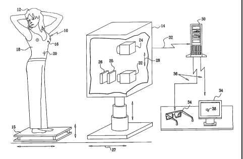
Reference is now made to Fig. 1, which illustrates a system for 3D non-
thermographic and thermographic imaging of a portion of a human body, in
accordance
with a preferred embodiment of the present invention. The system may be used
as a
diagnostic tool, for example for medical diagnosis such as diagnosis of
tumors, and
specifically in the diagnosis of cancerous breast tumors.
As seen in Fig. 1, a body part 10 of a person 12 is located in front of an
imaging device 14. The person 12, may be standing, sitting or in any other
suitable
position relative to imaging device 14. Person 12 may initially be positioned
or later be
repositioned relative to imaging device 14 by positioning device 15, which
typically
comprises a platform moving on a rail, by force of an engine, or by any other
suitable
force. Additionally, a tumor 16 may exist in body part 10 of person 12.
Typically, body
part 10 comprises a breast, and tumor 16 comprises a breast tumor such as a
cancerous
tumor.
In accordance with a preferred embodiment of the present invention,
person 12 may be wearing a clothing garment 18, such as a shirt. Preferably,
clothing
garment 18 may be non-penetrable or partially penetrable to visible
wavelengths such as
400-700 nanometers, and may be penetrable to wavelengths that are longer than
visible
wavelengths, such as IR wavelengths. Additionally, a reference mark 20 may be
located
close to person 12, preferably directly on the body of person 12 and in close
proximity
to body part 10. Optionally and preferably, reference mark 20 is directly
attached to
body part 10. Reference mark 20 may typically comprise a piece of material, a
mark
drawn on person 12 or any other suitable mark, as described hereinbelow.
Imaging device 14 typically comprises at least one non-thermographic
imaging system 22 that can sense at least visible wavelengths and at least one
thermographic imaging system 24 which is sensitive to infra-red (IR)
wavelengths,
typically in the range of as 3-5 micrometer and/or 8-12 micrometer. Typically
imaging
systems 22 and 24 are capable of sensing reference mark 20 described
hereinabove.
Optionally, a polarizer 25 may be placed in front of non-thermographic
imaging system 22. As a further alternative, a color filter 26, which may
block at least a
8
CA 02573246 2007-01-05
WO 2006/003658
PCT/1L2005/000707
portion of the visible wavelengths, may be placed in front of non-
thermographic
imaging system 22.
Typically, at least one non-thermographic imaging system 22 may
comprise a black-and-white or color stills camera, or a digital camera such as
CCD or
CMOS. Additionally, at least one non-thermographic imaging system 22 may
comprise
a plurality of imaging elements, each of which may be a three-dimensional
imaging
element.
Optionally, imaging device 14 may be repositioned relative to person 12
by positioning device 27. As a further alternative, each of imaging systems 22
and 24
may also be repositioned relative to person 12 by at least one positioning
device 28.
Positioning device 27 may comprise an engine, a lever or any other suitable
force, and
may also comprise a rail for moving imaging device 14 thereon. Preferably,
repositioning device 28 may be similarly structured.
Data acquired by non-thermographic imaging system 22 and
thermographic imaging system 24 is output to a computing device 30 via a
communications network 32, and is typically analyzed and processed by an
algorithm
running on the computing device. The resulting data may be displayed on at
least one
display device 34, which is preferably connected to computing device 30 via a
communications network 36. Computing device 30 typically comprises a PC, a PDA
or
any other suitable computing device. Communications networks 32 and 36
typically
comprise a physical communications network such as an intemet or intranet, or
may
alternatively comprise a wireless network such as a cellular network, IR
communication
network, a radio frequency (RF) communications network, a blue-tooth (BT)
communications network or any other suitable communications network.
In accordance with a preferred embodiment of the present invention
display 34 typically comprises a screen, such as an LCD screen, a CRT screen
or a
plasma screen. As a further alternative display 34 may comprise at least one
visualizing
device comprising two LCDs or two CRTs, located in front of a user's eyes and
packaged in a structure similar to that of eye-glasses. Preferably, display 34
also
displays a pointer 38, which is typically movable along the X, Y and Z axes of
the
displayed model and may be used to point to different locations or elements in
the
displayed data.
9
CA 02573246 2007-01-05
WO 2006/003658
PCT/1L2005/000707
Reference is now made to Figs. 2A-4, which illustrate various stages in
method of 3D non-thermographic and thermographic imaging of a portion of a
human
body, in accordance with a preferred embodiment of the present invention.
As seen in Fig. 2A, person 12 comprising body part 10 is located on a
positioning device 15 in front of an imaging device 14, in a first position 40
relative to
the imaging device. First image data of body part 10 is acquired by at least
one non-
thermographic imaging system 22, optionally through polarizer 25 or as an
alternative
option through color filter 26. Additionally, at least second image data of
body part 10
is acquired by at least one non-thermographic imaging system 22, such that
body part
10 is positioned in at least a second position 42 relative to imaging device
14.
The second relative position 42 may be configured by repositioning
person 12 using positioning device 15 as seen in Fig. 2A, by repositioning
imaging
device 14 using positioning device 27 as seen in Fig. 2B or by repositioning
non-
thermographic imaging system 22 using positioning device 28 as seen in Fig.
2C. As a
further alternative, the second relative position 42 may be configured by
using two
separate imaging devices 14 as seen in Fig. 2D or two separate non-
thermographic
imaging systems 22 as seen in Fig. 2E.
In a further stage of the method in accordance with a preferred
embodiment of the present invention, person 12 comprising body part 10 is
located on a
positioning device 15 in front of an imaging device 14, in a first position 44
relative to
the imaging device. First thermographic image data of body part 10 is acquired
by at
least one thermographic imaging system 24. Additionally, at least second
thermographic
image data of body part 10 is acquired by at least one thermographic imaging
system
24, such that body part 10 is positioned in at least a second position 42
relative to
imaging device 14.
The second relative position 46 may be configured by repositioning
person 12 using positioning device 15 as seen in Fig. 3A, by repositioning
imaging
device 14 using positioning device 27 as seen in Fig. 3B, or by repositioning
thermographic imaging system 24 using positioning device 28 as seen in Fig.
3C. As a
further alternative, the second relative position 46 may be configured by
using two
separate imaging devices 14 as seen in Fig. 3D or two separate thermographic
imaging
systems 24 as seen in Fig. 3E.
CA 02573246 2013-11-07
It will be appreciated that the non-thermographic image data acquisition
described in Figs. 2A-2E may be performed before, after or concurrently with
the
thermographic image data acquisition described in Figs. 3A-3E.
Image data of body part 10 may be acquired by thermographic imaging
system 24, by separately imaging a plurality of narrow strips of the complete
image of
body part 10. Alternatively, the complete image of body part 10 is acquired by
thermographic imaging system, and the image is sampled in a plurality of
narrow strips
or otherwise shaped portions for processing. As a further alternative, the
imaging of
body part 10 may be performed using different exposure times.
The thermographic and non-thermographic image data obtained from
imaging device 14 is analyzed and processed by computing device 30 as
illustrated in
Fig. 4.
In stage 50, image data acquired from non-thermographic imaging
system 22 is processed by computing device 30 to build a non-thermographic
three-
dimensional model of body part 10 of person 12, using algorithms and methods
that are
well known in the art, such as the method described in U.S. Patent No.
6,442,419. The
non-thermographic three-dimensional model, preferably includes spatial
information,
typically the X, Y and Z coordinates of the body part 10, as well as the
location of
reference marker 20. Additionally, the non-thermographic three-dimensional
model
preferably includes information relating to the color, hue and tissue texture
of body part
10. An exemplary non-thermographic three-dimensional model and the process of
building such a model are illustrated in Fig. 5.
Thermographic image data acquired from thermographic imaging system
24 is processed by computing device 30 in stage 52 to build a thermographic
three-
dimensional model of body part 10 of person 12, using algorithms and methods
that are
well known in the art, such as the method described in U.S. Patent No.
6,442,419. The
thermographic three-dimensional model preferably includes spatial temperature
information, typically the X, Y and Z coordinates of the temperature of body
part 10
and of reference marker 20. An exemplary thermographic three-dimensional model
and
the process of building such a model are illustrated in Fig. 6.
It is appreciated that the thermographic three-dimensional model may be
built before, after or concurrently with the non-thermographic three-
dimensional model.
11
CA 02573246 2013-11-07
The three-dimensional models built in stages 50 and 52 as described
hereinabove are combined into a single three-dimensional model in stage 54.
Correct
positioning of the two models in the combined three-dimensional model may be
achieved by accurately positioning reference marker 20 in the two models, by
comparing X, Y and Z coordinates or using any other suitable method. An
exemplary
combined three-dimensional model as built in stage 54 is illustrated in Fig.
7.
In stage 56, computing device 30 extracts information included in the
combined three-dimensional model, such as information regarding temperature,
temperature changes in a certain point and a comparison of temperatures in
different
points in body part 10. Additionally, computing device 30 may extract, compute
and
display a comparison of size or temperature between body part 10 and another
body part
of person 12, such as the two breasts of person 12.
In an additional or alternative stage 58, the computing device 30 may
compare and display differences between a plurality of three-dimensional
models of the
same body part 10 of a person 12, the plurality of models being based on data
acquired
at a plurality of different time points. Typically, the information compared,
computed
and displayed includes information about temperature, dimensions such as
length,
width, height and depth, shape, volume, color, hue and tissue texture. The
information
may be displayed graphically or textually, and may be described as a change in
percentage or in absolute value.
As shown in stage 60, the output of any of stages 54, 56 and 58 is
displayed on display 34. Pointer 38 is also displayed, and may be used to
point to
sections or elements of the displayed model, along any of the X, Y and Z
coordinates.
Optionally and preferably, an algorithm is provided to facilitate the display
of sectional
views of the three-dimensional model or of specific tissue layers of the
modeled body
part 10.
It will be appreciated by persons skilled in the art that the scope of the
present invention includes both combinations and subcombinations of the
various
features described hereinabove.
12