Note: Descriptions are shown in the official language in which they were submitted.
CA 02573710 2007-01-11
WO 2006/008505 PCT/GB2005/002820
ELECTROCHEMICAL SENSOR FOR IN-VIVO OR EX-VIVIO MEASUREMENTS OF THE CARBON
DIOXIDE PARTIAL PRESSURE OF LIVING TISSUE
The invention relates to a physiological sensor, in particular for the partial
pressure of carbon dioxide (pCO2), for example in vivo or ex vivo, e.g. in or
on the
surfaces of body tissues or organs.
Ischemia is a medical term for a shortage of blood supply to an organ. If
severe, it can lead to death of the affected tissue (infarction). A sensor can
be
provided to measure tissue pCO2, which is a parameter that increases
significantly
during the early and reversible stages of ischemia. Such a sensor preferably
provides the ability to identify the onset of ischemia events through real-
time data.
Ischemia is the most prevalent cause of death in the western world. Thus, for
example, myocardial infarction, cerebral infarction and other conditions
characterised by hypoperfusion to one or more organs are major factors in
mortality.
Reperfusion, reversal of ischemia, is frequently possible if an ischemia is
detected in time. Thus, early detection of ischemia followed by appropriate
chemical treatment (e.g. with an agent such as streptokinase, urokinase or t-
PA
which serves to lyse thrombi or emboli) or surgical intervention can save the
affected organ as well as the patient's life.
While the heart may be monitored continuously for ischemias using an
electrocardiograph (ECG), other organs may become severely ischemic and incur
irreversible damage before any symptom is detected. Indeed many organs are
"silent" when it comes to ischemia. The phenomenon of silent myocardial
infarction
is now well recognised. Furthermore, liver and kidney may be severely ischemic
without alerting symptoms before the organ damage is irreversible.
It is known that there is a distinct correlation between pCO2 in or on the
surface of an organ and the presence of an ischemia in that organ. During
tissue
metabolic acidosis, e.g. during the anaerobic metabolism that occurs in an
ischemia
in any organ or tissue, large quantities of carbon dioxide are formed. CO2 is
in
practical terms freely cell-membrane permeable and since in the ischemia blood
flow to transport away the CO2 is absent or restricted, CO2 build up in the
ischemic
tissue will occur and pCO2 in or on the ischemic tissue will increase.
Generally, in
CA 02573710 2013-09-10
- 2 -
the healthy body, the maximum pCO2 in blood (venous blood) is 7-10 kPa and the
maximum
pCO2 in healthy (aerobic) tissue is some 1-6 kPa higher, although the maxima
may vary from
organ to organ, e.g. 8-12 kPa for kidney, 7-11 kPa for liver, 8-12 kPa for
intestinal serosa,
and 12-19 kPa for intestinal mucosa. Where oxygen supply falls below the
critical oxygen
A simple sensor particularly suitable for pCO2 measurement, especially as part
of a
technique for monitoring for ischemias, is described in WO 00/04386.
The sensor comprises a closed chamber bounded, at least partially, by a
substantially
water-tight, carbon dioxide-permeable membrane. The chamber contains at least
two
electrodes and a film of substantially electrolyte-free liquid, such as de-
ionised water. The
liquid contacts the membrane and both electrodes, so that carbon dioxide
crossing the
membrane increases the concentration of bicarbonate ions in, and hence the
conductivity of,
The inventors have identified that in some circumstances even a substantially
water-
tight membrane may allow fluid transport across the membrane if there is a
sufficiently large
osmotic gradient across the membrane. For example, if the sensor is used in
vivo, a
sufficiently large osmotic pressure may be caused across the membrane by the
difference in
The present invention seeks to address this newly-identified problem.
Viewed from a first aspect, the invention provides a physiological sensing
device for
CA 02573710 2007-01-11
WO 2006/008505 PCT/GB2005/002820
- 3 -
Thus, according to the invention, the liquid in the chamber contains a non-
ionic excipient. In this way, the osmolarity of the liquid in the chamber can
be
increased to prevent egress of the liquid across the membrane, without
affecting the
electrical characteristics of the liquid.
The excipient should have at least isotonic concentration, i.e. should be
is osmotic with an aqueous solution of 0.9% w/v NaCl. Thus, the osmolality of
the
excipient in the chamber may be greater than that of 0.9% w/v aqueous NaCl,
preferably greater than that of 1.8% w/v aqueous NaC1 (twice isotonic
concentration). Osmolalities greater than that of 4.5% w/v aqueous NaC1 (five
times
isotonic concentration), or even greater than that of 9% w/v aqueous NaCl (ten
times
isotonic concentration) may be used.
Any suitable excipient may be used that is insert to the bicarbonate reaction
in the chamber. The excipient should also be soluble in the liquid, for
example
water. The excipient is also desirably an accepted pharmaceutical excipient
for
intravenous use and with low viscosity for simple filling of the chamber. The
excipient should preferably be sterilizable and storage stable. Desirably, the
excipient should inhibit microbiological growth.
A suitable excipient is polyethylene glycol (PEG) and the presently preferred
excipient is propylene glycol.
By substantially electrolyte-free, it is meant that the liquid has an ionic
osmolality no greater than that at 37 C of an aqueous 5 mM sodium chloride
solution, preferably no more than that of a 500 M sodium chloride solution,
more
especially no more than that of a 10-5 to l06 M HC1 solution.
Preferably, the liquid in contact with the electrodes is aqueous and
especially
preferably it is water, substantially electrolyte-free as defined above. Other
solvents
that react with CO2 to increase or decrease their conductance, e.g. by the
production
or neutralization of ions, may likewise be used. In practice, however,
deionized or
distilled water with or without the addition of a strong acid (e.g. HC1) to a
concentration of 0.1 to 100 M, preferably 0.5 to 50 M, more especially about
1
M, has been found to function particularly well. The function of this small
addition of acid is generally to maintain the pH of the liquid at 6 or below
to avoid
CA 02573710 2007-01-11
WO 2006/008505
PCT/GB2005/002820
- 4 -
significant contributions to conductance by hydroxyl ions and to maintain the
linearity of the measurements of pCO2.
The primary components of the pCO2 sensor are an electrode chamber, a
CO2-permeable membrane forming at least part of the wall of the electrode
chamber,
first and second electrodes having surfaces within said chamber (or providing
internal surfaces to said chamber), and a liquid (generally substantially
electrolyte-
free water) in the electrode chamber in contact with the membrane and the
first and
second electrodes. The sensor includes or is connectable to an AC power
supply, a
conductance (or resistance) determining device, a signal generator (which may
be
part of the determining means) and optionally a signal transmitter.
The mechanism by which pCO2 is determined using the sensor device of the
invention is straightforward. In a pure protic solvent, e.g. water, the
electrical
resistance is high because of the paucity of ionic species. Addition of CO2
results in
formation (with water) of II+ and HC0-3 ions and thus a reduction in the
electrical
resistance. Since the only factor responsible for reduction in resistance in
the sensor
is CO2 passing through the membrane, the change in resistance enables pCO2 to
be
measured.
From the equilibrium constant for the H20 + CO2 to II+ + HC0-3 equilibrium,
CO2 concentration is equal to apCO2 (where a. at 25 C is 0.310). The
electrical
conductivity for protons is GB+ = 349.8 S.cm2/mol, that for hydroxyls is GOH-
=
198.3 S.cm2/mol and that for bicarbonate is GH003- = 44.5 S. cm2/mol. The
concentrations of H+ and Off vary inversely, and the concentrations of H+ and
HCO3- are directly proportional to pCO2. The total conductance of the solution
is
thus effectively proportional to pCO2 since the contribution of Off is
minimal. The
conductivity of the solution Gsolution is thus given by
Gsolution = 011-4H+]Gx+ + 00x40ff]Goll- + OHCO3- [HCO3]GHCO3-
where OH-, 00H- and OHCO3- are the activity coefficients for the three ionic
species.
Table 1 below shows, by way of example, measured pCO2 and pH values
and corresponding calculated values for H+, Off and HCO3- concentrations
showing
the increase of H+ and HCO3- with increasing pCO2.
CA 02573710 2007-01-11
WO 2006/008505 PCT/GB2005/002820
- 5 -
Sample number pCO2 (kPa) pH [H1 [OH] [HC031
(mmo1/1) (mmo1/1)
(mmo1/1)
1 6.38 5.141 7.23E-06 1.38E-09 7.23E-06
2 9.64 5.060 8.71E-06 1.15E-09 8.71E-06
3 15.37 4.891 1.29E-05 7.78E-10 1.29E-05
4 25.88 4.760 1.74E-05 5.75E-10 1.74E-05
31.48 4.664 2.17E-05 4.61E-10 2.17E-05
(pCO2 and pH measured with A standard blood gas analyser, ABL System
625 at 37 C)
5 The
electrical conductivity is measured in the solvent film in the sensor of
the invention. This can be done by applying a constant voltage (or current) to
the
electrodes and measuring the current (or voltage) changes which correspond to
changes in conductivity as CO2 enters the solvent through the membrane.
Preferably however an alternating sine wave function voltage with a constant
peak
value is applied and the voltage drop across the electrodes is measured. The
solution conductivity is then equal to the current passed through the
electrode
divided by the voltage drop across the electrodes.
The pCO2 sensor may function by applying an alternating electrical potential
to the electrodes whereby to cause an alternating current in the liquid. The
liquid
should be reactive with carbon dioxide to alter its conductance. The
electrical
potential may have a frequency of 20 to 10,000 Hz, preferably 100 to 4,000 Hz.
The pCO2 sensors of the invention are provided with or are connectable to an
electrical power source arranged to apply an alternating electrical potential
across
the electrodes with a frequency of 100 to 10,000 Hz. The frequency is
preferably
greater than 1 kHz. The frequency is preferably less than 5 kHz, more
preferably
less than 2 kHz. At frequencies below 100 Hz, the sensitivity of pCO2
determination is lower due to electropolarization and moreover the instrument
response time becomes overly slow, while at frequencies above 10 kHz
sensitivity is
again less due to the low impedance of the capacitances in the sensor.
The power source may be an AC power source or alternatively a DC source
in conjunction with an oscillator, i.e. a combination which together
constitutes an
AC power source.
CA 02573710 2007-01-11
WO 2006/008505
PCT/GB2005/002820
- 6 -
The power supply is preferably such that the maximum current density
through the liquid at the electrodes is no more than 50 Aim2, preferably no
more
than 30 A/m2, more preferably no more than 20 A/m2, in particular no more than
10
A/m2, and most preferably about 1 A/m2 or below. Higher current density values
of
20 A/m2 or greater should only be used at the higher frequencies, e.g. 1-10
kHz.
The smallest maximum current density is determined by detection limits, but
values
down to 10-8 A/m2 are usable. The smallest maximum current density however
will
generally be at least 0.1 RA/m2.
By operating at such current densities and voltage frequencies, and by
appropriate construction, the sensor can determine the conductance/resistance
of the
liquid into which the CO2 migrates without any significant loss of accuracy
arising
as a result of the electropolarization of the electrodes.
For particularly high accuracy, the potential or current across the electrodes
(and hence the resistance or conductance of the liquid between the electrodes)
is
determined using a lock-in amplifier set to the same frequency as that of the
voltage
generator or electrical power source.
Furthermore it is preferred to incorporate in the detection a high pass filter
to
screen out current with a frequency less than 100 Hz, preferably less than 150
Hz.
The filter is preferably a passive filter, for example a capacitor and a
resistor.
The power source and the detector circuitry may, if desired, be included in
the sensor of the invention. In this case, if it is desired that the sensor be
wireless, it
will preferably also be provided with means enabling the signal to be detected
remotely, e.g. a transmitter, for example a RF transmitter. In this way the
sensor
may be implanted, for example in an at-risk patient.
A further electrode may be provided that is electrically connected to the
patient, for example to the patient's skin. The signal from this further
electrode may
be processed with the signal from the sensor in order to compensate for
electromagnetic noise from the patient.
Electropolarization effects are considerably reduced by increasing the
surface area of the electrodes in contact with the liquid, e.g. by siting the
electrodes
in wells disposed away from the plane of the membrane or by using non-planar
electrode surfaces, e.g. rough or textured surfaces. In general therefore it
is
CA 02573710 2007-01-11
WO 2006/008505 PCT/GB2005/002820
- 7 -
,
desirable to have as large a ratio of surface area of electrode to liquid
contact as
possible, and as shallow as possible a liquid depth over as much as possible
of its
area of contact with the membrane. In this way the response time is reduced,
electropolarization is reduced, lower frequencies may be used and stray
capacitance
effects are considerably reduced.
Increased electrical resistance relative to the resistance at the electrodes
may
be achieved by restricting the cross sectional area of the electrical path
through the
liquid between the electrodes at a zone in which the liquid is in contact with
the
membrane, e.g. by decreasing the depth of the liquid for a part of the path
between
the electrodes, and/or by ensuring a relatively large area of contact between
each
electrode and the liquid.
The resistance of the liquid at the membrane and between the electrodes may
be increased by the use of structural elements to define liquid channels
across the
membrane between the electrodes, e.g. by disposing the membrane across or
adjacent an insulating chamber wall portion in which such channels are formed,
for
example by etching. Likewise a porous spacer may be disposed between the
membrane and the chamber wall to defme the depth of the liquid.
Indeed, such spacers are important to use where, under the pressure
conditions experienced in use, the membrane is sufficiently flexible and the
liquid
depth behind the membrane sufficiently small, for the measured conductance to
vary
with pressure.
In a preferred arrangement, the sensor comprises:
a sensor body having a longitudinal axis;
at least two electrodes spaced in a direction transverse to the longitudinal
axis of the sensor body;
a plurality of support members extending outwardly from the axis of the
sensor body and defining between adjacent support members at least one liquid
channel that provides a fluid pathway between the electrodes; and
a gas-permeable membrane supported by the support members and providing
an outer wall of the liquid channel(s).
This arrangement provides a compact configuration of the sensor with a
longitudinal geometry that is suited to insertion in an organ. Furthermore,
the
CA 02573710 2007-01-11
WO 2006/008505 PCT/GB2005/002820
- 8 -
support members are able to provide physical support to the membrane, as well
as
defining liquid channels of small cross-sectional area that allow accurate
measurement.
In order to reduce the electropolarisation effect mentioned above, the
electrodes may be located in a recess in the sensor body that has a greater
cross-
sectional area than the liquid channels. In this way, the current density
around the
electrodes is reduced by the greater volume for liquid.
The electrodes of the sensor may extend longitudinally, for example parallel
to the longitudinal axis of the sensor body.
Similarly, the liquid channel(s) may be transverse, for example
perpendicular, to the longitudinal axis of the sensor body. In a preferred
arrangement, the sensor comprises a plurality of liquid channels. For example,
the
sensor may comprise at least three liquid channels.
The support members may be transverse to the longitudinal axis of the sensor
body. For example, the support members may be perpendicular to the
longitudinal
axis of the sensor body in the circumferential direction. In a preferred
arrangement,
the support members are in the form of rings formed about the longitudinal
axis of
the sensor body. The cross-section of the support members may be any suitable
shape. It has been found in particular that support members with a
substantially
triangular, in particular sawtooth, cross-section are particularly easily
formed by
injection moulding. Alternatively, a substantially rectangular cross-section
may be
used. The support members may be formed integrally with the sensor body, for
example by injection moulding. The sensor preferably comprises at least four
support members.
The sensor body and/or the sensor may be generally cylindrical. The
membrane may be arranged to surround the sensor body.
The described geometry may be applied to any suitable sensor. In the
preferred arrangement, the sensor is a pCO2 sensor.
Where the sensor is constructed with the liquid film in place, the electrodes
are preferably of, or plated with, an inert material such that the resistivity
of the
liquid will not change significantly with storage. Suitable materials include
platinum (especially black platinum), gold, silver, aluminium and carbon. Gold
is
CA 02573710 2007-01-11
WO 2006/008505 PCT/GB2005/002820
- 9 -
particularly preferred. In general inert electrodes which do not generate
solvated
ions are preferred.
The membrane may be any material which is permeable to CO2, and
substantially impermeable to the solvent of the liquid, any electrolyte and
water.
Polytetrafluoroethylene, e.g. Teflon , silicone rubber, polysiloxane,
polyolefins or
other insulating polymer films may be used, e.g. at thicknesses of 0.5 to 250
gm.
The thicker the membrane, in general the slower the response time of the
sensor will
be. However the thinner the membrane the greater the risk of non -uniformities
or of
perforation or other damage. Conveniently however the thickness of the
membranF
will be 1 to 100 p.m, preferably 50 to 100 gm.
The walls of the chamber of the sensor of the invention may be of any
suitable material, e.g. plastics. Preferably the material should be capable of
withstanding conditions normally used in sterilisation, e.g. radiation
sterilization (for
example using gamma radiation) or thermal sterilization (for example using
temperatures of about 121 C as used in autoclave sterilisation). In the case
of
thermal sterilization, the liquid will generally be sterile filled into the
sensor after
sterilization. The walls of the chamber and the membrane may be of the same
material, e.g. Teflon , machined to have self-supporting walls and a thinner
gas-
permeable membrane.
The sensors of the invention are generally relatively inexpensive and so,
unlike prior art sensors, may be single-use devices. Moreover the electrode
chamber
can be made extremely small without difficulty (unlike the prior art glass
electrode
containing sensors for which miniaturization poses insuperable impedance
problems).
This arrangement provides a sensor, in particular, a pCO2 sensor, which can
be inserted easily into the tissue of an animal, including a human, which can
be
retained in the tissue during monitoring and which can be removed easily when
monitoring is complete.
The device is sufficiently small that it will not cause undue disturbance to
the
tissue to be monitored. Consequently, the device may have a maximum diameter
of
2 mm, preferably 1 mm.
CA 02573710 2007-01-11
WO 2006/008505 PCT/GB2005/002820
- 10 -
The sensors according to the invention are readily produced having a size
and configuration particularly suited to measuring pCO2 on the surface of or
in an
organ, duct or tissue, e.g. brain, heart, liver, kidney, gut or muscle. This
is of
particular interest as it allows the functioning of the organ, duct or tissue
to be
monitored, e.g. during and after transplant, in intensive care, following
injury, etc.
and so allows early detection of ischemias.
The partial pressure determined by the sensor may be a quantified value or it
may simply be an indication that pCO2 is above or below one or more threshold
values indicative of ischemia or non-ischemia, values which may be varied
according to the location of the pCO2 measurement site.
The sensor may be used for a single measurement of pCO2 or, more
preferably, may be used for continuous or repeated monitoring, especially of
an at-
risk patient, for example a patient in intensive care, undergoing or
recovering from
an organ or tissue transplant operation, assessed as having unstable angina,
recovering from a coronary artery bypass operation, suffering trauma (e.g. of
skeletal muscle), or suffering from hypovolemia (e.g. shock).
The device may comprise a plurality of sensors for respective physiological
parameters. For example, the device may comprise an array of sensors. Such
sensors may measure one or more of the partial pressure of carbon dioxide, the
partial pressure of oxygen, temperature, pH or glucose concentration, for
example.
In the presently preferred embodiment, the device comprises a temperature
sensor
and a pCO2 sensor.
An embodiment of the invention will now be described, by way of example
only, with reference to the accompanying drawings, in which:
Figure 1 is a schematic diagram of a complete sensing system incorporating
the sensor of the invention;
Figure 2 is a schematic diagram illustrating the measurement principle for
the sensor in the system of Figure 1;
Figure 3 is a partially cutaway view of a sensor according to the invention;
Figure 4 is a cross-sectional view along line A-A of Figure 3;
Figure 4a is a magnified view of the detail indicated by the circle in Figure
4;
and
CA 02573710 2007-01-11
WO 2006/008505 PCT/GB2005/002820
- 11 -
Figure 5 is a view of the sensor of Figure 3 with the membrane removed.
In accordance with the invention, a pCO2 sensing system comprises a
disposable sensor unit 1, an electronic surface unit 2, and a monitor unit 3,
as shown
in Figure 1.
The disposable sensor unit 1 is delivered packaged and sterilised. It consists
of a membrane-protected conductometric sensor 4 with a diameter of less than 1
millimetre, and a temperature probe 5 integrated in the sensor unit. ' Wires 6
connect
the sensor 4 and probe 5 electrically by means of a connector to the
electronic
surface unit 2. Alternatively, a wireless connection may be provided between
the
sensor unit 1 and the surface unit 2.
The electronic surface unit 2 sends and receives signals to and from the
sensor unit 1. It is placed on the patient's skin, performs signal processing
and
transmits the conditioned signal to the monitor unit 5.
The monitor unit 3 is based on a portable personal computer 7 with PCMCIA
input/output card 8 and Labview software (available from National Instruments
Corporation of Austin, Texas).
The pCO2 sensor 4 is used for measurements of the level (partial pressure) of
CO2 (pCO2) in a fluid, according to the measurement principle illustrated in
Figure
2. The measurement chamber consists of two small cavities 9 with one electrode
10
positioned in each. The two cavities 9 are connected by one or more
passageways
11 enclosed by a semi-permeable membrane 12, i.e. a membrane that only allows
transport of CO2 in and out of the volume of the sensor 4. The whole volume is
filled with de-ionised water and 5% propylene glycol. The conductivity in the
water
depends upon the pCO2, and by measuring the conductivity between the
electrodes
10 in the volume, information about pCO2 may be extracted.
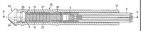
As shown in Figures 3 to 5, the sensor unit 1 comprises an injection moulded
plastics support 23, which is substantially cylindrical and surrounded by the
semi-
permeable membrane 12. The support 23 has a conical tip 24 at its distal end
and a
body portion 25 which extends proximally from the tip 24. On the body portion
25
are mounted, by gluing, two gold electrodes 10. The electrodes 10 extend
longitudinally along opposed sides of the body portion 25 and are received in
respective recesses in the body portion 25.
CA 02573710 2007-01-11
WO 2006/008505 PCT/GB2005/002820
- 12 -
Between the tip 24 and the body portion 25, a frustoconical projection 26 is
provided for securing the membrane 12 by frictional fit. A corresponding
projection
26 is provided at the proximal end of the body portion 25. The membrane 12 may
be glued to the support 23, but it is important that the glue used to secure
the
membrane 12 and electrodes 10 is selected such that it does not bleed ions
into the
water-filled chamber formed between the body portion 25 of the support 23 and
the
membrane 12. Furthermore, the sealing faces of the support 23 may be made
selectively hydrophobic in order to avoid the formation of a water film into
which
ions may bleed.
The membrane 12 may also be secured to the support 23 by means of crimp
connection and a soft gasket, if necessary. The membrane 12 may act as the
gasket,
particularly where the membrane 12 is formed of silicone rubber. A heat shrink
sleave may be used to form the crimp connection. Alternatively, metal crimp
rings
may be used in locations corresponding to those of the sealing projections 26.
The body portion 25 of the support 23 is provided with a plurality of ribs 27,
which are formed with a saw tooth profile for easy moulding. The ribs 28
provide
mechanical support to the membrane 12 and also defme the fluid passageways 11
required for the sensor 4 to function effectively. Between each electrode 10
and the
fluid passageways formed between the ribs 27 is provided a reservoir 9 formed
by
the recess in which the electrode 10 is located. The reservoir 9 provides a
region of
relatively low current density around the electrodes 10 in order to reduce
electropolarisation effects.
During manufacture, the membrane 12 is fixed onto the support 23, while
immersed in the de-ionised water and propylene glycol solution, so that the
chamber
bounded by the membrane 12, the electrodes 10, and the ribs 27 is completely
filled
with liquid. Thus, this chamber forms a pCO2 sensor as shown schematically in
Figure 2.
It is possible for the sensor 1 to include more than one sensing chamber. For
example, two parallel electrodes 10 separated by a wall member may be provided
on
each side of the support 23. A sensing chamber is thereby formed between one
electrode 10 on one side of support 23 via the fluid passageways 11 between
the ribs
27 on the top of the support 23 to one of the electrodes 10 on the other side
of the
CA 02573710 2007-01-11
WO 2006/008505 PCT/GB2005/002820
- 13 -
support 23. A corresponding sensing chamber is provided between the remaining
electrodes 10 and the fluid passageways 11 on the bottom of the support 11. An
electrode 10 from each of these chambers may be electrically connected to the
corresponding electrode from the other chamber, such that the electrical
signal from
the sensor reflects the conductivity of both chambers.
Embedded in the proximal end of the support 23 is a temperature sensor 5 in
the form of a thermocouple. The temperature sensor 5 is used both for pCO2
corrective calculations and for the measured tissue temperatures to be
displayed on
the monitor 3, which is informative for medical diagnosis. The temperature
sensor 5
has a minimum measuring range of 33-42 C and a minimum accuracy of +1- 0.2 C.
A ribbon cable 6 is electrically and mechanically connected to the electrodes
10 and the temperature sensor 5. The electrodes 10 are formed as extensions of
the
conductors of the ribbon cable 6. Alternatively, the electrodes may be formed
by
plating onto the support 23. Where the cable 6 and the connection to the
support 23
are sufficiently strong, the cable 6 can be used to pull the sensor unit 1
from its
position of use. Alternatively, a Kevlar line may be provided, for example
incorporated with the ribbon cable 6, to provide a strong external mechanical
connection.
The membrane 12 may extend proximally from the support 23 with the cable
6 to form a catheter around the cable 6. Alternatively, a separate catheter 28
may be
provided. In this case, the catheter 28 is bonded to the support 23 proximally
of the
electrodes 10 and the membrane 12.
The catheter tip with the integrated sensor 4 is placed 0.5 - 4 cm into organ
tissue during surgical procedures to monitor ischemia during a period of up to
two
weeks. The sensor may be used in orthopaedic and reconstructive surgery, and
in
organs such as the liver, kidneys, heart muscle, brain and intestines. An
insertion
tool(not shown) may be used for the placement of the sensor 4, and there may
be a
fixation aid to keep the sensor tip in position.
The sensor unit 1 has a maximum diameter of 1 mm and the maximum
distance from the catheter tip to the sensor element is 2 mm. The sensor 4 has
a
minimum pCO2 measuring range of 2-25 Ic.Pa, with a minimum detectable pCO2
difference of 0.2 kPa. The maximum response of the sensor 4 is 20 seconds. The
CA 02573710 2007-01-11
WO 2006/008505 PCT/GB2005/002820
- 14 -
maximum allowable measurement current is in any area of the fluid chamber is
such
that j<lmA/cm2 while the measuring input voltage is not more than 50 mV RMS.
The electrodes 10 are gold plated and their total area is approximately 0.3
mm2. The measurement frequency fmeas should be higher than 100 Hz. At lower
frequencies, polarisation effects in the measurement chamber dominate the
measurements. At frequencies above 10 kHz, the low impedance of the
capacitances become a significant issue. The measurement resistance R measure
is in
the range of 500 kOhm to 7 MOhm.
The sensor 4 is electrically connected to an electronic surface unit 2 located
on the patient skin by the ribbon cable 6, which has a length between 5 cm and
1
metre. The maximum diameter of the cable/catheter is 1 mm and the preferred
length of the cable/catheter is 25 cm. The cable/catheter is soft and flexible
so that it
does not excessively disturb the neighbouring tissue and organs. The
cable/catheter
and its connections are also sufficiently robust to withstand the strong
pulling forces
which may be caused by both normal and "abnormal" use.
During sterilisation, storage and transport the sensor unit 1 is covered by
deionised, sterile and endotoxin-free water to make sure that there is
substantially no
net loss of water from the sensor reservoir.
As shown in Figures 1 and 2, the electronic surface unit 2 comprises a sine
generator 13 which provides a voltage of at least 5 Volts and a current supply
of
50mV, and is powered by batteries 14. A filter 15 is provided for filtering or
averaging the input of the lock-in amplifier 16. A passive filter can be used
which
reduces the current consumption. A pre-amplifier 17 is combined with a servo
mechanism to remove DC current from the signal to reduce electrolysis effects.
According to the servo arrangement, the output of the pre-amplifier is fed
back to its
input via a low pass filter. Thus, only DC components of the output are fed
back
and cancel any DC current drawn through the pC0 2 sensor. In this way, it is
ensured
that there is no DC current through the pCO2 sensor which would degrade the
electrodes. The op-amp used in this stage consumes minimal current and has a
large
CM:MR value. At the same time, the bias current is minimal. A lock-in
amplifier 16
amplifies the AC signal from the sensor 4. This may be built with op-amps or
using
an IC package with at least 1% accuracy for the signal detection at
frequencies lower
CA 02573710 2007-01-11
WO 2006/008505 PCT/GB2005/002820
- 15 -
than lkHz. A galvanic division 19 such as an optocoupler or a coil coupler is
provided to prevent noise transfer from the monitor unit 3 and associated
cabling 18.
The optocoupler is normally favoured due to the noise signal ratio. A
temperature
signal amplification and conditioning unit 20 is provided to amplify the
signal from
the temperature sensor 5. The electronic unit 2 is powered by a rechargeable
and
changeable standard type battery 14. The battery capacity is sufficient for 14
days
continuous monitoring. The surface unit 2 is also provided with an on/off
indicator
LED 21, and a battery status indicator (not shown). Communication between the
surface unit 2 and the monitor 3 is analogue through a shielded cable 18.
However,
the surface unit 2 may include an analogue to digital converter such that
communication between the surface unit 2 and the monitor 3 may be digital, for
example by digital wire transmission or digital wireless transmission. The
cable 18
is at least 4 m long and light and flexible.
As shown in Figures 1 and 2, an AC current is generated by sine generator
13 and fed to one of the pCO2 sensor electrodes 10 and to a lock-in amplifier
16.
The high-pass signal from the other pCO2 electrode 10 is passed through a
filter 15
to a low noise amplifier 17 and from there to the lock-in amplifier 16 where
it is
compared to the reference signal generated by the sine generator 13. Out of
phase
components, i.e. undesired components, of the signal are rejected and the
remaining
portion of the signal is amplified. The amplified signal is proportional to
pCO2 (or
conductance) and is passed on for recordal or further manipulation to the
monitor 3.
The surface unit 2 may also be electrically connected to a reference electrode
(not shown) that is electrically connected to the patient's skin. The signal
from the
reference electrode can be used to compensate the signals from the sensor unit
1 for
the effect of electromagnetic noise generated by the patient.
A single surface unit 2 may receive signals from several sensor units 1 and
provide a multiplexed output to the monitor unit 3.
The monitor unit 3 comprises a portable PC 7 including CD RW and IR port,
and a PCMCIAI/0 card 8 which can collect signals from at least 4 different
surface
units 2 simultaneously. The PCMCIA card 8 may have an integrated non-galvanic
coupling. The power supply 22 for the monitor unit 3 is of a medically
approved
type operating on both 110V and 230V.
CA 02573710 2007-01-11
WO 2006/008505 PCT/GB2005/002820
- 16 -
The software functions of the monitor unit 3 may be implemented in
Labview, a software package available from National Instruments of Austin,
Texas
and capable of handling up to 4 different surface units simultaneously. The
software
provides the facility for calibration of the sensor(s) with three calibration
points and
a second order calibration function. The software can be modified to support
any
other number of calibration points and type of calibration function. The
software
also has the facility to smooth the signal from the sensor 4 over defined time
intervals. It is possible to have at least two alarm levels for the
measurement values
and two alarm levels for their gradients. The measurement value gradients are
calculated for individually defined time intervals. The alarm is both visible
and
audible. It is possible to stop an alarm indication while keeping the other
alarms
active. The monitor 3 can log all measured values, parameter settings and
alarms
throughout a session. With a 30 second logging interval there should be a
storage
capacity for at least 10 two week sessions on the hard disc. The session log
can be
saved to a writeable CD in a format readably by Microsoft Excel.
In summary, a physiological sensing device for the measurement of pCO2
includes a closed chamber bounded, at least partially, by a carbon dioxide
permeable
membrane. There are two electrodes within the chamber. The chamber contains a
substantially electrolyte-free liquid in contact with electrodes and the
membrane.
The liquid contains a non-ionic excipient in order to prevent egress of water
due to
an osmotic gradient across the membrane in use.