Note: Descriptions are shown in the official language in which they were submitted.
CA 02574186 2007-01-17
WO 2006/020954 PCT/US2005/028923
ISOLATION OF ENDOTHELIAL PROGENITOR CELL SUBSETS AND METHODS FOR
THEIR USE
RELATED APPLICATIONS
[0001] The application claims priority under 35 U.S.C. 119(e) to United
States
Provisional Patent Application 60/601,188 filed August 13, 2004.
FIELD OF THE INVENTION
[0002] The present invention relates generally to methods of isolating
endothelial
progenitor cells for the treatment of cardiovascular disease.
BACKGROUND OF THE INVENTION
[0003] The development of new blood vessels in response to tissue ischemia
constitutes a natural host reaction intended to maintain tissue perfusion
required for
physiologic organ function. This natural angiogenesis is impaired in advanced
age, diabetes
and hypercholesterolemia. In each of these conditions, there is a reduction in
endogenous
expression of vascular endothelial growth factor (VEGF) and exogenous VEGF
administration leads to enhanced neovascularization.
[0004] Ischemic tissue injury triggers a series of events, including
mobilization and
recruitment of circulating progenitor cells (CPC) to the injury site. In
models of post-ischemic
angiogenesis, a subpopulation of CPC, namely endothelial progenitor cells
(EPC),
incorporate into neovessels. Moreover, in animal models, as well as in
clinical settings of
acute myocardial infarction (aMI), systemic administration of EPC contributes
to.
revascularization of the myocardium and is associated with improved myocardial
function.
[0005] Since their original description, bone marrow-derived EPC have become a
focal
point in regenerative therapy following evolving vascular damage. Because
numbers of
CPC, which are normally low in peripheral blood, increase significantly after
an ischemic
event, a causal link between vascular damage and CPC-mediated repair has been
postulated. In animal models of angiogenesis following ischemia, bone marrow-
derived EPC
incorporate into neovessels. Moreover, local and systemic levels of angiogenic
growth
factors, including VEGF, rise after ischemia and are associated with increased
numbers of
circulating CPC.
[0006] The obvious therapeutic potential of exogenous growth factor
administration has
been successfully assessed in animals and humans. In various animal models,
mobilization
of EPC after vascular damage by administration of VEGF, granulocyte macrophage
colony
CA 02574186 2007-01-17
WO 2006/020954 PCT/US2005/028923
stimulating factor (GM-CSF), granulocyte colony stimulating factor (G-CSF),
fibroblast
growth factor 1(FGF-1), stromal derived factor 1 (SDF-1) or a statin drug,
positively
correlated with increased numbers of circulating EPC and improved therapeutic
neovascularization. Direct evidence for the vasculogenic potential of EPC has
been
provided by studies in which EPC transplanted in mice with hind limb ischemia
incorporated
into newly formed blood vessels (Kalka C. et al., "Transplantation of ex vivo
expanded
endothelial progenitor cells for therapeutic neovascularization," Proc. Nati.
Acad. Sci.
97:3422-7, 2000). In a murine model of myocardial infarction (MI) intravenous
injection of
human CD34+ CPC contributed to revascularization of the myocardium, and was
associated
with salvage of myocardial function (Kocher A.A. et al., "Neovascularization
of ischemic
myocardium by human bone marrow-derived angioblasts prevents cardiomyocyte
apoptosis,
reduces remodeling and improves cardiac function," Nat. Med. 7:412-3, 2001).
Moreover,
intracoronary infusion of autologous EPC into the infarct artery in patients
with aMI resulted
in increased myocardial viability in the infarct area (Assmus B. et al.,
"Transplantation of
progenitor cells and regeneration enhancement in acute myocardial infarction
[TOPCARE-
AMI]," Circulation 106:3009-17, 2002).
[0007] Current progenitor cell research is focused on the clinical application
of CPC in
therapeutic neovascularization. Therefore, future large-scale therapeutic
application of CPC
will require an understanding of the phenotypic and functional properties of
these cells. It
has been demonstrated that phenotypically diverging subsets of CPC can be
distinguished.
The cell surface markers CD34, CD133 and VEGFR-2 (KDR, flk-1) have been used
as CPC
markers for single- and dual-parameter flow cytometric analysis of CPC, which
leads to
enrichment of CD34+ progenitor cells (PC). The shortcomings of this approach
are technical
limitations and include restrictions in determining the identity and
relationship between CPC
subsets when they are defined by single and dual parameter detection.
[0008] Interestingly, the methods used for CPC detection and isolation also
determine
the outcome of CPC functional assays. When isolated by flow cytometry and
cultured under
angiogenic conditions, CD34+ CPC form spindle-shaped cells, which, over time,
organize in
capillary-like structures. Moreover, these cells express markers specific for
mature
endothelial cells (EC) such as CD31, E-selectin and Tie-2.
[0009] Alternatively CPC have been isolated based on in vitro culture of
mononuclear
cells on fibronectin- or gelatin-coated plates in the presence of angiogenic
growth factors.
Isolated adherent cells that were low density lipoprotein (LDL) negative and
exhibited lectin-
binding ability were called CPC. Although these cells promote angiogenesis in
vivo, they
have monocytic features and their angiogenicity is actually caused by their
production of
angiogenic factors, such as VEGF, hepatocyte growth factor (HGF), G-CSF and GM-
CSF.
2
CA 02574186 2007-01-17
WO 2006/020954 PCT/US2005/028923
Thus, while these LDL-, lectin-binding cells do not directly form EC, they can
modulate
angiogenesis.
[0010] Based on the foregoing, both heterogenous and homogenous populations of
endothelial progenitor cells present an opportunity for treatment of
cardiovascular disease.
Therefore, methods for sorting and isolating specific populations of cells
suitable for use in
regenerative therapy are needed.
SUMMARY OF THE INVENTION
[0011] The present invention describes methods for the isolation of human
peripheral
blood endothelial progenitor cells yielding cells which can form blood vessels
or induce
angiogenesis and inflammation-mediating cells using four-parameter
fluorescence activated
cell sorting. Additionally, the present invention provides for biodegradable
implants
containing endothelial progenitor cells having the ability to induce
angiogenesis and/or
chemotaxis for inflammatory cells. In one embodiment of the present invention,
a subset of
human peripheral blood endothelial progenitor cells, CD34+ CPC, is identified
which gives
rise to both blood vessel-forming/angiogenic cells and inflammation-mediating
cells.
[0012] In one embodiment of the present invention, a method is provided for
isolation of
endothelial progenitor cells comprising identifying lineage-committed cells
from a source of
progenitor cells by contacting the progenitor cells with a plurality of
fluorochrome-labeled
antibodies specific for the cell markers selected from the group consisting of
CD3, CD14,
CD16/56, CD19 and CD31; depleting the lineage-committed cells by fluorescence
activated
cell sorting to form a population of lineage-negative cells; reacting the
lineage-negative cells
with a plurality of fluorochrome-labeled antibodies specific for the cell
markers selected from
the group consisting of CD34, CD133 and KDR wherein each antibody is labeled
with a
fluorochrome with a unique emission wavelength; and sorting the labeled
lineage-
negative cells by three-color fluorescence activated cell sorting to form a
population of
endothelial progenitor cells.
[0013] In an embodiment of the present invention, the source of progenitor
cells is a
mammalian source, including a human source such as peripheral blood.
[0014] In another embodiment of the present invention, the antibodies useful
for
identifying lineage-committed cells include antibodies specific for the cell
markers CD3,
CD14, CD16/56, CD19 and CD31.
[0015] In yet another embodiment of the present invention, the reacting step
comprises
reacting the lineage-negative cells with antibodies specific for the cell
markers CD34, CD133
and KDR. In another embodiment of the present invention, the resulting
endothelial
3
CA 02574186 2007-01-17
WO 2006/020954 PCT/US2005/028923
progenitor cells express CD34. In yet another embodiment of the present
invention the
resulting endothelial progenitor cells are CD34CD133-KDR-.
[0016] In one embodiment of the present invention, the endothelial progenitor
cells are
blood vessel generating cells, inflammation-mediating cells or both. In
another embodiment
of the present invention, the blood vessel-generating cells are CD34+CD133"KDR-
endothelial progenitor cells. In yet another embodiment of the present
invention, the
endothelial progenitor cells are inflammation-mediating cells which can
express interleukin-8.
[0017] In another embodiment of the present invention, the endothelial
progenitor cells
induce angiogenic responses in surrounding blood vessels.
[0018] In one embodiment of the present invention, a therapeutic composition
for
inducing angiogenesis at a treatment site is provided comprising a
biodegradable matrix
having CD34+ endothelial progenitor cells disposed therein. In another
embodiment of the
present invention, the biodegradable biocompatible matrix contains
CD34+CD133"KDR-
endothelial progenitor cells.
[0019] In another embodiment of the present invention, a therapeutic
composition
having a chemotactic effect on inflammation-mediating cells at a treatment
site is provided
comprising a biodegradable biocompatible matrix having CD34+ endothelial
progenitor cells
disposed therein. In another embodiment of the present invention, the
biodegradable
biocompatible matrix contains CD34+CD133"KDR- endothelial progenitor cells.
[0020] In yet another embodiment of the present invention, the biodegradable
biocompatible matrix is selected from the group consisting of solubilized
basement
membrane, autologous platelet gel, collagen gels or collagenous substrates
based on
elastin, fibronectin, laminin, extracellular matrix and fibrillar proteins.
[0021] In one embodiment of the present invention, an isolated population of
endothelial progenitor cells is provided wherein the isolated population is
lineage-negative,
CD34+CD133-KDR-.
[0022] In an embodiment of the present invention, the isolated population of
endothelial
progenitor cells comprises blood vessel-generating cells. In another
embodiment of the
present invention, the isolated population of endothelial progenitor cells
comprises
inflammation-mediating cells which can express interleukin-8. In another
embodiment of the
present invention, the isolated population of endothelial progenitor cells
induce angiogenic
responses in surrounding blood vessels.
4
CA 02574186 2007-01-17
WO 2006/020954 PCT/US2005/028923
BRIEF DESCRIPTION OF THE FIGURES
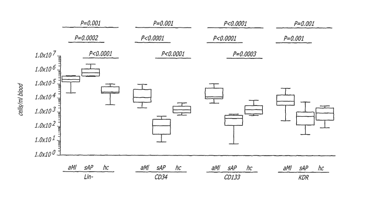
[0023] FIG. 1 depicts the numbers of circulating progenitor cells (CPC)
subsets,
determined by flow cytometry, in peripheral blood from patients with acute
myocardial infarct
(aMI, n=10), stable angina pectoris (sAP, n=10) and healthy controls (hc, n=9)
defined as
Lin-, CD34, CD133 or KDR according to the teachings of the present invention.
[0024] FIG. 2 depicts the in vivo behavior of human CPC subsets, either CD34+
or
CD133+ sorted in Matrigel pellets implanted in nude mice after 14 days
according to the
teachings of the present invention. (A) bare Matrigel ; (B) Matrigel
containing CD34+ cells,
(C) Matrigel containing CD133+ cells. Arrows indicate representative network
structures
formed by spindle-shaped cells, considered potential EPC. Objective
magnification 40x.
[0025] FIG. 3 depicts morphological detection of human endothelial progenitor
markers
on isolated CPC enclosed in Matrigel pellets implanted in nude mice according
to the
teachings of the present invention. In panels A-C human endothelial cells (EC)
were
detected with lectin Ulex europeus-1 agglutinin (UEA-1 conjugated to TRITC), a
human- and
EC-specific lectin; (A) human umbilical vein endothelial cell (HUVEC) positive
control; (B)
murine EC cell line (H5V) negative control; (C) cells in Matrigel seeded with
human CD34+
EPC bound UEA-1 lectin. UEA-1-binding cells were spindle shaped and made
contacts
(arrows). In panels D-E the EC phenotype was confirmed by (D) staining for
human CD34
(EPC, EC), and (E) CD31 (EC). Positive endothelial cells are indicated by
arrows. The
inserts show additional examples of human blood vessels. Objective
magnification 40x.
[0026] FIG. 4 depicts the murine angiogenic and inflammatory response to human
CPC
subsets, implanted in nude mice in Matrigel , isolated according to the
teachings of the
present invention. The formation of murine blood vessels in Matrigel were
detected using
monoclonal antibodies specific for murine CD31 (A-C). Murine
monocytes/macrophages
were detected with monoclonal antibodies specific to those cells (D-F). The
insert in panel E
shows the lack of human CD68+ macrophages in the section. Objective
magnification 40x.
[0027] FIG. 5 depicts detection of interleukin-8 (IL-8) in CD34+ cells,
isolated according
to the teachings of the present invention, implanted in nude mice in Matrigel
. Interleukin-8
expression was determined immediately after sorting (A) and 14 days after
implantation (B).
Objective magnification 40x.
[0028] FIG. 6 depicts flow cytometric analysis of CPC subsets in peripheral
blood from
patients with aMI (n=10) and healthy controls (hc, n=9) according to the
teachings of the
present invention. The lineage-negative (Lin )(A) cell population were stained
with
antibodies to CD34, CD133 and KDR and cells expressing each of the three
markers were
CA 02574186 2007-01-17
WO 2006/020954 PCT/US2005/028923
gated and analyzed for the expression of the remaining two markers. (B) CD133
analysis;
(C) CD34 analysis; (D) KDR analysis.
[0029] FIG. 7 depicts the number of CPC in peripheral blood from aMI patients
(n=10)
and healthy controls (hc, n=9) as subsets defined as Lin , CD34+, CD133+ or
KDR+. Dots
represent individuals; horizontal lines represent the median; fold increase
based on means.
P=p value.
[0030] FIG. 8 depicts cell numbers, determined by flow cytometric analysis, of
the
seven CPC subsets defined according to the teachings of the present invention
in aMI
patients (n=10) and healthy controls (hc, n=9). Horizontal lines represent the
median; fold
increase based on means.
[0031] FIG. 9 depicts flow cytometric sorting of four CD34' CPC subsets, or
combinations of subsets according to the teachings of the present invention.
(A) CD34+CD133+KDR"; (B) CD34+CD133+KDR; (C) CD34+CD133-KDR-; (D) CD34+CD133-
KD R+.
[0032] FIG. 10 depicts the presence of human CD31-positive cells in CD34+ CPC-
loaded Matrigel , according to the teachings of the present invention, 14 days
after
implantation in nude mice. (A) clusters of human CD31-positive cells with an
immature
phenotype; (B) Human CD31-positive cells in vessel-like structures.
[0033] FIG. 11 depicts the presence of murine CD31-positive vasculature in
CD34+CD133-KDR- CPC-loaded Matrigel , according to the teachings of the
present
invention, 14 days after implantation in nude mice. Vessels of different sizes
ranging from
capillaries (arrows) to small (asterisk) and large (inset) vessels were
detected. Both images
(large and inset) were taken at the same magnification of the same section of
the Matrigel
pellet.
[0034] FIG. 12 depicts the quantification of murine CD31-positive blood
vessels/nm2 in
Matrigel loaded with CD34+ CPC from four CD34+ subsets according to the
teachings of the
present invention, 14 days after implantation in nude mice. (A) capillaries,
(B) small blood
vessels and (C) large blood vessels. ++-, CD34+CD133+KDR-; +--, CD34+CD133-
KDR"; +++,
CD34+CD133+KDR+; +-+, CD34+CD133"KDR+; B, bare Matrigel .
DETAILED DESCRIPTION OF THE INVENTION
[0035] The present invention describes methods for the isolation of human
peripheral
blood endothelial progenitor cells yielding cells which can form blood vessels
or induce
angiogenesis and inflammation-mediating cells using four-parameter
fluorescence activated
cell sorting. Additionally, the present invention provides for biodegradable
implants
6
CA 02574186 2007-01-17
WO 2006/020954 PCT/US2005/028923
containing endothelial progenitor cells having the ability to induce
angiogenesis and/or
chemotaxis for inflammatory cells. In one embodiment of the present invention,
a subset of
human peripheral blood endothelial progenitor cells, CD34' circulating
progenitor cells
(CPC), is identified which gives rise to both blood vessel-forming/angiogenic
cells and
inflammation-mediating cells. It is the non-binding hypothesis of the present
inventors that a
subset of CD34+ CPC, CD34+CD133"KDR- cells, are responsible for these
activities
[0036] Over the past several years CPC have become a focal point in
cardiovascular
regenerative therapy, especially since therapeutic mobilization of CPC by
growth factor
administration and transplantation of these cells into the infarcted region
have proven
beneficial for patients with ischemic conditions. Previously, a subset of
CPCs, endothelial
progenitor cells (EPC), have been designated as key players in
neovascularization.
However, there is accumulating evidence that EPC are phenotypically and
functionally a
heterogeneous population with endothelium-forming capacity. This heterogeneous
population of CPC therefore provides a source of EPCs with different
functionalities. For the
purposes of describing the invention in this specification, circulating
progenitor cells and
endothelial progenitor cells refer to the same cell population.
[0037] The present inventors have unexpectedly discovered a subset of CPCs,
which
are lineage-negative and express CD34, but not CD133 and KDR, which are
responsible for
forming blood vessels. The CD designation refers to a "cluster of
differentiation" antigen
which systematically identifies antigens present on leukocyte cell surfaces.
CD34 is a
transmembrane glycoprotein constitutively expressed on endothelial cells and
on
hematopoietic stem cells. CD133 is a hematopoietic stem cell antigen also
known as
prominin. KDR is the precursor to the human vascular endothelial growth factor
receptor 2
(VEGFR2) and is also known as Flk-1. It had been previously thought that this
blood-vessel
forming population of CPCs was KDR+.
[0038] Stem and progenitor cells lack certain markers that are characteristic
of more
mature, lineage-committed (Lin+) cells. Lineage-specific markers include, but
are not limited
to, CD3, CD8, CD10, CD14, CD16/56, CD19, CD20, CD31 and CD33. In an embodiment
of
the present invention, Lin+ cells express CD3, CD14, CD16/56, CD19 and CD31.
In another
embodiment of the present invention, Lin cells do not express CD3, CD14,
CD16/56, CD19
and CD31.
[0039] The in vivo behavior of human CPC expressing the widely accepted CPC
markers CD34, CD133 and KDR was studied by transcription profiling and fine
dissection of
EPC phenotypes based on the expression pattern of these markers. CPCs from
three
groups of patients were studied: (i) acute myocardial infarct (aMI) patients
who had
undergone successful reperfusion therapy, (ii) healthy volunteers and (iii)
patients with stable
7
CA 02574186 2007-01-17
WO 2006/020954 PCT/US2005/028923
angina undergoing treatment with statin drugs. Statin therapy has been
reported to increase
the levels of CPCs as early as 7 days after the initiation of treatment and
many aMI patients
are on statin therapy.
[0040] Peripheral blood mononuclear cells from each group of patients, sorted
for the
lineage-negative population and expressing either of the CPC markers CD34,
CD133 or
KDR, can be subdivided into a total of seven discrete subsets based on a three-
parameter
assessment (three-color fluorescence activated cell sorting) of cell
expression of CD34,
CD133 and KDR. These seven subsets were present in the circulation of healthy
subjects
and in stable angina patients undergoing treatment with statin drugs and were
all increased
in cell number after aMI.
[0041] The mobilization of all three CPC subsets (i.e. CD34+, CD133+ and KDR+)
after
aMl indicates non-preferential recruitment of progenitor cells (PC) (FIG. 6A).
Moreover,
expression analysis of genes involved in endothelial cell differentiation and
function revealed
no major differences in gene expression within CPC of the same subset between
aMI
patients and healthy controls. These findings suggest that increased
mobilization of CPC
after aMi is not a consequence of altered expression makeup of these cells,
but rather of
external factors enhancing CPC detachment from the bone marrow. Additionally,
vascular
endothelial growth factor (VEGF), produced in the ischemic lesion, induces
expression of
matrix metalloproteinase-9 in the bone marrow. This process results in release
of soluble Kit
ligand, which drives the mobilization of cKit+ stem and progenitor cells to
the circulation.
[0042] To study the behavior of these three CPC subsets in vivo, the present
inventors
established a model in which human CPC, enclosed in a biodegradable matrix
such as
Matrigel (BD Biosciences), are allowed to mature in a murine host. Matrigel
is a
biodegradable and biocompatible solubilized basement membrane matrix. Other
biodegradable matrices as are known to those persons skilled in the art can be
used within
the scope of the present invention. Other examples of biodegradable matrices
include, but
are not limited to, autologous platelet gel, collagen gels or collagenous
substrates based on
elastin, fibronectin, laminin, extracellular matrix and fibrillar proteins.
The use of a
biodegradable matrix has a number of advantages, one of which is local
confinement of
CPC, which makes it possible to observe relatively low numbers of target
cells. Additionally,
soluble factors produced by CPC can freely diffuse through the biodegradable
matrix and
reach the host environment. Therefore, not only can differentiation of human
CPC be
monitored, but the impact of these cells on host-derived angiogenesis (i.e.
sprouting of
surrounding murine blood vessels) is readily visible. Additionally, in an
embodiment of the
present invention, biodegradable matrices are useful in the implantation of
CPC into
mammals for the treatment of diseases that will benefit from the localized
transplantation of
8
CA 02574186 2007-01-17
WO 2006/020954 PCT/US2005/028923
progenitor cells. Finally, the interplay between human CPC and murine
inflammatory cells
can be studied, thus providing indications regarding the role of EPC in
inflammatory
remodeling after ischemia.
[0043] In one embodiment of the present invention, CD34+ cells, encapsulated
in
Matrigel implants, but not CD133+ cells or KDR+ cells, formed mature
endothelial cells (EC)
(FIG. 2B). Gene expression analysis revealed that CD31 transcripts were
present in all
three subsets, suggesting endothelium-forming potential. CD31 has served as a
surrogate
for endothelial cells with a monocytic phenotype, or monocytes with angiogenic
potential,
whereby the presence of CD31 transcripts as a proof for endothelial commitment
is
attenuated. Von Willebrand Factor transcripts were detectable in CD34+ cells
and CD133+
cells, but not always in KDR+ cells, indicating that the differentiation of
the former two
subsets may indeed be skewed towards the endothelial lineage.
[0044] In another embodiment of the present invention, the CD34+ population
which
gives rise to mature endothelial cells also expresses Tie-2, a tyrosine kinase
receptor. Gene
transcripts for CD34, Tie-1, Tie-2, VEGF and KDR, which are characteristic of
CPC, were
present in CD34+ cells and at low levels in CD133+ cells but were absent in
KDR+ cells,
indicating a stronger commitment of the former two subsets to the endothelial
lineage. Tie-2
expression was found only in the CD34+ subset, which was the only subset
giving rise to
endothelial cells in vivo. Since Tie-2 is essential for endothelial cell
survival and capillary
morphogenesis, the presence of this molecule may be instrumental for
endothelial cell
formation in this subset in vivo.
[0045] In order to distinguish discrete, phenotypically distinct CPC subsets
of lineage-
negative cells, three-parameter flow cytometry analysis of the CPC markers
CD34, CD133
and KDR was established. The advantage of this strategy, as compared to
previous
techniques, consists of an unbiased inclusion of all CPC subtypes in the
analysis. This
unbiased inclusion is accomplished by the use of uncommitted progenitor cells,
enriched for
lineage-negative (Lin") cells rather than CD34 pre-selection, as the starting
population for
analysis. Because this approach allows the dissection of CPC populations
implanted in vivo
(i.e. CD34+, CD133+, KDR+ cells), probable CPC subsets responsible for the
observed in
vivo effects are identified. Using this technique, seven phenotypically
distinct CPC subsets
within the major CD34+, CD133+ and KDR+ cell populations were identified
(FIGS. 6C,D).
[0046] A recurrent CPC subset in all three major population is the
CD34+CD133+KDR+
or triple-positive subset. These cells have been previously described, after
preselection of
CD34+ cells, and were shown to harbor EPC. In the hands of the present
inventors these
cells did not contribute to EC formation in vivo, possibly due to their low
frequency.
9
CA 02574186 2007-01-17
WO 2006/020954 PCT/US2005/028923
[0047] Within the KDR+ population three more EPC subsets were identified:
CD34+CD133"KDR+ cells, CD34-CD133"KDR+ cells and CD34-CD133+KDR+ cells.
CD34+KDR+ cells have been described as potential hematopoietic stem cells or
adult
hemangioblasts. CD133 is a marker of primitive progenitors but not of mature
endothelial cells. Therefore, the CD34+CD133'KDR+ cells detected may be more
matured cells. The significance of CD34-CD133"KDR+ cells is as yet difficult
to
determine, since KDR is expressed on a variety of progenitor cells. The
potential
identity of CD34"CD133+KDR+ cells can be inferred from the observation that
CD133+KDR+ cells are EPC recruited to the circulation upon vascular trauma.
Moreover, this subset resembles - as far as expression of these three markers
is
concerned - a mesenchymal stem cell population from the bone marrow described
by Reyes et a/. (Reyes M., et al. Origin of endothelial progenitors in human
postnatal
bone marrow. J. Clin. Invest. 109:337-46, 2002), who have also demonstrated
the
endothelial cell forming capacity of these cells. Irrespective of their
phenotype,
however, the combination of these four subsets (i.e., CD34+/CD133+/KDR+, CD34"
/CD133+/KDR+, CD34-/CD133-/KDR+, and CD34+/CD133"/KDR+) did not result in EC
differentiation as demonstrated in Example 3 (FIGS. 2 and 3).
[0048] Similarly to KDR+ cells, CD133+ cells alone do not form endothelial
cells in vivo.
The CD133+ population shares with the KDR+ population the CD34"CD133+KDR+
subset,
previously discussed. The remaining two subsets within the CD133+ population
are CD34"
CD133+KDR" and CD34+CD133+KDR-. CD34'CD133+ cells may be precursors of
CD34+CD133+ cells, based on their capacity to give rise to CD34+ hematopoietic
progenitor
cells. For this latter phenotype (CD34+CD133+), functions of hematopoietic
progenitor cells,
EPC and vascular lymphatic cell progenitors have been proposed. However, in
the in vivo
system described in Example 3 the combination of the four CD133+ PC subsets
did not give
rise to endothelial cells, suggesting that the constellation of factors may
not have been
adequate to induce endothelial cell differentiation.
[0049] A summary of the phenotype and behavior of the seven CPC subsets is
found
below in Table 1.
CA 02574186 2007-01-17
WO 2006/020954 PCT/US2005/028923
Table 1
Subset aMl In vivo behavior Function
CD34 CD133 KDR
+ + + EC
+ + _ T induce human EC, EPC, HSC, HPC
angiogenesis,
+ - - T inflammation undefined progenitor
+ - + T HPC, hemangioblast
_ + + undefined progenitor
+ no response undefined progenitor
_ + - T undefined progenitor
Presence (+) or absence (-) of cell phenotype markers in the seven subsets.
[0050] In one embodiment of the present invention, the CD34+ population was
the only
one of the seven identified CPC subpopulations to form mature EC in vivo, as
demonstrated
by binding to the lectin Ulex europeus-1 agglutinin (UEA-1), expression of
CD34+ and
CD31+, spindle shape and organization in networks. The CD34+ population shares
with the
CD133+ population the CD34+CD133+KDR- subset and with the KDR+ population the
CD34+CD133"KDR+ subset. Whereas these two subsets did not contribute to
endothelial cell
differentiation in the context of the CD133+ and KDR+ populations
respectively, they did do
so in combination with the CD34+CD133-KDR" subset, which is unique to the
CD34+
population. Expression of CD34 on peripheral blood mononuclear cells (MNC) was
the
criterion by which Asahara et al. (Asahara T. et al., Isolation of putative
progenitor
endothelial cells for angiogenesis. Science 275:964-967, 1997) observed that
cells with this
phenotype, if grown on fibronectin and under angiogenic conditions, could give
rise to
endothelial cells.
[0051] In an embodiment of the present invention, the in vivo endothelial cell-
forming
capacity of CD34+ cells is due to the presence of CD34+CD133"KDR" cells, and
optionally,
expression of Tie-2 within this subset. In another embodiment of the present
invention, the
combined presence of CD34+CD133"KDR+ cells and CD34+CD133+KDR" cells, possibly
in
combination with the CD34+CD133"KDR- subset, may be required for endothelial
differentiation.
[0052] Unexpectedly, CD34+ CPC subsets, besides differentiating into EC, also
stimulated ingrowth of murine blood vessels into Matrigel . Although
angiogenic by its
composition of extracellular matrix components and growth factors, Matrigel
itself (bare
Matrigel ) did not induce ingrowth of murine blood vessels during a 14 day
observation
period (Example 3 and Example 5).
11
CA 02574186 2007-01-17
WO 2006/020954 PCT/US2005/028923
[0053] The implantation of CD34+ CPC results in neovascularization in two
ways. First,
human CD34+ CPC differentiate into human endothelium. Secondly, the human
CD34+ CPC
induce vascular ingrowth by the host. Not all the CD34+ CPC subsets induced
host
neovascularization equally. The CD34+CD133"KDR- subset and the CD34+CD133+KDR"
subset were important for the induction of host neovascularization. Large
vessels were
primarily seen in the CD34+CD133-KDR" subset and to a lesser extent in the
CD34+CD133+KDR" subset. Furthermore, a combination of these two subsets was
not
synergistic and did not lead to higher levels of neovascularization than
either subset alone.
Additionally, the combination of the CD34+CD133-KDR- and the CD34+CD133"KDR+
subsets
resulted in only the formation of capillaries and not in formation of small
and large blood
vessels. Induction of primarily capillaries may be useful in treating ischemic
heart disease.
[0054] Thus far, CPC have been viewed as cells that could directly give rise
to new
blood vessels and thus to contribute to neovascularization after damage. The
unexpected
observation by the present inventors demonstrate that CPC can exert a
modulatory function
on local vasculature, enhancing sprouting angiogenesis. This finding provides
new
perspectives for improved therapeutic neovascularization.
[0055] In another embodiment of the present invention, CD34+ CPCs isolated
according the teachings of the present invention recruit inflammatory cells of
the
monocyte/macrophage lineage to the Matrigel microenvironment. Bare Matrigel
exerted
little attraction of murine macrophages, indicating that only a low-grade
foreign body reaction
against Matrigel was mounted. Since the inflammatory responses to Matrigel
loaded with
CD133+ cells or KDR+ cells did not exceed those of bare Matrigel , these
subsets did not
modulate macrophage infiltration by themselves. In comparison, macrophage
infiltration of
Matrigel loaded with CD34+ cells was markedly higher, indicating that an
additional
macrophage-attracting effect of these cells was superimposed on the effect of
Matrigel .
Therefore the function of CPC may stretch beyond that of differentiation to
endothelial cells
and may have additional therapeutic implications. In yet another embodiment of
the present
invention, progenitor cells recruited by damage signals from the ischemic
myocardium may
not only contribute to neovascularization by directly differentiating to
endothelial cells and
promoting sprouting of local blood vessels, but may also recruit inflammatory
cells to the
damaged area.
[0056] Pro-inflammatory chemoattractants are produced by the CD34+ progenitor
cells.
While all three CPC subsets, CD34*, CD133+ and KDR+, contained transcripts for
the
inflammation-associated cytokines/chemokines tumor necrosis factor-a (TNF-a)
and
macrophage inflammatory protein-la (MIP-1a), only the CD34+ subpopulation
responsible
for recruiting inflammatory cells expressed high levels (3-fold increased over
KDR+ cells) of
12
CA 02574186 2007-01-17
WO 2006/020954 PCT/US2005/028923
human interleukin-8 (IL-8). Moreover, expression of human IL-8 by single cells
persisted for
14 days after Matrigel implantation.
[0057] The surprising observations by the present inventors demonstrates a
need for
revising the existing definition of CPC, which has previously been based
solely on
expression of markers such as CD34, CD133 and KDR, because various subsets
with
varying expression patterns of these molecules exist, which are not equally
able to
differentiate into endothelial cells. Acute MI leads to a mobilization of all
detected progenitor
cell subtypes, demonstrated by similar gene expression patterns in CPC subsets
from
healthy individuals and aMI patients, indicating that CPC do not respond
adaptively to
damage signals but rather are passively released from the bone marrow.
Finally, CD34+
progenitor cells harbor angiogenic as well as immunomodulatory potential,
which may be
exploited for the generation of new therapeutic strategies using the teachings
of the present
invention.
[0058] In an embodiment of the present invention, four-parameter fluorescence
activated cell sorting is used to identify CPC which yield both blood-vessel
forming cells and
inflammation mediated cells. The four parameters of the present invention are
lineage,
CD34, CD133 and KDR.
[0059] In an embodiment of the present invention, a source of cells containing
the
desired cell population is separated into a desired population and an
undesired population
by exposing the cells to a cocktail, or mixture, of antibodies, either
monoclonal or polyclonal,
that define the desired cells. The antibodies are conjugated with fluorescent
labels which
allow a fluorescence-activated cell sorter to identify cells to which one or
more of the
antibodies have bound. Individual antibodies can be conjugated with a variety
of fluorescent
labels (fluorochromes) which are well known to those persons skilled in the
art. In one
embodiment of the present invention, the antibodies can be linked to one or
more than one
fluorochrome having the same or unique fluorescence emission wavelengths. Each
fluorescence emission wavelength corresponds to a color. Exemplary
fluorochromes
include, but are not limited to, Texas Red (Molecular Probes, Eugene, OR),
allophycocyanin, phycoerythrin, fluorescein isothiocyanate, rhodamine,
SpectralRed
(Southern Biotech, Birmingham, AL), Cy-Chrome, and others.
[0060] In an embodiment of the present invention, a source of endothelial
progenitor
cells or circulating progenitor cells are contacted with a cocktail of
antibodies that define
lineage-committed (Lin+) cells. In an embodiment of the present invention,
this cocktail of
antibodies are all conjugated to the same fluorochrome. In another embodiment
of the
present invention, the lineage-committed markers include, but are not limited
to, CD3, CD8,
CD10, CD14, CD16/56, CD19, CD20, CD31 and CD33. In one embodiment of the
present
13
CA 02574186 2007-01-17
WO 2006/020954 PCT/US2005/028923
invention, Lin+ cells express CD3, CD14, CD16/56, CD19 and CD31. In another
embodiment of the present invention, lineage-uncommitted (progenitor, Lin )
cells do not
express CD3, CD14, CD16/56, CD19 and CD31.
[0061] In an embodiment of the present invention, Lin" cells are isolated by
contacting a
source of CPC with a cocktail of fluorochrome-labeled antibodies and sorting
the cells on a
fluorescence activated cell sorter such that a sterile purified population of
cells is obtained.
Protocols and methods for fluorescence-activated cell sorting are readily
available and well
known to persons skilled in the art.
[0062] In another embodiment of the present invention, isolated progenitor
cells are
obtained by contacting Lin- cells with fluorochrome-labeled antibodies to
CD34, CD133 and
KDR and sorting the labeled cells to identify a population of Lin cells
expressing CD34 but-
not expressing CD133 or KDR. In one embodiment of the present invention this
sorting step
is conducted under sterile conditions.
[0063] In one embodiment of the present invention, the isolated progenitor
cells are
useful for inducing new blood vessel formation in a patient. New blood vessels
can be
formed by vasculogenesis (formation of blood vessels from embryonic
precursors),
angiogenesis (in-growth of blood vessels from the surrounding tissue) or the
formation of
neovascularization (formation of new blood vessels where they had not been
previously)
including forming blood vessels from endothelial progenitor cells linking to
existing blood
vessels. There are numerous conditions in which a mammal may be in need of
forming new
blood vessels such as injury due to trauma, surgery or acute or chronic
diseases. In a non-
limiting example, the mammal may have a wound that requires healing. In
another non-
limiting example, the patient may have undergone cardiovascular surgery,
cardiovascular
angioplasty, carotid angioplasty, or coronary angioplasty, which are all
conditions requiring
new blood vessel formation. In another non-limiting example, patients who have
had a
myocardial infarction, such as an aMI, are in need of new blood vessel
formation. Other
conditions which may require new blood vessel formation include sickle cell
anemia and
thalassemia.
[0064] In another embodiment of the present invention, the isolated progenitor
cells can
be administered to the mammal in need of forming new blood vessels by any
route or
method that allows the preferential migration of the cells to the site in need
of new blood
vessel formation. Exemplary routes of administration include, but are not
limited to, systemic
administration such as intravenous injection, localized implantation such as
localized
intramuscular or subcutaneous injection of the progenitor cells in
biocompatible solutions or
biodegradable biocompatible matrices. Biocompatible solutions are known to
those skilled in
the art. Examples of biodegradable biocompatible matrices include, but are not
limited to,
14
CA 02574186 2007-01-17
WO 2006/020954 PCT/US2005/028923
solubilized basement membrane, autologous platelet gel, collagen gels or
collagenous
substrates based on elastin, fibronectin, laminin, extracellular matrix and
fibrillar proteins.
[0065] These examples are meant to illustrate one or more embodiments of the
present
invention and are not meant to limit the invention to that which is described
below.
Example 1
Identification of seven CPC subsets by four-parameter flow cytom
[0066] The phenotypic heterogeneity of endothelial progenitor cells (EPC)
based on
patterns of combined expression of three circulating progenitor cells (CPC)
markers, CD34,
CD133 and KDR was analyzed using four-parameter (three-color) flow cytometric
analysis.
[0067] Mononuclear cells were isolated from heparinized blood by lymphoprep
density
gradient centrifugation (Nycomed, Oslo, Norway). Because the number of
circulating
progenitor cells (PC), irrespective of phenotype, is low, lineage-negative
(Lin ) cells, i.e.
uncommitted, potential PCs, were enriched from total peripheral blood
mononuclear cells
(MNC) by high-speed flow cytometry sorting, whereas Lin+ cells were discarded
(FIG. 6A).
Total MNC were stained with a cocktail of phycoerythrin (PE)-labeled
monoclonal antibodies
(moAbs) against CD3 (T cells), CD14 (monocytes), CD19 (B cells), CD16/56 (NK
cells) and
CD31 (mature endothelial cells) (all from IQ Corp., Groningen, The
Netherlands). Lin cells
were sorted in basal endothelial medium (Becton Dickinson, Erembodegem-Aalst,
Belgium)
by high speed flow cytometry using a MoFlo cell sorter (Cytomation, Fort
Collins, CO). The
obtained Lin- populations were typically 95-98% free of Lin+ cells and
accounted for an
average 11.3 % of all MNC (range 0.6-22.4%), whereas in healthy controls (hc,
n=9) an
average 3.0% of the MNC were Lin (range 1.0-5.5%; P=0.0003) (Figure 6A).
[0068] To determine expression patterns of the CPC markers CD34, CD133 and
KDR,
sorted Lin- cells were subjected to three-color staining using CD34-
allophycocyanin (APC)
(clone 581, IQ Corp.), CD133-PE (Miltenyi Biotech, Germany) and rabbit
polyclonal anti-
KDR-fluorescein isothiocyanate (FITC) (Sigma Chemical Co.).
[0069] Within the Lin" population, cells expressing one of the three CPC
markers CD34,
CD133 and KDR were gated, followed by analysis of the expression of the
remaining two
markers (FIG 6B: CD34, FIG 6C: CD34, FIG 6D: KDR). Using this approach, seven
CPC
subsets were detected based on combined expression of CD34, CD133 and KDR.
Triple-
negative cells were not considered EPC. All seven subsets were present in aMl
patients
and healthy controls.
[0070] The CD34} population consisted mainly of CD34+CD133+KDR- cells (aMI,
mean
62% of all CD34+ cells, range 33-89%; healthy controls, mean 38%, range 24-
50%) and
CD34+CD133"KDR- cells (aMI, mean 37% of all CD34+ cells, range 10-61%; healthy
controls,
CA 02574186 2007-01-17
WO 2006/020954 PCT/US2005/028923
mean 60%, range 0.1-0.8%), whereas the triple-positive subset and
CD34+CD133"KDR+
subset accounted for less than 1% of this subpopulation (FIG. 6C).
[0071] In the CD133+ population in aMi patients, CD34+CD133+KDR- cells (mean
52%
of all CD133+ cells, range 33-89%) and CD34-CD133+KDR" cells (mean 28% of all
CD133+
cells, range 0.1-85%) dominated, whereas in healthy controls CD34+CD133+KDR-
cells
(mean 38%, range 13-76%, one outlier 1%) and CD34-CD133+KDR+ cells (mean 58%,
range
12-73%) were the dominating subsets (FIG. 6B).
[0072] Differences between aMI patients and healthy controls were also present
in the
KDR+ population, which in aMI patients consisted mainly of CD34-CD133-KDR+
cells (mean
65%, range 1-95%) and CD34"CD133+KDR+ cells (mean 33%, range 3-71%) (FIG. 6D).
In
healthy controls CD34"CD133+KDR+ cells dominated (mean 93%, range 52-99%),
whereas
the CD34-CD133-KDR+ subset encompassed only about 5% of all KDR+ cells (FIG.
6D).
Example 2
CPC numbers in aMI patients and healthy controls
[0073] Ten aMI patients and nine healthy control volunteers were compared with
regard to EPC numbers to determine whether the number of cells in the seven
CPC subsets
correlated with the event of aMI. A possible correlation between CPC numbers
and aMI is
reflected in the number of Lin- cells (within which CPC were detected).
Numbers of Lin- cells
were compared between the two subject groups. In aMi patients the number of
Lin cells
averaged 2.6x10⁵ cells/mL blood (range 0.2-4.7x10⁵ cells/mL blood), which was
significantly
higher (P=0.001) than in healthy controls (mean 0.5x10⁵ cells/mL blood, range
0.04-1.4x10⁵
cells/mL blood) (FIG. 7), equivalent with a 5.2-fold higher number of Lin
cells in aMI patients
as compared to controls.
[0074] The numbers of CPC expressing CD34, CD133 or KDR were compared in aMI
patients and healthy controls. CPC numbers in all three subsets were
significantly higher in
aMI patients than in healthy controls. In the CD34 subset an 8.6-fold higher
cell number was
found in aMI patients (mean 2.6x10⁴ cells/mL blood, range 2.1-11.0x10⁴
cells/mL blood,
P=0.005) relative to healthy controls (mean 0.3x10⁴ cells/mL blood, range 0.06-
1.2x10⁴
cells/mL blood). In the CD133 subset an 11.6-fold higher cell number was found
in aMI
patients (mean 3.5x10⁴ cells/mL blood, range 0.5-13.5x10⁴ cells/mL blood,
P<0.0001)
relative to healthy controls (mean 0.3x10⁴ cells/mL blood, range 0.07-0.7x10⁴
cells/mL
blood). Finally, in the KDR subset a 6.2-fold higher cell number was found in
aMi patients
(mean 1.3x10⁴ cells/mL blood, range 0.03-5.8x10⁴ cells/mL blood, P=0.005)
relative to
healthy controls (mean 0.2x10⁴ cells/mL blood, range 0.009-0.8x10⁴ cells/mL
blood).
16
CA 02574186 2007-01-17
WO 2006/020954 PCT/US2005/028923
[0075] To establish whether aMl triggered the mobilization of a specific CPC
subset,
possibly for repair of damaged myocardial blood vessels, the number of CPC
within the
seven subsets was determined. In all seven subsets, irrespective of their
phenotype,
significantly higher CPC numbers were present in aMl patients than in healthy
controls (FIG.
8). At the patient level, outliers in CPC numbers in specific subsets were
readily apparent,
although in these patients CPC numbers were not consistently higher in all
seven CPC
subsets.
[0076] Because numbers of CPC, irrespective of their phenotype, were increased
in
aMI patients, suggesting a causal link between cardiovascular damage and CPC
recruitment, correlations between CPC numbers and disease parameters were
sought
(Table 2). Fifteen aMI patients and 10 patients with stable angina pectoris
were included in
this analysis. Cardiovascular disease (CVD) history indicates previous
episodes of CVD in
patients. Number of aMI indicates the number of the current aMI episode. In
addition to the
risk factors for aMI listed in Table 2, age (>60 years) and male gender were
considered risk
factors for aMI, resulting in a total of six possible risk factors. The
cumulative risk factors
indicate the number of risk factors out of these six possible risk factors,
present in a given
patient.
[0077] There was no correlation between CPC numbers in various subsets and
age,
cumulative number of risk factors or ischemic time. Moreover, there was also
no correlation
between EPC numbers and serum lactate dehydrogenase (LDH), creatinine
phsophokinase
myocard band (CKMB) or troponin.
17
Table 2. Demographic and post aMI clinical characteristics of aMi patients
MI risk factors
Indic age gender CVD aMI CVD cum. ischemic LDH CKMB troponin post aMI 0
history number smoking family hyper- hyper- risk time (h) (U/I) U/I) Ug/1)
medication
history tens. cholest. factors
aMl 48 m 1 2 yes no no yes 3 3.2 1600 165 2392 B,AS,S,O
aMI 45 m 0 1 yes yes no yes 4 2.67 473 63 40 B,AS,S
aMl 41 m 1 1 yes yes no no 3 3 991 168 733.7 B,AS,S
aMI 55 m 0 1 yes no no no 2 5 573 54 346 B,N,AS,S
aMi 43 m 1 1 yes yes yes yes 5 7 940 80 216.4 B,AS,S,O
aMI 69 f 0 1 yes yes no yes 4 4 592 88 0.2 B,AS,S,O
aMl 63 f 1 1 yes yes no yes 4 8.5 825 97 503.6 B,AS,S
aMI 52 m 0 1 yes no no no 2 6.5 829 86 27.1 B,AS
aMI 59 m 1 1 no no yes no 2 6.5 1975 264 1392 B,AS o
N
aMl 56 f 1 1 yes no yes n 2 6.5 716 46 83.6 B,AS,S,O Ln
aMI 58 m 1 1 no yes no no 2 2 924 125 1085.6 B,AS,AI,S CD
aMl 58 f 0 1 yes yes no yes 3 2 427 48 176.4 B,AS,S 01
aMI 40 m 0 1 yes no no yes 3 2 907 97 352.3 B,AS,AI,S o
aMI 57 m 0 1 no yes no yes 3 2 219 3 0 AS,S o
aMI 44 m 0 1 yes yes no yes 4 3 1144 114 1255.2 B,AS,S
sAP 51 m I I no yes no yes 2 - - - - B,AS,S,C,AI,TI
sAP 58 m 1 0 no yes yes yes 4 - - - - B,AS,S,C,AI
sAP 55 m 1 1 ? ? no no ? - - - - AS,S
sAP 64 m 1 1 yes yes no yes 3 - - - - B,AS,S,AI,O
sAP 67 m 1 0 no no no no 0 - - - - B,S
sAP 71 f 1 1 no yes no yes 2 - - - - B,AS,S,C,ARB,O
sAP 62 m 1 0 no yes no no I - - - - AS,TI,S,C,AI
sAP 53 m 1 1 no no yes yes 2 - - - - B,AS,S,AI
sAP 63 m 1 0 no yes yes yes 4 - - - - B,S,C,O
sAP 72 m 1 0 no yes no yes 3 - - - - B,S,O
Post aMl medication: beta blockers (B), acetyl salicylate (AS), nitroglycerine
(N), ace inhibitor (AI), statins (S), ticlopidin (TI), Ca antagonist (C),
angiotensin
receptor blocker (ARB) or others (0). lschemic time = period between onset of
chest pain and intervention.
CA 02574186 2007-01-17
WO 2006/020954 PCT/US2005/028923
Example 3
Blood vessel-forming activity of CPC subsets
[0078] This experiment investigated the behavior of CPC subsets in vivo,
primarily with
respect to differentiation into mature endothelial cells (EC). CPC expressing
either CD34,
CD133 or KDR were sorted in 200 ,uL Matrigel and supplemented with 10 ng
basic
fibroblast growth factor (b-FGF, Chemicon, Temecula, CA) and 12 U heparin (Leo
Pharma,
Ballerup, Denmark) at 5,000 to 15,000 cells per implant and implanted
subcutaneously in
nude mice. Bare Matrigel contains the b-FGF and heparin supplement. After 14
days, the
Matrigel pellets were explanted, partly snap-frozen in liquid nitrogen for
immunohistochemistry, or fixed in 2% paraformaldehyde in 0.1 M sodium
phosphate buffer,
dehydrated and embedded in resin (Technovit 8100, Heraeus Kulzer, Wehrheim,
Germany).
For overall morphologic evaluation, 2,um sections of resin-embedded Matrigel
pellets were
stained with toluidin blue. Morphologic analysis of the implants showed that
by 14 days after
implantation, high cellularity was present in Matrigel seeded with CD34+
cells (FIG. 2B).
Cellularity was markedly lower in CD133'-Ioaded Matrigel (FIG. 2C) and
minimal in
Matrigel seeded with KDR+ cells or bare Matrigel (FIG. 2A). Network
structures composed
of spindle shaped cells, strongly resembling capillary networks, were abundant
in Matrigel
seeded with CD34+ cells but scarce or virtually absent in Matrigel seeded
with CD133+
cells, KDR+ cells, or bare Matrigel .
[0079] To determine whether these network structures were formed by human CPC,
Matrigel sections were stained with UEA-1, which binds specifically to human,
but not to
murine, EC (FIGS. 3A,B). Human umbilical vein endothelial cells (HUVEC) were
used as a
positive control for UEA-1 staining (FIG. 3A) and H5V cells (murine EC line)
as a negative
control (FIG. 3B). Network structures in CD34' implants were positive for UEA-
1 (FIG. 3C);
however, UEA-1-positive cells were not present in Matrigel seeded with the
other subsets
or in bare Matrigel .
[0080] Because the UEA-1 staining demonstrated the human origin of network
structures in Matrigel in vivo, but cannot differentiate between progenitor
and mature EC,
the explants were stained with the EPC/EC marker CD34 and the EC maturation
marker
CD31. In Matrigel seeded with CD34+ EPC, spindle-shaped cells, occasionally
arranged in
network structures and similar to the cells observed after staining with UEA-
1, were positive
for human CD34 (FIG. 3D). CD31 was present on cells with similar morphology in
Matrigel
seeded with CD34+ cells (FIG. 3E). Neither CD34, nor CD31 was detectable in
Matrigel
seeded with CD133+ CPC, KDR+ CPC or bare Matrigel .
19
CA 02574186 2007-01-17
WO 2006/020954 PCT/US2005/028923
Example 4
Induction of angiogenic and inflammatory responses to CPC subsets in vivo
[0081] Because not all cells detected in Matrigel seeded with CD34+ cells
stained for
human EC markers, it was investigated whether they were murine EC. Using
immunohistochemistry, murine blood vessels and inflammatory cells were
detected using rat
monoclonal antibodies directed against murine CD31 (Southern Biotech,
Birmingham, AL)
and monocytes/macrophages (MoMa, Serotech, Oxford, UK). Strong host-derived
angiogenic responses were detected towards Matrigel seeded with human CD34+
cells
(FIGS. 4B,E). However, Matrigel containing human CD133+ cells triggered
weaker murine
angiogenic responses (FIGS. 4C,F) whereas the angiogenic reaction upon
implantation of
Matrigel seeded with KDR+ cells or bare Matrigel was marginal (FIGS. 4A,D).
A strong
influx of murine MoMa+ inflammatory cells towards human CD34+ cells and weak
responses
against the other 2 subsets (FIGS. 4D-F) were observed in all 5 tested
subjects (2 aMl
patients, 3 healthy controls) and did not differ between CPC from aMI patients
and healthy
controls. No differentiation towards human monocytes (CD14+ cells) or
macrophages
(CD68+, FIG. 4E inset) was detected.
Example 5
Effects of CD34+ subsets on Angiogenesis
[0082] CD34+ CPC were isolated as described in Example 1 and propidium iodine
staining was included to ensure that only living cells were isolated (FIG. 9).
Four CD34+
subsets were further isolated from the CD34+ population: CD34+CD133"KDR- (+--
);
CD34+CD133+KDR" (++-); CD34+CD133"KDR+ (+-+); and CD34+CD133+KDR+ (+++). The
four CD34+ CPC subsets were resuspended in supplemented Matrigel and
implanted in
nude mice as described in Example 3. Single subsets or mixed populations were
implanted
in mice according to the Experimental Setup in Table 3.
Table 3
Injected Subset(s)
Experimental GrQup' N GD34/CD133/KDIR
1 ++ - 3
2 +-- 3
3 ++-/+-- 3
4 +++/+-- 3
+ - - / + - + 3
CA 02574186 2007-01-17
WO 2006/020954 PCT/US2005/028923
[0083] For overall histologic evaluation, 21um sections were prepared from the
resin-
embedded Matrigel pellets and stained with toluidin blue. The number of blood
vessels
were counted and corrected for the area of investigated tissue, resulting in
the number of
vessels per square micrometer. Blood vessels are defined as follows: large
vessels
containing erythrocytes, surrounded by smooth muscle; medium vessels
consisting of
erythrocytes and smooth muscle; and capillaries.
[0084] The specific binding of lectins to endothelium was used to detect the
presence
of both human and murine endothelial cells. The lectin UEA-1 binds
specifically to human
endothelium and BS-1 lectin (Bandeiraea simplicifolia-1, Sigma) selectively
binds to murine
endothelium. Additionally, antibodies directed specifically against human and
murine CD31,
a marker for endothelial cells, were used. Detection was achieved using the
ABC kit (Vector
labs) and amino-ethyl carbazole (AEC). The number of CD31-positive cells was
counted
and corrected for the area of tissue.
[0085] CD34+ CPC-containing Matrigel had increased vascularization when
compared
to bare Matrigel . Staining these Matrigel pellets for human CD31
demonstrated the
presence of large CD31-positive cell clusters representing immature
endothelial cells (FIG.
10A). These clusters were present primarily in the CD34+CD133"KDR" and
CD34+CD133+KDR- subsets. A small number of human CD31-positive cells
associated with
vessel-like structures were seen in the CD34+CD 1 33-KDR- subset (FIG. 10B).
[0086] While the human CPCs had not differentiated into human blood vessels,
the
CD34+ human CPC subsets did provide and inductive effect of host murine
neovascularization. There was an increased incidence of murine CD31-positive
vasculature
in the CD34+ CPC-loaded Matrigel with vessels ranging in size from
capillaries to large
vessels (FIG. 11).
[0087] The number of murine CD31-positive vessels/mm2 (including capillaries,
small
and large vessels was determined for five experimental groups listed in Table
3 (FIG. 12).
The criteria for identification of blood vessels are: capillaries have 1-2
endothelial cells;
small vessels have 3-5 endothelial cells; and large vessels have more than 5
endothelial
cells and may also have vascular smooth muscle surrounding the vessel. The
CD34+ CPC
subsets induced higher levels of host murine vascularization over bare
Matrigel controls.
Highest levels of vascularization were seen in the CD34+CD133-KDR" and
CD34+CD133+KDR" subsets. Large vessels were primarily seen in the
CD34+CD133"KDR-
subset and to a lesser extent in the CD34+CD133+KDR- subset. Furthermore, a
combination
of these two subsets was not synergistic and did not lead to higher levels of
neovascularization than either subset alone. Additionally, the combination of
the
21
CA 02574186 2007-01-17
WO 2006/020954 PCT/US2005/028923
CD34+CD133-KDR- and the CD34+CD133-KDR+ subsets resulted in only the formation
of
capillaries and not in formation of small and large blood vessels.
Example 6
CPC transcription profiles of aMI patients and healthy controls
[0088] Previous studies indicate that CPC can express a panel of markers
related to
their development and function. Although the above described four-parameter
flow
cytometric analysis allows simultaneous assessment of three CPC markers on
single CPC,
this approach does not cover all CPC markers known so far. Therefore
quantitative RT-PCR
was used to investigate the presence and expression level of a number of
transcripts related
to CPC development, maturation and function and to determine which factors
mediated the
observed pro-angiogenic and pro-inflammatory effects seen in CD34+ CPC.
[0089] Total RNA was isolated from 103-104 CPC from the desired phenotype
(CD34+,
CD133+ and KDR+) and random hexamers and copy DNA was synthesized.
Primer/probe
sets (TaqMan, Applied Biosystems, Foster City, CA) for human GAPDK, beta-2-
microglobulin (B2M), beta-actin, c-abl, CD34, CD133, Tie-1, Tie-2, flt-1, KDR,
VEGF, CD31,
VE-cadherin, von Willebrand factor (vWF), interleukin-8 (IL-8), tumor necrosis
factor-a (TNF-
a), granulocyte macrophage colony stimulating factor (GM-CSF), macrophage
inflammatory
protein-1 a(MIP-1 a), macrophage chemoattractant protein-1 (MCP-1), MCP-2 and
MCP-3
were used for CPC transcript analysis. Triplicate RT-PCR reactions were
performed on
equal amounts of cDNA using the following parameters: 2 min 50 C, 10 min 95 C,
and 45
cycles consisting of 15 sec denaturation (95 C) and 1 min annealing/extension
(60 C). The
variation (SD) of combined cDNA synthesis and PCR was less than 0.5 CT (cycle
threshold)
for the GAPDH housekeeping mRNA. Cycle threshold values were normalized to
beta-2-
microglobulin using the OCT method and differences in expression levels
between patients
and controls or between subsets are expressed as fold variance of expression,
calculated as
2-mcT (Livak KJ et al. Analysis of relative gene expression data using real-
time quantitative
PCR and the 2- C Method. Methods. 25:402-8, 2001).
[0090] The results of RT-PCT to determine the presence of mRNA transcripts of
14
markers potentially involved in CPC maturation and function are presented in
Table 4.
Because there was inter-individual variance with respect to gene expression,
the expression
of a gene in a given CPC subset was defined as the presence of a PCR product
(i.e. a CT
value < 45) in at least 3/5 subjects. Based on this definition, gene
expression profiles in aMI
patients and healthy controls were similar.
22
CA 02574186 2007-01-17
WO 2006/020954 PCT/US2005/028923
Table 4. Gene expression profiling in CPC subsets
Healthy control aMl HUVEC
Marker KDR CD133 CD34 KDR CD133 CD34
GAPDH + + + + + + +
62M + + + + + + +
B-Act + + + + + + +
c-abl n.d. + + + + + +
Tie-I n.d. + + n.d. + + +
Tie-2 n.d. n.d. + n.d. n.d. + +
CD34 n.d. + + n.d. + + +
CD133 n.d. + + n.d. n.d. + n.d.
fit-1 + n.d. + + n.d. + +
KDR n.d. n.d. n.d. n.d. n.d. n.d. +
VEGF + + + + + + +
CD31 + + + + + + +
VE-cadh n,d. n.d. n.d. n.d. n.d. n.d. +
vWF n.d. + + + + + +
103-104 CPC were sorted from 5 aMI patients and 5 healthy controls. When a PCR
product was detected in at least 3/5 individuals, gene expression was
considered to be
present in the respective subset (+). n.d. = not detected. RNA isolated from
1000
HUVEC (human umbilical vein endothelial cells) was used as a positive control.
[0091] When comparing the three CPC subsets, gain of gene expression was
apparent
in the order KDR 4 CD133 4 CD34. Tie-1, CD34 and, in healthy controls, CD133
and vWF
transcripts were present in the CD133 subset, but not in the KDR subset. FIt-1
transcripts,
which were detectable in the KDR+ subset, were absent in CD133+ cells. In the
CD34+
subset, transcripts of Tie-2, flt-1, CD133 and, in aMI patients, CD34 were
gained in
comparison to KDR+ and CD133+ cells.
[0092] Transcripts of the inflammation-associated molecules GM-CSF, MCP-1, CMP-
2
and MCP-3 were below PCT detection limits in all subsets. TNF-a and MIP-la
transcripts
were present in similar amounts in CD34+ CPC and KDR+ CPC. Interleukin-8 (IL-
8)
transcripts were present in CD34+ transcripts from all included individuals.
In KDR' CPC,
however, IL-8 transcripts were found in only 2 of 5 individuals. Moreover, IL-
8 transcript
levels were 3-fold higher in the CD34+ subsets than in the KDR+ subset.
Interleukin-8
transcripts were found in CD34+ CPC both directly after sorting and after 14
day implantation
in Matrigel in nude mice (FIG. 5).
[0093] Unless otherwise indicated, all numbers expressing quantities of
ingredients,
properties such as molecular weight, reaction conditions, and so forth used in
the
specification and claims are to be understood as being modified in all
instances by the term
"about." Accordingly, unless indicated to the contrary, the numerical
parameters set forth in
23
CA 02574186 2007-01-17
WO 2006/020954 PCT/US2005/028923
the following specification and attached claims are approximations that may
vary depending
upon the desired properties sought to be obtained by the present invention. At
the very
least, and not as an attempt to limit the application of the doctrine of
equivalents to the scope
of the claims, each numerical parameter should at least be construed in light
of the number
of reported significant digits and by applying ordinary rounding techniques.
Notwithstanding
that the numerical ranges and parameters setting forth the broad scope of the
invention are
approximations, the numerical values set forth in the specific examples are
reported as
precisely as possible. Any numerical value, however, inherently contains
certain errors
necessarily resulting from the standard deviation found in their respective
testing
measurements.
[0094] The terms "a" and "an" and "the" and similar referents used in the
context of
describing the invention (especially in the context of the following claims)
are to be construed
to cover both the singular and the plural, unless otherwise indicated herein
or clearly
contradicted by context. Recitation of ranges of values herein is merely
intended to serve as
a shorthand method of referring individually to each separate value falling
within the range.
Unless otherwise indicated herein, each individual value is incorporated into
the specification
as if it were individually recited herein. All methods described herein can be
performed in
any suitable order unless otherwise indicated herein or otherwise clearly
contradicted by
context. The use of any and all examples, or exemplary language (e.g. "such
as") provided
herein is intended merely to better illuminate the invention and does not pose
a limitation on
the scope of the invention otherwise claimed. No language in the specification
should be
construed as indicating any non-claimed element essential to the practice of
the invention.
[0095] Groupings of alternative elements or embodiments of the invention
disclosed
herein are not to be construed as limitations. Each group member may be
referred to and
claimed individually or in any combination with other members of the group or
other
elements found herein. It is anticipated that one or more members of a group
may be
included in, or deleted from, a group for reasons of convenience and/or
patentability. When
any such inclusion or deletion occurs, the specification is herein deemed to
contain the
group as modified thus fulfilling the written description of all Markush
groups used in the
appended claims.
[0096] Preferred embodiments of this invention are described herein, including
the best
mode known to the inventors for carrying out the invention. Of course,
variations on those
preferred embodiments will become apparent to those of ordinary skill in the
art upon
reading the foregoing description. The inventor expects skilled artisans to
employ such
variations as appropriate, and the inventors intend for the invention to be
practiced otherwise
than specifically described herein. Accordingly, this invention includes all
modifications and
24
CA 02574186 2007-01-17
WO 2006/020954 PCT/US2005/028923
equivalents of the subject matter recited in the claims appended hereto as
permitted by
applicable law. Moreover, any combination of the above-described elements in
all possible
variations thereof is encompassed by the invention unless otherwise indicated
herein or
otherwise clearly contradicted by context.
[0097] Furthermore, numerous references have been made to patents and printed
publications throughout this specification. Each of the above cited references
and printed
publications are herein individually incorporated by reference in their
entirety.
[0098] In closing, it is to be understood that the embodiments of the
invention disclosed
herein are illustrative of the principles of the present invention. Other
modifications that may
be employed are within the scope of the invention. Thus, by way of example,
but not of
limitation, alternative configurations of the present invention may be
utilized in accordance
with the teachings herein. Accordingly, the present invention is not limited
to that precisely
as shown and described.