Note: Descriptions are shown in the official language in which they were submitted.
CA 02574429 2007-01-18
WO 2006/023203 PCT/US2005/026147
1
AN OCCLUDABLE INTRAVASCULAR CATHETER FOR DRUG DELIVERY AND
METHOD OF USING THE SAME
Field of the Invention
The invention relates to the treatment and correction of venous insufficiency.
More
particularly the invention relates to a minimally invasive procedure using a
catheter-based
system to treat the interior of a blood vessel. The invention has particular
application to
varicose veins although it is not limited thereto.
Backffound of the Invention
The human venous system of the lower limbs consists essentially of the
superficial
venous system and the deep venous system with perforating veins connecting the
two systems.
The superficial system includes the longor great saphenous vein and the short
saphenous vein.
The deep venous system includes the anterior and posterior tibial veins which
unite to form the
popliteal vein, which in turn becomes the femoral vein when joined by the
short saphenous
vein.
The venous systems contain numerous one-way valves for directing blood flow
back to
the heart. Venous valves are usually bicuspid valves, with each cusp forming a
sac or reservoir
for blood which, under pressure, forces the free surfaces of the cusps
together to prevent
retrograde flow of the blood and allow antegrade flow to the heart. An
incompetent valve is a
valve which is unable to close because the cusps do not form a proper seal and
retrograde flow
of blood cannot be stopped.
Incompetence in the venous system can result from vein dilation. Separation of
the
cusps of the veilous valve at the commissure may occur as a result. Two venous
diseases which
often involve vein dilation are varicose veins and chronic venous
insufficiency.
The varicose vein condition includes dilatation and tortuosity of the
superficial veins of
the lower limb, resulting in unsightly discoloration, pain and ulceration.
Varicose veins often
involve incompetence of one or more venous valves, which allow reflux of blood
from the deep
venous system to the superficial venous system or reflux within the
superficial system.
CA 02574429 2007-01-18
WO 2006/023203 PCT/US2005/026147
2
Varicose veins are compatible with long life and rarely cause fatal
complications, but the
condition significantly decreases the quality of life. Patients complain
primarily of leg fatigue,
dull, aching pains, ankle swelling and ulcerations. Occasionally, thrombosis
occurs in dilated
subcutaneous channels, resulting in local pain, induration, edema,
inflammation, and disability.
In addition to those problems, the high visibility of the unattractive rope-
like swellings and
reddish skin blotches causes considerable distress for both men and women.
Lastly, varicose
eczema, which is a local reddened swollen and itching skin condition can occur
and can spread to
distant parts of the body (called an "Id reaction").
Phlebosclerosis, the destruction of venous channels by the injection of
sclerosing agents,
has been used to treat varicose veins since 1853, when Cassaignae and Ebout
used ferric
chloride. Sodium salicylate, quinine, urea, and sodium chloride have also been
used, but the
agent more recently favored is sodium tetradecyl sulfate. In order for
phlebosclerosis to be
effective, it is necessary to evenly dispense the sclerosing agent throughout
the wall of the vein
without using toxic levels of the sclerosing agent. This is not particularly
difficult for the
smaller veins. However, it is quite difficult or nearly impossible in larger
veins. When a larger
vein is injected with a sclerosing agent, the sclerosing agent is quickly
diluted by the
substantially larger volume of blood which is not present in smaller veins.
The result is that the
vein is sclerosed (injured) only in the vicinity of the injection. If the
procedure is continued, and
the injections are far apart, the vein often assumes a configuration
resembling sausage links. The
problem cannot be cured by injecting a more potent solution of sclerosing
agent, because the
sclerosing agent may become toxic at such a concentration.
U.S. Patent Number 5,676,962 discloses an injectable micro foam containing a
sclerosing
agent. The microfoam is injected into a vein where it expands and,
theoretically, achieves the
same results as a larger quantity of sclerosing agent without the toxicity.
Such foam is presently
manufactured under the trademark Varisolve by Provensis, Ltd., London,
England. Recent
clinical trials of the foam indicate a success rate of 81%.
Until recently, the preferred procedure for treating the great saphenous vein
was surgical
stripping. This highly invasive procedure involves making a 2.5 cm incision in
the groin to
expose the saphenofemoral junction, where the great saphenous vein and its
branches are doubly
ligated en masse with a heavy ligature. The distal portion of the vein is
exposed through a 1-cm
CA 02574429 2007-01-18
WO 2006/023203 PCT/US2005/026147
3
incision anterior to the medial malleolus, and a flat metal or plastic
stripper is introduced to exit
in the proximal saphenous vein. The leg is held vertically for 30 seconds to
empty the venous
tree before stripping the vein from the ankle to the groin. If the small
saphenous vein is also
incompetent, it is stripped at the same time from an incision posterior to the
lateral malleolus to
the popliteal space. After stripping the veins, the leg is held in the
vertical position for three to
four minutes to permit broken vessel ends to retract, constrict, and clot.
After the stripping procedure, collateral veins are removed by the avulsion-
extraction
technique. By working through small (5 to 8 mm) transverse incisions, segments
of vein 10 to
20 cm long can be removed by dissecting subcutaneously along the vein with a
hemostat, and
then grasping, avulsing, and removing the vein. With practice, long segments
of vein in all
quadrants can be removed through these small incisions. No attempt is made to
ligate the
branches or ends of the veins, since stripping has shown it to be unnecessary.
Bleeding is
controlled by elevation and pressure for two to four minutes. As many as 40
incisions are made
in severe cases, but their small size and transverse direction permit closure
with a single suture.
Before closure of the incisions, a rolled towel is rolled repeatedly from the
knee to the
ankle and from the knee to the groin to express any clots that may have
accumulated. The groin
incision is approximated with three 5-0 nylon mattress sutures and all other
incisions are closed
with a single suture.
As can be readily appreciated, the stripping and avulsion-extraction
procedures are
relatively invasive and require significant anesthesia. It can therefore be
appreciated that it
would be desirable to provide an alternative, less invasive procedure which
would accomplish
the same results as stripping and avulsion-extraction.
Recently, a number of patents have issued disclosing the treatment of varicose
veins
with RF energy. Illustrative of these recent patents are: U.S. Patent
#6,200,312 entitled
"Expandable Vein Ligator Catheter Having Multiple Electrode Leads"; U.S.
Patent #6,179,832
entitled "Expandable Catheter Having Two Sets of Electrodes"; U.S. Patent
#6,165,172 entitled
"Expandable Vein Ligator Catheter and Method of Use"; U.S. Patent #6,152,899
entitled
"Expandable Catheter Having Improved Electrode Design, and Method for Applying
Energy";
U.S. Patent #6,071,277 entitled "Method and Apparatus for Reducing the Size of
a Hollow
Anatomical Structure"; U.S. Patent #6,036,687 entitled "Method and Apparatus
for Treating
CA 02574429 2007-01-18
WO 2006/023203 PCT/US2005/026147
4
Venous Insufficiency"; U.S. Patent #6,033,398 entitled "Method and Apparatus
for Treating
Venous Insufficiency Using Directionally Applied Energy"; U.S. Patent
#6,014,589 entitled
"Catheter Having Expandable Electrodes and Adjustable Stent"; U.S. Patent
#5,810,847 entitled
"Method and Apparatus for Minimally Invasive Treatment of Chronic Venous
Insufficiency";
U.S. Patent #5,730,136 entitled "Venous Pump Efficiency Test System And
Method"; and U.S.
Patent #5,609,598 entitled "Method and Apparatus for Minimally Invasive
Treatment of
Chronic Venous Insufficiency". These patents generally disclose a catheter
having an electrode
tip which is switchably coupled to a source of RF energy. The catheter is
positioned within the
vein to be treated, and the electrodes on the catheter are moved toward one
side of the vein. RF
energy is applied to cause localized heating and corresponding shrinkage of
the adjacent venous
tissue. After treating one section of the vein, the catheter can be
repositioned to place the
electrodes to treat different sections of the vein.
Although this procedure has gained acceptance and is less invasive than the
stripping
and avulsion-extraction procedures, there are several disadvantages to it. In
particular, RF
treatment is actually quite slow and painful and the patient must be
sufficiently anaestlletized
along the entire length of the veins to be treated. In addition, repositioning
the catheter is time
consuming thus requiring anesthesia for a prolonged period. Moreover, the RF
treatment is
incomplete, as only a portion of the vein wall is actually treated, i.e. the
portion contacting the
electrode. The partially treated vein may eventually recanalize. Furthermore,
tributary veins
remain unaffected and must be treated separately. In addition, for even and
consistent
cauterization, RF treatment requires that the practitioner be keenly aware of
the procedure time.
If RF energy is applied for too long, it can cause undesired bums. If RF
energy is not applied
long enough, the treatment is ineffective.
In addition to RF treatment, laser treatment has been used with some success.
Laser
treatment shares many of the disadvantages of RF treatment. In particular, as
with the RF
devices, the practitioner must be very careful as to the intensity and
duration of the treatment to
assure that the treatment is effective but without causing undesired burns.
Parent application Serial Number 09/898,867 discloses an apparatus for
delivering an
intravascular drug such as a sclerosing agent (or a microfoam sclerosing
agent) to a varicose vein.
The apparatus includes a catheter having three concentric tubes. The innermost
tube has a guide
CA 02574429 2007-01-18
WO 2006/023203 PCT/US2005/026147
wire lumen and an inflation lumen. The distal end of the innermost tube has an
integral
inflatable occlusion balloon in fluid communication with the inflation lumen.
The intermediate
tube has a lumen through which the innermost tube extends. The distal end of
the intermediate
tube has a self-expanding balloon with a plurality of fluid pores in fluid
communication with the
intermediate tube lumen. The outer tube has a lumen through which the
intermediate tube
extends. Sclerosing agent is dispensed through the intermediate tube to pores
located at the
distal end of the intermediate tube or in the self-expanding balloon. Veins
are sclerosed as the
self-expanding balloon is pulled through and ultimately out of the vein.
While particular methods and apparatus were disclosed in the parent
application for
occluding the blood vessel, dispensing sclerosing agent, and locating
tributaries, it will be
appreciated that it would be desirable to have additional maimers of
accomplishing the same.
Summary of the Invention
In accordance with the present invention,
Additional features and advantages of the invention will become apparent to
those
skilled in the art upon reference to the detailed description taken in
conjunction with the
provided figures. I
Brief Description of the Drawings
Figure lA is a side elevational schematic view of one embodiment of the
invention with
multiple elution holes along the length of the catheter.
Figure 1B is a transverse cross sectional view taken along the line 1B-1B of
Figure 1A.
Figure 1 C is a fragmentary longitudinal cross sectinal view taken along the
line 1 C-1 C of
Figure lB.
Figure 1 C is a fragmentary longitudinal cross sectional view taken along the
line 1 C-1 C
of Figure 1B.
Figure 2 is a schematic view showing one embodiment of non uniform elution
hole
spacing in a catheter.
Figure 3 is a schematic view showing one embodiment of non uniform elution
hole size
in a catheter.
Figures 4A and 4B are side elevational fragmentary schematic views of two
embodiments of a porous elution region on an infusion catheter.
CA 02574429 2007-01-18
WO 2006/023203 PCT/US2005/026147
6
Figure 4C is a cross sectional view taken along the line 4C-4C of Figure 4A.
Figure 4D is a cross sectional view taken along the line 4D-4D of Figure 4B.
Figure 5A is a side elevational schematic cross sectional view of one
embodiment of a
catheter showing a movable occluder in the first position.
Figure 5B is a side elevational cross section as in Figure 5A, showing the
movable
occluder in a second position.
Figures 6A to 6C depict another embodiment of a catheter comprising a movable
occluder in closed, partially open and open positions, respectively.
Figures 7A to 7C depict sequential steps in the operation of another
embodiment of a
catheter comprising a movable occluder.
Figure 8 illustrates one embodiment of a catheter comprising stops in the side
lumen.
Figure 9 shows one embodiment of the invention where occlusion surfaces are
centrally
aligned.
Figure 10 show one embodiment of the invention where occlusion surfaces are
eccentrically aligned.
Figure 11 is a cross sectional schematic view of one embodiment of an occluder
with a
polygonal cross sectional shape.
Figure 12 is a side elevational schematic fragmentary view of the proximal
manifold
having an occluder position indicator.
Figures 13A and 13B are schematic views as in Figure 12, of various coinbined
occluder
actuator/indicators.
Figures 14A to 14C are longitudinal cross sectional schematic views of one
embodiment
of an alternative movable occluder.
Figures 15A and 15B are cross sectional schematic views of one embodiment of a
distally anchored elastomeric occluder.
Figures 16A and 16B illustrate another embodiment of a distally anchored
elastomeric
occluder.
Figures 17A and 17B are longitudinal cross sectional views of one embodiment
of the
invention comprising an inflatable occlusion tube in a deflated and inflated
state, respectively;
CA 02574429 2007-01-18
WO 2006/023203 PCT/US2005/026147
7
Figures 17C and 17D are transverse cross sectional views of the catheters of
Figures
17A and 17B, respectively.
Figures 18A and 18B are schematic transverse cross sectional views of one
embodiment
of the invention with a coaxially positioned occlusion tube.
Figures 19A and 19B are schematic axial cross sectional views of one
embodiment of the
invention with a concentric, eccentrically positioned occlusion tube.
Figures 20A and 20B are schematic views of one embodiment of the invention
comprising a catheter with slit elution holes.
Figures 21A and 21B are schematic views showing various embodiments of slit
elutions
holes.
Figures 22A to 22B illustrate one embodiment of the invention comprising H-
shaped
slits on the catheter.
Figures 22C and 22D are cross-sectional views of the catheter depicted in
Figures 22A
and 22B in a closed and open configuration, respectively.
Figures 23A to 23C are schematic views of anotller embodiment, comprising a
catheter
with a slotted overtube.
Figures 24A to 24E are schematic views of another embodiment, comprising a
catheter
with segmented elastic coverings.
Figure 25A and 25B are schematic views of another embodiment of the invention,
comprising a gate-type valve-controlled elution hole.
Figure 26 is a schematic cross sectional view of one embodiment of the
invention
comprising a single filter within a side lumen of a catheter.
Figure 27 is a schematic cross sectional view of one embodiment of the
invention
comprising multiple discrete filters within a side luinen of a catheter.
Figure 28 is a side elevational view of one embodiment of the invention,
comprising a
catheter sheath introducer and a catheter with markers for indicating catheter
position.
Figures 29A depicts another embodiment of the invention comprising a catheter
with a
rotatable flow control;
Figures 29B and 29C are transverse cross sectional views of the catheter from
Figure
29A in a closed and open configuration, respectively.
CA 02574429 2007-01-18
WO 2006/023203 PCTIUS2005/026147
8
Figures 30A and 30B are schematic illustrations of one embodiment of the
invention
comprising a catheter with an inflatable balloon tip and a bladder tube
occluder.
Figures 3 1A and 31B are frontal elevational and longitudinal cross sectional
views of the
catheter in Figures 30A and 30B.
Figures 32A and 32B are schematic longitudinal and axial cross sectional view
depicting
the configuration of the side lumen and elution holes.
Figure 33 is a cross sectional view of the catheter along the distal catheter
body and
balloon assembly.
Figures 34A to 34D are cross sectional views of the balloon assembly.
Figure 35 depicts an elevational view of one embodiment of the invention with
access
conduits in the trifurcated fitting of the catheter.
Detailed Description of the Preferred Embodiment
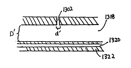
Referring now to Figures lA to 1C one embodiment of the invention is depicted
comprising an infusion catheter 1300 capable of generally simultaneous
infusion of the
treatment agent through a plurality of holes 1302 located along the length of
the catheter 1300.
The catheter 1300 comprises a proximal end 1304 with at least one access port
1306, 1308,
1310, a catheter body 1312, and a distal end 1314 with a blood vessel occluder
1316.
In one embodiment, each access port 1306, 1308, 1310 is in fluid communication
with a
lumen running generally along the length of the catheter body. In some
embodiments, a lumen
may be in fluid communication with multiple access ports. In one embodiment,
at least one
access port 1306 is in fluid communication with an infusion lumen allow
infusion of a treatment
agent into the catheter 1300 and out through the holes 1302 of the catheter
body 1312. In one
embodiment, one access port 1310 and lumen 1320 is provided to allow
manipulation of the
blood vessel occluder 1316 from the proximal end 1304 of the catheter 1300.
The inflation
lumen 1320 may be integral with the outer catheter wall 1322 or be defmed
within a separate
tubular wall (not shown) within the infusion lumen 1318.
In one embodiment, the catheter 1300 is configured so that the fluid elution
from the
holes 1302 generally occurs in a particular predetermined pattern when the
fluid is injected
through the catheter 1300 at a specific viscosity and pressure or pressure
range. In one
embodiment of the invention, the pattern of fluid elution is determined by at
least one of several
CA 02574429 2007-01-18
WO 2006/023203 PCTIUS2005/026147
9
factors, including but not limited to: 1) the hydraulic diameter D' of the
infusion lumen of the
catheter; 2) the hydraulic diameter d' of each elution hole; 3) the spacing s'
between each elution
hole; 4) the overall treatment length L' of the catheter; 5) the viscosity of
the agent used for
treatment; and 6) the compressibility of the treatment agent. The term
"hydraulic diameter", as
used herein, shall be given its ordinary meaning and shall also include the
equivalent diameter of
a structure when estimating pressure loss or head loss in non-circular lumena
using data made
for circular lumena. The term "treatment length" as used herein shall mean the
portion of the
catheter generally from about the most proximal elution hole 1324 to about the
most distal
elution hole 1326.
In one embodiment, the fluid distribution from the catheter 1300 is generally
even along
the treatment length of the catheter 1300. In another embodiment, the pattern
of fluid
distribution from the catheter 1300 provides for increased elution of agent at
the distal end 1314
of the treatinent length. The change in elution along the treatment length may
be a gradual ramp
or stepped. In another embodiment, the fluid distribution pattern provides
greater elution at the
proximal end 1304 of the treatment length. In another embodiment, the catheter
1300 provides
a customized distribution pattern adapted to provide increased flow at one or
more locations
along the treatment length which is adapted to correspond to the location of
,the venous
tributaries when the occluder has been positioned as described herein. In
another embodiment,
the catheter 1300 provides a customized distribution pattern adapted to
provide increased flow
at the venous tributaries and about the saphenofemoral junction. One skilled
in the art will
understand that the catheter may be configured for any of a variety of elution
or distribution
patterns.
The diameter D' of the infusion lumen 1318 of the catheter 1300 generally
ranges from
about 0.03" to about 0.20". In certain embodiments, the diameter d' ranges
from about 0.05" to
about 0.09". In one embodiment, the diameter d' is about 0.072".
The overall treatment length L' of the catheter generally ranges from about 10
cm to
about 175 cm. In certain embodiments, the treatment length L' is within the
range of from about
20 cm to about 100 cm. In another embodiment, the treatment length L' is
within the range of
from about 20 cm to about 44 cm.
CA 02574429 2007-01-18
WO 2006/023203 PCT/US2005/026147
The viscosity at body temperature of the treatment agent is generally within
the range of
from about 1.00E-04 (lb*s/in~2) to about 1.00E-08 (lb*s/in~2). In certain
embodiments, the
viscosity of the treatment agent is within the range of from about 1.00E-06
(lb*s/in~2) to about
1.00E-08 (lb*s/in~2). In one embodiment, the viscosity is about 1.74E-07
(lb*s/in~2).
Viscosities outside of the foregoing ranges may also be used, taking into
account the pore sizes,
infusion lumen length and diameter, as long as the desired delivery
performance (e.g. delivery
rate) is achieved. Sclerosing agents used for treating veins are generally
incompressible, but
compressible agents may also be used.
In one embodiment, the spacing s' between the elution holes 1302 ranges from
about
0.01 cm to about 10 cm. The spacing s' between the elution holes 1302 may
range from about
0.50 cm to about 5 cm. In other embodiments, the spacing s' between the
elution holes 1302 is
about 0.50 cm to about 3 cm. In another embodiment, the spacing s' between the
elution holes
1302 is about 0.50 cm to about 2 cm.
Figure 2 shows that the spacing between the elution holes 1032 may vary along
the
length of the catheter. Portions of the catheter with increased spacing s" may
exhibit a reduced
elution rate compared to portions of the catheter with decreased spacing s"',
for a given hole
diameter. Variations in the spacing of elution holes may be used to achieve
variations in the
elution patterns of the catheter. The elution pattern is defined by the
elution rates at different
segments of the infusion catheter. For example, an even elution pattern
generally has similar
elution rates along the all the segments catheter, while a distal elution
pattern provides increased
elution rate in at least one segment of the catheter located distally.
Increased elution in a
particular zone or region of the catheter may be provided by increasing the
total cross sectional
area of the elution holes in that region, such as by either increasing the
elution hole density or
the elution hole diameters or both in that region.
The diameter d' of the elution holes 1032 may be selected for the desired
elution pattern
by considering the catheter and sclerosing agent characteristics described
previously and the
pressure drop-off along the catheter length. In one embodiment of the
invention, the elution
hole diameter is about 0.001" to about 0.015". In another embodiment, the
elution hole diameter
is about 0.002" to about 0.010". In one embodiment, based upon a 6-French
catheter with a
length greater than 40 cm, elution hole spacing between 1 cm and 2 cm and
sclerosing agent
CA 02574429 2007-01-18
WO 2006/023203 PCTIUS2005/026147
11
characteristics described previously, an elution hole diameter of about 0.004"
or less is capable
of providing a generally uniform fluid elution along the length of the
infusion catheter 1300.
Other elution hole diameters may also be used, depending on the desired
elution pattern for the
infusion catheter and the catheter and sclerosing agent characteristics used.
Figure 3 shows that the diameters of the elution holes 1300 need not be
uniform.
Larger elution hole diameters d" will generally have a higher elution rate
than smaller elution
hole diameters d"', but other factors, such as the pressure drop-off along the
catheter, will also
effect the relative elution rates between the elution holes. In one embodiment
of the invention,
elution holes located in the distal portion of the catheter generally have a
greater diameter than
elution holes in the more proximal portions of the catheter to compensate for
the pressure drop
along the length of the delivery zone and produce a relatively constant
delivery profile. The
cross sectional shape of the elution holes can be circular, oval, square,
triangular or any
polygonal or closed shape. The cross sectional shape of the elution holes need
not be uniform
throughout the longitudinal length of the elution hole. In one embodiment,
variations in elution
hole diameter and elution hole spacing are used to alter the elution pattern.
In one embodiment of the invention, the diameters d' of the elution holes 1302
each have
an effective hydraulic diameter less than the fluid distribution lumen D' that
connects the
elution holes 1302. In a further embodiment, the total fluid resistance of the
plurality of elution
holes 1302 is generally equal or greater than fluid resistance of the infusion
lumen 1318 or
lumena of the catheter. In still a fiuther embodiment of the invention, the
total fluid resistance
of the plurality of elution holes 1302 is substantially greater than the fluid
resistance of the
catheter infusion lumen 1318. By providing elution holes 1032 with a total
fluid resistance
substantially greater than the infusion lumen 1318, uniform elution along the
catheter 1300 may
be achieved. The total fluid resistance of the infusion lumen should generally
be less than about
80 percent of the total fluid resistance of the elution holes, and in certain
devices less than about
50 percent of the total fluid resistance of the elution holes. The hydraulic
diameters of the
elution holes 1302, however, are not limited to consideration of the factors
described above.
The wall thickness of the infusion catheter 1300 may also contribute to the
total fluid
resistance of the plurality of elution holes 1032. The wall thickness
essentially corresponds to
the length of a capillary tube, creating resistance to flow which may at least
theoretically be
CA 02574429 2007-01-18
WO 2006/023203 PCTIUS2005/026147
12
determined by well known relationships such as Poiseuille's law. For example,
a 6-French
catheter made of Versamid polyamide resin may have a wall thickness within
the range of
about 0.006" to 0.015". Where the elution holes have a hydraulic diameter of
about 0.004" or
less, the wall thickness, which defmes the length of the elution holes 1302,
may contribute to
the fluid resistance of the elution hole 1302. In one embodiment of the
invention, the catheter
has a wall thickness of about 0.003" to about 0.100". In another embodiment,
the catheter has a
wall thickness of about 0.004" to about 0.060". In another embodiment, the
catheter has a wall
thickness of about 0.005" to about 0.030". In still another embodiment, the
catheter has a wall
thickness of about 0.004" to about 0.020".
The elution rate at a given segment of the catheter is affected by spacing s'
and hole
diameter d" of elution holes 1302, the distance of the segment from the
proximal end of the
catheter, as well as the spacing s' and diameter d' of the other catheter
segments. One skilled in
the art will understand that these characteristics, and other characteristics
described previously,
can be altered to achieve a different elution pattern.
Figures 4A to 4D illustrates one embodiment of the invention, where the
medicament is
eluted from the catheter 1330 through at least one catheter portion comprising
a porous or
permeable region 1332. The porous region comprises a plurality of small
openings 1334
through which the medicament may elute. In one embodiment, the region has a
porosity of
about 2 microns to about 40 microns. In another embodiment, the region has a
porosity of
about 4 microns to about 20 microns. In another embodiment, the region has a
porosity of
about 6 microns to about 12 microns. In one embodiment, the region has a
porosity of about 8
microns which is preferably capable of resisting clogging from blood
constituents. The porosity
of the porous or permeable regions need not be uniformly porous between
regions or within the
same region.
A porous portion 1332 may comprise a full circumference of catheter, as shown
in
Figures lA and 4C, or a portion of the circumference, as shown by segments
1336, 1338 in
Figures 4B and 4D. The infusion catheter may comprise a single porous portion,
multiple
contiguous porous portions or multiple porous portions separated by non-porous
portions.
Multiple porous portions may be arranged serially along the longitudinal
length of the catheter
as shown by segments 1336, in parallel where the porous portions are
longitudinal strips 1338
CA 02574429 2007-01-18
WO 2006/023203 PCT/US2005/026147
13
along the length of the catheter, or any combination thereof. In another
embodiment, a
combination of porous regions and elution holes may be used to provide the
desired elution
pattern for the catheter. The porous material may include, but is not limited
to, a ceramic,
ultrahigh molecular weight polyolefin, a perforated polymer film, porous or
microporous
membranes, polyethersulfone, TYVEK (spun-bonded polyethylene), GORTEX
(expanded
PTFE), woven or knit mesh or fabric, and other porous materials.
In one embodiment of the invention, a system for controlling or altering the
flow of
medicament at an elution hole, a series of elution holes, or a porous region
is provided. Multiple
elution control systems may be used in the same catheter to provide control
over multiple
portions of the catheter. A control system may also be capable of protecting
the elution hole
from clogging with blood components by exposing the elution hole only during
periods of
desired elution and protecting the elution holes at other times. Several
embodiments of the
control system are described below.
Figures 5A and 5B show one embodiment of the invention, where the fluid
control
system comprises a separate or side lumen 1340 generally along the length of
the infusion
catheter 1342. At least one inner hole 1344a-1344d is provided between the
infusion lumen
1346 and side lumen 1340, and at least one outer hole 1348a-1348f or porous
segment from the
side lumen 1340 to the exterior of the catheter is also provided. An elution
hole occluder 1350
capable of resisting flow through the inner hole 1344, outer hole 1348 or
both.
Medicament from the infusion lumen 1346 is capable of flowing through the
inner holes
1344a-1344d, intersecting the side lumen 1340, and passing through the outer
holes 1348a-
1348f to exit from the catheter 1342 when the occluder 1350 is in a first,
open position or has
been withdrawn from the catheter. The inner holes 1344a-1344d and outer holes
1340a-1340f
need not be aligned, and the number of inner 1344 and outer holes 1348 need
not be equal. Inner
hole 1344a and outer hole 1348a depict aligned holes whiles inner hole 1344d
and 1348f depict
non-aligned holes.
Any inner hole 1344 and outer hole 1348 capable of providing flow out of the
catheter
1342 defmes an elution hole or pathway. Any inner hole 1344 or outer hole 1348
may defme
more than one elution hole or pathway. For example, inner hole 1344c is
capable of flow to
outer holes 1348c-1348e. The cross-sectional areas of the inner holes and
outer holes need not
CA 02574429 2007-01-18
WO 2006/023203 PCT/US2005/026147
14
be equal and may vary within the same hole. In one embodiment, an inner hole
1344d has a
greater diameter than outer hole 1348f. In one embodiment, a greater number of
outer holes may
be desired to create a more uniform elution pattern. In one embodiment,
increased elution from
outer holes that are closer to the inner holes can be reduced by decreasing
the alignment between
the inner holes and the outer holes to increase the tortuosity of the flow
path and provide a
more even distribution pattern from the outer holes.
The cross sectional shape of the elution holes can be circular, oval, square,
triangular or
any polygonal or closed shape. The cross sectional shape of the elution holes
need not be
uniform througllout the longitudinal length of the elution hole. In certain
embodiments, the inner
holes have a circular diameter of about 0.002" and the outer holes have a
rectangular shape, with
a length of about 0.022" as measured along the longitudinal axis of the
catlleter, and a width of
about 0.007". In one embodiment, a rectangular outer hole configuration where
the width of the
hole is about equal to the diameter of the occluder is used to provide better
flow around some
occluder configurations.
In one embodiment, the movable occluder 1350 is located generally along the
length of
the side lumen 1340, such as coaxially within the side lumen 1340. In one
embodiment, the
movable occluder 1350 comprises at least one narrow connector portion 1352
with a narrow
diameter and at least one blocking portion 1354 which, in the illustrated
embodiment, comprises
an enlarged diameter or width that is capable of forming a seal with the side
lumen. Movable
occluders with a uniform diameter may also be used, but such occluders may
exhibit increased
resistance to sliding compared to occluders with variable diameters.
In sealing with the side lumen 1340, the enlarged portion 1354 may block an
inner hole,
an outer hole or both. Figure 5A illustrates an occluder 1350 blocking inner
hole 1344c and
outer hole 1348f but not inner hole 1344d or outer holes 1348c to 1348e. By
axially advancing
the occluder 1350 either proximally or distally in the side lumen 1340, the
relative position of
the blocking portions 1354 and the corresponding elution holes may be changed
and the effluent
flow path may be selectively opened or closed. Not every hole needs to be
blockable by the
elution hole occluder. In one embodiment, the enlarged portions have
longitudinal lengths that
are at least as long as the diameter of the holes to resist medicament flow
through the hole. The
enlarged portions of the occluder may also be provided with longer lengths to
decrease the
CA 02574429 2007-01-18
WO 2006/023203 PCT/US2005/026147
precision with which the occluder is positioned within the side lumen in order
to resist or
occlude flow through the holes. The occluder and/or side lumen may also be
provided with a
lubricious coating or treatment to facilitate sliding of the occluder within
the side lumen. Such
coatings may include PTFE, parylene, or others known in the art. The occluder
and/or side
lumen may also be coated or treated to alter the sealing characteristic
between the occluder and
the side lumen.
In one embodiment, the side lumen has an internal diameter of about 0.025" and
the
occluder comprises a valving wire with narrow portions having a primary
diameter of 0.015"
and at least one enlarged portion with a diameter of about 0.022" to about
0.024" by about
0.200" length. When the enlarged portion of the occluder is positioned next to
an inner hole or
outer hole, the elution hole or pathway defined by the inner hole and outer
hole is "closed" and
flow from the infusion lumen out of the catheter is blocked or resisted. When
the enlarged
portion of the valving wire is positioned away from a pair of inner and outer
holes, the pair of
holes is "open" and medicament is able to flow througll the holes and out of
the catheter.
In another embodiment, the occluder comprises a movable ribbon having narrow
portions and wider portions that is capable of reversibly occluding the
elution holes.
Alternatively, the occluder may comprise a rotatable element, such as an
elongate tubular body
having side wall apertures aligned to permit or block fluid communication
between the central
lumen 1346 and one or more ports on the exterior wall of the catheter.
In one embodiment, the occluder is configured to generally open all of the
elution holes
or porous segment simultaneously. This allows the user to quickly initiate the
fluid elution
along the entire length of catheter, so that the dilution of the medicament by
flowing blood is
reduced. The risk of plugging or blocking the elution holes with clotted blood
components may
also be reduced by quickly opening generally all the elution holes.
In certain embodiments of the invention, illustrated in Figures 6A to 6C, the
length and
number of the narrow portions and enlarged portions of the occluder are
configured or arranged
such that the occluder 1356 is capable of opening individual or a first group
of the elution holes
1358 while a second group of elution holes 1360 remain closed. By providing
the ability to
open a limited number of elution holes while maintaining closure of other
elution holes, the user
CA 02574429 2007-01-18
WO 2006/023203 PCT/US2005/026147
16
can control the location of the effective elution zone and further customize
the treatment
procedure.
In one embodiment, the first position of the occluder 1356, depicted in Figure
6A, keeps
all elution holes 1358, 1360 closed. In the second position illustrated in
Figure 6B, the increased
length of the enlarged portions 1362 allows the occluder to keep holes 1360 in
a first zone
closed while the shorter length of enlarged portions 1364 allow the opening of
holes 1358 in a
second zone. In the third occluder position in Figure 6C, all the holes 1358,
1360 in both the
first and second zones are open. The spacing of the elution holes on the
catheter may affect the
additional number of occlusion patterns available.
In certain embodiments, the elution holes can be opened sequentially along the
length of
the delivery zone to provide and then closed, a moving elution zone without
repositioning the
catheter, or to allow a single catheter length to be used for treating
patients requiring different
delivery zone lengths. One example of the latter configuration comprises a
catheter having a 44
cm delivery zone that is only partially inserted into a patient's leg because
only a 24 cm
delivery zone was required. The catheter will not leak sclerosant from the
proxima120 cm that
lies external to the patient where the occluder is configured and positioned
to only open the
elution holes in the distal 24 cm of the catheter. In another embodiment, the
occluder is
configured so that the elution holes are opened in groups rather than
individually, by either
arranging the elution holes circumferentially in the same longitudinal region
of the catheter, or by
provide the enlarged portions of the occluder with sufficient length or
particular spacing to
simultaneously block multiple holes.
Figures 7A to 7D depict one embodiment, where the occluder is further
configured to
open an elution hole or group of holes and then close the elution holes prior
to, during or after
opening another group of elution holes. The occluder 1366 comprises a narrow
segment 1368
that allows medicament flow through the elution holes 1370 adjacent to it. In
one embodiment,
the narrow segment 1368 is movable along the treatment length of the catheter
to open the
elution holes, two at a time. This particular embodiment may require a longer
catheter length
that extends beyond the occlusion balloon of the catheter to accommodate the
distal end of the
occluder. One skilled in the art will understand that the occluder may be
configured to provide
CA 02574429 2007-01-18
WO 2006/023203 PCT/US2005/026147
17
any of a variety of opening and closing patterns in the catheter by altering
the length, position
and number of narrow and enlarged portions on the occluder.
In one embodiment, an infusion catheter with an occluder capable of
sequentially
opening the elution holes may also be advantageous when infusing foam-based
medicaments,
including but not limited to sodium tetradecyl sulfate. The inventors have
found that when
elution holes with cross-section areas comprising a significant fraction of
the infusion lumen
cross-sectional area are used, it is common for liquid and foam-based
medicainents to
preferentially elute from the first hole that the foam encounters as it enters
the catheter. In
simple catheter constructions, this is typically the most proximal elution
hole. Foam is
typically disposed to elution in this manner because of its compressibility.
During elution, the
pressure of injection causes the foam to be compressed until it encounters an
opening in the
catheter, where it expands into the lower-pressure environment outside the
catheter. To
compensate for the increased elution of medicament at the proximal end of the
catlleter
treatinent zone, a catheter with a sequentially opening elution hole
controller may be used. In
one embodiment, to provide infusion of medicament along the entire length of
the treatment
zone, the most distal elution holes or elution zones are opened first, so that
the medicament will
elute from these distal areas. The adjacent proximal elution holes and/or
elution zones are then
sequentially opened to allow elution in a more proximal fashion. By using a
sequentially
opening catheter, a medicament that elutes primarily from the first-
encountered elution hole
may be dispensed evenly across the entire length of the catheter treatment
zone. In one
embodiment, elution control may be accomplished by proximally retracting a
valving wire, but
other control structures can also be used.
It may be advantageous for the catheter user to be able to elute a bplus of
medicament at
a specific location in the body, in addition to the even elution across the
treatment zone of the
catheter. Bolus treatment may be accomplished with a catheter comprising two
elution
systems: a) an "even-elution" system as previously described using a series of
elution holes or
pores which simultaneously or sequentially elute over a prescribed portion of
the infusion
catheter, and b) one or a series of sequentially-openable larger openings that
will elute
medicament (either foam or liquid) at a bolus delivery zone. Before, during or
after performing
CA 02574429 2007-01-18
WO 2006/023203 PCT/US2005/026147
18
an even elution, the operator may use the second system of larger holes to
deliver a single or
multiple boluses to specific areas in the blood vessel.
Figure 8 shows one embodiment comprising one or more stops 1372 and/or detents
in
the infusion catheter 1374 to facilitate alignment of the valving wire 1376
within the side lumen
1378. The stops may restrict the sliding range of the wire 1376 and can
prevent accidental
removal of the wire 1376 from the side lumen 1378. The stops 1372 and/or
detents may be
located within the side lumen 1378 and/or in the proximal portion of the
catheter 1374 at or
about the infusion ports. Alternatively, the stop may be provided within or in
the vicinity of a
proximal manifold on the catheter to simplify manufacturing as will be
appreciated by those of
skill in the art. In one embodiment of the invention, the infusion catheter is
supplied with a set
of different valving wires that are insertable into the side luinen before or
during the procedure,
to allow further adjustment to the elution pattern of the catheter.
Figure 9 illustrates one einbodiment of the invention, where the narrow
portions 1380
of the occluder 1382 are generally aligned with the enlarged portions 1384
along the same
longitudinal axis such that when an elution hole is open, fluid from the inner
hole must pass
around at least a portion of the occluder with the narrow. diameter to flow
into the outer holes
1386. In another embodiment, depicted in Figure 10, the primary portions 1380
of the
occluder 1382 are joined eccentrically with the enlarged portions 1384, so
that the primary
portions 1380 offer less resistance to flow through the outer elution holes
1386.
In one embodiment, shows in Figure 11, the cross sectional shape of the
occluder 1394
does not match the shape of the side lumen 1396. In one embodiment, by
providing an occluder
1394 with a non-circular or oval cross-sectional shape, surface friction
between the occluder
1394 and the side lumen 1396 may be reduced. In one embodiment, an occluder
1394 with a
polygonal cross section is provided, where the edges 1398 of each polygon face
are capable of
providing sealing contact with the side lumen wall 1400, but the overall
reduced friction allows
the user to quickly move or remove the occluder 1394. In the illustrated
embodiment, a four-
cornered (square) wire 1394 is used in a circular side lumen 1396 as an
occluder. At least one
sealing line at one of the wire corners 1398 is capable of forming sealing
contact with the side
lumen 1396. Although potential leakage paths 1402 may exist along the
longitudinal length of
side lumen 1396 because of the lack of complete surface-to-surface contact
between the wire
CA 02574429 2007-01-18
WO 2006/023203 PCTIUS2005/026147
19
and the side lumen walls, the length of the leakage paths are likely to be of
sufficient length so as
to substantially reduce or prevent elution of medicament or intrusion of blood
components at
the side lumen 1396.
In one example, an infusion catheter comprising a side lumen and an array of
ten elution
holes, with one hole per centimeter over a nine centimeter length, is
provided. The side lumen
contains a single square wire of at least about 9 cm length. In one
embodiment, a smaller-
diameter pull wire is engaged the proximal end of the square wire, to allow
manipulation of the
square wire from the proximal end of the catheter. In an alternate embodiment,
to simplify
manufacture of the square wire occluder, a square wire with a length at least
sufficient to extend
from through the proximal end of the catheter to the distal end of the
catlleter treatment segment
is used as an occluder. In one embodiment, short segments of the wire may have
cross-sections
closer to or matching that of the side lumen to limit the extent of lengthwise
leakage, without
significantly increasing the net sliding friction of moving or withdrawing the
wire from the
catheter.
Figures 12, 13A and 13B depict optional indicators on the catheter to provide
information regarding the position of the occluder, the open/close status of
the elution holes, or
both. In one embodiment, shown in Figure 12, the indicator 1404 is a marker
such as a colored
bank carried by the occluder 1406 another that is capable of moving within a
window 1408. In
another embodiment, schematically illustrated in Figure 13B the indicator
comprises a dial
turned relative to an index mark by a rack-and-pinion or friction drive. One
skilled in the art will
understand that other mechanisms for indicating the position of the occluder
or status of the
elution holes may be used. In one embodiment of the invention, shown in
Figures 13A and 13B,
the indicator 1410, 1412 is incorporated or combined with an occluder actuator
1414, 1416 for
manipulating the position of the occluder. The occluder actuator may comprise
a slider 1414,
lever, or turning knob 1416 attached to the occluder. The occluder actuator
may also comprise a
servo motor that is electronically controllable by the user. One skilled in
the art will understand
that other mechanisms for moving the occluder may also be used.
Figures 14A to 14C depict one embodiment of the invention, the movable
occluder
comprises an elastomeric cord 1418 within the side lumen 1420 of the catheter
1422. Such a
cord may comprise latex, silicone rubber, natural rubber, neoprene and other
chloroprene
CA 02574429 2007-01-18
WO 2006/023203 PCT/US2005/026147
variants, polyurethane, ethylene-propylene, polyvinyl chloride, polyamide,
polyamide
elastomer, copolymer of ethylene and vinyl acetate, polyethylene, polyimide,
polyethylene
terephthalate, fluorine resin, polyisobutylenes or other thermoset elastomers,
polyisoprene, or
any of a variety of resilient inaterials known in the art. The cord may have a
cross-sectional
shape that is square, rectangular, oval, circular, polygonal or any of a
variety of other shapes
that are capable of forming a seal with the side lumen. The cord may be solid,
hollow or have a
core comprising the same or different material. In one embodiment, at least
one portion or
segment of the elastomeric cord has a native diameter that is larger than the
inside diameter of
the side lumen 1420, to provide enhanced occlusion of the elution holes 1424.
As shown in
Figure 14B and 14C, by pulling on the proximal end 1426 of the cord 1418 and
causing
longitudinal lengthening, the cord 1418 is capable of deforming and reducing
its cross-sectional
area, as shown in the proximal end 1426 in Figures 14B. This reduction in
diameter allows the
cord to be removed from the side lumen and opens the elution holes 1424.
In another embodiment shown in Figures 15A and 15B, the distal end 1428 of the
cord
1430 is anchored in the side lumen 1432 so that the cord 1430 resists removal
from the side
lumen 1432 when a pulling force is applied to its proximal end 1434, but is
capable of
decreasing in diameter or cross sectional area sufficiently to allow flow
through the elution holes
1436. Anchoring may be accomplished using any of a variety of techniques, such
as adhesives,
solvent or thermal bonding, mechanical interfit, cross pins or others known in
the art. In one
embodiment, upon cessation of the proximal pulling force, the cord 1430 is
generally able to
revert back to its previous length and diameter and reversibly re-close the
elution holes. In
another embodiment, upon pulling the cord 1430, the cord plastically deforms
and some or all of
the elution holes 1436 remain at least partially open after cessation of the
pulling force. In one
embodiment, illustrated in Figure 16, the elastomeric cord 1438 comprises
narrow segments
1440 and enlarged segments 1442 or increasing the sealing characteristics of
the cord 1438 at the
elution holes 1444 and/or to reduce the tensile force needed to move or remove
the cord 1438 in
the side lumen 1446. In one embodiment, the elastomeric cord and/or side lumen
is coated or
treated to alter the friction between the cord and lumen.
Figures 17A to 17D depict another embodiment of the invention, in which a
hollow
flow regulating tube 1450, having a central lumen 1452 is positioned within
the side lumen 1448.
CA 02574429 2007-01-18
WO 2006/023203 PCT/US2005/026147
21
The tube 1450 has an open proximal end and a closed distal end. The proximal
end may be
provided with a releasable connector such as a Luer fitting for connection to
a source of inflation
media. Alternatively, the central lumen may be in direct communication with a
variable volume
chamber in the proximal manifold or hand piece for the catheter.
The outside diameter of the flow regulating tube 1450 is moveable from a
first, reduced
diameter to a second enlarged diameter upon introduction of inflation media
into the central
lumen 1452. The outside diameter of the tube 1450 in the first, relaxed
configuration is less than
the inside diameter of the lumen within which it resides, such as side lumen
1448. In this
configuration, a medicament or other agent in the infusion lumen 1456 is
capable of flowing past
or around the hollow tube 1450 to exit out of the elution hole 1454. See
Figure 18A.
Introduction of inflation media into central lumen 1452 causes an enlargement
of the outside
diameter of the tube 1450 such that it occludes the flow path between the
infusion lumen 1456
and the exterior of the catheter body. See Figure 18B.
The flow regulating tube 1450 thus provides a movable wall which may be
advanced
between a first orientation in which flow is permitted to occur and a second
orientation in which
flow is inhibited. Introduction of intermediate pressures into the central
lumen 1452 may be
utilized to regulate flow at intermediate flow rates, or permit flow only to
occur when the
driving pressure within the infusion lumen 1456 exceeds a predetermined
threshold.
Although the flow regulating tube 1450 is described as located within the side
lumen
1448, valves or flow regulators which are responsive to changes in pressure
may be
incorporated into the catheter of the present invention in any of a variety of
ways. For
example, the inflatable tube 1450 may be positioned within the inflation lumen
1456, and the
side lumen 1448 may be eliminated or utilized for another purpose. The
inflatable tube 1450
may be configured to have an axial length less than the length of the infusion
zone, such that, for
example, it occludes only a relatively proximal portion of the catheter body.
In one
implementation, the flow regulating tube 1450 has an axial length of no
greater than 2 or 3 or 4
times the inflated diameter, such that it operates as an inflatable valve
positioned in-between the
proximal most elution hole and the source of infusion media. In general,
however, it appears
desirable for the axial length of the flow regulating tube 1450 to be at least
as long as the infusion
CA 02574429 2007-01-18
WO 2006/023203 PCT/US2005/026147
22
zone, such that in the inflated configuration, the flow regulating tube 1450
physically occludes
each elution hole 1454.
The escape of material from the infusion lumen 1456 through each elution hole
1454
may be accomplished by providing an inflatable tube 1450 at any point between
that elution
hole 1454 and the source of infusion media. However, it also appears desirable
to block each
elution hole 1454 to prevent blood or other body fluid from entering the
catheter in a retrograde
flow direction, prior to the time that the sclerosant or other infusion media
is infused from the
catheter into the patient. Thus, in accordance with the present invention,
there is provided a
method and related device for introducing a catheter into a patient, the
catheter having a
plurality of elution holes 1454, and preventing the introduction of body fluid
into the catheter
through the elution holes. The introduction of body fluid into the catheter is
inhibited by the
positioning of a movable wall across the elution hole. The moveable wall is
moveable between a
first position in which it occludes the elution hole 1454, and a second
position in which the
infusion lumen 1456 is in communication with the exterior of the catheter
through the elution
hole 1454. In the illustrated embodiment, the moveable wall is the surface of
an inflatable tube,
although other structures for moving a wall between a first position and a
second position may
also be utilized.
Although the present embodiment has been described primarily in terms of a
hollow
flow regulating tube 1450 having a reduced outside diameter in its relaxed
configuration, the
device may alternatively be constructed such that the hollow flow regulating
tube 1450 resides
in an enlarged cross sectional diameter in it relaxed configuration. This
configuration would
provide a"normally closed" valve system, in which the outside diameter of the
flow regulating
tube 1450 would normally occlude the elution hole 1454. In this construction,
drawing a
negative pressure on the central lumen 1452 could be utilized to reduce the
cross sectional area
of the flow regulating tube 1450, thereby placing the elution hole.1454 into
communication with
the infusion lumen 1456.
The tube 1450 may comprise any of a variety of materials that may be expanded
under
pressure, such as latex, silicone rubber, natural rubber, neoprene and other
chloroprene variants,
polyurethane, ethylene-propylene, polyvinyl chloride, polyamide, polyamide
elastomer,
copolymer of ethylene and vinyl acetate, polyethylene, polyimide, polyethylene
terephthalate,
CA 02574429 2007-01-18
WO 2006/023203 PCT/US2005/026147
23
fluorocarbon resin, polyisobutylenes and other thermoset elastomers,
polyisoprene, or any of a
variety of materials known in the art that is capable of radial expansion when
fluid in the hollow
portion 1452 of the tube 1450 is pressurized.
In one embodiment, depicted in Figures 18A and 18B, the elastomeric tube 1450
is
positioned concentrically within, or is allowed to "float" within the side
lumen 1448 in both the
inflated and deflated states. In another embodiment, shown in Figures 19A and
19B, the
elastomeric tube 1450 in the deflated state is positioned eccentrically in the
side lumen 1448
using a sealant, adhesive, thermal welding or other bonding technique known in
the art. Figure
19B shows that when tube 1450 is fully expanded, it can assume a more
concentric position in
the side lumen 1448. In one embodiment, an eccentric position may provide a
larger or more
predictable effective flow path past the elastomeric tube 1450 compared to a
concentrically
positioned or free floating tube 1450.
The ratio of the first, reduced dianeter of the flow regulating tube 1450 to
the inside
diameter of the lumen within which it resides can be varied widely, depending
upon the desired
performance characteristics, taking into account the viscosity and desired
flow rate of the
infused media. In general, the deflated diaineter of the tube 1450 will be no
greater than about
75% of the inside diameter of the side lumen 1448. In certain constructions,
the deflated outside
diameter of the flow regulating tube will be no more than about 65%, and, in
certain
implementations, no greater than about 60% of the inside diameter of the lumen
within which it
is contained.
In certain constructions, the hollow elastomeric tube 1450 has a deflated
outside
diameter ranging from about 0.008" to about 0.100". In certain embodiments,
the tube 1450 has
a deflated outside diameter ranging from about 0.010" to about 0.050". The
elastomeric tube has
a deflated internal diameter generally within the range of from about 0.003"
to about 0.080". In
a preferred embodiment, the elastomeric tube has an outer diameter of about
0.015" and an inner
diameter of about 0.006", for use in a lumen having an inside diameter of
about 0.025".
The inflation pressure sufficient to occlude the elution holes may range from
about 10
pounds per square inch (psi) to about 1000 psi. In certain embodiments, the
occlusion pressure
is about 50 psi to about 500 psi. In another embodiment, the occlusion
pressure is about 100
psi to about 600 psi. In one embodiment, where the occluder comprises an
elastomeric tube
CA 02574429 2007-01-18
WO 2006/023203 PCT/US2005/026147
24
with an outer diameter of about 0.015" and an inner diameter of about 0.006"
in a 0.025" side
lumen, the tube has an occlusion pressure at about 100 psi to about 200 psi.
The tube diameter, wall thickness, wall compliance, and other tube
characteristics may
be varied along the length of the bladder tube. One skilled in the art may
alter these
characteristics to provide different occlusion characteristics across a
pressure range. In one
example, a bladder tube may be designed to sequentially deflate from distal to
proximal over a
pressure range from 200 psi to 100 psi. Distal to proximal deflation may be
accomplished, for
example, by providing a first wall thickness for the elastomeric tube 1450 in
the proximal end
and a second, greater wall thickness for the elastomeric tube 1450 near the
distal end. Wall
thickness may be graduated continuously from the proximal end to the distal
end.
Alternatively, deflation may be accomplished initially at the proximal end by
providing the
greater wall thickness at the proximal end. As will be apparent to those of
skill in the art in
view of the disclosure herein, the inflation characteristics of the foregoing
constructions will be
the reverse of the deflation characteristics, such that portions of the flow
regulating tube with a
relatively lesser wall thickness will inflate at a lower pressure than
portions of the flow
regulating tube with a greater wall thickness. The sequential expansion during
inflation may
occur smoothly across the length of the flow regulating tube, or in a
segmented fashion. In
another example, the bladder tube may comprise dimples in the bladder tube
that evert and
occlude elution holes at a particular pressure threshold.
In one embodiment of the invention utilizing an inflatable flow regulator form
of
occluder, the occluder comprises an inflatable tube in a catheter with outer
hole diameters of
about 150 microns or greater and inner holes diameters of about 200 microns or
less. In another
embodiment, the catheter comprises outer hole diameters of about 400 microns
or less and inner
hole diameters of about 5 thousandths of an inch (200 microns) or more. In one
embodiment,
the outer holes have diameters of about 200 microns or more and inner holes of
about 20
microns to about 250 microns. In another embodiment, the outer holes have
diameters of about
20 microns to about 250 microns and the inner holes have diameters of about
200 microns or
more. In one embodiment, at least either the outer holes or inner holes have a
diameter of about
8 microns to about 175 microns. In a preferred embodiment, the catheter
comprises outer holes
with diameters of about 300 microns or greater and inner holes with diameters
of about 50
CA 02574429 2007-01-18
WO 2006/023203 PCT/US2005/026147
microns to about 175 microns. The inner holes may have the same, a smaller, or
a larger
diameter than the corresponding outer hole.
The elastomeric tube may be pressurized with a pressure controller comprising
variable
volume container such as a syringe. The syringe may have a capacity of about
0.25cc to about
25cc, and may be is attachable such as by a Luer connector to the proximal end
of the inflatable
tube. In certain embodiments, the syringe has a capacity of about lcc to about
5cc. In a
preferred embodiment, the syringe has a capacity of about lcc to about 2cc.
The plunger of the syringe may be controlled directly by the operator or
through a lever
or knob with detent. In another embodiment, the pressure controller comprises
an electronically
controlled pump and pressure release valve. One skilled in the art will
understand that any of a
variety of pressure controllers may be used. In one embodiment, the syringe or
catheter fixrther
comprises a stopcock for maintaining pressure in the elastomeric tube without
further effort by
the user. In another embodiment, the plunger or tube controller further
comprises a latch for
maintaining the position of the plunger. In a preferred embodiment, the tube
controller provides
a two-position control of the tube wlzere the tube is either inflated or
deflated. In another
embodiment, the pressure controller is capable of providing multiple degrees
of tube
pressurization. A controller providing multiple degrees of tube pressurization
may be useful to
provide variable flow patterns or varying degrees of flow through the elution
holes to further
control the flow rate of medicament out of the catheter.
In one embodiment of the invention, the hollow elastomeric tube is pressurized
with a
gaseous medium. In one embodiment, the tube is pressurized with a liquid
medium. A liquid
medium may be preferred to decrease the risk of an air embolus in the venous
system that may
travel to the lungs or other sites and block tissue perfusion.
In one embodiment of the invention, the elastomeric or bladder tube comprises
silicone
or other porous material that is sufficiently permeable so that any trapped
gas in the tube can be
expelled by inflating the tube with a liquid to at least about 100 psi. Under
such a pressure, the
gases diffuse out through the permeable tube and/or into the liquid medium. In
another
embodiment, the bladder tube comprises a material such as neoprene that is
generally permeable
to gas but not to a liquid, such that when pressurized with a liquid, gases
are allowed to escape
through the pores of the material but liquid is retained. In another
embodiment, any trapped gas
CA 02574429 2007-01-18
WO 2006/023203 PCT/US2005/026147
26
in the tube is expelled by inflating the tube with a liquid to at least about
40 psi. In another
embodiment, any trapped gas in the tube is expelled by inflating the tube with
a liquid to at least
about 200 psi.
In one embodiment, the catheter and/or syringe further comprises an indicator
of elution
hole occlusion by the bladder tube, or pressure in the bladder tube. In one
embodiment, the
indicator comprises markings on the pressure controller, such as the syringe
or syringe plunger.
In one embodiment, a pressure indicator independent of the pressure controller
or pressure
actuator is provided in the catheter. An independent pressure indicator may be
advantageous
over other mechanisms of pressure status in situations where leakage or
failure of the bladder
tube has occurred. For example, in a catheter where the bladder tube has
ruptured, a plunger
position marker on a syringe will indicate that a leaking bladder tube is
fully pressurized, while
an independent pressure indicator may accurately show that the bladder tube is
unpressurized
even though the plunger is fully depressed. In one embodiment, a poppet-type
pressure
indicator is attached to the catheter to indicate pressurization of the
bladder tube. In another
embodiment, a MEMS type pressure sensor is provided on the catheter to
indicate the pressure
status of the bladder tube. One skilled in the art will understand that any of
a variety of
pressure detection mechanisms may be used for a pressure indicator for the
bladder tube.
In accordance witll another embodiment of the invention, the elution holes of
the
catheter 1458 comprise a plurality of slits in the outer catheter wall 1462
through which
medicament is able to pass. Figures 20A through 21B show embodiments where the
slits are
provided in a "u" configuration, to produce an aperture with a hinged cover.
The cover is
normally closed and capable of resisting entry of blood components into the
aperture to prevent
clogging. When sufficient pressure is placed on the medicament within the
infusion lumen 1464
of the catheter 1458, the cover 1460 will deform and open to allow the
medicament to exit the
catheter 1458.
In one embodiment, the angle a' of the slit between the external surface of
the catheter to
the inner surface of the catheter to form the cover 1460 is at a 90 degree
angle to the surface of
the catheter. In another embodiment, the slit angle a" may be anywhere from
about 1 degree to
about 179 degrees to the catheter surface. Figure 22A to 22D shows that the
slits may
comprise any of a variety of configurations, including but not limited to
simple lines, H-shapes
CA 02574429 2007-01-18
WO 2006/023203 PCT/US2005/026147
27
1466, S-shapes 1468, X-shapes 1470, star-shapes or U-shapes. One skilled in
the art will
understand that any of a variety of slit shapes may be used. Each slit on the
catheter need not
have the same shape, size or angular orientation. By changing the size or
shape of the slits
and/or by selecting the catheter wall thickness and material at the slit
location, among other
factors, one skilled in the art may configure the slit to open at a desired
pressure or range of
pressures.
One advantage of slit-based elution holes is the higher pressure required to
open the slit
valves. The higher opening pressure reduces the influence that the infusion
pressure may have
on the elution or flow pattern along the length of the catheter, due to the
pressure drop along the
length of the catheter. For example, in a catheter where there is a viscous
pressure drop from
the most proximal elution hole to the most distal elution hole of 20 psi and
the slits open at a
pressure of about 80 psi, if the pressure at the most proximal hole is 100
psi, the flow rate out
of the most distal elution whole will be approximately 80/100ths or 80% of the
flow rate out of
the most proximal elution hole, because the pressure at the most distal hole
will be about 80 psi.
Where the catheter slits are configured to open at 100 psi (and making a
simplifying assumption
that flow is proportional to pressure once the slit is opened), if the
pressure at the most
proximal elution slit is 200 psi, the pressure at the most distal slit is 180
psi. The resulting flow
from the most distal slit would be about 180/200ths or 90% of that at the most
proximal slit.
By altering the configuration of the slits, a catheter may be configured to
provide an even elution
pattern, or any other elution pattern, independent of the location of the
slits along the catheter.
Figures 23A to 23C depict one embodiment of the invention with an elastic
covering
1472 over the elution holes 1474 to prevent blood components from entering and
clogging the
holes. In one embodiment, the elastic covering comprises flaps or slits 1476
that form normally
closed valves overlying the outer catheter wall 1478. When medicament in the
infusion lumen
1480 is eluted from the catheter under pressure, the slit valves 1476 open to
allow the fluid to
egress, but close when the elution flow stops. In one embodiment, shown in
Figures 23B and
23C, the slits 1476 in the elastic covering 1472 are positioned directly over
the elution holes
1474 to provide a short path for the medicament to exit the catheter. In
another embodiment,
the slits in the elastic covering are not located directly over the elution
holes so that the
medicament takes a longer path from the elution hole to reach a slit. A longer
path may be
CA 02574429 2007-01-18
WO 2006/023203 PCT/US2005/026147
28
advantageous to further reduce blood ingress into the elution holes. In one
embodiment, the
number of slits does not match the number of elution holes on the catheter and
allows for a
distribution of the medicament that differs from that provided by the elution
holes of the
catheter. In one embodiment, the elastic covering is integral with the other
portions of the
catheter. In another embodiment, the elastic covering is attachable to the
catheter just prior to
insertion of the catheter into the patient. The user may be provided with a
variety of elastic
coverings each configured to provide a different elution pattern. The user can
select and attach
the desired elastic covering best suited to the anatomy of the patient.
In one embodiment of the invention, as shown in Figure 23A, a single
contiguous elastic
covering 1472 is located over the treatment portion of the catheter. In
another embodiment,
multiple short lengths of elastic covering, such as elastic rings, are used
over the elution holes.
Figures 24A to 24E shows still another embodiment of the invention, comprising
multiple
short lengths 1482 of elastic covering over the elution holes 1474, but where
the elastic
coverings lack slits so that the medicament flows out of the edges 1484, 1486
of the elastic
coverings 1482. In Figures 24D and 24E, where multiple short circumferential
bands 1482 of
elastic coverings are engaged to the catheter, the medicament can flow out of
the proximal 1484
and distal ends 1486 of each elastic band 1482.
Figures 25A and 25B illustrate one embodiment of the invention comprising
miniature
gate-type valves 1488 incorporated into the catheter wall 1490 so that the
flow through the
elution holes 1492 can be individually changed or adjusted under active
control by the clinician
to achieve a variety of elution patterns and to maintain a closed
configuration when elution is
not taking place to prevent clogging from ingress of blood components into the
elution holes
1492. In one embodiment such valves 1488 may be created using micro-machining
techniques.
In one embodiment, the valve head comprises a ball or pin with a diameter of
about 0.002" to
about 0.080". In a preferred embodiment, a 0.020" diameter ball or pin 1494
may be positioned
against a valve seat 1496 to close the elution hole 1492 with a small
compression spring 1498
made from stainless steel wire. In one embodiment, the gate-type valve is
contained within a
machine or molded housing incorporating a valve seat 1496. The balls or pins
1494 may be
made from tungsten carbide, stainless steel, glass or sapphire. In one
embodiment, the springs
1498 may be made from 0.002" wire wound to a 0.018" outside diameter spring
with a 0.02'
CA 02574429 2007-01-18
WO 2006/023203 PCT/US2005/026147
29
length. The valve is opened by exerting a pulling force on a control wire 1500
attached to the
proximal end 1502 of the valve head 1494. The control wire extends proximally
to a control
such as a slider switch, trigger or rotatable know which may be carried by the
proximal
manifold. The spring will close the valve when insufficient pulling force is
exerted. One skilled
in the art will understand that a variety of gate-type valve configurations
and sizes may be used
to achieve the desired catheter characteristics.
In one embodiment of the invention, shown in Figures 26 and 27, the elution
holes 1510
of catheter 1504 are protected from clogging by blood components by a filter
1506 located
within the side lumen 1508 of the catheter 1504. The filter comprises a
permeable rod or string
with a porosity of about 8 microns or less that is capable of excluding blood
components. Such
materials include but are not limited to Gore-tex ePTFE, DuPont Tyvek spun-
bonded
polyolefin or Millipore microporous filter media, or any of a variety of
porous organic or
inorganic filter media known in the art. In one embodiment, a filter substrate
with hydrophobic
properties may be used to enhance exclusion of the aqueous blood components
from the elution
holes. In another embodiment, a filter substrate with hydrophilic properties
may be used.
Hydrophilic filters may be advantageous because they preserve foam-based
medicaments as the
foam passes through the filter, rather than break down the foam into fluid and
gaseous
components.
Figure 26 depicts one embodiment of the invention, where a single filter
substrate 1506
is provided generally along the entire length of the side lumen 1508. In
another embodiment,
multiple discreet filter units 1512 are provided for the elution holes 1510.
The number of inner
holes 1514 and outer holes 1516 served by a single filter unit 1512 need not
be equal, as shown
by the holes 1514, 1516 in Figure 27. Discreet filter units may decrease the
amount of lateral
flow of treatment agent in the side lumen, thereby providing greater control
of elution rate at
any given catheter segment. One with skill in the art will understand that a
catheter side lumen
may be configured with both the filter and an elution hole controller.
In one embodiment of the invention, shown in Figure 28, one or more
visualization
markers are provided, such as on the exterior surface of the catheter 1518.
Used in conjunction
with the catheter sheath introducer 1520, the user is able to determine the
location of the
treatment zone relative to external fiducial markers on the body and whether
any elution holes
CA 02574429 2007-01-18
WO 2006/023203 PCT/US2005/026147
1522 of a partially inserted catheter 1518 are being blocked by the catheter
sheath introducer
1520. In one embodiment, the user is able to view the exposed markers located
proximally on
the catheter body 1524, relative to another landmark on the introducer 1520,
such as the most
proximal end 1526 of the introducer 1520. One marker region 1528 on the
catheter body 1524
informs the user that the proximal elution holes of the catheter 1518 are
within the introducer
1520. Interval markers 1532 convey to the user the distance from the
introducer to some
defined position on the catheter. This defined position may be the most
proximal elution hole,
the most distal elution hole, the blood vessel occluder position, or any of a
variety of sites on
the catheter. Knowledge of the catheter position relative to the introducer
allows the user to
properly position the infusion catheter to the patient's anatomy and to
provide the desired
elution pattern.
Figure 29 depicts another embodiment of the invention, comprising a catheter
1534
with a rotatable control tube 1536 overlying the elution holes 1538 of the
catheter. In one
embodiment, the control tube 1536 has a plurality of windows 1540 arranged
along the length of
the tube 1536 and is rotatable to at least two positions, as indicated by
proximal markers 1542.
In a first position, shown in Figure 29B, at least one elution hole 1538 is
occluded by the
control tube 1536 as the windows 1540 are not in alignment with the elution
holes 1538. In a
second position in Figures 29C, at least one of the elution holes 1538 that
were occluded in the
first position is exposed as a window 1540 in the control tube 1536 is rotated
to a location
overlying the elution hole 1538 to allow elution of treatment agent through
the elution hole
1538. Depending on the sizes and locations of the elution holes and the
control tube windows,
the control tube of the catheter may provide multiple positions that each
allow a different
elution pattern. Not every elution hole requires a corresponding window, as
some holes may be
open in all control tube positions. The proximal end of the control tube 1536
may have a
resistance lock capable of reversibly securing the relative position of the
control tube and the
catheter.
In another embodiment of the invention, comprising a catheter with a slidable
control
tube overlying the elution holes of the catheter and is slidable in a
direction along the
longitudinal axis of the catheter. The control tube has an extended position
whereby the control
tube is positioned over the elution holes to protect the elution holes from
clogging and other
CA 02574429 2007-01-18
WO 2006/023203 PCT/US2005/026147
31
damage, and a withdrawn position that provides for elution of medicament out
of the elution
holes. The control tube is also capable of intermediate positioning between
the the extended and
withdrawn positions. Intermediate positioning between the extended and
withdrawn positions
may be configured for smooth sliding or segmented sliding. With segmented
sliding, slight
resistance to movement is created along regular or desired intermediate
positions to provide
predictable positioning of the control tube. The resistance may be created by
spaced
protrusions and indentations between the control tube and catheter that are
capable of forming a
friction fit. The proximal end of the control tube may have a resistance lock
capable of
reversibly securing the relative position of the control tube and the
catlleter.
In one embodiment of the invention, the catheter system further comprises a
sterilizing
filter in the flow path between the medicainent source and the elution holes
that is capable of
filtering particles size as small as about 0.2 microns. A sterilizing filter
may be particularly
advantageous when the medicament comprises a foam. Techniques for producing
foam-based
medicaments often require the user to generate the foam at the time of the
procedure by mixing
the medicament with ambient air, which may contain particulates and
biologically active
materials. A sterilizing filter may be an integrally formed part of the
catheter, or it may be
attachable to the catheter, which is then attached to the medicament source
for infusion into the
catheter.
Figures 30A and 30B depict a preferred einbodiment of the invention, with an
infusion
catheter 1544 comprising a proximal end 1546, a catheter body 1548 and a
distal end 1550. The
proximal end 1546 of the catheter 1544 comprises a trifurcated fitting 1552
with three access
ports 1554, 1556, 1558, each port providing access to a lumen in the body 1548
of the catheter
1544. As shown in Figures 31A and 31B, the fitting 1552 and body 1548 of the
catheter
comprises an infusion lumen 1560, a side lumen 1562 and an inflation lumen
1564. As shown in
Figures 32A and 32B, the catheter body 1548 comprises at least one inner
elution hole 1566
and outer elution hole 1568 that allow fluid from the infusion lumen 1560 to
exit the catheter.
The side lumen 1562 is integral with the outer catheter wall 1572 and is
positioned between at
least some of the inner and outer elution holes. Figures 31B depicts the side
lumen 1562
containing a bladder tube 1570 that is capable of blocking flow through the
elution holes 1566,
1568 when the bladder tube 1570 is in an inflated state. The proximal end of
the access ports
CA 02574429 2007-01-18
WO 2006/023203 PCT/US2005/026147
32
1554, 1556, 1558 may comprise a mechanical coupling 1574 for attaching other
medical devices
to the infusion catheter. Such devices include but are not limited to
syringes, needles,
stopcocks, mechanical actuators, pressure sensors, fluid samplers,
intravascular ultrasound
devices and other devices known in the art. In one example, shown in Figures
30A and 30B, a
high pressure stopcock 1578 is attached to the access port 1556 contiguous
with the bladder
tube and a low pressure stopcock 1580 is attached to the access port
contiguous with the
inflation lumen. A high-pressure stopcock typically used in vascular
interventions is capable of
operating at up to 1000 psi; low-pressure stopcocks are typically rated at 200
psi or less. In
some embodiinents of the invention, the devices described above may be
integrally formed with
the proximal end of the catheter in any of a variety of combinations. The
mechanical coupling
may comprise any of a variety of mechanical couplings known in the art,
including but not
limited to Luer adapters. The components comprising the proximal end of the
catheter may be
joined or engaged using a UV-cure adhesive or sealant as is known in the art.
In one
embodiment, a stopcock is integrally formed in the catlleter between the
access port and the
lumen of the catheter body to restrict fluid movement in and/or out of a
catheter lumen through
the access port. As shown in Figures 30A and 30B, a proximal end of an access
port may
further comprise a hemostasis valve or fluid seal 1582 for preventing leakage
of bodily fluids out
of the access port
Figures 31A and 31B depict one preferred embodiment of the invention (but
without
any attached stopcocks). Proximally, the bladder tube 1570 and balloon
inflation lumen 1564
are surrounded by lumen seals 1584 that resist retrograde leakage of fluid
from the infusion
lumen 1560 around the bladder tube 1570 and inflation tube 1564. The bladder
tube courses
distally and enters the side lumen of the catheter body.
Figures 32A and 32B depict a portion of the catheter body 1548 comprising a
side
lumen 1562 for housing the bladder tube (not shown), the infusion lumen 1560
and the elution
holes 1566, 1568. The inner hole 1566 lies within an inner wall 1586 of the
catheter and the
outer hole 1568 that lies in the outer wall 1572 of the catheter, adjacent to
the side lumen 1562.
The elution holes 1566, 1568 are capable of being blocked by a bladder tube
located in the side
lumen 1562. In the preferred embodiment, the inner hole 1566 has a circular
cross section and a
diameter of about 0.0020". Each inner hole 1566 is aligned with an outer hole
1568, each outer
CA 02574429 2007-01-18
WO 2006/023203 PCT/US2005/026147
33
hole 1568 having a length of about 0.0070" as measured along the longitudinal
length of the
catheter 1544, and a width of about 0.0220". Each pair of holes 1566, 1568 is
spaced about 2
cm apart along the length of the catheter 1544. In one embodiment, the most
proximal pair of
holes is located about 32 cm distal from where the distal end of the
trifurcated fitting is engaged
to the proximal end of the catheter body. The catheter body generally
comprises from about ten
to about twenty-two pairs of elution hole, depending on the length of the
catheter.
Figure 33 depicts a preferred embodiment of the distal end of the catheter
body 1572
and its attachment to the proximal end of the inflatable balloon blood vessel
occluder 1588. The
inflatable tube 1570 terminates just distal to the end 1590 of the side lumen
1562, the distal end
of the tube 1570 comprising an enlarged bulb 1592 that seals off the end 1590
of the side lumen
from the rest the distal end of the catheter body. In other embodiments of the
invention, a
sealant, adhesive or melting process known in the art is used to seal off the
end of the inflatable
tube 1570 and side lumen 1562. The balloon inflation lumen inserts into a
conduit 1594 of a
coupling joint 1596 that attaches the inflatable balloon 1588 to the distal
end of the catheter
body.
Figures 34A to 34D depict a preferred embodiment of the balloon assembly 1598
attached to the distal end of the catheter body. The balloon assembly 1598
comprises a
proximal coupler 1596 or sleeve, a balloon support 1600, a tubular balloon
material 1588 and a
distal tip 1602. The coupler 1596 engages the inflation tube 1570 from the
catheter body 1548
and provides a bonding surface 1604 to circumferentially bond the tubular
balloon material 1588
between the coupler bonding surface 1604 and the distal end of the catheter
body lumen. In one
embodiment, the proximal 1606 and distal ends 1608 of the tubular balloon
material 1588 are
further reinforced by silk thread 1610 or a ferrule. A hermetic seal is
provided between the
catheter body, tubular balloon material 1588 and coupler 1594 using a sealant
or adhesive
known in the art, preferably a UV-bondable compound. A hermetic seal is also
provided with
the balloon inflation tube 1584 such that increased pressure in the inflation
tube 1584 is
transmittable to the inflation space 1612 within the tubular balloon material
1588. Distally, the
coupler 1594 engages the balloon support 1600, which provides a stiffened core
for anchoring
the balloon 1588, and provides for symmetrical inflation of the balloon 1588
and to resist
buckling and folding of the balloon 1588 as it is introduced into a body lumen
or a introducer. In
CA 02574429 2007-01-18
WO 2006/023203 PCT/US2005/026147
34
the preferred embodiment, the stiffened core 1600 comprises a cut wire, where
the proximal end
of the wire is engaged to the sleeve by crimping. The distal end of the wire
1600 is crimped to
the proximal end of the catheter tip 1602. The tip 1602 comprises an elongate
member that
provides a blunt, atraumatic tip to the infusion catheter that minimizes
vessel trauma as the
infusion catheter is inserted into the body. The elongate member is also used
to seal the distal
end of the tubular balloon material 1588 to form the inflation space of the
balloon assembly. In
one embodiment, distal tip 1602 comprises an LED, illuminated fiber-optic
line, radio-opaque
material, magnetized material or other positioning identification markers to
provide the in-situ
localization of the distal tip during the procedure by methods previously
described.
In one embodiment of the invention, a method for using a longitudinal infusion
catheter
is provided. The patient is placed on a flat surface and prepped and draped in
the usual sterile
fashion. The venous anatomy is evaluated and the insertion site is marked and
selected.
Tributary sites and other sites that may require additional therapy are
identified and the
distance measured relative to the insertion site or other similar site.
Catheter integrity and
function is verified by checking balloon inflation and infusion of saline,
heparinized saline or
other sterile fluid into the infusion lumen of the catheter. In one
embodiment, the balloon is
pressurized to at least about 100 psi with a syringe to purge the gaseous
fluid in the distal
balloon. Functionality of the elution hole controller, if provided, is
checked. Local or general
anesthesia is achieved as needed. Local anesthesia may be achieved with the
injection of 1%
lidocaine at the insertion site using a syringe with a 20 gauge to 25 gauge
needle. An 18 gauge
needle on a 5mL syringe is then inserted into the anesthetized skin while
aspirating. When
venous blood return is confirined, the needle is held in place as the syringe
is removed. In one
embodiment, a "J" wire is inserted through the needle. Resistance is checked
during the wire
insertion. If resistance is encountered, the needle is repositioned and wire
insertion is repeated.
If no resistance is encountered, wire position is maintained as the needle is
removed over the
wire. A vessel dilator and catheter introducer sheath is passed over the wire
and optionally
secured to the skin or the limb by a strap, suture or other anchoring
mechanism known in the
art. The wire and vessel dilator are removed from the catheter introducer
sheath and replaced
with the infusion catheter. In one embodiment, a catheter lock on the
introducer secures the
position of the catheter relative to the introducer. The limb to be treated
may be raised to
CA 02574429 2007-01-18
WO 2006/023203 PCT/US2005/026147
facilitate drainage of blood out of the vein. The position of the catheter
distal tip is verified and
the distal balloon is inflated, or alternatively, the distal vein occluder is
activated. A 5 mL
syringe with isotonic saline is attached to the balloon inflation lumen of the
catheter and the
plunger is fully depressed. Balloon inflation and/or blood flow across the
balloon is evaluated
by radiographic or other means. In one embodiment, a bolus of heparin is
injected into the
catheter through the infusion lumen access port while the elution holes are
open to verify and
maintain patency of the elution holes. In one embodiment, radio-contrast agent
is injected into
the blood vessel under radiographic visualization to confirm the vessel
anatomy. Radio-opaque
interval markers may be positioned about the leg to facilitate localization of
any areas of interest
visualized by the radio-contrast agent.
The sclerosing agent is prepared as needed and a 20mL syringe filled with the
agent is
attached to the infusion lumen access port. A pressure dressing may be applied
to the
treatment area to enhance vessel wall contact during the infusion of treatment
agent. In one
embodiment, the infusion catheter is configured for a first elution pattern or
location and an
amount of agent is dispensed from the syringe and into the vessel. The treated
limb may be
optionally lowered to a horizontal position to facilitate even distribution of
the agent during
injection. The position of the limb may also be altered with respect to the
level of the heart to
facilitate movement of the injected migration to areas requiring enhanced
sclerosing effect. In
instances where a foam-based sclerosing agent is used, the treated limb may be
placed in initially
in an elevated position to enliance drainage of venous blood from the limb,
then placed below
the heart during injection to facilitate migration of the foam-based
sclerosant to the
saphenofemoral junction to provide increased sclerosing effect. In one
embodiment, the catheter
is reconfigured for another elution pattern or location and additional agent
is injected into the
vessel. The reconfiguration of the catheter and dispensing of agent is
repeated as needed. In one
embodiment, treatment effect is evaluated between injections and additional
treatment sites may
be identified. The catheter is reconfigured to elute agent at the additional
sites and additional
treatment agent is injected. In one embodiment, heparin boluses or other anti-
coagulation agent
are infused through the infusion lumen and elution holes of the catheter
between injections of
the sclerosing agent or radio-contrast agent to maintain patency of the
infusion catheter. The
distal balloon of the catheter is deflated and the catheter is withdrawn from
the patient. The
CA 02574429 2007-01-18
WO 2006/023203 PCT/US2005/026147
36
introducer is removed from the insertion site and hemostasis is achieved by
placing one or more
non-absorbable sutures to close the insertion site. The insertion site is
cleaned with alcohol and
dressed. A pressure dressing or wrap is applied around treated limb as needed.
In one embodiment of the invention, a method for using an infusion catheter
with an
occludable bladder tube is provided. The patient is placed on a flat surface
and prepped and
draped in the usual sterile fashion. The venous anatomy is evaluated and the
insertion site is
marked and selected. Tributary sites and other sites that may require
additional therapy are
identified and the distance measured relative to the insertion site or other
siinilar site. Catheter
integrity and function is verified by checking balloon inflation and infusion
of saline, heparinized
saline or other sterile fluid into the infusion lumen of the catheter. In one
embodiment, the
balloon is pressurized to at least about 100 psi with a syringe to purge the
gaseous fluid in the
distal balloon. Integrity of the bladder tube is assessed by inflating the
bladder tube and
verifying occlusion of the elution holes by the bladder tube. The bladder tube
is deflated and
reopening of the elution holes is rechecked. Local or general anesthesia is
achieved as needed.
Local anesthesia may be achieved with the injection of 1% lidocaine at the
insertion site using a
syringe with a 20 gauge to 25 gauge needle. An 18 gauge needle on a 5mL
syringe is then
inserted into the anesthetized skin while aspirating. When venous blood return
is confirmed, the
needle is held in place as the syringe is removed. In one embodiment, a "J"
wire is inserted
through the needle. Resistance is checked during the wire insertion. If
resistance is encountered,
the needle is repositioned and wire insertion is repeated. If no resistance is
encountered, wire
position is maintained as the needle is removed over the wire. A vessel
dilator and catheter
introducer sheath is passed over the wire and optionally secured to the skin
or the limb by a
strap, suture or other anchoring mechanism known in the art. The bladder tube
is reinflated to
occlude the elution holes. The wire and vessel dilator are removed from the
catlleter introducer
sheath and replaced with the infusion catheter. In one embodiment, a catheter
lock on the
introducer secures the position of the catheter relative to the introducer.
The position of the
catheter distal tip is verified and the distal balloon is inflated. A 5 mL
syringe with isotonic
saline is attached to the balloon inflation lumen of the catheter and the
plunger is fully
depressed. Balloon inflation and/or blood flow across the balloon is evaluated
by radiographic
or other means. In one embodiment, a bolus of heparin is injected into the
catheter through the
CA 02574429 2007-01-18
WO 2006/023203 PCT/US2005/026147
37
infusion lumen access port while the elution holes are open to verify and
maintain patency of
the elution holes. In one embodiment, radio-contrast agent is injected into
the blood vessel
under radiographic visualization to conflrm the vessel anatomy. The bladder
tube, is deflated
prior to injection of heparin and/or radio-contrast agent and reinflated after
injection. Radio-
opaque interval markers may be positioned about the leg to facilitate
localization of any areas of
interest visualized by the radio-contrast agent. In another embodiment,
Doppler ultrasound is
used to confirm vessel occlusion. In one embodiment, use of Doppler ultrasound
is preferred
because it reduces the need to deflate and reinflate the bladder tube.
Reductions in the use of the
bladder tube during the procedure may decrease the exposure of the elution
holes to the vessel
and decrease the risk of occlusion.
The sclerosing agent is prepared as needed and a 20mL syringe filled with the
agent is
attached to the infusion lumen access port. In one embodiment, a pressure
dressing is applied
to the treatment area to enhance vessel wall contact during the infusion of
treatment agent. The
bladder tube is deflated and an amount of agent is dispensed from the syringe
and into the
vessel. The bladder tube is reinflated. In one embodiment, the operator
reconfigures and/or
repositions the catheter for another elution pattern or location, deflates the
bladder tube, injects
additional agent into the vessel, and reinflates the bladder tube. The cycle
is repeated as needed
to achieve the desired treatment parameters. In one embodiment, treatment
effect is evaluated
between injections,and additional treatment sites may be identified. In one
embodiment, heparin
boluses or other anti-coagulation agent are infused through the infusion lumen
and elution holes
of the catlleter after injections of the sclerosing agent or radio-contrast
agent to maintain patency
of the infusion catheter. The distal balloon of the catheter is deflated and
the catheter ~ is
withdrawn from the patient. The introducer is removed from the insertion site
and hemostasis
is achieved by placing one or more non-absorbable sutures to close the
insertion site. The
insertion site is cleaned with alcohol and dressed. A pressure dressing or
wrap is applied
around treated limb as needed. ,
In one embodiment of the invention a kit or system for performing
sclerotherapy is
provided. In one embodiment, the kit comprises an infusion catheter with an
elution zone along
at least a 15 cm longitudinal length of the catheter, an infusion syringe and
a distal balloon
inflation syringe. In another embodiment, the kit comprises an infusion
catheter with a plurality
CA 02574429 2007-01-18
WO 2006/023203 PCT/US2005/026147
38
of longitudinally arranged elution lumena, 5mi solution of 1% lidocaine with
1:100,000
epinephrine, an 18-gauge needle and 5niL syringe, a J-wire, a catheter sheath
introducer, a vessel
dilator, a treatment agent foaming device, a foam sterilizing filter, a
bladder tube syringe, a
balloon inflation syringe and a treatment agent infusion syringe. In another
embodiment of the
invention, the kit or system comprises an infusion catheter capable of
accepting a movable wire
occluder and a plurality of insertable wire occluders of different
configurations.
In one embodiment of the invention, the catheter with a side lumen may be
fabricated as
a single, integral structure, with the side luinen coinprising a longitudinal
hole within the sidewall
of the catheter. Such a catheter may be manufactured as a dual-lumen catheter
by processes
including but not limited to extrusion with a dual-air mandrel extrusion tip
and die, or extrusion
with an air-mandrel tip for the main catheter lumen and a removable wire
mandrel for the smaller
side lumen. If a wire mandrel, typically made from copper or silver-plated
copper, is used to
form a lumen, the wire is typically removed from cut lengths of catheter
tubing by stretching
and breaking the wire to remove the wire from the lumen. One skilled in the
art will understand
that other such techniques may be used to form catheter tubing with one or
more lumena.
The catheter tubing may be made from PTFE, FEP, PFA, Pebax , polyurethane,
nylon,
PVC, TPE, polyester and any of a variety of other polymers known in the art.
In one
embodiment, a catheter material with hydrophobic properties may be preferred,
because such
materials tend to stabilize foam medicaments better than hydrophilic
materials. A single
material may be used to form the catheter tubing, or more than one material
may be used. In
another embodiment, multiple materials are used to form the catheter tubing.
In one
embodiment, the inner wall material is different from the outer wall material
of the infusion
catheter. In one embodiment, a tube of a second material may be disposed
within the wall of the
catheter. In one example, the side lumen of the catheter is first formed by
extrusions, then the
remaining portions of the catheter are then extruded with the pre-formed side
lumen. In one
embodiment, the pre-formed side lumen preferably comprises a material that has
a higher
melting temperature than the material from which the other portion of the
catheter tube is
extruded, to reduce melting and/or distortion of the side lumen during the
catheter tube
extrusion. In one example of a dual-lumen catheter tube, a tubing of FEP or
PTFE with an
CA 02574429 2007-01-18
WO 2006/023203 PCT/US2005/026147
39
inside diameter of 0.025" and an outside diameter of 0.031" is used for the
side lumen, which
can be incorporated into the wall of an extruded catheter tubing of
polyurethane.
In one embodiment of the invention, the elution holes may be formed through
thermal
punching, wherein a heated wire punch of the desired diameter is pushed
through the sidewall of
the catheter and withdrawn, leaving a hole. In one embodiment, the temperature
of the wire
punch is controlled so that when the catheter material is displaced, but
adjacent regions of the
catheter do not undergo significant melting. In one preferred embodiment, the
wire punch is
tapered to add stiffness and strength to the wire punch while having the
capability of forming
smaller holes. For example, a wire may be tapered from 0.008" to 0.00 1" and
pushed through
the sidewall of the catheter so that the wire penetrates slightly beyond the
inner surface of the
catheter, resulting in a hole of about 0.002" at the smallest point. The wire
punch can have any
of a variety of cross-sectional shapes, including but not limited to circles,
ovals, squares,
rectangles, other polygons, or a combination thereof.
In one embodiment of the invention, a laser is used to drill from the exterior
surface of
the catheter, through the side lumen and to the infusion lumen to form the
inner holes and outer
holes. Small holes, of about 8 microns or less, may be drilled with lasers.
Pulse lasers capable
of delivering very high power levels for very short periods are preferably
used, but such lasers
are not required. High power levels and short pulse durations result in
ablation, evaporation,
and/or photodissociation of the catheter materials rather than melting. Such
pulses can be
provided with Q-switched YAG lasers at natural frequencies or a multiple
thereof, or by
excimer lasers, such as xenon fluoride lasers. With high-powered laser
drilling, hole size may be
controlled by using near-field focusing, beam apertures, and/or focal-length
control. In one
embodiment, holes may be of substantially constant diameter or may vary in
diameter through
the wall of the catheter. Larger holes may be formed by defocusing the beam,
near-field focusing
a larger aperture, and/or by moving either the catheter or the laser beam to
remove material and
form a larger hole.
In one embodiment, where infusion catheters comprise inner holes and outer
holes, the
inner and outer holes may be made with different sizes and different methods.
In one
embodiment, the outer holes may also be formed by catheter manufacturing
techniques such as
traditional punching, grinding or drilling. The wall thickness of the catheter
in the selected
CA 02574429 2007-01-18
WO 2006/023203 PCT/US2005/026147
location of the hole may also be reduced by skiving, where a portion of the
catheter wall
thickness is sliced off.
In one embodiment, if the infusion catheter is configured with inner holes
that are
generally aligned with the outer holes, the inner holes and outer holes may be
drilled or punched
at the same time as the outer holes.
In one embodiment, wherein the infusion catheter is configured so that the
inner holes
are not aligned with the outer holes, the inner holes can be formed by laser
drilling or thermal
punching through the outer catheter wall. The hole through the o,uter catheter
wall may be
closed off by thermal sealing or by the use of a sealant, such as a solvent,
solvent cement, UV-
cure adhesive, epoxy or any of a variety of adhesive materials. In one
embodiment, non-aligned
inner holes and outer holes may be fonned by extruding the catheter tube over
a preformed side
lumen tube having pre-drilled or pre-punched inner hole lumena.
In one embodiment of the invention, the catheter is constructed with the use
of rigid
ferrules of metal or hard plastic at the distal end and proximal end of the
inflatable occlusion
balloon. To maintain a catheter of a small size with the desired flexibility
and stiffness to be
introduced to the desired location in the body, the catheter body tubing
preferably has thickness
of about 0.010" or more to resist collapsing from the pressure of the fiber
winding. In other
embodiments of the invention, the catheter body tubing has a wall thickness of
about 0.004" to
about 0.012". In one embodiment, thin metal tabing, such as stainless steel
extra-thin-wall
hypodermic tubing, may be used as a ferrule onto which the balloon is tied and
bonded. In one
embodiment, silk thread or a plastic ferrule is used to bond the balloon.
These ferrules may be
bonded to the inflation tubing and sealed within the catheter outer tubing by
a sealant, including
but not limited to an acrylic adhesive or UV-curable urethane. Such a
construction is preferable
because it is conducive to good manufacturing practice ("GMP"), as it allows
the balloon-ferrule
subassembly to be fabricated separately and tested prior to incorporation into
the catheter
assembly.
To bond the parts of the infusion catheter during the manufacturing process,
any of a
variety of sealants and adhesives may be used, in addition to welding or other
techniques known
in the art. In the preferred embodiment of the invention, a UV-cure adhesive
is used to bond the
subparts of the catheter. To access inner areas of the catheter for bonding,
access holes may be
CA 02574429 2007-01-18
WO 2006/023203 PCT/US2005/026147
41
provided in the catheter. Figures 32A and 33 depict embodiments of the
invention with access
conduits 1614 for injecting adhesive into the catheter. Figure 35 shows access
conduits 1614
placed in the access ports 1556, 1558 of the trifurcated fitting 1546 in
Figures 31A and 31B.
The access conduits 1614 allow insertion of the adhesive or sealant around the
bladder tube and
balloon inflation tube and prevent retrograde leakage of the infusion lumen
contents from out of
these access ports. After sealing is complete, these access conduits may be
closed by thermal
sealing or by the use of a sealant, such as a solvent, solvent cement, W-cure
adhesive, epoxy or
any of a variety of adhesive materials.
To limit the flow of adhesive or sealant into unintended portions of the
catheter during
the manufacturing process, dams may be used in the catheter design to aid the
manufacturing
process without reducing the functionality of the catheter. In one example in
Figure 33, a distal
dam 1616 surrounds the balloon inflation tabe 1564 distal to the most distal
elution hole 1566.
The distal dam 1616 resists any retrograde flow of adhesive or sealant used to
seal the balloon
assembly that may affect the function of the catheter. The distal end of the
side lumen
terminates distal to the distal dam.
There have been described and illustrated herein several embodiments of
inetllods and
apparatus for treating the interior of a blood vessel. While particular
embodiments of the
invention have been described, it is not intended that the invention be
limited thereto, as it is
intended that the invention be as broad in scope as the art will allow and
that the specification
be read likewise. Thus, it will be appreciated that,the methods and apparatus
of the invention
may be used in different combinations. It will therefore be appreciated by
those skilled in the
art that yet other modifications could be made to the provided invention
without deviating from
its spirit and scope as so claimed. For all of the embodiments described
above, the steps of the
methods need not be performed sequentially.