Note: Descriptions are shown in the official language in which they were submitted.
DEMANDES OU BREVETS VOLUMINEUX
LA PRESENTE PARTIE I)E CETTE DEMANDE OU CE BREVETS
COMPREND PLUS D'UN TOME.
CECI EST LE TOME DE _2
NOTE: Pour les tomes additionels, veillez contacter le Bureau Canadien des
Brevets.
JUMBO APPLICATIONS / PATENTS
THIS SECTION OF THE APPLICATION / PATENT CONTAINS MORE
THAN ONE VOLUME.
THIS IS VOLUME 1 OF 2
NOTE: For additional volumes please contact the Canadian Patent Office.
CA 02574706 2007-01-19
WO 2006/020005 PCT/US2005/025138
SALIVARY TRANSCRIPTOME DIAGNOSTICS
Field of the Invention
The present invention relates generally to the detection and diagnosis of
human
disease states and methods relating thereto. More particularly, the present
invention
concerns probes and methods useful in diagnosing, identifying and monitoring
the
progression of disease states through measurements of gene products in saliva.
Government Interests
Pursuant to 35 U.S.C. 202(c) it is acknowledged that the U.S. Government has
certain rights ia the invention described herein, which was made in part with
funds from the
National Institutes of Health, Grant Number UO1 DE15018 and RO1 DE15018.
Background of the Invention
Saliva is not a passive "ultrafiltrate" of serum (Rehak, N.N. et al. 2000 Clin
Claena
Lab Med 38:335-343), but contains a distinctive composition of enzymes,
hormones,
antibodies, and other molecules. In the past 10 years, the use of saliva as a
diagnostic fluid
has been successfully applied in diagnostics and predicting populations at
risk for a variety
of conditions (Streckfus, C.F. & Bigler, L.R. 2002 Oral Dis 8:69-76).
Diagnostic
biomarkers in saliva have been identified for monitoring caries,
periodontitis, oral cancer,
salivary gland diseases, and systemic disorders, e.g., hepatitis and HIV
(Lawrence, H.P.
2002 J Can Dent Assoc 68:170-174.).
Human genetic alterations are detectable both intracellularly and
extracellularly
(Sidransky, D. 1997 Science 278:1054-1058). Nucleic acids have been identified
in -most
bodily fluids including blood, urine and cerebrospinal fluid, and have been
successfully
adopted for using as diagnostic biomarkers for diseases (Anker, P. et al. 1999
Cancer
Metastasis Rev 18:65-73; Rieger-Christ, K.M. et al. 2003 Cancer 98:737-744;
Wong, L.J.
et al. 2003 Cancer Res 63:3866-3871). Recent interest has developed to detect
nucleic acid
marlcers in saliva. To date, most of the DNA or RNA in saliva was found to be
of viral or
bacterial origin (Stamey, F.R. et al. 2003 J Virol Methods 108:189-193;
Mercer, D.K. et al.
2001 FEMS Microbiol Lett 200:163-167). There are a limited number of reports
demonstrating tumor cell DNA heterogeneity in saliva of oral cancer patients
(Liao P.H. et
al. 2000 Oral Oncol 36:272-276; El-Naggar, A.K. et al. 2001 JMol Diagsa 3:164-
170). We
have not found published evidence of human mRNA detectable in saliva.
-1-
CA 02574706 2007-01-19
WO 2006/020005 PCT/US2005/025138
More than 1.3 million new cancer cases are expected to be diagnosed in 2004 in
the
United States (Cancer facts and figures 2004. Atlanta: American Cancer
Society, 2004).
Cancer will cause approximately 563,700 deaths of American this year, killing
one person every minute. These numbers have been steadily increasing over the
past ten years, despite
advances in cancer treatment. Moreover, for some cancers such as oral cavity
cancer, the
overall 5-year survival rates have not improved in the past several decades,
remaining low
at approximately 30-50% (Epstein, J.B. et al. 2002 J Can Dent Assoc 68: 617-
621; Mao, L.
et al. 2004 Cancer Cell 5: 311-316). A critical factor in the lack of
prognostic
improvement is the fact that a significant proportion of cancers initially are
asymptomatic
lesions and are not diagnosed or treated until they reach an advanced stage.
Early detection
of cancer is the most effective means to reduce death from this disease.
The genetic aberrations of cancer cell lead to altered gene expression
patterns,
wllich can be identified long before the resulting cancer phenotypes are
manifested.
Changes that arise exclusively or preferentially in cancer, compared with
normal tissue of
same origin, can be used as molecular biomarkers (Sidransky, D. 2002 Nat Rev
Cancer
2:210-219, 2002). Accurately identified, biomarkers may provide new avenues
and
constitute major targets for cancer early detection and cancer risk
assessment. A variety of
nucleic acid-based biomarkers have been demonstrated as novel and powerful
tools for the
detection of cancers (Hollstein, M. et al. 1991 Science 253:49-53; Liu, T. et
al. 2000 Genes
Claf omosomes Cancer 27:17-25; Groden, J. et al. 1991 Cell 66:589-600).
However, most
of these markers have been identified either in cancer cell lines or in biopsy
specimens from
late invasive and metastatic cancers. We are still limited in our ability to
detect cancer in
its earliest stages using biomarkers. Moreover, the invasive nature of a
biopsy malces it
unsuitable for cancer screening in high-risk populations. This suggests an
imperative need
for developing new diagnostic tools that would improve early detection. The
identification
of molecular markers in bodily fluids that would predict the development of
cancer in its
earliest stage or in precancerous stage would constitute such a tool.
Suinmary of the Invention
The purpose of this study is to determine the transcriptome profiles in cell-
free
saliva obtained from normal subjects. Higli-density oligonucleotide
microarrays were used
for the global transcriptome profiling. The salivary transcriptome patterns
were used to
generate a reference database for salivary transcriptome diagnostics
applications.
-2-
CA 02574706 2007-01-19
WO 2006/020005 PCT/US2005/025138
Saliva, like other bodily fluids, has been used to monitor human health and
disease.
This study shows that informative human mRNA exists in cell-free saliva.
Salivary mRNA
provides potential biomarkers to identify populations and patients at high
risk for oral and
systemic diseases. High-density oligonucleotide microarrays were used to
profile salivary
mRNA. The results demonstrated that there are thousands of human mRNAs in cell-
free
saliva. Quantitative PCR (Q-PCR) analysis confirmed the present of mRNA
identified by
our microarray study. A reference database was generated based on the mRNA
profiles in
normal saliva. In one embodiment of the invention, Salivary Transcriptome
Diagnostics
(STD) is used in disease diagnostics as well as normal health surveillance.
In another embodiment, a practical, user-friendly, room temperature protocol
for the
optimal preservation of salivary RNA for Salivary Transcriptome Diagnostics
was
developed.
Brief Description of the I)rawinlzs
Figurel. Detection of gene specific RNA in cell-free saliva using RT-PCR. (A)
RNA stability in saliva tested by RT-PCR typing for actin-(3 (ACTB) after
storage for 1, 3,
and 6 months (lanes 2, 3, 4 respectively). Lane 1, molecular weight marker
(100 bp
ladder); Lane 5, negative control (omitting templates). (B) glyceraldehyde-3-
phosphate
dehydrogenase (GAPDH), ribosomal protein S9 (RPS9) and ACTB were detected
consistently in all 10 cases. Lanes 1, 2 and 3 are saliva RNA, positive
control (human total
RNA, BD Biosciences Clontech, Palo Alto, CA, USA) and negative controls
(omitting
templates), respectively.
Figure 2. Amplification of RNA from cell-free saliva for microarray study. (A)
Monitoring of RNA amplification by agarose gel electrophoresis. Lanes 1 to 5
are 1 kb
DNA ladder, 5 1 saliva after RNA isolation (undetectable), 1 l two round
amplified
cRNA (range from 200 bp to -4 kb), cRNA after fragmentation (around 100 bp)
and
Ambion RNA Century Marker, respectively. (B) ACTB can be detected in every
main step
during salivary RNA amplification. The agarose gel shows expected single band
(153 bp)
of PCR product. Lane 1 to 8 are 100 bp DNA ladder, total RNA isolated from
cell-free
saliva, 1 st round cDNA, 1 st round cRNA after RT, 2nd round cDNA, 2nd round
cRNA
after RT, positive control (human total RNA, BD Biosciences Clontech, Palo
Alto, CA,
USA) and negative control (omitting templates), respectively. (C) Target cRNA
analyzed
by Agilent 2100 bioanalyzer before hybridization on microarray. Only one
single peak in a
narrow range (50-200 bp) was detected demonstrating high purity of products.
-3-
CA 02574706 2007-01-19
WO 2006/020005 PCT/US2005/025138
Figure 3. Receiver operating characteristic (ROC) curve analysis for the
predictive
power of combined salivary mRNA biomarkers. The final logistic model included
four
salivaiy mRNA biomarkers: interleukin 1(3 (IL1B), ornithine decarboxylase
antizyme 1
(OAZ1), spermidine/sperniine Nl-acetyltransferase (SAT) and interleukin 8 (IL-
8). Using
a cut-off probability of 50%, we obtained sensitivity of 91% and specificity
of 91% by
ROC. The calculated area under the ROC curve was 0.95.
Figure 4. Classification and regression trees (CART) model assessing the
salivary
mRNA predictors for oral squamous cell carcinoma (OSCC). IL-8 (cutoff value =
3.14E-
18), chosen as the initial split, produced two child groups from the parent
group containing
the total 64 samples. Normal-1 group was further partitioned by SAT (cutoff
value =
1.13E-14), while cancer-1 group was further partitioned by H3F3A (cutoff value
= 2.07E-
16). The 64 samples involved in this study were classified into the final
cancer or normal
group by CART. The overall sensitivity is 90.6% (29/32, in normal group) and
specificity
is 90.6% (29/32, in cancer group) for OSCC classification.
Figure 5. Detection and quantification of human mRNA in RNAlaterTM -treated
saliva. (A). RT-PCR was used to detect transcripts from three genes, beta-
actin (ACTB),
glyceraldehyde-3-phosphate dehydrogenase (GAPDH) and interleukin 8(IL-8). (B).
RNA
quantification by using Ribogreen kit (Molecular Probes) showed higher RNA
yield from
RNAlaterTM processed sample other than the Superase-In (Ambion) processed
samples.
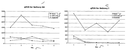
Figure 6. Quantitative PCR (qPCR) to quantify the salivary GAPDH and IL-8.
Detailed Descri-ption of the Preferred Embodiment
The present invention concerns the early detection, diagnosis, and prognosis
of
human disease states. Markers of a disease state, in the form of expressed RNA
molecules
of specified sequences or polypeptides expressed from these RNA molecules from
the
saliva of individuals with the disease state, are disclosed. These markers are
indicators of
the disease state and, when differentially expressed relative to expression in
a normal
subject, are diagnostic for the presence of the disease state in patients.
Such marlcers
provide considerable advantages over the prior art in this field. Since they
are detected in
saliva samples, it is not necessary to suspect that an individual exhibits the
disease state
(such as a tumor) before a sample may be taken, and in addition, the drawing
of a saliva
sample is much less invasive and painful to the patient than tissue biopsy or
blood drawing.
The detection methods disclosed are thus suitable for widespread screening of
asymptomatic individuals.
-4-
CA 02574706 2007-01-19
WO 2006/020005 PCT/US2005/025138
EXAMPLE 1
RNA Profiling Of Cell-Free Saliva Usint! Microarray Technolo%!y
Materials & Methods
Normal Subjects
Saliva samples were obtained from ten normal donors from the Division of
Otolaryngology, Head and Neck Surgery, at the Medical Center, University of
California,
Los Angeles (UCLA), CA, in accordance with a protocol approved by the UCLA
Institutional Review Board. The following inclusion criteria were used: age >
30 years; no
history of malignancy, immunodeficiency, autoimmune disorders, hepatitis, HIV
infection
or smoking. The study population was composed of 6 males and 4 females, with
an
average age of 42 years (range from 32 to 55 years).
Saliva Collection and Processing
Saliva samples were collected between 9 ain and 10 am in accordance with
published protocols (Navazesh, M. 1993 Ann N Y Acad Sci 694:72-77). Subjects
were
asked to refrain from eating, drinking, smoking or oral hygiene procedures for
at least one
hour prior to saliva collection. Saliva samples were centrifuged at 2,600 x g
for 15 min at
4 C. Saliva supernatant was separated from the cellular phase. RNase inhibitor
(Superase-
In, Ambion Inc., Austin, TX, USA) and protease inhibitor (Aprotinin, Sigma,
St. Louis,
MO, USA) were then added into the cell-free saliva supernatant.
RNA Isolation from Cell-fi ee Saliva
RNA was isolated from cell-free saliva supernatant using the modified protocol
from the manufacturer (QIAamp Viral RNA kit, Qiagen, Valencia, CA, USA).
Saliva (560
L), mixed well with AVL buffer (2,240 L), was incubated at room temperature
for 10
min. Absolute ethanol (2,240 L) was added and the solution passed through
silica
columns by centrifugation at 6,000 x g for 1 min. The columns were then washed
twice,
centrifuged at 20,000 x g for 2 min, and eluted with 30 L Rnase-free water at
9,000 x g for
2 min. Aliquots of RNA were treated with RNase-free DNase (DNase I-DNA-free,
Ambion Inc., Austin, TX, USA) according to the manufacturer's instructions.
The quality
of isolated RNA was examined by RT-PCR for three house-keeping gene
transcripts:
glyceraldehyde-3- phosphate dehydrogenase (GAPDH), actin-(3 (ACTB) and
ribosomal
protein S9 (RPS9). Primers were designed using PRIMER3 software
(genome.wi.mit.edu)
and were synthesized commercially (Fisher Scientific, Tustin, CA, USA) as
follows: 5'
TCACCAGGGCTGCTTTTAACTC3' (SEQ ID NO: 1) and
-5-
CA 02574706 2007-01-19
WO 2006/020005 PCT/US2005/025138
5'ATGACAAGCTTCCCGTTCTCAG3' (SEQ ID NO: 2) for GAPDH;
5'AGGATGCAGAAGGAGATCACTG3' (SEQ ID NO: 3) and
5'ATACTCCTGCTTGCTGATCCAC3' (SEQ ID NO: 4) for ACTB;
5'GACCCTTCGAGAAATCTCGTCTC3' (SEQ ID NO: 5) and
5'TCTCATCAAGCGTCAGCAGTTC3' (SEQ ID NO: 6) for RPS9. The quantity of RNA
was estimated using Ribogreen RNA Quantitation Kit (Molecular Probes, Eugene,
OR,
USA).
Target cRNA Preparation
Isolated RNA was subjected to linear amplification according to published
method
(Ohyama, H. et al. 2000 Biotechniques 29:530-536). In brief, reverse
transcription using
T7-oligo-(dT)24 as the primer was performed to synthesize the first strand
cDNA. The first
round of in vitro transcription (IVT) was carried out using T7 RNA polymerase
(Ambion
Inc., Austin, TX, USA). The BioArrayTM High Yield RNA Transcript Labeling
System
(Enzo Life Sciences, Farmingdale, NY, USA) was used for the second round IVT
to
biotinylate the cRNA product; the labeled cRNA was purified using GeneChip
Sample
Cleanup Module (Affymetrix, Santa Clara, CA, USA). The quantity and quality of
cRNA
were determined by spectrophotometry and gel electrophoresis. Small aliquots
from each
of the isolation and amplification steps were used to assess the quality by RT-
PCR. The
quality of the fragmented cRNA (prepared as described by Kelly, J.J. et al.
2002 Anal
Biochein 311:103-118) was assessed by capillary electrophoresis using the 2100
Bioanalyzer (Agilent Technologies, Palo Alto, CA, USA).
HG-U133A Microarray Analysis
The Affymetrix Human Genome U133A Array, which contains 22,215 human gene
cDNA probe sets representing approximately 19,000 genes (i.e., each gene may
be
represented by more than one probe sets), was applied for gene expression
profiling. The
array data were normalized and analyzed using Microarray Suite (MAS) software
(Affymetrix). A detection p-value was obtained for each probe set. Any probe
sets with p
< 0.04 was assigned "present", indicating the matching gene transcript is
reliably detected
(Affymetrix, 2001). The total number of present probe sets on each array was
obtained and
the present percentage (P%) of present genes was calculated. Functional
classification was
performed on selected genes (present on all ten arrays, p < 0.01) by using the
Gene
Ontology Mining Tool (netaffx.com).
-6-
CA 02574706 2007-01-19
WO 2006/020005 PCT/US2005/025138
Quantitative Gene Expression Analysis by Q-PCR
Q-PCR was performed using iCyclerTM thermal Cycler (Bio-Rad, Hercules, CA,
USA). A 2 L aliquot of the isolated salivary RNA (without amplification) was
reverse
transcribed into cDNA using MuLV Reverse Transcriptase (Applied Biosystems,
Foster
City, CA, USA). The resulting cDNA (3 L) was used for PCR amplification using
iQ
SYBR Green Supermix (Bio-Rad, Hercules, CA, USA). The primers were synthesized
by
Sigma-Genosys (Woodlands, TX, USA) as follows: 5'
GTGCTGAATGTGGACTCAATCC3' (SEQ ID NO: 7) and 5'
ACCCTAAGGCAGGCAGTTG3' (SEQ ID NO: 8) for interleukin 1-beta (IL1B); 5'
CCTGCGAAGAGCGAAACCTG 3' (SEQ ID NO: 9) and 5'
TCAATACTGGACAGCACCCTCC 3' (SEQ ID NO: 10) for stratifin (SFN); 5'
AGCGTGCCTTTGTTCACTG 3' (SEQ ID NO: 11) and 5'
CACACCAACCTCCTCATAATCC 3' (SEQ ID NO: 12) for tubulin-alpha, ubiquitous (K-
ALPHA-1). All reactions were performed in triplicate with conditions
customized for the
specific PCR products. The initial aniount of cDNA of a particular template
was
extrapolated from a standard curve using the LightCycler software 3.0 (Bio-
Rad, Hercules,
CA, USA). The detailed procedure for quantification by standard curve has been
previously described (Ginzinger, D. 2002 Exp Heinatol 30:503-512).
Results
RNA Isolation and Aynplification
On average, 60.5 13.1 ng (n=10) of total RNA was obtained from 560 L cell-
free
saliva samples (Table 1). RT-PCR results demonstrated all 10 saliva samples
contain
mRNAs that encode for house keeping genes: GAPDH, ACTB and RPS9. The mRNA of
these genes could be preserved without significant degradation for more than 6
months at -
80oC (Fig. 1). After two rounds of T7 RNA linear amplification, the average
yield of
biotinylated cRNA was 42.2 3.9 g with A260/280=2.067 (Tablel).
-7-
CA 02574706 2007-01-19
WO 2006/020005 PCT/US2005/025138
Tablel. Gene expression profiling in cell-free saliva obtained from ten normal
donors
Subject Gender Age RNA cRNA Present Probes' Probe P d
(ng) (Fig)
1 F 53 60.4 44.3 3172 14.24
2 M 42 51.6 40.8 2591 11.62
3 M 55 43.2 34.8 2385 10.70
4 M 42 48.2 38.0 2701 12.12
M 46 60.6 42.7 3644 16.35
6 M 48 64.8 41.8 2972 13.34
7 F 40 75.0 44.3 2815 12.63
8 M 33 77.8 49.3 4159 18.66
9 F 32 48.8 41.4 2711 12.17
F 32 79.8 44.4 4282 19.22
Mean n 42 8.3 60.5~13.1 42.2~3.94 3143 665.0 14.11 2.98
aTotal RNA quantity in 560 L cell-free saliva supernatant
bThe cRNA quantity after two rounds of T7 amplification
cNumber of probes showing present call on HG U133A microarray (detection
5 p<0.04)
dPresent percentage (P%) = Number of probes assigned present call / Number of
total probes (22,283 for HG U133A microarray)
[0024] The cRNA ranged from 200 bp to 4 kb before fragmentation; and was
concentrated to approximately 100 bp after fragmentation. The quality of cRNA
probe was
10 confirmed by capillary electrophoresis before the hybridizations. ACTB mRNA
was
detectable using PCR/RT-PCR on original sample and products from each
amplification
steps: first cDNA, first in vitro transcription (NT), second cDNA and second
NT (Fig. 2).
Microarray Profiling of Salivary mRNA
[0025] Salivary mRNA profiles of ten normal subjects were obtained using HG
U133A array which contains 22,283 cDNA probes. An average of 3,143 665.0
probe sets
(p < 0.04) were found on each array (n=10) with assigned present calls. These
probe sets
represent approximately 3,000 different mRNAs. The average present call
percentage was
14.11 2.98% (n=10). A reference database which includes data from the ten
arrays was
generated. The probe sets representing GAPDH, ACTB and RPS9 assigned present
calls
on all 10 arrays. There were totally 207 probe sets representing 185 genes
assigned present
calls on all 10 arrays with detection p < 0.01. These genes were categorized
on the basis of
their known roles in biological processes and molecular functions (Table 2).
The major
functions of the 185 genes are related to cell growth/maintenance (119 genes),
molecular
-8-
CA 02574706 2007-01-19
WO 2006/020005 PCT/US2005/025138
binding (118 genes) and cellular structure composition (95 genes). These were
termed as
"Normal Salivary Core Transcriptome (NSCT)".
Table 2: Biological processes and molecular functions of 185 genes in cell-
fiee saliva from
ten normal donors (data obtained by using Gene Ontology Mining Tool)
Biological processa Gennes, Molecular functiona Gene, nb
b
Cell growth and/or maintenance 119 Binding 118
Metabolism 93 Nucleic acid binding 89
Biosynthesis 70 RNA binding 73
Protein metabolism 76 Calcium ion binding 12
Nucleotide metabolism 10 Other binding 23
Other metabolisms 18
Cell organization and biogenesis 2 Structural molecule 95
Homeostasis 3 Ribosomal constituent 73
Cell cycle 5 Cytoskeleton constituent 17
Cell proliferation 11 Muscle constituent 2
Transport 5
Cell motility 8 Obsolete 15
Transporter 4
Cell communication 34 Enzyme 20
Response to external stimulus 19 Signal transduction 10
Cell adhesion 3 Transcription regulator 7
Cell-cell signaling 5 Translation regulator 5
Signal transduction 17 Enzyme regulator 9
Cell adhesion molecule 1
Obsolete 8
Development 18 Molecular function unknown 6
Death 2
Biological process unknown 11
aOne gene may have multiple molecular functions or participate in different
biological processes.
bNumber of genes classified into a certain group/subgroup.
Q-PCR Validation and Quantitation Analysis
[0026] Real time quantitative PCR (Q-PCR) was used to validate the presence
of human mRNA in saliva by quantifying selected genes from the 185 "Normal
Salivary
Core Transcriptome" genes. IL1B, SFN and K-ALPHA-1 were randomly selected and
assigned present calls on all 10 arrays, for validation. Q-PCR results showed
that mRNA of
IL1B, SFN and K-ALPHA-1 were detectable in all 10 original, unamplified, cell-
free
saliva. The relative amounts (in copy number) of these transcripts (n=10)
were: 8.68 x 10³
~ 4.15 x 10³ for IL1B; 1.29 x 10⁵ 1.08 x10⁵ for SFN; and 4.71 x 10⁶ 8.37 x
105 for K-
-9-
CA 02574706 2007-01-19
WO 2006/020005 PCT/US2005/025138
ALPHA-1. The relative RNA expression levels of these genes measured by Q-PCR
were
similar to those measured by the microarrays.
[0027] Saliva meets the demands of an inexpensive, non-invasive and
accessible bodily fluid to act as an ideal diagnostic medium. Specific and
informative
biomarkers in saliva are greatly needed to serve for diagnosing disease and
monitoring
lluman health (Bonassi, S. et al. 2001 Mutat Res 480-481:349-358; Streckfus,
C.F. et al.
2002 Oral Dis 8:69-76; Sidransky, D. 2000 Nat Reviews 3:210-219). Knowing the
constituents in saliva is essential for using this medium to identify
potential biomarkers for
disease diagnostics (Pusch, W. et al. 2003 Pharnzacogenomics 4:463-476). Prior
to this
invention, one criticism was the idea that informative molecules are generally
present in
low amounts in saliva. However, with new amplification techniques and highly
sensitive
assays, this may no longer be a limitation (Xiang, C.C. et al 2003 Nucleic
Acids Res
31:e53). In the present Example, the human RNA was successfully isolated from
cell-free
saliva supernatant. The quality of salivary mRNA was proved to be sufficient
for use in
RT-PCR, Q-PCR and microarray experiments.
[0028] Distinct difference exists between saliva and other bodily fluids
(e.g.,
blood) in that saliva natarally contains microorganisms (Sakki, T. &
Knuuttila, M. 1996
Eur J Oral Sci 104:619-622). In addition, some extraneous substances (e.g.,
food debris)
make the composition of saliva more complex. Therefore, it is simpler and more
accurate
to use the fluid/supernatant phase of saliva, instead of the whole saliva as
medium for
detecting biomarkers. In this Example, the conditions for separating the
pellet and saliva
supernatant were optimized to avoid mechanical rupture of cellular elements
which would
contribute to the RNA detected in the fluidic cell-free phase (St. Jolm,
M.A.R. et al. 2004,
in press ). These results demonstrate that it is feasible and efficient to use
cell-free saliva
for transcriptome analysis. While it is a novel finding that human mRNAs exist
in cell-free
saliva supernatant, nucleic acids have long been detected in other cell-free
bodily fluids and
subsequently used for disease diagnostics (Sidransky, D. 1997 Science 278:1054-
1058).
For example, specific oncogene, tumor suppressor gene and microsatellite
alterations have
been identified in patients' serum (Anker, P. et al. 2003 Int J Cancer 103:149-
152).
Moreover, tumor nzRNAs have been isolated and amplified from serum of patients
with
different malignancies (Kopreski, M.S. et al. 1999 Clin Cancer Res 5:1961-
1965;
Fleischhacker, M. et al 2001 Ann NYAcad Sci 945:179-188). It has been widely
accepted
-10-
CA 02574706 2007-01-19
WO 2006/020005 PCT/US2005/025138
that these genomic messengers detected extracellularly can serve as biomarkers
for diseases
(Sidransky, D. 1997 Science 278:1054-1058).
[0029] To our knowledge, this is the first report where human mRNA in saliva
is globally profiled. Using microarray technology, we discovered that
approximately 3,000
different human mRNAs exist in cell-free saliva of each normal subject. The
salivary
transcriptome pattern in cell-free saliva from normal populations is
envisioned to serve as a
health-monitoring database. It should be noted that we now know the human
genome
composed of more than 30,000 genes (Venter J.C. et al. 2001 Science 291:1304-
1351) and
the probe sets on HG U133A microarray used in this Example represent only -
19,000
human genes, additional gene transcripts not detectable by the HG U133A
microarray, are
predicted to exist in the cell-free saliva and can be detected using our
invention. The
identified gene transcripts in this Example, particularly the Normal Salivary
Core
Transcriptome (NSCT) mRNAs, represent the comtnon transcriptome of normal cell-
free
saliva. We envision that different, informative and diagnostic transcriptome
can be
identified in saliva from patients with various disease conditions. Therefore,
human
salivary mRNA is envisioned to be used as diagnostic biomarkers for oral and
systemic
diseases that are manifested in the oral cavity.
[0030] In one embodiment of the invention the salivary transcriptome
diagnostics is used to monitor health of normal patients. In another
embodiment, the
salivary transcriptome diagnostics is used to detect markers for diseases for
early diagnosis
for cancers (e.g., prostate, colon, breast, lung, oral, etc.), as well as for
systemic diseases,
such as autoimmune diseases, diabetes, osteoporosis; neurological diseases,
such as
Alzheimer's disease, Parkinson's disease, etc.
EXAMPLE 2
Salivary Transcriptome Diagnostics for Oral Cancer Detection
[0031] Purpose: Oral fluid (saliva) meets the demand for non-invasive,
accessible and highly-efficient diagnostic medium. Our discovery that a large
panel of
human RNA can be reliably detected in saliva gives rise to a novel clinical
approach,
Salivary Transcriptome Diagnostics. In this Example we evaluate the diagnostic
value of
this new approach by using oral squamous cell carcinoma (OSCC) as the proof-of-
principle
disease.
[0032] It has been shown that identical mutation present in the primary tumor
can be identified in the bodily fluids tested from affected patients
(Sidransky, D. 1997
-11-
CA 02574706 2007-01-19
WO 2006/020005 PCT/US2005/025138
Science 278:1054-1059). Cancer related nucleic acids in blood, urine and
cerebrospinal
fluid (CSF) has been used as biomarkers for cancer diagnosis (Anker, P. et al
1999 Cancer
Metastasis Rev 18:65-73; Rieger-Christ, K.M. et al. 2003 Cancer 98:737-744;
Wong, L.J.
et al. 2003 Cancer Res 63:3866-3871). More recently, n1RNA biomarkers in serum
or
plasma have been targets for RT-PCR-based detection strategies in patients
with cancers
(Kopreski, M.S. et al. 2001 Ann N Y Acad Sci 945:172-178; Bunn, P.J., Jr. 2003
J Clin
Oncol 21:3891-3893). Parallel to the increasing number of such biomarkers in
bodily
fluids is the growing availability of technologies using more powerful and
cost-efficient
methods that enable mass screening for genetic alterations. Our discovery by
microarray
teclmology that a large panel of human mRNA exists in saliva (Example 1)
provides a
novel clinical approach, Salivary Transcriptome Diagnostics, for applications
in disease
diagnostics as well as for normal healtll surveillance. It is a high
throughput, robust and
reproducible approach to harness RNA signatures from saliva. Moreover, using
saliva as a
diagnostic fluid meets the demands for inexpensive, non-invasive and
accessible diagnostic
methodology (Lawrence, H.P. 2002 J Can Dent Assoc 68:170-174, 2002). In this
Example, we tested the hypothesis that distinct mRNA expression patterns can
be identified
in saliva from cancer patients, and the differentially expressed transcripts
can serve as
biomarkers for cancer detection. The proof-of-principle disease in this study
is oral
squamous cell carcinoma (OSCC). The rationale is that oral cancer cells are
iinrnersed in
the salivary milieu and genetic heterogeneity has been detected in saliva from
patients with
OSCC (El-Naggar, A.K et al. 2001 J Mol Diagn 3:164-170; Liao, P.H et al. 2000
Oral
Oncol 36:272-276, 2000).
[0033] Experimental Desi~n: Unstimulated saliva was collected from patients
(n=32) with primary T1/T2 OSCC and normal subjects (n=32) with matched age,
gender
and smoking history. RNA isolation was performed from the saliva supernatant,
followed
by two-round linear amplification using T7 RNA polymerase. Human Genome U133A
microarrays were applied for profiling human salivary transcriptome. The
different gene
expression patterns were analyzed by combining a t test comparison and a fold-
change
analysis on ten matched cancer patients and controls. Quantitative PCR (qPCR)
was used
to validate the selected genes that showed significant difference (P<0.01) by
microarray.
The predicting power of these salivary mRNA biomarkers were analyzed by
receiver
operating characteristic curve and classification models.
-12-
CA 02574706 2007-01-19
WO 2006/020005 PCT/US2005/025138
[0034] Results: Microarray analysis showed 1,679 genes which exhibited
significantly different expression level in saliva between cancer patients and
controls
(P<0.05). Seven cancer-related RNA biomarkers, that exhibited at least 3.5-
fold elevation
in OSCC saliva (P<0.01), were consistently validated by qPCR on saliva samples
from
OSCC patients (n=32) and controls (n=32). These salivary RNA biomarkers are
transcripts
of interleukin 8(IL-8), interleukin 1-beta (IL1B), dual specificity
phosphatase 1(DUSP1),
H3 histone, family 3A (HA3A), ornithine decarboxylase antizyme 1 (OAZ1), S100
calcium
binding protein PS(100P) and spermidine/spermine N1-acetyltransferase (SAT).
The
combinations of these biomarkers yielded sensitivity (91%) and specificity
(91%) in
distinguishing OSCC from the controls.
[0035] Conclusions: The utility of salivary transcriptome diagnostics was
successfully demonstrated in this study for oral cancer detection. This novel
clinical
approach is envisioned as a robust, high-throughput and reproducible tool for
early cancer
detection. Salivary transcriptome profiling is envisioned to be applied to
evaluate other
major diseases as well as normal health surveillance.
Patients And Methods
[0036] Patient Selection. Oral squamous cell carcinoma (OSCC) patients were
recruited from Medical Centers at University of California, Los Angeles
(UCLA);
University of Southern California (USC), Los Angeles, CA; and University of
California
San Francisco (UCSF), San Francisco, CA. Thirty-two patients with documented
primary
T1 or T2 OSCC were included in this study. All patients had recently been
diagnosed with
primary disease, and had not received any prior treatment in the form of
chemotherapy,
radiotherapy, surgery, or alternative remedies. An equal number of age and sex
matched
subjects with comparable smoking histories were selected as a control group
(St. John,
M.A.R et al. 2004 IL-6 and IL-8: Potential Biomarkers for Oral Cavity and
Oropharyngeal
SCCA. Archives of Otolaryngology-Head & Neck Surgery, in press). Among the two
subject groups, there were no significant differences in terms of mean age:
OSCC patients,
49.8+7.6 years; normal subjects, 49.1 5.9 years (Student's t test P > 0.80);
gender (P >
0.90); or smoking history (P > 0.75). No subjects had a history of prior
malignancy,
immunodeficiency, autoimmune disorders, hepatitis, or HIV infection. All
subjects signed
the Institutional Review Board approved consent form agreeing to serve as
saliva donors
for the experiments.
-13-
CA 02574706 2007-01-19
WO 2006/020005 PCT/US2005/025138
[0037] Saliva collection and RNA isolation. Unstimulated saliva samples were
collected between 9 am and 10 am with previously established protocols
(Navazesh, M.
1993 Ann N YAcad Sci 694:72-77). Subjects were asked to refrain from eating,
drinking,
smoking or oral hygiene procedures for at least one hour prior to the
collection. Saliva
samples were centrifuged at 2,600 x g for 15 min at 4 C. The supematant was
removed
from the pellet and treated with RNase inhibitor (Superase-In, Ambion Inc.,
Austin, TX).
RNA was isolated from 560 1 of saliva supematant using QlAamp Viral RNA kit
(Qiagen,
Valencia, CA). Aliquots of isolated RNA were treated with RNase-free DNase
(DNasel-
DNA-free, Ambion Inc., Austin, TX) according to the manufacturer's
instructions. The
quality of isolated RNA was examined by RT-PCR for three cellular maintenance
gene
transcripts: glyceraldehyde-3-phosphate dehydrogenase (GAPDH), actin-(3 (ACTB)
and
ribosomal protein S9 (RPS9). Only those samples exhibiting PCR products for
all three
genes were used for subsequent analysis.
[0038] Microarray analysis. Saliva from ten OSCC patients (7 male, 3 female,
age=52 9.0) and from ten gender and age matched normal donors (age=49 5.6)
was
used for microarray study. Isolated RNA from saliva was subjected to linear
amplification
by RiboAmpTM RNA Amplification kit (Arcturus, Mountain View, CA). The RNA
amplification efficiency was measured by using control RNA of known quantity
(0.1 g)
running in parallel with the 20 samples in five independent runs. Following
protocols
described in Example 1, the Affymetrix Human Genome U133A Array (HG U133A,
Affymetrix, Santa, Clara, CA) was applied for gene expression analysis.
[0039] The arrays were scanned and the fluorescence intensity was measured by
Microarray Suit 5.0 software (Affimetrix, Santa Clara, CA) and then were
imported into
DNA-Chip Analyzer software for normalization and model-based analysis (Li, C.
& Wong,
W.H. 2001 PNAS USA 98:31-36). S-plus 6.0 (Insightful, Seattle, WA) was used to
carry
out all statistical tests. Three criteria were used to determine
differentially expressed
transcripts. First, we excluded probe sets on the array that were assigned as
"absent" call in
all sainples. Second, a two-tailed student's t test was used for comparison of
average gene
expression signal intensity among the OSCCs (n=10) and controls (n=10). The
critical
alpha level of 0.05 was defined for statistical significance. Third, fold
ratios were
calculated for those gene transcripts that showed statistically significant
difference (P <
0.05). Only those gene transcripts that exhibited at least 2-fold change will
be included for
further analysis.
-14-
CA 02574706 2007-01-19
WO 2006/020005 PCT/US2005/025138
[0040] Quantitative PCR validation. qPCR was performed to validate a subset
of differently expressed transcripts identified by microarray analysis. Using
MuLV reverse
transcriptase (Applied Biosystems, Foster City, CA) and random hexamers as
primer (ABI,
Foster City, CA), we synthesized cDNAs from the original and un-amplified
salivary RNA.
The qPCR reactions were performed in an iCyclerTM PCR system (Bio-Rad,
Hercules, CA,
USA), iQ SYBR Green Supermix (Bio-Rad, Hercules, CA). Primer sets were
designed by
using PRIMER3 software. All of the reactions were performed in triplicate with
customized conditions for specific products. The initial amount of cDNA/RNA of
a
particular template was extrapolated from the standard curve as described
previously
(Ginzinger, D.G. 2002 Exp Hematol 30:503-512). This validation completed by
testing all
of the samples (n = 64) including those 20 previously used for inicroarray
study. Wilcoxon
Signed Rank test was used for statistical analysis.
[0041] Receiver operating characteristic (ROC) curve analysis and prediction
models. Utilizing the RT-qPCR results, ROC curve analyses (Grunkemeier, G.L. &
Jin, R.
2001 Ann Thorac Surg 72:323-326) were conducted by S-plus 6.0 to evaluate the
predictive
power of each of the biomarkers. The optimal cutpoint was determined for each
biomarker
by searching for those that yielded the maximum corresponding sensitivity and
specificity.
ROC curves were then plotted on the basis of the set of optimal sensitivity
and specificity
values. Area under the curve was coinputed via numerical integration of the
ROC curves.
The biomarker that has the largest area under the ROC curve was identified as
having the
strongest predictive power for detecting OSCC.
[0042] Next, multivariate classification models were constructed to determine
the best combination of salivary markers for cancer prediction. Firstly, using
the binary
outcome of the disease (OSCC) and non-disease (normal) as dependent variables,
a logistic
regression model was constructed controlling for patient age, gender, and
smoking history.
The backward stepwise regression (Renger, R. & Meadows, L.M. 1994 Acad Med
69:738)
was used to find the best final model. Leave-one out cross validation was used
to validate
the logistic regression model. The cross validation strategy first removes one
observation
and then fits a logistic regression model from the remaining cases using all
markers.
Stepwise model selection was used for each of these models to remove variables
that do not
improve the model. Subsequently, the marker values were used for the case that
was left
out to compute a predicted class for that observation. The cross validation
error rate was
then the number of samples predicted incorrectly divided by the number of
samples. The
-15-
CA 02574706 2007-01-19
WO 2006/020005 PCT/US2005/025138
ROC curve was then computed for the logistic model by a similar procedure,
using the
fitted probabilities from the model as possible cut-points for computation of
sensitivity and
specificity.
[0043] Secondly, a tree-based classification model, classification and
regression
trees (CART), was constructed by S-plus 6.0 using the validated mRNA
biomarkers as
predictors. CART fits the classification model by binary recursive
partitioning, in which
each step involves searching for the predictor variable that results in the
best split of the
cancer versus the normal groups (Lemon, S.C. et al. 2003 Ann Behav Med 26:172-
181).
CART used the entropy function with splitting criteria determined by default
settings for S-
plus. By this approach, the parent group containing the entire samples (n=64)
was
subsequently divided into cancer groups and normal groups. The initial tree
was pruned to
remove all splits that did not result in sub-branches with different
classifications.
Results
[0044] On average, 54.2 :L 20.1 ng (n=64) of total RNA was obtained from 560
l saliva supernatant. There was no significant difference in total RNA
quantity between
the OSCC and the age and gender matched controls (t test, P = 0.29, n =64). RT-
PCR
results demonstrated that all of the saliva samples (n=64) contain transcripts
from three
genes (GAPDH, ACTB and RPS9), which were used as quality controls for human
salivary
RNAs (see Example 1). A consistent amplifying magnitude (658 47.2, n=5)
could be
obtained after two rounds of RNA amplification. On average, the yield of
biotinylated
cRNA was 39.3 6.0 g (n=20). There were no significant differences of the
cRNA
quantity yielded between the OSCC and the controls (t test, P = 0.31, n =20).
[0045] The HG U133A microarrays were used to identify the difference in
salivary profiles RNA between cancer patients and matched normal subjects.
Among the
10,316 transcripts included by the previously described criteria, 1,679
transcripts with P
value less than 0.05 were identified . Among these transcripts, 836 were up-
regulated and
843 were down-regulated in the OSCC group. These transcripts observed were
unlikely to
be attributable to chance alone (x2 test, P<0.0001) considering the false
positives using
P<0.05. Using a predefined criteria of a change in regulation > 3-fold in all
10 OSCC
saliva specimens, and a more stringent cutoff of P value < 0.01, we identified
17 transcripts
as presented in Table 3. These 17 salivary mRNAs were all up-regulated in OSCC
saliva,
whereas there were no mRNAs found down-regulated using the same filtering
criteria. The
biological functions of these genes are presented in Table 3.
-16-
CA 02574706 2007-01-19
WO 2006/020005 PCT/US2005/025138
Table 3. Salivary mRNA up-regulated (> 3-fold, P < 0.01) in OSCC identified by
microarray.
Gene Gene Name GenBank Locus Gene functions
Symbol Acc. No.
B2M Beta-2-microglobulin NM_004048 15q21- anti-apoptosis, antigen
q22.2 presentation
DUSPI Dual specificity NM_004417 5q34 protein modification,
phosphatase 1 signal transduction,
oxidative stress
FTHI Ferritin, heavy NM_002032 11q13 iron ion transport, cell
olype tide 1 proliferation
GOS2 Putative lymphocyte NM_015714 1q32.2- cell growth and/or
G0/Gl switch gene q41 maintenance,
regulation of cell
cycle
GADD45 Growth arrest and NM_015675 19p13.3 kinase cascade,
B DNA-damage- apoptosis
inducible, beta
H3F3A H3 histone, family 3A BE869922 1 41 DNA binding activity
HSPCO16 Hypothetical protein BG167522 3p21.31 unknown
HSPCO16
IER3 Immediate early NM_003897 6p2l.3 embryogenesis,
response 3 morphogenesis,
apoptosis, cell growth
and maintenance
IL1B Interleukin 1, beta M15330 2q14 signal transduction,
proliferation,
inflammation,
apoptosis
ILS Interleukin 8 NM_000584 4ql3-q21 angiogenesis,
replication, calcium-
mediated signaling
pathway, cell
adhesion, chemotaxis,
cell cycle arrest,
immune response
MAP2K3 Mitogen-activated AA780381 17q11.2 signal transduction,
protein kinase kinase 3 protein modification
OAZI Omithine D87914 19p13.3 polyamine
decarboxylase biosynthesis
antizyme 1
PRGI Proteoglycan 1, NM_002727 10q22.1 proteoglycan
secretory granule
RGS2 Regulator of G-protein NM_002923 1q31 oncogenesis, g-protein
signaling 2, 24 kDa signal transduction
SIOOP S100 calcium binding NM_005980 4p16 protein binding,
protein P calcium ion binding
-17-
CA 02574706 2007-01-19
WO 2006/020005 PCT/US2005/025138
Gene Gene Name GenBank Locus Gene functions
Symbol Acc. No.
SAT Spermidine/spermine NM_002970 Xp22.1 enzyme, transferase
N1-acetyltransferase activity
EST, Highly similar BG537190 iron ion homeostasis,
Ferritin light chain ferritin complex
The human Genome U133A microarrays were used to identify the difference in
RNA expression patterns in saliva from ten cancer patients and ten matched
normal
subjects. Using a criteria of a change in regulation >3-fold in all OSCC
saliva specimens,
and a cutoff of P value <0.01, 17 mRNA were identified, showing significant up-
regulation
in OSCC saliva
[0046] Quantitative PCR was performed to validate the microarray findings on
an enlarged sample size including saliva from 32 patients with OSCC and 32
matched
controls. Nine candidates of salivary mRNA biomarkers: DUSPI, GADD45B, H3F3A,
ILIB, IL8, OAZ1, RGS2, SIOOP and SAT were selected based on their reported
cancer
association (Table 3). Table 4 presents their quantitative alterations in
saliva from OSCC
patients determined by qPCR. The results confirmed that transcripts of 7 of
the 9 candidate
mRNA (78%), DUSP1, H3F3A, IL1B, IL8, OAZ1, SIOOP and SAT, were significantly
elevated in the saliva of OSCC patient (Wilcoxon Signed Rank test, P < 0.05).
The
statistically significant differences in the amount of RGS2 (P = 0.149) and
GADD45B (P =
0.116) by qPCR was not detected. The validated seven genes could be classified
in three
ranks by the magnitude of increase: high up-regulated mRNA including IL8 (24.3-
fold);
moderate up-regulated mRNA including H3F3A (5.61-fold), ILIB (5.48) and SlOOP
(4.88-
fold); and low up-regulated mRNA including DZISPl (2.60-fold), OAZ1 (2.82-
fold) and
SAT (2.98-fold). The detailed statistics of the area under the receiver
operator
characteristics (ROC) curves, the threshold values, and the corresponding
sensitivities and
specificities for each of the seven potential salivary mRNA biomarkers for
OSCC are listed
in Table 5. The data showed IL-8 mRNA performed the best among the seven
potential
biomarkers for predicting the presence of OSCC. The calculated area under the
ROC curve
for IL-8 was 0.85. With a tlireshold value of 3.19E-18 mol/L, IL-8 mRNA in
saliva yields a
sensitivity of 88% and a specificity of 81% to distinguish OSCC from the
normal.
-18-
CA 02574706 2007-01-19
WO 2006/020005 PCT/US2005/025138
Table 4. Quantitative PCR validation of selected 9 transcripts in saliva
(n=64)a
Gene Mean
s mbol Primer sequence (5' to 3') Validated value fold
y increase
F: CCTACCAGTATTATTCCCGACG (SEQ ID NO: 13)
DUSP1 R: TTGTGAAGGCAGACACCTACAC (SEQ ID NO: Yes 0.039 2.60
14)
F: AAAGCACCCAGGAAGCAAC (SEQ ID NO: 15)
H3F3A R: GCGAATCAGAAGTTCAGTGGAC (SEQ ID NO: Yes 0.011 5.61
16)
F: GTGCTGAATGTGGACTCAATCC (SEQ ID NO: 17)
IL1B Yes 0.005 5.48
R: ACCCTAAGGCAGGCAGTTG (SEQ ID NO: 18)
F: GAGGGTTGTGGAGAAGTTTTTG (SEQ ID NO: 19)
R,8 Yes 0.000 24.3
R: CTGGCATCTTCACTGATTCTTG (SEQ ID NO: 20)
F: AGAGAGAGTCTTCGGGAGAGG (SEQ ID NO: 21)
OAZ1 Yes 0.009 2.82
R: AGATGAGCGAGTCTACGGTTC (SEQ ID NO: 22)
F: GAGTTCATCGTGTTCGTGGCTG (SEQ ID NO: 23)
S100P Yes 0.003 4.88
R: CTCCAGGGCATCATTTGAGTCC (SEQ ID NO: 24)
F: CCAGTGAAGAGGGTTGGAGAC (SEQ ID NO:25)
SAT Yes 0.005 2.98
R: TGGAGGTTGTCATCTACAGCAG (SEQ ID NO: 26)
GADD4 F: TGATGAATGTGGACCCAGAC (SEQ ID NO: 27)
No 0.116
SB
R: GAGCGTGAAGTGGATTTGC (SEQ ID NO: 28)
F: CCTGCCATAAAGACTGACCTTG (SEQ ID NO: 29)
RGS2 No 0.149
R: GCTTCCTGATTCACTACCCAAC (SEQ ID NO: 30)
qPCR were performed to validate the microarray findings on an enlarged sample
size including saliva from 32 patients with OSCC and 32 matched control
subjects. Nine
potential salivary mRNA biomarkers were selected from the 17 candidates shown
in Table
3. Seven of them were validated by qPCR (P<0.05). Sample includes 32 saliva
from
OSCC patients and 32 from matched normal subjects.
Wilcoxon's Signed Rank test: if P < 0.05, validated (Yes); if P> 0.05, not
validated
(No).
-19-
CA 02574706 2007-01-19
WO 2006/020005 PCT/US2005/025138
Table 5. Receiver operator characteristic (ROC) curve analysis of OSCC
associated
salivary mRNA biomarkers
Biomarker Area under Threshold/Cutof Sensitivity Specificity Selected
ROC Curve f (M) (%) / References
DUSP1 0.65 8.35E-17 59 75 (34)
H3F3A 0.68 1.58E-15 53 81 (54)
IL1B 0.70 4.34E-16 63 72 (44)
1L8 0.85 3.19E-18 88 81 (55)
OAZ1 0.69 7.42E-17 100 38 (37)
S 100P 0.71 2.11E-15 72 63 (40)
SAT 0.70 1.56E-15 81 56 (35)
Utilizing the qPCR results, we conducted ROC curve analyses to evaluate the
predictive power of each of the biomarkers. The optimal cutpoint was
determined yielding
the maximum corresponding sensitivity and specificity. The biomarker that has
the largest
area under the ROC curve was identified as having the strongest predictive
power for
detecting OSCC.
To demonstrate the utility of salivary mRNAs for disease discrimination, two
classification/prediction models were examined. A logistic regression model
was built
based on the four of the seven validated biomarkers, IL1B, OAZ1, SAT and IL-8,
which in
combination provided the best prediction (Table 6). The coefficient values
were positive
for these four markers, indicating that the synchronized rise in their
concentrations in saliva
increased the probability that the sample was obtained from an OSCC subject.
The leave-
one-out cross-validation error rate based on logistic regression models was
19% (12/64).
All but one (out of the 64) of the models generated in the leave-one-out
analysis used the
same set of four markers found to be significant in the full data model
specified in Table 6.
The ROC curve was computed for the logistic regression model. Using a cutoff
probability
of 50%, a sensitivity of 91% and a specificity of 91% were obtained. The
calculated area
under the ROC curve was 0.95 for the logistic regression model (Figure 3).
-20-
CA 02574706 2007-01-19
WO 2006/020005 PCT/US2005/025138
Table 6. Salivary mRNA biomarkers for OSCC selected by logistic regression
model
Biomarker Coefficient Value Standard Error P value
Intercept -4.79 1.51 0.001
ILIB 5.10E+19 2.68E+19 0.062
OAZ1 2.18E+20 1.08E+20 0.048
SAT 2.63E+19 1.10E+19 0.020
IL-8 1.36E+17 4.75E+16 0.006
The logistic regression model was built based on the four of seven validated
biomarkers (IL1B, OAZ1, SAT and IL-8) that, in combination, provided the best
prediction.
The coefficient values are positive for these four markers, indicating that
the synchronized
increase in their concentration in saliva increases the probability that the
sample was
obtained from an OSCC subject.
A second model, the "classification and regression trees (CART) model", was
generated (Figure 4). The fitted CART model used the salivary mRNA
concentrations of
IL-8, H3F3A and SAT as predictor variables for OSCC. IL-8, chosen as the
initial split, with
a threshold of 3.14E-18 mol/L, produced two child groups from the parent group
containing
the total 64 samples. 30 samples with the IL-8 concentration < 3.14E-18 mol/L
were
assigned into "Normal-1" group, whereas 34 with IL-8 concentration > 3.14E-18
mol/L
were assigned into "Cancer-1". The "Normal-1" group was further partitioned by
SAT with
a threshold of 1.13E-14 mol/L. The resulting subgroups: "Normal-2", contained
25
samples with SAT concentration < 1.13E-14 mol/L; and "Cancer-2", contained 5
samples
with SAT concentration _ 1.13E-14 mol/L. Similarly, the "Cancer-1" group was
further
partitioned by H3F3A with a threshold of 2.07E-16 mol/L. The resulting
subgroups:
"Cancer-3", contained 27 samples with H3F3A concentration _ 2.07E-16 mol/L;
and
"Normal-3" group, contained 7 samples with H3F3A concentration < 2.07E-16
mol/L.
Consequently, the 64 saliva samples involved in this study were classified
into the
"Cancer" group and the "Normal" group by CART analysis. The "Normal" group was
composed of the samples from "Normal-2" group and those from "Normal-3" group.
There
were a total of 32 samples assigned in the "Normal" group, 29 from normal
subjects and 3
from cancer patients. Thus, by using the combination of IL-8, SAT, and H3F3A
for OSCC
prediction, the overall sensitivity is 90.6% (29/32). The "Cancer" group was
composed of
the samples from "Cancer-2" group and "Cancer-3" group. There were a total of
32
samples assigned in the final "Cancer" group, 29 from cancer patients and 3
from normal
-21-
CA 02574706 2007-01-19
WO 2006/020005 PCT/US2005/025138
subjects. Therefore, by using the combination of these three salivary mRNA
biomarkers for
OSCC prediction, the overall specificity is 90.6% (29/32).
[0049] The goal of a cancer-screening program is to detect tumors at a stage
early enough that treatment is likely to be successful. Screening tools are
needed that
exhibit the combined features of high sensitivity and high specificity.
Moreover, the
screening tool must be sufficiently noninvasive and inexpensive to allow
widespread
applicability. Significant development of biotechnology and improvement in our
basic
understanding of the cancer initiation and progression now enable to identify
tumor
signatures, such as oncogenes and tuinor-suppressor gene alterations, in
bodily fluids that
drain from the organs affected by the tumor (Sidransky, D. 1997 Science
278:1054-1059).
The results presented in this Example show that salivary transcriptome
diagnostics is a
suitable tool for the development of noninvasive diagnostic, prognostic and
follow-up tests
for cancer.
[0050] Previous studies have shown that human DNA biomarkers can be
identified in saliva and used for oral cancer detection (El-Naggar, A.K et al.
2001 J Mol
Diagn 3:164-170; Liao, P.H. et al. 2000 Oral Oncol 36:272-276). The presence
of human
mRNA in saliva expands the repertoire of diagnostic analytes for translational
and clinical
applications. However, RNA is more labile than DNA and is presumed to be
highly
susceptible to degradation by RNases. Furthermore, RNase activity in saliva is
reported to
be elevated in patients with cancer (Kharchenko, S.V. & Shpakov, A.A. 1989 Izv
Akad
Nauk SSSR Biol 58-63). It has thus been commonly presumed that human mRNA
could not
survive extracellularly in saliva. Surprisingly, using RT-PCR, the inventors
consistently
detected human mRNA in saliva, thus opening the door to saliva-based
expression
profiling. Using the described collection and processing protocols, the
presence of control
RNAs was confirmed in all saliva (patients and controls) by RT-PCR/qPCR. The
quality of
RNA could meet the demand for PCR, qPCR and microarray assays. In this
Example, we
employed prompt addition of RNase inhibitors to freshly collected oral fluids
followed by
ultra low temperature storage (-80 C).
[0051] Our reported findings will bring substantial interests to the field of
cancer and disease diagnostics. The interests stem not only from the fact that
a saliva-based
diagnostic and screening test for cancer is a simple and attractive concept,
but also from the
fact that conventional diagnostic cancer tests tend to be imperfect. Using
oral cancer as an
example, the clearly disappointing survival rate may most probably attribute
to diagnostic
-22-
CA 02574706 2007-01-19
WO 2006/020005 PCT/US2005/025138
delay (Wildt, J. et al. 1995 Clin Otolaiyngol 20:21-25). Since most oral
cancers arise as
asymptomatic small lesions at their early stage, only when the clinician or
patient notes
abnormal tissues do formal diagnosis procedures begin (Epstein, J.B. et al.
2002 J Can
Dent Assoc 68:617-621). Microscopic level for the progressive cancer is often
too late for
successful intervention (Fong, K.M. et al. 1999 in: In: S. S. HD and G. AF
(eds.),
Molecular Pathology of Early Cancer, pp. 13-26: IOS Press). It is also
impractical to use
imaging techniques for cancer screening, since they are time-consuming and
expensive.
These techniques are typically used for confirmation because of their
insensitivity for small
lesions (Myers, L.L. & Wax, M.K. 1998 J Otolaiyngol 27:342-347). Studies have
demonstrated that good positive predictive value can be achieved by oral
cancer tissue
staining with toluidine blue (Mashberg, A. & Samit, A. 1995 CA Cancef J Clin
45:328-
351). However, extensive experience is required in applying this technique and
in
interpreting its results. Exfoliative cytology may be a less invasive method
for oral cancer
detection (Rosin, M.P. et al. 1997 Cancer Res 57:5258-5260). But exfoliated
cancer cells
tend to correlate with tumor burden, with lower rates of detection seen in
those with
minimal or early disease. The salivary mRNA biomarkers identified in this
study provides
a new avenue for OSCC detection. Salivary transcriptome diagnostics meets the
demand
for a noninvasive diagnostic tool with sufficient predictive power.
[0052] For normal individuals, the salivary RNA sources are likely to be from
one of the following three sources: salivary glands (parotid, submandibular,
sublingual as
well as minor glands), gingival crevicular fluids and oral mucosal cells
(lining or
desquamated). For oral cancer patients, the detected cancer-associated RNA
signature is
likely to originate from the matched tumor and/ or a systemic response (local
or distal) that
further reflects itself in the whole saliva coming from each of the three
major sources
(salivary glands, gingival crevicular fluid and oral mucosal cells). It is
conceivable that
disease-associated RNA can find its way into the oral cavity via the salivary
gland or
circulation through the gingival crevicular fluid. A good example is the
elevated presence
of HER-2 proteins in saliva of breast cancer patients (Streckfus, C. et al.
2000 Clin Cancer
Res 6:2363-2370). For oral cancer, the local tumor is the source of elevated
salivary
mRNAs. We have recently selected the most significantly elevated oral cancer
tissue
transcript, IL8, and confirmed its protein level (by ELISA) is also
significantly elevated in
saliva of oral cancer patients (St. John, M.A.R. et al. 2004 IL-6 and IL-8:
Potential
Biomarkers for Oral Cavity and Oropharyngeal SCCA. Archives of Otolaryngology-
Head
-23-
CA 02574706 2007-01-19
WO 2006/020005 PCT/US2005/025138
& Neclc Surgery, in press). Chen et al. have previously independently
demonstrated the
elevation of IL8 protein expression in head and neck cancer tissues (Chen, Z.
et al. 1999
Clin Cancer Res 5:1369-1379). These data jointly support the concordant
alteration of oral
cancer associated expression changes in the tumor tissues and saliva, at the
mRNA and
protein levels.
[0053] In addition to IL8, six other cancer-associated genes were identified
as
being upregulated in saliva from oral cancer patients, such as DUSP, H3F3A,
OAZ1, SAT,
S100P and IL-lB. DUSP1 gene encodes a dual specificity phosphatase and has
been
implicated as a mediator of tumor suppressor PTEN signaling pathway (Unoki, M.
&
Nakamura, Y. 2001 Oncogene 20:4457-4465). The expression of DUSP1 has been
shown
to decrease in ovarian tumors and a novel single-nucleotide polymorphism (SNP)
in the
DUSP1 gene has been identified (Suzuki, C. et al. 2001.IHum Genet 46:155-157).
H3F3A
mRNA is commonly used as a proliferative marker and its level has been shown
to be
upregulated in prostate cancers and colon cancers (Bettuzzi, S. et al. 2000
Cancer Res
60:28-34; Torelli, G. et al. 1987 Cancer Res 47:5266-5269). OAZ1 is predicted
as a tumor
suppressor based on its known inhibitory function to omithine decarboxylase
(ODC) (Tsuji,
T. et al. 2001 Oncogene 20:24-33). However, it has been reported that OAZ1
mRNA is
upregulated in prostate cancers (Bettuzzi, S. et al. 2000 Cancer Res 60:28-
34).
Interestingly, the expression of SAT that is also involved in polyamine
metabolism has
been shown to be significantly higher in prostate cancers (Bettuzzi, S et al.
2000 Cancer
Res 60:28-34). S100P is known to be associated with prostate cancer
progression and its
overexpression is associated with an immortalization of human breast
epithelial cells in
vitro and early stages of breast cancer development in vivo (Gribenko, A. et
al. 1998
Protein Sci 7:211-215; Guerreiro Da Silva, I.D. et al. 2000 Int J Oncol 16:231-
240;
Mousses, S. et al. 2002 Cancer Res 62:1256-1260; Mackay, A. et al. 2003
Oncogene
22:2680-2688). Recent study shows that differential expression of S100P is
associated
with pancreatic carcinoma (Logsdon, C.D. et al. 2003 Cancef= Res 63:2649-2657;
Crnogorac-Jurcevic, T. et al. 2003 J Pathol 201:63-74). The expression of IL-
lB is also
associated with cancers. The serum level of IL-1B has been shown to be higher
in patients
with squamous cell carcinoma of oral cavity (Jablonska, E. et al. 1997 Pathol
Oncol Res
3:126-129). Also, it has been reported that the level of IL-lB is
significantly increased in
the ascitic fluid of women with ovarian cancer (Chen, C.K. et al. 1999 JFormos
Med Assoc
98:24-30). Genetic polymorphisms of IL-1B have been reported to have potential
-24-
CA 02574706 2007-01-19
WO 2006/020005 PCT/US2005/025138
associations with the risk of diseases, such as gastric cancer and breast
cancer (Hamajima,
N. & Yuasa, H. 2003 Nippon Koshu Eisei Zasshi 50:194-207; El-Omar, E.M. et al.
2003
Gastroenterology 124:1193-1201).
[0054] Saliva is increasingly being used as an investigational aid in the
diagnosis of systemic diseases, such as HIV (Malamud, D. 1997 Am J Med 102:9-
14),
diabetes mellitus (Guven, Y. et al. 1996 J Clin Periodontol 23:879-881), and
breast cancer
(Streckfus, C. et al. 2000 Clin Cancer Res 6:2363-2370). Most importantly, the
concepts,
techniques and approach of multiple biomarkers applied in the present Examples
could
easily be modified to screen and monitor other diseases. For oral cancer, one
of the most
important applications of the salivary transcriptome diagnostics approach is
to detect the
cancer conversion of oral premalignant lesions. The overall malignant
transformation rates
range from 11 to 70.3% (Lee, J.J. et al. 2000 Clin Cancef= Res 6:1702-1710;
Silverman, S.,
Jr. & Gorsky, M. 1997 Oral Surg Oral Med Oral Patlaol Oral Radiol Endod 84:154-
157).
Analysis of the DNA content in cells of oral leukoplakia was demonstrated to
be useful for
predicting the risk of oral cancer (Sudbo, J. et al. 2001 N Engl J Med
344:1270-1278).
However, it is still a post-biopsy methodology. We envision that "Salivary
Transcriptome
Diagnostics", will provide new opportunities for early diagnostics of oral
cancer and other
human diseases.
EXAMPLE 3
Practical Room Temperature StoraLye Protocol for Salivary RNA
[0055] A practical, user-friendly, room temperature protocol for the optimal
preservation of salivary RNA for diagnostic applications was developed. This
embodiment
of the invention provides salivary RNA of highest quality and quantity for
Salivary
Transcriptome Diagnostics.
[0056] Detection and quantification of human niRNA was performed in
RNALaterTM-treated saliva. Saliva was mixed with 1 or 2 volume(s) of
RNAlaterTM (Lane
1 or 2). Total RNA from 140 L saliva supematant was isolated using Qiagen
kit.
Aliquots of isolated RNA were treated with DNAse I (Ambion). RT-PCR was used
to
detect transcripts from three genes, beta-actin (ACTB), glyceraldehyde-3-
phosphate
dehydrogenase (GAPDH) and interleukin 8(IL-8) (Figure 5A). RNA quantification
by
using Ribogreen0 kit (Molecular Probes) showed higher RNA yield from
RNAlaterTM
processed sample other than the Superase-In (Ambion) processed samples (Figure
5B).
Using 1 volume of RNAlaterTM (Ll) or 2 volumes of RNAlaterTM (L2) yielded -10-
fold
-25-
CA 02574706 2007-01-19
WO 2006/020005 PCT/US2005/025138
and -3.3-fold more RNA than the Superase-In (S), respectively. These data were
reproduced in samples collected from one same individual in different time-
points and in
samples collected from 5 different individuals at the same time-point.
[0057] Quantitative PCR (qPCR) was performed to quantify the salivary
GAPDH and IL-8. Saliva sample was split into aliquots that were processed with
RNAlaterTM (1:1 ratio) or Superase-In. Saliva without treatment (None) was
used as
control. Samples were kept at room temperature for 24 hrs and then stored in 4
C. Total
RNA were isolated from 140 L saliva supernatant in a consecutive 5 days. RT-
qPCR
were performed from day one to day five to quantify cDNA/RNA encoded by GAPDH
and
IL-8. Data presented in Figure 6 indicates that RNAlaterTM has a better
protective effect on
salivary RNA integrity. The term "RNAlaterTM" is a trademark of Ambion, Inc.
(USP
6,528,641 and USP 6,204,375).
[0058] Human salivary mRNA were profiled by using HG U133 plus 2.0 arrays
(Affymetrix). The numbers in the Table 7 represent the number of mRNAs that
were
assigned present by MAS 5.0 and Dchip 1.3.
Table 7. Number of present mRNAs on microarrays
RNALaterTM Superase In
MAS 5.0 5,566 2,868
Dchip 1.3 10,202 7,566
[0059] Data indicates that more mRNAs were recovered by RNAlaterTM-
processed sample than "Superase-In"-processed sample.
[0060] This embodiment of the invention is envisioned to be used in any
setting
where RNA preservation in saliva is desired (e.g., pediatrician's, family
doctor's, dentist's,
other health care providers' offices, community clinics, home-care kits). The
preserved
RNA is then shipped to a diagnostic center for specific RNA-based screening or
diagnostics
as described in Examples 1 and 2. We envision kits for collecting saliva, such
as, for
example, described in USP Nos.: 6,652,481; 6,022,326; 5,393,496; 5,910,122;
5,376,337;
4,019,255; and 4,768,238, combined with RNAlaterTM--type RNAse inhibiting
composition.
[0061] Having now fu.lly described the invention, it will be understood to
those
of ordinary skill in the art that the same can be performed with a wide and
equivalent range
-26-
CA 02574706 2007-01-19
WO 2006/020005 PCT/US2005/025138
of conditions, formulations, and other parameters without affecting the scope
of the
invention or any embodiment thereof. All patents and publications cited herein
are fully
incorporated by reference hereby in their entirety.
-27-
DEMANDES OU BREVETS VOLUMINEUX
LA PRESENTE PARTIE DE CETTE DEMANDE OU CE BREVETS
COMPREND PLUS D'UN TOME.
CECI EST LE TOME 1 DE 2
NOTE: Pour les tomes additionels, veillez contacter le Bureau Canadien des
Brevets.
JUMBO APPLICATIONS / PATENTS
THIS SECTION OF THE APPLICATION / PATENT CONTAINS MORE
THAN ONE VOLUME.
THIS IS VOLUME 1 OF 2
NOTE: For additional volumes please contact the Canadian Patent Office.