Note: Descriptions are shown in the official language in which they were submitted.
CA 02574917 2007-01-24
WO 2006/012727 PCT/CA2005/001030
1
TITLE OF THE INVENTION
[0001] CAPTURE PROBE DESIGN FOR EFFICIENT HYBRIDISATION
FIELD OF THE INVENTION
[0002] The present invention relates to methods for selecting and designing
optimal probe
for improving the sensitivity of detection of a target as well as methods of
detection. The
present invention also provides capture probes, microarrays, kits comprising
such probes.
BACKGROUND OF THE INVENTION
[0003] Over the last decade, DNA microarrays have become useful tools in
genomic
studies and drug discovery (Debouck et al., 1999, Nat. Genet., 21:48-50;
Duggan et al.,
1999, Nat. Genet., 21:10-14; Marton et al., 1998, Nat. Med. 4:1293-1301).
Unlike other
hybridisation formats, microarrays allow significant miniaturisation, as
thousands of different
DNA fragments or oligonucleotide probes may be spotted onto a solid support,
generally a
glass slide. Other kinds of solid supports like plastic surfaces and porous
microspheres may
also be used. Therefore, microarrays are ideal for extensive gene profiling
studies and
multiplexed detection of nucleic acids for diagnostic purposes. While
microarrays have
been widely used in gene expression profiling, they also offer a great
potential for the
detection and identification of single nucleotide polymorphisms (SNPs) and for
the
diagnosis of infectious and genetic diseases. Examples of useful applications
include
cancer prognostics (Cardoso, 2003, Breast Cancer Res., 5:303-304; Cromer et
al., 2004,
Oncogene, 23:2484-2498), applications in forensic science (Verpoorte, 2002,
Electrophoresis, 23:677-712), detection of microbes and their associated
antimicrobial
resistance genotypes (Mikhailovich et al., 2001, J. Clin. Microbiol., 39:2531-
2540; Davies et
a/., 2002, FEMS Microbiol. Lett., 217:219-224), and detection of bio-weapon
pathogens
(Stenger et al., 2002, Curr. Opin. Biotechnol., 13:208-212).
[0004] While DNA probes longer than 70 nucleotides give reproducible
hybridisation
signals (Kane et al., 2000, Nucleic Acids Res., 38:4552-4557; Wang et al.,
2002, FEMS
Microbiol. Lett., 213:175-182), only short oligonucleotides (15-20 bases long)
allow efficient
discrimination of SNPs (Urakawa et al., 2003, Appl. Environ. Microbiol.,
69:2848-2856; Guo
et al., 1994, Nucleic Acids Res., 22:5456-5465). However, the hybridisation
efficiency of
shorter probes (i.e. less than 70 nucleotides) is still unpredictable, and
false-negative
results are often observed when short surface-bound DNA probes are used on
microarrays.
CA 02574917 2007-01-24
WO 2006/012727 PCT/CA2005/001030
2
Many parameters are suspected to influence the hybridisation efficiency of
target DNA to
immobilised oligonucleotide DNA probes. These parameters include steric
hindrance,
secondary structure of the target DNA, and binding capacity of the surface-
bound probe.
[0005] Steric h indrance may vary w ith p robe d ensity a nd s pacer I ength,
a s well a s with
hydrophobicity and charge of the solid support (Chizhikov et aL, 2001, Appl.
Environ.
Microbiol., 67:3258-3263). The secondary structure of the target DNA was shown
to
influence the efficiency of hybridisation and may be relieved by using helper
oligonucleotides hybridising beside the probe (Wang et al., 2002, FEMS M
icrobiol. Lett.,
213:175-182). The influence of the target secondary structure may be partially
circumvented by selecting probes for their signal intensity and
reproducibility (Peplies et al.,
2003, Appl. Environ. Microbiol., 69:1397-1407) or for their theoretical
thermodynamic
behaviour (Matveeva et aL, 2003, Nucleic Acids Res., 31:4211-4217). In
addition, the use
of single-stranded nucleic acid targets, instead of denatured, double-stranded
amplicons,
has been f ound to increase the s ensitivity of hybridisation reactions using
short capture
probes suggesting that the complementary strand may interfere with the
hybridisation of
nucleic acid targets to the capture probes (Peplies et al., 2003, Appl.
Environ. Microbiol.,
69:1397-1407; Tao et al., 2003, Mol. Cell. Probes, 17:197-202; Gao et al.,
2003, Analytical
Letters, 33:2849-2863; Nikiforov et al., 1994, PCR Methods Appl., 3:285-291).
Moreover,
the design of oligonucleotide probes that are both sensitive and specific
enough to
discriminate SNPs is not easily predictable by the capture probe Tm (Wang et
al., 2002,
FEMS Microbiol. Lett., 213:175-182; Reyes-Lopez et al., 2003, Nucleic Acids
Res., 31:779-
789). Thus, oligonucleotide design is done either empirically (Southern et
al., 1994, Nucleic
Acids Res., 22:1368-1373; Antipova et al., 2002, Genome Biol.,
3:research0073.1-
research0073.4) or by using software based on heuristic algorithms (Lockhart
et al., 1996,
Nat. Biotechnol., 14:1675-1680).
[0006] There thus remains a need to improve the selection and design of
optimal
oligonucleotide capture probes for microarray hybridisation.
[0007] The present invention seeks to meet these and other needs.
[0008] The present description refers to a number of documents, the content of
which is
herein incorporated by reference in its entirety.
CA 02574917 2007-01-24
WO 2006/012727 PCT/CA2005/001030
3
SUMMARY OF THE INVENTION
[0009] The present invention provides methods for selecting and designing
optimal nucleic
acid-based probe for improving the sensitivity of detection of a nucleic acid-
based target.
[0010] The present invention also provides capture probes allowing improvement
in the
sensitivity of detection of a target.
[0011] The present invention further provides detection methods based on the
capture
probes disclosed herein as well as microarrays and kits comprising such
material.
[0012] In one aspect thereof, the present invention relates to a method of
detecting at least
one nucleic acid-based target, the method may comprise, contacting the target
with a solid
support-anchored oligonucleotide-based capture probe which may be able to bind
a region
located between nucleotide no. 1 and nucleotide no. n or between nucleotide
no. m and
nucleotide no. q of the target,
wherein n may be defined according to (calculated by) the formula n = 0.4q
(i.e.,
(n/q) x100 = 40%),
wherein m may be defined according to (calculated by) the formula m = 0.6q
(i.e.,
(m/q) x 100 = 60%),
wherein q represents the total nucleotide number of the target (i.e., q
corresponds to
the last nucleotide of the target).
[0013] Upon hybridisation of the capture probe and target described herein,
the
unhybridised portion of the target which extends away (overhang) from the
solid support to
which it is linked may be about 40% or less of the total length (e.g., in
nucleotides) of the
target.
[0014] In accordance with the present invention, when the capture probe binds
to a region
located between nucleotide no. 1 and nucleotide no. n of the target the
capture probe may
be linked to the support by its 5' end. Further in accordance with the present
invention,
when the capture probe binds to a region located between nucleotide no. m and
nucleotide
no. q of the target, the capture probe may be linked to the support by its 3'
end.
[0015] The target may be captured therefore by a 5' anchored capture probe
which may
bind a region that lies closer to the 5' end of the target. This capture probe
may bind, for
example, a region located within 40 percent of the length of the entire
captured strand on its
CA 02574917 2007-01-24
WO 2006/012727 PCT/CA2005/001030
4
5' side.
[0016] For example, detection methods which use a capture probe which targets
a region
on a target nucleic acid strand so that, upon hybridisation of the probe and
target, the
longest part of the target strand may be oriented toward (is proximal to) the
solid support to
which the probe may be bound is encompassed herewith. In such methods, about
at least
60% of a target's length may be proximal to the solid support to which the
probe is bound.
Therefore about, at least 40% of the target's length may be extending away
from the
support. The length of the probe is not intended herein to substantially
influence any of the
the percentages discussed herein. For example, the probe may overlap the
desired region
of the target described herein as well as a region outside of the desired
region.
[0017] Unless it is specifically mentioned otherwise, it is to be understood
herein that the
nucleotide numbering is attributed based on the 5' to 3' nomenclature. For
example,
nucleotide no. 1 represents the first nucleotide encountered starting from the
5' end of a
target, whether the target is the sense strand or the anti-sense strand of a
double-stranded
nucleic acid. Similarly, nucleotide numbering of n, m and q are attributed
based on the 5' to
3' nomenclature.
[0019] It is also being understood herein that n, m and q are either integers
or fractions
which have been rounded to the closest integer. When n and/or m are for
example 0.5, 1.5,
etc., n and/or m are attributed the next upper integer, e.g., 1, 2, etc.
[0019] Using the method and probes of the present invention has been found to
advantageously generate a higher detection signal in comparison to a signal
measured for
a second capture probe which binds to a region outside of the region located
between
nucleotide no. 1 and nucleotide no. n or between nucleotide no. m and
nucleotide no. q of
the target. For example, the signal intensity measured for the capture probe
of the present
invention is generally higher than the signal intensity which is measured for
a second probe
located outside of the desired region. Generally, the closer the region of the
target to which
the probe binds is to nucleotide no.1 or nucleotide no. q of the target, the
higher is a signal
obtained with the method.
[0020] The method of the present invention may also comprise a step of
detecting a
complex formed by an hybridised capture probe and a target.
[0021] In accordance with the present invention, the target may comprise a
detectable label
CA 02574917 2007-01-24
WO 2006/012727 PCT/CA2005/001030
(marker) such as a fluorescent label which generates, for example, a
fluorescence signal
which may be measured and/or quantified using methods, reagents and equipments
known
in the art.
[0022] Also in accordance with the present invention, the target may be from
between 50
5 and 1000 nucleotides long. If desired the target may be longer than 1000
nucleotides. The
proper location of the probe is also determined, according to t he method of
the present
invention.
[0023] In accordance with the present invention, the unhybridised portion
(overhang) of the
target may be, for example, less than 1000 nucleotides (i.e., for target
longer than 1000
nucleotides, e.g., 2500 nucleotides). The unhybridised portion may be, for
example, less
than 750 nucleotides (e.g., less than 500 nucleotides, less than 300
nucleotides, less than
250 nucleotides, less than 200 nucleotides, less than 100 nucleotides, less
than 50
nucleotides and even 0 (i.e., no overhang)). However, depending on the total
length of the
target, the method may even be applied to targets having an unhybridised
portion of more
than 1000 nucleotides.
[0024] In accordance with the present invention, the target may be a 'sing le-
stranded
nucleic acid. Further in accordance with the present invention, the target may
be a
denatured double-stranded nucleic acid. The target may be, for example, a PCR
amplicon,
genomic DNA, cDNA, RNA, etc. The target nucleic acid may be, for example,
amplified
DNA or reverse transcribed and PCR-amplified RNA. The target nucleic acids may
be
amplified by techniques (nucleic acid amplification technology) known in the
art, such as,
for example, PCR, RT-PCR (reverse transcription polymerase chain reaction),
ligase chain
reaction (LCR), transcription-mediated amplification (TMA), strand
displacement
amplification (SDA), etc.
[0025] In accordance with the present invention, the target product (e.g., PCR
amplicon,
DNA fragment, etc.) may be from about 50 to about 1000 nucleotides long (bases
(nucleotides) or base pairs (bp)). The complex formed by the target and the
probe may be
detected (e.g., upon hydridisation) by methods known in the art. A detectale
label (a
fluorescent label, a fluorophore, etc.) may allow detection of the target. For
example, a
target DNA may be labelled with a fluorophore during PCR amplification (see
Example 1).
In addition, detection may be done, for example, using fluorescence,
colorimetry, a physical
process such as; plasmon resonance surface, microbalance, cantilever, mass
spectrometry, electrochemistry, polymeric biosensors or any other detection
methods. The
CA 02574917 2007-01-24
WO 2006/012727 PCT/CA2005/001030
6
signal may be detected and quantified using equipment known in the art
including those
described herein.
[0026] The method of the present invention may be more particularly applied to
targets
having an unhybridised portion which may be susceptible of being in contact
with a
substantially complementary sequence.
[0027] In accordance with the present invention, the method may be applied to
hydridisation techniques which may need to be carried out about 15 minutes or
more (e.g.,
more than 30 minutes).
[0028] Further in accordance with the present invention, the method may be
used for
capture probe having, for example, a AG of between 0 and -10 kcal/mol.
[0029] The method of the present invention may also be applied for the
detection of at least
two different types of target which are able to be captured by the probe. In
such instance,
the signal obtained for a first complex formed by a capture probe hybridised
with a first type
of target may be compared with the signal obtained for a second complex formed
by the
capture probe hybridised with a second type of target. A higher signal
obtained for one of
the first or second complex may be indicative, for example, of a higher degree
of identity
between the capture probe and the target which gives the highest signal.
[0030] In accordance with the present invention, the target may comprise, for
example,
DNA, RNA, or nucleic acid analogs (e.g. PNA (peptide nucleic acids), LNA
(locked nucleic
acids)) etc. More particularly, the target may comprise, for example,
deoxyribonucleotides,
ribonucleotides, modified deoxyribonucleotides (nucleotide or base analogs) or
modified
ribonucleotides (ribonucleotide or base analogs).
[0031] Similarly, the capture probe may also comprise, for example, DNA, RNA,
nucleic
acid analogs (e.g. PNA (peptide nucleic acids), LNA (locked nucleic acids)).
The capture
probe may therefore comprise deoxyribonucleotides, ribonucleotides, modified
deoxyribonucleotides or modified ribonucleotides.
[0032] Suitable nucleotide or base analogs includes for example, 2'-
deoxylnosine (dl or
inosine), dideoxyribonucleotides (ddNTPs), 7-deaza-8-aza-G, phosphorothioate
nucleic
acids, peptide nucleic acids (PNA), locked nucleic acids (LNA), (3-2)-a-L-
threose nucleic
acids (TNA), 5-bromo-2-deoxyuridine (BrdU), 2,6-diaminopurine,
deoxyribonucleotide
CA 02574917 2007-01-24
WO 2006/012727 PCT/CA2005/001030
7
triphosphate (dDapTP), 5-iodocytosine deoxyribonucleoside triphosphate
(IdCTP), 5-
bromo-uracil, 5-methyl-cytosine, 5-bromocytosine, 3-methyl 7-propynyl
isocarbostyril
nucleoside, 3-methyl isocarbostyril nucleoside, 5-methyl isocarbo-styril
nucleoside, 7-
nitroindole 2'-deoxyribonucleoside d(7-Ni), Iso-dC and Iso-dG.
[0033] The method of the present invention may use, for example, a solid
support which is
made from a material that is able to bind nucleic acids or analogs. The solid
support may
be selected, for example, from the group consisting of glass, plastic,
silicon, gold particles,
beads (microspheres), membranes, dextran, gels, etc. The capture probe may be
part of a
microarray.
[0034] The surface chemistry of the solid support may be modified with a
chemical
functional group able to allow association of the capture probe with the
support. The
surface may be modified, for example, by generating or grafting amine,
aldehyde, or epoxy
moieties. Probes and surfaces may also be modified by the grafting of spacers
or linkers of
various compositions, lengths, and structures (e.g. dendrimeric structures,
grafting to poly-
L-lysine films on glass, in situ DNA synthesis via photolithography). Probes
may be spotted
using an arrayer or any other technique known in the art. After spotting, the
slides may be
prepared for hybridisation experiments using standard procedures known to
those skilled in
the art (see Example 1). For example, when capture probes comprises DNA bound
to a
glass slide, an aldehyde coating may be used.
[0035] In accordance with the present invention, the method of detection used
herein may
be a passive hybridisation method or an active hybridisation method (e.g. flow-
through
hybridisation using active mass transport such as microfluidic or fluidic
systems). For
example, hybridisation may be carried out in a passive chamber and microarrays
may be
scanned and analysed using confocal microscopy (see Example 1).
[0036] Examples of target detection using methods and probes of the present
invention are
given herein. The examples of probes and targets etc. mentioned herein are not
intended
to be restrictive, i.e., other target such as fragments generated by enzymatic
restriction or
other amplicons or probes of other sequences may suitably be used without
departing from
the scope of the invention.
[0037] The present invention also relates in an aspect thereof, to the
detection of the ermB
gene of Staphylococcus aureus. For example, methods for the detection of a PCR
amplicon of the ermB gene are encompassed herewith. The method may be useful
for
CA 02574917 2007-01-24
WO 2006/012727 PCT/CA2005/001030
8
example, to the diagnosis of an infection of an individual with S. aureus and
also for the
determination of the antibiotic resistance profile of the bacteria.
[0038] In accordance with the present invention, a PCR amplicon generated from
the ermB
gene may be, for example, (inclusively) 550 nucleotides long or less (e.g.,
450 nucleotides
long or less, etc).
[0039] For example, the ermB capture probe may bind to a region located
between
nucleotide no. 1 and nucleotide no. 220 or between a region located between
nucleotide
no. 330 and nucleotide no. 550 of a PCR amplicon of 550 nucleotides long.
[0040] More particularly, according to the present invention, when the capture
probe are
designed to bind to a region located between nucleotide no. 1 and nucleotide
no. 220 of the
target, the capture probe may be linked to the support by a probe 5' end.
Additionally,
when the capture probe binds to a region located between nucleotide no. 330
and
nucleotide no. 550 of the target, the capture probe may be linked to the
support by a probe
3' end. In these particular examples, the unhybridised portion of the target
which extends
away (i.e., overhang) from the solid support is 220 nucleotides long or less.
[0041] In accordance with the present invention, a ermB PCR amplicon may be
generated
by standard PCR or by asymmetrical PCR using a primer pair selected, for
example, from
the group consisting of a primer pairs comprising SEQ ID NO.: 1, SEQ ID NO.:
2, SEQ ID
NO.: 3 and SEQ ID NO.: 4 (and including primers consisting of SEQ ID NO.: 1,
SEQ ID
NO.: 2, SEQ ID NO.: 3 or SEQ ID NO.: 4). The capture probe may thus comprise a
sequence which may be selected from the group consisting of SEQ ID NO.:14, SEQ
ID
NO.:15, SEQ ID NO.:16, SEQ ID NO.:17 and analogs thereof or any other probe
which is
able to bind a portion of the target located in the desired region.
[0042] Of course, any of the primer pair mentioned herein will be selected so
that at least
one of the primers may bind to a sense strand of the target and one of the
primers may bind
to an anti-sense strand of the same target.
[0043] Further in accordance with the present invention, the ermB capture
probe may also
comprise a sequence selected from the group consisting of SEQ ID NO.:13, SEQ
ID
NO.:14, SEQ ID NO.:15, SEQ ID NO.:16, SEQ ID NO.:17, SEQ ID NO.:18 (and
including
primers consisting of SEQ ID NO.:13, SEQ ID NO.:14, SEQ ID NO.:15, SEQ ID
NO.:16,
tSEQ ID NO.:17, SEQ ID NO.:18) and analogs. In such instance and in accordance
with the
CA 02574917 2007-01-24
WO 2006/012727 PCT/CA2005/001030
9
present invention, the target may be selected so that the probe binds a region
located
between nucleotide no. 1 and nucleotide no. n or between nucleotide no. m and
nucleotide
no. q of the target.
[0044] The present invention relates in a further aspect thereof, to the
detection of the tuf
gene of a Staphylococcus species. For example, the Staphylococcus species may
be
Staphylococcus hominis. Staphylococcus hominis may be obtained from the ATCC
under
no. 27844. The method may be useful for example, in the diagnosis of an
infection of an
individual with S. hominis.
[0045] In accordance with the present invention, the PCR amplicon generated
from the tuf
gene may be, for example, 600 nucleotides long or less (e.g., 550 nucleotides
long or less,
etc).
[0046] In accordance with the present invention the tuf capture probe may bind
to a region
located between nucleotide no. 1 and nucleotide no. 240 or a region located
between
nucleotide no. 360 and nucleotide no. 600 of a PCR amplicon of 600 nucleotides
long or
less.
[0047] More particularly, when the capture probe binds to a region located
between
nucleotide no. 1 and nucleotide no. 240 of the target, the capture probe may
be linked to
the support by the probe's 5' end. Additionally, when the capture probe binds
to a region
located between nucleotide no. 360 and nucleotide no. 600 of the target, the
capture probe
may be linked to the support by the probe's 3' end. In these specific
examples, the
unhybridised portion of the target which extends away (overhang) from a solid
support is
240 nucleotides long or less.
[0048] In accordance with the present invention, a tuf PCR amplicon may be
generated by
standard PCR or by asymmetrical PCR using a primer pair selected, for example,
from the
group consisting of a primer pair comprising SEQ ID NO.: 5, SEQ I D NO.: 6
(including
primers consisting of SEQ ID NO.: 5 or SEQ I D NO.: 6) and analogs thereof. I
n such
cases, the capture probe may comprise SEQ ID NO.:19 or an analog thereof or
any other
probe which is able to bind a portion of the target located in the desired
region.
[0049] Further in accordance with the present invention, the tuf capture probe
may also
comprise a sequence selected from the group consisting of SEQ ID NO.:19, SEQ
ID
NO.:20, SEQ ID NO.:21, SEQ ID NO.:22 (including probes consisting of SEQ ID
NO.:19,
CA 02574917 2007-01-24
WO 2006/012727 PCT/CA2005/001030
SEQ ID NO.:20, SEQ ID NO.:21, or SEQ ID NO.:22) and analogs thereof. In such
instance
and in accordance with the present invention, the target may therefore be
selected so that
the probe may bind a region located between nucleotide no. 1 and nucleotide
no. n or
between nucleotide no. m and nucleotide no. q of the target.
5 [0050] The present invention also relates in an additional aspect to
detection of the blasNv
gene of Escherichia coli, for example, E. coli strain CCRI-1 192. The method
may be
therefore particularly useful in the diagnosis of an infection of an
individual with E. coli and
also in the determination of the antibiotic resistance profile of the
bacteria.
[0051] In accordance with the present invention, the PCR amplicon generated
from the
10 blasHv gene gene may be, for example, (inclusively) 1000 nucleotides long
or less, 800
nucleotides long or less, etc.
[0052] In accordance with the present invention, when such amplicon is used,
the blasyv
capture probe may bind to a region located between nucleotide no. 1 and
nucleotide no.
400 or between a region located between nucleotide no. 600 and nucleotide no.
1000 of a
PCR amplicon of 1000 nucleotides long.
[0053] More particularly, the blasHv capture probe binds to a region located
between
nucleotide no. 1 and nucleotide no. 400 of the target, the capture probe may
be linked to
the support by a probe 5' end thereof. Additionally and i n accordance with
the present
invention, when the capture probe binds to a region located between nucleotide
no. 600
and nucleotide no. 1000 of the target, the capture probe may be linked to the
support by a
probe 3' end. In such specific examples, the unhybridised portion of the
blasNV gene which
extends away (overhang) from a solid support is 400 nucleotides long or less.
[0054] In accordance with the present invention, a blasyv PCR amplicon may be
generated
by standard PCR or by asymmetrical PCR using a primer pair selected, for
example, from
the group consisting of a primer pair comprising SEQ ID NO.: 7, SEQ ID NO.: 8,
SEQ ID
NO.: 9, SEQ ID NO.: 10, SEQ ID NO.: 11, SEQ ID NO.: 12, and analogs thereof.
In such
cases, the capture probe may comprise SEQ ID NO.:23 or an analog thereof or
any other
probe which is able to bind a portion of the target located in the desired
region.
[0055] In a further aspect, the present invention relates to a method for
increasing the
efficiency of detection of a nucleic acid-based target. The method may
comprise contacting
the target with a solid support-anchored oligonucleotide-based capture probe
(e.g. single-
CA 02574917 2007-01-24
WO 2006/012727 PCT/CA2005/001030
11
stranded). The probe may be substantially complementary to a portion of a
region located
between nucleotide no. 1 and nucleotide no. n or between nucleotide no. m and
nucleotide
no. q of the target,
wherein n may be defined according to the formula n = 0.4q,
wherein m may be defined according to the formula m 0.6q,
wherein q is the total nucleotide number of the target,
wherein when the capture probe is binding a region located between nucleotide
no.
1 and nucleotide no. n of the target, the capture probe may be anchored to the
solid
support by its 5' end thereof,
wherein when the capture probe is binding a region located between nucleotide
no.
m and nucleotide no. q of the target, the capture probe may be anchored to the
solid
support by its 3' end thereof, and
wherein the capture probe generates a higher (e.g., more intense) signal in
comparison to a signal measured for a second capture probe which binds to a
region outside of the desired region (i.e., a region located between
nucleotide no. 1
and nucleotide no. n or between nucleotide no. m and nucleotide no. q of the
target).
[0056] The target to which the present method may be applied, encompass, for
example, a
target which, following binding (hydridisation) to the probe, has an
unhybridised portion
susceptible of being in contact with a substantially complementary sequence.
Such as for
example, the complementary strand or a double-stranded target.
[0057] In accordance with the present invention, the method may further
comprise a step of
detecting a complex formed by a (hybridized) capture probe and target.
[0058] In accordance with the present invention, the signal intensity measured
for target
bound to the capture probe of the present invention is higher than a signal
intensity
measured for a target (similar or the same) which hybridizes with another
probe located
outside of the region.
[0059] In accordance with the present invention, the closer the region of the
target to which
the probe binds is to nucleotide no.1 or nucleotide no. q of the target, the
higher may be the
signal obtained.
[0060] The method of increasing the detection of targets of the present
invention may be
applied for example to a target which may contain 1000 nucleotides long and
more and
CA 02574917 2007-01-24
WO 2006/012727 PCT/CA2005/001030
12
which may have following binding to the probe of the present invention, an
unhybridised
portion (overhang) of about 400 nucleotides long or less. Additionally, the
method may be
applied to a target which may comprise 625 nucleotides and more and which may
have an
unhybridised portion (overhang) of about 250 nucleotides long or less.
Alternatively, the
method of the present invention may be applied to a target which may comprise
400
nucleotides and more and which may have an unhybridised portion (overhang) of
about 150
nucleotides long or less. Also alternatively, the method of the present
invention may be
applied to a target which may comprise 150 nucleotides and more and which may
have an
unhybridised portion (overhang) of about 60 nucleotides long or less.
[0061] The sequence of the genes mentioned herein may be found, for example,
at the
following GenBank accession numbers: AF239773 for the gene ermB, AF298802 for
the
gene tuf, and AF124984 for the gene blasNv. Theses sequences as well as any
other
mentioned herein are incorporated herein by reference.
[0062] In yet a further aspect, the present invention relates to an
oligonucleotide-based
capture probe for detection of a nucleic acid-based target, the capture probe
may be able to
bind to a substantially complementary target nucleotide sequence, whereby upon
hybridisation of the capture probe and the target, a length (in number of
nucleotides) of an
unhybridised portion of the target which extends away from a solid support to
which the
capture probe is a nchored, may be a bout 40% or less of the total length (in
number of
nucleotides) of the target.
[0063] In accordance with the present invention, the probe may be, for
example, single-
stranded.
[0064] Also in accordance with the present invention, the probe may be
generated in situ.
[0065] Further in accordance with the present invention, the capture probe may
be for
example, from about 10 to about 70 nucleotides long, such as for example, from
about 10
to about 50 nucleotides long, or for example, from about 10 to about 30
nucleotides long or
from about 10 to about 25 nucleotides long.
[0066] In accordance with the present invention, the capture probe may be
anchored to the
support by its 5' end and may be substantially complementary to a nucleotide
sequence of
the target that is located (inclusively) between nucleotide no. 1 and
nucleotide no. n,
CA 02574917 2007-01-24
WO 2006/012727 PCT/CA2005/001030
13
wherein n is defined according to the formula n = 0.4q, and
wherein q is the total nucleotide number of the target.
[0067] Further in accordance with the present invention, the capture probe may
be
anchored to the support by its 3' end and may be substantially complementary
to a
nucleotide sequence of the target that is located between (inclusively)
nucleotide no. m
and nucleotide no. q of the target,
wherein m is defined according to the formula m = 0.6q, and
wherein q is the total nucleotide number of the target.
[0068] Capture probes of the present invention may either bind to a sense
strand of a
target or to an anti-sense strand of a target.
[0069] In accordance with the present invention, the capture probe and the
region to which
it binds may be of the same length (size, number of nucleotides) or may
substantially be of
the same length (i.e., may be slightly longer or slightly shorter).
[0070] It is to be understood herein that capture probes which are able to
bind (under
conditions that promote hybridisation between the target and the probe) at
least a portion of
a first target and a portion of a second target, where the portions are less
than 100%
identical to one another are encompassed herein. A differential signal
intensity may
therefore be measured between the first and second target upon hybridisation
with the
capture probe thereof are also encompassed herewith.
[0071] In addition, a capture probe which has a higher percentage of
complementary to the
portion of the first target than to the portion of the second target may be
used in methods of
the present invention and are therefore, also encompassed herein. For example,
the
portion of the first target and the portion of the second target may be from
about 40 % to
99.99 % identical (or similar) and will therefore bind to the probe with
different ability.
[0072] The capture probe may further comprise a spacer and/or a linker at the
extremity
(either at the 5' end or at the 3' end) which is to be anchored.
[0073] Examples of capture probes of t he present invention include, for
example, those
which may bind to the ermB gene of Staphylococcus aureus, such as for example
a PCR
amplicon generated from the ermB gene.
CA 02574917 2007-01-24
WO 2006/012727 PCT/CA2005/001030
14
[0074] In accordance with the present invention, the ermB PCR amplicon which
may be
detected with capture probes of the invention may be about 550 nucleotides
long or less,
e.g., 450 nucleotides long or less, etc.
[0075] Capture probes which may bind to a region located between nucleotide
no. I and
nucleotide no. 220 or to a region located.between nucleotide no. 330 and
nucleotide no.
550 of such examplary ermB PCR amplicon are encompassed by the present
invention.
[0076] When the ermB capture probe binds to a region located between
nucleotide no. 1
and nucleotide no. 220 of the target, the capture probe is generally linked to
the support by
its 5' end thereof.
[0077] In contrast, when the ermB capture probe binds to a region located
between
nucleotide no. 330 and nucleotide no. 550 of the target the capture probe
generally linked
to the support by its 3' end thereof.
[0078] Additionally, ermB capture probes which upon hydridisation with the
ermB PCR
amplicon leave, an unhybridised portion which extends away from a solid
support of less
than 220 nucleotides long, are encompassed by the present invention.
[0079] For example, PCR amplicons generated with a primer pair selected from
the group
consisting of a primer pair comprising SEQ ID NO.: 1, SEQ ID NO.: 2, SEQ ID
NO.: 3 and
SEQ ID NO.: 4 are efficiently detected by the capture probes of the present
invention.
[0080] For example, a capture probe which may comprise a sequence selected
from the
group consisting of SEQ ID NO.:14, SEQ ID NO.:15, SEQ ID NO.:16, SEQ ID NO.:17
and
analogs thereof may suitably be used to detect ermB PCR amplicons referred
herein.
[0081] Other ermB capture probes, including those which comprise a sequence
selected
from the group consisting of SEQ ID NO.:13, SEQ ID NO.:14, SEQ ID NO.:15, SEQ
ID
NO.:16, SEQ ID NO.:17, SEQ ID NO.:18 and analogs thereof, may be suitably used
when
the target is selected, for example, so that the probe is able to bind a
region located
between nucleotide no. 1 and nucleotide no. n or between nucleotide no. m and
nucleotide
no. q of the target.
[0082] Other examples of capture probes of the present invention include, for
example,
those which binds a tuf gene from a Staphylococcus species, such as
Staphylococcus
CA 02574917 2007-01-24
WO 2006/012727 PCT/CA2005/001030
hominis, and including, for example a PCR amplicon generated from the tuf
gene.
[0083] In accordance with the present invention, the tuf PCR amplicon which
may be
detected by methods of the present invention may be about 600 nucleotides long
or less,
e.g., 550 nucleotides long or less, etc.
5 [0084] Capture probes which may bind to a region located between nucleotide
no. 1 and
nucleotide no. 240 or between a region located between nucleotide no. 360 and
nucleotide
no. 600 of such exemplary tuf PCR amplicon are encompassed by the present
invention.
[0085] When the tuf capture probe binds to a region located between nucleotide
no. 1 and
nucleotide no. 240 of the target, the capture probe is generally linked to the
support by its 5'
10 end thereof.
[0086] In contrast, when the capture probe binds to a region located between
nucleotide
no. 360 and nucleotide no. 600 of the target the capture probe is generally
linked to the
support by its 3' end thereof.
[0087] Additionally, tuf capture probes which upon hydridisation with the tuf
PCR amplicon,
15 leave an unhybridised portion which extends away from a solid support of
less than 240
nucleotides long are encompassed by the present invention.
[0088] For example, tuf PCR amplicons generated with a primer pair selected
from the
group consisting of a primer pair comprising SEQ ID NO.: 5, SEQ ID NO.: 6 and
analogs
thereof are efficiently detected by the probes of the present invention.
[0089] For example, a capture probe which may comprise a sequence selected
from the
group consisting of SEQ ID NO.:19 or analogs thereof may suitably be used to
detect tuf
PCR amplicons referred herein. .
[0090] Other tuf capture probes, including those which comprises a sequence
selected
from the group consisting of SEQ ID NO.:19, SEQ ID NO.:20, SEQ ID NO.:21, SEQ
ID
NO.:22 and analogs thereof, may be suitably used when the target is selected,
for example,
so that the probe is able to bind a region located between nucleotide no. 1
and nucleotide
no. n or between nucleotide no. m and nucleotide no. q of the target.
[0091] Further examples of capture probes of the present invention include,
for example,
CA 02574917 2007-01-24
WO 2006/012727 PCT/CA2005/001030
16
those which binds blaSHv gene of Escherichia coli such as for example, the
CCRI-1 192
strain of E. coli. Targets encompassed by the present invention include a
blasyv PCR
amplicon.
[0092] In accordance with the present invention, the blasHV PCR amplicon which
may be
detected with methods of the present invention, may be about 1000 nucleotides
long or
less, e.g., 800 nucleotides long or less, etc.
[0093] Capture probes which may bind to a region located between nucleotide
no. 1 and
nucleotide no. 400 or between a region located between nucleotide no. 600 and
nucleotide
no. 1000 of such examplary b/asyvPCR amplicon.
[0094] When the capture probe binds to a region located between nucleotide no.
1 and
nucleotide no. 400 of the target, the capture probe is generally linked to the
support by its 5'
end thereof.
[0095] In contrast, when the capture probe binds to a region located between
nucleotide
no. 600 and nucleotide no. 1000 of the target, the capture probe is generally
linked to the
support by its 3' end thereof.
[0096] Additionally, blasHV capture probes which upon hydridisation with the
blasHv PCR
amplicon leave an unhybridised portion which extends away from a solid support
of less
than 400 nucleotides long are encompassed by the present invention.
[0097] For example, blasHv PCR amplicons generated with a primer pair selected
from the
group consisting of a primer pair comprising SEQ ID NO.: 7, SEQ ID NO.: 8, SEQ
ID NO.:
9, SEQ ID NO.: 10, SEQ ID NO.: 11, SEQ ID NO.: 12, and analogs thereof are
efficiently
detected by the probes of the present invention.
[0098] For example, a capture probe which may comprise a sequence selected
from the
group consisting of SEQ ID NO.: 23 or analogs thereof m ay suitably be used to
detect
blasHvPCR amplicons referred herein.
[0099] In a further aspect, the present invention relates to probes, arrays
and kits
comprising the sequences defined herein.
[00100] In an additional aspect, the present invention provides an array
comprising
CA 02574917 2007-01-24
WO 2006/012727 PCT/CA2005/001030
17
at least one capture probe of the present invention.
[00101] In yet an additional aspect, the present invention provides kits
comprising at
least one capture probe of the present invention.
[00102] Probes which have been selected by methods of the present invention
may
be used with various hybridisation reagents, buffers and conditions. For
example, probes
and detection methods of the present invention may suitably be used in
combination with
hybridisation facilitators which may enhance hybridisation kinetics (e.g.
betaine, formamide,
tetramethyl ammonium chloride (TMAC)) or other reagents which may be used to
reduce
the hybridisation time and/or increase the sensitivity of the reactions
required for detection
of hybrids (a probe/target complex).
[00103] The capture probe and method of the present invention are to be used
for
detection of a nucleic acid-based target from a pluricellular organism which
may be present,
for example, in heterogenous forms (i.e., varies from an organism to another
or from a gene
of an organism to another).
[00104] The capture probe and method of the present invention may also be used
for
detection of a nucleic acid-based target from amicroorganism (e.g. algae,
bacteria,
archaea, virus, fungi, yeast or parasite), which may be present, for example,
in
heterogenous forms (i.e., varies from an organism to another or from a gene of
an organism
to another).
[00105] The capture probe of the present invention may also be used for
epidemiological purposes such as strain typing or species (subspecies) typing.
[00106] The capture probe and method of the present invention may therefore be
used for molecular diagnostic purposes, single nucleotide polymorphism
detection, allelic
heterogeneity determination, genotyping, isotyping, strain typing or
epidemiological typing
or in any methods which may require a higher level of sensitivity and a high
discriminatory
power.
[00107] The present invention relates in one aspect thereof to a method for
designing an oligonucleotide-based capture probe for the detection of a
nucleic acid-based
target, the method may comprise:
CA 02574917 2007-01-24
WO 2006/012727 PCT/CA2005/001030
18
- identifying a region located between nucleotide no. 1 and nucleotide no. n
or
between nucleotide no. m and nucleotide no. q of the target, wherein n is
defined
according to the formula n 0.4q (i.e., (nlq) x100 = 40%), wherein m is defined
according to the formula m 0.6q (i.e., (mlq) x 100 = 60%), and wherein q is
the
total nucleotide number of the target and
- providing a single-stranded oligonucleotide-based capture probe
substantially
complementary to a portion of the region,
whereby when the capture probe is binding a region located between nucleotide
no. 1
and nucleotide no. n of the target, the capture probe is to be anchored to a
solid support
by its 5' end thereof,
whereby when the capture probe is binding a region located between nucleotide
no. m
and nucleotide no. q of the target the capture probe is to be anchored to a
solid support
by a its 3' end thereof and
whereby the target after binding to the probe has an unhybridised portion
susceptibl of
being in contact with a substantially complementary sequence.
[00108] In accordance with the present invention the capture probe designed
according to the present method may generate a higher signal in comparison to
a signal
measured for a second capture probe which binds to a target region outside of
the region
located between nucleotide no. I and nucleotide no. n or between nucleotide
no. m and
nucleotide no. q of the target.
[00109] In accordance with the present invention, the capture probe may be
100%
complementary to the portion of the target to which it binds. Also in
accordance with the
present invention, the capture probe may be from 90 % to 99.99 % complementary
of the
target t o w hich i t b inds. A dditionally, t he c apture p robe may b e from
70 % t o 9 9.99 %
complementary to the portion.
[00110] A probe analog or variant is to be understood herein as having at
least about
70 % identity with a desired probe completer.
[00111] The expression "substantially complementary" is to be understood
herein as
referring to sequences which are complementary and which comprises at least
about 70%
of sequences being complementary to one another.
[00112] Further scope and applicability will become apparent from the detailed
CA 02574917 2007-01-24
WO 2006/012727 PCT/CA2005/001030
19
description given hereinafter. It should be understood however, that this
detailed
description, while, indicating preferred embodiments of the invention, is
given by way of
illustration only, since various changes and modifications within the spirit
and scope of the
invention will become apparent to those skilled in the art.
[00113] It is to be understood herein, that if a "range" or "group of
substances" is
mentioned with respect to a-particular characteristic (e.g., temperature,
concentration, time
and the like) of the present invention, the present invention relates to and
explicitly
incorporates herein each and every specific member and combination of sub-
ranges or
sub-groups therein whatsoever. Thus, any specified range or group is to be
understood as
a shorthand way of referring to each and every member of a range or group
individually as
well as each and every possible sub-ranges or sub-groups encompassed therein;
and
similarly with respect to any sub-ranges or sub-groups therein. Thus, for
example,
- with respect to a length of 1000 nucleotides long or less, is to be
understood as,
specifically incorporating herein each and every individual lenght, e.g., a
length
of 999, 592, 585, 273, 129, 93, etc.; Therefore, unless specifically
mentioned,
every range mentioned herein is to be understood as being inclusive. For
example, when a region is located between nucleotide no. 1 and nucleotide no.
n, it is to be understood that the region includes nucleotide no. 1 and n.
Similarly, an expression such as, "550 nucleotides long or less" includes a
length of 550, etc.
- with respect to reaction time, a time of 1 minute or more is to be
understood as
specifically incorporating herein each and every individual time, as well as
sub-
range, above 1 minute, such as for example 1 minute, 3 to 15 minutes, 1
minute to 20 hours, 1 to 3 hours, 16 hours, 3 hours to 20 hours etc.;
- and similarly with respect to other parameters such as concentrations,
elements, etc...
[00114] It is in particular to be understood herein that the sequences,
regions,
.portions defined herein each include each and every individual sequences,
regions,
portions described thereby as well as each and every possible sub-sequences,
sub-
regions, sub-portions whether such sub-sequences, sub-regions, sub-portions is
defined
as positively including particular possibilities, as excluding particular
possibilities or a
combination thereof; for example an exclusionary definition for a region may
read as
follows: "provided that when said region is comprised between nucleotide no. X
and
CA 02574917 2007-01-24
WO 2006/012727 PCT/CA2005/001030
nucleotide no.Y said probe may not be anchored by a probe's 3' end". Another
example of
a negative limitation is the following; provided that the target is no shorter
than 50
nucleotides (i.e., is 50 nucleotides long or longer). Yet another example of a
negative
limitation is the following: provided that the length of the probe is no
shorter than 10
5 nucleotides (i.e, is 10 nucleotides and longer). Yet a further example of a
negative
limitation is the following; a sequence comprising SEQ ID NO.: X with the
exclusion of a
gene encoding X.; etc.
[00115] It is also to be understood herein that "g" or "gm" is a reference to
the gram
weight unit and "C", or " C " is a reference to the Celsius temperature unit.
10 BRIEF DESCRIPTION OF THE DRAWINGS
[00116] Having thus generally described the invention, reference will be made
to the
accompanying drawings, showing by way of illustration only an illustrative
embodiment
thereof and in which:
[00117] Figure 1 shows the position of capture probes and PCR primers on the
ErmB
15 gene PCR amplicons of either 402 base pairs (bp) (Panel A) or 433 bp (Panel
B). Arrows
represent primers used for generating these amplicons. Dashed boxes represent
5' amino-
modified probes. Brackets indicate the length of the 5' overhanging tail
(overhang) of the
target strand captured by each capture probe,
[00118] Figure 2 illustrates the correlation between intensity of the
fluorescence
20 signal for 16 hours hybridisations and the length of the 5' overhang of the
captured ermB
amplicon strand. Panel A shows results for capture probes A-S-ErmBH272 and A-S-
ErmBH272a hybridising to both ermB amplicons (i.e. 402- and 433-bp amplicons).
Panel B
shows hybridisation of probes A-S-ErmBH370 and A-S-ErmBH370a also hybridising
to both
ermB amplicons. Panel C shows hybridisation of probes A-S-ErmBH459 and A-S-
ErmBH459a hybridising to both ermB amplicons. For all panels, each value
represents the
mean of three replicates. The standard deviation for these replicates is also
shown,
[00119] Figure 3 illustrates the hybridisation kinetics for the six
oligonucleotide
capture probes targeting ErmB. Arrays were hybridised for 15, 30, 60, 180 and
960 minutes
(16 hours) to the denatured double-stranded 433- bp ermB amplicon. (Panel A)
Hybridisation to capture probe A-S-ErmBH272a (164 nucleotides from the 5'
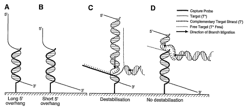
end). (Panel
B) Hybridisation to capture probe A-S-ErmBH370 (151 nucleotides from the 5'
end). (Panel
CA 02574917 2007-01-24
WO 2006/012727 PCT/CA2005/001030
21
C) Hybridisation to capture probe A-S-ErmBH459 (62 nucleotides from the 5'
end). (Panel
D) Hybridisation to capture probe A-S-ErmBH272 (249 nucleotides from the 5'
end). (Panel
E) Hybridisation to capture probe A-S-ErmBH370a (262 nucleotides from the 5'
end).
(Panel F) Hybridisation to capture probe A-S-ErmBH459a (351 nucleotides from
the 5'
end). For all panels, each value is the mean of three replicates. The standard
deviation for
these replicates is also shown. The scale for the fluorescence intensity axis
is different for
each panel to better illustrate the shape of the graphs. The letter (A) or (B)
attributed
beside each'of the tested probe refers to amplicons generated with PCR primers
of Fig.IA
or Fig. 1 B, respectively,
[00120] Figure 4 illustrates idealised interactions between an immobilised DNA
probe
and the two strands of the target amplicon. The target strand (T*) hybridises
to the DNA
probe, leaving a 5' overhang of variable length depending on the location of
the region of
the captured amplicon strand targeted by the probe. (Panel A) T* hybridised to
the DNA
probe, leaving a long 5' overhang of the captured product strand targeted by
the probe.
(Panel B) T* hybridised to the DNA probe, leaving a short 5' overhang of the
captured.
product strand targeted by the probe. (Panel C) The free complementary strand
(T') of the
target product hybridised to the overhanging tail of T*, generating a branch
migration that
caused destabilisation of the secondary complex. (Panel D) The free T*
(T*free) hybridised
to the free region of T', generating an antagonistic branch migration that
prevented the first
branch migration from breaking the secondary complex,
[00121] Figure 5 illustrates hybridisation to a microarray of capture probes
of single-
stranded target amplicon strand (T*) generated by asymmetrical PCR followed by
hybridisation with the complementary amplicon strand (T'). T* was hybridised
for 10 h to the
ermB array. Non-hybridised T* (T*free) was then washed away, and the array was
hybridised another 16 h with an equimolar quantity of the complementary strand
T' (grey
boxes) or with hybridisation buffer only (black boxes). Slides were washed
prior to
fluorescence detection. A significant decrease in signal intensity was
observed when the
complementary strand T' was hybridised for 16 hours compared to the control
hybridisation
using buffer only. (Panel A) Hybridisation to the lower (anti-sense or non-
coding) strand of
the 433-bp ermB amplicon followed by hybridisation with the upper (sense)
strand of the
same amplicon. (Panel B) Hybridisation to the upper strand of the 433-bp
amplicon followed
by hybridisation to the lower strand of the same amplicon. For both panels,
each result is
the mean of three replicates,
[00122] Figure 6 shows the correlation between the fluorescence intensity and
the
CA 02574917 2007-01-24
WO 2006/012727 PCT/CA2005/001030
22
length of the 5' overhang of captured tuf probes hybridised to different area
of the 523-bp
tuf PCR product amplified from Staphylococcus hominis. Probes A-S-TShoH520
(complementary to the lower strand) and A-S-TShoH520a (complementary to the
upper
strand) target the same region of the S. hominis product. Each value is the
mean of three
replicates. Staridard deviation for these replicates is also shown,
[00123] Figure 7 shows the correlation between the fluorescence intensity and
the
length of the 5' overhang of the captured blaSyõ probe A-S-Shvl H691
hybridised to different
blaSHV p roducts of 182 t o 715 b p. Each value i s the mean of three
replicates. Standard
deviation for these replicates is also shown,
[00124] Figure 8 shows the position of capture probes and PCR primers on the
tuf
gene PCR amplicons of 523 bp. Arrows represent primers while dashed boxes
represent 5'
amino-modified probes. Brackets indicate the length in nucleotides of the 5'
overhanging tail
of the target strand captured by each capture probe, and;
[00125] Figure 9 shows the position of PCR primers and a capture probe on the
blasHõgene PCR amplicons of 182 to 715 bp. Arrows represent primers used for
generating
these amplicons. The single dashed box represents a 5' amino-modified probe.
Brackets
indicate the length in nucleotides of the 5' overhanging tail of the target
strand captured by
the capture probe for each different PCR amplicons generated.
[00126] Other objects, advantages and features of the present invention will
become
apparent upon reading of the following non-restrictive description of
preferred embodiments
with reference to the accompanying drawing which is exemplary and should not
be
interpreted as limiting the scope of the present invention.
DETAILED DESCRIPTION OF THE INVENTION
[00127] The present invention is illustrated in further details by the
following non-
limiting examples.
EXAMPLES
EXAMPLE 1:
[00128] Correlation between the efficiency of microarray DNA hybridisation and
the
length of the 5' overhang of captured ermB amplicon strands.
CA 02574917 2007-01-24
WO 2006/012727 PCT/CA2005/001030
23
Materials and Methods
Microarray production
[00129] Twenty-mer oligonucleotide probes bearing a 5' amino-linker were
synthesised by Biosearch Technologies (Novato, CA, USA). Capture probe
sequences
used in the present invention are described in Table 1. The amino linker
modification
allowed covalent attachment of probes onto aldehyde-coated glass slides (CEL
Associates,
Pearland, TX, USA). Oligonucleotide probes were diluted 2-fold in ArrayltTM
MicroSpotting
Solution Plus (Telechem International, Sunnyvale, CA, USA) to a final
concentration of 5
pM. Oligonucleotides were spotted in triplicate using a VIRTEK SDDC-2 arrayer
(Bio-Rad
Laboratories, Hercules, CA, USA) with SMP3 pins from Telechem International.
After
spotting, slides were dried overnight, washed by immersion in 0.2% sodium
dodecyl sulfate
(SDS; Laboratoire Mat, Quebec, QC, Canada) for 2 min, and rinsed in ultrapure
water for 2
min. Slides were boiled in ultrapure water for 5 min for washing out the
unbound
oligonucleotides. Imine bonds between the glass surface and probes were
reduced to a
stable amide link by immersion for 20 min into a sodium borohydride solution
(1 g sodium
borohydride; Sigma, St.Louis, MO, USA), 300mL phosphate-buffered saline (PBS;
also
from Sigma), and 100 mL ethanol. Slides were then washed in 0.2% SDS for I min
and
rinsed in ultrapure water for 1 min. Slides were finally dried by
centrifugation for 5 min under
vacuum with a Savant SpeedVacTM Plus (Thermo Savant, NY, USA) and stored in a
dry
oxygen-free and dark environment. All above chemical treatments of the slides
were
performed at room temperature.
PCR amplification and amplicon labelling
[00130] Fluorescent dyes (label) were incorporated during PCR amplification.
Cy3 or
Cy5 dUTP (Amersham Biosciences, Baie d'Urfe, QC, Canada) were mixed at
concentrations of 0.02 pM in a 50-pL PCR mixture containing 0.05 mM dATP, 0.05
mM
dCTP, 0.05 mM dGTP, 0.02 mM dTTP, 5 mM KCI, 1 mM Tris-HCI (pH 9.0), 0.01%
Triton X-
100, 2.5 mM MgCl2, 0.5 unit of Taq DNA polymerase (Promega, Madison, WI, USA),
1 ng
purified genomic DNA, and 0.2 pM of each of the two primers. To test the
effect of
oligonucleotide probe position on the captured target DNA strand on
hybridisation
efficiency, we amplified by PCR two overlapping portions (402 and 433 bp) of
the
Staphylococcus aureus ermB gene (Figure 1). The ermB gene was amplified from
genomic
DNA isolated from the erythromycin-resistant S. aureus strain CCRI-1277. The
402-bp
CA 02574917 2007-01-24
WO 2006/012727 PCT/CA2005/001030
24
product was produced using primers ErmB225 and ErmB601, while the 433-bp
product was
amplified by PCR using primers ErmB109 and ErmB512 (Table 1). Thermal cycling
for PCR
amplification (180 s at 94 C, followed by 40cycles of 5 s at 95 C, 30 s at 55
C, and 30 s at
72 C) was carried out on an MJ Research PTC-200 DNA Engine thermal cycler
(Bio-Rad
Laboratories). PCR products were purified using the QlAquick PCR purification
kit
(Qiagen, Mississauga, ON, Canada). The dye incorporation was measured with an
Ultrospec 2000 Spectrophotometer (Amersham Biosciences) at 550 nm for Cy3 and
at 650
nm for Cy5. Concentration of the amplified product was determined at 260 nm
using the
Ultrospec 2000.
[00131] Asymmetric PCR was performed using the PCR conditions described above,
except that the upper strand of the 433-bp product was obtained using a 20:1
ratio of
ErmB109 and ErmB512 primers, respectively (Figure 1). An asymmetrical PCR was
performed to produce the lower strand using a 20:1 ratio of ErmB512 and
ErmB109,
respectively (Figure 1). Each asymmetric PCR was verified on a 1.5% agarose
gel to
ensure the production of single-stranded DNA and quantified using the
Ultrospec 2000 at
260 nm. The concentration of single-stranded DNA was adjusted to 1 pM and
hybridised to
the microarray to confirm the absence of the complementary strand.
DNA microarray hybridisation and data acquisition
[00132] Prehybridisation and hybridisation were performed in 15 x 13 mm
HybriWellT"' self-sticking hybridisation chambers (Grace Bio-Labs, Bend, OR,
USA).
Microarrays were first prehybridised for 30 min at room temperature with 1X
hybridisation
solution (6X standard saline phosphate-EDTA [SSPE; EM Science, Gibbstown, NJ,
USA],1 % bovine serum albumin [BSA], 0.01 % polyvinylpyrrolidone [PVP], 0.01 %
SDS, and
25% formamide [all from Sigma]). Cy-dUTP-labeled PCR products were denatured
at 95 C
for 5 min a nd t hen quickly c hilled o n i ce. F ive microliters o f d
enatured I abeled p roducts
were mixed with 10 pL of 2X hybridisation buffer (12X SSPE, 2% BSA, 0.02% PVP,
and
0.02% SDS) and 5 pL formamide (final concentration of 25%). Prehybridisation
solution
was removed from the chamber and replaced by the labeled PCR products
resuspended in
hybridisation solution. The hybridisation was carried out at 22 C for 15 min
and up to 16 h.
After hybridisation, microarrays were washed with 2X SSPE containing 0.1 /a
SDS for 5 min
at room temperature and rinsed once with 2X SSPE for 5 min, Microarrays were
dried by
centrifugation at 1350 x g for 3 min. Slides were scanned using a ScanArray
4000XL
confocal scanner (Packard Bioscience Biochip Technologies, Billerica, MA,
USA), and
fluorescent signals were analyzed using its software.
CA 02574917 2007-01-24
WO 2006/012727 PCT/CA2005/001030
Results
[00133] We tested whether the region of the product targeted by an
oligonucleotide
capture probe influenced hybridisation efficiency. To achieve this goal, we
initially used the
5 ermB bacterial antibiotic resistance gene as genetic target. This gene
encodes an adenine
N-6-methyltransferase, which confers resistance to macrolides, lincosamides,
and
streptogramin B(Roberts et al., 1999, Antimicrob. Agents Chemother., 43:2823-
2830). We
generated two overlapping ermB PCR products, each targeted by six 20-mer
capture
probes located at different areas of the products (Figure 1). Three of these
probes (A-S-
10 ErmBH272, A-S-ErmBH370, and A-S-ErmBH459) were designed to be complementary
to
the lower strand of both products, while the three other probes (A-S-
ErmBH272a, A-S-
ErmBH370a, and A-S-ErmBH459a) targeted the same region but hybridised to the
upper
strand of both products. For these perfectly complementary oligonucleotides,
both strands
have the same Tm.and secondary structure, and have also been shown to behave
15 identically for hybridisation in solution (Rafalski, 1988, Anal. Biochem.,
173:383-386).
Therefore, variations in the performance of hybridisation between capture
probes targeting
the same region located on the opposite strand of a product may be attributed
to a bias
correlated with the efficiency of hybridisation onto solid support.
[00134] The Cy3-labeled 402- and 433-bp products were hybridised overnight to
the
20 ermB array that contained the six different capture probes (Figure 1).
After washing and
analysis, it was observed that the fluorescence signal for each capture probe
after a 16
hours hybridisation was not identical. Plotting the fluorescence intensities
of hybridisation
against the regions of the product recognized by capture probes revealed a
correlation
between the fluorescence intensity and the length of the free 5' overhanging
portion of the
25 captured strand (Figure 2). For each of the six capture probes, the
strongest hybridisation
signal was always observed forthe probe targeting a region closest to the 5'
end of the
upper or lower targeted strand. These probes hybridised the closest to the 5'
end of the
complementary strand of the product, thus leaving the shortest overhanging 5'
end. Both
target ermB products (402- and 433-bp) behaved similarly with respect to
fluorescence
intensity a nd position o f t he capture p robe. Also, n o s ignificant d
ifference was observed
between the upper and lower strands. This is illustrated in Figure 2B by
hybridisation with
oligonucleotides A-S-ErmBH370 of the 433-bp product which is 151 nucleotides
from the 5'
end, and A-S-ErmBH370a of the 402-bp product which is 146 nucleotides from the
5' end,
showing that when the 5' overhang lengths were similar, the fluorescence
intensities were
also similar regardless of the product size or the target strand.
CA 02574917 2007-01-24
WO 2006/012727 PCT/CA2005/001030
26
[00135] Despite the fact that for the same oligonucleotide capture probe the
key
determinant for hybridisation intensity appears to be the length of the 5'
overhang of the
hybridised target DNA strand, some probes worked better than others. For
example, probe
A-S-ErmBH272a (5' overhang length of 48 nucleotides) produced a hybridisation
signal six
times stronger than probe A-S-ErmBH459 (5' overhang length of 62 nucleotides).
One
explanation may be that the area covered by probe A-S-ErmBH459 may be less
available
for hybridisation or less stable once hybridised than the area covered by
probes A-S-
ErmBH272 and A-S-ErmBH272a (Figure 2). This behavior may be attributed either
to the
secondary structure of the target strand or to thermodynamic properties of the
probes. It is
salient to point out that the AG of the secondary structure from probe 'A-S-
ErmBH459 is -
14.2 kcal/mol, which represents a much higher energy than that for the other
probes used
in this study (i.e. -5.3 kcal/mol for probe A-S-ErmBH272 and -3.5 kcal/mol for
probe A-S-
ErmBH370). Nonetheless, even if probe A-S-ErmBH459 gave a lower hybridisation
signal,
its intensity correlated with the length of the 5' overhang (Figure 2 C).
[00136] Thus, capture probes (P) targeting (able to bind) the 5' end of the
captured
target strand (T*) gave strong and reproducible hybridisation signals, while
probes targeting
(able to bind) the 3' extremity of the captured target strand gave no or very
weak
hybridisation signals after overnight hybridisation. One plausible explanation
is that T*
hybridised by its 3' end is less stable than the same strand hybridised closer
to its 5' end.
To verify this hypothesis, hybridisation kinetics were assessed by hybridising
the 433- bp
labeled products with the ermB array for 15, 30, 60, 180 and 960 min (16 h).
Probes
targeting regions close to the 5' end of either strand of the product showed a
fluorescent
signal increasing with hybridisation time (Figure 3, Panels A, B and C).
Probes targeting
regions leaving a longer 5' overhang of either strand of the products
exhibited very different
hybridisation kinetics (Figure 3, Panels D, E and F). Indeed, we observed an
increase of the
hybridisation signal in the first 30 min of hybridisation, but thereafter
fluorescence intensity
decreased over time until it reached background levels. This kinetics of
hybridisation during
the first 30 minutes is also observed for probes targeting the 5' end of the
captured strand.
It may be surmised that during the first 30 minutes of the reaction, local
higher
concentration of capture probe (P) favoured hybridisation of T* on P. This
hybridisation
behaviour appears to follow a classical equilibrium equation:
kl
T*+ P ; T*P
k2
CA 02574917 2007-01-24
WO 2006/012727 PCT/CA2005/001030
27
where k1 is the hybridisation constant and k2 the dissociation constant. This
hybridization
kinetics suggests that the longer the hybridisation period the more important
is the negative
impact of a long 5' overhang.
[00137] The hybridisation kinetics following the first 30 minutes, which is
dependent
on the position of the probe on the captured strand, may be explained by the
topology of
the T*P d uplex. When a p robe r ecognises a n area c loser t o the 3' e nd o
f the c aptured
target strand T*, most of the overhanging 5' end of non hybridised DNA is
exposed to the
liquid phase above the glass surface (Fig. 4A). On the other hand, when it
hybridises to an
area close to the 5' end of the captured strand target, most of T* (3' end) is
directed
towards the glass surface (Fig. 4B). In the first conformation, the
overhanging tail of T* may
be available for reassociation with its complementary strand; T', a process
that may
destabilises the probe-target duplex (T*P).
[00138] To test the ability of the nonhybridised complementary strand (T') to
destabilise the T*P duplex, we carried out experiments with single-stranded
products.
Microarrays were hybridised for 10 h with the amplified 433-bp ermB product
lower strand
(T*) generated by asymmetrical PCR. After washing out the nonhybridised T*
still in solution
(T*free), the hybridisation was carried out for an additional 16 h, either
with hybridisation
buffer only or with an equimolar amount of the complementary upper strand T'.
In the
presence of only single-stranded target DNAs (T*), the region at which the
oligonucleotide
probe hybridises no longer influences the hybridisation intensity (Figure 5).
For example,
probe A-S- ErmBH272, which leaves a 5' overhang of 249 nucleotides, hardly
captures any
of the target DNA when the double-stranded product is used as target (Figure
2A).
However, this same probe efficiently captured the complementary single-
stranded DNA.
produced by asymmetrical PCR (Figure 5A). Similar results were observed for
hybridisation
with the upper product strand. The intensity of fluorescence decreased
dramatically when
the complementary T' lower (anti-sense) strand was included in the assay
(Figure 5B). The
addition of the complementary strand T' reduced the intensity of hybridisation
close to
background levels, suggesting that T*P duplex destabilisation occurs in the
presence of the
complementary strand. Displacement of T* from P by reassociation with T'
probably
proceeds through a sequential displacement pathway also known as a zipper
effect
(Reynaldo et al., 2000, J. Mol. Biol., 297:511-520). Hybridisation between the
captured T*
strand and its complementary strand T' in solution will occur first at the
exposed overhang
tail of the captured T* and will be followed by a branch migration mechanism
towards the
3'end. Such a mechanism was used to build a DNA-fuelled nanomolecular machine
(Yurke
et al., 2000, Nature, 406: 605-608; Alberti et al., 2003, Proc. Natl. Acad.
Sci. U S A, 100:
CA 02574917 2007-01-24
WO 2006/012727 PCT/CA2005/001030
28
1569-1573). In those studies, the authors used the complementary DNA strand
(called "fuel
DNA") to close and open double-stranded DNA structures. In the experiment
described
above, the complementary strand T' seems to act as the "fuel" DNA, pulling the
captured
target strand T* from the probe (Fig. 4C). A longer 5' overhang increases the
probability of
collision between the complex T*P and free T' and thus leads to a faster
destabilisation
effect. This may explain the hybridisation bias observed with long 5'
overhangs but does not
explain why a short 5' overhang end generates a hybridisation signal that
increases over
time (Fig. 3 A, B, C).
EXAMPLE 2:
[00139] Correlation between the efficiency of microarray DNA hybridisation and
the
length of the 5' overhang of captured tuf amplicon strands.
[00140] Material and methods are the same as those used in Example 1 except
that
primers and capture probes targeting the tuf gene encoding the elongation
factor Tu were
used (see Table 1). The tuf gene was amplified from genomic DNA isolated from
Staphylococcus hominis subsp. hominis strain ATCC 27844. A 523-bp product was
produced using primers TshoH240 and TstaG765. Thermal cycling for PCR
amplification
was as described in Example 1.
[00141] Figure 8 shows the position of capture probes and PCR primers on the
tuf
gene PCR amplicons of 523 bp. Arrows represent primers while dashed boxes
represent 5'
amino-modified probes. Brackets indicate the length in nucleotides of the 5'
overhanging tail
of the target strand captured by each capture probe. Results with the tuf gene
were similar
to those obtained with ermB (Figure 6). Capture probes gave stronger
hybridisation signal
when the 5' overhanging tail was short and showed near background signals when
the 5'
tail reached a length over 250 nucleotides for tuf (Figure 6). Thus, different
capture probes
seem to follow similar hybridisation methods, irrespective of the target
sequences.
[00142] To demonstrate that methods predicted in Example 1 are applicable to
other
DNA targets, we have tested the hybridisation efficiency of different capture
probes
(according to the region to which they hybridise) on the highly conserved tuf
gene. As
described in Example 1, capture probes gave stronger hybridisation signal when
the 5'
overhang was short. In example 2, capture probes showed near background
signals when
the 5' overhang reached a length over 250 nucleotides (Figure 6).
CA 02574917 2007-01-24
WO 2006/012727 PCT/CA2005/001030
29
EXAMPLE 3:
[00143] Correlation between the efficiency of microarray DNA hybridisation and
the
length of the 5' overhang of captured blasHv amplicon strands.
[00144] Material and methods are the same as those used in Example 1 except
that
primers and capture probes targeting the blasyv gene encoding a(3-lactamase
were used
(see Table 1). The blasHv gene was amplified from genomic DNA isolated from
Escherichia
coli strain CCRI-1 192. Different products were generated by combining the
reverse primer
shv763 with five different primers used to produce different lengths of 5'
overhangs: (i)
primer shv604 amplified a 182-bp product; (ii) primer shv449 amplified a 337-
bp product;
(lii) primer shv368 amplified a 418-bp product; (iv) primer shv313 amplified a
473-bp
product; and (v) primer shvseq7l amplified a 715-bp product (Table 1). Thermal
cycling for
PCR amplification was as described in Example 1. ,
[00145] Figure 9 shows the position of PCR primers and a capture probe on the
b/asHvgene PCR amplicons of 182 to 715 bp. Arrows represent primers used for
generating
these amplicons. The single dashed box represents a 5' amino-modified probe.
Brackets
indicate the length in nucleotides of the 5' overhanging tail of the target
strand captured by
the capture probe for each different PCR amplicons generated. Results obtained
with the
blasHv gene are shown in Figure 7. Products were amplified using the same
reverse primer
but using different forward primers. This allowed the amplification of
products having a
variable forward length, while its reverse length remained constant. After
hybridisation of
each product to the microarray, we plotted the signal in function of the
length of the 5' tail
for the probes targeting the upper strand and in function of the length of the
3' tail for the
probes targeting. the lower strand. The increase of the length of the 5' tail
reduced the signal
(correlation coefficient between -0.66 and -0.85), whereas the increase of the
3' tail had no
major effect on the hybridisation signal (correlation coefficient between 0.12
and 0.20)
(Figure 7). Those results suggest that, while the length of the 5' tail has a
significant impact
on the hybridisation signal observed, the length of the 3' tail seems less
important (data not
shown).
[00146] Therefore, the results for blasyv were similar to those obtained with
ermB in
Example 1. Capture probes targeting blasyv gave stronger hybridisation signal
when the 5'
. overhanging tail was short and showed near background signals when the 5'
end reached a
CA 02574917 2007-01-24
WO 2006/012727 PCT/CA2005/001030
length over 600 nucleotides for blasHv (Figure 7). Thus, different capture
probes seem to
follow similar hybridisation methods, irrespective of the target sequences.
[00147] The hybridisation behaviour and efficiency of oligonucleotides arrayed
onto a
solid support has been investigated herein. As described herein, we observed
that the
5 position of a capture probe on a given product has an impact on the observed
hybridisation
signal. The hybridisation behaviour of a double-stranded product DNA on short
oligonucleotides immobilised by their 5' end gave counter-intuitive and
unexpected results.
Indeed, one would assume weaker hybridisation signal when a 5' end immobilised
probe
binds the target molecule close to its 5' end, because of steric hindrance
caused by a
10 longer 3' overhanging tail. However, our results show that the increase of
the 3' end has no
major effect on hybridisation signal, whereas the hybridisation signal
strength is inversely
correlated with the length of the 5' overhanging tail of the target molecule
when hybridised
with a probe immobilised via its 5' end.
[00148] This hybridisation behavior may be explained by the topology of the
T*P
15 duplex. When a probe recognizes an area closer to the 3' end of the
captured target strand
T*, most of the overhanging 5' end of nonhybridised DNA is exposed to the
liquid phase
above the glass surface (Figure 4). On the other hand, when it hybridises to
an area close
to the 5' end of the captured strand target, most of T* (3' end) is directed
towards the glass
surface. In the first conformation, the protruding tail of T* may be available
for reassociation
20 with its complementary strand (T '), a process that may destabilise the
probe-target duplex
(T*P) as shown when asymmetrical products were used. This hybridisation
behavior may
also be observed with 3' immobilised probes, although probes anchored to a
support by a 3'
end are not commonly used.
[00149] Displacement of T* from P by reassociation with T' may proceed through
a
25 sequential displacement pathway also known as a zipper effect (Reynaldo et
al., 2000, J.
Mol. Biol., 297:511-520). Hybridisation between the captured T* strand and its
complementary strand T' in solution would occur first at the exposed overhang
tail of
captured T* and would be followed by a branch migration mechanism. Such a
mechanism
was used recently to build a DNA-fueled nanomolecular machine (Yurke et al.,
2000,
30 Nature, 406:605-608; Alberti and Mergny, 2003, Proc. Nat. Acad. Sci. USA,
100:1569-
1573). In those studies, the authors used the complementary DNA strand (called
fuel DNA)
to close and open double-stranded DNA structures. The complementary strand T'
may act
as the fuel DNA, thereby pulling the captured target strand T* from the probe
(Figure 4).
CA 02574917 2007-01-24
WO 2006/012727 PCT/CA2005/001030
31
[00150] By using asymmetrical PCR, we have shown that the captured product
strand is displaced by the target complementary strand T' independently of the
area the
probe targets on the product (Figure 5). This suggests that some elements
stabilise T*P
when the hybridisations were performed in the presence of both T* and T'. One
possible
model would be that T*free forms a quaternary complex (T*T'T*free P) with the
ternary
complex (T'T*P) captured on the glass surface. In accordance with the random
walk theory
for branch migration (Lee et aL, 1970, J. Mol. Biol. 48:1-22), the branch
point between T*T'
and T'T*free duplexes of the T*T'T*free P complex may move in either
direction. The
random walk would continue until one of two helices becomes shorter than the
minimum
length of a stable duplex (Reynaldo et al., 2000, J. Mol. Biol., 297:511-520).
This means
that t he I onger t he d uplex p art o f t he h elix is, t he m ore I ikely i
t i s t o d isplace t he o ther
competing duplex (e.g. if T*T' forms a longer helix, it would destabilise the
complex T'T free
and vice-versa).
[00151] A nucleation step would occur first with encounter between T' and the
overhanging part of the captured T*. A double helix would rapidly be formed
until it reaches
the branch point made by the complex T*P (Radding et a/.,'1977, J. Mol. Biol.,
116: 825-
839). At that point,, it is proposed that strand displacement by branch
migration would start
with the two complexes T*P and T'T*. Simultaneously, the T*free would form a
double-
stranded helix with the overhanging part of T' associated with T*P (Fig. 4 C
and D), thereby
forming an antagonist migration fork. When the 5' overhang of T* DNA is longer
(Fig. 4C),
the double helix formed with T' will be longer than the double helix formed
between T*free
and the overhanging part of T. Branch mechanism competition between the two
duplexes
would be in favour of the reassociation of captured T* with T', pulling the
target T* away
from the probe P. In contrast, when the 5' overhanging tail is short (Fig.
4D), the competing
forming helix T*freeT' would be long enough to favour reassociation of T*free
with T',
thereby depleting locally the T' and thus stabilising the T*P complex. Over
time, diffusion of
T* in close proximity with free probes P, would feed the hybridisation of the
target T* with
the captured probe P, increasing -the fluorescent signal (Fig. 3 A, B, C). The
results
presented herein provide evidence that kinetic effects involving re-
association of the
complementary nucleic acid strand may be associated with destabilisation of
the capture
probe/nucleic acid target duplex and that this kinetic effect may be governed
by the position
of the complementary sequence on the targeted nucleic acids. The results
presented herein
therefore delineate key predictable parameters that govern the hybridisation
efficiency of
capture probes attached onto solid supports. These parameters allow selection
of optimal
capture probes for the detection of nucleotide polymorphisms. The kinetic
effects and
CA 02574917 2007-01-24
WO 2006/012727 PCT/CA2005/001030
32
reassociation of the target to the PCR product's complementary strand may lead
to
destabilisation of the capture probe/DNA target duplex (complex) and that this
kinetic effect
may be governed by the position of the complementary sequence on the targeted
nucleic
acid.
[00152] A correlation between the length of the overhang of the target and the
efficiency of hybridization has been demonstrated herein. Evidence that the
presence f the
complementary strand is associated with the poor hybridisation efficiency of
5' immobilised
probes targeting the 3' end of a product, thereby leaving a long 5' overhang
has also been
evidenced herein. On the other hand, probes targeting a region of the target
which is
located toward the 5' end of the same product, hybridised more efficiently.
Therefore, the
hybridisation efficiency of oligonucleotides anchored onto a solid support has
been found to
be highly dependent of their location on a target single-stranded nucleic
acid. The results
presented herein show that capture probes anchored by their 5' end and
targeting a region
that lies within about a 40% portion of the 5' end of the captured nucleic
acid strand provide
more efficient hybridisations as compared to those targeting the remaining 60%
portion at
the 3' end (Figures 2, 6 and 7). Conversely, capture probes anchored by their
3' end and
targeting a region that lies within a 40% portion of the 3' end of the
captured nucleic acid
strand provide more efficient hybridisations as compared to those targeting
the remaining
60% portion at the 5' end. Evaluation of the hybridisation signal for each
probe revealed an
inverse correlation between the length of the free overhanging end (either the
5' end or the
3' end depending on which end of the probe is anchored on the support) of the
target and
the hybridisation signal intensity. Therefore, hybridised targets having their
longest portion
(e.g., at least 60%) proximal the solid support have been found to be more
stable and to
give a better (more intense) hydridisation signal.
[00153] Results presented herein teach methods for the efficient design of
capture
probes, which help to improve the sensitivity and specificity of microarray
detection.
Methods used i n the selection and design of probes, thus ensure efficient and
sensitive
detection of either target single-stranded nucleic acids or denatured double-
stranded
nucleic acids such as PCR amplicons. This study demonstrates the importance of
choosing
the appropriate nucleic acid region to ensure efficient and sensitive
detection of a target
such as single-stranded nucleotide-based target which may come into contact
with a
nucleotide sequence substantially complementary to the unhybridized portion of
the target
(which extends away form the support), or double-stranded DNA fragments such
as PCR
products using short capture probes. This is particularly important for SNP
detection. In
addition, efforts are ongoing to develop novel amplification and labeling
systems for efficient
CA 02574917 2007-01-24
WO 2006/012727 PCT/CA2005/001030
33
production of single-stranded DNA products that would circumvent the
competition between
complementary strands.
[00154] Although the present invention has been described hereinabove by way
of
embodiments thereof, it may be modified, without departing from the spirit and
nature of the
subject invention as defined in the appended claims.
Table 1. Oligonucleotide primers and probes used in this invention.
Primers SN sD Nucleotide sequence (5' --> 3') b Target Product length
gene
ErmB225 1 TCGTGTCACTTTAATTCACCAAGATA ermB 402 bp
ErmB601 2 TTTTTAGTAAACAGTTGACGATATTC ermB (SEQ ID NOs 1+2)
ErmB109 3 GGAACAGGTAAAGGGCATTTAACGAC ermB 433 bp
ErmB512 4 CTGTGGTATGGCGGGTAAGTTTTATTAAG ermB (SEQ ID NOs 3+4)
TShoH240 5 GCTTTAGAAGGCGATGCTCAATACG tuf 523 bp
TStaG765 6 TIACCATTTCAGTACCTTCTGGTAA tuf (SEQ ID NOs 5+6)
shv604 7 CAGCTGCTGCAGTGGATGGT blaSHV 182 bp
(SEQ ID NOs 7+12)
shv449 8 AGATCGGCGACAACGTCACC blasHv 337 bp
(SEQ ID NOs 8+12)
shv368 9 TTACCATGAGCGATAACAGC blaSHV 418 bp
(SEQ ID NOs 9+12)
shv313 10 AGCGAAAAACACCTTGCCGAC bla 473 bp
sHV (SEQ ID NOs
10+12)
715 bp
shvseq7l 11 AGCCGCTTGAGCAAATTAAACTA blaSHV (SEQ ID NOs
11+12)
shv763 12 GTATCCCGCAGATAAATCACCAC blaSHV
Capture probes a
A-S-ErmBH272 13 CAAACAGAGGTATAAAATTG ermB
A-S-ErmBH370 14 TGATTGTTGAAGAAGGATTC ermB
A-S-ErmBH459 15 TTGCTTAAGCTGCCAGCGGA ermB
A-S-ErmBH272a 16 CAATTTTATACCTCTGTTTG ermB
A-S-ErmBH370a 17 GAATCCTTCTTCAACAATCA ermB
A-S-ErmBH459a 18 TCCGCTGGCAGCTTAAGCAA ermB
A-S-TShoH713 19 ATACGTTTTATCAAAAGATGAAG tuf
A-S-TStaGH554 20 TACTGGTGTAGAAATGTTC tuf
A-S-TShoH520a 21 GAAGTTTCTTTGATACCAAT tuf
A-S-TShoH520 22 ATTGGTATCAAAGAAACTTC tuf
A-S-shvl H691 23 CCCCGCTCGCCAGCTCCGGT blaSHV
a A-S stands for the 5' modifications: A is an amino group and S is a hexa-
ethyleneglycol
spacer.
b Nucleotide nomenclature is as follows: A: Adenine; C: Cytosine; G: Guanine;
I: Inosine;
T: Thymine
CA 02574917 2007-01-24
WO 2006/012727 PCT/CA2005/001030
34
DESCRIPTION OF SEQUENCES
2) INFORMATION FOR SEQ ID NO: 1
(i) SEQUENCE CHARACTERISTICS:
(A) LENGTH: 26 bases
(B) TYPE: Nucleic acid
(C) STRANDEDNESS: Single
(D) TOPOLOGY: Linear
(ii) MOLECULE TYPE: DNA
(xi) SEQUENCE DESCRIPTION: SEQ ID NO: 1
TCGTGTCACT TTAATTCACC AAGATA 26
2) INFORMATION FOR SEQ ID NO: 2
(i) SEQUENCE CHARACTERISTICS:
(A) LENGTH: 26 bases
(B) TYPE: Nucleic acid
(C) STRANDEDNESS: Single
(D) TOPOLOGY: Linear
(ii) MOLECULE TYPE: DNA
(xi) SEQUENCE DESCRIPTION: SEQ ID NO: 2
TTTTTAGTAA ACAGTTGACG ATATTC 26
2) INFORMATION FOR SEQ ID NO: 3
(i) SEQUENCE CHARACTERISTICS:
(A) LENGTH: 26 bases
(B) TYPE: Nucleic acid
(C) STRANDEDNESS: Single
(D) TOPOLOGY: Linear
(ii) MOLECULE TYPE: DNA
(xi) SEQUENCE DESCRIPTION: SEQ ID NO: 3
GGAACAGGTA AAGGGCATTT AACGAC 26
2) INFORMATION FOR SEQ ID NO: 4
(i) SEQUENCE CHARACTERISTICS:
(A) LENGTH: 29 bases
CA 02574917 2007-01-24
WO 2006/012727 PCT/CA2005/001030
(B) TYPE: Nucleic acid
(C) STRANDEDNESS: Single
(D) TOPOLOGY: Linear
5 (ii) MOLECULE TYPE: DNA
(xi) SEQUENCE DESCRIPTION: SEQ ID NO: 4
CTGTGGTATG GCGGGTAAGT TTTATTAAG 29
2) INFORMATION FOR SEQ ID NO: 5
(i) SEQUENCE CHARACTERISTICS:
(A) LENGTH: 25 bases
(B) TYPE: Nucleic acid
(C) STRANDEDNESS: Single
(D) TOPOLOGY: Linear
(ii) MOLECULE TYPE: DNA
(xi) SEQUENCE DESCRIPTION: SEQ ID NO: 5
GCTTTAGAAG GCGATGCTCA ATACG 25
2) INFORMATION FOR SEQ ID NO: 6
(i) SEQUENCE CHARACTERISTICS:
(A) LENGTH: 25 bases
(B) TYPE: Nucleic acid
(C) STRANDEDNESS: Single
(D) TOPOLOGY: Linear
(ii) MOLECULE TYPE: DNA
(xi) SEQUENCE DESCRIPTION: SEQ ID NO: 6
TIACCATTTC AGTACCTTCT GGTAA 25
2) INFORMATION FOR SEQ ID NO: 7
(i) SEQUENCE CHARACTERISTICS:
(A) LENGTH: 20 bases
(B) TYPE: Nucleic acid
(C) STRANDEDNESS: Single
(D) TOPOLOGY: Linear
(ii) MOLECULE TYPE: DNA
CA 02574917 2007-01-24
WO 2006/012727 PCT/CA2005/001030
36
(xi) SEQUENCE DESCRIPTION: SEQ ID NO: 7
CAGCTGCTGC AGTGGATGGT 20
2) INFORMATION FOR SEQ ID NO: 8
(i) SEQUENCE CHARACTERISTICS:
(A) LENGTH: 20 bases
(B) TYPE: Nucleic acid
(C) , STRANDEDNESS: Single
(D) TOPOLOGY: Linear
(ii) MOLECULE TYPE: DNA
(xi) SEQUENCE DESCRIPTION: SEQ ID NO: 8
AGATCGGCGA CAACGTCACC 20
,
2) INFORMATION FOR SEQ ID NO: 9
(i) SEQUENCE CHARACTERISTICS:
(A) LENGTH: 20 bases
(B) TYPE: Nucleic acid
(C) STRANDEDNESS: Single
(D) TOPOLOGY: Linear
(ii) MOLECULE TYPE: DNA
(xi) SEQUENCE DESCRIPTION: SEQ ID NO: 9
TTACCATGAG CGATAACAGC 20
2) INFORMATION FOR SEQ ID NO: 10
(i) SEQUENCE CHARACTERISTICS:
(A) LENGTH: 21 bases
(B) TYPE: Nucleic acid
(C) STRANDEDNESS: Single
(D) TOPOLOGY: Linear
(ii) MOLECULE TYPE:-DNA
(xi) SEQUENCE DESCRIPTION: SEQ ID NO: 10
AGCGAAAAAC ACCTTGCCGA C 21
CA 02574917 2007-01-24
WO 2006/012727 PCT/CA2005/001030
37
2) INFORMATION FOR SEQ ID NO: 11
(i) SEQUENCE CHARACTERISTICS:
(A) LENGTH: 23 bases
(B) TYPE: Nucleic acid
(C) STRANDEDNESS: Single
(D) TOPOLOGY: Linear
(ii) MOLECULE TYPE: DNA
(xi) SEQUENCE DESCRIPTION: SEQ ID NO: 11
AGCCGCTTGA GCAAATTAAA CTA 23
2) INFORMATION FOR SEQ ID NO: 12
(i) SEQUENCE CHARACTERISTICS:
(A) LENGTH: 23 bases
(B) TYPE: Nucleic acid
(C) STRANDEDNESS: Single
(D) TOPOLOGY: Linear
(ii) MOLECULE TYPE: DNA
(xi) SEQUENCE DESCRIPTION: SEQ ID NO: 12.
GTATCCCGCA GATAAATCAC CAC 23
2) INFORMATION FOR SEQ ID NO: 13
(i) SEQUENCE CHARACTERISTICS:
(A) LENGTH: 20 bases
(B) TYPE: Nucleic acid
(C) STRANDEDNESS: Single
(D) TOPOLOGY: Linear
(ii) MOLECULE TYPE: DNA
(xi) SEQUENCE DESCRIPTION: SEQ ID NO: 13
CAAACAGAGG TATAAAATTG 20
2) INFORMATION FOR SEQ ID NO: 14
(i) SEQUENCE CHARACTERISTICS:
(A) LENGTH: 20 bases
(B) TYPE: Nucleic acid
(C) STRANDEDNESS: Single
CA 02574917 2007-01-24
WO 2006/012727 PCT/CA2005/001030
38
(D) TOPOLOGY: Linear
(ii) MOLECULE TYPE: DNA
(xi) SEQUENCE DESCRIPTION: SEQ ID NO: 14
TGATTGTTGA AGAAGGATTC 20
2) INFORMATION FOR SEQ ID NO: 15
(i) SEQUENCE CHARACTERISTICS:
(A) LENGTH: 20 bases
(B) TYPE: Nucleic acid
(C) STRANDEDNESS: Single
(D) TOPOLOGY: Linear
(ii) MOLECULE TYPE: DNA
(xi) SEQUENCE DESCRIPTION: SEQ ID NO: 15
TTGCTTAAGC TGCCAGCGGA 20
2) INFORMATION FOR SEQ ID NO: 16
(i) SEQUENCE CHARACTERISTICS:
(A) LENGTH: 20 bases
(B) TYPE: Nucleic acid
(C) STRANDEDNESS: Single
(D) TOPOLOGY: Linear
(ii) MOLECULE TYPE: DNA
(xi) SEQUENCE DESCRIPTION: SEQ ID NO: 16
CAATTTTATA CCTCTGTTTG 20
2) INFORMATION FOR SEQ ID NO: 17
(i) SEQUENCE CHARACTERISTICS:
(A) LENGTH: 20 bases
(B) TYPE: Nucleic acid
(C) STRANDEDNESS: Single
(D) TOPOLOGY: Linear
(ii) MOLECULE TYPE: DNA
(xi) SEQUENCE DESCRIPTION: SEQ ID NO: 17
CA 02574917 2007-01-24
WO 2006/012727 PCT/CA2005/001030
39
GAATCCTTCT TCAACAATCA 20
2) INFORMATION FOR SEQ ID NO; 18
(i) SEQUENCE CHARACTERISTICS:
(A) LENGTH: 20 bases
(B) TYPE: Nucleic acid
(C) STRANDEDNESS: Single
(D) TOPOLOGY: Linear
(ii) MOLECULE TYPE: DNA
(xi) SEQUENCE DESCRIPTION: SEQ ID NO: 18
TCCGCTGGCA GCTTAAGCAA 20
2) INFORMATION FOR SEQ ID NO: 19
(i) SEQUENCE CHARACTERISTICS:
(A) LENGTH: '23 bases
(B) TYPE: Nucleic acid
(C) STRANDEDNESS: Single
(D) TOPOLOGY: Linear
(ii) MOLECULE TYPE: DNA
(xi) SEQUENCE DESCRIPTION: SEQ ID NO: 19
ATACGTTTTA TCAAAAGATG AAG 23
2) INFORMATION FOR SEQ ID NO:20
(i) SEQUENCE CHARACTERISTICS:
(A) LENGTH: 19 bases
(B) TYPE: Nucleic acid
(C) STRANDEDNESS: Single
(D) TOPOLOGY: Linear
(ii) MOLECULE TYPE: DNA
(xi) SEQUENCE DESCRIPTION: SEQ ID NO: 20
TACTGGTGTA GAAATGTTC 19
2) INFORMATION FOR SEQ ID NO: 21
CA 02574917 2007-01-24
WO 2006/012727 PCT/CA2005/001030
(i) SEQUENCE CHARACTERISTICS:
(A) LENGTH: 20 bases
(B) TYPE: Nucleic acid
(C) STRANDEDNESS: Single
5 (D) TOPOLOGY: Linear
(ii) MOLECULE TYPE: DNA
(xi) SEQUENCE DESCRIPTION: SEQ ID NO: 21
GAAGTTTCTT TGATACCAAT 20
2) INFORMATION FOR SEQ ID NO: 22
(i) SEQUENCE CHARACTERISTICS:
(A) LENGTH: 20 bases
(B) TYPE: Nucleic acid
(C) STRANDEDNESS: Single
(D) TOPOLOGY: Linear
(ii) MOLECULE TYPE: DNA
(xi) SEQUENCE DESCRIPTION: SEQ ID NO: 22
ATTGGTATCA AAGAAACTTC 20
2) INFORMATION FOR SEQ ID NO: 23
(i) SEQUENCE CHARACTERISTICS:
(A) LENGTH: 20 bases
(B) TYPE: Nucleic acid
(C) STRANDEDNESS: Single
(D) TOPOLOGY: Linear
(ii) MOLECULE TYPE: DNA
(xi) SEQUENCE DESCRIPTION: SEQ ID NO: 23,
CCCCGCTCGC CAGCTCCGGT 20