Note: Descriptions are shown in the official language in which they were submitted.
CA 02575202 2012-05-10
COMPOUND MODULATION TRANSFER FUNCTION FOR LASER
SURGERYAND OTHER OPTICAL APPLICATIONS
10
BACKGROUND OF THE INVENTION
[0004] This invention generally relates to goal functions or visual
function diagnostic
metrics, and in particular provides methods, devices, and systems for
mitigating or treating
vision conditions such as presbyopia, often by determining a compound
modulation transfer
function.
[0005] Presbyopia normally develops as a person ages, and is
associated with a natural
progressive loss of accommodation, sometimes referred to as "old sight." The
presbyopic eye
often loses the ability to rapidly and easily refocus on objects at varying
distances. There may
also be a loss in the ability to focus on objects at near distances. Although
the condition
progresses over the lifetime of an individual, the effects of presbyopia
usually become
noticeable after the age of 45 years. By the age of 65 years, the crystalline
lens has often lost
almost all elastic properties and has only limited ability to change shape.
Residual
accommodation refers to the amount of accommodation that remains in the eye. A
lower
degree of residual
1
CA 02575202 2007-01-22
WO 2006/020422
PCT/US2005/026859
accommodation contributes to more severe presbyopia, whereas a higher amount
of residual
accommodation correlates with less severe presbyopia.
[0006] Known methods and devices for treating presbyopia seek to provide
vision approaching
that of an emmetropic eye. In an emmetropic eye, both distant objects and near
objects can be
seen due to the accommodation properties of the eye. To address the vision
problems associated
with presbyopia, reading glasses have traditionally been used by individuals
to add plus power
diopter to the eye, thus allowing the eye to focus on near objects and
maintain a clear image.
This approach is similar to that of treating hyperopia, or farsightedness.
[0007] Presbyopia has also been treated with hi-focal eyeglasses, where one
portion of the lens
is corrected for distance vision, and another portion of the lens is corrected
for near vision.
When peering down through the bifocals, the individual looks through the
portion of the lens
corrected for near vision. When viewing distant objects, the individual looks
higher, through the
portion of the bi-focals corrected for distance vision. Thus with little or no
accommodation, the
individual can see both far and near objects.
[0008] Contact lenses and intra-ocular lenses (IOLs) have also been used to
treat presbyopia.
One approach is to provide the individual with monovision, where one eye
(usually the primary
eye) is corrected for distance-vision, while the other eye is corrected for
near-vision.
Unfortunately, with monovision the individual may not clearly see objects that
are intermediately
positioned because the object is out-of-focus for both eyes. Also, an
individual may have trouble
seeing with only one eye, or may be unable to tolerate an imbalance between
their eyes. In
addition to monovision, other approaches include bilateral correction with
either hi-focal or
multi-focal lenses. In the case of bi-focal lenses, the lens is made so that
both a distant point and
a near point can be focused. In the multi-focal case, there exist many focal
points between near
targets and far targets.
[0009] Surgical treatments have also been proposed for presbyopia. Anterior
sclerostomy
involves a surgical incision into the sclera that enlarges the ciliary space
and facilitates
movement of the lens. Also, scleral expansion bands (SEBs) have been suggested
for increasing
the ciliary space. Problems remain with such techniques, however, such as
inconsistent and
unpredictable outcomes.
[0010] In the field of refractive surgery, certain ablation profiles have been
suggested to treat
the condition, often with the goal of increasing the range of focus of the
eye, as opposed to
restoring accommodation in the patient's eye. Many of these ablation profiles
can provide a
2
CA 02575202 2012-05-10
single excellent focus of the eye, yet they do not provide an increased depth
of focus such
that optimal distance acuity, optimal near acuity, and acceptable intermediate
acuity occur
simultaneously. Shapes have been proposed for providing enhanced distance and
near vision,
yet current approaches do not provide ideal results for all patients.
[0011] To evaluate the effectiveness of a refractive correction, such as
with a spectacle
lens, contact lens, intra-ocular lens, or laser refractive surgery procedure,
it may be desirable
to consider a merit function, or gauge of optical quality, that can determine
such
effectiveness. Gauges of optical quality are discussed in corresponding US
Patent No.
7,293,873. Merit functions may be used in evaluating post-corrective
measurements, and in
predicting the effect or outcome of a proposed corrective procedure. While the
merit function
may be objective, it may also desirable that the merit function have a good
correlation with
subjective test results such as visual acuity, contrast acuity, and the like.
The following
optical metrics can be or have been used as possible optical metrics or merit
functions: high
order (HO) root mean square (RMS) error; Strehl ratio; modulation transfer
function (MTF)
at specific spatial frequencies; volume under MTF surface up to a certain
spatial frequency;
compound MTF; encircled energy; and wavefront refractions. Other goal
functions or visual
function diagnostic metrics are available for characterizing lenses and other
optical systems,
including visual acuity such as logMAR, refractive error such as sphere and
cylinder, and
contrast sensitivity (CS). However, many of the currently used goal functions
are difficult
and cumbersome to implement with current clinical methods, and are
insufficient in utilizing
currently available clinical data and in providing guidance to the
administration and
diagnosis of reported visual difficulties.
[0012] In light of the above, it would be desirable to have improved
methods, devices,
and systems for treatment and/or mitigation of optical defects, based on
improved goal
functions such as a compound modulation transfer function. The goal functions
should be
easily implemented with existing clinical data, and with clinical data that is
currently being
generated by present measurement techniques. Optionally, it would be desirable
to have
improved methods, devices, and systems for treatment and/or mitigation of
presbyopia and
other optical defects. It may be desirable to provide improved prescriptions
in the form of
practical
3
CA 02575202 2012-12-10
customized or optimized prescription shapes for treating or mitigating vision
conditions such
as presbyopia in a particular patient.
BRIEF SUMMARY OF THE INVENTION
[0013] There are provided devices, systems, and methods that use
improved goal
functions for mitigating or treating vision conditions in a patient. The goal
function can
reflect optical quality throughout a vergence range. The goal function may
also comprise a
ratio of an optical parameter of the eye with a diffraction theory parameter.
Relatedly, the
goal function may also comprise at least one parameter selected from the group
consisting of
Strehl Ratio (SR), modulation transfer function (MTF), point spread function
(PSF),
encircled energy (EE), MTF volume or volume under MTF surface (MTFV), compound
modulation transfer function (CMTF), and contrast sensitivity (CS).
[0014] In one aspect, there is provided a method for determining an
optical surface shape
that mitigates or treats a vision condition of an eye of a particular patient.
The method can
include determining an optical surface shape for the particular patient using
a set of patient
parameters for the specific patient with a compound modulation transfer
function (CMTF).
The compound modulation transfer function can include a combination of
modulation
transfer functions (MTF' s) at a plurality of distinct frequencies. In some
aspects, the CMTF
is normalized to a diffraction limited MTF. In related aspects, the MTF's at
the plurality of
distinct frequencies can be combined in a linear combination. In some aspects,
a CMTF can
be calculated according to the following formula
n
CMTF
n 1-1
where n is the number of MTF curves, a, is the reciprocal of the ith
diffraction-limited MTF,
and h, is the ith MTF curve. In related aspects, a CMTF can be calculated
according to the
following formula
F(v) = (ai MTh + a2 MTF2 + a3 MTF3)I3
where MTFI, MTF2, and MTF3 comprise MTF values ranging from about 5
cycles/degree to
about 20 cycles/degree, from about 15 cycles/degree to about 45 cycles/degree,
and from
4
CA 02575202 2012-12-10
about 30 cycles/degree to about 75 cycles/degree, respectively. In some
aspects, MTF1,
MTF2, and MTF3 comprise MTF values of 10 cycles/degree, 20 cycles/degree and
30
cycles/degree, respectively. In still other aspects, weighting coefficients
al, a2, a3 can be
chosen so that 1/ai, 1/a2, 1/a3 are the diffraction-limited MTF at these
spatial frequencies,
respectively. In yet other aspects, one MTF at a spatial frequency can
correspond to one
angular extend of features of targets, and the compound MTF can be calculated
as linear
combination of MTF at different spatial frequencies normalized by a
diffraction-limited
MTF. In some aspects, the CMTF can be used to predict visual outcome.
[0015] In yet other aspects, the CMTF can be calculated according to
the following
formula
1 n
CMTF (v) = -Ea, MTF (v)
n
where nu is visual vergence and a; is the reciprocal of the i-th diffraction-
limited MTF. In
some aspects, the CMTF can include three MTF curves at 10, 20 and 30 cycles
per degree. In
further aspects, the CMTF can have a value of about 1, which can be an ideal
case. In related
aspects, the CMTF can have a value ranging from about 0.2 to about 0.3. In
still further
aspects, the CMTF can be calculated over a vergence of 3 diopters. In still
further related
aspects, the MTF's at the plurality of distinct frequencies can include MTF's
at 10, 20, and
30 cycles per degree. In other related aspects, the MTF's at the plurality of
distinct
frequencies can include MTF's at 15, 30, and 60 cycles per degree. In some
related aspects,
the MTF's at the plurality of distinct frequencies can include MTF's at 30,
45, and 60 cycles
per degree. In yet another related aspect, the MTF's at the plurality of
distinct frequencies
can include at least one MTF ranging from about 5 cycles/degree to about 20
cycles/degree,
at least one MTF ranging from about 15 cycles/degree to about 45
cycles/degree, and at least
one MTF ranging from about 30 cycles/degree to about 75 cycles/degree. In some
aspects,
the CMTF can be used in an optimization routine as a goal function. In still
other related
aspects, MTFI, MTF2, and MTF3 can include MTF values of 10 cycles/degree, 20
cycles/degree and 30 cycles/degree, respectively, and the vision condition can
include
presbyopia.
5
CA 02575202 2012-12-10
[0016] In one aspect, there is provided a method for treating or
mitigating a vision
condition of an eye in a particular patient. The method can include selecting
a gauge of
optical quality appropriate for the vision condition of the eye; inputting a
set of patient
parameters specific for the particular patient; determining an optical surface
shape for the
particular patient using a set of patient parameters for the specific patient
with a compound
modulation transfer function (CMTF), the compound modulation transfer function
comprising a combination of modulation transfer functions (MTF's) at a
plurality of distinct
frequencies; and mitigating or treating the vision condition of the eye in the
patient by
administering to the patient a procedure selected from the group consisting
of: ablating a
corneal surface of the patient to provide a corneal surface shape that
corresponds to the
optical surface shape; providing the patient with a contact lens or spectacle
lens that has a
shape that corresponds to the optical surface shape; and providing the patient
with an intra-
ocular lens that has a shape that corresponds to the optical surface shape.
The gauge of
optical quality can include a compound modulation transfer function (CMTF)
parameter.
[0017] In one aspect, there is provided a system for establishing an
optical surface shape
that mitigates or treats a vision condition of an eye in a particular patient.
The system can
include an input that accepts a set of patient parameters; and a module that
determines an
optical surface shape for the particular patient based on the set of patient
parameters, using a
gauge of optical quality appropriate for the vision condition of the eye. The
gauge of optical
quality can include a compound modulation transfer function (CMTF) parameter,
the
compound modulation transfer function parameter based on a CMTF comprising a
combination of modulation transfer functions (MTF's) at a plurality of
distinct frequencies.
[0018] In one aspect, there is provided a system for reprofiling a
surface of a cornea of an
eye of a particular patient from a first shape to a second shape having
correctively improved
optical properties. The system can include an input that accepts a set of
patient parameters; a
module that determines an optical surface shape for the particular patient
based on the set of
patient parameters, using a gauge of optical quality appropriate for a vision
condition of the
eye; a processor that generates an ablation profile; and a laser system that
directs laser energy
6
CA 02575202 2012-12-10
onto the cornea according to the ablation profile so as to reprofile a surface
of the cornea
from the first shape to the second shape, wherein the second shape corresponds
to the
determined optical surface shape. The gauge of optical quality can include a
compound
modulation transfer function (CMTF) parameter, the compound modulation
transfer function
parameter based on a CMTF comprising a combination of modulation transfer
functions
(MTF's) at a plurality of distinct frequencies.
[0019] There are also provided improved devices, systems, and methods
for mitigating or
treating presbyopia and other vision conditions. Embodiments of the present
invention can
establish a prescription that mitigates or treats presbyopia in a particular
patient. In some
embodiments, an optically optimized shape may be generated based on patient
data input.
Typically, the shape will represent a compromise between improved near vision
and
improved distance vision. These optimized shapes can be derived numerically
using input
patient parameters such as pupil size, residual accommodation, and desired
vergence.
Presbyopia-mitigating shapes may be scaled (or otherwise varied) in response
to patient data
such as one or more pupil diameters. Appropriate scaling may be determined at
least in part
from prior patient data from patients having differing pupil sizes and/or
differing shapes.
Advantageously, presbyopia-mitigating prescriptions may be derived from,
scaled using,
and/or optimized to provide at least one desired optical power (and/or
manifest power), often
to provide a plurality of optical powers at differing viewing conditions,
thereby taking
advantage of changes in pupil size when viewing objects under differing
viewing conditions
such as at differing distances and lighting conditions.
[0020] In a first aspect, there is provided a method for treating
existing or potential
presbyopia of a patient. The patient has an eye with a pupil, a change in
viewing distance
with the eye inducing a change in pupil dimension. The method comprises
measuring a first
dimension of the pupil at a first viewing distance, and determining a first
desired power for
the eye at the first viewing distance. A prescription for the eye is
determined such that the
prescription provides the first desired power when the pupil has the first
dimension, and such
7
CA 02575202 2012-12-10
that the prescription effects a desired change in power in response to the
change in pupil
dimension, the desired change in power mitigating the presbyopia.
[0021] In many embodiments, a rate of the desired change in power for
the change in
pupil dimension comprises from about 0.25 D/mm to about 5.0 D/mm. When the
patient is
about 45 years old or less, and the rate may comprise from about 0.25 D/mm to
about 1.0
D/mm. When the patient is about 60 years old or less the rate may comprise
from about 1.0
D/mm to about 5.0 D/mm. A second desired optical power for the eye may be
determined at
a second viewing distance. At least a third desired optical power for the eye
may also be
determined, each optical power having an associated viewing condition, with a
rate of an
incremental desired change in power for an incremental change in pupil size
varying within a
pupil size range of the patient. The change in pupil dimension of the patient
may be
measured by measuring a second pupil dimension of the pupil at the second
viewing
distance, and/or the rate of the desired change in optical power for the
change in pupil
dimension may be assumed to be consistent for a plurality of patients.
[0022] The eye may have a residual accommodation range, and the first
desired power
for the eye may be determined so that the eye adjusts within the residual
accommodation
range when viewing at the first viewing distance with the first desired
optical power.
Optionally, particularly when the patient is about 60 years old or less, the
first desired power
for the eye and/or the desired change in power may be adjusted in response to
an anticipated
shrinkage of the pupil with age and/or anticipated reduction of residual
accommodation.
[0023] The prescription may be determined at least in part by
iteratively optimizing a
goal function, by scaling a refractive shape, and/or by analytically or
numerically deriving an
optical shape providing a plurality of desired optical powers at an associated
plurality of
viewing conditions.
[0024] In a system aspect, there is provided a system for treating existing
or potential
presbyopia of a patient. The patient has an eye with a pupil, a change in
viewing distance
with the eye inducing a change in pupil dimension. The system comprises a
pupilometer for
8
CA 02575202 2012-12-10
measuring a first dimension of the pupil while the eye is viewing at a first
viewing distance.
A prescription generating module has an input accepting a desired power for
the eye and the
first dimension. The module determines a prescription for the eye providing a
first desired
power when the pupil has the first dimension, the prescription effecting a
desired change in
power in response to the change in pupil dimension. The desired change in
power mitigates
the presbyopia.
[0025] The prescription generating module may comprise an optimizer
module that
determines the prescription based on the pupil diameter and the desired power
using a goal
function appropriate for the presbyopia; a scaling module that scales a
central portion of a
prescription shape based on the pupil dimension such that the prescription
shape ameliorates
presbyopia, and such that the central portion has a dimension between about
0.35 and about
0.55 of the pupil dimension; and/or a prescription calculating module
calculating a
presbyopia-mitigating prescription for the eye in response to the pupil
dimension and the
change in pupil dimension so that the eye has the first desired power suitable
for the first
viewing distance and so that the eye has a second desired power for a second
viewing
distance. Optionally, a laser may impose the prescription on the eye,
typically by ablating
corneal tissue.
[0026] In another aspect, there is provided a method for determining a
prescription that
mitigates or treats presbyopia in a particular patient. The method comprises
selecting a goal
function appropriate for presbyopia of an eye, inputting a set of patient
parameters specific
for the particular patient, and determining an optical shape for the
particular patient
appropriate for differing viewing conditions based on the set of patient
parameters per the
goal function so as to mitigate or treat the presbyopia in the patient.
[0027] The goal function may also be based on geometrical optics.
Similarly, the goal
function can be determined using ray tracing. In this context, the phrase 'ray
tracing' has a
meaning identical to 'geometrical optics'. The set of patient parameters can
include at least
one parameter selected from the group consisting of pupil size, residual
accommodation,
9
CA 02575202 2012-12-10
power need, and vergence. In this context the phrase "power need" has a
meaning identical to
"vergence."
[0028] The prescription may comprise an optical shape determined by
inputting a set of
patient parameters specific for the particular patient into an optimizer. The
shape is derived
for the particular patient per a goal function so as to mitigate or treat the
presbyopia in the
patient. An initial optical shape can be input, the initial shape often being
radially symmetric.
Relatedly, the radially symmetric shape may be decomposed into a set of
polynomials having
at least two independent variables. Further, one of the at least two
independent variables can
be the ratio of the customized shape diameter to pupil diameter. The iterative
optimization
may be selected from the group consisting of Downhill Simplex method,
Direction set
method, and Simulated Annealing method, or the like. The set of patient
parameters can
include at least one parameter selected from the group consisting of pupil
size, residual
accommodation, and power need.
[0029] Optionally, the presbyopia may be treated by administering to the
patient a
procedure selected from the group consisting of ablating a cornea of the
patient to provide a
corneal shape that corresponds to the optical shape, providing the patient
with a contact lens
or spectacle lens that has a shape that corresponds to the optical shape, and
providing the
patient with an intra-ocular lens that has a shape that corresponds to the
optical shape. The
optical shape may be determined based at least in part on an expansion such as
a regular
polynomial (Even-Power-Term polynomials ("EPTP") or non-EPTP), a Zemike
polynomial,
a Fourier series, and a discrete shape entirety. The expansion may be a 3rd
order or 4th order
non-EPTP expansion, or a 6th or 8th order EPTP expansion. The optical shape
may be
determined based at least in part on a presbyopia-add to pupil ratio (PAR),
the PAR ranging
from about 0.2 to about 1Ø
[0030] In another system aspect, there is provided a system for
establishing a prescription
that mitigates or treats presbyopia in a particular patient, where the system
includes an input
that accepts a set of patient parameters, and a module that determines an
optical shape for the
CA 02575202 2012-12-10
particular patient based on the set of patient parameters, using a goal
function appropriate for
presbyopia of an eye.
10031] The module may include data processing software and/or hardware,
and may be
optionally integrated with other data processing structures. The module may
comprise an
optimizer module that determines the prescription for the particular patient
based on the set
of patient parameters, using a goal function appropriate for presbyopia of an
eye. A processor
may generate an ablation profile, and a laser system can direct laser energy
onto the cornea
according to the ablation profile so as to reprofile a surface of the cornea
from the first shape
to the second shape, the second shape corresponding to the determined optical
shape. Pupil
diameters may be measured for input under one or more of the following
conditions: when
focusing on a near object; when focusing on a distant object; under photopic
conditions;
under mesopie conditions; under scotopic conditions. The prescription shape
may be
aspherical when the central portion of the prescription shape is aspherical;
the prescription
shape may be spherical when the central portion of the prescription shape is
spherical; the
prescription shape may be aspherical when the central portion of the
prescription shape is
spherical; and/or the prescription shape may be spherical when the central
portion of the
prescription shape is aspherical, with healing and LASIK flap effects and the
like optionally
varying the final shape of the eye. The dimension of the prescription shape
central portion
may comprise a diameter of the central portion and may remain within a range
between about
0.4 and about 0.5 of the pupil diameter of the particular patient, or within a
range between
about 0.43 and about 0.46 of the pupil diameter of the particular patient; a
power of the
central portion is optionally between about 1.5 diopters and about 4.0
diopters (ideally being
about 3.1 diopters).
[0032] In another aspect, there is provided a method for treating
presbyopia of an eye of
a patient. The method comprises identifying a first pupil size of the eye
under a first viewing
condition. A second pupil size of the eye is identified under a second viewing
condition. A
presbyopia-mitigating prescription is calculated for the eye in response to
the pupil sizes so
11
CA 02575202 2012-12-10
that the eye has a first power suitable for the first viewing condition at the
first size and so
that the eye has a second power suitable for the second viewing condition at
the second size.
[0033] Calculating the prescription may comprise determining a first
effective power of
the eye with the first pupil size and calculating a second effective power of
the eye with the
second pupil size. The first and second pupil diameters may be measured from
the eye of the
patient while the eye is viewing with the first and second viewing conditions,
respectively.
The prescription often comprises a prescription shape, and the method may
include altering
the refraction of the eye according to the prescription shape. The refraction
of the eye can be
altered using at least one of a laser, a contact lens, an intraocular lens,
and a spectacle. One or
more additional pupil diameters of the eye may be determined under one or more
associated
viewing condition, and the prescription can be calculated so that the eye has
appropriate
powers suitable for viewing at each additional viewing condition.
[0034] The prescription may be derived by determining at least one
coefficient of a set of
Zernike polynomials. Calculating the prescription often comprises determining
a plurality of
selected Zernike coefficients of spherical aberration at various orders. The
eye at the first
viewing condition may be viewing at a first viewing distance, and the eye at
the second
viewing condition may be viewing at a second viewing distance which is less
than the first
distance, with the second power being more negative than the first power. The
eye at the first
viewing condition can have a power between 0.25D and -0.25D, and the eye at
the second
viewing condition may have a power between -0.5D and -3.0D.
[0035] In another aspect, there is provided a method for deriving a
prescription for an
eye. The method comprises determining a polynomial expansion from a wavefront
of an eye,
and calculating a plurality of effective powers based on a plurality of
expansion coefficients
of the polynomial expansion at different viewing pupil sizes. The prescription
may be
generated so as to provide a plurality of desired effective powers at said
pupil sizes.
12
CA 02575202 2012-12-10
[0036] In yet another aspect, there is provided a method for determining
an effective
power of an eye under a viewing condition. The method comprises determining a
plurality of
coefficients of a Zernike polynomial expansion from a wavefront of an eye
while the eye has
a first pupil size, and determining a second pupil size of the pupil under the
viewing
condition. The effective power of the eye is calculated from at least one of
the coefficients of
the Zernike polynomial from a relationship between effective power and pupil
size.
[0037] In yet another aspect, there is provided a system for correcting
refraction of an
eye, the system comprising at least one input for a first pupil size of the
eye under a first
viewing condition and a second pupil size of the eye under a second viewing
condition. A
prescription calculating module calculates a presbyopia-mitigating
prescription for the eye in
response to the pupil sizes so that the eye has a first power suitable for the
first viewing
condition at the first size and so that the eye has a second power suitable
for the second
viewing condition at the second size.
[0038] In another aspect, there is provided a system for deriving a
prescription for an eye,
the system comprising a polynomial expansion module having an input for a
wavefront of an
eye and an output for a polynomial expansion. An effective power module has an
input
coupled to the output of the polynomial expansion module and an output. The
effective
power module determines an effective power from the polynomial expansion. A
prescription
module is coupled to the effective power module. The prescription module
generates the
prescription so as to provide a plurality of different desired effective
powers at an associated
plurality of different viewing pupil sizes.
[0039] In yet another aspect, there is provided a system for determining
an effective
power of an eye under a viewing condition, the system comprising a first input
for a plurality
of coefficients of a Zernike polynomial expansion from a wavefront of an eye
while the eye
has a first pupil size. A second input accepts a second pupil size of the
pupil under the
viewing condition. An effective power calculating module calculates the
effective power of
12a
CA 02575202 2012-12-10
the eye from at least one of the coefficients of the Zernike polynomial and a
relationship
between effective power and pupil size.
[0039a] In another aspect, there is provided a system for establishing an
optical surface
shape that mitigates or treats a vision condition of an eye in a particular
patient, the system
comprising: (a) an input that accepts a set of patient parameters; and (b) a
module comprising
a tangible medium embodying machine-readable code. The code directs at least
one
processor to: (i) determine a cutoff spatial frequency based on a pupil
dimension of the eye of
the patient; and (ii) determine the optical surface shape for the particular
patient based on the
set of patient parameters, using a gauge of optical quality appropriate for
the vision condition
of the eye. The gauge of optical quality comprises a compound modulation
transfer function
(CMTF) parameter, the compound modulation transfer function parameter based on
a CMTF
comprising a combination of modulation transfer functions (MTF's) at a
plurality of distinct
frequencies, wherein each of the distinct frequencies does not exceed the
cutoff spatial
frequency.
10039b] In another aspect, there is provided a system for reprofiling a
surface of a cornea
of an eye of a particular patient from a first shape to a second shape having
correctively
improved optical properties, the system comprising: (a) an input that accepts
a set of patient
parameters; and (b) a module comprising a tangible medium embodying machine-
readable
code. The code directs at least one processor to: (i) determine an optical
surface shape for the
particular patient based on the set of patient parameters, using a gauge of
optical quality
appropriate for a vision condition of the eye; (ii) determine a cutoff spatial
frequency based
on a pupil dimension of the eye of the patient; and (iii) generate an ablation
profile. The
system further comprises (c) a laser system configured to direct laser energy
onto the cornea
according to the ablation profile so as to reprofile a surface of the cornea
from the first shape
to the second shape, wherein the second shape corresponds to the determined
optical surface
shape. The gauge of optical quality comprises a compound modulation transfer
function
(CMTF) parameter, the compound modulation transfer function parameter based on
a CMTF
comprising a combination of modulation transfer functions (MTF's) at a
plurality of distinct
12b
CA 02575202 2012-12-10
frequencies, wherein each of the distinct frequencies does not exceed the
cutoff spatial
frequency.
[0039e] In another aspect, there is provided a system for evaluating the
optical quality of
an optical system of a patient, comprising: an input that accepts a set of
parameters
associated with the patient; and a module comprising a tangible medium
embodying
machine-readable code. The code directs at least one processor to: (i)
determine a cutoff
spatial frequency based on a pupil dimension of the eye of the patient; and
(ii) evaluate the
optical quality of the optical system of the patient based on the set of
parameters using a
gauge of optical quality. The gauge of optical quality comprises a compound
modulation
transfer function (CMTF) parameter, the compound modulation transfer function
parameter
based on a CMTF comprising a combination of modulation transfer functions
(MTF's) at a
plurality of distinct frequencies, wherein each of the distinct frequencies
does not exceed the
cutoff spatial frequency.
[0039d] In another aspect, there is provided a system for evaluating the
optical quality of
an optical system, comprising: an input that accepts a set of parameters
associated with the
system; and a module comprising a tangible medium embodying machine-readable
code. The
code directs at least one processor to: (i) determine a cutoff spatial
frequency based on a
pupil dimension of the eye of the patient; and (ii) evaluate the optical
quality of the optical
system based on the set of parameters using a gauge of optical quality. The
gauge of optical
quality comprises a compound modulation transfer function (CMTF) parameter,
the
compound modulation transfer function parameter based on a CMTF comprising a
combination of modulation transfer functions (MTF's) at a plurality of
distinct frequencies,
wherein each of the distinct frequencies does not exceed the cutoff spatial
frequency.
[0039e] In another aspect, there is provided a system for identifying a
treatment lens that
mitigates or treats a vision condition of an eye in a particular patient, the
system comprising:
an input that accepts a patient parameter specific for the patient; and a
module comprising a
tangible medium embodying machine-readable code. The code directs at least one
processor
12c
CA 02575202 2012-12-10
to: (i) determine a cutoff spatial frequency based on a pupil dimension of the
eye of the
patient; and (ii) determine a prescription for the eye and that identifies the
treatment lens, the
treatment lens shaped to impose the prescription on the eye when the treatment
lens is
administered to the eye, the prescription based on the patient parameter using
a gauge of
optical quality appropriate for the vision condition of the eye, the gauge of
optical quality
comprising a compound modulation transfer function parameter, the compound
modulation
transfer function parameter based on a compound modulation transfer function
comprising a
combination of modulation transfer functions at a plurality of distinct
frequencies, wherein
each of the distinct frequencies does not exceed the cutoff spatial frequency.
[0040] For a fuller understanding of the nature and advantages of the
present invention,
reference should be had to the ensuing detailed description taken in
conjunction with the
accompanying drawings.
BRIEF DESCRIPTION OF THE DRAWINGS
[0041] Fig. 1 illustrates a laser ablation system according to an
embodiment of the
present invention.
[0042] Fig. 2 illustrates a simplified computer system according to an
embodiment of the
present invention.
[0043] Fig. 3 illustrates a wavefront measurement system according to an
embodiment of
the present invention.
[0044] Fig. 3A illustrates another wavefront measurement system according
to an
embodiment of the present invention.
[0045] Fig. 4A illustrates an example of the compound MTF (upper panel)
versus its
corresponding individual MTF curves at 15, 30, and 60 cycles per degree (lower
panel).
12d
CA 02575202 2012-12-10
[0046] Fig. 4B illustrate an example of the compound MTF (upper panel)
versus its
corresponding individual MTF curves at 10, 20, and 30 cycles per degree (lower
panel).
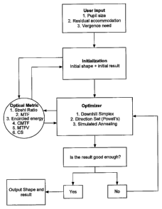
[0047] Fig. 5 is a flow chart illustrating exemplary method steps for
optimizing a optical
prescription that treats or corrects a vision condition.
12e
CA 02575202 2007-01-22
WO 2006/020422
PCT/US2005/026859
[0048] Fig. 6 illustrates a data flow process for shape optimization for
correction or treatment
of a vision condition.
[0049] Fig. 7 illustrates a comparison of Direction Set method and Downhill
Simplex method.
[0050] Figs. 8A and 8B illustrate alternative prescriptions optimized for an
eye of a particular
patient, and their characteristics.
[0051] Fig. 8C illustrates a comparison of optimizer values using even-term
polynomials and
all power term polynomials for pupil sizes of 4mm, 5mm, and 6mm.
[0052] Figs. 9A-D, show alternative presbyopia-mitigating prescriptions
optimized for an eye
of a particular patient.
[0053] Fig. 10 illustrates effects of random noise on prescriptions optimized
for an eye of a
particular patient.
[0054] Figs. 11A-C compare optimized prescriptions to alternative treatments
for differing
pupil sizes.
[0055] Figs. 12A-C compare optimized prescriptions to alternative treatments
for a range of
viewing distances.
[0056] Fig. 13 illustrates simulated viewing charts viewed at differing
distances to compare
optimized prescriptions to alternative treatments.
[0057] Figs. 14-16 illustrate graphical interface computer screen displays for
a prescription
optimizer and system.
[0058] Figs. 17 and 18 illustrate pupil sizes and changes at differing viewing
conditions for a
particular patient.
[0059] Fig. 19 graphically illustrates optimizer values for differing levels
of residual
accommodation.
[0060] Fig. 20 illustrates effects of pupil change and residual accommodation
on optimized
prescriptions for a particular patient.
[0061] Figs. 21A-C illustrate effects of pupil change and residual
accommodation on
optimized prescriptions for a particular patient.
13
CA 02575202 2007-01-22
WO 2006/020422
PCT/US2005/026859
[0062] Figs. 22-24 compare optical properties and results of eyes corrected
with a optimized
prescription to alternative treatments.
[0063] Fig. 25 schematically illustrates a system for determining a
prescription for a particular
patient and delivering that treatment using laser refractive surgery.
[0064] Fig. 26A illustrates a relationship between accommodation and pupil
size when healthy
eyes adjust to differing viewing distances.
[0065] Fig. 26B illustrates one exemplary relationship between effective power
of an eye and
pupil size for a patient, as can be provided from the presbyopia prescriptions
of the present
invention by generating an optical shape which effects desired changes in
power with changes in
pupil size of a particular patient under differing viewing conditions.
[0066] Fig. 26C illustrates a relationship between manifest power and pupil
diameter, for
example, as measured from patients having differing pupil diameters who have
been successfully
treated with a presbyopia-mitigating prescription. Such a relationship may be
used to identify a
desired change in optical power with changes in pupil diameter for a specific
patient.
[0067] Figs. 27A-B graphically illustrate optical properties of an eye
relevant to presbyopia.
[0068] Fig. 28 schematically illustrates a presbyopia-mitigating shape having
a central add
region.
[0069] Figs. 29 and 30 schematically illustrates residual accommodation and
presbyopia
treatments for increasing a focal range.
[0070] Figs. 31-37 graphically illustrate results from presbyopia-mitigating
treatments for a
population of individual patients.
[0071] Fig. 38 graphically illustrates accommodation through a range of
differing patient ages.
[0072] Fig. 39 schematically illustrates another system for determining a
presbyopia-
mitigating prescription for a particular patient and delivering that treatment
using laser refractive
surgery.
[0073] Figs. 40 and 41 graphically illustrate a presbyopia-mitigating
prescription derived so as
to provide appropriate effective powers at two differing viewing conditions
for a particular
patient.
14
CA 02575202 2007-01-22
WO 2006/020422
PCT/US2005/026859
[0074] Figs. 42 and 43 graphically illustrate a presbyopia-mitigating
prescription derived so as
to provide appropriate effective powers at three differing viewing conditions
for a particular
patient.
[0075] Figs. 44 and 45 graphically illustrate a presbyopia-mitigating
prescription derived so as
to provide appropriate effective powers at four differing viewing conditions
for a particular
patient.
[0076] Figs. 46A and 46B graphically illustrate different presbyopia-
mitigating prescriptions
which provide differing effective power variation characteristics during pupil
size changes under
differing viewing conditions.
[0077] Figs. 47 and 48 graphically illustrate effects of different pupil sizes
on derived
presbyopia-mitigating prescriptions and their optical characteristics.
[0078] Fig. 49 illustrates simulated eye-chart letters as viewed with a
presbyopic eye treated
with a presbyopia-mitigating prescription derived for a particular patient.
[0079] Figs. 50A and 50B illustrate an exemplary power/pupil correlation and
corresponding
presbyopia prescription.
DETAILED DESCRIPTION OF THE INVENTION
[0080] Although the methods, devices, and systems of the present invention are
described
primarily in the context of a laser eye surgery system, it should be
understood that the techniques
of the present invention may be adapted for use in other eye treatment
procedures and systems
such as contact lenses, intra-ocular lenses, radial keratotomy, collagenous
corneal tissue thermal
remodeling, removable corneal lens structures, glass spectacles, corneal ring
implants, and the
like.
[0081] Turning now to the drawings, Fig. 1 illustrates a laser eye surgery
system 10 of the
present invention, including a laser 12 that produces a laser beam 14. Laser
12 is optically
coupled to laser delivery optics 16, which directs laser beam 14 to an eye E
of patient P. A
delivery optics support structure (not shown here for clarity) extends from a
frame 18 supporting
laser 12. A microscope 20 is mounted on the delivery optics support structure,
the microscope
often being used to image a cornea of eye E.
[0082] Laser 12 generally comprises an excimer laser, ideally comprising an
argon-fluorine
laser producing pulses of laser light having a wavelength of approximately 193
nm. Laser 12
CA 02575202 2007-01-22
WO 2006/020422
PCT/US2005/026859
will preferably be designed to provide a feedback stabilized fluence at the
patient's eye, delivered
via delivery optics 16. The present invention may also be useful with
alternative sources of
ultraviolet or infrared radiation, particularly those adapted to controllably
ablate the corneal
tissue without causing significant damage to adjacent and/or underlying
tissues of the eye. Such
sources include, but are not limited to, solid state lasers and other devices
which can generate
energy in the ultraviolet wavelength between about 185 and 205 nm and/or those
which utilize
frequency-multiplying techniques. Hence, although an excimer laser is the
illustrative source of
an ablating beam, other lasers may be used in the present invention.
[0083] Laser system 10 will generally include a computer or programmable
processor 22.
Processor 22 may comprise (or interface with) a conventional PC system
including the standard
user interface devices such as a keyboard, a display monitor, and the like.
Processor 22 will
typically include an input device such as a magnetic or optical disk drive, an
internet connection,
or the like. Such input devices will often be used to download a computer
executable code from
a tangible storage media 29 embodying any of the methods of the present
invention. Tangible
storage media 29 may take the form of a floppy disk, an optical disk, a data
tape, a volatile or
non-volatile memory, RAM, or the like, and the processor 22 will include the
memory boards
and other standard components of modern computer systems for storing and
executing this code.
Tangible storage media 29 may optionally embody wavefront sensor data,
wavefront gradients, a
wavefront elevation map, a treatment map, a corneal elevation map, and/or an
ablation table.
While tangible storage media 29 will often be used directly in cooperation
with a input device of
processor 22, the storage media may also be remotely operatively coupled with
processor by
means of network connections such as the internet, and by wireless methods
such as infrared,
Bluetooth, or the like.
[0084] Laser 12 and delivery optics 16 will generally direct laser beam 14 to
the eye of patient
P under the direction of a computer 22. Computer 22 will often selectively
adjust laser beam 14
to expose portions of the cornea to the pulses of laser energy so as to effect
a predetermined
sculpting of the cornea and alter the refractive characteristics of the eye.
In many embodiments,
both laser beam 14 and the laser delivery optical system 16 will be under
computer control of
processor 22 to effect the desired laser sculpting process, with the processor
effecting (and
optionally modifying) the pattern of laser pulses. The pattern of pulses may
by summarized in
machine readable data of tangible storage media 29 in the form of a treatment
table, and the
treatment table may be adjusted according to feedback input into processor 22
from an
automated image analysis system in response to feedback data provided from an
ablation
16
CA 02575202 2012-12-10
monitoring system feedback system. Optionally, the feedback may be manually
entered into
the processor by a system operator. Such feedback might be provided by
integrating the
wavefront measurement system described below with the laser treatment system
10, and
processor 22 may continue and/or terminate a sculpting treatment in response
to the
feedback, and may optionally also modify the planned sculpting based at least
in part on the
feedback. Measurement systems are further described in U.S. Patent No.
6,315,413.
[0085] Laser beam 14 may be adjusted to produce the desired sculpting
using a variety of
alternative mechanisms. The laser beam 14 may be selectively limited using one
or more
variable apertures. An exemplary variable aperture system having a variable
iris and a
variable width slit is described in U.S. Patent No. 5,713,892. The laser beam
may also be
tailored by varying the size and offset of the laser spot from an axis of the
eye, as described
in U.S. Patent Nos. 5,683,379, 6,203,539, and 6,331,177.
[0086] Still further alternatives are possible, including scanning of
the laser beam over
the surface of the eye and controlling the number of pulses and/or dwell time
at each
location, as described, for example, by U.S. Patent No. 4,665,913; using masks
in the optical
path of laser beam 14 which ablate to vary the profile of the beam incident on
the cornea, as
described in U.S. Patent No. 5,807,379; hybrid profile-scanning systems in
which a variable
size beam (typically controlled by a variable width slit and/or variable
diameter iris
diaphragm) is scanned across the cornea; or the like. The computer programs
and control
methodology for these laser pattern tailoring techniques are well described in
the patent
literature.
17
CA 02575202 2012-12-10
[0087] Additional components and subsystems may be included with laser
system 10, as
should be understood by those of skill in the art. For example, spatial and/or
temporal
integrators may be included to control the distribution of energy within the
laser beam, as
described in U.S. Patent No. 5,646,791. Ablation effluent evacuators/filters,
aspirators, and
other ancillary components of the laser surgery system are known in the art.
Further details of
suitable systems for performing a laser ablation procedure can be found in
commonly
assigned U.S. Patent Nos. 4,665,913, 4,669,466, 4,732,148, 4,770,172,
4,773,414, 5,207,668,
5,108,388, 5,219,343, 5,646,791 and 5,163,934. Suitable
17a
CA 02575202 2007-01-22
WO 2006/020422
PCT/US2005/026859
systems also include commercially available refractive laser systems such as
those manufactured
and/or sold by Alcon, Bausch & Lomb, Nidek, WaveLight, LaserSight, Schwind,
Zeiss-Meditec,
and the like. Basis data can be further characterized for particular lasers or
operating conditions,
by taking into account localized environmental variables such as temperature,
humidity, airflow,
and aspiration.
[0088] Fig. 2 is a simplified block diagram of an exemplary computer system 22
that may be
used by the laser surgical system 10 of the present invention. Computer system
22 typically
includes at least one processor 52 which may communicate with a number of
peripheral devices
via a bus subsystem 54. These peripheral devices may include a storage
subsystem 56,
comprising a memory subsystem 58 and a file storage subsystem 60, user
interface input devices
62, user interface output devices 64, and a network interface subsystem 66.
Network interface
subsystem 66 provides an interface to outside networks 68 and/or other
devices, such as the
wavefront measurement system 30.
[0089] User interface input devices 62 may include a keyboard, pointing
devices such as a
mouse, trackball, touch pad, or graphics tablet, a scanner, foot pedals, a
joystick, a touchscreen
incorporated into the display, audio input devices such as voice recognition
systems,
microphones, and other types of input devices. User input devices 62 will
often be used to
download a computer executable code from a tangible storage media 29 embodying
any of the
methods of the present invention. In general, use of the term "input device"
is intended to
include a variety of conventional and proprietary devices and ways to input
information into
computer system 22.
[0090] User interface output devices 64 may include a display subsystem, a
printer, a fax
machine, or non-visual displays such as audio output devices. The display
subsystem may be a
cathode ray tube (CRT), a flat-panel device such as a liquid crystal display
(LCD), a projection
device, or the like. The display subsystem may also provide a non-visual
display such as via
audio output devices. In general, use of the term "output device" is intended
to include a variety
of conventional and proprietary devices and ways to output information from
computer system
22 to a user.
[0091] Storage subsystem 56 can store the basic programming and data
constructs that provide
the functionality of the various embodiments of the present invention. For
example, a database
and modules implementing the functionality of the methods of the present
invention, as
described herein, may be stored in storage subsystem 56. These software
modules are generally
18
CA 02575202 2007-01-22
WO 2006/020422
PCT/US2005/026859
executed by processor 52. In a distributed environment, the software modules
may be stored on
a plurality of computer systems and executed by processors of the plurality of
computer systems.
Storage subsystem 56 typically comprises memory subsystem 58 and file storage
subsystem 60.
[0092] Memory subsystem 58 typically includes a number of memories including a
main
random access memory (RAM) 70 for storage of instructions and data during
program execution
and a read only memory (ROM) 72 in which fixed instructions are stored. File
storage
subsystem 60 provides persistent (non-volatile) storage for program and data
files, and may
include tangible storage media 29 (Fig. 1) which may optionally embody
wavefront sensor data,
wavefront gradients, a wavefront elevation map, a treatment map, and/or an
ablation table. File
storage subsystem 60 may include a hard disk drive, a floppy disk drive along
with associated
removable media, a Compact Digital Read Only Memory (CD-ROM) drive, an optical
drive,
DVD, CD-R, CD-RW, solid-state removable memory, and/or other removable media
cartridges
or disks. One or more of the drives may be located at remote locations on
other connected
computers at other sites coupled to computer system 22. The modules
implementing the
functionality of the present invention may be stored by file storage subsystem
60.
[0093] Bus subsystem 54 provides a mechanism for letting the various
components and
subsystems of computer system 22 communicate with each other as intended. The
various
subsystems and components of computer system 22 need not be at the same
physical location but
may be distributed at various locations within a distributed network. Although
bus subsystem 54,
is shown schematically as a single bus, alternate embodiments of the bus
subsystem may utilize
multiple busses.
[0094] Computer system 22 itself can be of varying types including a personal
computer, a
portable computer, a workstation, a computer terminal, a network computer, a
control system in
a wavefront measurement system or laser surgical system, a mainframe, or any
other data
processing system. Due to the ever-changing nature of computers and networks,
the description
of computer system 22 depicted in Fig. 2 is intended only as a specific
example for purposes of
illustrating one embodiment of the present invention. Many other
configurations of computer
system 22 are possible having more or less components than the computer system
depicted in
Fig. 2.
[0095] Referring now to Fig. 3, one embodiment of a wavefront measurement
system 30 is
schematically illustrated in simplified form. In very general terms, wavefront
measurement
system 30 is configured to sense local slopes of a gradient map exiting the
patient's eye. Devices
19
CA 02575202 2007-01-22
WO 2006/020422
PCT/US2005/026859
based on the Hartmann-Shack principle generally include a lenslet array to
sample the gradient
map uniformly over an aperture, which is typically the exit pupil of the eye.
Thereafter, the local
slopes of the gradient map are analyzed so as to reconstruct the wavefront
surface or map.
[0096] More specifically, one wavefront measurement system 30 includes an
image source 32,
such as a laser, which projects a source image through optical tissues 34 of
eye E so as to form
an image 44 upon a surface of retina R. The image from retina R is transmitted
by the optical
system of the eye (e.g., optical tissues 34) and imaged onto a wavefront
sensor 36 by system
optics 37. The wavefront sensor 36 communicates signals to a computer system
22' for
measurement of the optical errors in the optical tissues 34 and/or
determination of an optical
tissue ablation treatment program. Computer 22' may include the same or
similar hardware as
the computer system 22 illustrated in Figs. 1 and 2. Computer system 22' may
be in
communication with computer system 22 that directs the laser surgery system
10, or some or all
of the components of computer system 22, 22' of the wavefront measurement
system 30 and
laser surgery system 10 may be combined or separate. If desired, data from
wavefront sensor 36
may be transmitted to a laser computer system 22 via tangible media 29, via an
I/O port, via an
networking connection 66 such as an intranet or the Internet, or the like.
[00971 Wavefront sensor 36 generally comprises a lenslet array 38 and an image
sensor 40. As
the image from retina R is transmitted through optical tissues 34 and imaged
onto a surface of
image sensor 40 and an image of the eye pupil P is similarly imaged onto a
surface of lenslet
array 38, the lenslet array separates the transmitted image into an array of
beamlets 42, and (in
combination with other optical components of the system) images the separated
beamlets on the
surface of sensor 40. Sensor 40 typically comprises a charged couple device or
"CCD," and
senses the characteristics of these individual beamlets, which can be used to
determine the
characteristics of an associated region of optical tissues 34. In particular,
where image 44
comprises a point or small spot of light, a location of the transmitted spot
as imaged by a beamlet
can directly indicate a local gradient of the associated region of optical
tissue.
[0098] Eye E generally defines an anterior orientation ANT and a posterior
orientation POS.
Image source 32 generally projects an image in a posterior orientation through
optical tissues 34
onto retina R as indicated in Fig. 3. Optical tissues 34 again transmit image
44 from the retina
anteriorly toward wavefront sensor 36. Image 44 actually formed on retina R
may be distorted
by any imperfections in the eye's optical system when the image source is
originally transmitted
by optical tissues 34. Optionally, image source projection optics 46 may be
configured or
adapted to decrease any distortion of image 44.
CA 02575202 2012-12-10
[0099] In some embodiments, image source optics 46 may decrease lower
order optical
errors by compensating for spherical and/or cylindrical errors of optical
tissues 34. Higher
order optical errors of the optical tissues may also be compensated through
the use of an
adaptive optic element, such as a deformable mirror (described below). Use of
an image
source 32 selected to define a point or small spot at image 44 upon retina R
may facilitate the
analysis of the data provided by wavefront sensor 36. Distortion of image 44
may be limited
by transmitting a source image through a central region 48 of optical tissues
34 which is
smaller than a pupil 50, as the central portion of the pupil may be less prone
to optical errors
than the peripheral portion. Regardless of the particular image source
structure, it will be
generally be beneficial to have a well-defined and accurately formed image 44
on retina R.
[0100] In one embodiment, the wavefront data may be stored in a computer
readable
medium 29 or a memory of the wavefront sensor system 30 in two separate arrays
containing
the x and y wavefront gradient values obtained from image spot analysis of the
Hartmann-
Shack sensor images, plus the x and y pupil center offsets from the nominal
center of the
Hartmann-Shack lenslet array, as measured by the pupil camera 51 (Fig. 3)
image. Such
information contains all the available information on the wavefront error of
the eye and is
sufficient to reconstruct the wavefront or any portion of it. In such
embodiments, there is no
need to reprocess the Hartmann-Shack image more than once, and the data space
required to
store the gradient array is not large. For example, to accommodate an image of
a pupil with
an 8 mm diameter, an array of a 20 x 20 size (i.e., 400 elements) is often
sufficient. As can be
appreciated, in other embodiments, the wavefront data may be stored in a
memory of the
wavefront sensor system in a single array or multiple arrays.
[0101] While the methods of the present invention will generally be
described with
reference to sensing of an image 44, a series of wavefront sensor data
readings may be taken.
For example, a time series of wavefront data readings may help to provide a
more accurate
overall determination of the ocular tissue aberrations. As the ocular tissues
can vary in shape
over a brief period of time, a plurality of temporally separated wavefront
sensor
measurements can avoid relying on a single snapshot of the optical
characteristics as the
21
CA 02575202 2012-12-10
basis for a refractive correcting procedure. Still further alternatives are
also available,
including taking wavefront sensor data of the eye with the eye in differing
configurations,
positions, and/or orientations. For example, a patient will often help
maintain alignment of
the eye with wavefront measurement system 30 by focusing on a fixation target,
as described
in U.S. Patent No. 6,004,313. By varying a position of the fixation target as
described in that
reference, optical characteristics of the eye may be determined while the eye
accommodates
or adapts to image a field of view at a varying distance and/or angles.
[0102] The location of the optical axis of the eye may be verified by
reference to the data
provided from a pupil camera 52. In the exemplary embodiment, a pupil camera
52 images
pupil 50 so as to determine a position of the pupil for registration of the
wavefront sensor
data relative to the optical tissues.
[0103] An alternative embodiment of a wavefront measurement system is
illustrated in
Fig. 3A. The major components of the system of Fig. 3A are similar to those of
Fig. 3.
Additionally, Fig. 3A includes an adaptive optical element 53 in the form of a
deformable
mirror. The source image is reflected from deformable mirror 98 during
transmission to
retina R, and the deformable mirror is also along the optical path used to
form the transmitted
image between retina R and imaging sensor 40. Deformable mirror 98 can be
controllably
deformed by computer system 22 to limit distortion of the image formed on the
retina or of
subsequent images formed of the images formed on the retina, and may enhance
the accuracy
of the resultant wavefront data. The structure and use of the system of Fig.
3A are more fully
described in U.S. Patent No. 6,095,651.
[0104] The components of an embodiment of a wavefront measurement system
for
measuring the eye and ablations may comprise elements of a VISX WaveScan ,
available
from VISX, INCORPORATED of Santa Clara, California. One embodiment includes a
WaveScan with a deformable mirror as described above. An alternate embodiment
of a
wavefront measuring system is described in U.S. Patent No. 6,271,915. It is
appreciated that
any wavefront aberrometer could be employed for use with the present
invention.
22
CA 02575202 2012-12-10
[0105] The present invention is useful for enhancing the accuracy and
efficacy of
photorefractive keratectomy (PRK), laser in situ keratomileusis (LASIK), laser
assisted
epithelium keratomileusis (LASEK), and the like. The present invention can
provide
enhanced optical correction approaches by improving the methodology for
scaling an optical
shape, or by generating or deriving new optical shapes, and the like.
[0106] The techniques of the present invention can be readily adapted
for use with
existing laser systems, including the VISX Excimer laser eye surgery systems
commercially
available from VISX of Santa Clara, California. Other suitable laser systems
are
manufactured by Alcon, Bausch & Lomb, Wavelight, Schwind, Zeiss-Meditec,
Lasersight,
Nidek and the like. By
22a
CA 02575202 2007-01-22
WO 2006/020422
PCT/US2005/026859
providing improved corneal ablation profiles for treating optical defects, the
present invention
may allow enhanced treatment of patients who have heretofore presented
difficult or complicated
treatment problems. When used for determining, deriving, and/or optimizing
prescriptions for a
particular patient, the systems and methods may be implemented by calculating
prescriptions for
a range of patients, for example, by calculating discrete table entries
throughout a range of
patient characteristics, deriving or empirically generating parametric patient
characteristic/prescription correlations, and the like, for subsequent use in
generating patient-
specific prescriptions.
[0107] When designing a prescriptive shape for an eye treatment, it is useful
to select a
mathematical gauge of optical quality appropriate for the vision condition for
use as a goal
function. This allows for quantification and optimization of the shape, and
for comparison
among different shapes. The present invention provides methods for
establishing a customized
optical shape for a particular patient based on a set of patient parameters
per the goal function.
By incorporating iterative optimization algorithms, it is also possible to
generate a shape having
an optimized level of optical quality for the particular patient.
[0108] Selecting A Goal Function Appropriate For A Vision Condition
[0109] The goal function relates to optical quality, and it can be, for
example, based on, or a
function of (or related to) optical metrics such as Strehl ratio (SR),
modulation transfer function
(MTF), point spread function (PSF), encircled energy (EE), MTF volume or
volume under MTF
surface (MTFV), or contrast sensitivity (CS); and optionally to new optical
metrics which are
appropriate to vision conditions such as presbyopia; for instance, compound
modulation transfer
function (CMTF) as described below. In optical terms, the goal function should
make sense.
That is to say, minimization or maximization of the goal function should give
a predictable
optimized optical quality of the eye. The goal function can be a function with
a certain number
of free parameters to be optimized (minimized) through an optimization, or
minimization,
algorithm.
[0110] Although there are many types of goal functions available for use with
the present
invention, the discussion below generally touches on two broad schools of goal
functions. In a
Diffraction Theory based approach, the shape is considered as a wave
aberration. Typically, a
Fourier transform is employed for calculating optical quality related
parameters, such as Strehl
ratio (SR), modulation transfer function (MTF), MTF volume or volume under MTF
surface
(MTFV), compound modulation transfer function (CMTF), or contrast sensitivity
(CS), encircled
23
CA 02575202 2007-01-22
WO 2006/020422
PCT/US2005/026859
energy (EE) (based on point spread function), as well as special cases that
combine one or more
of these parameters, or values of the parameters in specific situations (such
as MTF at spatial
frequency or encircled energy at a field of view), or integration of any
parameters (volume of
MTF surface at all frequencies or up to a cutoff frequency, for example 60
cycles/degree or 75
cycles/degree, because 60 cycles/degree is the retina cone's limiting spatial
frequency). In a
Geometrical Optics approach, or the so-called ray tracing approach, the
optical effect is based on
ray tracing. With both the Diffraction Theory and the Geometrical Optics
approaches,
polychromatic point spread function with Stiles-Crawford effect, chromatic
aberrations as well
as retina spectral response function can be used.
[0111] Monochromatic point spread function (PSF) has been used for describing
optical
defects of optical systems having aberrations. Due to the simple relationship
between wave
aberrations and the PSF for an incoherent light source, Fourier transform of
the generalized pupil
function has been used in the calculation of point spread functions. Most
optical applications,
however, do not use a monochromatic light source. In the case of human vision,
the source is
essentially white light. Thus, there are limitations associated with the use
of monochromatic PSF
as a goal function.
[0112] Polychromatic point spread function (PSF) with correct chromatic
aberrations, Stiles-
Crawford effect as well as retina response function, can be used for optical
modeling of human
eyes. Here, chromatic aberrations arise because light composed of different
wavelengths will
focus either in front of the retina or behind it. Only portions of the light
will focus exactly on the
retina. This gives the eye an extended depth-of-focus, i.e., if one has
focusing error of some
amount, the eye is still capable of focusing at least for some wavelengths.
Therefore, chromatic
aberrations in fact help the correction of presbyopia. If the depth-of-focus
is sufficiently large,
there would be no presbyopia problem. Unfortunately, the chromatic aberrations
are not large
enough and it also varies with the wavelength. Stiles-Crawford effect, also
known as pupil
apodization, is due to the waveguide property of the retinal cones. Light from
the pupil
periphery has a slightly less chance of being detected by the retina because
the ray of light might
not reach the bottom of the cone, due to a slight incident angle. As for the
retinal spectral
response function, it is known that the cones, which are responsible for
daylight vision, have
different sensitivity to different wavelengths. Only green light is absorbed
by the eye almost
completely. Both blue light and red light are absorbed by the eye partially.
[0113] Once the PSF is calculated, calculation of the Strehl ratio is
straightforward. Strehl
ratio can be defined as the ratio of the peak of the point spread function
(PSF) of an optical
24
CA 02575202 2007-01-22
WO 2006/020422
PCT/US2005/026859
system to the peak of a diffraction-limited optical system with the same
aperture size. An
example of a Strehl ratio is shown in Fig. 27A. A diffraction-limited optical
system is typically
a system with no aberrations, or optical errors. It can be an ideal or perfect
optical system,
having a Strehl ratio of 1.
[0114] The goal function can also be a function of modulation transfer
function (MTF).
Modulation transfer function can be used to predict visual performance.
Typically, the MTF at
one spatial frequency corresponds to one angular extend of features of
targets. The modulation
transfer function (MTF) can be calculated with the following formulations:
MTF(u,v) = FT[PSF(x,y)]
MTF(u,v) = Re[GPF(x,y) 4 GPF(x,y)]
where u and v represent spatial frequencies, Re represents the real part of a
complex number, FT
represents a Fourier Transform, GPF represents a generalized pupil function,
and x and y
represent position or field of view. An example of an MTF is shown in Fig.
27B.
[0115] Modulation transfer function (MTF) is a measure for how much spatial
details are
transferred from pupil space to imaging space (retina in the case of human
eye). MTF can be
related to contrast sensitivity (CS). Mathematically MTF can be defined as the
Fourier transform
of the point spread function as
h(u,v) = y)exp[¨i27c (ux + vy)]clxdy, ,
where i(x,y) is the point spread function (PSF). Calculation of PSF can be
done with the Fourier
transform of the generalized pupil function.
[0116] MTF at a specific spatial frequency can represent the percentage of the
sinusoidal wave
of a specific spatial frequency that is preserved after going through the
optical system. MTF at
cycles/degree and at 60 cycles/degree are considered as important because 30
cpd
corresponds to 20/20 visual acuity and 60 cpd corresponds to 20/10 visual
acuity, the highest
25 spatial resolution the cones in the retinal can process. MTF at other
spatial frequencies may also
be useful.
[0117] The volume under the MTF surface up to a certain spatial frequency
(such as 60 cpd)
can be meaningful as it includes all spatial frequency information. In some
cases, it is desirable
CA 02575202 2007-01-22
WO 2006/020422
PCT/US2005/026859
to use the volume under MTF surface within a band (i.e. from one specific
spatial frequency to
another specific spatial frequency).
Compound Modulation Transfer Function
[0118] Compound MTF can be calculated as a linear combination of MTF at
certain spatial
frequencies, normalized at diffraction-limited MTF, and can be represented by
the following
formula
"
CMTF = 1 ¨Ecc ;hi ,
n "
where n is the number of MTF curves, al is the reciprocal of the ith
diffraction-limited MTF, and
hi is the ith MTF curve. The selection of certain spatial frequencies can
depend on the
importance of each frequency. For example, in the case of presbyopia, 20/40
vision may be
more important than 20/20 as the distance vision is often compromised by the
improved near
vision. Figs 4A and 4B show an examples of an CMTF curve as well as it's
individual MTF
curves at different specific spatial frequencies. In a perfect optical system,
CMTF is equal to
one.
[0119] In a related embodiment, the compound MTF can be calculated as
F(v) = (aiMTFI + a 2MTF2 + a 3MTF3)/ 3
where MTFi, MTF2, and MTF3 are the MTF values at 10 cycles/degree, 20
cycles/degree and 30
cycles/degree, respectively. These correspond to Snellen eye chart of 20/60,
20/40 and 20/20
visions, respectively. The weighting coefficients al, a2, a3 can be chosen so
that 1/ai, 1/a2, 1/a3
are the diffraction-limited MTF at these spatial frequencies, respectively.
Therefore, in the
diffraction-limited case, the compound MTF F(v) can have a maximal value of
unity.
[0120] Where MTF at one spatial frequency corresponds to one angular extend of
features of
targets, compound MTF can be calculated as linear combination of MTF at
different spatial
frequencies normalized by a diffraction-limited MTF, and can similarly be used
to predict visual
outcome. Another general formula for the calculation of CMTF as a function of
visual vergence
(nu) is
1 "
CMTF (v) = ¨ Ea i./14TFi(v)
77 1=1
26
CA 02575202 2007-01-22
WO 2006/020422
PCT/US2005/026859
where ai is the reciprocal of the i-th diffraction-limited MTF. This formula
can provide CMTF
for all possible vergence. In some cases, three MTF curves at 10, 20 and 30
cycles per degree
are used. An ideal value of CMTF can be about 1. Good values can be about 0.2
or about 0.3.
In a healthy eye, the spatial frequency limit can be about 60 cycles per
degree due to the
configuration of retina cones. However, in the treatment of presbyopia, for
example, it may not
be necessary to provide a treatment corresponding to this limit, as the
treatment will often
involve a compromise of good distance and near sight. Optionally, a minimum
distance vision
gauge desired target may be provided, with near sight being optimized and, as
needed,
compromised.
[0121] Fig. 4A illustrates an example of the compound MTF over a vergence of 3
diopters
(upper panel) versus its corresponding individual MTF curves at 15, 30, and 60
cycles per degree
(lower panel). Fig. 4B illustrates an example of the compound MTF over a
vergence of 3
diopters (upper panel) versus its corresponding individual MTF curves at 10,
20, and 30 cycles
per degree (lower panel). Compound MTF can correlate well with visual acuity
and contrast
sensitivity at the same time, at least optically. In some embodiments, the
compound modulation
transfer function is determined for individual MTF curves at 30, 45, and 60
cpd. The selection of
the individual MTF curve values can involve a linear combination based on the
optical response
of the eye.
[0122] In general, there can be two different types of cutoff spatial
frequencies, and each
involves a factors that affect acuity. Cutoff spatial frequency can correspond
to the maximum
spatial frequency, above which information can no longer be used. Whereas most
individuals
can discern information from objects having very low spatial frequency, as the
spatial frequency
increases, it is typically increasingly more difficult for an individual to
discern information from
such objects. At some threshold, an increased spatial frequency no longer
yields increased
information.
[0123] A first type of cutoff spatial frequency is related to aperture
dimension. In this case, a
system having a larger aperture (e.g. an eye with a larger pupil) will
correspond to a larger cutoff
spatial frequency. Conversely, a system having a smaller aperture (e.g. an eye
with a smaller
pupil) will correspond to a smaller cutoff spatial frequency. Often, such
cutoff spatial
frequencies will be linearly dependent on a pupil dimension, for example the
pupil diameter.
Smaller pupil sizes typically correspond to an extended, or larger, depth of
focus. Relatedly,
27
CA 02575202 2007-01-22
WO 2006/020422
PCT/US2005/026859
smaller pupil sizes often result in lower resolution. Assuming there are no
aberrations, a larger
pupil size is thought to confer increased resolution.
[0124] A second type of cutoff spatial frequency typically depends on the
spacing of cones on
the retina of the eye. With this type of cutoff spatial frequency, the
standard value is 30 cpd,
which corresponds to 20/20 vision. Another value, 60 cpd, corresponds to 20/10
vision and is
often considered a physiological limit. In such cases, the retinal cones are
very closely spaced.
The spacing of retinal cones will vary among individuals.
[0125] In the example of presbyopia treatment, it may be desirable to maintain
a lower spatial
frequency. In some cases, presbyopia will involve a compromise between
distance and near
vision. It may be difficult to achieve high spatial resolution, thus enhancing
the desirability of
emphasizing lower and medium spatial frequency information. In other words,
high spatial
frequency information may be sacrificed in order to improve the combination of
near and
distance vision.
[0126] As noted above, a compound modulation transfer function can include
individual MTF
curves at various combinations of spatial frequencies, such as 15, 30, and 60
cycles per degree
and 10, 20, and 30 cycles per degree. An individual MTF can have a value
ranging from about 5
cycles per degree to about 75 cycles per degree. In many instances, at least
one individual MTF
of a CMTF will range from about 10 cycles per degree to about 30 cycles per
degree, and can
often be about 20 cycles per degree. Where a CMTF includes three individual
MTF's, a first
individual MTF can range from about 5 cycles per degree to about 20 cycles per
degree, a second
individual MTF can range from about 15 cycles per degree to about 45 cycles
per degree, and a
third individual MTF can range from about 30 cycles per degree to about 75
cycles per degree.
In some circumstances, the upper limit of an individual MTF can be about 60
cycles per degree.
[0127] In some cases, the CMTF will be based on an average of the individual
MTF curves. In
some embodiments, the present invention provides compound modulation transfer
functions that
correspond to three, four, five, or any number of individual modulation
transfer functions. For
example, a CMTF can include from about 2 to about 7 individual MTF's. A CMTF
can also
include from about 3 to about 6 individual MTF's.
[0128] Individual MTF's can correspond to a curve through a certain vergence.
Typically, a
target at a far distance corresponds to a small vergence value. As a target
moves closer to the
eye, the vergence increases. The individual MTF's can be based on a value
ranging from about
zero to about three diopters.
28
CA 02575202 2007-01-22
WO 2006/020422
PCT/US2005/026859
[0129] The individual MTF's can be selected based on any number of criteria,
such as
empirical data or clinical observations. Relatedly, individual MTF's can be
chosen for pure
testing purposes. The CMTF can provide a parameter to evaluate the
effectiveness of a treatment
for a vision condition, such as presbyopia. Often, the CMTF will correlate
with a particular
visual outcome.
[0130] To establish an optically optimized shape appropriate for a vision
condition, at least one
of the goal functions, such as Strehl ratio, encircled energy, or MTF, MTF
volume or volume
under MTF surface (MTFV), compound modulation transfer function (CMTF), or
contrast
sensitivity (CS) should be maximized. For improved vision condition treatment,
the optical
metric can be maximized in all target vergence, that is, for targets at all
distances. Furthermore,
it is also desirable to minimize the fluctuation of the goal function.
Therefore, the goal function,
which is incorporated into the optimization algorithm of the optimizer, can be
defined as
di'
0(c1,c2,...,PAR)=(1+a)(1+PV) ____________________________
F(v)dv
where 0 is the goal function; cl, c2, ... are the polynomial coefficients; PAR
is presbyopia-add to
pupil ratio (described below); v is the vergence; F(v) is one of the optical
metrics; a is the
standard deviation of F(v), PV is the peak-to-valley of F(v); and vo is the
end point of the
vergence range, which may be (for example) between 15 and 100 cm, such as 40
cm. Because
idv is a constant, either a smaller a or a larger IF(v)dv can minimize the
goal function 0.
[0131] The formulas given here are examples of the many formulae that can be
used as the
goal function. The basic approach will often be to provide a goal function
that is optimized to
give as practical a solution as possible for correction or treatment of the
vision condition.
[0132] The compound MTF may reflect to what extent information is being
modulated when
passing through an optical system. For example, CMTF can represent the
percentage of
information at different spatial frequencies that is retained.
Selecting An Iterative Optimization Algorithm
[0133] Any of a number of optimization algorithms may be used by the optimizer
to maximize,
minimize, or otherwise globally or locally optimize the goal function. Because
many numerical
algorithms use function minimization concept, it is often convenient, but not
necessarily
required, to use minimization of the goal function. As examples, N-dimensional
minimization
29
CA 02575202 2012-12-10
algorithms such as the Downhill Simplex method, the Direction Set method, and
the
Simulated Annealing method can be used to optimize the goal function.
Likewise, the
algorithm described by Press et al., in "Numerical Recipes in C++", Cambridge
University
Press, 2002 can also be used. Algorithms such as those listed above are often
used for
function optimization in multi-dimensional space.
10134] The Downhill Simplex method starts with an initialization of N+1
points or
vertices to construct a simplex for an N-dimensional search, and in every
attempt tries to
reflect, stretch, or shrink the simplex by geometrical transformation so that
a close-to-global
minimum or pre-defined accuracy can be found. When Gaussian random noise of
standard
deviation of 0.02 1.tm in optical path difference (OPD) is added, the
algorithm still converges,
with no degradation.
101351 In the case of Direction Set method, also known as Powell's
method, N one-
dimensional vectors are initialized and the N-dimensional search is split in
such a way that a
one N-dimensional vector is chosen and the minimization is done in that
direction while
other variables (N-1 dimensions) are fixed. This process is continued until
all dimensions are
covered. A new iteration is initiated until the pre-determined criterion is
met. The Direction
Set method can use a separate one-dimensional minimization algorithm such as a
Golden
section search.
CA 02575202 2012-12-10
[0136] The Simulated Annealing method, which is useful for dealing with
a large number
of uncertainties, starts with an initial configuration. The objective is to
minimize E (analog to
energy) given the control parameter T (analog to temperature). Simulated
Annealing is
analogous to annealing, is a recent, proven method to solve otherwise
intractable problems,
and may be used to solve the ablation equation in laser ablation problem. This
is more fully
described in PCT Application No. PCT/US01/08337, filed March 14, 2001, and in
U.S.
Patent No. 6,673,062, issued January 6, 2004. Simulated annealing is a method
that can be
used for minimizing (or maximizing) the parameters of a function. It is
particularly suited to
problems with very large, poorly behaved function spaces. Simulated annealing
can be
applied in the same way regardless of how many dimensions are present in the
search space.
It can be used to optimize any conditions that can be expressed numerically,
and it does not
require a derivative. It can also provide an accurate overall minimum despite
local minima in
the search space, for example.
[0137] Fig. 5 shows the flow chart of an overall method for shape
optimization for a
vision condition treatment. Each functional block may contain one or more
alternatives. To
create a add-on shape W(r) for a vision condition treatment, an iterative
function
minimization algorithm
30a
CA 02575202 2007-01-22
WO 2006/020422
PCT/US2005/026859
can be employed such that the goal function, which could be a function of any
suitable optical
metrics (e.g. CMTF) is itself optimized to solve for an unknown shape. The
shape can be
expanded into a set of even power term polynomials (EPTP) or non-EPTP (i.e.
all power term
polynomials). EPTP refers to polynomials that only have the even power terms,
for instance,
F(r) = ar2 + br4 + cr6. The goal function should have good correlation with
visual performance,
at least optically. Point spread function can be calculated to obtain
additional and/or alternative
optical metrics. The vision condition prescription can refer to an optical
surface that can be used
to treat or mitigate the vision condition. It can correspond to, for example,
the shape of a
spectacle lens, a contact lens, an intra-ocular lens, a tissue ablation
profile for refractive surgery,
and the like.
[0138] Another representation of the data flow process is depicted in the flow
chart in Fig. 6,
which shows data flow for shape optimization for presbyopia correction. Again,
each functional
block may contain one or more alternatives.
[0139] It is desirable that the optimizer provide satisfactory outcome in
terms of attributes such
as result, convergence, and speed. Fig. 7 shows a comparison of Direction Set
method and
Downhill Simplex method for the following inputs: pupil size 5.6 mm, vergence
3D and
vergence step 0.1D. Direction Set method uses 17 iterations and Downhill
Simplex method uses
152 iterations. Each Direction Set method iteration takes longer than each
Downhill Simplex
method iteration. The optimizer value for the Direction Set method is 2.8
while that for the
Downhill Simplex method is 2.658. Shape for left panel is as
¨0.9055r2+6.4188r4-
2.6767r6+0.5625r8 with ratio of 0.7418.
[0140] Both algorithms seem to converge to a similar shape, although the
depths of the shapes
are different. Considering the difference in the pupil ratio, however, the
actual shapes within
70% of the pupil radius are quite close. When the vergence step is smaller,
each iteration can
take a longer time, but the overall number of iterations often tends to become
smaller.
Inputting An Initial Prescription Into an Optimizer
[0141] The initial prescription, often comprising an optical surface shape,
may be defined by
an expansion such as a polynomial (EPTP, non-EPTP), a Zernike polynomial, a
Fourier series, or
a discrete shape entirety. A discrete shape entirety can also be referred to
as a direct surface
representation by numerical grid values. The prescription shape may be assumed
to be circularly
or radially symmetric, with the aim of approaching an emmetropic eye. The
symmetric shape
can be decomposed into a set of polynomials, such that it has one or more
independent variables.
31
CA 02575202 2007-01-22
WO 2006/020422
PCT/US2005/026859
One of the variables can be the presbyopia-add to pupil ratio (PAR), or the
ratio of the shape
diameter to the pupil diameter. When a central power add region is employed
(as described
below), the PAR can be the ratio of the radius of the presbyopia-add to the
radius of the pupil. It
will also be appreciated that the ratios discussed herein can be based on area
ratios or on
diameter or radius ratios. It should be assumed that when diameter or radius
ratios are discussed,
that discussion also contemplates area ratios. In certain cases, the PAR can
range from about 0.2
to about 1Ø Relatedly, in some cases the methods of the present invention
can constrain the
PAR to range from about 0.2 to about 1Ø The other variables can be the
coefficients of each
polynomial term. For example,
Shape(r) = ar + br2 + cr3 + dr 4 + + fr6
[0142] The diameter of the shape can be larger than the pupil size, but if so
special
considerations may need to be taken. For example, it may be necessary to only
consider the net
shape within the pupil.
[0143] The polynomials can be normal polynomials or polynomials with even
power terms
only. For example, even-power-term polynomials (EPTP) up to the 6th or 8th
order can be used
to obtain a practically good output, that is, a practical optimal shape for
the particular patient.
Residual accommodation can also play an active role in presbyopia correction.
In a related
instance, normal presbyopes can be treated with the prescription obtained in
this approach
together with a prescription for the correction of the refractive error.
[0144] As an example, a circularly or radially symmetric, pupil-size dependent
shape for
presbyopia-add can be assumed for emmetropic presbyopes. The shape can then be
expanded to
polynomials up to the 6th or 8th order. With the optimization procedure, it is
found that
polynomial expansion of the shape up to the 6th or 8th order can be used to
obtain a practical
optimal shape for presbyopia correction.
[0145] In a wavefront with aberrations, denoted by W(r, , 0), the wavefront
can be thought of as
an optimal shape for vision correction. The polychromatic PSF can be expressed
as
P SF = E R(A) FFT(Põ(r) exp[¨ j-27r [W (r , 0) + aD(A) +V (1) + RA(1)]1) 2
A
where R(k) is the retina spectral response function and can be approximated to
32
CA 02575202 2007-01-22
WO 2006/020422
PCT/US2005/026859
R(2)
and P(r) is the pupil apodization function (Stiles-Crawford) and can be
written as
P5 (r) =10 R2
and D(k) is chromatic aberration at wavelength k and is close to
D(2) = ¨21.587 + 92.872 ¨134.9822 + 67.407k3
and v(l is the vergence induced aberration at distance 1 meters, and RA (1) is
the residual
accommodation induced aberrations with a different sign as compared to V(/).
When there are
no aberrations, RA(1) can cancel v(l as long as there is enough residual
accommodation in the
eye. Here, the central wavelength k is taken as 0.55 gm (as all wavelength
units in the above
formulae are in gm). The pupil apodization strength parameter p is taken as
0.06. a is the
conversion factor from diopter to optical path difference (OPD). FFT denotes a
fast Fourier
transform and I*1 denotes the module of a complex number.
[0146] The polychromatic point spread function, or PPSF, can be the point
spread function of
an eye as calculated with consideration of the polychromatic nature of the
incident light.
Further, the chromatic aberrations, the Stiles-Crawford effect, as well as the
retinal spectral
response function can also be considered.
[0147] The vergence induced aberration, or VIA, can be equal to the reciprocal
of the vergence
distance. When a target at a certain distance is viewed by the eye, it is the
same as viewing the
target at infinity but the eye has an additional aberration, the vergence
induced aberration.
[0148] For emmetropic eyes, it may be desirable that the wavefront that is
optimized be
circularly symmetric. Therefore, it can be decomposed into a set of
polynomials (non-EPTP) as
W (r)= ar + br2 + cr3 + dr4 +er5+ = = =
[0149] However, if it is desirable that the edge of the shape be smoother, it
may be
advantageous to decompose the wavefront into a set of even-power-term
polynomials (EPTP) as
W (r)= ar2 + br4 + cr6 + dr' + = = =
[0150] Using even power term polynomials (EPTP) also can help to establish a
surface shape
that is more round at the center, which creates certain manufacturing or
ablation efficiencies.
[0151] It may also be useful to denote another parameter, t, to be the ratio
of the radius of the
wavefront R to the radius of the pupil Ro. This is because both D(2,) and V(Z)
can have the same
33
CA 02575202 2007-01-22
WO 2006/020422
PCT/US2005/026859
size as the pupil and W(r) usually has a smaller size. When the calculated t
is larger than 1, the
shape can become larger than the pupil. In this case, only the portion of the
shape up to the pupil
size is used for optical quality evaluation.
[0152] As depicted in Fig. 8A, although normal polynomials can give slightly
better optimizer
values than even-power-term polynomials, the prescription may be harder to
realize. Fig. 8A
illustrates a comparison of shapes with normal polynomials (left panel) and
with even-power-
term polynomials (right panel). The shape on the right panel can be expanded
as ¨1.6154r +
1.7646r2+ 1.2646r3+ 1.9232r4+ 0.1440r5+ 0.1619r6 with a ratio of 0.8 and the
shape on the left
panel can be expanded as ¨1.1003r2+8.2830r4+0.7305r6-2.2140r8 with a ratio of
0.9106. Both
were determined using Downhill Simplex method for a pupil size of 5.6 mm and
vergence of 3D
with 0.1D step, without residual accommodation. The left panel shows an
optimal shape for 6
normal polynomial terms and the right panel shows an optimal shape with 4 EPTP
terms. It has
been found that polynomials up to the 8th power (4 EPTP terms) appear to give
highly
satisfactory results.
[0153] Fig. 8B shows another comparison of EPTP and non-EPTP expansions. The
left panel
shows an optimized shape based on an 8th order expansion (EPTP), whereas the
right panel
shows an optimized shape based on a 3rd order expansion (non-EPTP). In
general, shapes
derived from an EPTP have a smoother shape with a flat central zone. This flat
central zone can
correspond to good distance visual performance.
[0154] Another comparison of EPTP and non-EPTP expansions is provided in Fig.
8C, which
shows optimized (minimized) values with EPTP and non-EPTP expansion for a 4,
5, and 6 mm
pupil over a 3D vergence distance. In general, non-EPTP optimization gives a
slightly smaller
(more optimized) value than EPTP. Sixth-order EPTP appears to give the
smallest value for 4
mm and 5 mm pupils and eighth-order EPTP appears to give the smallest value
for a 6 mm pupil.
Third-order non-EPTP appears to give the smallest value for 4 mm and 5 mm
pupils and fourth-
order non-EPTP appears to give the smallest value for a 6mm pupil.
[0155] Using an even-power-term polynomial (EPTP) expansion can result in a
smoother
shape than a non-EPTP expansion. This smooth shape can be the minimal
requirement for good
distance vision. In general, 6th-order or 8th-order EPTP expansion and 3rd-
order or 4th-order non-
EPTP expansion result in good optimized value. Without residual accommodation,
larger pupils
can be more difficult to optimize than smaller pupils. This is shown, for
example, in Fig. 11A.
34
CA 02575202 2007-01-22
WO 2006/020422
PCT/US2005/026859
[0156] The optimized multi-focal shape appears to give much more balanced
results for the
correction of presbyopia than bi-focal and multi-focal shapes.
[0157] In addition to using a general polynomial expansion for the optimal
surface, it is also
possible to use other means of surface expansion. For example, Zernike
polynomial expansions
may be used. The following formula presents an example of a Zernike polynomial
expansion
W(r) = EciZ1(r,6)
where radially symmetric terms such as Z4, Z12, and Z24 can be used, and ci
are free parameters.
[0158] Another way of surface expansion is by means of spectral expansion, or
Fourier
expansion. The following formula presents an example of a Fourier expansion.
W(r)= EciFi(r)
where ci are free parameters. Fourier expansion is based on the premise that
any surface can be
decomposed into a set of sinusoidal harmonics with different spatial
frequencies. It may not be
necessary to expand the surface to very high spatial frequencies.
[0159] Discrete surface, or discrete shape entirety, is another type of
expansion that can be
used in the present invention. Discrete surface can be represented by the
following formula
W(r) = Wti , (i =1,2,..., M; j =1,2,..., M)
where Wii are free parameters (M x M).
Inputting A Set Of Patient Parameters Into an Optimizer
[0160] The set of patient parameters can also be referred to as the set of
user input parameters.
The input parameters may provide certain patient characteristics, such as
pupil size and its
variations, desired power, and residual accommodation which can be modeled by
factors such as
gender, age, and race, or which can be measured by instruments.
[0161] Residual accommodation can be measured in diopters. Vergence can also
be measured
in diopters and typically is inversely related to distance, such that a
distance of infinity
corresponds to a vergence of zero. Similarly, a normal reading distance of 1/3
meters can
correspond to a vergence of 3 diopters, and a farther distance of 10 meters
can correspond to 0.1
diopters.
CA 02575202 2007-01-22
WO 2006/020422
PCT/US2005/026859
[0162] It can be useful to model the residual accommodation in the
optimization process. The
visual quality of the shape can be optimized given a certain set of conditions
such as vergence,
residual accommodation, and chromatic aberrations. However, even without a
direct correlation
between optical surface and the visual quality, it may be convenient to use
the minimum root-
mean-square (RMS) error to determine the accommodation during different visual
vergence. For
instance, if no aberrations are present, and there is 2D of residual
accommodation, such a patient
uses 0.5 D of residual accommodation when visualizing a target at 2 meters.
Relatedly, the
patient uses all 2D of residual accommodation to view a target at 0.5 meters.
The patient would
have difficulty viewing targets closer than 0.5 meters, as the residual
accommodation is
exhausted and no longer available. People with larger pupils or smaller
residual accommodation
may be harder to treat.
[0163] When aberrations or additional add-on shapes are present, the amount of
residual
accommodation for different visual vergence may vary. For example, in a
patient having 0.5D
residual accommodation, with an add-on shape of exactly 1D added to the eye,
the eye may not
need to accommodate until viewing a target at a distance of one meter. Here,
the 1D add-on can
cover the first diopter of visual vergence, either entirely or partially. At a
large distance, the
visual quality may be worse because the eye cannot accommodate in the reverse
direction. The
techniques of the present invention can be adapted to enhance an optimizer
value at low
vergence when residual accommodation is assumed.
[0164] When a more complicated add-on shape is used, one way to determine the
accommodation is to calculate the available residual accommodation which would
minimize the
overall RMS.
[0165] Shape optimization can be customized for a patient. The customization
can include the
patient's pupil sizes at different lighting and viewing conditions, such as
bright far viewing,
bright near viewing, dim far viewing, and dim near viewing. The optimization
can also be based
on the patient's residual accommodation, or predicted residual accommodation
based on the
patient's age, or the patient's vision preference due to for example, their
employment or other
requirements. That is to say, the customization can put more emphasis on far,
near, or
intermediate viewing. Similarly, the customization can put more emphasis on
dim lighting
condition, bright lighting condition or scotopic lighting condition. Further,
the optimization can
be based on how long the patient wishes to have the correction last. In many
ways, presbyopia
correction can be a management of compromise. If a patient needs to have
excellent correction,
36
CA 02575202 2007-01-22
WO 2006/020422
PCT/US2005/026859
he or she might need re-treatment after a couple of years as he or she gets
older, when residual
accommodation diminishes and/or the pupil size becomes smaller.
Inputting A Set Of Initial Conditions Into an Optimizer
[0166] The output result, or optical surface shape, can be sensitive to the
choice of the initial
condition. In the case of Downhill Simplex method, the initial condition can
be the initial N+1
vertices as well as the corresponding initial optimizer values for an N-
dimensional problem. In
other words, the conditions can be the initial vertices, as well as the value
associated with these
vertices, for N independent variables. In the case of the Direction Set
method, the initial
condition can be the initial N direction's unit vector and an initial point
for an N-dimensional
problem.
[0167] When both or either the initial values for the polynomial coefficients
and the pupil ratio
are set low, the resulting actual numbers may often be low, especially for the
case of pupil ratio.
In one example, the initial condition is chosen to be 1.75 for all
coefficients and 0.26 for pupil
ratio. Figs. 9A-9D show a variety of shapes determined using different initial
conditions, as
calculated by the Downhill Simplex method. Pupil size of 5.6 mm and vergence
of 3D with
0.1D step are assumed. Shape for Fig. 9A is 4.12r-0.235r2+0.08r3-
6.9r4+4.81r5+2.157r6; for Fig.
9B it is 2.6165r2+4.1865r4+6.9123r6-9.6363r8; for Fig. 9C it is
1.7926r+5.0812r2-2.163r3-
2.3766r4-1.1226r51.6845r6; and for Fig. 9D it is ¨1.5178r2+7.2303r4-2.4842r6-
1.7458r8+1.8996r10
.
[0168] For the initial conditions, totally random input and fixed ratios may
not necessarily help
the algorithm to converge to a global minimum or maximum.
Implementing An Optimizer To Establish A Customized Optical Shape For The
Particular
Patient Per The Goal function So As To Treat Or Mitigate A Vision Condition In
The
Particular Patient
[0169] The iterative optimization algorithm can be employed to calculate a
shape that
optimizes the optical quality for the particular patient. For example, in the
case of presbyopia the
shape can be calculated to optimize distance vision and near vision. In other
words, the
corrective optical surface shape corresponds to the set of output parameters
provided by the
optimizer. The output parameters can be the coefficients of polynomials
describing the shape, as
well as the ratio of diameter of the shape to that of the pupil diameter.
These output parameters
37
CA 02575202 2007-01-22
WO 2006/020422 PCT/US2005/026859
can define the final customized or optimized optical surface shape. This
approach provides a
numerical way for general optimization of the optical surface shape for
correction or treatment of
a vision condition, such as presbyopia. Whether it is for refractive surgery,
contact lens,
spectacle lens, or intra-ocular lens, the approach can be very beneficial. For
presbyopes with
refractive error, the optimal shape can be combined with the shape that
corrects for the refractive
error, for example the patient's measured wavefront error.
[0170] In order to model such deviation in practice, Gaussian distributed
noise can be added
into the shape so that when noise is present the stability of the algorithm
can be tested. For
example, Gaussian noise of standard deviation of 0.02 pm OPD can be
introduced. This
corresponds to nearly 0.06 pm in tissue depth in the case of laser surgery.
This is larger than the
general RIVIS threshold for the Variable Spot Scanning (VSS) algorithm for
such a shape. Fig.
10 illustrates a comparison of the shapes calculated with a noise-free (dark)
condition and with a
0.02 lam standard deviation of Gaussian random noise in OPD on the wavefront.
The noise-free
case has an optimizer value of 3.008 with 184 iterations and the noisy case
has an optimizer
value of 2.9449 with 5000 iterations. Both use Downhill simplex method. Pupil
size is 5 mm
with 3D vergence and 0.1D step. Noise addition can also help to guarantee the
stability of the
algorithm.
[0171] It is also possible to test how the convergence, optimizer value, and
shape work with
different input pupil sizes. An example of results from such a test is shown
in Table 1. For
smaller pupil sizes, the shape can cover the whole pupil. That is to say, the
shape can be larger
than the pupil size. Also, the depth may tend to become smaller with smaller
pupils.
Pupil # A B C D T Value Depth
Iterations
6.0 234 -1.5688
12.0893 -0.5895 -2.6934 0.9866 2.6808 7.2881
5.8 316 -0.5212
4.4186 -0.8472 -0.0764 0.6870 2.8215 2.9980
5.6 152 -1.1003
8.2830 0.7305 -2.2140 0.9106 2.6580 5.7356
5.4 274 -0.5918
5.0881 1.2448 -1.1930 0.9124 2.7539 4.5651
5.2 269 -1.4101
5.3067 -0.4326 -0.4379 0.7944 2.7979 3.1210
5.0 186 0.4079
2.2298 0.0598 1.1958 0.9446 3.0080 3.8933
4.8 531 -3.4870
54.9625 48.5083 -125.31 1.8427 2.6772 4.0692
4.6 492 -1.3517
8.5336 -4.8138 1.6981 0.999 2.5871 4.1223
4.4 422 -2.1972
17.2673 32.1306 -44.903 1.5095 2.6924 3.4652
38
CA 02575202 2007-01-22
WO 2006/020422
PCT/US2005/026859
Pupil # A B C D T
Value Depth
Iterations
4.2 163 -
0.8345 4.2663 4.3575 -3.5136 1.1093 2.7196 2.9770
4.0 545 -4.8205
29.1525 7.9952 -23.086 1.5984 2.6822 2.7003
3.8 333 0.1519
0.6105 2.5097 -1.6318 0.7765 3.0533 1.6403
3.6 177 -
1.0422 1.4185 2.2061 -0.9600 0.9736 2.7533 1.7636
3.4 230 -
3.6844 19.0878 4.2289 5.3957 1.6909 2.7202 1.4760
3.2 219 -
1.2266 1.9391 0.8145 0.2914 1.0989 3.0486 1.0858
3.0 287 3.3482
-2.5793 0.8977 -0.3937 0.9941 2.9061 1.3286
2.8 257 -
0.2052 0.2657 0.0451 0.2494 0.7920 2.8933 0.3890
2.6 136 -
0.6749 1.8074 0.3418 -0.3918 1.0637 2.7377 0.8731
2.4 332 -
2.8455 16.408 -13.119 0.9270 1.5988 3.0920 0.7653
2.2 239 -
2.6435 2.2329 1.9556 -1.7776 0.8557 3.1667 0.6329
2.0 303 -
0.6398 0.9010 0.5835 -0.3601 0.9527 3.9384 0.5827
Table 1. Shapes for pupil dependency with 3D vergence and 0.1D step.
[0172] As determined by the approach of the present invention, one desirable
optical surface
shape has a central un-ablated zone and an outside zone that provides improved
near vision or
reading capability. Based on the example shown in Fig. 7, the central flat
zone can be about 1.96
mm in diameter. Because the healing effect may reduce the central zone, the
planned flat
ablation may need to go beyond 2 mm in order to get a healed flat zone of
about 1.96 mm. This
can be for a pupil size of about 5.6 mm (natural size). The present invention
can also consider
practical pupil dependency in the approach. In one example of the present
invention, the optical
zone can go to about 0.91 times the size of the pupil size, which is about 5.1
mm. Further, the
present invention may also incorporate a transition zone such as the VISX
standard transition
zone technique, as used in variable spot scanning (VS S). What is more, the
present invention
can also provide a clear mathematical description for the optical surface
shape outside of the un-
ablated zone.
[0173] Relatedly, Fig. 11C illustrates that there can be a dependency between
optimizer value
and pupil size. Fig. 11C also shows a preferred optimizer value (optimal). An
optimizer value
can be a value of the goal function after it is optimized. Theoretically, this
value should not be
smaller than unity. An optimization, or minimization, algorithm can be used to
find values of
free parameters such that the optimizer value is as close to unity as
possible.
39
CA 02575202 2007-01-22
WO 2006/020422
PCT/US2005/026859
[0174] The present invention can incorporate varying pupil sizes, although
presbyopes may
tend to have smaller pupil size variation. Because an optimal shape for a
fixed pupil size may no
longer be optimized if the pupil size changes, the present invention can
provide approaches that
can allow for pupil size variations. The final optical surface shape can be
one that gives an
optimal optical quality over a certain vergence range when the pupil size
varies over a range.
[0175] To demonstrate how effective a solution is in terms of optical metrics,
the MTF can be
shown at different spatial frequencies, as illustrated in Figs. 11A-C, which
provides optimizer
values for various corrections. Apparently the optimal curve gives the minimum
(optimized)
value for all pupil sizes. Eyes with larger pupils can be more difficult to
optimize. What is
more, carefully designed multi-focal correction can be close to optimal, as
further illustrated in
Figs. 11A-C. That is, the optimizer value for the multi-focal correction can
be close to that of
the optimized correction, hence the results are quite similar. This outcome is
also illustrated in
Fig. 13. The lower regression line in Fig. 11C can set the practical limit for
the optimizer value.
[0176] In another approach, to demonstrate how effective a solution is in
terms of optical
metrics, the compound MTF can be plotted, as shown in Figs. 9A-B. Here, the
compound MTF
for various treatments for a 5 mm pupil over a 3D vergence is plotted. It can
be beneficial to
optimally balance the level of compound MTF at every vergence distance or over
the desired
vergence. Fig. 9C shows a comparison of bi-focal and optimal corrections, with
a simulated eye
chart seen at different target distances, assuming a 5mm pupil with no
accommodation. The eye
chart has 20/100, 20/80, 20/60, 20/40, and 20/20 lines, respectively.
[0177] Fig. 10 is a simulated eye chart seen at different target distances,
and compares an
optimized case (bottom) to no correction (top line); reading glasses (second
line); bi-focal lenses
(inner half for reading and outer half for distance, third line); and multi-
focal lenses (pupil center
for reading with maximum power and pupil periphery for distance with zero
power and linear
power change in between, four line). The effects of the optimization can be
clearly seen by the
comparison. No accommodation or refractive error is assumed in any of the
cases. The eye
chart has 20/100, 20/80, 20/60, 20/40, and 20/20 lines.
[0178] Using the above approaches, it is possible to obtain a shape that is
not only larger than
the pupil size, but that can also be practically implemented. Often, only the
portion of the shape
inside the pupil may be evaluated for optical quality, although this is not a
requirement. For
example, the entire zone over the pupil can remain un-ablated, but there may
be a zone outside
the pupil that is ablated. In this way, distance vision is not affected, but
for near vision, there can
CA 02575202 2007-01-22
WO 2006/020422
PCT/US2005/026859
be an advantage from light coming outside of the pupil due to greatly deformed
periphery. A
goal function based on geometrical optics, or ray tracing, can be useful to
determine such shapes.
[0179] Residual accommodation can also affect the optimization result, because
it can remove
some of the ripples on the combined wavefront at any vergence.
[0180] The approaches of the present invention can be implemented on a variety
of computer
systems, including those with a 200MHz CPU with 64MB memory, and typically
will be coded
in a computer language such as C or C++. Simulations have successfully been
run on a laptop
computer with a 1.2GHz CPU with 256 MB memory. The techniques of the present
invention
can also be implemented on faster and more robust computer systems.
[0181] The present invention includes software that implements the optimizer
for practical
applications in a clinical setting. The optimizer will often comprise an
optimizer program code
embodied in a machine-readable medium, and may optionally comprise a software
module,
and/or a combination of software and hardware. As shown in Figs. 14-16, the
software interface
can comprise two primary panels: the parameter panel and the display panel.
The parameter
panel can be split into two sub-panels: optimization and verification. The
display panel can also
be split into two sub-panels: graph panel and image panel. The software can
also include a menu
bar, a tool bar, and a status bar. In the tool bar, small icons can be used
for easy access of
actions.
[0182] The optimization sub-panel can include a number of parameter units. For
example, a
first parameter unit can be the pupil information group. In the examples shown
in Figs. 14-16,
the user or operator can give four different pupil sizes for a specific eye.
More particularly, the
pupil information group includes the pupil size in (a) bright distance viewing
condition, (b)
bright near viewing condition (e.g. reading), (c) dim light distance viewing
condition, and (d)
dim light near viewing condition (e.g. reading). These different pupil sizes
can be used in the
optimization process.
[0183] A second parameter unit in the optimization sub-panel can be the
display group. In the
examples shown in Figs. 14-16, the user or operator has three different
choices for the display,
including (a) none, (b) shape, and (c) metric. The display group can provide
instruction to the
software regarding what kind of display is desired for each iteration. For
instance, none can
mean no display, shape can mean displaying the current shape, and metric can
mean displaying
the current optical metric curve over the desired vergence for this current
shape. The choices can
be changed during the optimization procedure, and in this sense it is
interactive.
41
CA 02575202 2007-01-22
WO 2006/020422
PCT/US2005/026859
[0184] A third parameter unit in the optimization sub-panel can be the optical
metric group. In
the examples shown in Figs. 14-16, the user has five different choices for the
metric, including
(a) Strehl ratio, (b) MTF at a desired spatial frequency, (c) encircled energy
at a desired field of
view, (d) compound MTF (CMTF) with a set of specific combinations, which could
be any
number of MTF curves at different spatial frequencies, and when the "auto"
check box is
checked, it can use a default CMTF with three frequencies, such as, for
example: 10, 20 and 30
cycles/degree, and (e) the MTF volume up to a specific spatial frequency. 25%
CMTF over the
vergence appears to be an example of a good target value for optimization.
[0185] A fourth parameter unit in the optimization sub-panel can be the
optimization algorithm
group. In the examples shown in Figs. 14-16, the user has three different
choices for the
optimization algorithm employed by the optimizer, including (a) the Direction
Set (Powell's)
method, (b) the Downhill Simplex method, and (c) the Simulated Annealing
method. The
optimizer can employ a standard or derived algorithm for function optimization
(minimization or
maximization). It can be a multi-dimensional, non-linear, and iterative
algorithm.
[0186] A number of other parameters can be included in the optimization sub-
panel. As shown
in Figs. 14-16, these other parameters can be implemented separately
(optionally as a
ComboBox) with a number of choices for each. These can include parameters such
as (a) the
number of terms of the polynomial expansion, (b) the frame size, (c) the PSF
type
(monochromatic, ROB, or polychromatic), (d) whether the shape is EPTP or non-
EPTP, (e) the
vergence requirement, (f) the vergence step, and (g) the residual
accommodation. The software
can include a StringGrid table that displays the polynomial coefficients, the
PAR value, the
optimizer value, as well as the current number of iterations. These numbers
can be updated
every iteration.
[0187] The verification sub-panel can include a number of parameter units. For
example, a
first parameter unit can be the "which" group. In the examples shown in Figs.
14-16, the
operator can use this group to select whether to use built-in eye chart
letters, or an entire eye
chart or a scene. A second parameter unit in the verification sub-panel can be
the left image
group. The user can make a selection in the left image group from PSF and
imported scene. A
third parameter unit is the right image group, wherein the user can make a
selection from
imported scene, and blur at near. The two image display groups are for the
left and right
subpanels in the image subpanel.
42
CA 02575202 2007-01-22
WO 2006/020422
PCT/US2005/026859
[0188] As further illustrated in Figs. 14-16, the ComboBox for letter can
provide a list of
different eye chart letters, and the VA ComboBox can provide the expected
visual acuity, from
20/12 to 20/250. The Contrast ComboBox can provide a list of contrast
sensitivity selections,
from 100% to 1%. Two check box can also be included. The Add check box, once
checked,
adds the presbyopia to the simulated eye. The Test check box, when checked,
performs the
distance (zero vergence). At the bottom, there is a slider with which all the
saved images (e.g.
PSF and convolved images) can be reviewed.
[0189] There are many factors that can affect the pupil size, and these
factors can be
considered optimization approaches of the present invention. For example, the
shape can be
customized for various lighting and accommodation conditions. As shown in Fig.
17, and
further discussed in Table 2, pupil size can change with lighting conditions.
Each of the
presbyopia-mitigating and/or treating methods, devices, and systems described
herein may take
advantage of these variations in pupil size. A pupil size of a particular
patient will often be
measured, and multiple pupil sizes under different viewing conditions may be
input for these
techniques.
Table 2
dim bright
distance 5 mm 3.5 mm
near 4 mm 2.5 mm
[0190] A patient can also have a task-related vision preference that
correlates with lighting
conditions, such as those described in Table 3, and the customization can be
based upon these
task-related preferences.
Table 3
cd/m2 lighting condition
30 subdued indoor lighting
60 display-only workplaces
120 typical office
240 bright indoor office
480 very bright; precision indoor tasks
960 usual indoors
1920 bright afternoon
[0191] Fig. 18 illustrates that pupil size can change with accommodation, and
Fig. 19
illustrates a comparison of corrections by providing optimizer values for
various
43
CA 02575202 2012-12-10
accommodations. With 3 or more diopters of residual accommodation, the
optimizer value
can achieve a limit of about 1.0, regardless of the pupil size. Typically, a
larger amount of
residual accommodation can correspond to a smaller optimizer value after
optimization. The
limit line can correspond to an optimizer value of about 5Ø In other words,
an optimizer
value of about 5.0 can be viewed as a good practical limit. Either there can
be a smaller
pupil, or a larger amount of residual accommodation, in order to optimize such
that all
vergence distances have good visual performance.
101921 Figs. 20 and 21 show optimizations under various accommodation
conditions.
Figs. 21A and 21B show CMTF and optimizer values when pupil size changes and
Residual
Accommodation (RA) are modeled. Fig. 21C shows simulated eye charts seen at
different
target distances after optimization, all assuming a 5mm maximum pupil size.
Each eye chart
has 20/100, 20/80, 20/60, 20/40, and 20/20 lines. The top line simulates no
accommodation
and no pupil size changes. The middle line assumes no accommodation but the
pupil size
changes from 5mm (dim distance) to 2.5mm (bright near). In the bottom line,
the simulation
assumes 1D accommodation with pupil size changes from 5mm (dim distance) to
2.5mm
(bright near).
[0193] Fig. 22 shows CMTF values for various corrections. A 5mm pupil
eye is
assumed, along with a smallest pupil size of 2.5mm (bright light reading
condition) and a 1D
residual accommodation. Fig. 23 compares bi-focal, optimal, and multi-focal
corrections,
under the assumption of a one diopter residual accommodation. These simulated
eye charts
are seen at different target distances after optimization. 1D accommodation
and a 5mm pupil
changes from 5mm (dim distance) to 2.5mm (bright near) are assumed. The eye
chart has
20/100, 20/80, 20/60, 20/40, and 20/20 lines, respectively. Fig. 24
illustrates a simulated eye
chart seen at different target distances. The data in this figure based on the
assumption that
the pupil size decreases from 5mm to 2.5mm, and there is a 1 diopter residual
accommodation in all cases.
44
CA 02575202 2012-12-10
[0194] The customized shape methods and systems of the present invention
can be used
in conjunction with other optical treatment approaches. For example, U.S.
provisional patent
application number 60/431,634, filed December 6, 2002 and U.S. provisional
patent
application number 60/468,387 filed May 5, 2003 describe an approach to
defining a
prescription shape for treating a vision condition in a particular patient.
The approach
involves determining a prescriptive refractive shape configured to treat the
vision condition,
the prescriptive shape including an inner or central "add" region and an outer
region. The
approach also includes
44a
CA 02575202 2007-01-22
WO 2006/020422
PCT/US2005/026859
determining a pupil diameter of the particular patient, and defining a
prescription shape
comprising a central portion, the central portion having a dimension based on
the pupil diameter,
the inner region of the prescriptive refractive shape, and an attribute of at
least one eye
previously treated with the prescriptive refractive shape.
[0195] Accordingly, the present invention can include a method for determining
a customized
shape that includes a scaled central portion as described above, the
customized shape giving
results at least as good or better than previously known methods.
Systems
[0196] The present invention also provides systems for providing practical
customized or
optimized prescription shapes that mitigate or treat vision conditions such as
presbyopia in
particular patients. The systems can be configured in accordance with any of
the above
described methods and principles.
[0197] For example, as shown in Fig. 25, a system 100 can be used for
reprofiling a surface of
a cornea of an eye 150 of a particular patient from a first shape to a second
shape having
correctively improved optical properties. System 100 can comprise an input 110
that accepts a
set of patient parameters, a module 120 that determines an optical surface
shape for the particular
patient based on the set of patient parameters, using a goal function
appropriate for a vision
condition of an eye, a processor 130 that generates an ablation profile, and a
laser system 140
that directs laser energy onto the cornea according to the ablation profile so
as to reprofile a
surface of the cornea from the first shape to the second shape, wherein the
second shape
corresponds to the prescription shape.
[0198] Referring to Fig. 26A, the present invention will often take advantage
of the fact that
the eye changes in two different ways with changes in viewing distance: the
lens changes in
shape so as to provide accommodation, and the pupil size simultaneously
varies.
Accommodation and pupillary constriction work in unison in normal healthy eyes
when shifting
from a far to a near viewing distance, and a fairly linear relation may exist
between at least a
portion of the overlapping constriction and accommodation ranges, but the
effect may vary
significantly among subjects (from 0.1 to 1.1 mm per diopter). Moreover, when
the stimulus for
accommodation is increased beyond the eye's ability to change its refraction,
the relationship
between accommodation of the lens and pupillary constriction may be
curvilinear as shown.
[0199] While they work in unison, pupillary constriction and accommodation are
not
necessarily linked. These two functions may proceed independently, and may
even work in
CA 02575202 2007-01-22
WO 2006/020422
PCT/US2005/026859
opposite directions, particularly when the patient is simultaneously subjected
to large variations
in light intensity with changes in viewing distance. Nonetheless,
prescriptions for presbyopia
can take advantage of the correlation between pupil dimension and viewing
distance for a
particular patient. The effective time span for a presbyopia-mitigating
prescription may also be
extended by accounting for gradual changes in pupil dimension over time (such
as the gradual
shrinkage of the pupil as one ages) with the concurrent gradual decrease in
the accommodation.
Details regarding constriction of the pupil were published in a book entitled
The Pupil by Irene
E. Loewenfeld (Iowa State University Press, 1993).
[0200] Referring now to Fig. 26B and 26C, if we assume that we can tailor a
beneficial overall
optical power for the eye as it changes to different pupil sizes, we may first
want to identify a
relationship between this desired optical power and pupil size. To determine
what powers would
be desirable for a particular patient at different viewing conditions, we
might measure both the
manifest sphere and corresponding pupil sizes of that patient at a variety of
different viewing
conditions. The manifest sphere may then be used as our desired or effective
power to be used
for treating presbyopia, as detailed below. The desired optical power might
also be determined
from the measured manifest, for example, with desired power being a function
of the manifest to
adjust for residual accommodation and/or anticipated aging effects or the
like. In either case,
these patient-specific measurements can be the basis for determining desired
powers for
associated pupil sizes of that patient, such as at the four points illustrated
in Fig. 26B. Fewer or
more points might also be used.
[0201] Alternatively, manifest sphere and pupil size for a population of
different patients who
have been successfully treated with a given presbyopia prescriptive shape may
be plotted, and a
correlation derived from this empirical data, as schematically illustrated in
Fig. 26C. Still
further approaches may be employed, including combinations where a population
of patients
having differing pupil sizes are used to derive an initial correlation, which
is subsequently
refined with multiple measurements from at least one patient (and often a
plurality of patients).
Regardless, the relationship between our desired optical power and the pupil
size can be
determined. As will be clear from the detailed description below, constriction
of the pupil at
differing viewing distances then allows the overall power of the eye to be
altered by the pupillary
constriction, despite a loss in the flexibility of the lens. For example, we
can employ a peripheral
portion of the ocular system having a different power than a central portion.
By understanding
the variations of these often aspherical optical systems with changing pupil
sizes, we can provide
good optical performance throughout a range of viewing distances.
46
CA 02575202 2007-01-22
WO 2006/020422
PCT/US2005/026859
[0202] The following description will first provide techniques and devices for
iteratively
optimizing refraction for treatment of presbyopia. This is followed by a brief
review of an
exemplary initial laser ablation shape for mitigation of presbyopia, which is
in turn followed by
an explanation of techniques for optimizing that shape (or other shapes),
often using empirical
and/or patient-specific information to scale the shape. Generalized analytical
and numerical
techniques for determining or selecting appropriate presbyopia mitigating
prescription shapes
will then be provided.
[0203] Defining A Scaled Prescription shape For A Vision Condition
Determining a prescriptive prescription shape
[0204] Certain prescriptive refractive shapes are effective in treating vision
conditions, and it is
possible to provide an efficient prescription shape by scaling a shape to the
particular patient
being treated. Optical shapes can be scaled based on data collected from
subjects previously
treated with a uniform prescriptive optical shape, such as measured manifest
powers for different
pupil sizes. Shapes may also be scaled based on the desired overall optical
power of the eye
under differing viewing conditions.
[0205] It is useful to select or construct an initial prescriptive refractive
shape appropriate for
the vision condition. For example, prescriptive treatment shapes such as those
shown in Fig. 28
have been found to provide a range of good focus to the eye so as to mitigate
presbyopia. This
particular prescriptive shape is the sum of two component shapes: a base curve
treatment
defining an outer region having a diameter of about 6.0 mm, and a refractive
add defining an
inner region having a diameter of about 2.5 mm. Prescriptive shapes such as
this can provide a
spherical power add ranging from between about 1.0 diopters to about 4.0
diopters at the inner
region. Further, the spherical power add can be about 3.1 diopters. Combining
the inner and
outer regions, the overall prescriptive refractive shape can be aspheric. It
is appreciated,
however, that the dimensions and properties of a prescriptive shape can vary
depending on the
intended purpose of the shape.
[0206] Treatment of presbyopia often involves broadening the focus range of
the eye.
Referring to Fig. 29, in an emmetropic eye a focal length of the optical
system results in a point
of focus 10 that produces a sharp image. At this point, the refractive power
of the cornea and
lens is matched to the length of the eye. Consequently, light rays 20 entering
the eye converge
on the retina 30. If there is a difference between the refractive power and
the length of the eye,
however, the light rays can converge at a point 40 in front of or behind the
retina, and the image
formed on the retina can be out of focus. If this discrepancy is small enough
to be unnoticed, it
47
CA 02575202 2007-01-22
WO 2006/020422 1
PCT/US2005/026859
is still within the focus range 50 or depth of focus. In other words, the
image can be focused
within a certain range either in front of or in back of the retina, yet still
be perceived as clear and
sharp.
[0207] As shown in Fig. 30, when an object is at a far distance 60 from the
eye, the light rays
20 converge on the retina 30, at focal point 10. When the object is moved to a
near distance 70,
the light rays 20' converge at a focal point 80 beyond the retina. Because the
image is outside of
the depth of focus 50, the image is perceived to be blurred. Through the
process of
accommodation, the lens changes shape to increase the power of the eye. The
power increase
brings the focal point 80 back toward the retina as the eye attempts to reduce
the blur.
[0208] In the presbyopic eye the accommodative mechanism may not work
sufficiently, and
the eye may not be able to bring the focal point to the retina 30 or even
within the range of focus
50. In these circumstances, it is desirable to have an optical system having a
broadened focus
range 50'. One way to achieve this is by providing an optical system with an
aspheric shape.
The aspheric shape, for example, can be ablated on a surface of the eye, the
surface often
comprising a stromal surface formed or exposed by displacing or removing at
least a portion of a
corneal epithelium, or a flap comprising corneal epithelium, Bowman's
membrane, and stroma.
Relatedly, the shape can be provided by a correcting lens. In some optical
systems, only a
portion of the shape may be aspheric. With an aspheric shape, there is not a
single excellent
point of focus. Instead, there is greater range of good focus. The single best
focus acuity is
compromised, in order to extend the range of focus. By extending the range of
focus 50 to a
broadened range of focus 50', there is an improvement in the ability to see
both distant and near
objects without the need of 3D or more in residual accommodation.
[0209] Without being bound by any particular theory, it is believed that the
power add of the
inner region depicted in Fig. 28 provides a myopic effect to aid near vision
by bringing the near
vision focus closer to the retina, while the outer region remains unaltered
for distance vision. In
this sense the application of this prescriptive shape is bifocal, with the
inner region being myopic
relative to the outer region. Put another way, the eye can use the inner
region for near vision,
and can use the whole region for distance vision.
[0210] In a laser ablation treatment, the prescriptive refractive ablation
shape can have fairly
abrupt changes, but post ablation topographies may show that healing of the
eye can smooth the
transitions. The shape can be applied in addition to any additional required
refractive correction
by superimposing the shape on a refractive corrective ablation shape. Examples
of such
48
CA 02575202 2012-12-10
procedures are discussed in U.S. patent application number 09/805,737, filed
March 13,
2001.
[0211] Alternative presbyopia shapes may also be scaled using the
techniques described
herein, optionally in combination with other patient customization
modifications, as can be
understood with reference to U.S. Provisional Patent Application Nos.
60/468,387 filed May
5, 2003, 60/431,634, filed December 6, 2002, and 60/468,303, filed May 5,
2003. Alternative
presbyopia shapes may include concentric add powers along a peripheral or
outer portion of
the pupil, along an intermediate region between inner and outer regions, along
intermittent
angular bands, or the like; asymmetric (often upper or lower) add regions,
concentric or
asymmetric subtrace or aspheric regions, and the like. The present application
also provides
additional customized refractive shapes that may be used to treat presbyopia.
[0212] Determining a pupil diameter of the particular patient
[0213] When scaling a refractive shape to treat a particular patient, it
is helpful to
determine the pupil diameter of the particular patient to be treated. Several
methods may be
used to measure the pupil diameter, including image analysis techniques and
wavefront
measurements such as Wavescan (VISX, Incorporated, Santa Clara, CA) wavefront
measurements. The size of the pupil can play a role in determining the amount
of light that
enters the eye, and can also have an effect on the quality of the light
entering the eye. When
the pupil is very constricted, a relatively small percentage of the total
light falling on the
cornea may actually be allowed into the eye. In contrast, when the pupil is
more dilated, the
light allowed into the eye may correspond to a greater area of the cornea.
Relatedly, the
central portions of the cornea have a more dominant effect on the light
entering the eye than
do the peripheral portions of the cornea.
49
CA 02575202 2012-12-10
[0214] Pupil size can have an effect on light quality entering the eye.
When the pupil size
is smaller, the amount of light passing through the central portion of the
cornea is a higher
percentage of the total light entering the eye. When the pupil size is larger,
however, the
amount of light passing through the central portion of the cornea is a lower
percentage of the
total light entering the eye. Because the central portion of the cornea and
the peripheral
portion of the cornea can differ in their refractive properties, the quality
of the refracted light
entering a small pupil can differ from that entering a large pupil. As will be
further discussed
below, eyes with different pupil sizes may require differently scaled
refractive treatment
shapes.
49a
CA 02575202 2007-01-22
WO 2006/020422 PCT/US2005/026859
[0215] An inner region of the prescriptive refractive shape
[0216] Experimental data from previously treated eyes can provide useful
information for
scaling a refractive treatment shape for a particular patient. For example, a
refractive shape for a
particular patient can be scaled based on certain characteristics or
dimensions of the shape used
to treat the eyes of the subjects. One useful dimension of the above-described
presbyopic
prescriptive shape is a size or diameter of inner region or refractive add. It
is possible to scale a
treatment shape for a particular patient based on the diameter of the
refractive add of the
prescriptive shape. Alternative techniques might scale a power of an inner,
outer, or
intermediate region, a size of an outer or intermediate region, or the like.
[0217] If the refractive add diameter is small, it can occupy a smaller
percentage of the total
refractive shape over the pupil. Conversely, if the refractive add diameter is
large, it can occupy
a greater percentage of the total refractive shape over the pupil. In the
latter case, because the
area of the periphery is relatively smaller, the distance power is diminished.
In other words, the
area of the add is taking up more of the total refractive shaped used for
distance vision.
[0218] An attribute of a set of eyes previously treated with the prescriptive
refractive
shape
[0219] As noted above, experimental data from previous prescriptive eye
treatments can be
useful in scaling a treatment for a particular individual. When scaling a
presbyopia treatment
shape, it is helpful to identify a pupil diameter measure from among a set of
previously treated
eyes having a fixed treatment size that corresponds to both good distance and
near sight. It is
possible to use acuity and power measurements from the set of treated eyes to
determine such a
pupil diameter. The fixed treatment size (e.g. 2.5 mm inner region) can then
be said to be
appropriate for this identified pupil diameter.
[0220] Figs. 31 and 32 illustrate the effect that pupil size can have on
distance acuity and near
acuity in subjects treated with a prescriptive refractive shape, for example a
shape having a 2.5
mm central add zone of -2.3 diopters. Referring to Fig. 31, pupil size values
were obtained from
a group of subjects as they gazed into infinity under mesopic or dim light
conditions. The 6-
month uncorrected distance acuity values were obtained from the same group of
subjects under
photopic conditions. Referring to Fig. 32, pupil size values were obtained
from a group of
subjects as they gazed at a near object under mesopic or dim light conditions.
The 6-month
uncorrected near acuity values were obtained from the same group of subjects
under photopic
conditions.
CA 02575202 2007-01-22
WO 2006/020422
PCT/US2005/026859
[0221] One way to determine an optimal pupil diameter measure is by
superimposing a near
acuity graph over a distance acuity graph, and ascertaining the pupil diameter
that corresponds to
the intersection of the lines.
[0222] Another way to determine a pupil diameter that corresponds to both good
distance and
near acuity is to define each of the slopes mathematically:
Near acuity = -2.103 + 0.37879 * Pupil size (Dim) (Fig. 27)
Distance acuity = 0.40001 - 0.0677 * Pupil size (Dim) (Fig. 26)
By setting the two equations from the graphs equal, it is possible to solve
for the intersection
point.
-2.103 + 0.37879 * Pupil size (Dim)= 0.40001 - 0.0677 * Pupil size (Dim)
Pupil size (Dim)=2.4/0.45= 5.33 mm
[0223] An optimum overlap can occur in a range from between about 4.0 mm to
about 6.0 mm.
Further, an optimum overlap can occur in a range from between about 5.0 mm to
about 5.7 mm.
These measurements may correspond to a pupil diameter measure from the set of
previously
treated eyes that corresponds to both good distance and near vision when the
diameter of the
central add region is 2.5 mm.
[0224] Defining a refractive shape for treating a particular patient acuity
as a function of pupil size
[0225] The present invention provides methods and systems for defining a
prescription for
treating a vision condition in a particular patient, with the prescription
optionally comprising a
refractive shape. Such a method can be based on the following features: (a) a
prescriptive
refractive shape configured to treat the vision condition, including an inner
region thereof; (b) a
pupil diameter of the particular patient, and (c) an attribute of a set of
eyes previously treated
with the prescriptive shape.
[0226] For example, the prescriptive shape can be the shape described in Fig.
28. The inner
region of the shape can be a refractive add, having a diameter of 2.5 mm. For
illustrative
purposes, a pupil diameter of the particular patient of 7 mm is assumed. The
attribute of a set of
previously treated eyes can be the pupil diameter of the eyes that corresponds
to both good
distance and near vision, such as the exemplary 5.3 mm treated pupil diameter
shown in Figs. 31
and 32. Thus, a ratio of the prescriptive refractive add to treated pupil
(PAR) can be expressed
as 2.5/5.3.
51
CA 02575202 2007-01-22
WO 2006/020422
PCT/US2005/026859
[0227] The PAR can be used in conjunction with the pupil diameter of the
particular patient to
scale the refractive shape. For example, a central portion of the scaled
refractive shape can be
calculated as follows.
central portion diameter = PAR * pupil diameter of particular patient
Given the example above, the diameter of a central portion of the scaled
refractive shape for
treating the particular patient is:
(2.5/5.3)*7 mm = 3.3 mm
[0228] In this example, this scaled central portion can correspond to the
diameter of the
refractive add of the defined refractive shape. It should be appreciated that
the refractive shape
and the central portion of the refractive shape can alternately be spheric or
aspheric. For
example, the refractive shape can be aspherical, and the central portion of
the refractive shape
can be aspherical; the refractive shape can be spherical and the central
portion of the refractive
shape can be spherical; the refractive shape can be aspherical, and the
central portion of the
refractive shape can be spherical; or the refractive shape can be spherical,
and the central portion
of the refractive shape can be aspherical.
[0229] As shown above, the PAR can be about 2.5/5.3, or 0.47. It will be
appreciated that the
PAR can vary. For example, the PAR can range from between about 0.35 and 0.55.
In some
embodiments, the PAR may range from about 0.2 to about 0.8. Optionally, the
PAR can range
from about 0.4 to about 0.5. Further, the PAR can range from about 0.43 to
about 0.46. It will
also be appreciated that the ratios discussed herein can be based on area
ratios or on diameter
ratios. It should be assumed that when diameter ratios are discussed, that
discussion also
contemplates area ratios.
[0230] Power as a function of pupil size
[0231] In another example, the attribute of a set of previously treated eyes
can be the pupil
diameter of the eyes that correspond to both good distance and near values for
spherical
manifest. A group of individuals with varying pupil sizes were treated with
the same
prescriptive refractive shape, the shape having a constant presbyopic
refractive add diameter of
approximately 2.5 mm. Pupil sizes were obtained on a Wavescan device. The
Spherical
Manifest at 6 months post-treatment is shown as a function of the pupil size
in Fig. 33. Here, the
spherical manifest represents the effective distance power as the result from
the total prescriptive
shape, including the inner region and outer regions of the shape.
52
CA 02575202 2007-01-22
WO 2006/020422
PCT/US2005/026859
[0232] As Fig. 33 illustrates, for a given prescriptive treatment shape, the
effect that the shape
has on the individual's manifest can depend on the individual's pupil
diameter. Depending on
the pupil size of the treated subject, the refractive add will have different
relative contribution to
the power. And due to the varying pupil sizes, the prescriptive refractive add
to treated pupil
ratio (PAR) may not be constant. Thus, with the same prescriptive treatment,
the effective power
can vary among different patients. In a simplified model, the power change
from the central
portion of the treated eye to the periphery can be assumed to be linear. This
simplification can
be justified by the data. The change in power can be represented by the
following formula,
expressed in units of diopters.
MRS (Effective Distance Power) = -2.87 + 0.42 * Pupil size (Dim) [diopters]
[0233] The rate change in effective power is 0.42D per mm for distance vision.
It has been
shown that the pupil diameter can change at a rate of approximately 0.45D per
mm. The add
power is -2.87 diopters.
[0234] Without being bound by any particular theory, it is thought that due to
the asphericity
of the central add, there can be a linear relationship between the effective
distance power and the
pupil diameter. Accordingly, is it possible to characterize the ratio of
effective distance power
versus pupil diameter with the following linear core equation, where Co and A
are constants.
Equation A: Effective Distance Power = Co + A(pupil_diameter)
[0235] In individuals having smaller pupil diameters, the contribution of the
outer region of the
prescriptive shape is diminished; the manifest refraction is more myopic and
the effective power
is smaller. And whereas a lower MRS value can correspond to a more myopic
refraction, a
higher MRS value can correspond to a less myopic refraction. The manifest
refraction, which
can be expressed in terms of power, is often proportional to distance vision,
which can be
expressed in terms of acuity or logarithm of the minimum of angle of
resolution (logMAR).
[0236] As discussed above, a PAR can be determined based on acuity
measurements as a
function of pupil size. In an analogous manner, it is possible to determine a
PAR based on
power measurements as a function of pupil size.
[0237] Skewing
[0238] The Effective Distance Power Equation A above represents one approach
to finding a
good approximation to customize the refractive shape size. In sum, the
intersection of a distance
version of the equation and a near version of the equation is solved to
determine a pupil diameter
53
CA 02575202 2007-01-22
WO 2006/020422 PCT/US2005/026859
measure, which forms the denominator for the PAR (prescriptive shape add
diameter/pupil
diameter of treated eye). By adjusting the PAR, it is possible to adjust the
shape to achieve
emmetropia or other refractive states.
[0239] Altering the size of the prescriptive shape add
[0240] Referring to Fig. 33, a treated pupil diameter of about 5.4 mm has a
spherical manifest
of about -0.6 diopters. If the size of the prescriptive shape add is made
bigger, the line can be
shifted downward. Consequently, the effect in a particular patient treated
with the scaled
refractive shape would be a more myopic spherical manifest of -2.0, for
example. On the other
hand, if the size of the add is made smaller, the line can be shifted upward,
and the effect would
be a spherical manifest of -0.2, for example. As the diameter of the add
decreases, the manifest
of the particular patient treated with the scaled refractive shape becomes
more skewed to better
distance sight. As the diameter of the add increases, the manifest becomes
more skewed to better
near sight.
[0241] Fixing the PAR
[0242] It is possible to set the near manifest for all patients by fixing the
PAR. Referring to the
example of Figs. 31 and 32 (where the Equation A intersection is about 5.3
mm), a ratio of
2.5/5.3 mm can rotate these near and distance lines toward horizontal, about
the 5.3 mm point.
In other words, an analysis of particular patients treated with a PAR of
2.5/5.3 is expected to
result in manifest versus pupil size plots having lines that are more
horizontally oriented. Thus
each patient would be expected to have similar near manifest. Alternatively,
it is possible to
choose a different point of rotation to optimize distance manifest over near
manifest, or vice
versa. For example, by choosing a 5.0 mm point for rotation, better near
manifest can be
provided at the expense of the distance manifest.
[0243] When comparing the graphs of Figs. 31 and 32 the distance acuity and
near acuity
slopes can vary. As shown in these figures, near vision changes at a slightly
higher rate than
distance vision. In other words, near vision appears to be more sensitive to
changes in pupil
diameter than distance vision. An adjustment was made to near measurements in
Fig. 32 to
offset a distance correction used during the measurement.
[0244] Non-linear models
[0245] The effective distance power versus pupil diameter can also be
expressed by the
following non-linear equation.
Equation B: Power = Co + A(pupil_diameter) + B(pupil_diameter)2 +
C(pupil_diameter)3 +
54
CA 02575202 2007-01-22
WO 2006/020422
PCT/US2005/026859
where Co, A, B, and C are constants. This equation is only one of many that
can be used to
model the desired relationship. Similar non-linear equations can be used to
model desired
effective power, as discussed below. Also, both linear and non-linear
equations can be used to
model target manifest, as discussed below.
[0246] Target manifest (acuity as a function of power)
[0247] The target manifest or desired power at a particular viewing distance
may or may not be
emmetropic (0 diopters). For example, near sight may be improved by a manifest
which is
slightly myopic. Following an analysis similar to that discussed above for
pupil size
dependency, an optimum target refraction can be calculated based on acuity as
a function of
power in a set of eyes treated with the prescribed refractive shape. Figs. 34
and 35 show the
distance and near acuity as a function of manifest, respectively. Distance and
near acuity versus
manifest can be expressed by the following non-linear equations.
Near _Acuity = Ao + A(Manifest)+ B(Manifest)2 + C(Manifest)3 +
Distance _Acuity = Ao + A(Manifest)+ B(Manifest)2 C(Manifest)3 +
[0248] Applying a first order approximation to the above equations, and using
measurements
from previous data, the near and distance acuity as a function of manifest can
be expressed as
follows.
Near _Acuity = 0.34 + 0.67 (Manifest)
Dist _Acuity = ¨0.04¨ 0.13(Manifest)
[0249] The intersection between the two functions can be solved as follows.
0.34 + 0.67 (Manifest ) = ¨0.04 ¨0.13 (Manifest )
(-0.04 ¨0.34)
Manifest = = ¨0.48 [Diopters ]
0.67 + 0.13
[0250] The point where the two lines meet is about -0.5D. Therefore, it can be
useful to set the
target manifest to -0.5D. The target manifest equations can be refined based
on additional data
collected from those patients that are treated with the refractive shape. As
noted above in
reference to Fig. 28, a prescriptive shape may be the sum of a base curve
treatment and a central
refractive add. It is possible to change the base shape to compensate for any
power offset
contributed by the central refractive add to the distance manifest.
CA 02575202 2007-01-22
WO 2006/020422
PCT/US2005/026859
[0251] PAR refinements applied to particular patients
[0252] As additional data is accumulated, it is possible to calculate the
higher order terms of
Equation B. More particularly, it is possible to calculate the higher order
terms from additional
subjects who have been treated with refractive shapes corresponding to
constant and linear term
adjustments. For example, a group of patients can be treated according to the
PAR of 2.5/5.3
discussed above, and based on their results, the PAR can be further refined.
[0253] A group of patients had adjustments made to their prescriptive
presbyopic shape based
on results from the analysis discussed above. The patients were treated with
shapes based on a
constant PAR of 2.5/5.6 as applied to the central add shape, with a target
manifest of -0.5D.
These adjustments rotate the equation about the 5.6 mm line toward horizontal
because the near
effect is a constant. For example, a 5 mm pupil patient has the same near
correction as a 6 mm
pupil patient, which means that their near acuity should be the same, i.e. a
plot of the near acuity
versus pupil size will be a substantially flat line. Figs. 36 and 37 show the
result of these
adjustment on this group of patients. As predicted, the lines rotated. The
distance acuity of 7 of
8 of these patients was 20/20 (logMAR 0) or better, and the 8th was 20/20+2.
Their near acuity
slopes have also flattened, with 7/8 patient having simultaneous 20/32 -2
acuity or better, and the
8th 20/40. Table 4 summarizes the acuity and power measures.
Table 4
Near acuity 0.19 0.1
Distance acuity -0.08 0.08
MRS -0.19 0.26
[0254] This PAR adjusted group has, which is a good result for a presbyopia
treatment.
[0255] Optimizing A Refractive Shape For A Vision Condition
[0256] It is possible to define customized refractive shapes such that they
are optimized to treat
a particular patient. In one approach to defining an optimized refractive
shape, the power of the
refractive shape may be based on the central power add of a prescriptive
shape, and the power
change requirement of the particular patient. Other approaches may involve
deriving an
appropriate prescription so as to provide a desired overall effective power of
the eye at different
viewing conditions, again by taking advantage of the changes in pupil size.
56
CA 02575202 2007-01-22
WO 2006/020422
PCT/US2005/026859
[0257] Determining a desired central power add of a prescriptive refractive
shape
configured to treat the vision condition
[0258] A prescriptive shape can be selected for treating the vision condition
of the particular
patient. For example, the prescriptive shape shown in Fig. 28 can be selected
for treating a
particular patient having presbyopia. As previously discussed, the central
power add of this
exemplary prescriptive shape can be about -3.1 diopters.
[0259] Determining a power change of a particular patient
[0260] The desired power change of a particular patient can vary widely, and
often depends on
the patient's desired treatment or a recommendation from a vision specialist.
For example, the
desired power change of a particular patient having presbyopia can be about -
2.5 diopters. The
desired power change may be linear or non-linear.
[0261] Determining a pupil diameter parameter of the particular patient
[0262] When defining a refractive shape for treating a vision condition in a
particular patient, it
is helpful to determine the pupil diameter parameter of the particular
patient. Pupil diameters
can be measured by, for example, a pupillometer. Pupil diameter parameters can
involve, for
example, the patient's pupil diameter as measured under certain distance and
lighting conditions,
such as under photopic conditions while the patient gazes at infinity
(distance-photopic). Pupil
diameter parameters can also involve pupil diameter measurements under other
conditions such
as distance-mesopic, distance-scotopic, near-photopic, near-mesopic, or near-
scotopic. Still
further additional measurements at other viewing conditions, such as at
intermediate distances
and/or moderate lighting conditions, may also be measured. Often, pupil
diameter parameters
will be based on two pupil diameter measurements. For example, a pupil
diameter parameter can
be the value of the particular patient's pupil diameter at distance-photopic
minus the patient's
pupil diameter at distance scotopic. According to this example, if the
distance-photopic pupil
diameter is 0.7 mm and the distance-scotopic pupil diameter is 0.2 mm, then
the pupil diameter
parameter is 0.7 mm minus 0.2 mm, or 0.5 mm.
[0263] Defining a refractive shape configured to treat the particular patient,
the power of
the refractive shape at a given diameter based on: the central power add of
the prescriptive
refractive shape, the power change requirement of the particular patient, and
the pupil
diameter parameter of the particular patient
[0264] When defining the refractive treatment shape, it can be beneficial to
base the power of
the refractive shape (Power/Shape Requirement) at a given diameter based on
the central power
add of the prescriptive refractive shape, and on the power change requirement
of the particular
57
CA 02575202 2007-01-22
WO 2006/020422
PCT/US2005/026859
patient. For example, the power of the refractive shape can be a function of a
given diameter, as
expressed in the following formula.
Power/Shape_Requirement = Co + A(pupil_diameter)
where Power/Shape Requirement is the power of the refractive shape at a
particular
Pupil_Diameter, Co is the central power add of the prescriptive refractive
shape, and A is
calculated as
A = (PRC-00)/PDP
where PRC is the power change requirement for the particular patient, and PDP
is the pupil
diameter parameter (obtained, for example, by subtracting the diameter of the
pupil measured
when the patient is gazing at infinity from the diameter of the pupil measured
when the patient is
looking at a near object under identical light conditions). Given the values
discussed above, the
Power/Shape_Requirement (PSR) can be calculated as follows.
PSR = -3.1 diopters + [(-2.5 diopters - -3.1 diopters)/0.5
mm)](pupil_diameter)
Or
PSR = -3.1 diopters + 1.2(pupil_diameter)
[0265] Other Pupil Diameter Parameters
[0266] It is also possible to calculate a pupil diameter parameter based on a
pupil diameter
change slope as measured under certain distance and lighting conditions, for
example, as the
patient gazes at infinity while the lighting conditions change from photopic
to scotopic
(distance-photopic to scotopic). Pupil diameter parameters can also involve
pupil diameter
change slopes such as near-photopic to scotopic, photopic-distance to near,
mesopic-distance to
near, or scotopic-distance to near.
[0267] The Effective Power
[0268] The effective power (e.g., linear power model or higher order model)
can be used to
calculate or derive a presbyopic shape, optionally based on the following
parameters.
F.1. Emmetropic at distance (photopic and mesopic lighting conditions)
a. This can determine a maximum diameter of the add
F.2. Near can have an effective power of -2.5D (or more, if desired by the
patient
F.3. The rate of change of power for the add-treatment combination can have
one
of the four:
i. The same power rate of change as the photopic ¨
Distance to near
58
CA 02575202 2007-01-22
WO 2006/020422
PCT/US2005/026859
ii. The same power rate of change as the mesopic ¨ Distance to near
iii. The same power rate of change as the scotopic ¨ Distance to near
iv. Non-linear rate of change similar to the above, but is optimized to give
better simultaneous distance and near vision.
[0269] For an eye gazing into infinity, under photopic conditions, the
theoretical pupil size at
emmetropia can vary within the population. Moreover, the pupil diameter can
further vary when
the eye is used for different tasks. For example, the pupil diameter can
decrease as the eye's
gaze changes from infinity to a near object. As the eye changes from a
distance gaze to a near
gaze, the typical pupil diameter decreases. This change in pupil diameter may
be linear with
convergence and sigmoid with accommodation. In an eye treated with an
exemplary prescriptive
shape, the pupil diameter at near gaze can typically have the inner region of
the prescriptive
shape as the dominant refractive component. Consequently, the change of pupil
size from larger
to smaller (distance gaze to near gaze) can be equivalent to a change in
power. In comparison,
the distance gaze pupil will have an effective power based on the combination
of the inner region
add and the outer region of the prescriptive shape, with the outer region
becoming a more
dominant refractive component. Therefore, each refractive shape can be
customized to each
particular individual because of the many different combinations available. By
changing the
power of the cornea, for example, from emmetropia at the "distance" pupil size
to within a range
of about -1.0 diopters to about -4.0 diopters myopic for "near" pupil size, it
may be possible to
mitigate presbyopia.
[0270] A general prescription may go as follows. First, measure the continuous
pupil size and/or
size change at different distances and lighting conditions, such as for at
least one (optionally two
or more, in some cases all) of: Distance ¨ Photopic; Distance ¨ Mesopic,
Distance ¨ Scotopic,
Near ¨ Photopic, Near ¨ Mesopic, and/or Near ¨ Scotopic. The pupil size can be
affected by the
lighting conditions as well as viewing distances. The refractive shape can
also include
adjustments and/or optimization for lighting. In photopic conditions, the
pupil is typically
constricted. In scotopic conditions, the pupil is usually dilated. Under
mesopic conditions, the
pupil can be variably dilated or constricted depending on the specific type of
mesopic condition.
Second, calculate the pupil diameter continuous rate of change for the
following combinations:
Distance ¨ photopic to scotopic, Near ¨ photopic to scotopic, Photopic ¨
Distance to near,
Mesopic ¨ Distance to near, and/or Scotopic ¨ Distance to near. It is possible
to design a shape
and ablation size such that patient is substantially emmetropic as pupil size
goes from larger
(distant) to smaller (near), typically within a range.
59
CA 02575202 2007-01-22
WO 2006/020422
PCT/US2005/026859
[0271] The presbyopic lens power can compensate focus such that the lens is
the inverse of the
rate of pupil change. To do this, the power can change (for example -3D) for
different pupil
diameters.
Power I Shape _ Re quirement
=Co+ A(pupil _diameter)+B(pupil _diameter) 2 C(pupil _diameter)3
[0272] The Power / Shape_Requirement in the above equation may be effective
power, and/or
may be manifest power. The power can change with changes in pupil diameter.
For a linear
power shape, the coefficient A can be calculated as follows.
d(power) A
d(pupil _diameter)
Solving for the linear coefficient,
A= PowerChange Requirement ¨Co
pupil _diameter _rate _of _change
[0273] The target manifest can be targeted to the patient's request or a
doctor's
recommendation by using the effective distance power equation as described
above in the "target
manifest" section.
[0274] Multifocal shapes
[0275] A good refractive shape (including a multi-focal shape) may be at or
near an optimum
compromise between distance and near sight. The near add has an "effective"
power - it may not
have a single power because of the multi-focal shape. The sum of the
peripheral and central add
may give the distance power ¨ again it may not have a single power because of
the multi-focal
shape.
[0276] The Age Dependent Presbyopic Shape
[0277] As discussed above, as one ages, accommodation decreases. This is shown
in Fig. 38.
At 60, accommodation can decrease significantly, even to nearly zero. Studies
have shown that
pupil sizes decrease as one gets older. As seen in the figure, the slope or
rate of change in
accommodation also changes with age. It is possible to optimize the pupil
dependencies to the
age related change in accommodation. The rate of distance and near acuities
for a central add
shape can be
CA 02575202 2007-01-22
WO 2006/020422
PCT/US2005/026859
Near acuity = -2.103 + 0.37879 * Pupil size (Dim)
Distance acuity = 0.40001 - 0.0677 * Pupil size (Dim)
[0278] According to these equations, as the pupil size decreases, the near
acuity gets better, at
a rate of 0.37 lines per millimeter. The distance acuity gets worse, but at
much slower rate of
0.07 lines per millimeter. Therefore, it is possible to optimize the treatment
parameters for the
patient's age by targeting the treatment for less myopia. It is possible to
allow a shift in the
centering of the "range" by taking the residual accommodation into account in
the customization
of the treatment.
[0279] It is possible that the optimum shape may be on a "linear" power
approximation as
discussed above, but it may consist of higher orders. Though the effective
power can be given
by the equation above, the shape can be constant over, for example, a central
2.5mm and have a
curvature gradient that will blend the central add to the peripheral region.
With this shape it may
be beneficial to choose the diameter of the central add to match the patients
near pupil such that
the near pupil will encompass only the central add when it's at its smallest,
and the gradient will
be customized to the patient's pupil size rate of change.
[0280] Hence, by modeling the residual accommodation, the range of pupil
change may be
shifted to optimize the "life" long presbyopic correction.
[0281] Systems
[0282] The present invention also provides systems for scaling refractive
shapes and providing
practical customized or optimized refractive shapes that mitigate or treat
presbyopia and other
vision conditions in particular patients. The systems can be configured in
accordance with any
of the above described methods and principles.
[0283] For example, as shown in Fig. 39, a system 1000 can be used for
reprofiling a surface
of a cornea of an eye 1600 of a particular patient from a first shape to a
second shape having
correctively improved optical properties. System 1000 can comprise an input
1100 that accepts a
prescriptive shape specific for treating the vision condition, an input 1200
that accepts a pupil
dimension of the particular patient, a module 1300 that scales a dimension of
a central portion of
a refractive shape based on the pupil dimension of the particular patient and
an attribute of at
least one eye previously treated with the prescriptive shape, a processor 1400
that generates an
ablation profile, and a laser system 1500 that directs laser energy onto the
cornea according to
the ablation profile so as to reprofile a surface of the cornea from the first
shape to the second
= shape, wherein the second shape corresponds to the refractive shape.
61
CA 02575202 2007-01-22
WO 2006/020422
PCT/US2005/026859
[0284] Calculating of Presbyopia Mitigating Prescriptions
[0285] Methods, Systems, and Devices described herein can be used to generate
prescriptions
for treatment of refractive errors, particularly for treatment of presbyopia.
Such treatments may
involve mitigation of presbyopia alone, or may treat a combination of
presbyopia with other
refractive disorders.
[0286] As described above, presbyopia is a condition where the degree of
accommodation
decreases with the increase of age. Most people have some degree of presbyopia
by the age of
about 45.
[0287] Treatments of presbyopia may involve passive and/or active procedures.
In passive
procedures, treatment or mitigation is performed in such a way that an
improved balance
between near vision and distance vision is provided and maintained. In an
active procedure,
restoration of full or partial accommodation is a goal. So far, active
procedures for the correction
of presbyopia have not been fully successful.
[0288] With passive procedures, it is desirable to provide an improved and/or
optimal balance
between near vision and distance vision. In order to do that, patients may
sacrifice some of their
distance vision to gain improved near vision. In addition, they may sacrifice
some contrast
sensitivity because of the introduction of the asphericity of the new optics
of the eye.
Fortunately, the sacrifice of distance vision and contrast sensitivity may be
mitigated by taking
advantage of a pupil shrinkage when the eye accommodates.
[0289] As described below, an analytical solution for a presbyopia shape can
be achieved
based on a desire for different powers at different pupil sizes. In order to
understand this, we can
take advantage of a concept of optical power that depends on the change of
pupil size and might
also depend on wavefront aberrations other than defocus terms. We will
concentrate on the pupil
size dependency in this description.
[0290] The following approach considers the correction as a "full pupil"
correction rather than
"partial pupil" correction as employed with a central add. Healing effect,
flap effect as well as
how the effective power correlates with the manifest refraction may be
addressed with empirical
studies, allowing these effects to be fed back into the following calculations
and/or a laser
ablation planning program as appropriate so as to provide optimized real-world
results.
[0291] Effective Power and Its Application to Presbyopia
62
CA 02575202 2007-01-22
WO 2006/020422
PCT/US2005/026859
[0292] As used herein, "effective power" means the optical power that best
matches the
manifest sphere at a certain pupil size. With wavefront based ocular
aberrations, the defocus-
dependent effective power can be written as
4V-j-c
Peff = 2 (1)
R 2
where R stands for the pupil radius in mm when c2 is the Zernike coefficient
given in microns in
order to get the effective power in diopters, and Peff is effective power.
When a wavefront map is
defined in radius R with a set of Zernike polynomials, when the pupil shrinks
the smaller map, if
re-defined with a new set of Zernike polynomials, will have a different set of
Zernike
coefficients than the original set. Fortunately, analytical as well as
algorithmatical solutions of
the new set of Zernike coefficients exist. If the original set of Zernike
coefficients is represented
by {ci} that corresponds to pupil radius rj, then the new set of Zernike
coefficients Oil that
corresponds to pupil radius r2 can be expressed by a recursive formula as
nI2¨i 0 1120+ j)+1 (21+ j)! " 011k +1 (-1)''2'(k/2+i)!
21 15 = e 2i E E bk
j=0 21+1 j!pi)! k=2(1,14) 21+1 (1 c 1 2¨ 0!(20!
step2
where e = r2/rb71 is the maximum radial order. As an example, if we set i = 1,
and n = 4, we
have the following formula
b20 [e20 _ 5 _ e 2 )e40 ]e 2
Therefore, a power profile with pupil size can be given as a condition to
obtain an optical surface
for presbyopia correction.
[0293] In order to obtain a presbyopia prescription (which will here be an
optical shape), let's
assume that we know the power profile or desired effective optical powers for
different viewing
conditions so as to mitigate presbyopia. From the power profile, we can in
general do an
integration to calculate the wavefront shape. In the following, we consider
three cases where
two, three, or four power points (different desired effective optical powers
for different
associated viewing conditions, often being different viewing distances and/or
pupil diameters)
are known.
[0294] Two-Power-Point Solution
63
CA 02575202 2007-01-22
WO 2006/020422
PCT/US2005/026859
[0295] Let's consider radially symmetric terms .Z2 . and Z40, when the pupil
radius is changed
from R to eR, where e is a scaling factor not larger than 1, since the new set
of Zernike
coefficients for the defocus term can be related to its original coefficients
as
= ¨ 5 (1¨ e 2 )c40 ie 2
(2)
[0296] Substituting c;) with b20, and R2 with e2R2 in Equation 1 using
Equation 2, we have
j(1¨ e 2 )c40 = _R 2 p (3)
[0297] Suppose we request power pc, at radius e0R, and p1 at radius ell?, an
analytical solution
of the original wavefront shape, which is represented by c2 and c40, can be
obtained as
co = (1¨ 4)190 ¨ (1¨ 4,)p1 R2
2
4-s5(e02 ¨ 4) =
(4)
co =
'01 R2
4
12V3(e02 ¨e)
[0298] As an example, let's consider a pupil with a dim distance size of 6mm,
requesting
effective power of OD at pupil size 6mm and bright reading pupil size of
4.5mm, requesting
effective power of-1.5D. Substituting e0 = 6/6 =1, el = 4.5/6 = 0.75, and po =
0 and
p1 = ¨1.5, we get c2 = 0 and c4 = ¨1.15. Figs. 40 and 41 show the presbyopia
shape and
effective power as a function of pupil size. It is very close to a linear
relationship.
[0299] Three-Power-Point Solution
[0300] Let's consider radially symmetric terms Z20, Z4 and Z60, when the
pupil radius is
changed from R to eR, where e is a scaling factor not larger than 1, since the
new set of Zernike
coefficients for the defocus term can be related to its original coefficients
as
b2o =[c _ F-
-05(1 ¨ e2 )C4 A51-(2 ¨ 5e2 +3e4)c6C1e2,
(5)
[0301] Substituting c2 with b2 , and R2 with e2R2 in Equation 1 using
Equation 5, we have
4A5c2 ¨1215-(1¨ e2)c4 +12.1 (2 ¨ 5e2 +3e4)c6o = _R2 p
(6)
64
CA 02575202 2007-01-22
WO 2006/020422
PCT/US2005/026859
[0302] Suppose we request power po at radius eoR, p1 at radius ell?, and p2
and radius e2R, an
analytical solution of the original wavefront shape, which is represented by
c2 , 4 and c60, can
be obtained as
0
= (1¨ ei )(1¨ e22 )(ei e22 )Po (1 eo2
e22 )(eo2 e; )Pi + ¨ e02 ¨ )(e02 ¨e) p2 R2
c2
4 ..s5(e12 ¨e) (e )(e02 e22
co = (5 _3ei2 _ 3e22 )(ei2 _ e22 (5
_3e22 )(e02 _ e22 )pi + (5 _ 3e02 _34)(4 _ ei2 )p2 R2
4 36 j(ei2 e22 )(e02 ei2 )(e02 e22 )
0(ei2 ¨e)
)po (e,02 e22 )pi (e02
)F2 R2
C6 =
36/J
(ei2 e22 )(eo2 ei2 )(e02 e22 )
(7)
[0303] As an example, let's consider a pupil with WaveScan pupil size of 6mm,
and dim
distance pupil size of 6mm, requesting effective power of OD and bright
reading pupil of 3.5mm,
requesting effective power of ¨1.5D. In between are the dim reading and bright
distance, with
combined pupil size of 4.5mm with effective power of ¨0.5D. Substituting e0 =
6/6 =1,
= 4.5/6 = 0.75 , and e2 = 3.5 / 6 = 0.583 as well as po =0, p1 =-0.6 and p2 =
¨1.5 , we get
c20 = 0, c4 = ¨0.31814 and co = 0.38365. Figs. 42 and 43 shows the
presbyopia shape and the
effective power as a function of pupil sizes.
[0304] Four-Power-Point Solution
[0305] Let's consider radially symmetric terms Z20, z40, z60 and z ¨80,
when the pupil radius is
changed from R to eR, where e is a scaling factor not larger than 1, since the
new set of Zernike
coefficients for the defocus term can be related to its original coefficients
as
1,20 ¨[C _ _ e 2 )c40 ATI (2 _ 5e2 +3e4 )C6 ¨ 15(10 ¨ 45e 2 +63e4 ¨
28e 6 )c80 ie 2 (8)
[0306] Substituting c20 with 2 ,b and R2 with e2R 2 in Equation 1 using
Equation 8, we have
4.N/c2 ¨12/5-(1¨ e2)c 4 +12-F7(2 ¨5e2 +3e4)c: ¨12(10 ¨ 45e2 +63e4 ¨ 28e6)c:
= ¨R 2P (9)
[0307] Suppose we request power po at radius eoR, p1 at radius ell?, p2 and
radius e2R, and p3
and radius e3R, an analytical solution of the original wavefront shape, which
is represented by
c2 , c4 , c: and c8 , can be obtained as:
CA 02575202 2007-01-22
WO 2006/020422
PCT/US2005/026859
o _ R2 a3P0 API Y3P2 83/23
4A52,
0 =A n2 a2P0 API 72P2 82/33
C4
252-a2 , (10)
0 n2 aiPo API YiP2 81P3
C6 = A
144-T72
0n2 aoPo API +70p2 a0P3
C8 = A
336X
where
X = (e02 _ ei2 )(e02 _ e22 )(e02 _ e32 )(ei2 _ e22 )(ei2 _ e32 )(e22 e32 )
(11)
a 0 =(e _ e22 )(e1.2 _ e32 )(e22 e32 ) (12)
13 0 = (e02 _ e22 )(e02 _ e32 )(e22 _ e32 ) (13)
y 0 = (e02 ei2 )(e02 e32 )(ei2 e32 ) (14)
8 0 = (e02 _ ei2 )(e02 _ e22 )(ei2 e22 ) (15)
a1 = [9 _ 4(ei2 e22 ^ e32 )ia 0 (16)
181 = [9 ¨ 4(e02 +e2 + 4)]13 0 (17)
= [9¨ 4(e02 ei2 ^ e32 )}y0 (18)
81 = [9 _ 4(e02 ei2 e22 )]8 0 (19)
a2 = [45 ¨ 35(4 +e2 + e32 ) + 2 1(4 e22 ei2 e32 e22 e32 )10G 0
(20)
132 = [45 ¨ 35(e02 +e2 + 4 )+ 21(e02e22 eo2e32 ^ e22e32)ip0 (21)
72 = [45 ¨ 35(e02 + 4 + 4)+21(4 ei2 e 02 e32 ei2 e32 )Iy 0
(22)
82 = [45 _ 3 *02 ei2 e22 ) 21(e024 e02 e22 ^ ei2 e22 )]8 0
(23)
a3 = (1 ¨ 4)(1 ¨ e 22 )(1 ¨ 4 )a 0 (24)
133 = (1 ¨ e02 )(1 ¨ e22 )(1 ¨ 4) pc, (25)
y3 = (1 ¨ e02 )(1 ¨ 4)(1¨ 4 )y0 (26)
83 = (1 ¨ e02 )(1 ¨ 4)(1¨ e22 )8 0 (27)
[0308] As an example, let's consider a pupil with WaveScan pupil size of 6mm,
and dim
distance pupil size of 6mm, requesting effective power of OD and bright
reading pupil size of
3.5mm, requesting effective power of ¨1.5D. We also request that the bright
distance pupil size
to be 5mm and dim reading pupil size of 4.5mm, with effective power of-0.2D
and ¨0.5D,
respectively. Substituting e0 = 6/6 = 1, e = 5 /6 = 0.833, e2 = 4.5/6 = 0.75
and
e3 = 3.5 /6 = 0.583 as well as p0 =0, p1 = ¨0.2 , p2 =-0.5 and p3 = ¨1.5 , we
get c2 = 0 ,
c4 = ¨0.2919, c: = 0.3523 and c8 = ¨0.105. Figs. 44 and 45 show the
presbyopia shape and the
effective power as a function of pupil sizes. Note that both the presbyopia
shape and the effective
66
CA 02575202 2007-01-22
WO 2006/020422
PCT/US2005/026859
power are similar to those shown in Figs. 42 and 43. However, the shape and
power given with
4-term solution is smoother and have a flatter power at larger pupil sizes.
[0309] It is also possible to use the same approach to obtain analytical
solutions for conditions
that use more than four power points. For example, when we use five power
points, we could
use up to 10th order of Zernike coefficients to describe the aspheric shape
that satisfies the power
profile defined with five power points. Similarly, six power points can define
an aspheric shape
using 12th order of Zernike coefficients. Because more power points can in
general make the
analytical solution more difficult, another way of approaching the solution is
by more complex
numerical algorithms. Due to the availability of the recursive formula, the
equations that lead to
analytical solutions might be converted to an eigen system problem, which does
have numerical
solutions, optionally making use of the methods of William H Press, Saul A.
Teukolsky, William
Vetterling, and Brian P. Flannery, in Numerical Recipes in C++, (Cambridge
University Press,
2002). Such a solution may be more accurate than use of discrete power point.
[0310] Discussion
[0311] The first thing we want to discuss is how many terms we should use in
determining the
presbyopia shape. In the two-power-term solution, we use the pupil sizes as
well as the
corresponding desired powers. Obviously, we can use this solution for a
somewhat "bifocal"
design with one distance pupil size and power (which should be zero to keep
the eye at
emmetropia) and one reading pupil size and its corresponding power. From Figs.
40 and 41, the
effective power follows a rather linear relationship with pupil size changes.
This may not be
ideal in that the distance power may tend to become myopic. With a 3-power-
term solution, we
have one more freedom to choose the power in a middle pupil size and in fact
the solution is
rather close to a 4-power-term solution when carefully designed.
Unfortunately, with a 3-power-
term solution, the bright distance pupil and the dim reading pupil tend to be
averaged and so do
the corresponding powers. This may become too inflexible to design an ideal
shape. Therefore,
the 4-power-term solution, which tends to give a more favorable reverse Z-
curve, should be used
in the practical implementation. The reverse Z-curve such as that shown in
Fig. 46A, a positive
power gradient region between two lower slope (or flat) regions within a pupil
size variation
range for a particular eye, may be a beneficial effective power characteristic
for presbyopia
mitigation.
[0312] Even with a 4-power-term solution, choosing effective powers in-between
dim distance
pupil and bright reading pupil should be carefully considered. For instance,
in order to satisfy
restaurant menu reading, we might want to increase the power for dim reading.
In this case, an
67
CA 02575202 2007-01-22
WO 2006/020422
PCT/US2005/026859
unfavorable S-curve would exist, as is also shown in Fig. 46A. Presbyopia-
mitigation shapes
corresponding to the S-curve and Z-curve shapes are shown in Fig. 46B. These
results were
generated for a 6mm pupil with the dim distance pupil at 6mm with a power of
OD, the bright
distance pupil at 5mm with power of ¨0.2D and ¨0.7D, the dim reading pupil at
4.5mm with a
power of ¨1.2D and the bright reading pupil at 3.5mm with a power of ¨1.5D. To
reduce the
fluctuation of effective power, we can also increase the power in bright
distance and in this case
the distance vision can be affected (in addition to the contrast drop due to
asphericity).
[0313] Another parameter we can set is desired reading power. Optionally we
can give the
patient full power; say 2.5D, so the treatment can be sufficient to treat
presbyopia for the
patient's life span. However, the natural pupil size decreases with increasing
age. Therefore, a
shape well suited to a patient at the age of 45 could become deleterious at
the age of 60.
Secondly, not everyone easily tolerates asphericity. Furthermore, too much
asphericity can
reduce the contrast sensitivity to a level that distance vision would
deteriorate. As such,
measurement of a patient's residual accommodation becomes beneficial in the
success of
presbyopia correction. In addition, the various pupil sizes at different
lighting conditions and
accommodation can be measured systematically and more accurately. Such
measurements may
employ, for example, a commercially available pupilometer sold by PROCYON
INSTRUMENTS
LIMITED of London, United Kingdom, under the model number Procyon P-2000 SA. A
wide
variety of alternative pupil measurement techniques might be used, including
visual
measurements, optionally using a microscope displaying a scale and/or reticule
of known size
superimposed on the eye, similar to those employed on laser eye surgery
systems commercially
available from VISX, INCORPORATED of Santa Clara, California.
[0314] The influence of high order aberrations on the effective power, as
described above
regarding the power map, may also be incorporated into the presbyopia-
mitigating shape
calculations. This may involve integration over the entire power map, i.e.,
the average power,
with appropriate adjustment so as to avoid overestimating power (that may
otherwise not agree
with the minimum root-mean-square (RMS) criterion) and so as to correlate with
patient data.
The influence of high order spherical aberrations on effective power
calculation should not be
entirely ignored. In particular, the influence on the depth of focus, and
hence to the blur range
during manifest refraction test, can be determined using clinical testing.
[0315] Taking advantage of the ability to calculate presbyopia shapes based on
effective
power, presbyopia-mitigating shapes can be derived and/or optimized based on
the following
considerations. First, image quality of the presbyopia shape at different
viewing conditions can
68
CA 02575202 2007-01-22
WO 2006/020422
PCT/US2005/026859
be evaluated. To do so, optimization of the shape itself can be pursued. This
can be done in
several ways, such as using diffraction optics (wave optics) or geometrical
optics (ray tracing).
Because we are dealing with aberrations of many waves, it may be impractical
to use point
spread function based optical metrics. However, since the aberration we
introduce belongs to
high orders only, wave optics may still work well. In fact, a comparison of
Zemax modeling
with three wavelengths and using verification tools (wave optics), as shown in
Fig. 16, with
7-wavelengths show almost identical results in both point spread function
(PSF) and modulation
transfer function (MTF). Fig. 47 shows some derived shapes for a 5mm and a 6mm
pupil, while
the corresponding MTF curves are shown in Fig. 48. The simulated bluffing of
eye chart letter E
for both cases is shown in Fig. 49. These letters graphically illustrate
verification of presbyopia
shape using a goal function with 7-wavelengths polychromatic PSF and a 20/20
target. The first
image shows a target at 10m. The second to the last image shows targets from
lm to 40cm,
separated by 0.1D in vergence. One diopter of residual accommodation is
assumed for each.
Even without optimization, the optical surface shown gives almost 20/20 visual
acuity over 1.5D
vergence.
[0316] The above approach is valid to apply in contact lens, intra-ocular
lens, as well as
spectacles, as well as refractive surgery. Such calculations for refractive
surgery may be
adjusted for the healing effect as well as the LASIK flap effect based on
empirical studies and
clinical experience.
[0317] As established above, it is possible to obtain analytical expressions
for the Zernike
coefficients of the first few spherical aberrations of different orders to
create an aspheric shape
for presbyopia correction based on one or more desired effective powers.
Healing effect, flap
effect, and the correlation of effective power with manifest refraction will
benefit from additional
patient data and empirical studies to further refine the presbyopia shape so
as to (for example)
more accurately plan the shape for future ablation.
[0318] Figs. 50A and 50B illustrate exemplary desired power curves and
treatment shapes
for mitigating presbyopia of a particular patient. The four power point
solution was used to
establish these shapes. For a 6mm pupil, the following table describes the
four conditions or set
points from which the shape was generated:
Conditions 6mm Pupil 5mm Pupil
Effective power Pupil size (mm) Effective power Pupil size (mm)
1 OD 6 OD 5
2 -0.5D 5 -0.5D 4.2
69
CA 02575202 2007-01-22
WO 2006/020422
PCT/US2005/026859
3 -1D 4.5 -1D 3.8
4 -1.5D 3.4 -1.55D 3.1
Table 5
[0319] Fig. 50A shows the effective power profiles, while Fig. 50B shows the
corresponding
presbyopia shapes. To model the healing and LASIK flap effect, we uniformly
boost the shape
by 15%. In addition to the added presbyopia shape, we also used -0.6D
physician adjustment in
the wavefront prescription generation to offset myopic bias to aim emmetropia
at normal
viewing condition (bright distance) after surgery.
[0320] Each of the above calculations may be performed using a computer or
other processor
having hardware, software, and/or firmware. The various method steps may be
performed by
modules, and the modules may comprise any of a wide variety of digital and/or
analog data
processing hardware and/or software arranged to perform the method steps
described herein.
The modules optionally comprising data processing hardware adapted to perform
one or more of
these steps by having appropriate machine programming code associated
therewith, the modules
for two or more steps (or portions of two or more steps) being integrated into
a single processor
board or separated into different processor boards in any of a wide variety of
integrated and/or
distributed processing architectures. These methods and systems will often
employ a tangible
media embodying machine-readable code with instructions for performing the
method steps
described above. Suitable tangible media may comprise a memory (including a
volatile memory
and/or a non-volatile memory), a storage media (such as a magnetic recording
on a floppy disk, a
hard disk, a tape, or the like; on an optical memory such as a CD, a CD-R/W, a
CD-ROM, a
DVD, or the like; or any other digital or analog storage media), or the like.
[0321] As the analytical solutions described herein some or all of these
method steps may be
performed with computer processors of modest capability, i.e., a 386 processor
from IntelTM may
be enough to calculate the Zernike coefficients, and even 286 processor may be
fine. Scaling of
Zernike coefficients was described by Jim Schweigerling, "Scaling Zernike
Expansion
Coefficients to Dfferent Pupil Sizes," J. Opt. Soc. Am. A 19, pp 1937-1945
(2002). No
special memory is needed (i.e., no buffers, all can be done as regular
variables or using
registers). Also, it can be written in any of a wide variety of computer
languages, with the
exemplary embodiment employing C++. This exemplary embodiment comprises code
which
performs the Zernike coefficient calculation, shape combination (combining a
regular aberration
treatment prescription as well as the presbyopia shape), and provides
graphical output for
CA 02575202 2007-01-22
WO 2006/020422
PCT/US2005/026859
reporting purpose. It was written in C++ with Borland C++ BuilderTM 6, and it
is run with a
laptop of 1.13GHz CPU having 512Mb of memory.
[0322] As noted above, a variety of output data can be generated by the
systems and methods
of the present invention. Such outputs may be used for a variety of research,
comparison,
prediction, diagnostic, and verification operations. The outputs may be
evaluated directly, or
they may be used as input into the system for further analysis. In some
embodiments, the
outputs will be used to model the effect of an ocular treatment prior to
application. In other
embodiments, the outputs will be used to evaluate the effect of an ocular
treatment after
application. The outputs may also be used to design ocular treatments.
Relatedly, it is possible
to create treatment tables based on outputs of the instant invention.
[0323] While the exemplary embodiments have been described in some detail, by
way of
example and for clarity of understanding, those of skill in the art will
recognize that a variety of
modification, adaptations, and changes may be employed. Hence, the scope of
the present
invention should be limited solely by the claims.
71