Note: Descriptions are shown in the official language in which they were submitted.
CA 02575443 2010-09-02
ILLUMINATION DEVICE FOR TRANSMITTING
ELECTROMAGNETIC ENERGY
BACKGROUND OF THE INVENTION
1. Field of the Invention
The present invention relates generally to electromagnetic energy devices and,
more particularly, to cutting, treatment and illumination devices that
transmit
electromagnetic energy toward target surfaces.
2. Description of Related Art
Electromagnetic energy devices are employed in a variety of applications. For
example, a simple incandescent light may be used to illuminate an area with
electromagnetic energy in a form of visible light. Another form of
electromagnetic
energy, such as a laser beam, may be used to illuminate an area, to identify a
target, or to
deliver concentrated energy to a target in order to perform various procedures
such as
melting, cutting, or the like.
Certain medical devices may deliver electromagnetic energy to a target surface
such as, for example, an eye, in order to correct a deficiency in visual
acuity. Other
medical devices may direct electromagnetic energy toward a surface of a tooth
to perform,
for example, a cutting operation. Endoscopic devices can be used to enhance
visualization
of internal parts of, for example, a human body in order to detect and/or
remove diseased
tissue. Constructions of these devices may vary, while underlying
functionalities or goals,
including, for example, the provision of efficient operation by supplying
optimal
1
CA 02575443 2007-01-26
WO 2006/012461 PCT/US2005/025957
illumination without obstructing a user's access or view and/or the provision
of reliable
operation to ensure reproducibility and favorable procedural results, are
often shared.
A need exists in the prior art to efficiently and reliably transmit various
types of
electromagnetic energy to and from target surfaces in order, for example, to
enhance
visualization and treatments of the target surfaces.
SUMMARY OF THE INVENTION
The present invention addresses this need by providing an illumination device
that
utilizes optical fibers to transmit electromagnetic energy toward a target
surface. As used
herein, "optical fiber" refers to any electromagnetic energy (e.g., light)
transmitting
medium (e.g., fiber) that is able to transmit light from one end of the fiber
to another end
of the fiber. The light transmission may be passive or it may include one or
more light
altering elements to influence the way light is emitted from the optical
fiber. Optical
fibers can be used to transmit any type of light, including visible light,
infrared light, blue
light, laser light, and the like. Optical fibers may be hollow or solid, and
may include one
or more reflectors within bodies of the fibers to control transmission and
emission of light
from the optical fibers.
An illumination device in accordance with the present invention includes a
unitary
distal end (output portion) and a split proximal end (input portion). As used
herein, "distal
end" refers to an end of an illumination device that is closest to a target
surface, and
"proximal end" refers to an end of an illumination device that is closest to a
power source
or other source of electromagnetic energy. The illumination device can include
a plurality
of different sized optical fibers depending on a particular application for
which the
illumination device is utilized. In illustrative embodiments, and as disclosed
herein, the
proximal end of the illumination device includes three proximal end members
configured
to accommodate three sets of optical fibers.
Another illumination device in accordance with the present invention includes
a
plurality of sets of optical fibers configured to emit electromagnetic energy
from the distal
end of the illumination device toward a target surface. The device further may
include at
least one optical fiber configured to receive electromagnetic energy from the
target surface
and transmit the energy to the proximal end of the illumination device. The
2
CA 02575443 2010-09-02
electromagnetic energy transmitted to the proximal end of the illumination
device can be
used as a signal for further analysis.
In another embodiment of the present invention, an illumination device
includes a
handpiece having a reflector. The reflector is constructed to reflect both
laser energy, such
as light provided by an erbium laser, and visible light, such as blue light,
toward a target
surface. In an illustrated embodiment, as disclosed herein, the reflector
includes a
plurality of mirrors to provide enhanced control of the emission of
electromagnetic energy
from the optical fibers toward a target surface and of the transmission of
electromagnetic
energy reflected from the target surface back through the illumination device
in the
opposite direction.
Any feature or combination of features described herein are included within
the
scope of the present invention provided that the features included in any such
combination
are not mutually inconsistent as will be apparent from the context, this
specification, and
the knowledge of one skilled in the art. For purposes of summarizing the
present
invention, certain aspects, advantages and novel features of the present
invention are
described herein. Of course, it is to be understood that not necessarily all
such aspects,
advantages or features will be embodied in any particular embodiment of the
present
invention. Additional advantages and aspects of the present invention are
apparent in the
following detailed description and claims that follow.
BRIEF DESCRIPTION OF THE DRAWINGS
FIG. 1 is a side elevation view of an apparatus according to an example of the
present invention;
3
CA 02575443 2007-01-26
WO 2006/012461 PCT/US2005/025957
FIG. 2 is a partial cut-away diagram of a portion of the apparatus illustrated
in FIG.
1;
FIG. 2a is an enlarged diagram of part of the portion illustrated in FIG. 2
depicting
a mixing chamber for spray air and spray water;
FIG. 3 is a cross-sectional view taken along line 3-3' of FIG. 2;
FIG. 4 is a cross-sectional view of a proximal member taken along line 4-4' of
FIG.
1;
FIG. 5 is a side view of a combination formed by a fiber tip and a tip ferrule
according to an example of the present invention
FIG. 6 is an on-axis top view of the fiber tip and tip ferrule combination of
FIG. 5;
and
FIG. 7 is a cross-sectional view of an illumination device and a handpiece
according to an example of the present invention.
DETAILED DESCRIPTION OF THE INVENTION
Reference will now be made in detail to the presently preferred embodiments of
the invention, examples of which are illustrated in the accompanying drawings.
Wherever
possible, the same or similar reference numbers are used in the drawings and
the
description to refer to the same or like parts. It should be noted that the
drawings are in
simplified form and are not to precise scale. In reference to the disclosure
herein, for
purposes of convenience and clarity only, directional terms, such as, top,
bottom, left,
right, up, down, over, above, below, beneath, rear, front, distal, and
proximal are used with
respect to the accompanying drawings. Such directional terms should not be
construed to
limit the scope of the invention in any manner.
Although the disclosure herein refers to certain illustrated embodiments, it
is to be
understood that these embodiments are presented by way of example and not by
way of
limitation. The intent of the following detailed description, although
discussing exemplary
embodiments, is to be construed to cover all modifications, alternatives, and
equivalents of
the embodiments as may fall within the spirit and scope of the invention as
defined by the
appended claims. The present invention may be utilized in conjunction with,
for example,
various medical and/or dental procedures that are conventionally used in the
art.
4
CA 02575443 2010-09-02
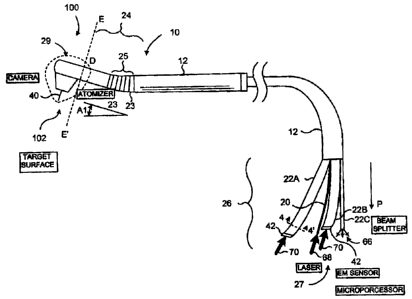
Referring to the figures, and specifically FIG. 1, an apparatus comprising an
illumination device 10 is illustrated. As presently embodied, illumination
device 10
includes an elongate body 12 having a generally tube-like structure that is
constructed to
contain a plurality of light transmitters, such as optical fibers and the
like, which are used
to transmit light to and/or from a handpiece 100 (i.e., a portion of the
illumination device
disposed distally from phantom line E-E' in FIG. 1). In the illustrated
embodiment, the
elongate body 12 surrounds and defines a hollow interior, such as lumen 14
(FIG. 3, infra)
as is more particularly described below. Illumination device 10 has a distal
end D and a
proximal end P, the distal end being the end closer to an end that is normally
held by a
user. Referring to the illustrated embodiment, a distal portion 24 of
illumination device 10
includes distal end D, and a proximal portion 26 includes proximal end P.
Elongate body 12 can comprise, for example, a hollow structure having one
portion that is flexible, and a distally-disposed portion that may be
substantially inflexible.
With continuing reference to the illumination device 10 of FIG. 1, a fraction
of distal
portion 24 is substantially inflexible, or is generally rigid and straight,
and a fraction 25 of
elongate body 12 is flexible. Corresponding structures can be found in FIGS.
6a and 6b of
U.S. Patent No. 6,389,193. In the illustrated embodiment of the present
invention, fixed ribs or joints 23 indicate the flexible portion of the
elongate body 12. In additional embodiments, parts or all of either the
length between and including distal portion 24 and proximal portion 26 are
flexible. Elongate body 12 can be made from any suitable material or
materials, such as
stainless steel, metal coil or plastic. As presently embodied, while being
flexible, the
flexible portion of elongate body 12 is set to form in a neutral position an
angle Al of
about 15 to 20 degrees, thereby disposing the fraction of distal portion in a
contra-angle
orientation relative to a part of the elongate body 12 adjacent to and
proximal of the flexible
portion. In a modified embodiment, a jointed section formed by joints 23 forms
the same
angle but is not flexible (i.e., is rigid) or is substantially non-flexible.
While the illumination
device 10 in FIG. I is illustrated as having a generally cylindrical cross-
section, the
illumination device 10 could also include one or more portions with different
cross-sectional
shapes including, for example, oval, rectangular, or triangular, and the like.
The illustrated illumination device 10 comprises an output portion 29 located
distally of phantom line E-E' in FIG. 1 that may be rotatable about a
longitudinal axis of
the distal portion 24. In modified embodiments, the output portion 29 may be
only
partially rotatable or entirely fixed relative to a distal end of distal
portion 24. As
5
CA 02575443 2007-01-26
WO 2006/012461 PCT/US2005/025957
presently embodied, the portion 29 can be rotated 360 degrees about the
longitudinal axis
of the distal portion 24. Referring to FIG. 2, it may be noted that a first
mirror 32 and a
second mirror 34 can operate to maintain an accurate coupling between output
ends of
fibers of the distal portion 24 and input ends of a sleeve 38 and tip 40
(e.g., a fiber tip)
independent of any rotational orientation of output portion 29, thereby
resulting in a
handpiece 100 that can be, in some embodiments, a 360-degree fully rotating
instrument.
As illustrated, the handpiece 100, which may be constructed of lightweight
(i.e. low mass)
materials such as exhibits, in some embodiments, a contra-angle design
constructed to
provide relatively high maneuverability and/or visibility of a working surface
(e.g., a
surgical field). The design further can feature a reduced profile when
compared with
conventional handpieces, thereby minimizing view-obstruction, which may be
caused by
other handpieces during procedures. When employed in medical applications such
as
dental applications, these characteristics of the present invention may
produce enhanced
patient and user comfort and, further, may provide improved efficiency,
accuracy, and
access to areas of for example an oral cavity. The sleeve 38 and tip 40 are
described
below with reference to FIGS. 2, 5 and 6. U.S. Patent No. 6,389,193, describes
an
embodiment of a rotating handpiece that maybe incorporated into the present
invention to
the extent compatible or modifiable by one skilled in the art to be compatible
and not
mutually exclusive. Additionally, other embodiments may be modified by one
skilled in
the art to be compatible and then incorporated into the present invention.
Illumination device 10 is illustrated having a plurality of proximal members
22A,
22B, and 22C. Proximal members 22A, 22B, and 22C have hollow interiors
configured to
accommodate one or more light transmitters or other tubular or elongate
structures having
cross-sectional areas less than the cross-sectional areas of the respective
hollow interiors.
Proximal members 22A, 22B, and 22C are arranged such that the hollow interiors
of each
of the proximal members is in communication with the lumen 14 (FIG. 3) of
elongate
body 12. This arrangement provides for a substantially continuous path for the
light
transmitters to extend from proximal end P to distal end D of elongate body
12. Although
the illustrated embodiment is provided with three proximal members, additional
embodiments could be provided with two, or four or more proximal members,
depending
on, for example, a number of light transmitters being used in the illumination
device 10.
In addition, the illustrated embodiment of illumination device 10 includes two
proximal
members 22A and 22B that have substantially equal diameters, and one proximal
member
6
CA 02575443 2010-09-02
22C that has a diameter that is less than either of the diameters of the other
two proximal
members.
Illumination device 10 is illustrated as being configured to be held by a
user. In an
exemplary embodiment, illumination device 10 is configured to direct
electromagnetic
energy from or in conjunction with the output portion 29 of handpiece 100
and/or to
receive energy that may be generated (e.g., reflected from a target) in
proximity to the
handpiece 100. The illumination device 10 can be used in medical, industrial,
dental, and
other applications. In one embodiment, the illumination device 10 is a device
for emitting
electromagnetic energy in dental applications. The electromagnetic energy
preferably
includes light, such as visible light, laser light (e.g., infrared laser
light) and the like. The
device can be used, for example, in dental hygiene procedures.
Illumination device 10 is typically connected to at least one external
electromagnetic energy source, such as a laser and/or one or more light
emitting diodes
(LEDs), and/or (in alternative embodiments) a lamp, so that electromagnetic
energy
generated by the electromagnetic energy source can be transmitted through
illumination
device 10 to the handpiece 100 and directed from the handpiece 100 to a target
(e.g., a
treatment surface such as a tooth). In modified embodiments, the
electromagnetic energy
source and/or other components may comprise parts or substantially all of that
described
in U.S. Patent No. 5,741,247 to the extent compatible or modifiable by one
skilled in the
art to be compatible and not mutually exclusive. Moreover, in other
embodiments wherein
fluid outputs and fluids (e.g., fluid outputs and fluids as described in U.S.
Patent No.
5,741,247) are implemented, the fluid outputs and fluids may comprise parts or
substantially all of any of that described in U.S. Patent Publication
2005/0256517, filed January
24, 2005 and entitled ELECTROMAGNETICALLY INDUCED TREATMENT
DEVICES AND METHODS, to the extent compatible or modifiable by one skilled in
the
art to be compatible and not mutually exclusive.
Distal portion 24 of the illumination device 10 of FIG. 1 can comprise, as
presently
embodied, a unitary structure having an inner lumen 14 (FIG. 3) forming a
distal portion
tube. Proximal members 22A, 22B, and 22C of the proximal portion 26 can each
have, in
an exemplary embodiment, a lumen in communication with the lumen 14 of distal
portion
24. Referring to elongate body 12 of FIG. 1, in an exemplary implementation
proximal
members 22A, 22B, and 22C can be integrally formed with distal portion 24 of
the
elongate body 12. In additional embodiments, one or more proximal members may
comprise separate elements that are joined or connected to elongate body 12 so
that the
7
CA 02575443 2007-01-26
WO 2006/012461 PCT/US2005/025957
proximal member lumens are in communication with the hollow interior or lumen
14 of
distal portion 24.
In a representative embodiment of elongate body 12, distal end D includes an
electromagnetic energy emitting internal output end 19 that, as presently
embodied,
coincides with phantom line E-E', and proximal end P includes an
electromagnetic energy
input end 27 (FIG. 1). Referring to proximal members 22A, 22B, and 22C of FIG.
1, each
proximal member can include a lumen dimensioned to accommodate one or more
light
transmitters or other tube- or fiber-like structures. In the illustrated
embodiment, proximal
members 22A and 22B each contain three energy-emitting fibers, such as optical
fibers,
and proximal member 22C can contain six energy-emitting fibers, such as
optical fibers.
In certain implementations, as a result of proximal member 22C being
illustrated as having
a smaller cross-sectional area relative to proximal members 22A and 22B, the
cross-
sectional area of each of the optical fibers (e.g., one, three, or six fibers)
in proximal
member 22C can be less than cross-sectional areas of the optical fibers in
proximal
members 22A and 22B. As illustrated in the embodiment of FIG. 4, which is a
cross-
sectional view along line 4-4' of FIG. 1, the proximal member 22A can comprise
three
optical fibers 16 that can be substantially fused together to define a unitary
light emitting
assembly or waveguide. In modified embodiments, the three optical fibers 16
may be
joined by other means or not joined. A structure similar to that of FIG. 4 may
describe
proximal member 22B, which may be similarly formed of fibers designated by
reference
numeral 17 in FIG. 3, which is a cross-sectional view taken along line 3-3' of
FIG. 2 near
distal end D of elongate body 12. Proximal member 22C can include six
relatively small
fibers 18, as likewise is shown in the cross-sectional view of FIG. 3. Fibers
18 are
illustrated as being separate from each other, but in additional embodiments,
two or more
of the fibers 18 can be fused or otherwise joined together at or near one or
more of the
proximal end P and the distal end D. Fibers 16, 17 and 18 can be manufactured
from
plastic using conventional techniques, such as extrusion and the like.
Another optical fiber 20 is illustrated in FIG. 1, passing between proximal
members 22A and 22B near the input end 27 of elongate body 12, and being
centrally
disposed relative to fibers 16, 17 and 18 near the internal output end 19
(FIG. 2) of
elongate body 12 as shown in FIG. 3. Optical fiber 20 is illustrated as a
power erbium
fiber that is structured to fit inside elongate body 12, although optical
fiber 20 may
comprise other structures in modified embodiments. As partially shown in FIG.
2, fibers
16, 17, 18 and 20 may terminate at the internal output end 19 (FIG. 2) located
inside the
8
CA 02575443 2010-09-02
elongate body 12. At the internal output end 19 (FIG. 2), the fibers 16, 17,
18, and 20 can
be arranged in a plane to form a planar surface. In an example, the fibers can
be cut and
polished in the same plane and arranged to be maintained in a substantially
fixed position
relative to one another and the handpiece 100. For example, tubing, such as
metal tubing,
can be used both at the inside of elongate body 12 and outside of elongate
body 12 to keep
part, and preferably all, of the fibers 16, 17, 18 and 20 in a fixed, straight
position.
At the input end 27, or proximal end P as illustrated in FIG. 1, fibers 16 and
17 of
respective proximal members 22A and 22B are configured to receive and transmit
light
from, for example, a laser and/or an LED, and/or, in alternative embodiments,
a lamp. As
presently embodied, blue light 70, for example blue light generated by one or
more blue
light LEDs, is received by proximal members 22A and 22B. In the illustrated
embodiment, two blue light LEDs are used as a source of blue light for
transmission
through fibers 16 and 17, each LED generating, for example, electromagnetic
energy at a
wavelength of about 470 nanometers (nm) and a power level of about 200
milliwatts
(mW) either in a continuous wave (CW) or pulsed mode. Blue light can be
particularly
useful in curing dental composites, whitening teeth, and detecting caries,
among other
things, when the illumination device 10 is used for dental care and hygiene.
Each of the
proximal members 22A and 22B is illustrated as including an optional light
altering
element such as, for example, a shutter mechanism or filter 42 to influence,
for example,
the transmission of blue light from the LEDs. In the illustrated embodiment,
each shutter
mechanism or filter 42 is structured to convert blue light into white, or any
other visible
light. This conversion may be accomplished by using or placing phosphoric
filters in front
of each of the proximal members 22A and 22B.
Proximal member 22C is configured to accommodate the six smaller optical
fibers
18, as described above. In the illustrated embodiment, optical fibers 18 are
configured to
collect or receive reflected and scattered light 64 (FIG. 2) from a treatment
output end 102
of handpiece 100 and to guide the reflected and scattered light 64 back toward
the input
end 27 (FIG. 1). The reflected and/or scattered light can be used as a
feedback signal 66,
which can be passed to a sensor or other suitable device for analysis. The
feedback signal
66 may be used, for example, by a microprocessor, to detect damage of an
optical surface
(e.g., a red light beam used for aiming may scatter and reflect back) or
fluorescence of
dental material (e.g., caries, bacteria, demineralization, and the like),
among other things.
9
CA 02575443 2010-09-02
The optical fiber 20, which may be an erbium fiber or other suitable laser
emitting
fiber, can be inserted into elongate body 12 such that a distal end of optical
fiber 20 is co-
planar (cf. plane coincident with phantom line E-E' of FIG. 2) with fibers 16,
17 and 18 at
distal end D of illumination device 10. In the illustrated embodiment, optical
fiber 20 is
centrally disposed along a central longitudinal axis of elongate body 12, as
shown in
FIGS. 2 and 3. In the illustrated configuration, fibers 16, 17 and 18 are
perimetrically
disposed around optical fiber 20, at least at the distal end D of illumination
device 10. The
concentric configuration of fibers 16, 17, 18 and 20 can be maintained for any
desired
distance of elongate body 12. In the illustrated embodiment, the concentric
configuration
is maintained until a region, (e.g., proximal portion 26) where proximal
members 22A,
22B, and 22C split from elongate body 12.
At the treatment output end 102 (FIG. 2), light is emitted from and collected
into
the handpiece 100. In the illustrated embodiment, light or other
electromagnetic radiation
is emitted from one or more of the fibers 16 and 17 at the internal output end
19 (FIG. 2),
and light is collected by fibers 18. In addition, light or other
electromagnetic radiation
from a laser, and/or an LED, and/or a lamp, can be emitted from optical fiber
20. In an
illustrative embodiment, electromagnetic radiation 68 (FIG. 1) is derived from
an erbium,
chromium, yttrium scandium gallium garnet (Er, Cr:YSGG) solid state laser,
which
generates electromagnetic energy having a wavelength of approximately 2.78
microns at
an average power of about 6 watts (W), a repetition rate of about 20 hertz,
and a pulse
width of about 150 microseconds. Moreover, electromagnetic radiation 68 may
further
comprise an aiming beam, such as light having a wavelength of about 655 rim
and an
average power of about 1 mW emitted in a continuous-wave (CW) mode. In one
embodiment, blue and white light are emitted from one or more of the fibers 16
and 17
toward a working surface, reflected light from the working surface is
collected by fibers
18, and erbium laser light is emitted from optical fiber 20. According to
another
embodiment, fibers 16, for example, may emit blue light and fibers 17 may emit
white
light. In other embodiments, appropriate light can be emitted by one or more
of the fibers
16 and 17, causing reflected white light and/or stimulated fluorescent light
to be collected
by fibers 18. In the above implementations, for example, the emitted light may
be directed
toward a working surface, such as a tissue surface, including a surface of a
tooth, to
perform one or more light sensitive procedures.
CA 02575443 2010-09-02
The present invention contemplates constructions and uses of visual
feedback implements (e.g., cameras) on (e.g., attached) or in a vicinity of
(e.g., on or near, attached or not, output ends) of electromagnetic energy
output devices (e.g., lasers and dental lasers), wherein such output devices,
constructions and uses can be, in whole or in part, including any associated
methods, modifications, combinations, permutations, and alterations of any
constructions(s) or use(s) described or referenced herein or recognizable as
included or includable in view of that described or referenced herein by one
skilled in the art, to the extent not mutually exclusive, as described in U.S.
Patent Publication 2006/0241574, filed January 10, 2005 and entitled
ELECTROMAGNETIC ENERGY DISTRIBUTIONS FOR
ELECTROMAGNETICALLY INDUCED DISRUPTIVE CUTTING, U.S.
Patent Publication 2005/0283 143, filed January 10, 2005 and entitled
TISSUE REMOVER AND METHOD. In some embodiments, the sensor may
comprise one or more visual feedback implements. The visual feedback
implement can be used, for example, (a) in a form that is integrated into a
handpiece or output end of an electromagnetic energy output device, (b) in
a form that is attached to the handpiece or electromagnetic energy output
device, or (c) in conjunction with (e.g., not attached to) the handpiece or
electromagnetic energy output device, wherein such handpieces and devices
can facilitate cutting, ablating, treatments, and the like. Treatments can
include low-level light treatments.
11
CA 02575443 2010-09-02
For example, one implementation may be useful for, among other things,
optimizing, monitoring, or maximizing a cutting effect of an electromagnetic
energy
emitting device, such as a laser handpiece. The laser output can be directed,
for example,
into fluid (e.g., an air and/or water spray or an atomized distribution of
fluid particles from
a water connection and/or a spray connection near an output end of the
handpiece) that is
emitted from the handpiece above a target surface. An apparatus including
corresponding
structure for directing electromagnetic energy into an atomized distribution
of fluid
particles above a target surface is disclosed, for example, in the above-
referenced U.S.
Patent No. 5,574,247. Large amounts of laser energy, for example, can be
imparted into
the fluid (e.g., atomized fluid particles), which can comprise water, to
thereby expand the
fluid (e.g., fluid particles) and apply disruptive (e.g., mechanical) cutting
forces to the
target surface. During a procedure, such as an oral procedure where access and
visibility
are limited, careful and close-up monitoring by way of a visual feedback
implement of (a)
interactions between the electromagnetic energy and the fluid (e.g., above the
target
surface) and/or (b) cutting, ablating, treating or other impartations of
disruptive surfaces to
the target surface, can improve a quality of the procedure.
In certain embodiments, visualization optical fibers (e.g., a coherent fiber
bundle)
can be provided that are configured to transmit light from the distal end D to
the proximal
end P, for routing images (e.g., working-surface images) acquired at or in a
vicinity of the
distal end by a visual feedback implement. According to some embodiments, the
visual
feedback implement can comprise an image-acquisition device (e.g., CCD or CMOS
camera) for obtaining or processing images from the distal end D. The visual
feedback
implement can be built-in or attached (e.g., removably attached) to the
handpiece and,
further, can be disposed at various locations on or in connection with the
handpiece
between the proximal end P and distal end D, or proximally of the proximal end
P.
According to this and any of the other embodiments described herein, one or
more of the
optical fibers 16, 17, 18 and 20, and the visualization optical fibers (not
shown), can be
arranged, for example, outside of the handpiece envelope. A few applications
for the
presently-described visual feedback implement may include periodontal pockets
(e.g.,
diagnostic and treatment), endodontics (e.g., visualization of canals), micro-
dentistry,
tunnel preparations, caries detection and treatment, bacteria visualization
and treatment,
general dentistry, and airborne-agent and gas detection applications.
12
CA 02575443 2007-01-26
WO 2006/012461 PCT/US2005/025957
According to another embodiment of the present invention, electromagnetic
radiation (e.g., one or more of blue light, white light, infrared light, a
laser beam,
reflected/scattered light, fluorescent light, and the like, in any
combination) may be
transmitted in one or both directions through one or more of the fibers 16,
17, 18, and 20,
in any combination. Outgoing and incoming beams of electromagnetic radiation
can be
separated or split, for example, according to one or more characteristics
thereof, at the
proximal end P (FIG. 1) using a beam splitter, such as a wavelength-selective
beam
splitter (not shown), in a manner known to those skilled in the art.
In certain embodiments of the invention, illumination device 10, as shown, for
example, in FIG. 1, may be useable in a person's hand or other suitable
holding device to
direct light toward a target surface. In other embodiments, the illumination
device 10,
which may comprise an optical fiber 20 oriented in a direction nominally
parallel to a
longitudinal axis of the illumination device 10, may be separate from but
configured to be
coupled to a handpiece 100 as illustrated in FIG. 7. Handpiece 100 (FIG. 7),
which, in the
illustrated embodiment, is structured to be held in a user's hand, can
comprise a treatment
output end 102 that is oriented at an angle relative to the longitudinal axis
of the
illumination device 10. Optical fiber 20 may terminate at an internal output
end
11 coinciding with phantom line F-F', of illumination device 10 in the
embodiment shown
in FIG. 7. In the illustrated embodiment, treatment output end 102 is oriented
at an
approximately ninety degree angle to the longitudinal axis of illumination
device 10. To
direct the emitted light from fibers 18 and 20 toward treatment output end
102, a reflector
30 is provided with handpiece 100. An embodiment of reflector 30 can comprise
a
parabolic mirror as described in U.S. Patent No. 6,389,193. In other
embodiments, such as
the embodiment of FIG. 2, reflector 30 may include a plurality of mirrors,
such as first
mirror 32 and second mirror 34. In still other embodiments, first and second
mirrors 32
and 34 may comprise parabolic, toroidal, or flat surfaces. In additional
embodiments, a
fewer or greater number of mirrors may be provided.
Referring again to FIG. 2, first mirror 32 is illustrated as being configured
to alter
light emitted from optical fiber 20. In other words, as presently illustrated,
first mirror 32
is configured to direct, for example, a beam 28 generated by a laser source
from the
internal output end 19 to the treatment output end 102. Second mirror 34, on
the other
hand, is illustrated as being configured to alter a path of light emitted from
one or more of
the fibers 16 and 17. In other words, mirror 34 can be configured to direct
one or more
beams of light, such as blue light or white light, from the internal output
end 19 to the
13
CA 02575443 2007-01-26
WO 2006/012461 PCT/US2005/025957
treatment output end 102. In addition, mirror 34 can be configured to direct
light 64,
which is reflected back from the target surface, toward fibers 18 (not visible
in FIG. 2) for
the provision of, for example, a signal that can be used for analysis, as
described above.
Either or both of mirrors 32 and 34 may be removable and replaceable.
With continuing reference to FIG. 2, handpiece 100 is also illustrated as
including
a tip 40 to direct electromagnetic energy (e.g., light), as indicated by
reference number 62,
that is emitted from optical fiber 20 toward a target surface. In addition, a
sleeve 38 may
be provided with handpiece 100, wherein sleeve 38 may partially,
substantially, or
completely (e.g., wherein sleeve 38 comprises a ring or cylindrical shape)
surround tip 40.
As presently embodied, sleeve 38 can be constructed of a material that is
substantially
transparent to permit light 60 emitted from fibers 16 and/or 17, such as white
light, to be
directed to a target surface. Light 60 may be used, for example, to illuminate
the target
surface. The illumination or the intensity of illumination of the target
surface may occur
continuously during the procedure being performed, or may be interrupted. In
addition,
such illumination may be automatically or manually controlled. First and
second mirrors
32 and 34 may also be constructed to focus one or more of the light beams into
tip 40. In
the illustrated embodiment, the first mirror 32 is constructed to focus the
erbium laser
beam emitted from optical fiber 20 into tip 40, and the second mirror 34 is
constructed to
focus the light emitted from fibers 16 and 17, such as blue light, white
light, or other light,
into sleeve 38. An embodiment of the handpiece 100 may comprise a plurality of
LEDs
(e.g., 2 or more, such as about 6 to 12, and in one implementation 10)
concentrically
disposed around the tip 40 in order to provide, according to certain
implementations, one
or more of a relatively bright, ultra-white and shadow-free illumination
system that may
significantly enhance maneuverability relative to, access to, and visibility
of, a working
surface. When employed in medical applications such as dental applications,
any one or
more of the above characteristics, such as enhanced illumination, may provide
for
significantly improved efficacy, accuracy and patient comfort.
In accordance with an aspect of the present invention, the tip 40 further may
be
surrounded by a tip ferrule 50. FIG. 5 is a magnified side elevation view
showing a
combination of the tip 40 and the tip ferrule 50. The tip ferrule 50 in the
illustrated
embodiment comprises a groove 52 that may be used to extract the tip ferrule
50 and,
consequently, the tip 40 from the handpiece 100. The tip ferrule 50 in the
illustrated
embodiment further comprises a plurality of ring-shaped projections 53 (see
also FIG. 2)
that make contact with an interior of the sleeve 38 of the handpiece 100.
Another
14
CA 02575443 2007-01-26
WO 2006/012461 PCT/US2005/025957
embodiment of the tip ferrule 50 replaces the plurality of ring-shaped
projections 53 with a
plurality of O-rings. The tip ferrule 50 may have at least one locking
shoulder 54 and, in
certain embodiments, may have a plurality of locking shoulders. In the
illustrated
embodiment, the locking shoulder is capable of providing a "click" or "snap"
feedback
when the tip ferrule 50 is fitted into a recess 58 (FIG. 2), which recess is
formed by
structure of one or more of the treatment end 102, the handpiece 100 and the
sleeve 38. In
modified embodiments, a locking shoulder can be formed, instead, by structure
of one or
more of the treatment end, the handpiece and the sleeve; and a recess can be
formed,
instead, in the tip ferrule, so that the locking shoulder is capable of
providing a "click" or
"snap" feedback when the tip ferrule is fitted into the recess. The click or
snap feedback
can facilitate the securing or locking of the tip ferrule 50 to the handpiece
100. Thus,
audible and/or tactile feedback in the form of a "click" or, in modified
embodiments, other
forms, can be provided to a user when an optical waveguide (e.g., tip 40),
which is secured
to the tip ferrule 50, is properly installed. As presently embodied, the tip
40 can be
secured to the tip ferrule 40 by way of inserting an adhesive into a cavity 51
or gap
disposed at a distal end of the tip ferrule 50, which cavity 51 is depicted in
FIG. 2 as a
distal portion of the tip ferrule 50 that surrounds but does not contact the
tip 40.
FIG. 6 is an on-axis top view of the tip ferrule 50 and tip 40 of FIG. 5. In
the
illustrated embodiment, the tip ferrule 50 comprises four locking shoulders
capable of
providing a "click" or "snap" feedback when the tip ferrule 50 is fitted into
the recess 58
(FIG. 2). According to one implementation one or more gaps 56 disposed between
locking shoulders may provide for a spring action capable, at least in part,
of producing
the click or snap feedback referred to above.
According to another aspect of the present invention, a facility may be
provided for
mixing spray air and spray water that may be directed toward a target surface.
An
illustration of an embodiment of a chamber for mixing spray air and spray
water in the
distal portion 24 of handpiece 100 is shown in FIG. 2a. A mixing chamber 80
(see also
FIG. 2) comprises an air intake 83, which is connected to, for example, tubing
(not shown)
in the elongate body 12 that supplies spray air. Similarly, a water intake 84
receives fluid
(e.g., water) from, for example, tubing (not shown) in the elongate body 12
that supplies
water. The air intake 83 and the water intake 84, which may have circular
cross-sections
about 250 im in diameter, join at an angle 82 that may approximate 110 in a
typical
embodiment. In certain embodiments, mixing may occur or begin to occur in a
neighborhood where the air intake 83 and water intake 84 join, and a spray
mixture 86 of
CA 02575443 2010-09-02
water and air (e.g., particles or atomized particles) may be ejected through a
fluid output
85. Fluid output 85 may have a circular cross-section measuring about 350 Am
in
diameter. A typical embodiment can comprise, for example, three such fluid
outputs
surrounding the tip 40 and tip ferrule 50 illustrated in FIG. 2. These fluid
outputs may, for
example, correspond to, comprise parts of, or comprise substantially all of,
any of fluid
outputs described in U.S. Patent Publication 2005/0256517, filed January 24,
2005 and
entitled ELECTROMAGNETICALLY INDUCED TREATMENT DEVICES AND
METHODS, to the extent compatible, or, in other embodiments, structures
described in the
referenced provisional patent application may be modified to be compatible
with the
present invention.
Handpiece 100 may further include another tip structure 36, such as a curing
tip, as
illustrated in FIG. 2. Tip structure 36 can be coupled with tip 40 or, as
presently
embodied, can replace tip 40. In an embodiment wherein the tip structure 36 is
coupled
with tip 40, the tip structure 36 may comprise a hollow center for
accommodating the tip
40 therethrough. In other embodiments, tip structure 36 can be coupled with or
can
replace both tip 40 and sleeve 38. In an embodiment wherein the tip structure
36 is
coupled with tip 40 and sleeve 38, the tip structure 36 may abut against an
output end of
sleeve 38 and further may comprise a hollow center for accommodating the tip
40.
While the tip structure 36 in the illustrated embodiment comprises a
cylindrical
shape (e.g., a solid cylinder) that surrounds a space of tip 40, which space
may or may not
be occupied, other embodiments may comprise a tip structure that only
partially surrounds
the space (occupied or not) of tip 40. When tip structure 36 is a curing tip,
the curing tip
can be positioned in handpiece 100 and configured to receive or collect light
(e.g., blue
light) emitted from, for example, fibers 16 to direct the light toward a
target surface and
obtain a desired effect, such as curing of dental composites. To increase an
amount of
light that is collected by tip structure 36, a diameter can be chosen for tip
structure 36 that
will optimize or maximize a characteristic (e.g., an amount) of light
collected. Tip 40 and
tip structure 36 can be formed of a plastic-like material, including a
plurality of plastic
materials, that is/are optically transparent to permit the light to be
effectively transmitted
therethrough to and from a target surface.
In an exemplary implementation, illumination device 10 may have a total length
of
between about 1 and about 2 meters. In one particular embodiment, illumination
device
can be about 1.6 meters long. Each proximal member 22A and 22B may have a
diameter between about 2 millimeters (mm) and about 5 mm, such as about 3 mm.
16
CA 02575443 2010-09-02
Typically, proximal members 22A, 22B, and 22C meet to define a unitary tubular
structure having an outer diameter between about 4 mm and about 5 mm, such as
about
4.5 mm (or about 3/16 of an inch). Proximal members 22A, 22B, and 22C may be
arranged so that the fibers contained therein define a central lumen having a
diameter
ranging from about 1 mm to about 2 mm, such as about 1.5 mm (or about 1/16 of
an inch).
This central lumen can be structured to accommodate a power erbium laser
fiber, such as
optical fiber 20 capable of transmitting, for example, concentrated infrared
electromagnetic energy. In the embodiment illustrated in FIG. 1, proximal
members 22A,
22B, and 22C are routed together to form a unitary structure at a distance of
approximately
centimeters (cm) from the proximal end P of elongate body 12. Power erbium
optical
fiber 20 may have a diameter of approximately 0.8 mm, and fibers 16 and,
optionally,
fibers 17, may have a diameter of about 1.5 mm. Fibers 18 may be about 0.5 mm
in
diameter. The internal output end 19 of illumination device 10 can include a
substantially
rigid, straight portion that is approximately 10 centimeters in length.
Illumination device
can include six larger-diameter fibers, such as six fibers 16, or optionally
can include
three larger-diameter fibers 16 and three larger-diameter fibers 17 as shown
in FIG. 3
concentrically arranged about a central lumen with six relatively smaller
diameter fibers
18 concentrically arranged about the same central lumen. The numerical
apertures of
fibers 16 (and, optionally, fibers 17) and 18 can be about 0.68.
Light provided by two high power blue LEDs, which light may comprise visible
electromagnetic energy relatively less concentrated than the infrared energy
referred to
above, may be directed into proximal members 22A and 22B to cure dental
composites,
whiten teeth, and/or detect dental caries. Each blue light LED can have a
power of
TM
approximately %2 W. One suitable example of a high-power blue LED is a Luxeon
Emitter, 5 W Dental, which emits light having a wavelength in a range of about
450 nm to
about 470 urn with a bandwidth of about 20 rim (Model No. LXHL-PRD5). If
illumination is desired at the target surface, two phosphoric filters can be
placed in a light
path between the blue light emitting LEDs and proximal members 22A and 22B.
The
phosphoric filters may be used as white-light shutters to provide white light
to the target
surface, as discussed above. The white light that is generated from filtering
the blue light
is typically reduced in power relative to the blue light. In the embodiment
illustrated in
FIG. 1, the white light is reduced to a range of about twenty percent to about
thirty percent
of the power of the blue light. Additional filters can be provided to alter
the white light, as
may be desired. In a preferred embodiment, a blue light filter is placed at
the proximal
17
CA 02575443 2010-09-02
end of each of the proximal members 22A and 22B. In other embodiments,
however, the
filters can be located at any location along the illumination device 10,
including at the
distal end.
By way of the disclosure herein, an illumination device has been described
that
utilizes electromagnetic energy to affect a target surface. In the case of
dental procedures,
the illumination device includes an optical fiber for transmitting laser
energy to a target
surface for treating (e.g., ablating) a dental structure, such as a tooth, a
plurality of optical
fibers for transmitting blue light for illumination, curing, whitening, and/or
diagnostics of
a tooth, a plurality of optical fibers for transmitting for example white
light to a tooth to
provide illumination of the target surface, and a plurality of optical fibers
for transmitting
light from the target surface back to a sensor for analysis. In the
illustrated embodiment,
the optical fibers that transmit blue light also transmit white light. In
accordance with one
aspect of the invention herein disclosed, an illumination device comprises an
illumination
tube having a feedback signal end and a double mirror handpiece.
In certain embodiments, the methods and apparatuses of the above embodiments
can be configured and implemented for use, to the extent compatible and/or not
mutually
exclusive, with existing technologies including any of the above-referenced
apparatuses
and methods. Corresponding or related structure and methods described in the
following
patents assigned to BioLase Technology, Inc. are referenced, wherein such
referencing
includes corresponding or related structure (and modifications thereof) in the
following
patents which may be (i) operable with, (ii) modified by one skilled in the
art to be
operable with, and/or (iii) implemented/used with or in combination with any
part(s) of,
the present invention according to this disclosure, that/those of the patents,
and the
knowledge and judgment of one skilled in the art: U.S. Patent No. 5,741,247;
U.S. Patent
No. 5,785,521; U.S. Patent No. 5,968,037; U.S. Patent No. 6,086,367; U.S.
Patent No.
6,231,567; U.S. Patent No. 6,254,597, U.S. Patent No. 6, 288,499; U.S. Patent
No.
6,350,123; U.S. Patent No. 6,389,193; U.S. Patent No. 6,544,256; U.S. Patent
No.
6,561,803; U.S. Patent No. 6,567,582; U.S. Patent No. 6,610,053; U.S. Patent
No.
6,616,447; U.S. Patent No. 6,616,451; U.S. Patent No. 6,669,685; and U.S.
Patent No.
6,744,790.
For example, one implementation may be useful for tailoring, optimizing or
maximizing an effect (e.g., cutting or ablating) of a laser. The laser output
(e.g., from a
18
CA 02575443 2010-09-02
power fiber) can be directed, for example, into fluid (e.g., an air and/or
water spray or an
atomized distribution of fluid particles from a water connection and/or a
spray connection
near the treatment output end 102) that is emitted from a fluid output of the
handpiece 100
at the treatment output end 102 above a target surface (e.g., one or more of
tooth, bone,
cartilage and soft tissue). The fluid output may comprise a plurality of fluid
outputs,
concentrically arranged around a power fiber, as described in, for example,
U.S.
Patent Publication No. 2005/0256517. The power fiber may comprise, for
example,
optical fiber 20, and in various implementations may be
coupled to an electromagnetic energy source comprising one or more of a
wavelength
within a range from about 2.69 to about 2.80 microns and a wavelength of about
2.94
microns. In certain implementations the power fiber may be coupled to one or
more of an
Er:YAG laser, an Er:YSGG laser, an Er, Cr:YSGG laser and a CTE:YAG laser, and
in
particular instances may be coupled to one of an Er, Cr:YSGG solid state laser
having a
wavelength of about 2.789 microns and an Er:YAG solid state laser having a
wavelength
of about 2.940 microns. An apparatus including corresponding structure for
directing
electromagnetic energy into an atomized distribution of fluid particles above
a target
surface is disclosed in the above-referenced U.S. Patent No. 5,574,247. Large
amounts of
laser energy, for example, can be imparted into the fluid (e.g., atomized
fluid particles),
which can comprise water, to thereby expand the fluid (e.g., fluid particles)
and apply
disruptive (e.g., mechanical) cutting forces to the target surface.
The optical fibers and/or tip ferrules referred to herein may comprise plastic
and/or be color coded to designate predetermined or predefined sizes, shapes
or other
properties. These materials may all be autoclavable. The tip ferrule and
corresponding
structure may comprise parts or substantially all of any of that described in
U.S. Patent No.
6,567,582, entitled FIBER TIP FLUID OUTPUT DEVICE to the extent compatible;
or, in
other embodiments, structures described in the referenced patents may be
modified to be
compatible with the device tip ferrule 50 disclosed in FIGS. 5 and 6.
While this invention has been described with respect to various specific
examples
and embodiments, it is to be understood that the invention is not limited
thereto and that it
can be variously practiced. Multiple variations and modification to the
disclosed
embodiments will occur, to the extent not mutually exclusive, to those skilled
in the art
upon consideration of the foregoing description. Additionally, other
combinations,
19
CA 02575443 2007-01-26
WO 2006/012461 PCT/US2005/025957
omissions, substitutions and modifications will be apparent to the skilled
artisan in view of
the disclosure herein. Accordingly, the present invention should not be
limited by the
disclosed embodiments, but is to be defined by reference to the appended
claims.