Note: Descriptions are shown in the official language in which they were submitted.
DEMANDE OU BREVET VOLUMINEUX
LA PRESENTE PARTIE DE CETTE DEMANDE OU CE BREVET COMPREND
PLUS D'UN TOME.
CECI EST LE TOME 1 DE 2
CONTENANT LES PAGES 1 A 238
NOTE : Pour les tomes additionels, veuillez contacter le Bureau canadien des
brevets
JUMBO APPLICATIONS/PATENTS
THIS SECTION OF THE APPLICATION/PATENT CONTAINS MORE THAN ONE
VOLUME
THIS IS VOLUME 1 OF 2
CONTAINING PAGES 1 TO 238
NOTE: For additional volumes, please contact the Canadian Patent Office
NOM DU FICHIER / FILE NAME:
NOTE POUR LE TOME / VOLUME NOTE:
CA 02575614 2007-01-29
WO 2006/015209 PCT/US2005/026976
DIFFERENTIATION OF STEM CELLS
1. CROSS-REFERENCE TO RELATED APPLICATIONS
1. This application claiins benefit of U.S. Provisional Application No.
60/592,027, filed
July 29, 2004. Application Serial No. 60/592,027, filed July 29, 2004, is
hereby incorporated
herein by reference in its entirety.
II. BACKGROUND
2. Pluripotent stem cells, such as human pluripotent stem cells, promise to
dramatically
alter and extend our ability to both understand and treat many of the chronic
illnesses that define
modern medicine. From drug discovery, to the generation of monoclonal
antibodies, to the
production of cell therapies, much of human cell biology expects to be
transformed by the ability
to generate specific cell types, such as human cell types at will. The medical
and industrial
application of pluripotent stem cells requires the ability to generate large
numbers of a single cell
type in vitro. Current strategies of directing cell differentiation through
treatment with known
morphogens, hormones or other chemicals have been successful in certain
instances but in no
case have they been able to generate the quality and volume of cells
necessary.for any practical
application outside the laboratory. There is a tremendous need for being able
to generate cell
types in vitro. The production of monoclonal antibodies through in vitro
immune systems, the
production of islets for diabetes treatment, and the production of neural
precursors for neural
related dysfunction are just a few of the human disease areas needing a steady
reliable
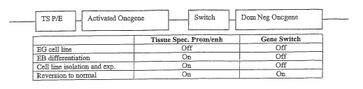
production of specific cell types. The economic significance of this project
is dramatic. The
monoclonal antibody application alone is a multibillion dollar industry. The
National Institutes
of Health estimates that the annual cost of diabetes to the United States is
$132 billion
(http://diabetes.niddk.nih.gov/dm/pubs/statistics/index.htm#14). Estimates for
the annual
national cost of neurodegenerative disease is over $100 billion
(http://www.
alzheimers.org/pubs/prog00.htm#The%20Impact%20ofb/o20Alzheimer%92s%20Di
sease).
3. The practical application of embryonic stem cell biology will require the
generation
of large numbers of homogeneous cell types. Large scale culture of
undifferentiated stem cells,
followed by directed differentiation, presents a series of challenges that
suggest a need for an
alternative solution. ES and EG lines require the addition of expensive
recombinant hormones
to the cell culture medium to maintain their growth and maintenance of the
undifferentiated
CA 02575614 2007-01-29
WO 2006/015209 PCTIUS2005/026976
1,,,;1 46h
II
state, sas Fibr'o~~1~as~~r~ and Leukemia Inhibitory Factor. In general, ES and
EG
lines are still cultured on feeder layers. They grow slowly, freeze and
recover poorly and are
difficult to passage. While.progress is being made in making ES and EG cell
culture easier, they
will always require substantial resources and a knowledgeable and dedicated
staff.
4. Directed differentiation presents additional problems. Differentiation can
be initiated
either by changing the hormonal milieu, forming embryoid bodies or a
combination of both.
Embryoid body formation is the most widely used and general process at
present. This method
appears to generate a wide variety of cells, resulting from the juxtaposition
of the various tissue
types within the embryoid body. Problems with this method revolve around
homogenous
formation. In a static culture, bodies of various sizes and shapes form,
resulting in a variable
differentiation process. Again, while laboratory scale methods, such as the
hanging drop, can
surmount these problems, they are problematic on a large scale. While the use
of hormones and
chemicals to direct differentiation, rather than embryoid body formation,
seems a more attractive
approach, our understanding of the complex interactions required for
organogenesis is
rudimentary. Filling in these gaps in our understanding will require
painstaking and difficult
analysis of embryological processes that are not easily accessible to
experimentation.
5. Disclosed herein are methods that can generate virtually any cell type in
vitro, as welT
as compositions used in the methods or derived from the methods. These cell
lines which are
generated can be cloned, characterized, frozen, and used in any quantity
necessary while, for
example, maintaining the advantages of a normal karyotype. The availability of
these cells will
enable the realization of many of the potential applications currently
envisioned for human stem
cells.
III. SUMMARY
6. Disclosed are methods and compositions related to production of cells and
cell lines.
IV. BRIEF DESCRIPTION OF THE DRAWINGS
7. The accompanying drawings, which are incorporated in and constitute a part
of this
specification, illustrate several embodiments and together with the
description illustrate the
disclosed compositions and methods.
8. Figure 1 shows a schematic for an example of a cassette for reversible
transformation
using sequential expression of activated, dominant negative pairs of a
transforming gene. Below
the schematic there is a temporal progression of which parts of the cassette
are activated during
the progression from a pluripotent stem cell to a differentiated cell.
-2-
CA 02575614 2007-01-29
WO 2006/015209 PCT/US2005/026976
4'les of plasmids that can be used for isolation of an
hepatocyte derived cell line from ACTEG1, a gonadal ridge derived pluripotent
stem cell.
10. Figure 3 shows a schematic of an example of a cassette for reversible
transformation
using an excisable activated oncogene.
11. Figure 4 shows the structure of ploxHBV-aRas, an example of a plasmid
which can
be used in the generation of a cassette as in Figure 3.
12. Figure 5 shows a schematic of an example of a cassette for reversible
transformation
using a temperature sensitive transforming gene.
13. Figure 6 shows a schematic of the pEGSH plasmid, as indicated by
Stratagene.
14. Figure 7 shows a diagram of a form of the disclosed tissue specific
reversible
transformation (TSRT) method.
15. Figure 8 shows a schematic of an example of a cassette for reversible
transformation
using a tetracycline regulated CMV promoter driving expression of a dominant
negative ras and
a tissue specific promoter driving expression of a-ras.
V. DETAILED DESCRIPTION
16. Before the present compounds, compositions, articles, devices, and/or
methods are
disclosed and described, it is to be understood that they are not limited to
specific synthetic
methods or specific recombinant biotechnology methods unless otherwise
specified, or to
particular reagents unless otherwise specified, as such may, of course, vary.
It is also to be
understood that the terminology used herein is for the purpose of describing
particular
embodiments only and is not intended to be limiting.
17. Numerous authors have written about the possible applications of human
pluripotent
stem cells (for example, Gearhart, J (1998) Science 282, 1061 - 1062; Pera,
MF, et al., (2000)
J. Cell Sci. 113, 5 - 10; Trounson, A (2001) Reprod Fertil Dev. 2001;13(7-
8):523-32; Sussman,
NL, Kelly, JH. (1994) US Patent 5,368,555). These range from target evaluation
and toxicity
testing in drug discovery to attempting to cure type I diabetes by implanting
new beta cells into
the pancreas. Each of these applications requires large quantities of
differentiated cells from a
controlled and renewable source. While previous technologies fail to meet this
requirement,
disclosed herein are compositions and methods capable of producing large
quantities of a desired
cell type in vitro in a controlled and reproducible way.
18. Human pluripotent stem cells promise to dramatically alter and extend our
ability to
treat many of the chronic illnesses that define modem medicine.
Neurodegenerative disease,
-3-
CA 02575614 2007-01-29
WO 2006/015209 PCT/US2005/026976
; ~ i .,;,
nee;~tlial~et~~, uto~~iimune disease, leukemia, and heart disease are all
examples of targets for cell-based therapies aimed at replacing and
regenerating damaged tissue.
19. This vision is primarily based on the success of using pluripotent stem
cells to
generate transgenic mice (Zambrowicz, BP, Sands, AT (2003) Nat. Rev. Drug
Disc. 2, 38 - 51).
The ability to alter stem cells in vitro and create mice with targeted
mutations has led to rapid
advancement in the understanding of gene regulation and function, as well as
mammalian
development. This, in turn, has led to an ability to mimic human disease in
mouse models,
facilitating the process of drug development. Work with pluripotent stem cells
in mice has
shown that they are capable of contributing to any tissue in the organism, and
that genes of
interest can be altered essentially at will, being turned off, deleted,
activated or expressed in
individual tissues, depending on the needs of the particular experiment.
20. While these results properly encourage enthusiasm for human pluripotent
stem cell
work, they also frame the central problem in generalizing this work from the
mouse to the
human. Because of the success of the transgenic mouse as a model, and its
ability to replicate
the complex interplay of tissues that leads to organotypic differentiation,
substantially less
attention has been devoted to defining conditions that reproduce
differentiation in vitro. Yet, in
order to realize the vision of cell-based therapies, substantial quantities of
specific cell types or
sets of cell types will need to be generated in vitro. It would be useful to
have differentiated
stem cells comprising an absolutely homogeneous population, that is, that they
be clonal or semi-
purified, in order to avoid the well documented propensity of pluripotent stem
cells to form
tumors when implanted in other than their normal environment (Andrew, PW
(2002) Philos.
Trans. R. Soc. Lond. B. Biol. Sci. 357, 405 - 417). Accordingly, disclosed are
homogenous
differentiated stem cells, clonal differentiated stem cells, semi-purified
differentiated stem cells,
and mixed differentiated stem cells. Also disclosed are populations of cells,
which can, but need
not be, clonal, can, but need not be, the same cell type, and can, but need
not be, a subset of all
cell types that could be produced. These populations can be used, for example,
for therapy, in in
vivo toxicity assays or in other types of in vitro assays such as drug
screening. Also disclosed
are semi-purified sets of a cell type which contain, at least 99, 98, 97, 96,
95, 94, 93, 92, 91, 90,
89, 88, 87, 86, 85, 84, 83 82, 81, 80, 79, 78, 77, 76, 75, 74, 73, 72, 71, 70,
65, 60, 55, 50, 45, 40,
35, 30, or 25 % of a particular cell type, such as any combination of any cell
disclosed herein,
any cell disclosed herein, or a hepatocyte.
21. Disclosed is a method for producing differentiated stem cells and/or one
or more
types of cells. Also disclosed are cells and/or cell types produced by the
disclosed method. The
-4-
CA 02575614 2007-01-29
WO 2006/015209 PCT/US2005/026976
metl~'od ~e~erail Ji~ri"volf'e Ac" b'htiiig stem cells under conditions that
promote differentiation
and selecting or screening for one or more cells and/or cell types. The stem
cells used can
comprise a nucleic acid segment comprising a transcriptional control element
operably linked to
a nucleic acid sequence encoding a marker. The selection or screening can be
on the basis of the
marker. The cells and/or cell types in which the marker is expressed can be
selected or screened
for, or the cells and/or cell types in which the marker is not expressed can
be selected or
screened for. In this way, particular cells and/or cell types can be obtained
from stem cells.
22. The transcriptional control element can be a tissue-, cell-, cell type-
and/or cell
lineage-specific transcriptional control element, which means that the
transcriptional control
element allows or promotes expression of nucleic acid sequences operably
linked to the
transcriptional control element in specified tissues, cells, cell types and/or
cell lineages,
respectively. Thus, in the disclosed method, the marker can be expressed in
tissues, cells, cell
types and/or cell lineages for which the transcriptional control eleinent is
specific. In this way,
particular cells, cells of particular tissues, particular cell types and/or
cells of particular cell
lineages can be obtained from stem cells.
23. The disclosed method has the advantage of providing a feature or
characteristic
(expression or non-expression of the marker) by which differentiated cells of
interest can be
selected or screened from stem cells and differentiated cells that are not of
interest. The concept
of the disclosed method is that the marker, operably linked to a
transcriptional control element,
will be.expressed (or not expressed) only or primarily when starting stem
cells have
differentiated into a desired type of cell or tissue (the type of tissue or
cell for which the
transcriptional control element is specific). Any cell, cell type, cell
lineage, and/or tissue of
interest can be targeted by choosing a transcriptional control element
relevant to the cell, cell
type, cell lineage, and/or tissue of interest.
24. A useful type of marker is a transformation agent, such as an oncogene. In
this case,
expression of the transformation agent can cause transformation of the cell.
The result can be
growth and/or preferential growth of cells expressing the transformation
agent. In the context of
differentiated stem cells, transformation, and the associated growth, can
allow selective and/or
preferential growth of cells expressing the transformation agent because most
other
differentiated stem cells will grow slowly if at all. Cells expressing (or not
expressing) the
marker can be selected by applying selective pressure relevant to the marker.
For example, many
genes and proteins are known that can be used to give cells a selective
advantage or
disadvantage. Cells expressing (or not expressing) the marker can be screened
by identifying
-5-
CA 02575614 2007-01-29
WO 2006/015209 PCT/US2005/026976
cells~ 4n ~ "i'marker. For exam le manenz es and rotems are
il.., ~ ~( P ~) p~ Y Ym p
known that constitute and/or produce a signal that can be detected. Such a
signal can be the
basis of cell identification.
25. The method can also involve reversal of the marker expression. This can be
accomplished by, for example, removal of all or part of the nucleic acid
segment, such as by
excision of all or part of the nucleic acid segment; inactivation of the
nucleic acid segment, the
transcriptional control element, and/or the marker; repression of the nucleic
acid segment, the
transcriptional control element, and/or the marker; and/or introduction and/or
expression of a
reversing agent. Excision of the nucleic acid segment can be accomplished in
numerous ways.
For example, the nucleic acid segment can be excised via site-specific
recombination using a
recombinase. A reversing agent can alter and/or reduce the effect of the
marker. For example,
where the marker is a transforming agent such as Ras, transformation of the
cells (the effect of
Ras) can be reversed by expression of a dominant negative Ras. Forms of the
disclosed method
that involve use of a transformation agent and subsequent reversal of
transformation can be
referred to as tissue specific reversible transformation (TSRT). Although TSRT
refers to tissue
specific reversible transformation, this is merely for convenience and it is
intended that TSRT
refers to tissue-, cell-, cell type- and/or cell lineage-specific expression
of the transforming agent.
26. As indicated, combinations of reversal operations can be used to
accomplish reversal.
For example, excision of the nucleic acid segment and expression of a
reversing agent can be
used together in the disclosed method. Removal of the nucleic acid segment is
a useful reversal
operation when a cell having minimal genetic alteration (compared to a natural
cell of the same
type, for example) is desired. This is desirable, for example, if the cells
are to be used
therapeutically.
27. Disclosed herein are strategies involving tissue-specific reversible
transformation for
establishing differentiated cell lines of any particular cell type, using stem
cells as a starting
material. Disclosed are methods that employ tissue specific expression of a
transforming gene,
which can be used to identify and culture the particular cell type. This
transforming event can, in
some fonns of the method, then be reversed, using one of a number of possible
processes,
leaving a clonal or semi-purified population of non-transformed,
differentiated cells, including
populations of different or semi-purified cells, or a clonal population of
cells, as discussed
herein.
28. Disclosed are compositions and methods involving modified stem cells, such
as
pluripotent stem cells, wherein the pluripotent stem cell contains, for
example, a marker whose
- 6 -
CA 02575614 2007-01-29
WO 2006/015209 PCT/US2005/026976
expli n control element, such as a tissue specific promoter, a
cell type specific promoter, a cell specific promoter, and/or a cell lineage
specific promoter. The
modified pluripotent stem cell can then be grown under conditions that allow
for cell
proliferation or embryoid body (EB) and differentiated cell formation as
discussed herein. When
the stem cell is allowed to form an EB the EB produces many different cell
types through
spontaneous differentiation. In some forms of the disclosed method, after the
EB is allowed to
form for a desired time, a selective pressure can be applied by, for example,
growing the cells in
the cognate selection media for the marker. While at this point, there are
many different cell
types (the number depends on the length of time the EB is allowed to develop
without selective
pressure), the selective pressure causes cells having the expressed marker to
be selectively
amplified or visualized. The cells having the selective marker are a desired
differentiated cell
type or types, because the marker can be designed to be preferentially or
selectively expressed in
the desired cell type or types from the tissue specific promoter. It is also
understood that in
certain systems, there can be more than one tissue specific promoter driven
marker. Having
multiple markers driven by different promoters, the selective stringency can
be increased for cell
types where the tissue specific promoter is not expressed exclusively in a
single tissue. It is also
understood that there can an additional identification step after the
selection step or steps in
which the desired cell is identified. These identified cells can then be
further isolated and
cultured.
29. After a period of time under the selective conditions (selective pressure,
for example)
can be removed to allow for increased cell proliferation, and then the
selective pressure can be
reapplied. Thus, iterative rounds of selection can occur, increasing the
stringency of selection.
The iterative rounds of selection can also occur in systems with more than one
type of marker
being expressed from the same tissue specific promoter. In some forms of the
method these
iterative rounds of selection can occur such that, for example, a first marker
is utilized and then a
second marker is utilized and then the first marker is utilized and the second
marker is utilized,
and so forth. After the selective pressure is completed, the desired
differentiated cells caii be
grown under non-selective conditions, at which point the marker and related
DNA can be
removed if desired. There are numerous ways for achieving this, including, for
example, the use
of recombinase technology, such as Cre-lox technology or temperature specific
mutant markers.
It is also understood that the marker can be integrated into the pluripotent
stem cell chromosome
or can be carried on extrachromosomal cassettes, such as a mammalian
artificial chromosome.
- 7 -
CA 02575614 2007-01-29
WO 2006/015209 PCT/1JS2005/026976
sR'alt 11~~ I-bompositions for establishing differentiated cell lines of
any particular cell type, using stem cells as a starting material. This
mechanism can employ
tissue specific expression of a marker, such as a transforming gene, which is
used to identify and
culture the particular cell type. This transforming event can then be
reversed, using one of a
number of possible processes, leaving a clonal or semi-purified population of
nontransformed,
differentiated cells.
31. For example, disclosed are compositions and methods related to the human
liver
specific promoter/enhancers from the hepatitis B virus core antigen driving
different variations
of the RAS gene. In some forms of the method, an activated RAS coupled to an
ecdysone
inducible dominant negative RAS as the reversing agent can be used. In some
forms of the
method, the HBV/RAS construct can be flanked with loxP sites that can be
excised with CRE
recombinase. Some forms of the method can use the generation of a temperature
sensitive (ts),
activated RAS.
32. Typically the marker construct can be transfected into a stem cell line,
such as a
human einbryonal germ (EG) cell line. Differentiation of the resultant cell
line can then be
initiated, for example, by the formation of embryoid bodies. In this way,
natural biological
processes result in development of the appropriate cell type. When a cell
becomes the desired
cell type, such as an hepatocyte, the tissue or cell specific promoter, such
as a liver specific
construct, will be activated and the marker will be expressed. The cell is,
for example,
transformed or marked by expression of the marker. A selective media can be
used, for
example, such as soft agar for transformed cells, and when placed in the
selective media only the
appropriately differentiated transformed cells in the EB will survive or have
selective advantage.
Transformed cells will preferentially or selectively grow out and form
colonies. Colonies can
be picked and re-plated for cloning. For use, the cells can be grown by
standard methods to the
desired quantity and configuration. At the appropriate time, the reversing
signal can be applied,
for example, either ecdysone for gene switches, CRE recombinase for lox
constructs or
temperature shift for ts construct, leaving a population of cells functionally
equivalent to primary
cultures.
33. For example, disclosed are pluripotent stem cells containing a nucleic
acid segment
comprising the structure P-I, wherein: P is a transcriptional control element;
and I is a sequence
encoding a marker, wherein the marker can comprise a transformation agent.
34. Disclosed are cells, wherein the marker is expressed from a heterologous
nucleic
acid, wherein the nucleic acid further comprises a suicide gene, wherein P is
a tissue specific
-8-
CA 02575614 2007-01-29
WO 2006/015209 PCT/US2005/026976
1:;;1~ ,=.=,g,, r' ~ , ;I:~:' I"' {I"õ,, ~fl ':' l~;~~ ,~:
trans~cr~pti~riall'~'l~rr~er'w~~rP causes I to be preferentially or
selectively expressed,
wherein the immortalization agent is a temperature permissive agent, wherein I
comprises the
SV401arge T antigen, wherein the nucleic acid segment is flanked by a site-
specific excision
sequence, wherein I is flanked by a site-specific excision sequence, wherein P
is flanked by a
site-specific excision sequence, and/or wherein P-I is flanked by a site-
specific excision
sequence, X, forming X-P-I-X.
35. Also disclosed are cells produced by excising the nucleic acid segment
from the stem
cells disclosed herein.
36. Disclosed are cells, wherein the nucleic acid segment comprising the
structure P-I is
excised using an adenovirus-mediated site-specific excision, and/or wherein
the excision of the
nucleic acid molecule comprising the structure P-I results in recombination of
the non-excised
nucleic acid molecule.
37. Disclosed are methods of deriving a population of conditionally immortal
cell types
from stem cells, comprising: transfecting a stem cell with a construct
containing one of the
nucleic acid molecules P-I disclosed herein, culturing the stem cells in an
environment such that
transcriptional control of element P is activated, whereby I is,preferentially
or selectively
expressed, and selecting cell types expressing I.
38. Disclosed are methods, further comprising the step of increasing the
purity of the
population of cells expressing I, wherein the step of increasing the purity
comprises creating a
clonal or semi-purified population of cells, further comprising excising the
nucleic acid, further
comprising freezing the selected cell type, and/or further comprising adding a
gene of interest to
the population of cells.
39. Disclosed are methods of deriving conditionally immortal cell types,
comprising
transfecting pluripotent stem cells with a construct containing one of the
nucleic acid molecules
P-I disclosed herein, activating control element P, whereby I is
preferentially or selectively
expressed, selecting cell types expressing I and excising the construct
containing the P-I nucleic
acid molecule, contacting the selected cell types with an environment such
that the ends of the
nucleic acid formerly containing the construct containing the P-I nucleic acid
molecule
recombine; and freezing of the selected cell type.
40. Disclosed are methods wherein the stem cell culture is allowed to
spontaneously
differentiate into an embryoid body.
41. Also disclosed are methods of deriving a cell culture, comprising
transfecting
pluripotent stem cells with a construct containing one of the nucleic acid
molecules P-I disclosed
-9-
CA 02575614 2007-01-29
WO 2006/015209 PCT/US2005/026976
i . , ~
here~in,~' co~itacfi$g:'t~i~'~Tein"&e~T5 ~ithlTb environment such that tran
scriptional control element P
is activated and I is preferentially or selectively expressed, culturing the
cells expressing I.
42. Disclosed are methods, further comprising cloning the cultured cells
expressing I.
43. Disclosed are methods of treating a patient comprising administering the
cells
disclosed herein, such as by transplanting the cells disclosed herein.
44. Disclosed are methods of assaying a composition for toxicity comprising
incubating
the composition with the cells produced by the method disclosed herein.
45. Disclosed are pluripotent stem cells containing a nucleic acid molecule
construct
comprising the structure P-I, wherein P is a tissue specific transcriptional
control element, P
causes I to be preferentially or selectively expressed; and I is a temperature
permissive
immortalization agent.
46. Disclosed are pluripotent stem cell containing a nucleic acid molecule
construct
comprising the structure X-P-I-X, wherein P is a tissue specific
transcriptional control element, P
causes I to be preferentially or selectively expressed, I is a temperature
permissive
immortalization agent; and X is a site-specific excision sequence.
47. Disclosed are cells, wherein P-I is excised, wherein P-I is excised at X
by an
adenovirus-mediated site-specific excision, and/or wherein the excision of P-I
allows
recombination of the nucleic acid formerly containing the construct containing
the P-I nucleic
acid molecule.
48. Derived are methods of deriving stem cell derived conditionally immortal
cell types,
comprising: transfecting pluripotent stem cells with a construct containing
the nucleic acid
molecule construct P-I disclosed herein, contacting the stem cells with an
environment such that
transcriptional control element P is activated and I is preferentially or
selectively expressed,
selection of stem cell derived cell types expressing I; and cloning and
freezing of a selected cell
type.
49. Disclosed are methods of deriving stem cell derived conditionally immortal
cell
types, comprising, transfecting pluripotent stem cells with a construct
containing the nucleic acid
molecule construct X-P-I-X disclosed herein contacting the stem cells with an
environment such
that transcriptional control element P is activated and I is preferentially or
selectively expressed,
selecting the stem cell derived cell types expressing I; and cloning and
freezing of a selected cell
type.
50. Disclosed are methods of deriving stem cell derived conditionally immortal
cell
types, comprising transfecting pluripotent stem cells with a construct
containing the nucleic acid
-10-
CA 02575614 2007-01-29
WO 2006/015209 PCT/US2005/026976
molIec~il~6i9e~:herein; contacting the stem cells witli an environment such
that transcriptional control element P is activated and I is preferentially or
selectively expressed,
selecting the stem cell derived cell types expressing I, excising of the
construct containing the P-
I nucleic acid molecule; and cloning and freezing of a selected cell type.
51. Disclosed are cells, wherein P and I are contained in the same vector or
wherein P
and I are contained in different vectors.
52. Disclosed are compositions and methods for generation of differentiated
cells from
stem cells. Particularly useful forms of the method involve site specific
recombination and a
tissue specific, reversible transformation (TSRT) process. The method can use,
for example,
flp/frt mediated recombination and a tissue specific promoter to activate, for
example, ras
transformation and identify the appropriate cell. Transformation can then be
reversed, using, for
example, tetracycline regulated expression of a dominant negative ras.
Stepwise application of
these techniques yields cells of any desired cell type that can be cloned,
banked and cultured
without extensive knowledge of their developmental program. Reversal of the
transformation
yields a veriflably uniform population of differentiated cells. The process is
outlined in the
Figure 7 using, as an example, a nucleic acid segnlent diagramed in Figure 8.
Any cell type can
be selected by switching out the tissue specific promoter (TS Promoter) in the
nucleic acid
segment. The a-MHC promoter is used in this example. The tissue specific
selector in Figure 8
consists of a tetracycline regulated CMV promoter driving dominant negative
ras and a tissue
specific promoter driving a-f-as. Formation of the tissue type of interest
activates the promoter
and transforms the cell. When desired, transformation is reversed by the
addition of tetracycline.
53. The method can use stem cells, such as human embryonic germ (EG) cell
lines, that
can be cultured under defined, feeder free conditions. In some forms of the
method, TSRT
process can be used in these cells can be used to identify and culture cell
types formed during
embryoid body differentiation and take advantage of the ability of a
transforming gene, such as
ras, expressed from a tissue specific promoter, to drive cell growth. These
cells can then be
cloned, characterized and frozen in Master Cell Banks for use as needed. When
the cells are
used, such as drug screening or cell therapy, the transformation process can
be reversed through
expression of a corresponding dominant negative ras. In this way, any required
cell type can be
identified, cultured to any desired mass, and quantitatively converted to an
untransformed
phenotype.
54. The disclosed method can involve, for example, the use of modified stem
cells
adapted for the method. For example, a- frt recombination site can be inserted
into a stem cell
-11-
CA 02575614 2007-01-29
WO 2006/015209 PCT/US2005/026976
line~~'s~ch allokihsertion of the tissue specific selectors into the same
known site for each selection. The selectors can be nucleic acid segments
containing, for
example, expression-regulated transformation agent. Independent isolates can
be characterized
to identify a stem cell line with an optimal integration site. The resulting
stem cell line can be
referred to as a frt insertion (FI) line. The frt insertion lines can be used
to create a tetracycline
regulated insertion site. The resulting tetracycline operator frt insertion
(TOFI) lines allow
regulated expression of a dominant negative transformation agent to reverse
the transformation.
55. Flp is a member of the lambda integrase family, named for its ability to
flip a DNA
segment in yeast (Branda and Dymecki, (2004) Talking about a revolution: the
impact of site
specific recombinases on genetic analyses in mice. Developmental Ce116, 7 -
28). It mediates
recombination through a specific recognition sequence, frt (flp recombinase
target). Insertion of
a frt sequence has been demonstrated to allow site specific integration of a
plasmid containing a
second frt sequence. Flp/frt has been demonstrated to work efficiently in
embryonic stem cells
(Dymecki, (1996) Flp recombinase promotes site specific DNA recombination in
embryonic
stem cells and transgenic mice. Proc. Natl. Acad. Sci. 93, 6191 - 6196).
56. By inserting a frt site (or other site specific recombination or insertion
site) into stem
cell lines, the selector construct, the tissue specific promoter attached to
ras, can be targeted to
the same site for any selection. This eliminates a problem with undirected
insertion of DNA
where the DNA integrates into a section of the genome that is turned on or off
as differentiation
progresses or into a functioning gene. Although not an insurmountable problem
in traditional
DNA insertion systems (it can generally be overcome by continued growth in the
selection
medium), the disclosed method provides an elegant solution. The disclosed
method can use
random insertion of the selector, but this requires more work since each
insert might need to be
assessed for insertional effects. Using a recombination site allows generation
of appropriate cell
once. This cell can then be used over and over, recombining into the same site
repeatedly to
select additional cell types. By recombining into an existing site, all
transfectants will be the
same and so an entire dish can be collected, avoiding the problems of repeated
cloning. Use of a
flp/frt system also maximizes the efficiency of transfection.
57. The disclosed method can be used to make any desired cell type based on,
for
example, the use of transcription control elements active in the desired cell
type. For example,
cardiomyocyte cells can be produced in the disclosed method by using, for
example, the alpha
myosin heavy chain (aMHC) promoter driving ras. An inserted tetracycline
regulated, dominant
negative ras can theii be used to reverse the transformation of the
cardiomyocyte cells.
-12-
CA 02575614 2007-01-29
WO 2006/015209 PCT/US2005/026976
Terr~ip~~rat~r e: Ws2tNOWdn~iio ~Yits -,f excision of the selector (nucleic
acid segment containing
the expression-regulated transformation agent) through regulated expression of
the flp
recombinase.
A. Compositions
1. Stem Cells
58. Stem cells are defined (Gilbert, (1994) DEVELOPMENTAL BIOLOGY, 4th Ed.
Sinauer Associates, Inc. Sunderland, MA., p. 354) as cells that are "capable
of extensive
proliferation, creating more stem cells (self-renewal) as well as more
differentiated cellular
progeny." These characteristics can be referred to as stem cell capabilities.
Pluripotential stem
cells, adult stem cells, blastocyst-derived stem cells, gonadal ridge-derived
stem cells, teratoma-
derived stem cells, totipotent stem cells, multipotent stem cells, embryonic
stem cells (ES),
embryonic germ cells (EG), and embryonic carcinoma cells (EC) are all examples
of stem cells.
59. Stem cells can have a variety of different properties and categories of
these
properties. For example in some forms stem cells are capable of proliferating
for at least 10, 15,
20, 30, or more passages in an undifferentiated state. In some forms the stem
cells can
proliferate for more than a year without differentiating. Stem cells can also
maintain a normal
karyotype while proliferating and/or differentiating. Stem cells can also be
capable of retaining
the ability to differentiate into mesoderm, endoderm, and ectoderm tissue,
including germ cells,
eggs and sperm. Some stem cells can also be cells capable of indefinite
proliferation in vitro in
an undifferentiated state. Some stem cells can also maintain a normal
karyotype through
prolonged culture. Some stem cells can maintain the potential to differentiate
to derivatives of
all three embiyonic germ layers (endoderm, mesoderm, and ectoderm) even after
prolonged
culture. Some stem cells can form any cell type in the organism. Some stem
cells can form
embryoid bodies under certain conditions, such as growth on media which do not
maintain
undifferentiated growth. Some stem cells can form chimeras through fusion with
a blastocyst,
for example.
60. Some stem cells can be defined by a variety of markers. For example, some
stem
cells express alkaline phosphatase. Some stem cells express SSEA-1, SSEA-3,
SSEA-4, TRA-
1-60, and/or TRA-1-81. Some stem cells do not express SSEA-1, SSEA-3, SSEA-4,
TRA-1-60,
and/or TRA-1-81. Some stem cells express Oct 4 and Nanog (Rodda et al., J.
Biol. Chem. 280,
24731-24737 (2005); Chambers et al., Cell 113, 643-655 (2003)). It is
understood that some
stem cells will express these at the mRNA level, and still others will also
express them at the
protein level, on for exarnple, the cell surface or within the cell.
-13-
CA 02575614 2007-01-29
WO 2006/015209 PCT/US2005/026976
!fi'" I~~1 Ii It , ~ o
~~fi ~~~s~c~d Ãn9t s~~'~xii Hills can have any combination of any stem cell
property or
category or categories and properties discussed herein. For example, some stem
cells can
express alkaline phosphatase, not express SSEA-1, proliferate for at least 20
passages, and be
capable of differentiating into any cell type. Another set of stem cells, for
example, can express
SSEA-1 on the cell surface, and be capable of forming endoderm, mesoderm, and
ectoderm
tissue and be cultured for over a year without differentiation. Another set of
stem cells, for
example, could be pluripotent stem cells that express SSEA-1. Another set of
stem cells, for
example, could be blastocyst-derived stem cells that express alkaline
phosphatase.
62. Stem cells can be cultured using any culture means which promotes the
properties of
the desired type of stem cell. For example, stem cells can be cultured in the
presence of basic
fibroblast growth factor, leukemia inhibitory factor, membrane associated
steel factor, and
soluble steel factor which will produce pluripotential embryonic stem cells.
See United States
Patents, 5,690,926; 5,670,372, and 5,453,357, which are all incorporated
herein by reference for
material at least related to deriving and maintaining pluripotential embryonic
stem cells in
culture. Stem cells can also be cultured on embryonic fibroblasts and
dissociated cells can be re-
plated on embryonic feeder cells. See for example, United States Patents,
6,200,806 and
5,843,780 which are herein incorporated by reference at least for material
related to deriving and
maintaining stem cells.
63. One category of stem cells is a pluripotential embryonic stem cell. A
pluripotential
embryonic stem cell as used herein means a cell which can give rise to many
differentiated cell
types in an embryo or adult, including the germ cells (sperm and eggs).
Pluripotent embryonic
stem cells are also capable of self-renewal. Thus, these cells not only
populate the germ line and
give rise to a plurality of terminally differentiated cells which comprise the
adult specialized
organs, but also are able to regenerate themselves.
64. One category of stem cells are cells which are capable of self renewal and
which can
differentiate into cell types of the mesoderm, ectoderm, and endoderm, but
which do not give
rise to germ cells, sperm or egg.
65. Another category of stem cells are stem cells which are capable of self
renewal and
which can differentiate into cell types of the mesoderm, ectoderm, and
endoderm, but which do
not give rise to placenta cells.
66. Another category of stem cells is an adult stem cell which is any type of
stem cell that
is not derived from an einbryo or fetus. Typically, these stem cells have a
limited capacity to
generate new cell types and are committed to a particular lineage, although
adult stem cells
-14-
CA 02575614 2007-01-29
WO 2006/015209 PCT/US2005/026976
~~e~ 8s have been described (for example, United States Patent
capB,'1Pod gen14A ia~Tl
Application Publication No 20040107453 by Furcht, et al. published June 3,
2004 and
PCT/US02/04652, which are both incorporated by reference at least for material
related to adult
stem cells and culturing adult stem cells). An example of an adult stem cell
is the multipotent
hematopoietic stem cell, which forms all of the cells of the blood, such as
erythrocytes,
macrophages, T and B cells. Cells such as these are referred to as
"pluripotent hematopoietic
stem cell" for its pluripotency within the hematopoietic lineage. A
pluripotent adult stem cell is
an adult stem cell having pluripotential capabilities (See for example, United
States Patent
Publication no. 20040107453, which is United States patent Application No.
10/467963.
67. Another category of stem cells is a blastocyst-derived stem cell which is
a pluripotent
stem cell which was derived from a cell which was obtained from a blastocyst
prior to the, for
example, 64, 100, or 150 cell stage. Blastocyst-derived stem cells can be
derived from the inner
cell mass of the blastocyst and are the cells commoi-Ay used in transgenic
mouse work (Evans
and Kaufinan, (1981) Nature 292:154-156; Martin, (1981) Proc. Natl. Acad. Sci.
78:7634-7638).
Blastocyst-derived stem cells isolated from cultured blastocysts can give rise
to permanent cell
lines that retain their undifferentiated characteristics indefinitely.
Blastocyst-derived stem cells
can be manipulated using any of the techniques of modern molecular biology,
then re-implanted
in a new blastocyst. This blastocyst can give rise to a full term animal
carrying the genetic
constitution of the blastocyst-derived stem cell. (Misra and Duncan, (2002)
Endocrine 19:229-
238). Such properties and manipulations are generally applicable to blastocyst-
derived stem
cells. It is understood blastocyst-derived stem cells can be obtained from pre
or post
implantation embryos and can be referred to as that there can be pre-
implantation blastocyst-
derived stem cells and post-implantation blastocyst-derived stem cells
respectively.
68. Another category of stem cells is a gonadal ridge-derived stem cell which
is a
pluripotent stem cell which was derived from a cell which was obtained from,
for example, a
human embryo or fetus at or after the 6, 7, 8, 9, or 10 week, post ovulation,
developmental stage.
Alkaline phosphatase staining occurs at the 5-6 week stage. Gonadal ridge-
derived stem cell can
be derived from the gonadal ridge of, for example, a 6-10 week human embryo or
fetus from
gonadal ridge cells.
69. Another category of stem cells are embryo derived stem cells which are
derived from
embryos of 150 cells or more up to 6 weeks of gestation. Typically embryo
derived stem cells
will be derived from cells that arose from the inner cell mass cells of the
blastocyst or cells
-15-
CA 02575614 2007-01-29
WO 2006/015209 PCT/US2005/026976
whiEli~k~il~ b'e UNM:~gohe'd6FAidg~ 2eh~, which can arise from the inner cell
mass cells, such as
cells which migrate to the gonadal ridge during development.
70. Other sets of stem cells are embryonic stem cells, (ES cells), embryonic
germ cells
(EG cells), and embryonic carcinoma cells (EC cells).
71. Also disclosed is another category of stem cells called teratoma-derived
stem cells
which are stem cells which was derived from a teratocarcinoma and can be
characterized by the
lack of a normal karyotype. Teratocarcinomas are unusual tumors that, unlike
most tumors, are
comprised of a wide variety of different tissue types. Studies of
teratocarcinoma suggested that
they arose from primitive gonadal tissue that had escaped the usual control
mechanisms. Such
properties and manipulations are generally applicable to teratoma-derived stem
cells.
72. Stem cells can also be classified by their potential for development. One
category of
stem cells are stem cells that can grow into an entire organism. Another
category of stem cells
are stem cells (which have pluripotent capabilities as defined above) that
cannot grow into a
whole organism, but can become any other type of cell in the body. Another
category of stem
cells are stem cells that can only become particular types of cells: e.g.
blood cells, or bone cells.
Other categories of stem cells include totipotent, pluripotent, and
multipotent stem cells.
73. The disclosed methods and compositions are generally described by
reference to
"stem cells" or "pluripotent stem cells." However, the disclosed methods are
not limited to use
of stem cells 'and pluripotent stem cells. It is specifically contemplated
that the disclosed
methods and compositions can use or comprise any type or category of stem
cell, such as adult
stem cells, blastocyst-derived stem cells, gonadal ridge-derived stein cells,
teratoma-derived
stem cells, totipotent stem cells, and multipotent stem cells, or stem cells
having any of the
properties described herein. The use of any type or category of stem cell,
both alone and in any
combination, with or in the disclosed methods and compositions is specifically
contemplated and
described.
2. Differentiation of Stem Cells in Vitro
74. Until recently, pluripotent stem cell work was confined almost entirely to
the mouse.
Although lines had been derived from several other species, the experimental
advantages of the
mouse served to concentrate most of the work there. A secondary consequence of
the mouse as
an experimental model has been to deemphasize work on establishing conditions
to facilitate in
vitro differentiation. The relative simplicity of creating transgenic mice has
discouraged the
uncertain and serendipitous work of defining cell culture conditions that
mimic the exceedingly
complex interaction of cells that leads to organotypic differentiation. With
the announcement of
-16-
CA 02575614 2007-01-29
WO 2006/015209 PCT/US2005/026976
, ,:,~, ~,,I , , ~-
hurr~~,rl~~plu~ r=t,,,~~~rit modulate differentiation in vitro has taken on
new
prominence.
75. Pluripotent stem cells maintained, for example, on feeder layers and with
appropriate
culture medium remain undifferentiated indefinitely. Reinoval from the feeder
layer and culture
in suspension leads to the formation of aggregates and other differentiated
cells (Kyba, M,
(2003) Meth. Enzymol. 365, 114 - 129). These aggregates begin to organize and
develop some
of the characteristics of blastocysts. These protoblastocysts are called
embryoid bodies (EB).
Within the EB, progressive rounds of proliferation and differentiation occur,
roughly following
the pattern of development. While a wide variety of tissue types can be
identified in EBs,
without outside direction, differentiation is disorganized and does not lead
to formation of
significant quantities of any one cell type (Fairchild, PJ, (2003) Meth.
Enzymol. 365, 169 - 186).
Numerous strategies have been devised to direct a larger proportion of cells
down any particular
developmental pathway (Wassarman, PM, Keller, GM. (2003) METHODS IN
ENZYMOLOGY,
Differentiation of Embryonic Stem Cells, vol. 365, Elsevier Academic Press,
New York, NY,
510p.). These have taken the form of treatment with known morphogens,
alteration of the
hormonal environment, culture of the cells on particular substrata, and
sequential application of
chemicals known to affect differentiation in vitro. All of these strategies
have been successful in
certain applications but in no case have they been able to generate cells that
are homogenously
one cell type.
76. In addition to the problem of homogeneity, another problem arises when one
considers the possibility of actually employing a particular cell type in a
secondary application.
For example, normal human hepatocytes for use in toxicity testing can be very
useful in drug
development. Human primary hepatocytes, cells derived directly from human
livers, are in
extremely short supply. Hepatocytes derived from a line of stem cells could
solve this problem
but would need to be available in significant numbers. Disclosed are
compositions and methods
capable of solving this problem.
77. In order for stem cell derived products to be applied in real
applications, large
quantities of identical cells need to be generated. Ideally, this can be a
general process that could
be applied broadly rather than necessitating tedious experimentation for each
cell type.
3. Cell Specific Generation
78. Tissue specific reversible selection, such as transformation provides a
useful process
for generating differentiated stem cells. The disclosed method allows
permanent lines of cells of
-17-
CA 02575614 2007-01-29
WO 2006/015209 PCT/US2005/026976
anyllg~p'ei~ic't~C~ Yde~YtpIi~'~~~'d16ultured, then allows the entire
population to revert to the
normal phenotype or be eliminated from the population.
79. Disclosed are compositions and methods for using tissue specific,
reversible
transformation of stem cell lines, which will develop into cell lines of any
desired cell type. The
disclosed methods use tissue specific expression of a transforming gene. Also
disclosed are
methods where the transformation is reversed via any number of strategies,
such as expression of
a dominant negative version of the transforming gene, depending on the context
of the desired
cell product. The disclosed compositions and methods avoid large scale
cultivation of stem
cells, as stem cells themselves need only be grown on a laboratory scale to
isolate the desired
cell type; they develop individual cell lines that can be cloned and
characterized as is currently
done in any large scale cell culture application and the lines can be
characterized and frozen;
they bypass pieces of biology that are poorly understood at present because
the compositions and
methods utilize the power of the biology as it is, rather than attempting to
duplicate these
complex processes on a large scale; and the cell lines will behave as most
transformed lines in
culture with general culture conditions, i.e., insulin, transferrin, selenium,
ordinary cell culture
medium, can be sufficient for most of these lines. It is understood that non-
transformation
methods as discussed herein can be used as well, and are interchangeable with
transformation
methods.
4. Modified Stem Cells
80. Disclosed are modified stem cells. A modified stem cell is a stem cell
that has a
genetic background different than the original background of the cell. For
example, a modified
stem cell can be a stem cell that expresses a marker from either an extra
chromosomal nucleic
acid or an integrated nucleic acid. The stem cell can be modified in a number
of ways including
through the expression of a marker. A marker can be anything that allows for
selection or
screening of the stem cell or a cell derived from the stem cell. For example,
a marker can be a
transformation gene, such as Ras, which provides a cell the ability to grow in
conditions in
which non-transformed cells cannot.
81. Cells can be put under a selective pressure which means that the cells are
grown or
placed under conditions designed to alter the cell population in some way
which is related to the
marker. For example, if the marker confers antibiotic resistance to the cells
that express the
marker, then the cell population can be put under conditions where the
antibiotic was present.
Only cells expressing the gene conveying antibiotic resistance can survive or
can have a survival
advantage relative to cells not expressing the antibiotic resistance gene.
Cells that express the
-18-
CA 02575614 2007-01-29
WO 2006/015209 PCT/US2005/026976
mar~e~ ge~ic a~~l'1~a~e ~~eI'ec~iWef~t~v~htage can in some forms of the method
be selectively
amplified relative to other cells not having the marker meaning they would
grow at a rate or
survive at a rate greater than the cells not having the marker. In some forms
of the method the
selection of the cells having the marker has a certain selective stringency.
The selective
stringency is the efficiency with which the marker identifies cells having the
marker from cells
that do not have the marker. For example, the selective stringency can be such
that the marker
producing cells have at least 2, 4, 8, 10, 15, 20, 25, 30, 40, 50, 75, 100,
200, 400, 500, 800, 1000,
2000, 4000, 10,000, 25000, 50,000 fold growth advantage over the non-marker
expressing cells.
In some forms of the method the selective stringency can be expressed as a
selective ratio of the
percent of cells expressing the marker that survive over a period of time, for
example, a passage,
over the percent of cells not expressing the marker that survive over the same
time period. For
example disclosed are markers that can confer a selective ratio of at least 1,
1.5, 2, 4, 8, 10, 15,
20, 25, 30, 40, 50, 75, 100, 200, 400, 500, 800, 1000, 2000, 4000, 10,000,
25000, 50,000, or 100,
000. The markers allow the cells expressing the markers to be selectively
grown or visualized
which means that the cells expressing the marker can be preferentially or
selectively grown or
identified over the cells not expressing the marker.
a) Markers
82. The marker or marker product can used to determine if the marker or some
other
nucleic acid has been delivered to the cell and once delivered is being
expressed. For example,
the marker can be the expression product of a marker gene or reporter gene.
Examples of useful
marker genes include the E. Coli lacZ gene, which encodes B-galactosidase,
adenosine
phosphoribosyl transferase (APRT), and hypoxanthine phosphoribosyl transferase
(HPRT).
Fluorescent proteins can also be used as markers and marker products. Examples
of fluorescent
proteins include green fluorescent protein (GFP), green reef coral fluorescent
protein (G-RCFP),
cyan fluorescent protein (CFP), red fluorescent protein (RFP or dsRed2) and
yellow fluorescent
protein (YFP).
(1) Negative Selection Markers
83. The marker can be a selectable marker. Examples of suitable selectable
markers for
marnrnalian cells are dihydrofolate reductase (DHFR), thymidine kinase,
neomycin, neomycin
analog G418, hydromycin, and puromycin. When such selectable markers are
successfully
transferred into a mammalian host cell, the transformed mammalian host cell
can survive if
placed under selective pressure. There are two widely used distinct categories
of selective
regimes. The first category is based on a cell's metabolism and the use of a
mutant cell line
-19-
CA 02575614 2007-01-29
WO 2006/015209 PCT/US2005/026976
k, < <uN ,,,,. õy
whi~~~(~~M =t~ty~~tti irid'e~ehdent of a supplemented media. Two examples are:
CHO
DHFR- cells and mouse LTK- cells. These cells lack the ability to grow without
the addition of
such nutrients as thymidine or hypoxanthine. Because these cells lack certain
genes necessary
for a complete nucleotide synthesis pathway, they cannot survive unless the
missing nucleotides
are provided in a supplemented media. An alternative to supplementing the
media is to
introduce an intact DHFR or TK gene into cells lacking the respective genes,
thus altering their
growth requirements. Individual cells which were not transformed with the DHFR
or TK gene
will not be capable of survival in non-supplemented media.
(2) Dominant Selection Markers
84. The second category is dominant selection which refers to a selection
scheme used in
any cell type and does not require the use of a mutant cell line. These
schemes typically use a
drug to arrest growth of a host cell. Those cells wliich have a novel gene
would express a
protein conveying drug resistance and would survive the selection. Exaniples
of such dominant
selection use the drugs neomycin, (Southern P. and Berg, P., J. Molec. Appl.
Genet. 1: 327
(1982)), mycophenolic acid, (Mulligan, R.C. and Berg, P. Science 209: 1422
(1980)) or
hygromycin, (Sugden, B. et al., Mol. Cell. Biol. 5: 410-413 (1985)). The three
examples
employ bacterial genes under eukaryotic control to convey resistance to the
appropriate drug
G418 or neomycin (geneticin), xgpt (mycophenolic acid) or hygromycin,
respectively. Other
examples include the neomycin analog G418 and puromycin.
(3) Transforming Genes
85. A transforming gene can be used as a marker. A transforming gene is any
sequence
that encodes a protein or RNA that causes a cell to have at least one property
of a cancer cell,
such as the ability to grow in soft agar. Other properties include loss of
contact inhibition and
independence from growth factors, for example. Also, changes in morphology can
occur in
transformed cells, such as the cells become less round. Transforming genes can
also be referred
to as transformation genes. Transforming genes, transformation genes, and
their products can be
referred to as transforming agents or transformation agents. Transformation
agents can also be
referred to as iunmortalization agents.
86. An oncogene can be a transforming gene aild typically a transforming gene
will be an
oncogene. An oncogene typically codes for a component of a signal transduction
cascade.
Typically the normal gene product of the oncogene regulates cell growth and a
inutation in the
protein or expression occurs which deregulates this activity or increases the
activity. Oncogenes
typically code for molecules in signal transduction pathways, such as the MAPK
pathway or Ras
-20-
CA 02575614 2007-01-29
WO 2006/015209 PCT/US2005/026976
patl~~~~; c~i7vth factors, growth factor receptors, transcription factors
(erbA: codes a thyroid hormone receptor (steroid receptor), rel: form pairwise
combinations that
regulate transcription (NF-kB), v-rel: avian reticuloendotheliosis, jun &
fos), protein kinases,
signal transduction, serine/threonine kinases, nuclear proteins, growth factor
receptor kinases, or
cytoplasmic tyrosine kinases. It is understood that many oncogenes in
combination can become
transforming. All sets of combinations of the disclosed oncogenes and
transforming genes
specifically contemplated. Some oncogenes, such as Ras, are transforming by
themselves.
87. Membrane associated transducing molecules can often be oncogenes. Membrane
associated transducing molecules, such as Ras, are indirectly activated by the
binding of other
molecules to nearby receptors. The activation of the nearby receptors causes
the oncogene to
become active that starts a signaling cascade which leads to changes in the
normal cell behavior.
Receptor tyrosine kinases can also be oncogenes. Receptor tyrosine kinases are
enzymes that
are capable of transferring phosphate groups to target molecules. When a
target molecule, such
as a growth factor, binds to the extracellular portion of the kinase a signal
is transmitted through
the cell membrane causing a signal transduction cascade. An example of this
type of oncogene
is the HER2 protein. Receptor-associated kinases are also membrane associated
enzymes but
they are activated by binding other nearby receptors. This binding causes the
kinase to
phosphorylate a target protein causing signal transduction to the nucleus. Src
is an example of
this type of oncogene. Transcription factors are proteins that bind to
specific sequences along
the DNA helix causing the bound genes to be expressed in the nucleus. An
example of this type
of oncogene is myc. Some transcription factors are repressors, such as Rb.
Telomerase is a
protein-RNA complex that maintains the termini of chromosomes. If telomerase
is not present
or present in low amounts, chromosomes shorten with each cell division until
serious damage
occurs. Telomerase is not expressed or present or lowly expressed or present
in most normal
cells, but is present in concentrations, higher than in a cognate
untransformed cell in most
transformed cells. Apoptosis regulating proteins are proteins functioning to
control programmed
cell death. When DNA is damaged or other insults occur, apoptosis can occur.
Many oncogenes
in their normal state function to block cell death, such as Bcl-2.
88. A non-limiting list of oncogenes is abl (Tyrosine kinase activity); abUbcr
(New
protein created by fusion); Af4/hrx (Fusion effects transcription factor
product of hrx); akt-2
(Encodes a protein-serine/threonine kinase Ovarian cancer 1); alk (Encodes a
receptor tyrosine
kinase); ALK/NPM (New protein created by fusion); amll (Encodes a
transcription factor);
amll/mtg8 (New protein created by fusion); axl (Encodes a receptor tyrosine
kinase); bcl-2, 3, 6
-21-
CA 02575614 2007-01-29
WO 2006/015209 PCT/US2005/026976
(Bl&V ~p~pto~is" ~~gl'aini~i~~' bcr/abl (New protein created by fiision); c-
myc (Cell
proliferation and DNA synthesis); dbl (Guanine nucleotide exchange factor);
dek/can (New
protein created by fusion); E2A/pbxl (New protein created by fusion); egfr
(Tyrosine kinase);
enUhrx (New protein created by fusion); erg/c16 (New protein created by
fitsion); erbB
(Tyrosine kinase); erbB-2 (originally neu) (Tyrosine kinase Breast); ets-1
(Transcription factor
for some promoters); ews/fli-1 (New protein created by fusion); fins (Tyrosine
kinase); fos
(Transcription factor for API); fps (Tyrosine kinase); gip (Membrane
associated G protein); gli
(Transcription factor); gsp (Membrane associated G protein); HER2/neu (New
protein created
by gene fusion); hoxl 1 (Over-expression of DNA binding protein); hrx/enl (New
protein created
by fusion); hrx/af4 (New protein created by fusion); hst (Encodes fibroblast
growth factor); IL-3
(Over expression of protein); int-2 (Encodes a fibroblast growth factor); jun
(Transcription
factor); kit (Tyrosine kinase); KS3 (Growth factor); K-sam (Encodes growth
factor receptors);
Lbc (Guanine nucleotide exchange factor); lck (Relocation of tyrosine kinase
to the T-cell
receptor gene); lmo-1, (2 Relocation of transcription factor near the T-cell
receptor gene); L-myc
(Cell proliferation and DNA synthesis); lyl-1 (Over-expression of DNA binding
protein); lyt- 10
(Relocation of transcription factor near the IgH. gene);1t-10/C alphal (New
protein created by
fusion); mas (Angiotensin receptor); mdm-2 (Encodes a p53 inhibitor) Sarcomas
1; MLH1
(Mismatch repair in DNA); mll (New protein created by gene fusion); MLM
(Encodes p16 a
negative growth regulator that arrests the cell cycle); mos (Serine/threoiune
kinase); MSH2
(Mismatch repair in DNA); mtg8/amll (New protein created by fusion); myb
(Encodes a
transcription factor with DNA binding domain); MYH11/CBFB (New protein created
by
fusion); neu (now erb-2) (Tyrosine kinase); N-myc (Cell proliferation and DNA
synthesis);
NPM/ALK (New protein created by fusion); nrg/rel (New protein created by
fusion); ost
(Guanine nucleotide axchange factor); pax-5 (Relocation of transcription
factor to the IgH gene);
pbxl/E2A (New protein created by fusion); pim-1 (Serine/threonine kinase);
PML/RAR (New
protein created by fusion); PMS1, 2 (Mismatch repair in DNA); PRAD-1 (Encodes
cyclin Dl
that is important in Gl of the cell cycle); raf (Serine/threonine kinase);
RAR/PML (New protein
created by fusion); rasH (Involved in signal transduction of the cell); rasK
(Involved in signal
transduction of the cell); rasN (Involved in signal transduction of the cell);
rel/nrg (New protein
created by fusion); ret (DNA rearrangements that encode a receptor tyrosine
kinase); rhom-1, 2
(Over-expression of DNA binding protein); ros (Tyrosine kinase); ski
(Transcription factor); sis
(Growtll factor); set/can (New protein created by gene fusion); Src (Tyrosine
kinase); tal-1, 2
-22-
CA 02575614 2007-01-29
WO 2006/015209 PCT/US2005/026976
(OvP-16xp~re9JJ~ h9ci+TPt'ibh''fd6toT); tan-1 (Over-expression of protein);
Tiam-1 (Guanine
nucleotide exchange factor); TSC2 (GTPase activator); trk (Recombinant fusion
protein).
89. An example of a transforming gene is the Ras gene, an example of which is
shown in
SEQ ID NO:2. The ras family of oncogenes is comprises 3 main members: - K-ras,
H-ras and
N-ras. All of three of the oncogenes are involved in a variety of cancers. The
K-ras oncogene is
found on chromosome 12p12, encoding a 21-kD protein (p2lras). P21 is involved
in the G-
protein signal transduction pathway. Mutations of the K-ras oncogene produce
constitutive
activation of the G-protein transduction pathway which results in aberrant
proliferation and
differentiation.
90. Activating K-ras mutations are present in greater than 50% of colorectal
adenomas
and carcinomas, and the vast majority occur at codon 12 of the oncogene. K-ras
mutations are
one of the most common genetic abnormalities in pancreatic and bile duct
carcinomas (greater
than 75%). K-ras mutations are also frequent in adenocarcinomas of the lung.
91. Likewise, the disclosed transforming genes could be paired with other
genes or sets
of transforming genes that have desirable properties in the particular
experiment. Different
transformation strategies will be useful in different instances. For example,
a cell transformed
with an, activated/dominant negative pair allows for multiple cycles of
reversion. These cells
then have the advantages of both primary cells and a cell line. Cells can be
expanded, arrested,
manipulated, then expanded again. Cells that are reverted using Cre/lox become
analogs of
primary cells, with only the 34 bp lox site remaining in the genome. These
cells could be useful
in a cell therapy setting.
b) Expression Systems
92. The nucleic acids that are delivered to cells typically contain expression
controlling
systems and often these expression controlling systems are tissues specific.
The cells contain an
expression controlling system which is tissue specific and possibly another
which is not
necessarily tissue specific. An expression controlling system is a system
which causes
expression of a target nucleic acid. For example, the inserted genes in viral
and retroviral
systems usually contain promoters, and/or enhancers to help control the
expression of the desired
gene product. A promoter is generally a sequence or sequences of DNA that
function when in a
relatively fixed location in regard to the transcription start site. A
promoter contains core
elements required for basic interaction of RNA polymerase and transcription
factors, and can
contain upstream elements and response elements. Sequences for affecting
transcription can be
referred to as transcription control elements.
-23-
CA 02575614 2007-01-29
WO 2006/015209 PCT/US2005/026976
~~' , "= '~1'~j4''is~~she Specific and Cell Specific Promoters
93. Differentiation is the process whereby a cell is directed to express a
particular set of
transcription factors that transcribe the family of genes characteristic of
that cell type. These
transcription factors then act combinatorially at the promoters of the
characteristic genes to bring
about expression of the cognate mRNA and protein. In this way, a limited
number of
transcription factor genes can specifically regulate a much larger set of
target genes (Alberts, B,
Bray, D, Lewis, J, Raff, M, Roberts, K, Watson, JD. (1994) MOLECULAR BIOLOGY
OF
THE CELL, 3rd Ed., Garland Publishing, New York, NY, 1294p).
94. Tissue specific promoters function most effectively only in a particular
biological
context (Kelly, JH, Darlington, GJ. (1985) Ann. Rev. Gen. 19, 273 - 296). For
example,
albuinin is the major protein product of the adult hepatocyte and is expressed
significantly only
in that cell type. This is accomplished through expression of the human
albumin gene, which
has a promoter and enhancer that drive expression of the albumin gene only in
the hepatocyte.
Numerous experiments in transgenic mice have demonstrated that heterologous
genes under the
control of the albumin promoter/enhancer are expressed almost exclusively in
the hepatocyte
(Pinkert, CA, et al., (1987) Genes Dev. 3, 268-76). Since cell types are
defined by the
expression of particular genes and proteins, every specific type has a
specific gene that is
expressed exclusively, or nearly exclusively, in that cell type. Rhodopsin is
expressed only in
the cells of the retina, cardiac myosin is expressed only in cardiomyocytes,
insulin is expressed
only in the beta cells of the pancreas. Each of these genes is driven by a
promoter which
functions only in that cell type.
(a) Cell Specific Genes Have Cell Specific Promoters
95. In Table 3, there is an exemplary list of genes, which are expressed in
whole or in
part in the specific type of tissue indicated. It is understood that each of
these genes has a 5'
upstream regions which contain regulatory elements which allow there specific
expression
patterns. Disclosed are nucleic acids comprising 100, 350, 500, 750, 1000,
1500, 2000, 2500,
3000, 4000, or 5000 bases of the 5' upstream region of each of these genes,
for example, linked
operatively to a transformation gene disclosed herein. Also disclosed are
methods of making
and using the 5' upstream regions of these genes including methods of
identifying and isolating
specific elements contained within these regions having the particular
properties disclosed
herein. Methods are well known, which allow for the identification of
regulatory elements.
96. Table 3 attached to this application.
-24-
CA 02575614 2007-01-29
WO 2006/015209 PCT/US2005/026976
11
~~ ==' (b) Specific Promoters
97. There are a number of cell specific promoters that can be used in the
disclosed
methods and compositions. Promoters can also be identified by identifying
regulatory regions
associated with transcripts of genes that are cell type specific or occur in a
subset of cell types.
98. For example for adipocyte regulatory sequences including promoters and
enhancers,
such as the sequences from the human adiponectin gene sequences from -908 to
+14 can be used
to identify adipocytes (SEQ ID NO:9) (Iwaki, M., et al. Diabetes 52, 1655 -
1663, 2003,
Genbank nos. Q15848 and NM 004797, all of which are herein incorporated at
least for material
related to the adiponectin gene and regulatory sequences including the
sequences and methods of
obtaining the same).
99. Another example are the hepatocyte cell regulatory sequences including
promoters
and enhancers, such as Human hepatitis B virus sequences from 1610 to 1810
(SEQ ID NO:22),
Human alpha-l-antitrypsin promoter sequences from -137 to -37 (SEQ ID NO:10),
and Human
albumin gene sequences from -434 to +12 (SEQ ID NO:11). (Gabriela Kramer, M.,
et al.
Molecular Therapy 7, 375 - 385 (2003) which is incorporated herein at least
for material related
to the hepatocyte regulatory sequences including the sequences and methods of
obtaining the
same).
100. Also disclosed heart cell regulatory sequences including promoters and
enhancers. For example, Human myosin light chain gene VLC1 sequences from -357
- +40
(SEQ ID NO:12) act in a heart cell specific way. (Kurabayashi, el al., J.
Biol. Chem. 265, 19271
- 19278, (1990) which is incorporated herein at least for material related to
the heart regulatory
sequences including the sequences and methods of obtaining the same).
101. Also disclosed are retina regulatory sequences such as promoters and
enhancers,
such as the regulatory sequences for the human rhodopsin gene, such as
sequences from -176 to
+70 plus 246 bp from -2140 to -1894. (SEQ ID NO:13) (Nie el al., J. Biol.
Chem. 271, 2667 -
2675, (1996) which is incorporated herein at least for material related to the
retina regulatory
sequences including the sequences and methods of obtaining the same).
102. Also disclosed are B cell regulatory sequences such as promoter and
enhancer
sequences, such as the sequences regulating the human immunoglobulin heavy
chain promoter
and enhancer elements (Maxwell, IH, et al. Cancer Res. 51, 4299 - 4304, (1991)
which is
incorporated herein at least for material related to the B cell regulatory
sequences including the
sequences and methods of obtaining the same).
- 25 -
CA 02575614 2007-01-29
WO 2006/015209 PCT/US2005/026976
I{õ ~ ,õ,
10~: ~'~~rs~l~,, 'o c~ 6 elic~'d~thelial cell regulatory sequences such as
promoter and
enhancer sequences, such as the-regulatory sequences for the human E selectin
gene , such as
sequences from -547 to +33. (SEQ IDNO:14) (Maxwell, IH, et al. Angiogenesis 6,
31- 38,
(2003) which is incorporated herein at least for material related to the
endothelial regulatory
sequences including the sequences and methods of obtaining the same).
104. Also disclosed are T cell regulatory sequences, such as promoter and
enhancer
sequences, such as the sequences for the human preT cell receptor, such as
sequence from -279
to +5 (SEQ ID NO:15) and can include the upstream enhancer elements (Reizis
and Leder, Exp.
Med., 194, 979 - 990, (2001) which is incorporated herein at least for
material related to the T
cell regulatory sequences including the sequences and methods of obtaining the
same).
105. Also disclosed are macrophage regulatory sequences, such as promoter and
enhancer sequences, such as sequences for the human HCgp-39 gene from -308 -
+2. (SEQ ID
NO:16) (Rehli, M., et al. J. Biol. Chem. 278, 44058 - 44067, (2003) which is
incorporated
herein at least for material related to the macrophage regulatory sequences
including the
sequences and methods of obtaining the same).
106. Also disclosed are regulatory sequences for kidney cells, such as
promoter and
enhancer sequences, such as regulatory sequences for the human uromodulin gene
such as
promoter sequences from -3.7kb of the gene. (SEQ ID NO:17) (Zbikowska, HM, et
al. Biochem.
J. 365, 7-11, (2002) which is incorporated herein at least for material
related to the kidney cell
regulatory sequences including the sequences and methods of obtaining the
same).
107. Also disclosed are brain regulatory sequences, such as proinoter and
enhancer
sequences, such as regulatory sequences for the Human glutamate receptor 2
gene (G1uR2), such
as sequences from -302 to +320 of the gene. (SEQ IDNO:18) (Myers, SJ, et al.
J.
Neuroscience 18, 6723 - 6739, (1998) which is incorporated herein at least for
material related
to the brain regulatory sequences including the sequences and methods of
obtaining the same).
108. Also disclosed are regulatory sequences for lung cells, such as promoters
and
enhancers, such as regulatory sequences for the human surfactant protein A2
(SP-A2), such as
sequences from -296 to +13 of the gene. (SEQ ID NO:19) (Young, PP, CR
Mendelson Am. J.
Physiol. 271, L287 - 289, (1996) which is incorporated herein at least for
material related to the
lung cell regulatory sequences including the sequences and methods of
obtaining the same).
109. Also disclosed are pancreas cell regulatory sequences, such as promoters
and
enhancers, such as the regulatory sequences for the human insulin gene, such
as sequences from
-279 of the gene. (SEQ ID NO:20) (Boam, DS, et al. J. Biol. Chem. 265, 8285 -
8296, (1990)
- 26 -
CA 02575614 2007-01-29
WO 2006/015209 PCT/US2005/026976
whi6li f1; 1ere'ui' ~~1I~6~a~~ ~'material related to the pancreas cell
regulatory
sequences including the sequences and methods of obtaining the same).
110. Also disclosed are skeletal muscle regulatory sequences, such as
promoters and
enhancers, such as regulatory sequences for the human fast skeletal muscle
troponin C gene,
such as sequences from -978 to +1 of the gene. (SEQ ID NO:21) (Gahlmann, R, L.
Kedes J.
Biol. Chem. 265, 12520 - 12528, (1990) which is incorporated herein at least
for material
related to the skeletal muscle regulatory sequences including the sequences
and methods of
obtaining the same).
111. Also disclosed are nucleic acids that contain a suicide gene, such as
those
disclosed herein, wherein the gene will kill the cell if it is turned on, for
example, and these
genes can be regulated in their expression. For example, the suicide gene can
also be included
within a cre-lox recombination site, so that after transformation has taken
place as disclosed
herein, and after the cell or set of cells has been selectively grown in
transformation media, and
the transformation gene will be excised by a recombinase, such as Cre, the
suicide gene will also
be excised. Then in non-transformation media containing the appropriate
conditions for turning
the suicide gene on will allow only those cells in which a recombination event
has occurred to
survive. There are many variations and combinations of this result with the
markers and
compositions and methods disclosed herein in combination.
(2) Viral Promoters and Enhancers
112. Preferred promoters controlling transcription from vectors in mammalian
host
cells can be obtained from various sources, for example, the genoines of
viruses such as:
polyoma, Simian Virus 40 (SV40), adenovirus, retroviruses, hepatitis-B virus
and most
preferably cytomegalovirus, or from heterologous maminalian promoters, e.g.
beta actin
promoter. The early and late promoters of the SV40 virus are conveniently
obtained as an SV40
restriction fragment which also contains the SV40 viral origin of replication
(Fiers et al., Nature,
273: 113 (1978)). The immediate early promoter of the human cytomegalovirus is
conveniently
obtained as a HindIII E restriction fragment (Greenway, P.J. et al., Gene 18:
355-360 (1982)).
Of course, promoters from the host cell or related species also are useful
herein.
113. Enhancer generally refers to a sequence of DNA that functions at no fixed
distance from the transcription sta.rt site and can be either 5' (Laimins, L.
et al., Proc. Natl. Acad.
Sci.78: 993 (1981)) or 3' (Lusky, M.L., et al., Mol. Cell Bio. 3: 1108 (1983))
to the
transcription unit. Furthermore, enhancers can be within an intron (Banerji,
J.L. et al., Ce1133:
729 (1983)) as well as within the coding sequence itself (Osborne, T.F., et
al., Mol. Cell Bio. 4:
- 27 -
CA 02575614 2007-01-29
WO 2006/015209 PCT/US2005/026976
n 10 and 300 bp in length, and they function in cis.
Enhancers function to increase transcription from nearby promoters. Enhancers
also often
contain response elements that mediate the regulation of transcription.
Promoters can also
contain response elements that mediate the regulation of transcription.
Enhancers often
determine the regulation of expression of a gene. While many,enhancer
sequences are now
known from mammalian genes (globin, elastase, albumin, a-fetoprotein asid
insulin), typically
one will use an enhancer from a eukaryotic cell virus for general expression.
Preferred examples
are the SV40 enhancer on the late side of the replication origin (bp 100-270),
the
cytomegalovirus early promoter enhancer, the polyoma enhancer on the late side
of the
replication origin, and adenovirus enhancers.
114. The promoter and/or enhancer can be specifically activated either by
light or
specific chemical events which trigger their function. Systems can be
regulated by reagents such
as tetracycline and dexamethasone. There are also ways to enhance viral vector
gene expression
by exposure to irradiation, such as gamma irradiation, or alkylating
chemotherapy drugs.
115. The promoter and/or enhancer region can act as a constitutive promoter
and/or
enhancer to maximize expression of the region of the transcription unit to be
transcribed. In
certain constructs the promoter"and/or enhancer region be active in all
eukaryotic cell types, even
if it is only expressed in a particular type of cell at a particular time. A
preferred promoter of this
type is the CMV promoter (650 bases). Other preferred promoters are SV40
promoters,
cytomegalovirus (full length promoter), and retroviral vector LTF.
116. It has been shown that all specific regulatory elements can be cloned and
used to
construct expression vectors that are selectively expressed in specific cell
types such as
melanoma cells. The glial fibrillary acetic protein (GFAP) promoter has been
used to
selectively express genes in cells of glial origin.
117. Expression vectors used in eukaryotic host cells (yeast, fungi, insect,
plant,
animal, human or nucleated cells) can also contain sequences necessary for the
termination of
transcription which can affect mRNA expression. These regions are transcribed
as
polyadenylated segments in the untranslated portion of the mRNA encoding
tissue factor protein.
The 3' untranslated regions also include transcription termination sites. It
is preferred that the
transcription unit also contain a polyadenylation region. One benefit of this
region is that it
increases the likelihood that the transcribed unit will be processed and
transported like mRNA.
The identification and use of polyadenylation signals in expression constructs
is well established.
It is preferred that homologous polyadenylation signals be used in the
transgene constructs. In
-28-
CA 02575614 2007-01-29
WO 2006/015209 PCT/US2005/026976
cert~~~'r~~~~r,af~iscrÃ~~i~~ ~ii~its, 1tnA" 16Iyadlaliylation region is
derived from the SV40 early
polyadenylation signal and consists of about 400 bases. It is also preferred
that the transcribed
units contain other standard sequences alone or in combination with the above
sequences
improve expression from, or stability of, the construct.
c) Reversible Transformation
118. Transformation is the process whereby a cell loses its ability to respond
to the
signals that would normally regulate its growth. This can take the form of a
loss of function
mutation, such as results in loss of a repressor of cell growth such as PTEN,
or a gain of function
mutation whereby a gene becomes permanently activated such as occurs in many
RAS
mutations. Many laboratories have shown that insertion of one or more of these
transforming
genes into a normal cell can free it of the usual constraints on its growth
and allow it to
proliferate (Downward, J. (2002) Nat. Rev. Cancer 3, 11 - 22). Reversible
transformation
activates the transforming gene in one instance, then shuts it off in another.
There are several
means to accomplish this reversal.
119. The combination of tissue specific promoter/enhancers with reversible
transforming genes allows the identification and culture of any specific cell
type from
differentiating stem cells. This system provides the dual advantages referred
to above in that it is
general and can be used to generate large quantities of specific cell types.
In fact, it allows the
establishment of permanent, clonal or semi-purified, differentiated cell lines
that can be
characterized and frozen. Upon reversal, the entire population reverts,
providing an unlimited
source of characterized, differentiated, normal cells.
(1) Dominant Negative Reversal
120. Many transforming genes, such as RAS, have another known mutant that is a
dominant negative. For example, dominant negative RAS sequesters RAF, another
protein
necessary for propagation of the RAS signal, such that RAS signaling is turned
off (Fiordalisi,
(2002) J Biol Chem.. 29, 10813-23). Using such activated/dominant negative
pairs of genes
provides a reversible system. Such pairs are known for RAS, SRC and p53, for
example
(Barone and Courtneidge, (1995) Nature. 1995 Nov 30;378(6556):509-12; Willis
A, et al.,
Oncogene. 2004 Mar 25;23(13):2330-8).
(2) Temperature Sensitive Mutant Reversal
121. Another mechanism to effect reversible transformation is with temperature
sensitive mutants (Jat, PS, et al., (1991) Proc. Natl. Acad. Sci. 88, 5096 -
5100). Temperature
sensitive (ts) proteins are stable at the permissive temperature but unstable
at the restrictive
-29-
CA 02575614 2007-01-29
WO 2006/015209 PCT/US2005/026976
terripeeJWai1r6:~~ a-iitn~e~ .0; 1 known transforming gene of the SV40 virus,
has several
ts mutants. When tsTAg is inserted into a normal cell, the cell is transformed
and proliferates at
32 C but arrests and reverts to normal at 39 C. Several such temperature
sensitive mutants are
known for SV40 T antigen and adenovirus E1A, for example (Fahnestock, ML,
Lewis, JB.
(1989) J. Virol. 63, 2348 - 2351).
(3) Recombinase Reversal
122. A third mechanism for reversible transformation is to, in fact,
reversibly insert the
transforming gene. Cre/lox and flp/frt are two such mechanisms for reversible
insertion (Sauer.
B. (2002) Endocrine 19, 221- 228; Schaft, J, et al., (2001) Genesis 31, 6-10).
If a gene is
transfected into a target cell capped on each end by lox recombination sites,
treatment of the cell
with CRE recombinase will excise the inserted sequence, leaving only a single
lox sequence.
Likewise, if a gene is transfected into a target call capped on each end by
frt treatment with flp
will excise the inserted sequence, leaving only the flp sequence.
123. Disclosed are compositions including cells that comprise one or more of
the
sequences disclosed herein, such as a cell comprising a transformation
sequence driven by the
insulin promoter, such as a purified or seini-purified or clonal population of
cells comprising the
recombinase sequence, such as a lox or flp sequence, remaining after a
recombination event, for
example, wherein the cell was a cell previously containing one or more of the
nucleic acids
disclosed herein.
5. Cells Produced by the Disclosed Methods and Compositions
124. The adult human body produces many different cell types. Information on
human
cell types can be found at
http: //encyclopedia. thefreedictionary. com/List%20of%20distinct%20ce11%20typ
es%20in%20the
%20adu1t%20human%20body ). These different cell types include, but are not
limited to,
Keratinizing Epithelial Cells, Wet Stratified Barrier Epithelial Cells,
Exocrine Secretory
Epithelial Cells, Hormone Secreting Cells, Epithelial Absorptive Cells (Gut,
Exocrine Glands
and Urogenital Tract), Metabolism and Storage cells, Barrier Function Cells
(Lung, Gut,
Exocrine Glands and Urogenital Tract), Epithelial Cells Lining Closed Intenial
Body Cavities,
Ciliated Cells with Propulsive Function, Extracellular Matrix Secretion Cells,
Contractile Cells,
Blood and Irnmune System Cells, Sensory Transducer Cells, Autonomic Neuron
Cells, Sense
Organ and Peripheral Neuron Supporting Cells, Central Nervous System Neurons
and Glial
Cells, Lens Cells, Pigment Cells, Germ Cells, and Nurse Cells. Also included
are any stem cells
and progenitor cells of the cells disclosed herein, as well as the cells they
lead to. Cells and cell
-30-
CA 02575614 2007-01-29
WO 2006/015209 PCT/US2005/026976
i.,.z= i.,, 9 IC,.,, ;. .,. ,,;; ~ ~1~e~, .,,.i ~~ .
typ~s bf i~ters p~u~e'~l ~ri ~, ~ ~i c~sed method can be identified by
reference to one or more
characteristics of such cells. Many such characteristics are known, some of
which are described
herein.
Cell Types
125. The usual estimate based on histological studies is that there are -200
distinct
kinds of cells in an adult human body that show alternate structures and
functions (David S.
Goodsell, The Machinery of Life, Springer-Verlag, New York, 1993; Bruce
Alberts, Dennis
Bray, Julian Lewis, Martin Raff, Keith Roberts, James D. Watson, The Molecular
Biology of the
Cell, Second Edition, Garland Publishing, Inc., New York, 1989; Arthur J.
Vander, James H.
Sherman, Dorothy S. Luciano, Human Physiology: The Mechanisms of Body
Function, Fifth
Edition, McGraw-Hill Publishing Company, New York, 1990). These represent
discrete
categories of cell types of markedly different character, not arbitrary
subdivisions along a
morphological continuum. Traditional classification is based on microscopic
shape and structure,
and on crude chemical nature (e.g., affinity for various stains), but newer
immunological
techniques have revealed, for instance, that there are more than 10 distinct
types of lymphocytes.
Pharmacological and physiological tests have revealed many different varieties
of smooth
muscle cells -- for example, uterine wall smooth muscle cells are highly
sensitive to estrogen and
(in late pregnancy) oxytocin, while gut wall smooth muscle cells are not.
126. Cells of the human body include Keratinizing Epithelial Cells, Epidermal
keratinocyte (differentiating epidermal cell), Epidermal basal cell (stem
cell), Keratinocyte of
fingernails and toenails, Nail bed basal cell (stem cell), Medullary hair
shaft cell, Cortical hair
shaft cell, Cuticular hair shaft cell, Cuticular hair root sheath cell, Hair
root sheath cell of
Huxley's layer, Hair root sheath cell of Henle's layer, External hair root
sheath cell, Hair matrix
cell (stem cell), Wet Stratified Barrier Epithelial Cells, Surface epithelial
cell of stratified
squamous epithelium of cornea, tongue, oral cavity, esophagus, anal canal,
distal urethra and
vagina, basal cell (stem cell) of epithelia of cornea, tongue, oral cavity,
esophagus, anal canal,
distal urethra and vagina, Urinary epithelium cell (lining bladder and urinary
ducts), Exocrine
Secretory Epithelial Cells, Salivary gland mucous cell (polysaccharide-rich
secretion), Salivary
gland serous cell (glycoprotein enzyme-rich secretion), Von Ebner's gland cell
in tongue (washes
taste buds), Mammary gland cell (milk secretion), Lacrimal gland cell (tear
secretion),
Ceruminous gland cell in ear (wax secretion), Eccrine sweat gland dark cell
(glycoprotein
secretion), Eccrine sweat gland clear cell (small molecule secretion),
Apocrine sweat gland cell
(odoriferous secretion, sex-hormone sensitive), Gland of Moll cell in eyelid
(specialized sweat
-31-
CA 02575614 2007-01-29
WO 2006/015209 PCT/US2005/026976
,,,, ,,~, ,,,,Sebac,. , ~'~ u
i ~}.'&h sebum secretion), Bowman's gland cell in )~~,~~~ gl~i~l''c~11 i n
inose (washes
glar
olfactory epithelium), Brunner's gland cell in duodenum (enzymes and alkaline
mucus), Seminal
vesicle cell (secretes seminal fluid components, including fructose for
swimming sperm),
Prostate gland cell (secretes seminal fluid components), Bulbourethral gland
cell (mucus
secretion), Bartholin's gland cell (vaginal lubricant secretion), Gland of
Littre cell (mucus
secretion), Uterus endometrium cell (carbohydrate secretion), Isolated goblet
cell of respiratory
and digestive tracts (mucus secretion), Stomach lining mucous cell (mucus
secretion), Gastric
gland zymogenic cell (pepsinogen secretion), Gastric gland oxyntic cell (HCl
secretion),
Pancreatic acinar cell (bicarbonate and digestive enzyme secretion), Paneth
cell of small
intestine (lysozyme secretion), Type II pneumocyte of lung (surfactant
secretion), Clara cell of
lung, Hormone Secreting Cells, Anterior pituitary cell secreting growth
hormone, Anterior
pituitary cell secreting follicle-stimulating hormone, Anterior pituitary cell
secreting luteinizing
hormone, Anterior pituitary cell secreting prolactin, Anterior pituitary cell
secreting
adrenocorticotropic hormone, Anterior pituitary cell secreting thyroid-
stimulating hormone,
Intermediate pituitary cell secreting melanocyte-stimulating hormone,
Posterior pituitary cell
secreting oxytocin, Posterior pituitary cell secreting vasopressin, Gut and
respiratory tract cell
secreting serotonin, Giit and respiratory tract cell secreting endorphin, Gut
and respiratory tract
cell secreting somatostatin, Gut and respiratory tract cell secreting gastrin,
Gut and respiratory
tract cell secreting secretin, Gut and respiratory tract cell secreting
cholecystokinin, Gut and
respiratory tract cell secreting insulin, Gut and respiratory tract cell
secreting glucagon, Gut and
respiratory tract cell secreting bombesin, Thyroid gland cell secreting
thyroid hormone, Thyroid
gland cell secreting calcitonin, Parathyroid gland cell secreting parathyroid
hormone, Parathyroid
gland oxyphil cell, Adrenal gland cell secreting epinephrine, Adrenal gland
cell secreting
norepinephrine, Adrenal gland cell secreting steroid hormones
(mineralcorticoids and gluco
corticoids), Leydig cell of testes secreting testosterone, Theca intema cell
of ovarian follicle
secreting estrogen, Corpus luteum cell of ruptured ovarian follicle secreting
progesterone,
Kidney juxtaglomerular apparatus cell (renin secretion), Macula densa cell of
kidney, Peripolar
cell of kidney, Mesangial cell of kidney, Epithelial Absorptive Cells (Gut,
Exocrine Glands and
Urogenital Tract), Intestinal brush border cell (with microvilli), Exocrine
gland striated duct cell,
Gall bladder epithelial cell, Kidney proximal tubule brush border cell, Kidney
distal tubule cell,
Ductulus efferens nonciliated cell, Epididymal principal cell, Epididymal
basal cell, Metabolism
and Storage Cells, Hepatocyte (liver cell), White fat cell, Brown fat cell,
Liver lipocyte, Barrier
Function Cells (Lung, Gut, Exocrine Glands and Urogenital Tract), Type I
pneumocyte (lining
- 32 -
CA 02575614 2007-01-29
WO 2006/015209 PCT/US2005/026976
;,,,, a~i, , ,,,
air e~.(centroacinar cell), Nonstriated duct cell (of sweat gland,
salivary gland, mammary gland, etc.), Kidney glomerulus parietal cell, Kidney
glomerulus
podocyte, Loop of Henle thin segment cell (in kidney), Kidney collecting duct
cell, Duct cell (of
seminal vesicle, prostate gland, etc.), Epithelial Cells Lining Closed
Internal Body Cavities,
Blood vessel and lymphatic vascular endothelial fenestrated cell, Blood vessel
and lymphatic
vascular endothelial continuous cell, Blood vessel and lymphatic vascular
endothelial splenic
cell, Synovial cell (lining joint cavities, hyaluronic acid secretion),
Serosal cell (lining peritoneal,
pleural, and pericardial cavities), Squamous cell (lining perilymphatic space
of ear), Squamous
cell (lining endolymphatic space of ear), Columnar cell of endolymphatic sac
with microvilli
(lining endolymphatic space of ear), Columnar cell of endolymphatic sac
without microvilli
(lining endolymphatic space of ear), Dark cell (lining endolymphatic space of
ear), Vestibular
membrane cell (lining endolymphatic space of ear), Stria vascularis basal cell
(lining
endolymphatic space of ear), Stria vascularis marginal cell (lining
endolymphatic space of ear),
Cell of Claudius (lining endolymphatic space of ear), Cell of Boettcher
(lining endolymphatic
space of ear), Choroid plexus cell (cerebrospinal fluid secretion), Pia-
arachnoid squamous cell,
Pigmented ciliary epithelium cell of eye, Nonpigmented ciliary epithelium cell
of eye, Comeal
endothelial cell, Ciliated Cells with Propulsive Function, Respiratory tract
ciliated cell, Oviduct
ciliated cell (in female), Uterine endometrial ciliated cell (in female), Rete
testis cilated cell (in
male), Ductulus efferens ciliated cell (in male), Ciliated ependymal cell of
central nervous
system (lining brain cavities), Extracellular Matrix Secretion Cells,
Ameloblast epithelial cell
(tooth enamel secretion), Planum semilunatum epithelial cell of vestibular
apparatus of ear
(proteoglycan secretion), Organ of Corti interdental epithelial cell
(secreting tectorial membrane
covering hair cells), Loose connective tissue fibroblasts, Comeal fibroblasts,
Tendon fibroblasts,
Bone marrow reticular tissue fibroblasts, Other (nonepithelial) fibroblasts,
Blood capillary
pericyte, Nucleus pulposus cell of intervertebral disc,
Cementoblast/cementocyte (tooth root
bonelike cementum secretion), Odontoblast/odontocyte (tooth dentin secretion),
Hyaline
cartilage chondrocyte, Fibrocartilage chondrocyte, Elastic cartilage
chondrocyte,
Osteoblast/osteocyte, Osteoprogenitor cell (stem cell of osteoblasts),
Hyalocyte of vitreous body
of eye, Stellate cell of perilymphatic space of ear, Contractile Cells, Red
skeletal muscle cell
(slow), White skeletal muscle cell (fast), Intermediate skeletal muscle cell,
Muscle spindle --
nuclear bag cell, Muscle spindle -- nuclear chain cell, Satellite cell (stem
cell), Ordinary heart
muscle cell, Nodal heart muscle cell, Purkinje fiber cell, Smooth muscle cell
(various types),
Myoepithelial cell of iris, Myoepithelial cell of exocrine glands, Blood and
Immune System
-33-
CA 02575614 2007-01-29
WO 2006/015209 PCT/US2005/026976
Cell; I~~..+r~lth~o6Y:';~r~eWb1'o;Migakaryocyte, Monocyte, Connective tissue
macrophage
(various types), Epidermal Langerhans cell, Osteoclast (in bone), Dendritic
cell (in lymphoid
tissues), Microglial cell (in central nervous system), Neutrophil, Eosinophil,
Basophil, Mast cell,
Helper T lymphocyte cell, Suppressor T lymphocyte cell, Killer T lymphocyte
cell, IgM B
lympliocyte cell, IgG B lymphocyte cell, IgA B lymphocyte cell, IgE B
lymphocyte cell, Killer
cell, Stem cells and committed progenitors for the blood and immune system
(various types),
Sensory Transducer Cells, Photoreceptor rod cell of eye, Photoreceptor blue-
sensitive cone cell
of eye, Photoreceptor green-sensitive cone cell of eye, Photoreceptor red-
sensitive cone cell of
eye, Auditory inner hair cell of organ of Corti, Auditory outer hair cell of
organ of Corti, Type I
hair cell of vestibular apparatus of ear (acceleration and gravity), Type II
hair cell of vestibular
apparatus of ear (acceleration and gravity), Type I taste bud cell, Olfactory
neuron, Basal cell of
olfactory epithelium (stem cell for olfactory neurons), Type I carotid body
cell (blood pH
sensor), Type II carotid body cell (blood pH sensor), Merkel cell of epidermis
(touch sensor),
Touch-sensitive primary sensory neurons (various types), Cold-sensitive
primary sensory
neurons, Heat-sensitive primary sensory neurons, Pain-sensitive primary
sensory neurons
(various types), Proprioceptive primary sensory neurons (various types),
Autonomic Neuron
Cells, Cholinergic neural cell (various types), Adrenergic neural cell
(various types), Peptidergic
neural cell (various types), Sense Organ and Peripheral Neuron Supporting
Cells, Inner pillar cell
of organ of Corti, Outer pillar cell of organ of Corti, Inner phalangeal cell
of organ of Corti,
Outer phalangeal cell of organ of Corti, Border cell of organ of Corti, Hensen
cell of organ of
Corti, Vestibular apparatus supporting cell, Type I taste bud supporting cell,
Olfactory
epithelium supporting cell, Schwann cell, Satellite cell (encapsulating
peripheral nerve cell
bodies), Enteric glial cell, Central Nervous System Neurons and Glial Cells,
Neuron cell (large
variety of types, still poorly classified), Astrocyte glial cell (various
types), Oligodendrocyte glial
cell, Lens Cells, Anterior lens epithelial cell, Crystallin-containing lens
fiber cell, Pigment Cells,
Melanocyte, Retinal pigmented epithelial cell, Germ Cells, Oogonium/oocyte,
Spermatocyte,
Spermatogonium cell (stem cell for spermatocyte), Nurse Cells, Ovarian
follicle cell, Sertoli cell
(in testis), Thymus epithelial cell
127. This list of cells is organized by cellular function and omits
subdivisions of
smooth muscle cells, neuron classes in the CNS, various related connective
tissue and fibroblast
types, and intermediate stages of maturing cells such as keratinocytes (only
the stem cell and
differentiated cell types are given). Otherwise, the catalog is represents an
exhaustive listing of
the -219 cell varieties found in the adult human phenotype (complexity theory
and phylogenetic
-34-
CA 02575614 2007-01-29
WO 2006/015209 PCT/US2005/026976
cor'~%sc~ns s~g'~g~~~ ~~iat' t~i' ~ri~i'i'~~ i~n~r number of cell types NCeu -
Ngeõei12 = 370 cell types for
humans with Nge1e N 105 genes) (S.A. Kauffinan, "Metabolic Stability and
Epigenesis in
Randomly Constructed Genetic Nets," J. Theoret. Biol. 22(1969):437-467; Stuart
A. Kauffinan,
The Origins of Order: Self-Organization and Selection in Evolution, Oxford
University Press,
New York, 1993).
Cell Markers
128. There are several identifying characteristics by which a cell can be
distinguished
and identified. Different cell types are unique in size, shape, density and
have distinct expression
profiles of intracellular, cell-surface, and secreted proteins. Described are
markers that can be
used to identify and define a differentiated cell provided herein. These
markers can be evaluated
using methods known in the art using antibodies, probes, primers, or other
such targeting means
known in the art. Examples of markers that are routinely used to identify and
distinguish
differentiated cell types are provided in Table 4.
TABLE 4. Markers Commonly Used to Identif and Characterize Differentiated Cell
Types
Marker Name Cell Type Significance
Blood Vessel
Fetal liver kinase-1 Endothelial Cell-surface receptor protein that identifies
(Flkl) endothelial cell progenitor; marker of cell-cell
contacts
Smooth muscle cell- Smooth muscle Identifies smooth muscle cells in the wall
of blood
specific myosin heavy vessels
chain
Vascular endothelial cadherin Smooth muscle Identifies smooth muscle cells in
cell the wall of blood vessels
Bone
Bone-specific alkaline Osteoblast Enzyme expressed in osteoblast; activity
indicates
phosphatase (BAP) bone formation
Hydroxyapatite Osteoblast Minerlized bone matrix that provides structural
integrity; marker of bone formation
Osteocalcin (OC) Osteoblast Mineral-binding protein uniquely synthesized by
osteoblast; marker of bone formation
Bone Marrow and Blood
Bone morphogenetic Mesenchymal stem Important for the differentiation of
committed
protein receptor and progenitor cells mesenchymal cell types from mesenchymal
stem
(BMPR) and progenitor cells; BMPR identifies early
mesenchymal lineages (stem and progenitor cells)
CD4 and CD8 White blood cell Cell-surface protein markers specific for mature
T
(WBC) lymphocyte (WBC subtype)
CD34 Hematopoietic stem Cell-surface protein on bone marrow cell,
cell (HSC), satellite, indicative of a HSC and endothelial progenitor;
endothelial CD34 also identifies muscle satellite, a muscle
progenitor stem cell
CD34+Scal+ Liri Mesencyhmal stem Identifies MSCs, which can differentiate into
profile cell (MSC) adipocyte, osteocyte, chondrocyte, and myocyte
-35-
CA 02575614 2007-01-29
WO 2006/015209 PCT/US2005/026976 'Oliiii
CD ~8" ~,,,=, - u~==~f~kb~en ih +S s Cell-surface molecule that identifies WBC
lineages.
Present on WBC Selection of CD34+/CD38- cells allows for
lineages purification of HSC populations
CD44 Mesenchymal A type of cell-adhesion molecule used to identify
specific types of inesenchymal cells
c-Kit HSC, MSC Cell-surface receptor on BM cell types that
identifies HSC and MSC; binding by fetal calf
serum (FCS) enhances proliferation of ES cells,
HSCs, MSCs, and hematopoietic progenitor cells
Colony-forming unit HSC, MSC CFU assay detects the ability of a single stem
cell
(CFU) progenitor or progenitor cell to give rise to one or more cell
lineages, such as red blood cell (RBC) and/or white
blood cell ()VBC) lineages
Fibroblast colony- Bone marrow An individual bone marrow cell that has given
rise
forming unit (CFU-F) fibroblast to a colony of multipotent fibroblastic cells;
such
identified cells are precursors of differentiated
mesenchymal lineages
Hoechst dye Absent on HSC Fluorescent dye that binds DNA; HSC extrudes the
dye and stains lightly compared with other cell
types
Leukocyte common WBC Cell-surface protein on WBC progenitor
antigen (CD45)
Lineage surface antigen HSC, MSC Thirteen to 14 different cell-surface
proteins that
(Lin) Differentiated RBC are markers of mature blood cell lineages; detection
and WBC lineages of Lin-negative cells assists in the purification of
HSC and hematopoietic progenitor populations
Mac-1 WBC Cell-surface protein specific for mature granulocyte
and macropha e(WBC subtypes)
Muc-18 (CD146) Bone marrow Cell-surface protein (inununoglobulin superfamily)
fibroblasts, found on bone marrow fibroblasts, which may be
endothelial important in hematopoiesis; a subpopulation of
Muc-18+ cells are mesenchymal precursors
Stem cell antigen (Sca- HSC, MSC Cell-surface protein on bone marrow (BM)
cell,
1) indicative of HSC and MSC Bone Marrow and
Blood cont.
Stro-1 antigen Stromal Cell-surface glycoprotein on subsets of bone
(mesenchymal) marrow stromal (mesenchymal) cells; selection of
precursor cells, Stro-1+ cells assists in isolating mesenchymal
hematopoietic cells precursor cells, which are multipotent cells that
give rise to adipocytes, osteocytes, smooth
myocytes, fibroblasts, chondrocytes, and blood
cells
Thy-1 HSC, MSC Cell-surface protein; negative or low detection is
suggestive of HSC
Cartila e
Collagen types II and Chondrocyte Structural proteins produced specifically by
IV chondrocyte
Keratin Keratinocyte Principal protein of skin; identifies differentiated
keratinocyte
Sulfated proteoglycan Chondrocyte Molecule found in connective tissues;
synthesized
by chondrocyte
Fat
Adipocyte lipid-binding Adipocyte Lipid-binding protein located specifically
in
protein (ALBP) adipocyte
Fatty acid transporter Adipocyte Transport molecule located specifically in
(FAT) adipocyte
-36-
CA 02575614 2007-01-29
WO 2006/015209 PCT/US2005/026976
Adiio'b~rteXI.$id Lipid-binding protein located specifically in
rotein (ALBP) adipocyte
Liver
Albumin Hepatocyte Principal protein produced by the liver; indicates
functioning of maturing and fully differentiated
hepatocytes
B-1 integrin Hepatocyte Cell-adhesion molecule iunportant in cell-cell
interactions; marker expressed during development
of liver
Nervous System
CD133 Neural stem cell, Cell-surface protein that identifies neural stem
HSC cells, which give rise to neurons and glial cells
Glial fibrillary acidic Astrocyte Protein specifically produced by astrocyte
rotein (GFAP)
Microtubule-associated Neuron, Dendrite-specific MAP; protein found
specifically
protein-2 (MAP-2) in dendritic branching of neuron
Myelin basic protein Oligodendrocyte Protein produced by mature
oligodendrocytes;
(MPB) located in the myelin sheath surrounding neuronal
structures
Nestin Neural progenitor Intermediate filament structural protein expressed
in rimitive neural tissue
Neural tubulin Neuron Important structural protein for neuron; identifies
differentiated neuron
Neurofilament (NF) Neuron Important structural protein for neuron; identifies
differentiated neuron
Noggin Neuron A neuron-specific gene expressed during the
development of neurons
04 Oligodendrocyte Cell-surface marker on irnmature, developing
oligodendrocyte
01 Oligodendrocyte Cell-surface marker that characterizes mature
oligodendrocyte
Synaptophysin Neuron Neuronal protein located in synapses; indicates
connections between neurons
Tau Neuron Type of MAP; helps maintain structure of the axon
Pancreas
Cytokeratin 19 (CK19) Pancreatic CK19 identifies specific pancreatic
epithelial cells
epithelium that are progenitors for islet cells and ductal cells
Glucagon Pancreatic islet Expressed by alpha-islet cell of pancreas
Insulin Pancreatic islet Expressed by beta-islet cell of pancreas
Pancreas Insulin- Pancreatic islet Transcription factor expressed by beta-
islet cell of
promoting factor-1 pancreas
(PDX-1)
Nestin Pancreatic Structural filament protein indicative of progenitor
progenitor cell lines including pancreatic
Pancreatic pol e tide Pancreatic islet Expressed by gamma-islet cell of
pancreas
Somatostatin Pancreatic islet Expressed by delta-islet cell of pancreas
Pluripotent
Stem Cells
Alpha-fetoprotein Endoderm Protein expressed during development of primitive
(AFP) endodeml; reflects endodermal differentiation
Pluri otent Stem Cells
Bone morphogenetic Mesoderm Growth and differentiation factor expressed during
protein-4 early mesoderm formation and differentiation
Brachyury Mesoderm Transcription factor important in the earliest phases
of inesoderm fom7ation and differentiation; used as
the earliest indicator of inesoderm formation
GATA-4 gene Endoderin Expression increases as ES differentiates into
endoderm
-37-
CA 02575614 2007-01-29
WO 2006/015209 PCT/US2005/026976
. ..,,.
Hep to yt n~ticle :;,I~ Eiid~ ~ ~ Transcription factor expressed early in
endoderm
factor-4 (HNF-4) formation
Nestin Ectoderm, neural Intermediate filaments within cells; characteristic
and pancreatic of primitive neuroectoderm formation
progenitor
Neuronal cell-adhesion Ectoderm Cell-surface molecule that promotes cell-cell
molecule (N-CAM) interaction; indicates primitive neuroectoderm
formation
Pax6 Ectoderm Transcription factor expressed as ES cell
differentiates into neuroepithelium
Vimentin Ectoderm, neural Intermediate filaments within cells; characteristic
and pancreatic of primitive neuroectoderm formation
progenitor
Skeletal Muscle/Cardiac/Smooth Muscle
MyoD and Pax7 Myoblast, myocyte Transcription factors that direct
differentiation of
myoblasts into mature myocytes
Myogenin and MR4 Skeletal myocyte Secondary transcription factors required for
differentiation of myoblasts from muscle stem cells
Myosin heavy chain Cardiomyocyte A component of structural and contractile
protein
found in cardiomyocyte
Myosin light chain Skeletal myocyte A component of structural and contractile
protein
found in skeletal myocyte
129. Cell surface antigens are routinely used as markers to identify and
distinguish
cells. Antigenic specificities exist for species (xenotype), organ, tissue, or
cell type for almost all
cells -- possibly involving as many as -104 distinct antigens. Examples of
cell surface antigens
that can be used to distinguish cell types are provided in Table 5.
TABLE 5. Human Cell Surface Antigens
B cell CDIC, CHST10, HLA-A, HLA-DRA, NT5E
Activated B Cells CD28, CD38, CD69, CD80, CD83, CD86, DPP4, FCER2, IL2RA,
TNFRSF8, TNFSF7
Mature B Cells CD19, CD22, CD24, CD37, CD40, CD72, CD74, CD79A, CD79B, CR2,
IL1R2, ITGA2,
ITGA3, MS4AI, ST6GALI
T cell CD160, CD28, CD37, CD3D, CD3G, CD3Z, CD5, CD6, CD7, FAS, KLRB1, KLRD1,
NT5E, ST6GAL1
Cytotoxic T Cells CD8A, CD8B1
Helper T Cells CD4
Activated T Cells ALCAM, CD2, CD38, CD40LG, CD69, CD83, CD96, CTLA4, DPP4, HLA-
DRA, IL12RB1,
IL2RA, ITGA1, TNFRSF4, TNFRSF8, TNFSF7
Natural Killer (NK) cell CD2, CD244, CD3Z, CD7, CD96, CHST10, FCGR3B, IL12RB1,
KLRB1, KLRC1, KLRD1,
LAG3, NCAMI
Monocyte/ macrophage ADAM8, C5R1, CD14, CD163, CD33, CD40, CD63, CD68, CD74,
CD86, CHIT1,
CHSTIO, CSFIR, DPP4, FABP4, FCGRIA, HLA-DRA, ICAM2, ILIR2, ITGAI, ITGA2,
S100A8,TNFRSF8,TNFSF7
Activated Macro ha es CD69, ENG, FCER2, IL2RA
Endothelial cell ACE, CD14, CD34, CD31, CDH5, ENG, ICAM2, MCAM, NOS3, PECAM1,
PROCR,
SELE, SELP, TEK, THBD, VCAM1, VWF.
Smooth muscle cell ACTA2, MYH10, MYH11, MYH9, MYOCD.
Dendritic cell CD1A, CD209, CD40, CD83, CD86, CR2, FCER2, FSCNI
Mast cell C5RI, CMAI, FCERIA, FCER2, TPSABI
Fibroblast stromal ALCAM, CD34, COL1A1, COLIA2, COL3A1, PH-4
Epithelial cell CD1D, K61RS2, KRT10, KRT13, KRT17, KRT18, KRT19, KRT4, KRT5,
KRT8, MUCI,
TACSTDI.
Adi oc e ADIPOQ, FABP4, RETN.
-38-
CA 02575614 2007-01-29
WO 2006/015209 PCT/US2005/026976
ii " ?+:::~ ~~0=: ?~ ,". ~~~? ~~s~ ~;~ ~~ct' ~1~i~c~E cells, antigens in the
Rh, Kell, Duffy, and Kidd blood
group systems are found exclusively on the plasma membranes of erythrocytes
and have not
been detected on platelets, lymphocytes, granulocytes, in plasma, or in other
body secretions
such as saliva, milk, or amniotic fluid (P.L. Mollison, C.P. Engelfriet, M.
Contreras, Blood
Transfusions in Clinical Medicine, Ninth Edition, Blackwell Scientific,
Oxford, 1993). Thus
detection of any member of this four-antigen set establishes a unique marker
for red cell
identification. MNSs and Lutheran antigens are also limited to erythrocytes
with two exceptions:
GPA glycoprotein (MN activity) also found on renal capillary endothelium (P.
Hawkins, S.E.
Anderson, J.L. McKenzie, K. McLoughlin, M.E.J. Beard, D.N.J. Hart,
"Localization of MN
Blood Group Antigens in Kidney," Transplant. Proc. 17(1985):1697-1700), and
Lub-like
glycoprotein which appears on kidney endothelial cells and liver hepatocytes
(D.J. Anstee, G.
Mallinson, J.E. Yendle, et al., "Evidence for the occurrence of Lub-active
glycoproteins in
huinan erythrocytes, kidney, and liver," International Congress ISBT-BBTS Book
of Abstracts,
1988, p. 263). In contrast, ABH antigens are found on many non-RBC tissue
cells such as kidney
and salivary glands (Ivan M. Roitt, Jonathan Brostoff, David K. Male,
Itnmunology, Gower
Medical Publishing, New York, 1989). In young embryos ABH can be found on all
endothelial
and epithelial cells except those of the central nervous system (Aron E.
Szulman, "The ABH
antigens in human tissues and secretions during embryonal development," J.
Histochem.
Cytochem. 13(1965):752-754). ABH, Lewis, I and P blood group antigens are
found on platelets
and lymphocytes, at least in part due to adsorption from the plasma onto the
cell membrane.
Granulocytes have I antigen but no ABH (P.L. Mollison, C.P. Engelfriet, M.
Contreras, Blood
Transfusions in Clinical Medicine, Ninth Edition, Blackwell Scientific,
Oxford, 1993).
131. Platelets also express platelet-specific alloantigens on their plasma
membranes, in
addition to the HLA antigens they already share with body tissue cells.
Currently there are five
recognized human platelet alloantigen (HPA) systems that have been defined at
the molecular
level. The phenotype frequencies given are for the Caucasian population;
frequencies in African
and Asian populations may vary substantially. For instance, HPA-lb is
expressed on the platelets
of 28% of Caucasians but only 4% of the Japanese population (Thomas J.
Kunicki, Peter J.
Newman, "The molecular immunology of human platelet proteins," Blood
80(1992):1386-
1404).
132. Lymphocytes with a particular functional activity can be distinguished by
various
differentiation markers displayed on their cell surfaces. For example, all
mature T cells express a
set of polypeptide chains called the CD3 complex. Helper T cells also express
the CD4
-39-
CA 02575614 2007-01-29
WO 2006/015209 PCT/US2005/026976
., ,,,,, ,,, ,.,., ,,,,
gly~Ãi~i~pt~iri; ~~~'.:cyte~t~~' ~.~r~!~~ppressor T cells express a marker
called CD8 (Wayne M.
Becker, David W. Deamer, The World of the Cell, Second Edition,
Benjamin/Cummings
Publishing Company, Redwood City CA, 1991). Thus the phenotype CD3+CD4+CD8-
positively
identifies a helper T cell, whereas the detection of CD3+CD4-CD8+ uniquely
identifies a
cytotoxic or suppressor T cell. All B lymphocytes express immunoglobulins
(their antigen
receptors, or Ig) on their surface and can be distinguished from T cells on
that basis, e.g., as Ig+
MHC Class II+.
133. Lymphocyte surfaces also display distinct markers representing specific
gene
products that are expressed only at characteristic stages of cell
differentiation. For example,
Stage I Progenitor B cells display CD34+PhiL"CD19"; Stage II, CD34-'PhiL+CD19-
; Stage III,
CD34+PhiL+CD19+; and finally CD34"PhiL+CD19+ at the Precursor B stage (Una
Chen,
"Chapter 33. Lymphocyte Engineering, Its Status of Art and Its Future," in
Robert P. Lanza,
Robert Langer, William L. Chick, eds., Principles of Tissue Engineering, R.G.
Landes Company,
Georgetown TX, 1997, pp. 527-561).
134. There are neutrophil-specific antigens and various receptor-specific
immunoglobulin binding specificities for leukocytes. For instance, monocyte
FcRI receptors
display the measured binding specificity IgGl~ IgG27gG3.. IgG4+, monocyte
FcRIII receptors
have IgGl'IgG2-IgG3'IgG4", and FcRIl receptors on neutrophils and eosinophils
show
IgG1.. IgG2-'TgG3.. IgG4+. Neutrophils also have (3-glucan receptors on their
surfaces (Vicki
Glaser, "Carbohydrate-Based Drugs Move CLoser to Market," Genetic Engineering
News, 15
April 1998, pp. 1, 12, 32, 34).
135. Tissue cells display specific sets of distinguishing markers on their
surfaces as
well. Thyroid microsomal-microvillous antigen is unique to the thyroid gland
(Ivan M. Roitt,
Jonathan Brostoff, David K. Male, Itnmunology, Gower Medical Publishing, New
York, 1989).
Glial fibrillary acidic protein (GFAP) is an immunocytochemical marker of
astrocytes (Carlos
Lois, Jose-Manuel Garcia-Verdugo, Arturo Alvarez-Buylla, "Chain Migration of
Neuronal
Precursors," Science 271(16 February 1996):978-981), and syntaxin lA and 1B
are
phosphoproteins found only in the plasma membrane of neuronal cells (Nicole
Calakos, Mark K.
Bennett, Karen E. Peterson, Richard H. Scheller, "Protein-Protein Interactions
Contributing to
the Specificity of Intracellular Vesicular Trafficking," Science 263(25
February 1994):1146-
1149). Alpha-fodrin is an organ-specific autoantigenic marker of salivary
gland cells (Norio
Haneji, Takanori Nakamura, Koji Takio, et al., "Identification of alpha-Fodrin
as a Candidate
Autoantigen in Primary Sjogren's Syndrome," Science 276(25 April 1997):604-
607). Fertilin, a
-40-
CA 02575614 2007-01-29
WO 2006/015209 PCT/US2005/026976
., , ., .. .,,..~', ,,,. ~,.., , .,,
me~Yl~~~"'~f;th' iA~ farr~ õy ~~ . 'uh3d on the plasma membrane of mammalian
sperm cells
(Tomas Martin, Ulrike Obst, Julius Rebek Jr., "Molecular Assembly and
Encapsulation Directed
by Hydrogen-Bonding Preferences and the Filling of Space," Science 281(18
September
1998):1842-1845). Hepatocytes display the phenotypic markers ALB+++GGT-CK19-
along with
connexin 32, transferrin, and major urinary protein (MUP), while biliary cells
display the
markers AFP-GGT+++CK19.. plus BD.1 antigen, alkaline phosphatase, and DPP4
(Lola M.
Reid, "Chapter 31. Stem Cell/Lineage Biology and Lineage-Dependent
Extracellular Matrix
Chemistry: Keys to Tissue Engineering of Quiescent Tissues such as Liver," in
Robert P. Lanza,
Robert Langer, William L. Chick, eds., Principles of Tissue Engineering, R.G.
Landes Company,
Georgetown TX, 1997, pp. 481-514). A family of 100-kilodalton plasma membrane
guanosine
triphosphatases implicated in clathrin-coated vesicle transport include
dynamin I (expressed
exclusively in neurons), dynamin II (found in all tissues), and dynamin III
(restricted to the
testes, brain, and lungs), each with at least four distinct isoforms; dynamin
II also exhibits
intracellular localization in the trans-Golgi network (Martin Sclulorf, Ingo
Potrykus, Gunther
Neuhaus, "Microinjection Technique: Routine System for Characterization of
Microcapillaries
by Bubble Pressure Measurement," Experimental Cell Research 210(1994):260-
267). Table 6
lists numerous unique antigenic markers of hepatopoietic (e.g., hepatoblast)
and hemopoietic
(e.g., erythroid progenitor) cells.
TABLE 6. Unique anti enic markers of he ato oietic and hemopoietic human
cells.
Hepatopoietic Cells a-fetoprotein, albumin, stem cell factor, hepatic heparin
sulfate-PGs
(e.g., Hepatoblasts) (syndecan/perlecans), IGF I, IGF II, TGF-a, TGF-a
receptor, a1 integrin, a5 integrin,
connexin 26, and connexin 32
Hematopoietic Cells OX43 (MCA 276), OX44 (MCA 371, CD37), OX42 (MCA 275,
CD118), c-Kit, stem cell
(e.g., Erythroid Progenitors) factor receptor, hemopoietic heparin sulfate-PG
(serglycin), GM-CSF, CSF, a4 integrin,
and red blood cell antigen
136. At least four major families of cell-specific cell adhesion molecules had
been
identified by 1998 -- the immunoglobulin (Ig) superfamily (including N-CAM and
ICAM-1), the
integrin superfamily, the cadherin family and the selectin family (see below).
137. Integrins are -200 kilodalton cell surface adhesion receptors expressed
on a wide
variety of cells, with most cells expressing several integrins. Most
integrins, which mediate
cellular connection to the extracellular matrix, are involved in attaclunents
to the cytoskeletal
substratum. Cell-type-specific examples include platelet-specific integrin
(anb(33), leukocyte-
specific (32 integrins, late-activation (aL(32) lymphocyte antigens, retinal
ganglion axon integrin
(a6(31) and keratinocyte integrin (a5(31) (Richard O. Hynes, "Integrins:
Versatility, Modulation,
and Signaling in Cell Adhesion," Cell 69(3 April 1992):11-25). At least 20
different heterodimer
integrin receptors were known in 1998.
-41-
CA 02575614 2007-01-29
WO 2006/015209 PCT/US2005/026976
.,,, ~ õ , :
~-:.~F 1f', j~'~t'8,: '' I-,,,,I'~~C;b~"dhena~ ~udlka~ family of 723-748-
residue transmembrane proteins
provides yet another avenue of cell-cell adhesion that is cell-specific
(Masatoshi Takeichi,
"Cadherins: A molecular family important in selective cell-cell adhesion,"
Ann. Rev. Biochem.
59(1990):237-252). Cadherins are linked to the cytoskeleton. The classical
cadherins include E-
(epithelial), N- (neural or A-CAM), and P- (placental) cadherin, but in 1998
at least 12 different
members of the family were known (Elizabeth J. Luna, Anne L. Hitt,
"Cytoskeleton-Plasma
Membrane Interactions," Science 258 (1992):955-964). They are concentrated
(though not
exclusively found) at cell-cell junctions on the cell surface and appear to be
crucial for
maintaining multicellular architecture. Cells adhere preferentially to other
cells that express the
identical cadherin type. Liver hepatocytes express only E-; mesenchymal lung
cells, optic axons
and neuroepithelial cells express only N-; epithelial lung cells express both
E- and P-cadherins.
Members of the cadherin family also are distributed in different
spatiotemporal patterns in
embryos, with the expression of cadherin types changing dynamically as the
cells differentiate
(Masatoshi Takeichi, "Cadherins: A molecular family important in selective
cell-cell adhesion,"
Ann. Rev. Biochem. 59(1990):237-252).
139. Carbohydrates are crucial in cell recognition. All cells have a thin
sugar coating
(the glycocalyx) consisting of glycoproteins and glycolipids, of which -3000
different motifs had
been identified by 1998. The repertoire of carbohydrate cell surface
structures changes
characteristically as the cell develops, differentiates, or sickens. For
example, a unique
trisaccharide (SSEA-1 or Le") appears on the surfaces of cells of the
developing embryo exactly
at the 8- to 16-cell stage when the embryo coinpacts from a group of loose
cells into a smooth
ball.
140. Carbohydrate motifs are in theory more combinatorially diverse than
nucleotide
or protein-based structures. While nucleotides and amino acids can
interconnect in only one way,
the monosaccharide units in oligosaccharides and polysaccharides can attach at
multiple points.
Thus two amino acids can make only two distinct dipeptides, but two identical
monosaccharides
can bond to form 11 different disaccharides because each monosaccharide has 6
carbons, giving
each unit 6 different attachment points for a total of 6 + 5 = 11 possible
combinations. Four
different nucleotides can make only 24 distinct tetranucleotides, but four
different
monosaccharides can make 35,560 unique tetrasaccharides, including many with
branching
structures (Nathan Sharon, Halina Lis, "Carbohydrates in Cell Recognition,"
Scientific
American 268(January 1993):82-89). A single hexasaccharide can make _1012
distinct
- 42 -
CA 02575614 2007-01-29
WO 2006/015209 PCT/US2005/026976
~ix:"Q7 for a hexapeptide; a 9-mer carbohydrate has a mole of
isomers (Roger A. Laine. Glycobiology 4(1994):1-9).
141. The CD44 family of transmembrane glycoproteins are 80-95 kilodalton cell
adhesion receptors that mediate ECM binding, cell migration and lymphocyte
homing. CD44
antigen shows a wide variety of cell-specific and tissue-specific
glycosylation patterns, with each
cell type decorating the CD44 core protein with its own unique array of
carbohydrate structures
(Jayne Lesley, Robert Hyman, Paul W. Kincade, "CD44 and Its Interaction with
Extracellular
Matrix," Advances in Immunology 54(1993):271-335; Tod A. Brown, Todd Bouchard,
Tom St.
John, Elizabeth Wayner, William G. Carter, "Human Keratinocytes Express a New
CD44 Core
Protein (CD44E) as a Heparin-Sulfate Intrinsic Membrane Proteoglycan with
Additional Exons,"
J. Cell Biology 113(April 1991):207-221). Distinct CD44 cell surface molecules
have been
found in lymphocytes, macrophages, fibroblasts, epithelial cells, and
keratinocytes. CD44
expression in the nervous system is restricted to the white matter (including
astrocytes and glial
cells) in healthy young people, but appears in gray matter accompanying age or
disease (Jayne
Lesley, Robert Hyman, Paul W. Kincade, "CD44 and Its Interaction with
Extracellular Matrix,"
Advances in Imniunology 54(1993):271-335). A few tissues are CD44 negative,
including liver
hepatocytes, kidney tubular epitllelium, cardiac muscle, the testes, and
portions of the skin.
142. The selectin family of -50 kilodalton cell adhesion receptor glycoprotein
molecules (Ajit Varki, "Selectin ligands," Proc. Natl. Acad. Sci. USA
91(August 1994):7390-
7397; Masatoshi Takeichi, "Cadherins: A molecular family important in
selective cell-cell
adhesion," Ann. Rev. Biochem. 59(1990):237-252) can recognize diverse cell-
surface antigen
carbohydrates and help localize leukocytes to regions of inflammation
(leukocyte trafficking).
Selectins are not attached to the cytoskeleton (Elizabeth J. Luna, Anne L.
Hitt, "Cytoskeleton-
Plasma Membrane Interactions," Science 258(6 November 1992):955-964).
Leukocytes display
L-selectin, platelets display P-selectin, and endothelial cells display E-
selectin (as well as L and
P) receptors. Cell-specific molecules recognized by selectins include tumor
mucin
oligosaccharides (recognized by L, P, and E), brain glycolipids (P and L),
neutrophil
glycoproteins (E and P), leukocyte sialoglycoproteins (E and P), and
endothelial proteoglycans
(P and L) (Ajit Varki, (1994). The related MEL-14 glycoprotein homing receptor
family allows
lymphocyte homing to specific lymphatic tissues coded with "vascular
addressin" -- cell-specific
surface antigens found on cells in the intestinal Peyer's patches, the
mesenteric lymph nodes,
lung-associated lymph nodes, synovial cells and lactating breast endothelium.
Homing receptors
also allow some lymphocytes to distinguish between colon and jejunum (Ted A.
Yednock,
- 43 -
CA 02575614 2007-01-29
WO 2006/015209 PCT/US2005/026976
Advances in hmnunology 44(1989):313-378; Lloyd
M. Stoolman, "Adhesion Molecules Controlling Lymphocyte Migration," Cell 56(24
March
1989):907-910). Selectin-related interactions, along with chemoattractant
receptors and with
integrin-Ig, regulate leukocyte extravasation in series, establishing a three-
digit "area code" for
cell localization in the body (Timothy A. Springer, "Traffic Signals on
Endothelium for
Lymphocyte Recirculation and Leukocyte Emigration," Annu. Rev. Physiol.
57(1995):827-872).
143. Finally, cells may be typed according to their indigenous transmembrane
cytoskeleton-related proteins. For example, erythrocyte membranes contain
glycophorin C(-25
kilodaltons, -3000 molecules/micron) and band 3 ion exchanger (90-100
kilodaltons, -10,000
molecules/micron2) (Elizabeth J. Luna, Anne L. Hitt, "Cytoskeleton-Plasma
Membrane
Interactions," Science 258(6 Noveinber 1992):955-964; M.J. Tanner, "The major
integral
proteins of the human red cell," Baillieres Clin. Haematol. 6(June 1993):333-
356); platelet
membranes incorporate the GP Ib-IX glycoprotein complex (186 kilodaltons);
cell membrane
extensions in neutrophils require the transmembrane protein ponticulin (17
kilodaltons); and
striated muscle cell membranes contain a specific laminin-binding glycoprotein
(156
kilodaltons) at the outermost part of the transmembrane dystrophin-
glycoprotein complex
(Elizabeth J. Luna, Anne L. Hitt, "Cytoskeleton-Plasma Membrane Interactions,"
Science 258(6
November 1992):955-964). There are also a variety of carbohydrate-binding
proteins (lectins)
that appear frequently on cell surfaces, and can distinguish different
monosaccharides and
oligosaccharides (Nathan Sharon, Halina Lis, "Carbohydrates in Cell
Recognition," Scientific
American 268(January 1993):82-89). Cell-specific lectins include the galactose
(asialoglycoprotein)-binding and fucose-binding lectins of hepatocytes, the
mannosyl-6-
phosphate (M6P) lectin of fibroblasts, the mannosyl-N-acetylglucosamine-
binding lectin of
alveolar macrophages, the galabiose-binding lectins of uroepithelial cells,
and several galactose-
binding lectins in heart, brain and lung (Nathan Sharon, (1993); Mark J.
Poznansky, Rudolph L.
Juliano, "Biological Approaches to the Controlled Delivery of Drugs: A
Critical Review,"
Pha.rmacological Reviews 36(1984):277-336; Karl-Anders Karlsson,
"Glycobiology: A Growing
Field for Drug Design," Trends in Pharmacological Sciences 12(July 1991):265-
272; N. Sharon,
H. Lis, "Lectins -- proteins with a sweet tooth: functions in cell
recognition," Essays Biochem.
30(1995):59-75).
144. Further description of cell types that can be produced in the disclosed
method is
provided below and elsewhere herein.
a) Keratinizing Epithelial Cells
-44-
CA 02575614 2007-01-29
WO 2006/015209 PCT/US2005/026976
11 ,~~;~'r~' '~jZiii9~RPit~e~41;1FCells include which includes Epidermal
keratinocytes
((differentiating epidermal cell)). The keratinocyte makes up approximately
90% of the cells of
the epidermis. The epidermis is divided into four layers based on keratinocyte
morphology:
which includes the basal layer (at the junction with the dermis), the stratum
granulosum, the
stratum spinosum, and the stratum corneum. Keratinocytes begin their
development in the basal
layer through keratinocyte stem cell differentiation. They are pushed up
through the layers of the
epidermis, undergoing gradual differentiation until they reach the stratum
comeum where they
form a layer of dead, flattened, highly keratinised cells called squames. This
layer forms an
effective barrier to the entry of foreign matter and infectious agents into
the body and minimizes
moisture loss. Keratinizing Epithelial Cells also include Epidermal basal
cells which are
epidermal stem cells. Keratinizing Epithelial Cells also include Keratinocytes
of fingernails and
toenails, Nail bed basal cells (a stem cell), Medullary hair sllaft cells,
Cortical hair shaft cells,
Cuticular hair shaft cells, Cuticular hair root sheath cells, Hair root sheath
cells of Huxley's layer,
Hair root sheath cells of Henle's layer, External hair root sheath cells, and
Hair matrix cells (a
stem cell). Also included are any stem cells and progenitor cells of the cells
disclosed herein, as
well as the cells they lead to.
b) Wet Stratified Barrier Epithelial Cells
146. The human Wet Stratified Barrier Epithelial Cells include surface
epithelial cells
of the stratified squamous epithelium of the cornea, tongue, oral cavity,
esophagus, ainal' canal,
distal urethra, and vagina, as well as basal cells (stem cells) of the
epithelia of cornea, tongue,
oral cavity, esophagus, anal canal, distal urethra and vagina, and urinary
epithelium cells (lining
the bladder and urinary tracks. Also included are any stem cells and
progenitor cells of the cells
disclosed herein, as well as the cells they lead to.
147. In zootoiny, epithelium is a tissue composed of epithelial cells. Such
tissue
typically covers parts of the body, like a cell membrane covers a cell. It is
also used to form
glands. The outermost layer of human skin and mucous membranes of mouths and
body cavities
are made up of dead squamous epithelial cells. Epithelial cells also line the
insides of the lungs,
the gastrointestinal tract, the reproductive and urinary tracts, and make up
the exocrine and
endocrine glands. Also included are any stem cells and progenitor cells of the
cells disclosed
herein, as well as the cells they lead to.
c) Exocrine Secretory Epithelial Cells
148. Exocrine secretory epithelial cells include Salivary gland mucous cells
(which
produce polysaccharide-rich secretions), Salivary gland serous cell
(glycoprotein-enzyme rich
- 45 -
CA 02575614 2007-01-29
WO 2006/015209 PCT/US2005/026976
sec~Oti;~hY,,rV6V~F,bih.6fg gladdl: a&-l %fi ibngue (washes taste buds),
Mammary gland cells (milk
secretion), Lacrimal gland cell (tear secretion), and Ceruminous gland cell in
ear (wax secretion),
Eccrine sweat gland dark cells, (Glycoprotein secretion) Eccrine sweat gland
clear cell (small
molecule secretion), Apocrine sweat gland cell (odoriferous secretion, sex-
hormone sensitive),
Gland of Moll cell in eyelid (specialized sweat gland), Sebaceous gland cell
(lipid-rich sebum
secretion), Bowman's gland cell in nose, Brunner's gland cell in duodenum
(enzymes and
alkaline mucus), Seminal vesicle cell (secretes seininal fluid components),
Prostate gland cell
(secretes seminal fluid components), Bulbourethral gland cell (mucus
secretion), Bartholin's
gland cell (vaginal lubricant secretion), Gland of Littre cell (mucus
secretion), Uterus
endometrium cell (carbohydrate secretion), Isolated goblet cell of respiratory
and digestive tracts
(mucus secretion), Stomach lining mucous cell (mucus secretion), Gastric gland
zymogenic cell
(pepsinogen secretion), Gastric gland oxyntic cell (HCl secretion), Pancreatic
acinar cell
(bicarbonate, and digestive enzyme secretion), Paneth cell of small intestine
(lysozyme
secretion), Type II pneumocyte of lung (surfactant secretion), and Clara cell
of lung. Also
included are any stem cells and progenitor cells of the cells disclosed
herein, as well as the cells
they lead to.
d) Hormone Secreting Cells
149. Hormone secreting cells include Anterior pituitary cells, Somatotropes,
Lactotropes, Thyrotropes, Gonadotropes, Corticotropes, Intermediate pituitary
cell, secreting
melanocyte-stimulating hormone, Magnocellular neurosecretory cells, secreting
oxytocin,
secreting vasopressin, Gut and respiratory tract cells secreting serotonin,
secreting endorphin,
secreting somatostatin, secreting gastrin, secreting secretin, secreting
cholecystokinin, secreting
insulin, secreting glucagon, secreting bombesin, Thyroid gland cells, thyroid
epithelial cell,
parafollicular cell, Parathyroid gland cells, Parathyroid chief cell, oxyphil
cell, Adrenal gland
cells, chromaffin cells, secreting steroid honnones (mineralcorticoids and
glucocorticoids),
Leydig cell of testes secreting testosterone, Theca interna cell of ovarian
follicle secreting
estrogen, Corpus luteum cell of ruptured ovarian follicle secreting
progesterone, Kidney
juxtaglomerular apparatus cell (renin secretion), Macula densa cell of kidney,
Peripolar cell of
kidney, and Mesangial cell of kidney. Also included are any stem cells and
progenitor cells of
the cells disclosed herein, as well as the cells they lead to.
-46-
CA 02575614 2007-01-29
WO 2006/015209 PCT/US2005/026976
Cells (Gut, Exocrine Glands and Urogenital
Tract)
150. Epithelial Absorptive Cells include, Intestinal brush border cell (with
microvilli),
Exocrine gland striated duct cell, Gall bladder epithelial cell, Kidney
proximal tubule brush
border cell, Kidney distal tubule cell, Ductulus efferens nonciliated cell,
Epididymal principal
cell, and Epididymal basal cell. Also included are any stem cells and
progenitor cells of the cells
disclosed herein, as well as the cells they lead to.
f) Metabolism and Storage cells
151. Metabolism and Storage cells include, Hepatocyte (liver cell), White fat
cell,
Brown fat cell, and Liver lipocyte. Also included are any stem cells and
progenitor cells of the
cells disclosed herein, as well as the cells they lead to.
g) Barrier Function Cells (Lung, Gut, Exocrine Glands and
Urogenital Tract)
152. Barrier Function Cells include Type I pneumocyte (lining air space of
lung),
Pancreatic duct cell (centroacinar cell), Nonstriated duct cell (of sweat
gland, salivary gland,
mammary gland, etc.), Kidney glomerulus parietal cell, Kidney glomerulus
podocyte , Loop of
Henle thin segment cell (in kidney), Kidney collecting duct cell, and Duct
cell (of seminal
vesicle, prostate gland, etc.). Also included are any stem cells and
progenitor cells of the cells
disclosed herein, as well as the cells they lead to.
h) Epithelial Cells Lining Closed Internal Body Cavities
153. Epithelial Cells Lining Closed Internal Body Cavities include Blood
vessel and
lymphatic vascular endothelial fenestrated cell, Blood vessel and lymphatic
vascular endothelial
continuous cell, Blood vessel and lymphatic vascular endothelial splenic cell,
Synovial cell
(lining joint cavities, hyaluronic acid secretion), Serosal cell (lining
peritoneal, pleural, and
pericardial cavities), Squamous cell (lining perilymphatic space of ear),
Squamous cell (lining
endolymphatic space of ear), Columnar cell of endolymphatic sac with
microvilli (lining
endolymphatic space of ear), Coluinnar cell of endolymphatic sac without
microvilli (lining
endolymphatic space of ear), Dark cell (lining endolymphatic space of ear),
Vestibular
membrane cell (lining endolymphatic space of ear), Stria vascularis basal cell
(lining
endolymphatic space of ear), Stria vascularis marginal cell (lining
endolymphatic space of ear),
Cell of Claudius (lining endolymphatic space of ear), Cell of Boettcher
(lining endolymphatic
space of ear), Choroid plexus cell (cerebrospinal fluid secretion), Pia-
arachnoid squamous cell,
Pigmented ciliary epithelium cell of eye, Nonpigmented ciliary epithelium cell
of eye, and
-47-
CA 02575614 2007-01-29
WO 2006/015209 PCT/US2005/026976
Co~r~~l~:e~dtith'if l~ciare any stem cells and progenitor cells of the cells
~.:.
disclosed herein, as well as the cells they lead to.
i) Ciliated Cells with Propulsive Function
154. Ciliated Cells with Propulsive Function include, Respiratory tract
ciliated cell,
Oviduct ciliated cell (in female), Uterine endometrial ciliated cell (in
female), Rete testis cilated
cell (in male), Ductulus efferens ciliated cell (in male), and Ciliated
ependymal cell of central
nervous system (lining brain cavities). Also included are any stem cells and
progenitor cells of
the cells disclosed herein, as well as the cells they lead to.
j) Extracellular Matrix Secretion Cells
155. Extracellular Matrix Secretion Cells include Ameloblast epithelial cell
(tooth
enamel secretion), Planum semilunatum epithelial cell of vestibular apparatus
of ear
(proteoglycan secretion), Organ of Corti interdental epithelial cell
(secreting tectorial membrane
covering hair cells), Loose connective tissue fibroblasts, Comeal fibroblasts,
Tendon fibroblasts,
Bone marrow reticular tissue fibroblasts, Other nonepithelial fibroblasts,
Blood capillary
pericyte, Nucleus pulposus cell of intervertebral disc,
Cementoblast/cementocyte (tooth root
bonelike cementum secretion), Odontoblast/odontocyte (tooth dentin secretion),
Hyaline
cartilage chondrocyte, Fibrocartilage chondrocyte, Elastic cartilage
chondrocyte,
Osteoblast/osteocyte, Osteoprogenitor cell (stem cell of osteoblasts),
Hyalocyte of vitreous body
of eye, and Stellate cell of perilymphatic space of ear. Also included are any
stein cells and
progenitor cells of the cells disclosed herein, as well as the cells they lead
to.
k) Contractile Cells
156. Contractile Cells include Red skeletal muscle cell (slow), White skeletal
inuscle
cell (fast), Intermediate skeletal muscle cell, nuclear bag cell of Muscle
spindle, nuclear chain
cell of Muscle spindle, Satellite cell (stem cell), Ordinary heart muscle
cell, Nodal heart muscle
cell, Purkinje fiber cell, Smooth muscle cell (various types), Myoepithelial
cell of iris, and
Myoepithelial cell of exocrine glands. Also included are any stem cells and
progenitor cells of
the cells disclosed herein, as well as the cells they lead to.
1) Blood and Immune System Cells
157. Blood and Immune System Cells include, Erythrocyte (red blood cell),
Megakaryocyte (platelet precursor), Monocyte, Connective tissue macrophage
(various types),
Epidermal Langerhans cell, Osteoclast (in bone), Dendritic cell (in lymphoid
tissues), Microglial
cell (in central nervous system), Neutrophil granulocyte, Eosinophil
granulocyte, Basophil
granulocyte, Mast cell, Helper T cell, Suppressor T cell, Cytotoxic T cell, B
cells, Natural killer
-48-
CA 02575614 2007-01-29
WO 2006/015209 PCT/US2005/026976
cel ~; Rieti~uln~~t'~y~ ~i~c~ ~tei~ '~el~~~iar~cl;;aonunitted progenitors for
the blood and immune system
(various types). Also included are any stem cells and progenitor cells of the
cells disclosed
herein, as well as the cells they lead to.
m) Sensory Transducer Cells
158. Sensory Transducer Cells include Photoreceptor rod cell of eye,
Photoreceptor
blue-sensitive cone cell of eye, Photoreceptor green-sensitive cone cell of
eye, Photoreceptor
red-sensitive cone cell of eye, Auditory inner hair cell of organ of Corti,
Auditory outer hair cell
of organ of Corti, Type I hair cell of vestibular apparatus of ear
(acceleration and gravity), Type
II hair cell of vestibular apparatus of ear (acceleration and gravity), Type I
taste bud cell,
Olfactory receptor neuron, Basal cell of olfactory epithelium (stem cell for
olfactory neurons),
Type I carotid body cell (blood pH sensor), Type II carotid body cell (blood
pH sensor), Merkel
cell of epidennis (touch sensor), Touch-sensitive primary sensory neurons
(various types), Cold-
sensitive primary sensory neurons, Heat-sensitive primary sensory neurons,
Pain-sensitive
primary sensory neurons (various types), and Proprioceptive primary sensory
neurons (various
types). Also included are any stem cells and progenitor cells of the cells
disclosed herein, as
well as the cells they lead to.
n) Autonomic Neuron Cells
.159. Autonomic Neuron Cells include Cholinergic neural cell (various types),
Adrenergic neural cell (various types), and Peptidergic neural cell (various
types). Also included
are any stem cells and progenitor cells of the cells disclosed herein, as well
as the cells they lead
to.
o) Sense Organ and Peripheral Neuron Supporting Cells
160. Sense Organ and Peripheral Neuron Supporting Cells include Inner pillar
cell of
organ of Corti, Outer pillar cell of organ of Corti, Inner phalangeal cell of
organ of Corti, Outer
phalangeal cell of organ of Corti, Border cell of organ of Corti, Hensen cell
of organ of Corti,
Vestibular apparatus supporting cell, Type I taste bud supporting cell,
Olfactory epithelium
supporting cell, Schwann cell, Satellite cell (encapsulating peripheral nerve
cell bodies), and
Enteric glial cell. Also included are any stem cells and progenitor cells of
the cells disclosed
herein, as well as the cells they lead to.
p) Central Nervous System Neurons and Glial Cells
161. Central Nervous System Neurons and Glial Cells include Neuron cells
(large
variety of types), Astrocyte glial cell (various types), and Oligodendrocyte
glial cell. Also
- 49 -
CA 02575614 2007-01-29
WO 2006/015209 PCT/US2005/026976
=, . =
. , = ~ ,,. , ,= ,, ; ,i,,.~ i! ,,, ,, ~õ ,,.,.i, ~=õ~
.=t~ellsa.=, ~,=,. ; g~e'nttor cells of the cells disclosed herein, as well as
the cells
they lead to.
q) Lens Cells
162. Lens Cells include Anterior lens epithelial cell, and Crystallin-
containing lens
fiber cell. Also included are any stem cells and progenitor'cells of the cells
disclosed herein, as
well as the cells they lead to.
r) Pigment Cell
163. Pigment Cells include Melanocyte and Retinal pigmented epithelial cell.
Also
included are any stem cells and progenitor cells of the cells disclosed
herein, as well as the cells
they lead to.
s) Germ Cells
164. Germ Cells include Oogonium/oocyte, Spermatocyte, and Spermatogonium cell
(stem cell for spermatocyte). Also included are any stem cells and progenitor
cells of the cells
disclosed herein, as well as the cells they lead to.
t) Nurse Cells
165. Nurse Cells include Ovarian follicle cell, Sertoli cell (in testis), and
Thymus
epithelial cell. Also included are any stem cells and progenitor cells of the
cells disclosed herein,
as well as the cells they lead to.
6. Characteristics and Techniques, for Compositions and Methods
a) Sequence Similarities
166. It is understood that as discussed herein the use of the terms homology
and
identity mean the same thing as similarity. Thus, for example, if the use of
the word homology
is used between two non-natural sequences it is understood that this is not
necessarily indicating
an evolutionary relationship between these two sequences, but rather is
looking at the similarity
or relatedness between their nucleic acid sequences. Many of the methods for
determining
homology between two evolutionarily related molecules are routinely applied to
any two or more
nucleic acids or proteins for the purpose of measuring sequence similarity
regardless of whether
they are evolutionarily related or not.
167. In general, it is understood that one way to define any known variants
and
derivatives or those that can arise, of the disclosed genes and proteins
herein, is through defining
the variants and derivatives in terms of homology to specific known sequences.
This identity of
particular sequences disclosed herein is also discussed elsewhere herein. In
general, variants of
genes and proteins herein disclosed typically have at least, about 70, 71, 72,
73, 74, 75, 76, 77,
-50-
CA 02575614 2007-01-29
WO 2006/015209 PCT/US2005/026976
i, 6 ,-
78, 6~78889 90, 91, 92 93 94 95, 96, 97, 98, or 99 percent
> > , > > > > > > > > > homology to the stated sequence or the native
sequence. Those of skill in the art readily
understand how to determine the homology of two proteins or nucleic acids,
such as genes. For
example, the homology can be calculated after aligning the two sequences so
that the homology
is at its highest level.
168. Another way of calculating homology can be performed by published
algorithms.
Optimal alignment of sequences for comparison can be conducted by the local
homology
algorithm of Smith and Waterman Adv. Appl. Math. 2: 482 (1981), by the
homology alignment
algorithm of Needleman and Wunsch, J. MoL Biol. 48: 443 (1970), by the search
for similarity
method of Pearson and Lipman, Proc. Natl. Acad. Sci. U.S.A. 85: 2444 (1988),
by computerized
implementations of these algorithms (GAP, BESTFIT, FASTA, and TFASTA in the
Wisconsin
Genetics Software Package, Genetics Computer Group, 575 Science Dr., Madison,
WI), or by
inspection.
169. The same types of homology can be obtained for nucleic acids by for
example the
algorithms disclosed in Zuker, M. Science 244:48-52, 1989, Jaeger et al. Proc.
Natl. Acad. Sci.
USA 86:7706-7710, 1989, Jaeger et al. Methods Enzymol. 183:281-306, 1989 which
are herein
incorporated by reference for at least xriaterial related to nucleic acid
alignment. It is understood
that any of the methods typically can be used and that in certain instances
the results of these
various methods may differ, but the skilled artisan understands if identity is
found with at least
one of these methods, the sequences can be said to have the stated identity,
and be disclosed
herein.
170. For example, as used herein, a sequence recited as having a particular
percent
homology to another sequence refers to sequences that have the recited
homology as calculated
by any one or more of the calculation methods described above. For example, a
first sequence
has 80 percent homology, as defined herein, to a second sequence if the first
sequence is
calculated to have 80 percent homology to the second sequence using the Zuker
calculation
method even if the first sequence does not have 80 percent homology to the
second sequence as
calculated by any of the other calculation methods. As another example, a
first sequence has 80
percent homology, as defined herein, to a second sequence if the first
sequence is calculated to
have 80 percent homology to the second sequence using both the Zuker
calculation method and
the Pearson and Lipman calculation method even if the first sequence does not
have 80 percent
homology to the second sequence as calculated by the Smith and Waterman
calculation method,
the Needleman and Wunsch calculation method, the Jaeger calculation methods,
or any of the
-51-
CA 02575614 2007-01-29
WO 2006/015209 PCT/US2005/026976
otheracul}atioti'~rri~tri~ae~~': As ~~'otIii'olli'er example, a first sequence
has 80 percent homology, as
defined herein, to a second sequence if the first sequence is calculated to
have 80 percent
homology to the second sequence using each of calculation methods (although,
in practice, the
different calculation methods will often result in different calculated
homology percentages).
b) Hybridization/Selective Hybridization
171. The term hybridization typically means a sequence driven interaction
between at
least two nucleic acid molecules, such as a primer or a probe and a gene.
Sequence driven
interaction means an interaction that occurs between two nucleotides or
nucleotide analogs or
nucleotide derivatives in a nucleotide specific inanner. For example, G
interacting with C or A
interacting with T are sequence driven interactions. Typically sequence driven
interactions occur
on the Watson-Crick face or Hoogsteen face of the nucleotide. The
hybridization of two nucleic
acids is affected by a number of conditions and parameters known to those of
skill in the art. For
example, the salt concentrations, pH, and temperature of the reaction all
affect whether two
nucleic acid molecules will hybridize.
172. Parameters for selective hybridization between two nucleic acid molecules
are
well known to those of skill in the art. For example, selective hybridization
conditions can be
defined as stringent hybridization conditions. For example, stringency of
hybridization is
controlled by both temperature and salt concentration of either or both of the
hybridization and
washing steps. For example, the conditions of hybridization to achieve
selective hybridization
can involve hybridization in high ionic strength solution (6X SSC or 6X SSPE)
at a temperature
that is about 12-25 C below the Tm (the melting temperature at which half of
the molecules
dissociate from their hybridization partners) followed by washing at a
combination of
temperature and salt concentration chosen so that the washing temperature is
about 5 C to 20 C
below the Tm. The temperature and salt conditions are readily determined
empirically in
preliminary experiments in which samples of reference DNA immobilized on
filters are
hybridized to a labeled nucleic acid of interest and then washed under
conditions of different
stringencies. Hybridization temperatures are typically higher for DNA-RNA and
RNA-RNA
hybridizations. The conditions can be used as described above to achieve
stringency, or as is
known in the art (Sambrook et al., Molecular Cloning: A Laboratory Manual, 2nd
Ed., Cold
Spring Harbor Laboratory, Cold Spring Harbor, New York, 1989; Kunkel et al.
Methods
Enzymol. 1987:154:367, 1987 which is herein incorporated by reference for
material at least
related to hybridization of nucleic acids). A preferable stringent
hybridization condition for a
DNA:DNA hybridization can be at about 68 C (in aqueous solution) in 6X SSC or
6X SSPE
-52-
CA 02575614 2007-01-29
WO 2006/015209 PCT/US2005/026976
followe'd by was7ur~g"~f"6806-f 'Wri~ge~rby of hybridization and washing, if
desired, can be
reduced accordingly as the degree of complementarity desired is decreased, and
further,
depending upon the G-C or A-T richness of any area wherein variability is
searched for.
Likewise, stringency of hybridization and washing, if desired, can be
increased accordingly as
homology desired is increased, and further, depending upon the G-C or A-T
richness of any area
wherein high homology is desired, all as known in the art.
173. Another way to define selective hybridization is by looking at the amount
(percentage) of one of the nucleic acids bound to the other nucleic acid. For
example, selective
hybridization conditions can be when at least about, 60, 65, 70, 71, 72, 73,
74, 75, 76, 77, 78, 79,
80, 81, 82, 83, 84, 85, 86, 87, 88, 89, 90, 91, 92, 93, 94, 95, 96, 97, 98,
99, 100 percent of the
limiting nucleic acid is bound to the non-limiting nucleic acid. Typically,
the non-limiting
primer is in for example, 10 or 100 or 1000 fold excess. This type of assay
can be performed at
under conditions where both the limiting and non-limiting primer are for
example, 10 fold or 100
fold or 1000 fold below their kd, or where only one of the nucleic acid
molecules is 10 fold or
100 fold or 1000 fold or where one or both nucleic acid molecules are above
their kd.
174. Another way to define selective hybridization is by looking at the
percentage of
primer that gets enzymatically manipulated under conditions where
hybridization is required to
promote the desired enzymatic manipulation. For example, selective
hybridization conditions
can be when at least about, 60, 65, 70, 71, 72, 73, 74, 75, 76, 77, 78, 79,
80, 81, 82, 83, 84, 85,
86, 87, 88, 89, 90, 91, 92, 93, 94, 95, 96, 97, 98, 99, 100 percent of the
primer is enzymatically
manipulated under conditions which promote the enzymatic manipulation, for
example if the
enzymatic manipulation is DNA extension, then selective hybridization
conditions can be when
at least about 60, 65, 70, 71, 72, 73, 74, 75, 76, 77, 78, 79, 80, 81, 82, 83,
84, 85, 86, 87, 88, 89,
90, 91, 92, 93, 94, 95, 96, 97, 98, 99, 100 percent of the primer molecules
are extended.
Preferred conditions also include those suggested by the manufacturer or
indicated in the art as
being appropriate for the enzyme performing the manipulation.
175. Just as with homology, it is understood that there are a variety of
methods herein
disclosed for determining the level of hybridization between two nucleic acid
molecules. It is
understood that these methods and conditions may provide different percentages
of hybridization
between two nucleic acid molecules, but unless otherwise indicated meeting the
parameters of
any of the methods would be sufficient. For example if 80% hybridization was
required and as
long as hybridization occurs within the required parameters in any one of
these methods it is
considered disclosed herein.
-53-
CA 02575614 2007-01-29
WO 2006/015209 PCT/US2005/026976
il '76"' . is';i; E~ ~~.~o8 a
'd Ytlio~~e of ski
~ ll in the art understand that if a composition or
method meets any one of these criteria for determining hybridization either
collectively or singly
it is a composition or method that is disclosed herein.
c) Nucleic Acids
177. There are a variety of molecules disclosed herein that are nucleic acid
based,
including for example the nucleic acids that encode, for exampl'e, Ras, as
well as any other
proteins disclosed herein, as well as various functional nucleic acids. The
disclosed nucleic
acids are made up of, for example, nucleotides, nucleotide analogs, or
nucleotide substitutes.
Non-limiting examples of these and other molecules are discussed herein. It is
understood that
for example, when a vector is expressed in a cell, that the expressed mRNA
will typically be
made up of A, C, G, and U. Likewise, it is understood that if, for example, an
antisense
molecule is introduced into a cell or cell environment through for example
exogenous delivery, it
is advantageous that the antisense molecule be made up of nucleotide analogs
that reduce the
degradation of the antisense molecule in the cellular environment.
(1) Nucleotides and Related Molecules
178. A nucleotide is a molecule that contains a base moiety, a sugar moiety
and a
phosphate moiety. Nucleotides can be linked together through their phosphate
moieties and
sugar moieties creating an intemucleoside linkage. The base moiety of a
nucleotide can be
adenin-9-yl (A), cytosin-l-yl (C), guanin-9-yl (G), uracil-1-yl (U), and
thymin-1-yl (T). The
sugar moiety of a nucleotide is a ribose or a deoxyribose. The phosphate
moiety of a nucleotide
is pentavalent phosphate. An non-limiting example of a nucleotide would be 3'-
AMP (3'-
adenosine monophosphate) or 5'-GMP (5'-guanosine monophosphate).
179. A nucleotide analog is a nucleotide which contains some type of
modification to
either the base, sugar, or phosphate moieties. Modifications to nucleotides
are well known in the
art and would include for example, 5-methylcytosine (5-me-C), 5-hydroxymethyl
cytosine,
xanthine, hypoxanthine, and 2-aminoadenine as well as modifications at the
sugar or phosphate
moieties.
180. Nucleotide substitutes are molecules having similar functional properties
to
nucleotides, but wliich do not contain a phosphate moiety, such as peptide
nucleic acid (PNA).
Nucleotide substitutes are molecules that will recognize nucleic acids in a
Watson-Crick or
Hoogsteen maruler, but which are linked together through a moiety other than a
phosphate
moiety. Nucleotide substitutes are able to conform to a double helix type
structure when
interacting with the appropriate target nucleic acid.
-54-
CA 02575614 2007-01-29
WO 2006/015209 PCT/US2005/026976
1" ~is''~ooss~l~ t~a ltiil~,,bther types of molecules (conjugates) to
nucleotides or
nucleotide analogs to enhance for example, cellular uptake. Conjugates can be
chemically
linked to the nucleotide or nucleotide analogs. Such conjugates include but
are not limited to
lipid moieties such as a cholesterol moiety (Letsinger et al., Proc. Natl.
Acad. Sci. USA,
1989,86, 6553-6556).
182. A Watson-Crick interaction is at least one interaction with the Watson-
Crick face
of a nucleotide, nucleotide analog, or nucleotide substitute. The Watson-Crick
face of a
nucleotide, nucleotide analog, or nucleotide substitute includes the C2, Nl,
and C6 positions of a
purine based nucleotide, nucleotide analog, or nucleotide substitute and the
C2, N3, C4 positions
of a pyrimidine based nucleotide, nucleotide analog, or nucleotide substitute.
183. A Hoogsteen interaction is the interaction that takes place on the
Hoogsteen face
of a nucleotide or nucleotide analog, which is exposed in the major groove of
duplex DNA. The
Hoogsteen face includes the N7 position and reactive groups (NH2 or 0) at the
C6 position of
purine nucleotides.
(2) Sequences
184. There are a variety of sequences related to, for example, Ras, as well as
any other
protein disclosed herein that are disclosed on Genbank, and these sequences
and others are
herein incorporated by reference in their entireties as well as for individual
subsequences
contained therein.
185. A variety of sequences are provided herein and these and others can be
found in
Genbank, at www.pubmed.gov. Those of skill in the art miderstand how to
resolve sequence
discrepancies and differences and to adjust the compositions and methods
relating to a particular
sequence to other related sequences. Primers and/or probes can be designed for
any sequence
given the information disclosed herein and known in the art.
(3) Primers and Probes
186. Disclosed are compositions including primers and probes, which are
capable of
interacting with the genes disclosed herein. The primers can be used to
support DNA
amplification reactions. Typically the primers will be capable of being
extended in a sequence
specific manner. Extension of a primer in a sequence specific manner includes
any methods
wherein the sequence and/or composition of the nucleic acid molecule to which
the primer is
hybridized or otherwise associated directs or influences the composition or
sequence of the
product produced by the extension of the primer. Extension of the primer in a
sequence specific
manner therefore includes, but is not limited to, PCR, DNA sequencing, DNA
extension, DNA
-55-
CA 02575614 2007-01-29
WO 2006/015209 PCT/US2005/026976
polymerization,f-erse transcription. Techniques and conditions that
arnplify the primer in a sequence specific manner are preferred. The primers
can be used for the
DNA amplification reactions, such as PCR or direct sequencing. It is
understood that the
primers can also be extended using non-enzymatic techniques, where for
example, the
nucleotides or oligonucleotides used to extend the primer are modified such
that they will
chemically react to extend the primer in a sequence specific manner. Typically
the disclosed
primers hybridize with the nucleic acid or region of the nucleic acid or they
hybridize with the
complement of the nucleic acid or complement of a region of the nucleic acid.
(4) Functional Nucleic Acids
187. Functional nucleic acids are nucleic acid molecules that have a specific
function,
such as binding a target molecule or catalyzing a specific reaction.
Functional nucleic acid
molecules can be divided into the following categories, which are not meant to
be limiting. For
example, functional nucleic acids include antisense molecules, aptamers,
ribozymes, triplex
forming molecules, RNAi, and external guide sequences. The functional nucleic
acid molecules
can act as affectors, inhibitors, modulators, and stiinulators of a specific
activity possessed by a
target molecule, or the functional nucleic acid molecules can possess a de
novo activity
independent of any other molecules.
188. Functional nucleic acid molecules can interact with any macromolecule,
such as
DNA, RNA, polypeptides, or carbohydrate chains. Thus, functional nucleic acids
can interact
with the mRNA of Ras or the genomic DNA of Ras or they can interact with the
polypeptide
Ras. Often functional nucleic acids are designed to interact with other
nucleic acids based on
sequence homology between the target molecule and the functional nucleic acid
molecule. In
other situations, the specific recognition between the functional nucleic acid
molecule and the
target molecule is not based on sequence homology between the functional
nucleic acid
molecule and the target molecule, but rather is based on the formation of
tertiary structure that
allows specific recognition to take place.
189. Antisense molecules are designed to interact with a target nucleic acid
molecule
through either canonical or non-canonical base pairing. The interaction of the
antisense
molecule and the target molecule is designed to promote the destruction of the
target molecule
through, for example, RNAseH mediated RNA-DNA hybrid degradation.
Alternatively the
antisense molecule can be designed to interrupt a processing function that
normally would take
place on the target molecule, such as transcription or replication. Antisense
molecules can be
designed based on the sequence of the target molecule. Numerous methods for
optimization of
-56-
CA 02575614 2007-01-29
WO 2006/015209 PCT/US2005/026976
antis@& ~ffic~c~nc~tB ~~firidÃrij'tl'i~ riihst accessible regions of the
target molecule exist.
Exemplary methods would be in vitro selection experiments and DNA modification
studies
using DMS and DEPC. It is preferred that antisense molecules bind the target
molecule with a
dissociation constant (kd)less than or equal to 10"6, 10-$, 10-10, or 10-12. A
representative sample
of methods and techniques which aid in the design and use of antisense
molecules can be found
in the following non-limiting list of United States patents: 5,135,917,
5,294,533, 5,627,158,
5,641,754, 5,691,317, 5,780,607, 5,786,138, 5,849,903, 5,856,103, 5,919,772,
5,955,590,
5,990,088, 5,994,320, 5,998,602, 6,005,095, 6,007,995, 6,013,522, 6,017,898,
6,018,042,
6,025,198, 6,033,910, 6,040,296, 6,046,004, 6,046,319, and 6,057,437.
190. Aptamers are molecules that interact with a target molecule, preferably
in a
specific way. Typically aptamers are small nucleic acids ranging from 15-50
bases in length that
fold into defined secondary and tertiary structures, such as stein-loops or G-
quartets. Aptamers
can bind small molecules, such as ATP (United States patent 5,631,146) and
theophiline (United
States patent 5,580,737), as well as large molecules, such as reverse
transcriptase (iJnited States
patent 5,786,462) and thrombin (United States patent 5,543,293). Aptamers can
bind very
tightly with kds from the target molecule of less than 10"12 M. It is
preferred that the aptamers
bind the target molecule with a kd less than 10-6, 10"8, 10-10, or 10-12.
Aptamers can bind the
target molecule with a very, high degree of specificity. For example, aptamers
have been isolated
that have greater than a 10000 fold difference in binding affinities between
the target molecule
and another molecule that differ at only a single position on the molecule
(United States patent
5,543,293). It is preferred that the aptamer have a kd with the target
molecule at least 10, 100,
1000, 10,000, or 100,000 fold lower than the kd with a background binding
molecule. It is
preferred when doing the comparison for a polypeptide for example, that the
background
molecule be a different polypeptide. For example, wlien determining the
specificity of Ras
aptamers, the background protein could be Serum albumin. Representative
examples of how to
make and use aptamers to bind a variety of different target molecules can be
found in the
following non-limiting list of United States patents: 5,476,766, 5,503,978,
5,631,146, 5,731,424
15,780,228, 5,792,613, 5,795,721, 5,846,713, 5,858,660, 5,861,254, 5,864,026,
5,869,641,
5,958,691, 6,001,988, 6,011,020, 6,013,443, 6,020,130, 6,028,186, 6,030,776,
and 6,051,698.
191. Ribozymes are nucleic acid molecules that are capable of catalyzing a
chemical
reaction, either intramolecularly or intermolecularly. Ribozymes are thus
catalytic nucleic acid.
It is preferred that the ribozymes catalyze intermolecular reactions. There
are a number of
different types of ribozymes that catalyze nuclease or nucleic acid polymerase
type reactions
-57-
CA 02575614 2007-01-29
WO 2006/015209 PCT/US2005/026976
~:..,
wlucNi"a~e~b'dsei{~ai~ utzyiff.~d~d i~iisnatural systems, such as hammerhead
ribozymes, (for
example, but not limited to the following United States patents: 5,334,711,
5,436,330,
5,616,466, 5,633,133, 5,646,020, 5,652,094, 5,712,384, 5,770,715, 5,856,463,
5,861,288,
5,891,683, 5,891,684, 5,985,621, 5,989,908, 5,998,193, 5,998,203, WO 9858058
by Ludwig and
Sproat, WO 9858057 by Ludwig and Sproat, and WO 9718312 by Ludwig and Sproat)
hairpin
ribozymes (for example, but not limited to the following United States
patents: 5,631,115,
5,646,031, 5,683,902, 5,712,384, 5,856,188, 5,866,701, 5,869,339, and
6,022,962), and
tetrahymena ribozymes (for example, but not limited to the following United
States patents:
5,595,873 and 5,652,107). There are also a number of ribozymes that are not
found in natural
systems, but which have been engineered to catalyze specific reactions de novo
(for example, but
not limited to the following United States patents: 5,580,967, 5,688,670,
5,807,718, and
5,910,408). Preferred ribozymes cleave RNA or DNA substrates, and more
preferably cleave
RNA substrates. Ribozymes typically cleave nucleic acid substrates through
recognition and
binding of the target substrate with subsequent cleavage. This recognition is
often based mostly
on canonical or non-canonical base pair interactions. This property makes
ribozymes
particularly good candidates for target specific cleavage of nucleic acids
because recognition of
the target substrate is based on the target substrates sequence.
Representative examples of how
to make and use ribozymes to catalyze a variety of different reactions can be
found in the
following non-limiting list of United States patents: 5,646,042, 5,693,535,
5,731,295, 5,811,300,
5,837,855, 5,869,253, 5,877,021, 5,877,022, 5,972,699, 5,972,704, 5,989,906,
and 6,017,756.
192. Triplex forming functional nucleic acid molecules are molecules that can
interact
with either double-stranded or single-stranded nucleic acid. When triplex
molecules interact
with a target region, a structure called a triplex is formed, in which there
are three strands of
DNA forming a complex dependant on both Watson-Crick and Hoogsteen base-
pairing. Triplex
molecules are preferred because they can bind target regions with high
affinity and specificity. It
is preferred that the triplex forming molecules bind the target molecule with
a kd less than 10"6,
10-$, 10"10, or 10-12. Representative examples of how to make and use triplex
forming molecules
to bind a variety of different target molecules can be found in the following
non-limiting list of
United States patents: 5,176,996, 5,645,985, 5,650,316, 5,683,874, 5,693,773,
5,834,185,
5,869,246, 5,874,566, and 5,962,426.
193. External guide sequences (EGSs) are molecules that bind a target nucleic
acid
molecule forming a complex, and this complex is recognized by RNase P, which
cleaves the
target molecule. EGSs can be designed to specifically target a RNA molecule of
choice. RNAse
-58-
CA 02575614 2007-01-29
WO 2006/015209 PCT/US2005/026976
P aio (tR~j~TA) within a cell. Bacterial RNAse P can be recruited to
cleave virtually any RNA sequence by using an EGS that causes the target
RNA:EGS complex
to mimic the natural tRNA substrate. (WO 92/03566 by Yale, and Forster and
Altman, Science
238:407-409 (1990)).
194. Similarly, eukaryotic EGS/RNAse P-directed cleavage of RNA can be
utilized to
cleave desired targets within eukaryotic cells. (Yuan et al., Proc. Natl.
Acad. Sci. USA 89:8006-
8010 (1992); WO 93/22434 by Yale; WO 95/24489 by Yale; Yuan and Altman, EMBO J
14:159-168 (1995), and Carrara et al., Proc. Natl. Acad. Sci. (USA) 92:2627-
2631 (1995)).
Representative examples of how to make and use EGS molecules to facilitate
cleavage of a
variety of different target molecules be found in the following non-limiting
list of United States
patents: 5,168,053, 5,624,824, 5,683,873, 5,728,521, 5,869,248, and 5,877,162.
195. It is also understood that the disclosed nucleic acids can be used for
RNAi or
RNA interference. It is thought that RNAi involves a two-step mechanism for
RNA interference
(RNAi): an initiation step and an effector step. For example, in the first
step, input double-
stranded (ds) RNA (siRNA) is processed into small fragments, such as 21-23-
nucleotide 'guide
sequences'. RNA ainplification appears to be able to occur in whole animals.
Typically then,
the guide RNAs can be incorporated into a protein RNA complex which is cable
of degrading
RNA, the nuclease complex, which has been called the RNA-induced silencing
complex (RISC).
This RISC complex acts in the second effector step to destroy mRNAs that are
recognized by
the guide RNAs through base-pairing interactions. RNAi involves the
introduction by any
means of double stranded RNA into the cell which triggers events that cause
the degradation of a
target RNA. RNAi is a form of post-transcriptional gene silencing. Disclosed
are RNA hairpins
that can act in RNAi. For description of making and using RNAi molecules see
See, e.g.,
Hammond et al., Nature Rev Gen 2: 110-119 (2001); Sharp, Genes Dev 15: 485-490
(2001),
Waterhouse et al., Proc. Natl. Acad. Sci. USA 95(23): 13959-13964 (1998) all
of which are
incorporated herein by reference in their entireties and at least form
material related to delivery
and making of RNAi molecules.
196. RNAi has been shown to work in a number of cells, including mammalian
cells.
For work in mammalian cells it is preferred that the RNA molecules which will
be used as
targeting sequences within the RISC complex are shorter. For example, less
than or equal to 50
or 40 or 30 or 29, 28, 27, 26, 25, 24, 23,,22, 21, 20, 19, 18, 17, 16, 15, 14,
13 , 12, 11, or 10
nucleotides in length. These RNA molecules can also have overhangs on the 3'
or 5' ends
relative to the target RNA which is to be cleaved. These overhangs can be at
least or less than or
-59-
CA 02575614 2007-01-29
WO 2006/015209 PCT/US2005/026976
equ~}l~t~;;1'~"~=;'"~,,IkG~i:~~;l~~;ii7.5 $i~~~iõ;id'~~';~'~~;;~r 20
nucleotides long. RNAi works in mammalian stem
cells, such as mouse ES cells.
d) Delivery of Compositions to Cells
197. There are a number of compositions and methods which can be used to
deliver
nucleic acids to cells, either in vitro or in vivo. These methods and
compositions can largely be
broken down into two classes: viral based delivery systems and non-viral based
delivery systems.
For example, the nucleic acids can be delivered through a number of direct
delivery systems
such as, electroporation, lipofection, calcium phosphate precipitation,
plasmids, viral vectors,
viral nucleic acids, phage nucleic acids, phages, cosmids, or via transfer of
genetic material in
cells or carriers such as cationic liposo.mes. Appropriate means for
transfection, including viral
vectors, chemical transfectants, or physico-mechauical methods such as
electroporation and
direct diffusion of DNA, are described by, for exaYnple, Wolff, J. A., et al.,
Science, 247, 1465-
1468, (1990); and Wolff, J. A. Nature, 352, 815-818, (1991). Such methods are
well known in
the art and readily adaptable for use with the compositions and methods
described herein. Jil
certain cases, the methods will be modified to specifically function with
large DNA molecules.
Further, these methods can be used to target certain diseases and cell
populations by using the
targeting characteristics of the carrier.
(1) Nucleic Acid Based Delivery Systems
198. Transfer vectors can be any nucleotide construction used to deliver genes
into
cells (e.g., a plasmid), or as part of a general strategy to deliver genes,
e.g., as part of
recombinant retrovirus or adenovirus (Ram et al. Cancer Res. 53:83-88,
(1993)).
199. As used herein, plasmid or viral vectors are agents that transport the
disclosed
nucleic acids, such as a Ras expressing ilucleic acid, into the cell without
degradation and
include a promoter yielding expression of the gene in the cells into which it
is delivered. The
vectors can be derived from eitller a virus or a retrovirus. Viral vectors
are, for example,
Adenovirus, Adeno-associated virus, Herpes virus, Vaccinia virus, Polio virus,
AIDS virus,
neuronal trophic virus, Sindbis and other RNA viruses, including these viruses
with the HN
backbone. Also preferred are any viral families which share the properties of
these viruses
which make them suitable for use as vectors. Retroviruses include Murine
Maloney Leukemia
virus, MMLV, and retroviruses that express the desirable properties of MMLV as
a vector.
Retroviral vectors are able to carry a larger genetic payload, i.e., a
transgene or marker gene, than
other viral vectors, and for this reason are a commonly used vector. However,
they are not as
useful in non-proliferating cells. Adenovirus vectors are relatively stable
and easy to work with,
-60-
CA 02575614 2007-01-29
WO 2006/015209 PCT/US2005/026976
havp.E~,i-,gh;('ti,ter,j;!A'AdMiNA0 -iQOVI 4;1n aerosol formulation, and can
transfect non-dividing
cells. Pox viral vectors are large and have several sites for inserting genes,
they are thermostable
and can be stored at room temperature. A viral vector can be used which has
been engineered so
as to suppress the immune response of the host organism, elicited by the viral
antigens.
Preferred vectors of this type will carry coding regions for Interleukin 8 or
10.
200. Viral vectors can have higher transaction abilities (ability to introduce
genes)
than chemical or physical methods to introduce genes into cells. Typically,
viral vectors contain,
nonstructural early genes, structural late genes, an RNA polymerase III
transcript, inverted
terminal repeats necessary for replication and encapsidation, and promoters to
control the
transcription and replication of the viral genome. When engineered as vectors,
viruses typically
have one or more of the early genes removed and a gene or gene/promoter
cassette is inserted
into the viral genome in place of the removed viral DNA. Constructs of this
type can carry up to
about 8 kb of foreign genetic material. The necessary functions of the removed
early genes are
typically supplied by cell lines which have been engineered to express the
gene products of the
early genes in trans.
(a) Retroviral Vectors
201. A retrovirus is an animal virus belonging to the virus family of
Retroviridae,
including any types, subfamilies, genus, or tropisms. Retroviral vectors, in
general, are
described by Verma, I.M., Retroviral vectors for gene transfer. In
Microbiology- 1985, American
Society for Microbiology, pp. 229-232, Washington, (1985), which is
incorporated by reference
herein. Examples of methods for using retroviral vectors for gene therapy are
described in U.S.
Patent Nos. 4,868,116 and 4,980,286; PCT applications WO 90/02806 and WO
89/07136; and
Mulligan, Science 260:926-932 (1993); the teachings of which are incorporated
herein by
reference.
202. A retrovirus is essentially a package which has packed into it nucleic
acid cargo.
The nucleic acid cargo carries with it a packaging signal, which ensures that
the replicated
daughter molecules will be efficiently packaged within the package coat. In
addition to the
package signal, there are a number of molecules which are needed in cis, for
the replication, and
packaging of the replicated virus. Typically a retroviral genome, contains the
gag, pol, and env
genes which are involved in the making of the protein coat. It is the gag,
pol, and env genes
which are typically replaced by the foreign DNA that it is to be transferred
to the target cell.
Retrovirus vectors typically contain a packaging signal for incorporation into
the package coat, a
sequence which signals the start of the gag transcription unit, elements
necessary for reverse
-61-
CA 02575614 2007-01-29
WO 2006/015209 PCT/US2005/026976
t,;'iOg site to bind the tRNA primer of reverse transcription,
terminal repeat sequences that guide the switch of RNA strands during DNA
synthesis, a purine
rich sequence 5' to the 3' LTR that serve as the priming site for the
synthesis of the second strand
of DNA synthesis, and specific sequences near the ends of the LTRs that enable
the insertion of
the DNA state of the retroviras to insert into the host genome. The removal of
the gag, pol, and
env genes allows for about 8 kb of foreign sequence to be inserted into the
viral genome, become
reverse transcribed, and upon replication be packaged into a new retroviral
particle. This amount
of nucleic acid is sufficient for the delivery of a one to many genes
depending on the size of each
transcript. It is preferable to include either positive or negative selectable
markers along with
other genes in the insert.
203. Since the replication machinery and packaging proteins in most retroviral
vectors
have been removed (gag, pol, and env), the vectors are typically generated by
placing them into a
packaging cell line. A packaging cell line is a cell line which has been
transfected or
transformed with a retrovirus that contains the replication and packaging
machinery, but lacks
any packaging signal. When the vector carrying the DNA of choice is
transfected into these cell
lines, the vector containing the gene of interest is replicated and packaged
into new retroviral
particles, by the machinery provided in cis by the helper cell. The genomes
for the machinery
are not,packaged because they lack the necessary signals.
(b) Adenoviral Vectors
204. The construction of replication-defective adenoviruses has been described
(Berkner et al., J. Virology 61:1213-1220 (1987); Massie et al., Mol. Cell.
Biol. 6:2872-2883
(1986); Haj-Ahmad et al., J. Virology 57:267-274 (1986); Davidson et al., J.
Virology 61:1226-
1239 (1987); Zhang "Generation and identification of recombinant adenovirus by
liposome-
mediated transfection and PCR analysis" BioTechniques 15:868-872 (1993)). The
benefit of the
use of these viruses as vectors is that they are limited in the extent to
which they can spread to
other cell types, since they can replicate within an initial infected cell,
but are unable to form
new infectious viral particles. Recombinant adenoviruses have been shown to
achieve high
efficiency gene transfer after direct, in vivo delivery to airway epithelium,
hepatocytes, vascular
endothelium, CNS parenchyma and a number of other tissue sites (Morsy, J.
Clin. Invest.
92:1580-1586 (1993); Kirshenbaum, J. Clin. Invest. 92:381-387 (1993);
Roessler, J. Clin.
Invest. 92:1085-1092 (1993); Moullier, Nature Genetics 4:154-159 (1993); La
Salle, Science
259:988-990 (1993); Gomez-Foix, J. Biol. Chem. 267:25129-25134 (1992); Rich,
Human
Gene Therapy 4:461-476 (1993); Zabner, Nature Genetics 6:75-83 (1994); Guzman,
-62-
CA 02575614 2007-01-29
WO 2006/015209 PCT/US2005/026976
Bout, Human Gene Therapy 5:3-10 (1994); Zabner,
Cell 75:207-216 (1993); Caillaud, Eur. J. Neuroscience 5:1287-1291 (1993); and
Ragot, J.
Gen. Virology 74:501-507 (1993)). Recombinant adenoviruses achieve gene
transduction by
binding to specific cell surface receptors, after which the virus is
internalized by receptor-
mediated endocytosis, in the same manner as wild type or replication-defective
adenovirus
(Chardonnet and Dales, Virology 40:462-477 (1970); Brown and Burlingham, J.
Virology
12:386-396 (1973); Svensson and Persson, J. Virology 55:442-449 (1985); Seth,
et al., J. Virol.
51:650-655 (1984); Seth, et al., Mol. Cell. Biol. 4:1528-1533 (1984); Varga et
al., J. Virology
65:6061-6070 (1991); Wickham et al., Cell 73:309-319 (1993)).
205. A viral vector can be one based on an adenovirus which has had the El
gene
removed and these virons are generated in a cell line such as the human 293
cell line. Both the
El and E3 genes can be removed from the adenovirus genoine.
(c) Arleiao-associated Viral Vectors
206. Another type of viral vector is based on an adeno-associated virus (AAV).
This
defective parvovirus is a preferred vector because it can infect many cell
types and is
nonpathogenic to humans. AAV type vectors can transport about 4 to 5 kb and
wild type AAV
is known to stably insert into chromosome 19. Vectors which contain this site
specific
integration property are preferred. An useful form of this type of vector is
the P4.1 C vector
produced by Avigen, San Francisco, CA, which can contain the herpes simplex
virus thymidine
kinase gene, HSV-tk, and/or a marker gene, such as the gene encoding the green
fluorescent
protein, GFP.
207. In another type of AAV virus, the AAV contains a pair of inverted
terminal
repeats (ITRs) which flank at least one cassette containing a promoter which
directs cell-specific
expression operably linked to a heterologous gene. Heterologous in this
context refers to any
nucleotide sequence or gene which is not native to the AAV or B 19 parvovirus.
208. Typically the AAV and B19 coding regions have been deleted, resulting in
a safe,
noncytotoxic vector. The AAV ITRs, or modifications thereof, confer
infectivity and site-
specific integration, but not cytotoxicity, and the promoter directs cell-
specific expression.
United states Patent No. 6,261,834 is herein incorporated by reference for
material related to the
AAV vector.
209. The disclosed vectors thus provide DNA molecules which are capable of
integration into a mammalian chromosome without substantial toxicity.
-63-
CA 02575614 2007-01-29
WO 2006/015209 PCT/US2005/026976
"flial and retroviral usually contain promoters, and/or
enhancers to help control the expression of the desired gene product. A
promoter is generally a
sequence or sequences of DNA that function when in a relatively fixed location
in regard to the
transcription start site. A promoter contains core elements required for basic
interaction of RNA
polymerase and transcription factors, and can contain upstream elements and
response elements.
(d) Large Payload Viral Vectors
211. Molecular genetic experiments with large human herpes viruses have
provided a
means whereby large heterologous DNA fragments can be cloned, propagated and
established in
cells permissive for infection with herpes viruses (Sun et al., Nature
genetics 8: 33-41, 1994;
Cotter and Robertson, Curr Opin Mol Ther 5: 633-644, 1999). These large DNA
viruses (herpes
simplex virus (HSV) and Epstein-Barr virus (EBV), have the potential to
deliver fragments of
human heterologous DNA > 150 kb to specific cells. EBV recombinants can
maintain large
pieces of DNA in the infected B-cells as episomal DNA. Individual clones
carried human
genomic inserts up to 330 kb appeared genetically stable The maintenance of
these episomes
requires a specific EBV nuclear protein, EBNA1, constitutively expressed
during infection with
EBV. Additionally; these vectors can be used for transfection, where large
amounts of protein
can be generated transiently in vitro. Herpesvirus amplicon systems are also
being used to
package pieces of DNA > 220 kb and to infect cells that can stably maintain
DNA as episomes.
212. Other useful systems include, for exainple, replicating and host-
restricted non-
replicating vaccinia virus vectors.
(2) Non-nucleic Acid Based Systems
213. The disclosed compositions can be delivered to the target cells in a
variety of
ways. For example, the compositions can be delivered througll electroporation,
or through
lipofection, or through calcium phosphate precipitation. The delivery
mechanism chosen will
depend in part on the type of cell targeted and whether the delivery is
occurring for example in
vivo or in vitro.
214. Thus, the compositions can comprise, in addition to the disclosed vectors
for
example, lipids such as liposomes, such as cationic liposomes (e.g., DOTMA,
DOPE,
DC-cholesterol) or anionic liposomes. Liposomes can further comprise proteins
to facilitate
targeting a particular cell, if desired. Administration of a composition
comprising a compound
and a cationic liposome can be administered to the blood afferent to a target
organ or inhaled
into the respiratory tract to target cells of the respiratory tract. Regarding
liposomes, see, e.g.,
Brigham et al. Am. J. Resp. Cell. Mol. Biol. 1:95-100 (1989); Felgner et al.
Proc. Natl. Acad. Sci
-64-
CA 02575614 2007-01-29
WO 2006/015209 PCT/US2005/026976
ZISA ''. 4,897,355. Furthennore, the compound can be
administered as a component of a microcapsule that can be targeted to specific
cell types, such as
macrophages, or where the diffusion of the compound or delivery of the
compound from the
microcapsule is designed for a specific rate or dosage.
215. In the methods described above which include the administration and
uptake of
exogenous DNA into the cells of a subject (i.e., gene transduction or
transfection), delivery of
the compositions to cells can be via a variety of mechanisms. As one example,
delivery can be
via a liposome, using commercially available liposome preparations such as
LIPOFECTIN,
LIPOFECTAMINE (GIBCO-BRL, Inc., Gaithersburg, MD), SUPERFECT (QIAGEN, Inc.
Hilden, Germany) and TRANSFECTAM (Promega Biotec, Inc., Madison, WI), as well
as other
liposomes developed according to procedures standard in the art. In addition,
the disclosed
nucleic acid or vector can be delivered in vivo by electroporation, the
technology for which is
available from Genetronics, Inc. (San Diego, CA) as well as by means of a
SONOPORATION
machine (ImaRx Pharmaceutical Corp., Tucson, AZ).
216. The materials can be in solution, suspension (for example, incorporated
into
microparticles, liposomes, or cells). These can be targeted to a particular
cell type via
antibodies, receptors, or receptor ligands. The following references are
examples of the use of
this technology to target specific proteins to tumor tissue (Senter, et al.,
Bioconjugate Chem.,
2:447-451, (1991); Bagshawe, K.D., Br. J. Cancer, 60:275-281, (1989);
Bagshawe, et al., Br. J.
Cancer, 58:700-703, (1988); Senter, et al., Bioconjugate Chem., 4:3-9, (1993);
Battelli, et al.,
Cancer Immunol. Immunother., 35:421-425, (1992); Pietersz and McKenzie,
Immunolog.
Reviews, 129:57-80, (1992); and Roffier, et al., Biochem. Pharmacol, 42:2062-
2065, (1991)).
These techniques can be used for a variety of other specific cell types.
Vehicles such as "stealth"
and other antibody conjugated liposomes (including lipid mediated drug
targeting to colonic
carcinoma), receptor mediated targeting of DNA through cell specific ligands,
lyinphocyte
directed tumor targeting, and highly specific therapeutic retroviral targeting
of murine glioma
cells in vivo. The following references are examples of the use of this
technology to target
specific proteins to tumor tissue (Hughes et al., Cancer Research, 49:6214-
6220, (1989); and
Litzinger and Huang, Biochimica et Biophysica Acta, 1104:179-187, (1992)). In
general,
receptors are involved in pathways of endocytosis, either constitutive or
ligand induced. These
receptors cluster in clathrin-coated pits, enter the cell via clathrin-coated
vesicles, pass through
an acidified endosome in which the receptors are sorted, and then either
recycle to the cell
surface, become stored intracellularly, or are degraded in lysosomes. The
internalization
-65-
CA 02575614 2007-01-29
WO 2006/015209 PCT/US2005/026976
~ ~ ;..,,
patlw'~~s' ~~~ o~'f~icff ons~; %uch as nutrient uptake, removal of activated
proteins,
clearance of macromolecules, opportunistic entry of viruses and toxins,
dissociation and
degradation of ligand, and receptor-level regulation. Many receptors follow
more than one
intracellular pathway, depending on the cell type, receptor concentration,
type of ligand, ligand
valency, and ligand concentration. Molecular and cellular mechanisms of
receptor-mediated
endocytosis has been reviewed (Brown and Greene, DNA and Cell Biology 10:6,
399-409
(1991)).
217. Nucleic acids that are delivered to cells which are to be integrated into
the host
cell genome, typically contain integration sequences. These sequences are
often viral related
sequences, particularly when viral based systems are used. These viral
integration systems can
also be incorporated into nucleic acids which are to be delivered using a non-
nucleic acid based
system of deliver, such as a liposome, so that the nucleic acid contained in
the delivery system
can be come integrated into the host genome.
218. Other general techniques for integration into the host genome include,
for
example, systems designed to promote homologous recombination with the host
genome. These
systems typically rely on sequence flanking the nucleic acid to be expressed
that has enough
homology with a target sequence within the host cell genome that recombination
between the
vector nucleic acid and the target nucleic acid takes place, causing the
delivered nucleic acid to
be integrated into the host genome. These systems and the methods necessary to
promote
homologous recombination are known to those of skill in the art.
(3) In Vivo/Ex Vivo
219. As described herein, the compositions can be administered in a
pharmaceutically
acceptable carrier and can be delivered to the subject cells in vivo and/or ex
vivo by a variety of
mechanisms well known in the art (e.g., uptake of naked DNA, liposome fusion,
intramuscular
injection of DNA via a gene gun, endocytosis and the like).
220. If ex vivo methods are employed, cells or tissues can be removed and
maintained
outside the body according to standard protocols well known in the art. The
compositions can be
introduced into the cells via any gene transfer mechanism, such as, for
example, calcium
phosphate mediated gene delivery, electroporation, microinjection or
proteoliposomes. The
transduced cells can then be infused (e.g., in a phannaceutically acceptable
carrier) or
homotopically transplanted back into the subject per standard methods for the
cell or tissue type.
Standard methods are known for transplantation or infusion of various cells
into a subject.
-66-
CA 02575614 2007-01-29
WO 2006/015209 PCT/US2005/026976
: i1
(1) Protein Variants
221. There are numerous variants of the disclosed proteins that are known and
herein
contemplated. In addition, to the known functional strain variants there are
derivatives of the
proteins which also function in the disclosed methods and compositions.
Protein variants and
derivatives are well understood to those of skill in the art and in can
involve amino acid
sequence modifications. For example, amino acid sequence modifications
typically fall into one
or more of three classes: substitutional, insertional or deletional variants.
Insertions include
amino and/or carboxyl terminal fusions as well as intrasequence insertions of
single or multiple
amino acid residues. Insertions ordinarily will be smaller insertions than
those of amino or
carboxyl terminal f-usions, for example, on the order of one to four residues.
Immunogenic
fusion protein derivatives, such as those described in the examples, are made
by fusing a
polypeptide sufficiently large to confer immunogenicity to the target sequence
by cross-linking
in vitro or by recoinbinant cell culture transformed with DNA encoding the
fusion. Deletions are
characterized by the removal of one or more amino acid residues from the
protein sequence.
Typically, no more than about from 2 to 6 residues are deleted at any one site
within the protein
molecule. These variants ordinarily are prepared by site specific mutagenesis
of nucleotides in
the DNA encoding the protein, thereby producing DNA encoding the variant, and
thereafter .
expressing the DNA in recombinant cell culture. Techniques for making
substitution mutations
at predetermined sites in DNA having a known sequence are well known, for
example M13
primer mutagenesis and PCR mutagenesis. Amino acid substitutions are typically
of single
residues, but can occur at a number of different locations at once; insertions
usually will be on
the order of about from 1 to 10 amino acid residues; and deletions will range
about from 1 to 30
residues. Deletions or insertions preferably are made in adjacent pairs, i.e.
a deletion of 2
residues or insertion of 2 residues. Substitutions, deletions, insertions or
any combination
thereof can be combined to arrive at a final construct. The mutations must not
place the
sequence out of reading frame and preferably will not create complementary
regions that could
produce secondary mRNA structure. Substitutional variants are those in which
at least one
residue has been removed and a different residue inserted in its place. Such
substitutions
generally are made in accordance with the following Tables 1 and 2 and are
referred to as
conservative substitutions.
222. TABLE 1:Amino Acid Abbreviations
Amino Acid Abbreviations
alanine A1aA
-67-
CA 02575614 2007-01-29
WO 2006/015209 PCT/US2005/026976
F. ,1E i.,,dk
ii-i ~ Acid Abbreviations
allosoleucine AIle
arginine ArgR
as ara ne AsnN
aspartic acid AspD
cysteine CysC
glutamic acid GluE
glutamine GInK
glycine GlyG
histidine HisH
isolelucine Ilel
leucine LeuL
lysine LysK
henylalanine PheF
proline ProP
pyroglutamic acidp Glu
serine SerS
threonine ThrT
tyrosine TyrY
tophan T W
valine Va1V
TABLE 2:Amino Acid Substitutions
Original Residue Exemplary Conservative Substitutions, others are known in the
art.
Ala ser
Arg lys, gln
Asn gln; his
Asp glu
Cys ser
Gln asn, lys
Glu asp
Gly pro
His asn;gln
Ile leu; val
Leu ile; val
Lys ar ; gln;
Met Leu; ile
Phe met; leu; tyr
Ser thr
Thr ser
Trp tyr
Tyr trp; he
Val ile; leu
223. Substantial changes in function or immunological identity are made by
selecting
substitutions that are less conservative than those in Table 2, i.e.,
selecting residues that differ
more significantly in their effect on maintaining (a) the structure of the
polypeptide backbone in
the area of the substitution, for example as a sheet or helical conformation,
(b) the charge or
hydrophobicity of the molecule at the target site or (c) the bulk of the side
chain. The
substitutions which in general are expected to produce the greatest changes in
the protein
properties will be those in which (a) a hydrophilic residue, e.g. seryl or
threonyl, is substituted
-68-
CA 02575614 2007-01-29
WO 2006/015209 PCT/US2005/026976
, õ=;; l , ~r::.; ~~,,, , ~e==. .; ,;a~ for a'h~lr~ipl ~ic iaes ri'u;
~.~:"leucyl, isoleucyl, phenylalanyl, valyl or alanyl; (b) a
cysteine or proline is substituted for (or by) any other residue; (c) a
residue having an
electropositive side chain, e.g., lysyl, arginyl, or histidyl, is substituted
for (or by) an
electronegative residue, e.g., glutamyl or aspartyl; or (d) a residue having a
bulky side chain, e.g.,
phenylalanine, is substituted for (or by) one not having a side chain, e.g.,
glycine, in this case, (e)
by increasing the number of sites for sulfation and/or glycosylation.
224. For example, the replacement of one amino acid residue with another that
is
biologically and/or chemically similar is known to those skilled in the art as
a conservative
substitution. For example, a conservative substitution would be replacing one
hydrophobic
residue for another, or one polar residue for another. The substitutions
include combinations
such as, for example, Gly, Ala; Val, Ile, Leu; Asp, Glu; Asn, Gln; Ser, Thr;
Lys, Arg; and Phe,
Tyr. Such conservatively substituted variations of each explicitly disclosed
sequence are
included within the mosaic polypeptides provided herein.
225. Substitutional or deletional mutagenesis can be employed to insert sites
for N-
glycosylation (Asn-X-Thr/Ser) or O-glycosylation (Ser or Thr). Deletions of
cysteine or other
labile residues also can be desirable. Deletions or substitutions of potential
proteolysis sites, e.g.
Arg, is accomplished for example by deleting one of the basic residues or
substituting one by
glutaminyl or histidyl residues.
226. Certain post-translational derivatizations are the result of the action
of
recombinant host cells on the expressed polypeptide. Glutaininyl and
asparaginyl residues are
frequently post-translationally deamidated to the corresponding glutamyl and
asparyl residues.
Alternatively, these residues can be deamidated under mildly acidic
conditions. Other post-
translational modifications include hydroxylation of proline and lysine,
phosphorylation of
hydroxyl groups of seryl or threonyl residues, methylation of the o-amino
groups of lysine,
arginine, and histidine side chains (T.E. Creighton, Proteins: Structure and
Molecular
Properties, W. H. Freeman & Co., San Francisco pp 79-86 [1983]), acetylation
of the N-terminal
amine and, in some instances, amidation of the C-terminal carboxyl.
227. It is understood that one way to define the variants and derivatives of
the
disclosed proteins herein is through defining the variants and derivatives in
terms of
homology/identity to specific known sequences. Specifically disclosed are
variants of these and
other proteins herein disclosed which have at least, 70% or 75% or 80% or 85%
or 90% or 95%
homology to the stated sequence. Those of skill in the art readily understand
how to determine
-69-
CA 02575614 2007-01-29
WO 2006/015209 PCT/US2005/026976
the l~io i'~ifli9gy pi'bte in's~! 'i'~~r~''ellnple, the homology can be
calculated after aligning the
two sequences so that the homology is at its highest level.
228. Another way of calculating homology can be performed by published
algorithms.
Optimal alignment of sequences for comparison can be conducted by the local
homology
algorithm of Smith and Waterman Adv. Appl. Math. 2: 482 (1981), by the
homology alignment
algorithm of Needleman and Wunsch, J. MoL Biol. 48: 443 (1970), by the search
for similarity
method of Pearson and Lipman, Proc. Natl. Acad. Sci. U.S.A. 85: 2444 (1988),
by computerized
implementations of these algorithms (GAP, BESTFIT, FASTA, and TFASTA in the
Wisconsin
Genetics Software Package, Genetics Computer Group, 575 Science Dr., Madison,
WI), or by
inspection.
229. The same types of homology can be obtained for nucleic acids by for
example the
algorithms disclosed in Zuker, M. Science 244:48-52, 1989, Jaeger et al. Proc.
Natl. Acad. Sci.
ZISA 86:7706-7710, 1989, Jaeger et al. Methods Enzymol. 183:281-306, 1989
which are herein
incorporated by reference for at least material related to nucleic acid
alignment.
230. It is understood that the description of conservative mutations and
homology can
be combined together in any combination, such as embodiments that have at
least 70%
homology to a particular sequence wherein the variants are conservative
mutations.
231. As this specification discusses various proteins and protein sequences it
is
understood that the nucleic acids that.can encode those protein sequences are
also disclosed.
This would include all degenerate sequences related to a specific protein
sequence, i.e. all
nucleic acids having a sequence that encodes one particular protein sequence
as well as all
nucleic acids, including degenerate nucleic acids, encoding the disclosed
variants and derivatives
of the protein sequences. Thus, while each particular nucleic acid sequence
may not be written
out herein, it is understood that each and every sequence is in fact disclosed
and described herein
through the disclosed protein sequence. It is also understood that while no
amino acid sequence
indicates what particular DNA sequence encodes that protein within an
organism, where
particular variants of a disclosed protein are disclosed herein, the known
nucleic acid sequence
that encodes that protein in the particular cell from which that protein
arises is also known and
herein disclosed and described.
232. It is understood that there are numerous amino acid and peptide analogs
which
can be incorporated into the disclosed compositions. For example, there are
numerous D amino
acids or amino acids which have a different functional substituent then the
amino acids shown in
Table 1 and Table 2. The opposite stereo isomers of naturally occurring
peptides are disclosed,
-70-
CA 02575614 2007-01-29
WO 2006/015209 PCT/US2005/026976
as ~ e~~l as~~tlieM e''11is6riier~ 1tilk analogs. These amino acids can
readily be incorporated
into polypeptide chains by charging tRNA molecules with the amino acid of
choice and
engineering genetic constructs that utilize, for example, amber codons, to
insert the analog
amino acid into a peptide chain in a site specific way (Thorson et al.,
Methods in Molec. Biol.
77:43-73 (1991), Zoller, Current Opinion in Biotechnology, 3:348-354 (1992);
Ibba,
Biotechnology & Genetic Engineering Reviews 13:197-216 (1995), Cahill et al.,
TIBS,
14(10):400-403 (1989); Benner, TIB Tech, 12:158-163 (1994); Ibba and Hennecke,
Bio/technology, 12:678-682 (1994) all of which are herein incorporated by
reference at least for
material related to amino acid analogs).
233. Molecules can be produced that resemble peptides, but which are not
connected
via a natural peptide linkage. For example, linkages for amino acids or amino
acid analogs can
include CH2NH--, --CH2S--, --CH2--CH2 --, --CH=CH-- (cis and trans), --COCH2 --
, --
CH(OH)CH2--, and --CHH2SO-(These and others can be found in Spatola, A. F. in
Chemistry
and Biochemistry of Amino Acids, Peptides, and Proteins, B. Weinstein, eds.,
Marcel Dekker,
New York, p. 267 (1983); Spatola, A. F., Vega Data (March 1983), Vol. 1, Issue
3, Peptide
Backbone Modifications (general review); Morley, Trends Pharm Sci (1980) pp.
463-468;
Hudson, D. et al., Int J Pept Prot Res 14:177-185 (1979) (--CHZNH--, CH2CH2--
); Spatola et al.
Life Sci 38:1243-1249 (1986) (--CH H2--S); Hann J. Chem. Soc Perkin Trans.
1307-314 (1982)
(--CH--CH--, cis and trans); Ahnquist et al. J. Med. Chem. 23:1392-1398 (1980)
(--COCH2--);
Jennings-White et al. Tetrahedron Lett 23:2533 (1982) (--COCH2--); Szelke et
al. European
Appln, EP 45665 CA (1982): 97:39405 (1982) (--CH(OH)CH2--); Holladay et al.
Tetrahedron.
Lett 24:4401-4404 (1983) (--C(OH)CH2--); and Hi-uby Life Sci 31:189-199 (1982)
(--CH2--S--);
each of which is incorporated herein by reference. A particularly preferred
non-peptide linkage is
--CH2NH--. It is understood that peptide analogs can have more than one atom
between the
bond atoms, such as b-alanine, g-aminobutyric acid, and the like.
234. Amino acid analogs and analogs and peptide analogs often have enhanced or
desirable properties, such as, more economical production, greater chemical
stability, enhanced
pharmacological properties (half-life, absorption, potency, efficacy, etc.),
altered specificity (e.g.,
a broad-spectrum of biological activities), reduced antigenicity, and others.
235. D-amino acids can be used to generate more stable peptides, because D
amino
acids are not recognized by peptidases and such. Systematic substitution of
one or more amino
acids of a consensus sequence with a D-amino acid of the same type (e.g., D-
lysine in place of L-
lysine) can be used to generate more stable peptides. Cysteine residues can be
used to cyclize or
-71-
CA 02575614 2007-01-29
WO 2006/015209 PCT/US2005/026976
atta6 or or~'pp~ticie'~''tg,"~e~~h~r'~ :'This can be beneficial to constrain
peptides into particular
conformations (Rizo and Gierasch, Ann. Rev. Biochem. 61:387 (1992),
incorporated herein by
reference).
f) Pharmaceutical Carriers/Delivery of Pharmaceutical Products
236. As described above, the compositions can also be administered in vivo in
a
pharmaceutically acceptable carrier. By "pharmaceutically acceptable" is meant
a material that is
not biologically or otherwise undesirable, i.e., the material can be
administered to a subject,
along with the nucleic acid or vector, without causing any undesirable
biological effects or
interacting in a deleterious manner with any of the other components of the
pharmaceutical
composition in which it is contained. The carrier would naturally be selected
to minimize any
degradation of the active ingredient and to minimize any adverse side effects
in the subject, as
would be well known to one of skill in the art.
237. The compositions can be administered orally, parenterally (e.g.,
intravenously),
by intramuscular injection, by intraperitoneal injection, transdermally,
extracorporeally, topically
or the like, including topical intranasal administration or administration by
inhalant. As used
herein, "topical intranasal administration" means delivery of the compositions
into the nose and
nasal passages through one or both of the nares and can comprise delivery by a
spraying
mechanism or droplet mechanism, or through aerosolization of the nucleic acid
or vector.
Administration of the compositions by inhalant can be through the nose or
mouth via delivery by
a spraying or droplet mechanism. Delivery can also be directly to any area of
the respiratory
system (e.g., lungs) via intubation. The exact amount of the compositions
required will vary
from subject to subject, depending on the species, age, weight and general
condition of the
subject, the severity of the allergic disorder being treated, the particular
nucleic acid or vector
used, its mode of administration and the like. Thus, it is not possible to
specify an exact amount
for every composition. However, an appropriate amount can be determined by one
of ordinary
skill in the art using only routine experimentation given the teachings
herein.
238. Parenteral administration of the composition, if used, is generally
characterized
by injection. Injectables can be prepared in conventional forms, either as
liquid solutions or
suspensions, solid forms suitable for solution of suspension in liquid prior
to injection, or as
emulsions. A more recently revised approach for parenteral administration
involves use of a
slow release or sustained release system such that a constant dosage is
maintained. See, e.g.,
U.S. Patent No. 3,610,795, which is incorporated by reference herein.
-72-
CA 02575614 2007-01-29
WO 2006/015209 PCT/US2005/026976
11
"w'r~rRIt~
'e~T~a~en~Y~3~aln~'be solution, suspension (for example, incorporated into
microparticles, liposomes, or cells). These can be targeted to a particular
cell type via
antibodies, receptors, or receptor ligands. The following references are
examples of the use of
this technology to target specific proteins to tumor tissue (Senter, et al.,
Bioconjugate Chem.,
2:447-451, (1991); Bagshawe, K.D., Br. J. Cancer, 60:275-281, (1989);
Bagshawe, et al., Br. J.
Cancer, 58:700-703, (1988); Senter, et al., Bioconjugate Chem., 4:3-9, (1993);
Battelli, et al.,
Cancer Immunol. Irnmunother., 35:421-425, (1992); Pietersz and McKenzie,
Immunolog.
Reviews, 129:57-80, (1992); and Roffler, et al., Biochem. Pharmacol, 42:2062-
2065, (1991)).
Vehicles such as "stealth" and other antibody conjugated liposomes (including
lipid mediated
drug targeting to colonic carcinoma), receptor mediated targeting of DNA
through cell specific
ligands, lymphocyte directed tumor targeting, and highly specific therapeutic
retroviral targeting
of murine glioma cells in vivo. The following references are examples of the
use of this
technology to target specific proteins to tumor tissue (Hughes et al., Cancer
Research, 49:6214-
6220, (1989); and Litzinger and Huang, Biochimica et Biophysica Acta, 1104:179-
187, (1992)).
In general, receptors are involved in pathways of endocytosis, either
constitutive or ligand
induced. These receptors cluster in clathrin-coated pits, enter the cell via
clathrin-coated
vesicles, pass through an acidified endosome in which the receptors are
sorted, and then either
recycle to the cell surface, become stored intracellularly, or are degraded in
lysosomes., The
internalization pathways serve a variety of functions, such as nutrient
uptake, removal of
activated proteins, clearance of macromolecules, opportunistic entry of
viruses and toxins,
dissociation and degradation of ligand, and receptor-level regulation. Many
receptors follow
more than one intracellular pathway, depending on the cell type, receptor
concentration, type of
ligand, ligand valency, and ligand concentration. Molecular and cellular
mechanisms of
receptor-mediated endocytosis has been reviewed (Brown and Greene, DNA and
Cell Biology
10:6, 399-409 (1991)).
(1) Pharmaceutically Acceptable Carriers
240. The compositions, including antibodies, can be used therapeutically in
combination with a pharmaceutically acceptable carrier.
241. Suitable carriers and their formulations are described in Renaington: The
Science
and Practice ofPlaarnaacy (19th ed.) ed. A.R. Gennaro, Mack Publishing
Company, Easton, PA
1995. Typically, an appropriate amount of a pharmaceutically-acceptable salt
is used in the
formulation to render the formulation isotonic. Examples of the
pharmaceutically-acceptable
carrier include, but are not limited to, saline, Ringer's solution and
dextrose solution. The pH of
-73-
CA 02575614 2007-01-29
WO 2006/015209 PCT/US2005/026976
;:i~ 1' ;11 'F
the uti~n abb~it 5~ to about 8, and more preferably from about 7 to about 7.5.
Further carriers include sustained release preparations such as semi-permeable
matrices of solid
hydrophobic polymers containing the antibody, which matrices are in the form
of shaped articles,
e.g., films, liposomes or microparticles. It will be apparent to those persons
skilled in the art that
certain carriers may be more preferable depending upon, for instance, the
route of administration
and concentration of composition being administered.
242. Pharmaceutical carriers are known to those skilled in the art. These most
typically would be standard carriers for administration of drugs to humans,
including solutions
such as sterile water, saline, and buffered solutions at physiological pH. The
compositions can
be administered intramuscularly or subcutaneously. Other compounds will be
administered
according to standard procedures used by those skilled in the art.
243. Pharmaceutical compositions can include carriers, thickeners, diluents,
buffers,
preservatives, surface active agents and the like in addition to the molecule
of choice.
Pharmaceutical compositions can also include one or more active ingredients
such as antimicrobial
agents, anti-inflammatory agents, anesthetics, and the like.
244. The pharmaceutical composition can be administered in a number of ways
depending on whether local or systemic treatment is desired, and on the area
to be treated.
Administration can be topically (including ophthalmically, vaginally,
rectally, intranasally), orally,
by inhalation, or parenterally, for example by intravenous drip, subcutaneous,
intraperitoneal or
intramuscular injection. The disclosed antibodies can be administered
intravenously,
intraperitoneally, intramuscularly, subcutaneously, intracavity, or
transdermally.
245. Preparations for parenteral administration include sterile aqueous or non-
aqueous
solutions, suspensions, and emulsions. Examples of non-aqueous solvents are
propylene glycol,
polyethylene glycol, vegetable oils such as olive oil, and injectable organic
esters such as ethyl
oleate. Aqueous carriers include water, alcoholic/aqueous solutions, emulsions
or suspensions,
including saline and buffered media. Parenteral vehicles include sodium
chloride solution,
Ringer's dextrose, dextrose and sodium chloride, lactated Ringer's, or fixed
oils. Intravenous
vehicles include fluid and nutrient replenishers, electrolyte replenishers
(such as those based on
Ringer's dextrose), and the like. Preservatives and other additives can also
be present such as,
for example, antimicrobials, anti-oxidants, chelating agents, and inert gases
and the like.
246. Formulations for topical administration can include ointments, lotions,
creams, gels,
drops, suppositories, sprays, liquids and powders. Conventional pharmaceutical
carriers, aqueous,
powder or oily bases, thickeners and the like may be necessary or desirable.
-74-
CA 02575614 2007-01-29
WO 2006/015209 PCT/US2005/026976
24~7:'' 0 c7gitioiiis'l W"Ml{RNhninistration include powders or granules,
suspensions or
solutions in water or non-aqueous media, capsules, sachets, or tablets.
Thickeners, flavorings,
diluents, emulsifiers, dispersing aids or binders may be desirable.
248. Some of the compositions can be administered as a pharmaceutically
acceptable
acid- or base- addition salt, formed by reaction with inorganic acids such as
hydrochloric acid,
hydrobromic acid, perchloric acid, nitric acid, thiocyanic acid, sulfuric
acid, and phosphoric acid,
and organic acids such as formic acid, acetic acid, propionic acid, glycolic
acid, lactic acid,
pyruvic acid, oxalic acid, malonic acid, succinic acid, maleic acid, and
fumaric acid, or by
reaction with an inorganic base such as sodium hydroxide, ammonium hydroxide,
potassium
hydroxide, and organic bases such as mono-, di-, trialkyl and aryl amines and
substituted
ethanolamines.
(2) Therapeutic Uses
249. Effective dosages and schedules for administering the compositions can be
determined empirically, and making such determinations is within the skill in
the art. The
dosage ranges for the adininistration of the compositions are those large
enough to produce the
desired effect in which the symptoms disorder are effected. The dosage should
not be so large as
to cause adverse side effects, such as unwanted cross-reactions, anaphylactic
reactions, and the
like. Generally, the dosage will vary with the age, condition, sex and extent
of the disease in the
patient, route of administration, or whether other drugs are included in the
regimen, and can be
determined by one of skill in the art. The dosage can be adjusted by the
individual physician in'
the event of any counterindications. Dosage can vary, and can be administered
in one or more
dose administrations daily, for one or several days. Guidance can be found in
the literature for
appropriate dosages for given classes of pharmaceutical products. For example,
guidance in
selecting appropriate doses for antibodies can be found in the literature on
therapeutic uses of
antibodies, e.g., Handbook of Monoclonal Antibodies, Ferrone et al., eds.,
Noges Publications,
Park Ridge, N.J., (1985) ch. 22 and pp. 303-357; Smith et al., Antibodies in
Human Diagnosis
and Therapy, Haber et al., eds., Raven Press, New York (1977) pp. 365-389. A
typical daily
dosage of the antibody used alone can range from about 1 g/kg to up to 100
mg/kg of body
weight or more per day, depending on the factors mentioned above.
g) Chips and Microarrays
250. Disclosed are chips where at least one address is the sequences or part
of the
sequences set forth in any of the nucleic acid sequences, peptides, or cells
disclosed herein. Also
disclosed are chips where at least one address is the sequences or portion of
sequences set forth
-75-
CA 02575614 2007-01-29
WO 2006/015209 PCT/US2005/026976
in a~iyf}af ~lie ~jpt~'~cI s~'qu~iic~"ged herein. For example, one could have
different 96 well
plates, one of which has liver cells, one of which has lung cells, and one of
which has heart cells
heart cells, for example, and ship these as a kit with reagents and media. The
end user, would
then add things to be tested, for example, into the wells. Another example
includes screening
using a high density array of chemicals on a film which is then washed with
various solutions
containing compositions, such as cells or other things, which then give an
indicator if they
interact with something on the chip.
251. Also disclosed are chips where at least one address is a variant of the
sequences
or part of the sequences set forth in any of the nucleic acid sequences,
peptides, or cells disclosed
herein. Also disclosed are chips where at least one address is a variant of
the sequences or
portion of sequences set forth in any of the peptide sequences disclosed
herein.
h) Computer Readable Media
252. It is understood that the disclosed nucleic acids and proteins can be
represented as
a sequence consisting of the nucleotides of amino acids. There are a variety
of ways to display
these sequences, for example the nucleotide guanosine can be represented by G
or g. Likewise
the amino acid valine can be represented by Val or V. Those of skill in the
art understand how
to display and express any nucleic acid or protein sequence in any of the
variety of ways that
exist, each of which is considered herein disclosed. Specifically contemplated
herein is, the
display of these sequences on computer readable mediums, such as, commercially
available
floppy disks, tapes, chips, hard drives, compact disks, and video disks, or
other computer
readable mediums. Also disclosed are the binary code representations of the
disclosed
sequences. Those of skill in the art understand what computer readable
mediums. Thus,
coinputer readable mediums on which the nucleic acids or protein sequences are
recorded,
stored, or saved.
253. Disclosed are computer readable media comprising the sequences and
information regarding the sequences set forth herein.
i) Kits
254. Disclosed herein are kits that are drawn to reagents that can be used in
practicing
the methods disclosed herein. The kits can include any reagent or combination
of reagent
discussed herein or that would be understood to be required or beneficial in
the practice of the
disclosed methods. For example, the kits could include nucleic acids encoding
the desired
molecules or modified ES cells discussed in certain forms of the methods, as
well as the buffers
and enzymes required to use them. Other examples of kits, include cells
derived by the methods
-76-
CA 02575614 2007-01-29
WO 2006/015209 PCT/US2005/026976
s,.,~i l,,,~. ~.~~ ,. P;"' = i"U ' I~,~,,~ 11',
des~r~d h~r~i~4 ~s~~ iY fo~ to~~city s~ening. These cells can represent a
variety of terminally
differentiated cells that give a relevant profile of the drug being screened.
The cells could, for
example, still comprise the marker or could have the marker excised. Since the
metliods allow
the use of a pluripotent cell as the starting cell, multiple cell types all
derived from a common
pluripotent cell and thus sharing a common genotype can be generated. Kits,
can include, for
example, plates, such as 96 well plates, which can be coated with the
compositions disclosed
herein.
B. Methods
1. Methods of Using Modified Stem Cells
255. The modified stem cells can be used to identify and select desired cell
types and
cultures of desired cell types. In general, the modified stem cells can be
cultured under
conditions allowing all cells to grow. Then the inodified stem cells can then
be put under a
selective pressure, such as movement into soft agar which will select for the
presence of a
transforming gene. Those cells which are expressing the selection gene, such
as transforming
gene, will continue to grow or can be identified. Because the modified stem
cell has been
engineered so that the selection gene is only expressed in a single cell type
or subset of cell types
only these cells will continue to proliferate or remains identifiable. Further
or alternative steps
of identification, such as through cell sorting for particular cell type
markers or visualization and
subsequent sub-culturing and cloning can produce a population of cells which
are a single cell
type and which if cloned, arose from a single ancestor cell, When the modified
stem cell is a cell
which can form an embryoid body under the appropriate conditions, then since
an embryoid
body can give rise to any cell type spontaneously, any desired cell type can
be obtained by
allowing the modified stem cell to go through spontaneous embryoid body
formation, with
subsequent selection, such as for a transforming gene, as discussed herein. It
is understood that
these methods and those disclosed herein, along with the compositions
disclosed can produce
any desired cell type, such as those disclosed herein. To initiate the
formation of embryoid
bodies, typically undifferentiated stem cells are passaged, via trypsin or
some other dissociation
method, into untreated plastic dishes in the absence of a feeder layer.
Without special treatment,
cells typically do not readily attach to plastic. In these condition, the stem
cells will divide to
form individual balls of cells with a hollow cavity.
2. Methods of Using Differentiated Cells
256. The methods for making the modified stem cells as disclosed herein can
produce
cells which are suitable for in vivo methods and/or ex vivo methods and/or in
vitro methods.
-77-
CA 02575614 2007-01-29
WO 2006/015209 PCT/US2005/026976
,,I ,,, II;;;" ,~, ~ ~
For!ex~nle, ~ t~v'~.'tedt'dr~ihu~a.ritR'egative transforming gene strategy,
for example, can be
best suited to in vitro applications but would not be as desirable for cell
therapy because the
marker, such as the transforming gene, would remain within the cell. On the
other hand
CRE/lox is suitable for cell therapy because the marker, such as a
transforming gene, is excised
from the final cell. Furtllermore, for in vivo mechanisms the marker can be
placed on an
extrachromosomal cassette, such as a mammalian artificial chromosome, which
can then be
removed entirely from the final cells using a variety of mechanisms.
a) Methods of Identifying Conditions for Differentiation
257. Disclosed are methods of using the disclosed cells in methods for
identifying and
optimizing conditions to differentiate stem cells. The process of
differentiation proceeds in a
stepwise fashion with cells progressing from one precursor cell to the next
before their final cell
type. An example can be found in the hematopoietic system where the primordial
stem cell
gives rise to various precursors which in turn generate additional precursors
before the
appearance of the final B cell or T cell. Disclosed are methods and
compositions which can be
used to define this progression, or any other, from precursor to final
product, and include the
disclosed reversible transformation system.
258. Most genes whose function is well understood are genes expressed in the
final
tissue. These genes are genes whose promoters would be useful in the disclosed
methods and
compositions, as they are terminal cell type promoters. A terminal cell type
is a cell type which
is no longer differentiates. Albumin is a good example of a gene expressed in
a terminal cell
type. Albumin is expressed only in the hepatocyte. Its promoter is driven by a
series of known
transcription factors, such as the CAAT/Enhancer binding protein (C/EBP) and
the forkhead
family of proteins (Schrem, H.,et al. Pharmacol. Rev. 54, 129 - 158, 2002.)
Using the disclosed
methods and compositions, such as the tissue specific reversible
transformation procedure, one
can identify cells that become hepatocytes within the mixture of other cells
derived from the
embryoid body. One can use the promoter from one of the albumin-controlling
transcription
factors as the tissue specific selector, and identify the cell immediately
preceding the hepatocyte.
This cell can then be isolated and using standard genomic techniques, genes
expressed in that
cell can be identified and additional selectors, genes which are uniquely
expressed in the cell,
can be identified. Repeating this procedure with each additional selector, we
can trace a lineage
back to the origin.
259. A variation on this can be used to define cell culture conditions for
each step in
the progression. Using, for example, a transforming gene, such as the
activated Ras gene, as the
-78-
CA 02575614 2007-01-29
WO 2006/015209 PCT/US2005/026976
mark I ~~ 'AA 'q~~a~i~i~ale id~ '~olomes appear in soft agar under various
culture
e ec
conditions. Using green fluorescent protein or lactate dehydrogenase would
also allow
quantitation. By varying the conditions of culture along with the selectors,
cell or linage specific
proinoters, one can maximize the number of cells that follow a particular
pathway at each stage,
or identify any other desired characteristic. Maximizing the yield at each
stage can allow, for
example, one to desigii a differentiation protocol that would lead to the
desired cell type without
the use of the selector.
b) Reconstituted Immune System
260. Disclosed herein are methods and compositions capable of generating and
modifying any desired human cell type. For example, disclosed is the in vitro
reconstitution of
the human immune system. Monoclonal antibodies currently are produced in mice
by a three-
step process. The mouse is first inoculated with the desired antigen. After a
few days, its spleen
is removed and the immune cells residing in the spleen are fused with a mouse
B cell lymphoma
line. This serves to immortalize the B cells in the spleen. These are then
cultured and the fusion
that is producing the appropriate antibody is selected.
261.. Mouse monoclonal antibodies are poor therapeutics in humans since they
are
recognized as foreign and destroyed. Monoclonal antibodies that are currently
being used for
therapies, such as Herceptin for breast cancer, are humanized or chimerized
to minimize these
problems, but they are not completely eliminated. Fully human monoclonal
antibodies are the
solution. Unfortunately, this would mean inoculating people with the antigen.
This has been
both unpopular and unsuccessful, in the few instances where it has been
attempted. As disclosed
herein, tissue specific, reversible transformation of stem cells will allow
the selection of a
matched set of human immune cells: B, T and macrophage lines. This can only be
accomplished
from stem cells since the B, T, and macrophage cells should be from the same
genetic
background in order to function correctly. When the appropriate cells are
established, they can
be cultured together to produce an ifa vitro immune system. Antigen incubated
in the system can
be processed and presented to the B cells correctly, expanding the cognate
cells. With time in
culture, these cells can proliferate preferentially or selectively, comprising
a larger percentage of
the total B cell population. These cells can then be cloned and the
appropriate antibody
producing cell can be selected. Because they are transformed, they can be
characterized, frozen,
and then expanded indefinitely, producing fully human monoclonal antibodies.
This system can
dramatically expand the applicability of monoclonal antibodies for therapy.
c) Toxicology Testing
-79-
CA 02575614 2007-01-29
WO 2006/015209 PCT/US2005/026976
:,,h~ ~õõ
~rr~i,~ceutical industry to drive down the staggering cost of
new drug discovery and development has forced an examination of the factors
that cause drug
candidates to fail. After efficacy problems, the most cominon reason for
failure is toxicity (van
de Waterbeemd, H, Gifford, E. (2003) Nat. Rev. Drug Disc. 2, 192 - 204). Even
more
problematic are compounds that go onto the market, only to be withdrawn due to
unrecognized
toxicities. Troglitazone and trovafloxacin are well known examples of
compounds which were
pulled or whose use was severely curtailed due to liver toxicity,
grepafloxacin had problems with
muscle toxicity, terfenadine and astemizole were pulled due to cardiac
toxicity (Suchard, J.
(2001) Int. J. Med. Toxicol. 4, 15 - 20).
263. Ideally, the toxic properties of new compounds can be recognized and
avoided
early in development. ACTIVTox, based on a human liver cell line, is designed
to provide a
high throughput, metabolically active platform for the development of
structure toxicity
relationships. Compounds are screened through a battery of tests at multiple
concentrations to
develop a structural ranking that can be used by the chemists to direct the
next round of
synthesis. In this way, the toxic properties of a compound can be minimized
while the
therapeutic properties are maximized.
264. By developing a panel of related cell lines, the idea of ACTIVTox can be
generalized. New compounds can be tested against a panel of matched, non-
transformed cell
lines in a high throughput system, raising the probability of success in
clinical trials. Using the
methods described herein, the panel can consist of cell lines, representing a
number of tissues,
matched as closely as possible. This could be accomplished by derivation of
the cells used in the
assay from the same parental stem cell line, e.g. an EG line, and reversibly
transformed by the
same mechanism. These cells would constitute a set of tissue samples from a
single individual,
minimizing problems with differences in genetic background.
265. Predictive toxicology using the disclosed method can also be performed
with a
larger cell collection. Disclosed are methods of toxicology testing on heart,
neuron, intestine,
kidney, liver, muscle, or lung lines. These lines can be produced and screened
in the same
toxicity assays using the same compounds, as those which are used for liver.
266. An example is beating heart cell cultures. A major concern among
pharmaceutical companies is the phenomenon known as QT prolongation, which can
lead to
heart arrythmias and possibly death (Belardinelli, L., et al. Trends in
Pharmocol. Sci. 24, 619 -
625, 2003). Several compounds, such as terfenadine, were withdrawn from the
market for this
serious side effect. Currently, it is difficult to test for QT prolongation
except in animals or
-80-
CA 02575614 2007-01-29
WO 2006/015209 PCT/US2005/026976
peofl~;i~ce i~ienon. Beating heart cell cultures would allow a direct test
for this problem.
267. By testing the same compounds in the same assays using many different
cell
types, a clear picture of the toxic potential of new compounds ca.n be
determined before testing
in humans. This will have a dramatic effect on the cost and speed of new drug
development
since clinical testing is by far the most expensive phase.
d) Specific Target Cells for Discovery Applications
(1) Dopamine Specific Neurons
268. Tissue specific reversible transformation also allows the development of
specific
cell types for drug discovery applications. Currently, new drugs are
frequently tested on cells
that have been genetically manipulated to contain the target of interest
because the natural target-
containing cell is unavailable. An example is dopaminergic neurons. Many
neuroactive drugs
are directed against the dopamine receptor, such as the tricyclic
antidepressants or dopamine
reuptake inhibitors for drug addiction. The availability of an unlimited and
reproducible supply
of the specific cell type of interest, such as dopaminergic neurons
uncontaminated by any other
cell type, are disclosed herein.
e) Knockouts for Target Validation
269. The use of the disclosed methods and compositions, such as tissue
specific
reversible transformation, in combination with gene targeted, homologous
recombination allows
the development of cells with a particular gene deleted or modified. A central
problem in drug
development is the validation of therapeutic targets. This is the
determination of whether a
particular protein, when blocked or activated by a drug, will in fact deliver
the desired
therapeutic effect. Knockout or knock in mice are frequently used in this
application
(Zambrowicz, BP, et al. Nat. Rev. Drug Disc. 2, 38 - 51, 2003). The disclosed
cells and cell
lines, which have been produced as disclosed herein, will provide similar
validation
opportunities in vitro. A specific example is the knockout of the human low
density lipoprotein
receptor. The LDL receptor is used as an entryway for a number of human
viruses, including the
human hepatitis B virus. Using the techniques of homologous recombination in
the cells
disclosed herein, such as stem cells, the LDL receptor gene can be damaged,
such that no LDL
receptor protein is synthesized. Using tissue specific reversible
transformation in these cells,
human hepatocytes without the LDL receptor can be created. These cells can be
used to examine
the role of the LDL receptor in HBV infection. If, for example, these cells
were uninfectable
with HBV, the LDL receptor would be declared to be a validated target for anti
HBV therapies.
-81-
CA 02575614 2007-01-29
WO 2006/015209 PCT/US2005/026976
,~r .,.j,,, ,. , ,f,:.,, ..,,,fi
Simh'a~tr~ae ~ s' C'il''be ~eei oteate gain of function or loss of function
mutations for
other purposes. Using the same example as above, the LDL receptor could be
activated in cells
that normally do not express this protein.
f) Ex Vivo Cell Therapy
(1) Liver Assist Device
270. Disclosed is a liver assist device based on the liver cell lines
disclosed herein.
There are about 5,0001iver transplantations carried out in the United States
each year. There are
currently about 17,000 on the waiting list. About 1500 die on the list each
year.
271. Currently, there is no means to support a patient who has entered into
end stage
liver disease, such as hemodialysis for kidney patients. Because of the
liver's ability to
regenerate, support for this short, crucial period can allow the patient to
survive, either until a
suitable organ is available or, in the best of circumstances, with their own
liver.
272. A liver assist device in animals and on 52 patients in the United States
and Great
Britain has been developed and tested (Sussman, NL, et al., (1992) Hepatology
16, 60-65;
Sussman, NL, et al., (1994) Artificial Organs 18, 390 - 396; Millis, JM, et
al., (2002)
Transplantation 74, 1735 - 1746). In this device, a hollow fiber cartridge, as
is used in kidney
dialysis, is filled with a human liver cell line that carries out the f-
unction of the liver. The cells
are separated from the patient's immune system by the cellulose acetate
fibers. Blood is pumped
through the lumen of the fibers, small molecules diffuse through the fibers to
the cells, where
they are appropriately metabolized. The device is safe and 'while trials of
sufficient power to
prove its effectiveness have not been carried out, anecdotal evidence suggests
that it is able to
save lives. Other similar devices, using animal hepatocytes, also appear to be
effective (Hui, T,
et al., (2001) J. Hepatobiliary Pancreat Surg. 8, 1 - 15).
273. A practical problem arises in the source of the hepatocytes to fill the
device. In
order to be effective, each device requires about 200 g of cells, 15 to 20% of
the total liver mass.
Hepatocytes, despite their regenerative capabilities in vivo, do not divide to
any extent in
culture, even after decades of research on this topic. The statistics
described in the opening
paragraph are not encouraging in using human livers to supply cells for
support devices.
Transplantation is totally organ limited. The use of animal livers can supply
sufficient cells but
requires the constant harvest of new organs and presents problems of
reproducibility and quality
control. This problem has been approached by employing a human liver cell
line, which is
immortalized and could be frozen in cell banks (Sussman, NL & Kelly, JH.
(1995) Scientific
- 82 -
CA 02575614 2007-01-29
WO 2006/015209 PCT/US2005/026976
AmLI'nS~a~ic~ 1WIe~i~i'neIE2;"d~'-77). These cells can supply ' a constantly
renewable,
reproducible and unlimited supply of devices.
274. Unfortunately, the tumor-derived source of these cells has presented
acceptance
and regulatory problems for its use in human therapy. The disclosed
hepatocytes produced from
the compositions and methods disclosed herein can circumvent this hurdle,
because after
reversion, they are no longer a cell line.
g) Genetically Matched Cell Lines
275. Genetically matched cell lines can be used for gene expression studies
and
proteomic studies since the genetic noise level can be dramatically reduced.
276. A major drawback to use of cells in culture, prior to the disclosed
cells, to study
gene expression is that the cells do not have the same genetic background.
Different sets of
genes are expressed at different levels in different individuals. This has
both a genetic and
environmental component. Moreover, most cells in culture are derived from
tumors, which are,
by definition, genetically abnormal and usually contain multiple inversions,
duplications and
completely duplicated or missing chromosomes.
277. A set of cells that were isolated from the same stem cell would be that
same as
having tissue samples from an individual. The genetic background of cells from
the liver and the
intestine, for example, would be the same. This allows for a much clearer
determination of
tissue specific expression of genes and proteins, since individual variability
is eliminated. The
disclosed methods and compositions can be used to produce genetically matched
cells of a
specific cell type from any cell disclosed herein, such as stem cells, from
any source, such as any
unique individual.
h) Identification of Developmental Pathways and Control
278. As described earlier, transcription factors act combinatorially to effect
tissue
specific gene expression. The disclosed compositions and methods can be used
to identify cell
stages that activate certain genes specific for a given cell type. Using the
hepatocyte as an
example, albumin is primarily a product of the adult hepatocyte. Several
transcription factors are
known to regulate its expression. One such factor is C/EBP, a factor in the
regulation of many
genes involved in intermediary metabolism (Darlington, GJ, (1998) J. Biol.
Chem. 273, 30057 -
30060). Using the promoter for C/EBP in the EG system, for example, one can
identify cells
that activate this gene. One of these is the hepatoblast, a precursor to the
hepatocyte. By then
selecting a gene whose expression regulates C/EBP, we can follow the
developmental pathway
backwards to the origin, stepwise.
-83-
CA 02575614 2007-01-29
WO 2006/015209 PCT/US2005/026976
279. As used in the specification and the appended claims, the singular fonns
"a," "an"
and "the" include plural referents unless the context clearly dictates
otherwise. Thus, for
example, reference to "a pharmaceutical carrier" includes mixtures of two or
more such carriers,
and the like.
280. Disclosed are the components to be used to prepare the disclosed
compositions as
well as the compositions themselves to be used within the methods disclosed
herein. These and
other materials are disclosed herein, and it is understood that when
combinations, subsets,
interactions, groups, etc. of these materials are disclosed that while
specific reference of each
various individual and collective combinations and permutation of these
compounds may not be
explicitly disclosed, each is specifically contemplated and described herein.
For example, if a
particular modified ES cell is disclosed and discussed and a number of
modifications that can be
made to a number of molecules including the modified ES cell are discussed,
specifically
contemplated is each and every combination and permutation of modified ES cell
and the
modifications that are possible unless specifically indicated to the contrary.
Thus, if a class of
molecules A, B, and C are disclosed as well as a class of molecules D, E, and
F and an example
of a combination molecule, A-D is disclosed, then even if each is not
individually recited each is
individually and collectively contemplated meaning combinations, A-E, A-F, B-
D, B-E, B-F, C-
D, C-E, and C-F are considered disclosed. Likewise, any subset or coinbination
of these is also
disclosed. Tlius, for example, the sub-group of A-E, B-F, and C-E would be
considered
disclosed. This concept applies to all aspects of this application including,
but not limited to,
steps in methods of making and using the disclosed compositions. Thus, if
there are a variety of
additional steps that can be performed it is understood that each of these
additional steps can be
performed with any specific embodiment or combination of embodiments of the
disclosed
methods.
281. It is understood that there are many different compositions and method
steps
disclosed herein and each and every combination and permutation for each
composition and
method as disclosed herein is contemplated and disclosed. For example, there
are lists of
transformation genes, promoters, cell types, recombinase combinations,
modified stem cells,
markers, cell specific genes, and each combination of each of these singularly
or in total, is
disclosed, which provides many thousands of specific embodiments and sets of
embodiments.
Once the lists and pieces are disclosed, the combinations are also disclosed
without specifically
reciting each combination.
-84-
CA 02575614 2007-01-29
WO 2006/015209 PCT/US2005/026976
~,,,, !i;,,,
28~:' iti't''r~h,or6~ =i;= Is~~~td~i'stood that unless specifically indicated
to the contrary or
unless understood as being contrary to the skilled artisan, where one specific
embodiment is
discussed, such as a Ras transformation gene, then all other transformation
genes are also
disclosed for that recitation or embodiment, and likewise for each composition
and method step
disclosed herein.
283. Ranges can be expressed herein as from "about" one particular value,
and/or to
"about" another particular value. When such a range is expressed, another
embodiment includes
from the one particular value and/or to the other particular value. Similarly,
when values are
expressed as approximations, by use of the antecedent "about," it will be
understood that the
particular value forms another embodiment. It will be further understood that
the endpoints of
each of the ranges are significant both in relation to the other endpoint, and
independently of the
other endpoint. It is also understood that there are a number of values
disclosed herein, and that
each value is also herein disclosed as "about" that particular value in
addition to the value itself.
For example, if the value "10" is disclosed, then "about 10" is also
disclosed. It is also
understood that when a value is disclosed that "less than or equal to" the
value, "greater than or
equal to the value" and possible ranges between values are also disclosed, as
appropriately
understood by the skilled artisan. For exanzple, if the value "10" is
disclosed the "less than or
equal to 10"as well as "greater than or equal to 10" is also disclosed. It is
also understood that
the throughout the application, data is provided in a nuinber of different
formats, and that this
data, represents endpoints and starting points, and ranges for any combination
of the data points.
For example, if a particular data point "10" and a particular data point 15
are disclosed, it is
understood that greater than, greater than or equal to, less than, less than
or equal to, and equal to
10 and 15 are considered disclosed as well as between 10 and 15. It is also
understood that each
unit between two particular units are also disclosed. For example, if 10 and
15 are disclosed,
then 11, 12, 13, and 14 are also disclosed.
284. As used throughout, by a "subject" is meant an individual. Thus, the
"subject"
can include, for example, domesticated animals, such as cats, dogs, etc.,
livestock (e.g., cattle,
horses, pigs, sheep, goats, etc.), laboratory animals (e.g., mouse, rabbit,
rat, guinea pig, etc.)
mammals, non-human mammals, primates, non-human primates, rodents, birds,
reptiles,
amphibians, fish, and any other animal. The subject can be a mammal such as a
primate or a
human.
285. "Treating" or "treatment" does not mean a complete cure. It means that
the
symptoms of the underlying disease are reduced, and/or that one or more of the
underlying
- 85 -
CA 02575614 2007-01-29
WO 2006/015209 PCT/US2005/026976
cellttl' to~g~l; l~~ b'icaA~eal~ ibauses or mechanisms causing the symptoms
are reduced.
It is understood that reduced, as used in this context, means relative to the
state of the disease,
including the molecular state of the disease, not just the physiological state
of the disease.
286. By "reduce" or other forms of reduce means lowering of an event or
characteristic. It is understood that this is typically in relation to some
standard or expected
value, in other words it is relative, but that it is not always necessary for
the standard or relative
value to be referred to. For example, "reduces phosphorylation" means lowering
the amount of
phosphorylation that takes place relative to a standard or a control.
287. By "inhibit" or other forms of inhibit means to hinder or restrain a
particular
characteristic. It is understood that this is typically in relation to some
standard or expected
value, in other words it is relative, but that it is not always necessary for
the standard or relative
value to be referred to. For example, "inhibits phosphorylation" means
hindering or restraining
the amount of phosphorylation that takes place relative to a standard or a
control.
288. By "prevent" or other forms of prevent means to stop a particular
characteristic or
condition. Prevent does not require comparison to a control as it is typically
more absolute than,
for example, reduce or inhibit. As used herein, something could be reduced but
not inhibited or
prevented, but something that is reduced could also be inhibited or prevented.
It is understood
that where reduce, inhibit or prevent are used, unless specifically indicated
otherwise, the use of
the other two words is also expressly disclosed. Thus, if inhibits
phosphorylation is disclosed,
then reduces and prevents phosphorylation are also disclosed.
289. The term "therapeutically effective" means that the amount of the
composition
used is of sufficient quantity to ameliorate one or more causes or symptoms of
a disease or
disorder. Such amelioration only requires a reduction or alteration, not
necessarily elimination.
The term "carrier" means a compound, composition, substance, or structure
that, when in
combination with a compound or composition, aids or facilitates preparation,
storage,
administration, delivery, effectiveness, selectivity, or any other feature of
the compound or
composition for its intended use or purpose. For example, a carrier can be
selected to minimize
any degradation of the active ingredient and to minimize any adverse side
effects in the subject.
290. Throughout the description and claims of this specification, the word
"comprise"
and variations of the word, such as "comprising" and "comprises," means
"including but not
limited to," and is not intended to exclude, for example, other additives,
components, integers or
steps.
-86-
CA 02575614 2007-01-29
WO 2006/015209 PCT/US2005/026976
'2~9~.''' ~~r'1 ~'~e~~~l~~ ~s~~se~ lierein also refers to individual cells,
cell lines, primary
culture, or cultures derived from such cells unless specifically indicated. A
"culture" refers to a
composition comprising isolated cells of the same or a different type.
292. A cell line is a culture of a particular type of cell that can be
reproduced
indefinitely, thus making the cell line "immortal."
293. A cell culture is a population of cells grown on a medium such as agar.
294. A primary cell culture is a culture from a cell or taken directly from a
living
organism, which is not immortalized.
295. The term "pro-drug" is intended to encompass compounds which, under
physiologic conditions, are converted into therapeutically active agents. A
common method for
making a prodrug is to include selected moieties which are hydrolyzed under
physiologic
conditions to reveal the desired molecule. In other embodiments, the prodrug
is converted by an
enzymatic activity of the host animal.
296. The term "metabolite" refers to active derivatives produced upon
introduction of
a compound into a biological milieu, such as a patient.
297. When used with respect to pharmaceutical compositions, the term "stable"
is
generally understood in the art as meaning less than a certain amount, usually
10%, loss of the
active ingredient under specified storage conditions for a stated period of
time. The time required
for a composition to be considered stable is relative to the use of each
product and is dictated by
the commercial practicalities of producing the product, holding it for quality
control and
inspection, shipping it to a wholesaler or direct to a customer where it is
held again in storage
before its eventual use. Including a safety factor of a few months time, the
minimum product life
for pharmaceuticals is usually one year, and preferably more than 18 months.
As used herein, the
term "stable" references these market realities and the ability to store and
transport the product at
readily attainable environmental conditions such as refrigerated conditions, 2
C to 8 C.
298. References in the specification and concluding claims to parts by weight,
of a
particular element or component in a composition or article, denotes the
weight relationship
between the element or component and any other elements or components in the
composition or
article for which a part by weight is expressed. Thus, in a coinpound
containing 2 parts by
weight of component X and 5 parts by weight component Y, X and Y are present
at a weight
ratio of 2:5, and are present in such ratio regardless of wliether additional
components are
contained in the compound.
-87-
CA 02575614 2007-01-29
WO 2006/015209 PCT/US2005/026976
.,,
Wh _.
29~: e
~ f'a onent, unless specifically stated to the contrary, s
i
' iEg .p ~
based on the total weight of the formulation or composition in which the
component is included.
300. In this specification and in the claims which follow, reference will be
made to a
number of terms which shall be defined to have the following meanings:
301. "Optional" or "optionally" means that the subsequently described event or
circumstance may or may not occur, and that the description includes instances
where said event
or circumstance occurs and instances where it does not.
302. "Primers" are a subset of probes which are capable of supporting some
type of
enzymatic manipulation and which can hybridize with a target nucleic acid such
that the
enzymatic manipulation can occur. A primer can be made from any combination of
nucleotides
or nucleotide derivatives or analogs available in the art which do not
interfere with the enzymatic
manipulation.
303. "Probes" are molecules capable of interacting with a target nucleic acid,
typically
in a sequence specific manner, for example through hybridization. The
hybridization of nucleic
acids is well understood in the art and discussed herein. Typically a probe
can be made from any
combination of nucleotides or nucleotide derivatives or analogs available in
the art.
304. Nucleic acid segments for use in the disclosed method can also be
referred to as
nucleic acid sequences and nucleic acid molecules. Unless the context
indicates otherwise,
reference to a nucleic acid segment, nucleic acid sequence, and nucleic acid
molecule is intended
to refer to an oligo- or polynucleotide chain having specified sequence and/or
function which can
be separate from or incorporated into or a part of any other nucleic acid.
305. Throughout this application, various publications are referenced. The
disclosures
of these publications in their entireties are hereby incorporated by reference
into this application
in order to more fully describe the state of the art to which this pertains.
The references
disclosed are also individually and specifically incorporated by reference
herein for the material
contained in them that is discussed in the sentence in which the reference is
relied upon.
D. Methods of Making the Compositions
306. The compositions disclosed herein and the compositions necessary to
perform the
disclosed methods can be made using any method known to those of skill in the
art for that
particular reagent or compound unless otherwise specifically noted.
1. Nucleic Acid Synthesis
307. For example, the nucleic acids, such as, the oligonucleotides to be used
as primers
can be made using standard chemical synthesis methods or can be produced using
enzymatic
-88-
CA 02575614 2007-01-29
WO 2006/015209 PCT/US2005/026976
~~ ,~,,,yf thtlcl. Such methods can range from standard enzymatic
G'S ar =an , y 01 ~ n e
metl~
digestion followed by nucleotide fragment isolation (see for example, Sambrook
et al.,
Molecular Cloning: A Laboratory Manual, 2nd Edition (Cold Spring Harbor
Laboratory Press,
Cold Spring Harbor, N.Y., 1989) Chapters 5, 6) to purely synthetic methods,
for example, by the
cyanoethyl phosphoramidite method using a Milligen or Beckman System 1Plus DNA
synthesizer (for example, Mode18700 automated synthesizer of Milligen-
Biosearch, Burlington,
MA or ABI Mode1380B). Synthetic methods useful for making oligonucleotides are
also
described by Ikuta et al., Ann. Rev. Biochena. 53:323-356 (1984),
(phosphotriester and phosphite-
triester methods), and Narang et al., Methods Enzymol., 65:610-620 (1980),
(phosphotriester
method). Protein nucleic acid molecules can be made using known methods such
as those
described by Nielsen et al., Bioconjug. Chem. 5:3-7 (1994).
2. Peptide Synthesis
308. One method of producing the disclosed proteins is to link two or more
peptides or
polypeptides together by protein chemistry techniques. For example, peptides
or polypeptides
can be chemically synthesized using currently available laboratory equipment
using either Fmoc
(9-fluorenylmethyloxycarbonyl) or Boc (tert -butyloxycarbonoyl) chemistry.
(Applied
Biosystems, Inc., Foster City, CA). One skilled,in the art can readily
appreciate that a peptide or
polypeptide corresponding to the disclosed proteins, for example, can be
synthesized by standard
chemical reactions. For example, a peptide or polypeptide can be synthesized
and not cleaved
from its synthesis resin whereas the other fragment of a peptide or protein
can be synthesized
and subsequently cleaved from the resin, thereby exposing a terminal group
which is functionally
blocked on the other fragment. By peptide condensation reactions, these two
fragments can be
covalently joined via a peptide bond at their carboxyl and amino termini,
respectively, to form an
antibody, or fragment thereof. (Grant GA (1992) Synthetic Peptides: A User
Guide. W.H.
Freeman and Co., N.Y. (1992); Bodansky M and Trost B., Ed. (1993) Principles
of Peptide
Synthesis. Springer-Verlag Inc., NY (which is herein incorporated by reference
at least for
material related to peptide synthesis). Alternatively, the peptide or
polypeptide can be
independently synthesized in vivo as described herein. Once isolated, these
independent peptides
or polypeptides can be linked to form a peptide or fragment thereof via
similar peptide
condensation reactions.
309. For example, enzymatic ligation of cloned or synthetic peptide segments
allow
relatively short peptide fragments to be joined to produce larger peptide
fragments, polypeptides
or whole protein domains (Abrahmsen L et al., Biochemistry, 30:4151 (1991)).
Alternatively,
- 89 -
CA 02575614 2007-01-29
WO 2006/015209 PCT/US2005/026976
!l;;;~a !,,,,, ,; ,
nati~e'~eh~rlic~a~ thc p'~ptides can be utilized to synthetically construct
large
peptides or polypeptides from shorter peptide fragments. This method consists
of a two step
chemical reaction (Dawson et al. Synthesis of Proteins by Native Chemical
Ligation. Science,
266:776-779 (1994)). The first step is the chemoselective reaction of an
unprotected synthetic
peptide--thioester with another unprotected peptide segment containing an
amino-terminal Cys
residue to give a thioester-linked intermediate as the initial covalent
product. Without a change
in the reaction conditions, this intermediate undergoes spontaneous, rapid
intramolecular
reaction to form a native peptide bond at the ligation site (Baggiolini M et
al. (1992) FEBS Lett.
307:97-101; Clark-Lewis I et al., J.Biol.Chem., 269:16075 (1994); Clark-Lewis
I et al.,
Biochemistry, 30:3128 (1991); Rajarathnam K et al., Biochemistry 33:6623-30
(1994)).
310. Alternatively, unprotected peptide segments can be chemically linked
where the
bond formed between the peptide segments as a result of the chemical ligation
is an unnatural
(non-peptide) bond (Schnolzer, M et al. Science, 256:221 (1992)). This
technique has been used
to synthesize analogs of protein domains as well as large amounts of
relatively pure proteins
with full biological activity (deLisle Milton RC et al., Techniques in Protein
Chemistry IV.
Academic Press, New York, pp. 257-267 (1992)).
3. Process for Making the Compositions
311. Disclosed are processes for making the compositions as well as making the
intermediates leading to the compositions. For example, disclosed are the
cells produced by the
disclosed methods. There are a variety of methods that can be used for making
these
compositions, such as synthetic chemical methods and standard molecular
biology methods. It is
understood that the methods of making these and the other disclosed
compositions are
specifically disclosed.
312. Disclosed are nucleic acid molecules produced by the process comprising
linking
in an operative way a nucleic acid comprising the sequences disclosed herein
and a sequence
controlling the expression of the nucleic acid.
313. Also disclosed are nucleic acid molecules produced by the process
comprising
linking in an operative way a nucleic acid molecule comprising a sequence
having 80% identity
to the sequences disclosed herein, and a sequence controlling the expression
of the nucleic acid.
314. Disclosed are nucleic acid molecules produced by the process comprising
linking
in an operative way a nucleic acid molecule comprising a sequence that
hybridizes under
stringent hybridization conditions to the disclosed sequences and a sequence
controlling the
expression of the nucleic acid.
-90-
CA 02575614 2007-01-29
WO 2006/015209 PCT/US2005/026976
cileic 1~,
tid molecules produced by the process comprising linking
in an operative way a nucleic acid molecule comprising a sequence encoding a
peptide disclosed
herein and a sequence controlling an expression of the nucleic acid molecule.
316. Disclosed are nucleic acid molecules produced by the process comprising
linking
in an operative way a nucleic acid molecule comprising a sequence encoding a
peptide having
80% identity to a peptide disclosed herein and a sequence controlling an
expression of the
nucleic acid molecule.
317. Disclosed are nucleic acids produced by the process comprising linking in
an
operative way a nucleic acid molecule comprising a sequence encoding a peptide
having 80%
identity to a peptide disclosed herein, wherein any change from the peptide
sequence are
conservative changes and a sequence controlling an expression of the nucleic
acid molecule.
318. Disclosed are cells produced by the process of transforming the cell with
any of
the disclosed nucleic acids. Disclosed are cells produced by the process of
transforming the cell
with any of the non-naturally occurring disclosed nucleic acids. Combinations
of different cells
produced by the methods described herein are also disclosed. Also combinations
of cells
produced by the methods described herein mixed with other cells are also
provided. These cells
can have various purities based on the particular need or application.
319. Disclosed are any of the disclosed peptides produced by the process of
expressing
any of the disclosed nucleic acids. Disclosed are any of the non-naturally
occurring disclosed
peptides produced by the process of expressing any of the disclosed nucleic
acids. Disclosed are
any of the disclosed peptides produced by the process of expressing any of the
non-naturally
disclosed nucleic acids.
320. Disclosed are animals produced by the process of transfecting a cell
within the
animal with any of the nucleic acid molecules disclosed herein. Disclosed are
animals produced
by the process of transfecting a cell within the animal any of the nucleic
acid molecules
disclosed herein, wherein the animal is a mammal. Also disclosed are animals
produced by the
process of transfecting a cell within the a.nimal any of the nucleic acid
molecules disclosed
herein, wherein the mammal is mouse, rat, rabbit, cow, sheep, pig, or primate.
321. Also disclose are animals produced by the process of adding to the animal
any of
the cells disclosed herein.
322. Disclosed are any of the stem cells disclosed herein produced by
transforming the
cells with the nucleic acids disclosed herein. Also disclosed are any of the
cells produced by the
-91-
CA 02575614 2007-01-29
WO 2006/015209 PCT/US2005/026976
, ,~= 1[ "' i~.~~
metho~l~ c~isclo'scr' r~iti, 1~ie'rnethods for isolating selecting a specific
cell type and
using the disclosed modified stem cells.
E. Methods of Using the Compositions
1. Methods of Using the Compositions as Research Tools
323. The disclosed compositions can be used in a variety of ways as research
tools.
324. The compositions can be used for example as targets in combinatorial
chemistry
protocols or other screening protocols to isolate molecules that possess
desired functional
properties related to the specific cell type.
325. The disclosed compositions can be used as discussed herein as either
reagents in
micro arrays or as reagents to probe or analyze existing microarrays. The
disclosed
compositions can be used in any known method for isolating or identifying
single nucleotide
polymorphisms. The compositions can also be used in any method for determining
allelic
analysis of for example, a particular gene in a particular cell type disclosed
herein. The
compositions can also be used in any known method of screening assays, related
to chip/micro
arrays. The compositions can also be used in any known way of using the
computer readable
embodiments of the disclosed compositions, for example, to study relatedness
or to perform
molecular modeling analysis related to the disclosed compositions.
2. Methods of Gene Modification and Gene Disruption
326. The disclosed compositions and methods can be used for targeted gene
disruption
and modification in any animal that can undergo these events. Gene
modification and gene
disruption refer to the methods, techniques, and compositions that surround
the selective
removal or alteration of a gene or stretch of chromosome in an animal, such as
a maminal, in a
way that propagates the modification through the germ line of the mammal. In
general, a cell is
transformed with a vector which is designed to homologously recombine with a
region of a
particular chromosome contained within the cell, as for example, described
herein. This
homologous recombination event can produce a chromosome which has exogenous
DNA
introduced, for example in frame, with the surrounding DNA. This type of
protocol allows for
very specific mutations, such as point mutations, to be introduced into the
genome contained
within the cell. Methods for performing this type of homologous recombination
are disclosed
herein. Similarly, a stem cell, such as a pluripotent stem cell, can be used
to knock out a gene to
create a transgenic animal and the same cell can be used in methods described
herein to create
cell lines that can be compared to the animal in various assays.
-92-
CA 02575614 2007-01-29
WO 2006/015209 PCT/US2005/026976 ,1111 327: '''(7~e'~~1f t~- .lre~'rrid~
c~G'aracteristics of performing homologous recombination in
mammalian cells is that the cells should be able to be cultured, because the
desired
recombination event occur at a low frequency.
328. Once the cell is produced through the methods described herein, an animal
can be
produced from this cell through either stem cell technology or cloning
technology. For example,
if the cell into which the nucleic acid was transfected was a stem cell for
the organism, then this
cell, after transfection and culturing, can be used to produce an organism
which will contain the
gene modification or disruption in germ line cells, which can then in turn be
used to produce
another animal that possesses the gene modification or disruption in all of
its cells. In other
methods for production of an animal containing the gene modification or
disruption in all of its
cells, cloning technologies can be used. These technologies generally take the
nucleus of the
transfected cell and either through fusion or replacement fuse the transfected
nucleus with an
oocyte which can then be manipulated to produce an animal. The advantage of
procedures that
use cloning instead of ES technology is that cells other than ES cells can be
transfected. For
example, a fibroblast cell, which is very easy to culture can be used as the
cell which is
transfected and has a gene modification or disruption event take place, and
then cells derived
from this cell can be used to clone a whole animal.
F. Specific Embodiments
329. Disclosed is a pluripotent stem cell containing a nucleic acid segment,
wherein
the nucleic acid segment comprises the structure P-I, wherein P is a
transcriptional control
element and I is a sequence encoding a marker, wherein the marker comprises a
transformation
agent.
330. Also disclosed is a differentiated cell produced by culturing a
pluripotent stem
cell under conditions in which the transcriptional control element is
activated, whereby I is
preferentially or selectively expressed, wherein the pluripotent stem cell
contains a nucleic acid
segment, wherein the nucleic acid segment comprises the structure P-I, wherein
P is a
transcriptional control element and I is a sequence encoding a marker, wherein
the marker
comprises a transformation agent.
331. Also disclosed is a method comprising introducing the differentiated cell
into a
subject, wherein the differentiated cell is produced by culturing a
pluripotent stem cell under
conditions in which the transcriptional control element is activated, whereby
I is preferentially or
selectively expressed, wherein the pluripotent stem cell contains a nucleic
acid segment, wherein
the nucleic acid segment comprises the structure P-I, wherein P is a
transcriptional control
-93-
CA 02575614 2007-01-29
WO 2006/015209 PCT/US2005/026976
ele'riie~t a~t1 i~ ~~seohiii~ ~ marker, wherein the marker comprises a
transformation
agent.
332. Also disclosed is a method of assaying a composition for toxicity, the
method
comprising incubating the composition with a differentiated cell, and
assessing the differentiated
cell for toxic effects, wherein the differentiated cell is produced by
culturing a pluripotent stem
cell under conditions in which the transcriptional control element is
activated, whereby I is
preferentially or selectively expressed, wherein the pluripotent stem cell
contains a nucleic acid
segment, wherein the nucleic acid segment comprises the structure P-I, wherein
P is a
transcriptional control element and I is a sequence encoding a marker, wherein
the marker
comprises a transformation agent.
333. Also disclosed is a method of assaying a compound for toxicity, the
method
comprising incubating the compound with a differentiated cell, and assessing
the differentiated
cell for toxic effects, wherein the differentiated cell is produced by
culturing a pluripotent stem
cell under conditions in which the transcriptional control element is
activated, whereby I is
preferentially or selectively expressed, wherein the pluripotent stem cell
contains a nucleic acid
segment, wherein the nucleic acid segment comprises the structure P-I, wherein
P is a
transcriptional control element and I is a sequence encoding a marker, wherein
the marker
comprises a transformation agent.
334. Also disclosed is a method of assaying a composition for an effect of
interest on a,
cell, the method comprising incubating the composition with a differentiated
cell, and assessing
the differentiated cell for the effect of interest, wherein the differentiated
cell is produced by
culturing a pluripotent stem cell under conditions in which the
transcriptional control element is
activated, whereby I is preferentially or selectively expressed, wherein the
pluripotent stem cell
contains a nucleic acid segment, wherein the nucleic acid segment comprises
the structure P-I,
wherein P is a transcriptional control element and I is a sequence encoding a
marker, wherein the
marker comprises a transformation agent.
335. Also disclosed is a method of assaying a compound for an effect of
interest on a
cell, the method comprising incubating the compound with a differentiated
cell, and assessing
the differentiated cell for the effect of interest, wherein the differentiated
cell is produced by
culturing a pluripotent stem cell under conditions in which the
transcriptional control element is
activated, whereby I is preferentially or selectively expressed, wherein the
pluripotent stem cell
contains a nucleic acid segment, wherein the nucleic acid segment comprises
the structure P-I,
-94-
CA 02575614 2007-01-29
WO 2006/015209 PCT/US2005/026976
;,...,,,b ~i.. , . .'+a+
wherei~[' Pi i; ~ a t"r~~fs~~r
~tD'tlo ~lal l &,ment and I is a sequence encoding a marker, wherein the
marker comprises a transformation agent.
336. Also disclosed is a method of deriving differentiated cells from stem
cells, the
method comprising culturing stem cells under conditions in which the
transcriptional control
element is activated, whereby I is preferentially or selectively expressed,
thereby deriving
differentiated cells, wherein the stem cells contain a nucleic acid segment,
wherein the nucleic
acid segment comprises the structure P-I, wherein P is a transcriptional
control element and I is a
sequence encoding a marker, wherein the marker comprises a transformation
agent, wherein I is
a heterologous nucleic acid sequence.
337. Also disclosed is a method of deriving stem cell derived conditionally
immortal
cell types, the method comprising culturing stem cells under conditions in
which the
transcriptional control element is activated, whereby I is preferentially or
selectively expressed,
thereby deriving stem cell derived conditionally immortal cell types, wherein
the stem cells
contain a nucleic acid segment, wherein the nucleic acid segment comprises the
structure P-I,
wherein P is a transcriptional control element and I is a sequence encoding a
marker, wherein the
marker comprises a transformation agent, wherein I is a heterologous nucleic
acid sequence.
338. Also disclosed is a method of deriving stem cell derived conditionally
immortal
cell types, the method comprising transfecting stem cells with a nucleic acid
segment comprising
the structure P-I, wherein P is a transcriptional control element and I is a
sequence encoding a
marker, wherein the marker comprises a transformation agent; culturing the
stem cells under
conditions in which the transcriptional control element is activated, whereby
I is preferentially or
selectively expressed, thereby deriving stem cell derived conditionally
immortal cell types.
339. Also disclosed is a method of deriving differentiated cells from stem
cells, the
method comprising transfecting stem cells with a nucleic acid segment
comprising the structure
P-I, wherein P is a transcriptional control element and I is a sequence
encoding a marker,
wherein the marker comprises a transformation agent; and culturing the stem
cells under
conditions in which the transcriptional control element is activated, whereby
I is preferentially or
selectively expressed, thereby deriving differentiated cells.
340. Also disclosed is a method of deriving differentiated cells from stem
cells, the
method comprising transfecting stem cells with a nucleic acid segment
comprising the structure
P-I, wherein P is a transcriptional control element and I is a sequence
encoding a marker; and
culturing the stem cells under conditions in which the transcriptional control
element is
activated, whereby I is preferentially or selectively expressed, wherein the
conditions in which
-95-
CA 02575614 2007-01-29
WO 2006/015209 PCT/US2005/026976
~' '11 ptia ~ ~I I''cti jYe~'e'~~~s f~ti
the ivated are conditions in which the stem cells
~ a~r sc~
differentiate thereby deriving differentiated cells.
341. Also disclosed is a pluripotent stem cell containing a nucleic acid
molecule
comprising the structure P-I, wherein: P is a transcriptional control element;
and I is a sequence
encoding a marker, wherein the marker comprises a transformation agent. Also
disclosed is a
cell produced by excising a nucleic acid from a stem cell, wherein the stem
cell contains a
nucleic acid molecule comprising the structure P-I, wherein: P is a
transcriptional control
element; and I is a sequence encoding a marker, wherein the marker comprises a
transformation
agent.
342. Also disclosed is a method of deriving a population of conditionally
inunortal cell
types from stem cells, comprising transfecting a stem cell with a construct
containing one of the
nucleic acid molecules P-I recited in claim 1; culturing the stem cells in an
environment such
that transcriptional control of element P is activated, whereby I is
preferentially or selectively
expressed; and selecting cell types expressing I.
343. Also disclosed is a method of deriving a population of conditionally
immortal cell
types from stem cells, comprising transfecting a stem cell with a construct
containing one of the
nucleic acid molecules P-I recited in claim 1; culturing the stem cells in an
environment such
that transcriptional control of element P is activated, whereby I is
preferentially or selectively
expressed; and selecting cell types expressing I.
344. Also disclosed is a method of deriving conditionally immortal cell types,
comprising transfecting pluripotent stem cells with a construct containing one
of the nucleic acid
molecules P-I; activating control element P, whereby I is preferentially or
selectively expressed;
selecting cell types expressing I and; excising the construct containing the P-
I nucleic acid
molecule; contacting the selected cell types with an environment such that the
ends of the
nucleic acid formerly containing the construct containing the P-I nucleic acid
molecule
recombine; and freezing of the selected cell type.
345. Also disclosed is a method of deriving a cell culture, comprising
transfecting
pluripotent stem cells with a construct containing one of the nucleic acid
molecules P-I;
contacting the stem cells with an environment such that transcriptional
control element P is
activated and I is preferentially or selectively expressed; and culturing the
cells expressing I,
wherein P is a transcriptional control element; and I is a sequence encoding a
marker, wherein
the marker comprises a transformation agent.
-96-
CA 02575614 2007-01-29
WO 2006/015209 PCT/US2005/026976
c~'~s~ 'a il '"potent stem cell containing a nucleic acid molecule
construct comprising the structure P-I, wherein P is a tissue specific
transcriptional control
element; P causes I to be preferentially or selectively expressed; and I is a
temperature
permissive immortalization agent.
347. Also disclosed is a pluripotent stem cell containing a nucleic acid
molecule
construct comprising the structure X-P-I-X, wherein P is a tissue specific
transcriptional control
element; P causes I to be preferentially or selectively expressed; I is a
temperature permissive
immortalization agent; and X is a site-specific excision sequence.
348. Also disclosed is a method of deriving stem cell derived conditionally
immortal
cell types, comprising transfecting pluripotent stenl cells with a construct
containing the nucleic
acid molecule construct P-I; contacting the stem cells witll an environment
such that
transcriptional control element P is activated and I is preferentially or
selectively expressed;
selecting of stem cell derived cell types expressing I; and cloning and
freezing of a selected cell
type, wherein P is a transcriptional control element; and I is a sequence
encoding a marker,
wherein the marker comprises a transformation agent.
349. Also disclosed is a method of deriving stem cell derived conditionally
iinmortal
cell types, comprising transfecting pluripotent stem cells with a construct
containing the nucleic
acid molecule construct X-P-I-X; contacting the stem cells with an environment
such that
transcriptional control element P is activated and I is preferentially or
selectively expressed;
selecting of stem cell derived cell types expressing I; and cloning and
freezing of a selected cell
type, wherein X is a site-specific recombination site, P is a transcriptional
control element; and I
is a sequence encoding a marker, wherein the marker comprises a transformation
agent.
350. Also disclosed is a method of deriving stem cell derived conditionally
immortal
cell types, comprising transfecting pluripotent stem cells with a construct
containing the nucleic
acid molecule construct X-P-I-X recited in claim 11; contacting the stem cells
with an
environment such that transcriptional control element P is activated and I is
preferentially or
selectively expressed; selecting of stem cell derived cell types expressing I;
excising of the
construct containing the P-I nucleic acid molecule; and cloning and freezing
of a selected cell
type, wherein X is a site-specific recombination site, P is a transcriptional
control element; and I
is a sequence encoding a marker, wherein the marker comprises a transformation
agent.
351. Also disclosed is a method of treating a patient comprising transplanting
cell
types derived from stem cells. Also disclosed is a method of treating a
patient comprising
transplanting cell types derived form stem cells. Also disclosed is a method
of assaying a
-97-
CA 02575614 2007-01-29
WO 2006/015209 PCT/US2005/026976
, õ~~.
conl~pd~itilbrY f~ tc1' r i~'cdrn~s~ng ~Cubating-the composition with cells
derived from stem
cells.
352. The nucleic acid segment can be a heterologous nucleic acid segment. The
nucleic acid segment can be an exogenous nucleic acid segment. The marker can
be
heterologous. I can be a heterologous nucleic acid sequence. P and I can be
contained in the
same vector. P and I can be contained in different vectors. The nucleic acid
segment can further
comprise a suicide gene. P can be a tissue specific transcriptional control
element. P can be a
cell type specific transcriptional control element. P can be a cell lineage
specific transcriptional
control element. P can be a cell specific transcriptional control element. P
can causes I to be
preferentially or selectively expressed.
353. The marker can comprise a temperature permissive immortalization agent.
The
transformation agent can be a temperature permissive agent. I can comprises
the SV40 large T
antigen. The nucleic acid segment can be flanked by a site-specific excision
sequence. I can be
flanked by a site-specific excision sequence. P can be flanked by a site-
specific excision
sequence. The nucleic acid segment can further comprise X, wherein X can be a
site-specific
excision sequence, wherein X flanks P-I, wherein the nucleic acid segment
comprises the
structure X-P-I-X. The nucleic acid segment can be excised at X. X can be a
loxP site.
354. The conditions in which the transcriptional control elenlent can be
activated can
be conditions in which the stem cell differentiates. The stem cell can
differentiate under the
conditions in which the transcriptional control element can be activated. The
transcriptional
control element can be activated by allowing the stem cells to spontaneously
differentiate into an
embryoid body. The nucleic acid segment can be excised from the differentiated
cell. The
nucleic acid seginent can be excised using an adenovirus-mediated site-
specific excision. The
nucleic acid segment can be excised using a recombinase. The recombinase can
be Cre. The
excision of the nucleic acid segment results in recombination of the nucleic
acid molecule from
which the nucleic acid segment can be excised.
355. The effect of the expression of I can be reversed. The effect of
expression of I
can be transformation of the differentiated cell, wherein reversal of the
effect of the expression
of I can be reversal of transformation of the differentiated cell. The effect
of the expression of I
can be reversed by expression of a dominant negative transformation agent. The
effect of the
expression of I can be reversed by excision of the nucleic acid segment. The
differentiated cell
can be a hepatocyte. The differentiated cell can be a stem cell derived
conditionally immortal
cell.
-98-
CA 02575614 2007-01-29
WO 2006/015209 PCT/US2005/026976
331' IE~Pi~J~r~e~ei'i~i~a~'e1c11'e~ 6an be introduced by administering the
differentiated cell
to the subject. The differentiated cell can be introduced by transplanting the
differentiated cell
into the subject. The conditions in which the transcriptional control element
can be activated can
be conditions in wliich the stem cells differentiate. The stem cells can
differentiate under the
conditions in which the transcriptional control element can be activated. The
transcriptional
control element can be activated by allowing the stem cells to spontaneously
differentiate into an
embryoid body.
357. The method can further comprise selecting cells expressing I. The method
can
further comprise increasing the purity of the cells expressing I. Increasing
the purity can
comprise creating a clonal or semi-purified population of cells. The method
can further
comprise excising the nucleic acid segment. The method can further comprise
cloning the
differentiated cells. The method can further comprise culturing the
differentiated cells. The
method can further comprise freezing the differentiated cells. The method can
further comprise
adding a gene of interest to the selected cells. The method can further
comprise excising the
nucleic acid seginent; and freezing of the selected cells. The ends of the
nucleic acid formerly
containing the nucleic acid segment can recombine when the nucleic acid
segment is excised.
The method can further comprise culturing the cells expressing I. The method
can further
comprise cloning the cultured cells expressing I. The method can further
comprise introducing
the differentiated cells into a subject.
358. The differentiated cell can be introduced by administering the
differentiated cell
to the subject. The differentiated cell can be introduced by transplanting the
differentiated cell
into the subject. The method can further comprise incubating a composition
with the
differentiated cells, and assessing the differentiated cells for toxic
effects. The method can
further comprise incubating a compound with the differentiated cells, and
assessing the
differentiated cells for toxic effects. The metliod can further coinprise
incubating a composition
with the differentiated cells, and assessing the differentiated cells for an
effect of interest. The
method can fitrther comprise incubating a compound with the differentiated
cells, and assessing
the differentiated cells for an effect of interest. The method can further
comprise selecting the
differentiated cells by selecting for the marker. The method can furtlier
comprise screening for
the differentiated cells be identifying cells expressing the marker. The stem
cells can
differentiate under the conditions in which the transcriptional control
element can be activated.
The transcriptional control element can be activated by allowing the stem
cells to spontaneously
differentiate into an embryoid body.
-99-
CA 02575614 2007-01-29
WO 2006/015209 PCT/US2005/026976
birMkO60essed from a heterologous nucleic acid. The nucleic acid
can further comprise a suicide gene. P can be a tissue specific
transcriptional control element. P
can cause I to be preferentially or selectively expressed. The immortalization
agent can be a
temperature permissive agent. I can comprise the SV40 large T antigen. The
nucleic acid
molecule can be flanked by a site-specific excision sequence. I can be flanked
by a site-specific
excision sequence. P can be flanked by a site-specific excision sequence. P-I
can be flanked by
a site-specific excision sequence, X, forming X-P-I-X. The nucleic acid
molecule comprising
the structure P-I can be excised using an adenovirus-mediated site-specific
excision. The
excision of the nucleic acid molecule comprising the structure P-I can result
in recombination of
the non-excised nucleic acid molecule.
360. The method can further comprise increasing the purity of the population
of cells
expressing I. Increasing the purity can comprise creating a clonal or semi-
purified population of
cells. The method can further comprise excising the nucleic acid. The method
can further
comprise freezing the selected cell type. The metliod can further comprise
adding a gene of
interest to the population of cells. Activating control eleinent P can
coinprise allowing the stem
cell culture to spontaneously differentiate into an embryoid body. The method
can further
comprise cloning the cultured cells expressing I.
361. P-I can be excised. P-I can be excised at X by an adenovirus-mediated
site-
specific excision. The excision of P-I can allow recombination of the nucleic
acid formerly
containing the construct containing the P-I nucleic acid molecule. P and I can
be contained in
the same vector. P and I can be contained in different vectors.
G. Examples
362. The following examples are put forth so as to provide those of ordinary
skill in
the art with a complete disclosure and description of how the compounds,
compositions, articles,
devices and/or methods claimed herein are made and evaluated, and are intended
to be purely
exemplary and are not intended to limit the disclosure. Efforts have been made
to ensure
accuracy with respect to numbers (e.g., amounts, temperature, etc.), but some
errors and
deviations should be accounted for. Unless indicated otherwise, parts are
parts by weight,
temperature is in C or is at ambient temperature, and pressure is at or near
atmospheric.
1. Example 1- Identification of a human hepatocyte cell line using an
activated/dominant negative transforming gene pair.
363. Identification of a human hepatocyte cell line starting from human EG
cells using
sequential expression of an activated and a dominant negative transforming
gene can be
- 100 -
CA 02575614 2007-01-29
WO 2006/015209 PCT/US2005/026976
;i 't. ,. .; ,d'. ." rr 1' '; i.. 1II;:;i" '' ;";;Ii ;:" .~~ .,.. n ,:..
per~or~etl ~'s fdl: ~u~rra ''c~l1s can be transfected with a construct
containing the human
hepatitis B virus core promoter/enhancer (SEQ IID NO:1) driving an activated H-
RAS gene
(SEQ IDNO:2) and also optionally containing an ecdysone inducible gene switch
promoter (SEQ
ID NO:3) driving a doininant negative H-RAS gene (SEQ ID NO:4) (Sandig et al.,
(1996) Gene
Therapy 3, 1002 - 1009; Saez et al., (2000) Proc. Natl. Acad. Sci. 97, 14512-
14517). The
activated H-RAS can be transcribed after differentiation of the EG cells.
Transformed
hepatocytes can be isolated in soft agar, cloned, expanded and frozen.
Cultures can be plated at
low density then treated with ponasterone A to induce the dominant negative
RAS and reverse
transformation. Cells are expected to arrest growth at subconfluent densities.
Their identity as
hepatocytes can be confirmed by production of albumin, cyplA and cyp3A.
364. This transformation can be performed using pHBV-aRAS and ACTEG1 cells to
produce hepatocyte cell lines that can be identified from embryoid bodies.
a) Methods
(1) Plasmids
365. The plasmid shown in Figure 2, pLS-RAS, contains a promoter enhancer from
the
hepatitis B virus driving transcription of an activated H-Ras and an ecdysone
inducible promoter
driving a dominant negative H-Ras. The Ras containing plasmids can be obtained
from Upstate,
Inc. Both the activated Ras and the dominant negative Ras plasmids can be
digested with Bg1II
and BamHI to remove the CMV promoter enhancer. Sequences corresponding to
nucleotides
1610 to 18 10 in the human hepatitis B virus can be isolated via PCR
amplification from pEco63
(ATCC). This segment can be ligated into the Bg1I1/BamHI cut, activated Ras
containing
plasmid to create pHBV-Ras (Figure 2). The sequence corresponding to the
ecdysone inducible
promoter of pEGSH (Stratagene, under license from Salk Institute), when
desired to be part of
the construct, can be obtained by PCR amplification and ligated into the
BglIl/BamHI cut,
dominant negative Ras containing plasmid to create pEcdys-Ras (Figure 2).
366. The sequences containing the ecdysone inducible promoter, the dominant
negative Ras and the polyA addition site can be amplified from pEcdys-Ras by
PCR. The
plasmid pLS-Ras can be constructed by blunt end ligating the PCR amplification
product into
pHBV-Ras linearized between the ampicillin resistance gene and the HBV
promoter/enhancer by
SspI digestion.
(2) Cell Culture
367. The human EG cell line ACTEG1 can be cultured on mouse STO feeder layers
in
KnockOut DMEM, 15% Knockout serum substitute (both from Invitrogen)
supplemented with
- 101 -
CA 02575614 2007-01-29
WO 2006/015209 PCT/US2005/026976
i ,:, r,, .,,,= ~ ~1:",< õ~~,,,, ,. l,,,,~~ R::' li;~ ,'s'~' (::;
glu~~in~, s8n' ~ial amino acids, forskolin or LIF, basic fibroblast growth
factor and leukemia inhibitory factor as described for other EG cell lines
(United States Patents
5,690,926; 5,670,372, and 5,453,357, de Miguel and Donovan, (2002) Meth.
Enzymol. 365, 353
- 363). Isolation of specific cell lines from EG cell lines can be achieved by
transfecting pHBV-
aRAS into ACTEG1 (A human gonadal ridge derived stem cell which is a
pluripotent stem cell)
via electroporation. Colonies can be selected for G418 resistance on Matrigel
plates. ACTEG-
RAS will be selected for further study.
368. To induce differentiation, cells can be removed from the Matrigel coated
plates
and aggregates can be formed via hanging drop culture. After two days,
embryoid bodies can be
collected and re-plated in Petri dishes that are not coated for cell culture.
Cultures can be re-fed
every two days. On day twelve, EBs can be collected, suspended in soft agar
containing
Amphioxus Cell Technologies Med3 with 5% defined calf serum. Within one week,
colonies
can be visible in the agar. Colonies can be picked, dispersed into Med3, 5%
serum and plated
into 24 well plates. Transformed colonies can form from most embryoid bodies.
These colonies
can be positive for markers of hepatocyte differentiation such as albuinin,
cyp1A, and cyp3A.
369. Medium from confluent cultures can be assayed for human albumin
production.
Cells can be trypsinized and counted using a hemocytometer. Cells can then be
suspended in
sufficient cell culture medium such that the density of the cells in the
suspension is
approximately three cells per milliliter. This suspension can then be
aliquoted into the wells of a
96 well plate, using 200 microliters per well. The resulting culture will have
less than one cell
per well. In this way, colonies that appear are known to have arisen from a
single cell. This
clonal population is then assured to have a homogeneous genetic background.
370. This same cloning step can be used to isolate cells of a particular cell
type from a
mixed population. If the colony arising in the soft agar is of mixed lineage,
cloning the cells as
described above will separate them into individual homogeneous populations.
These clones can
then be examined for the cell type off interest by any of a variety of
mechanisms. A usual
method is to measure a known secreted protein in the supernate of the culture.
For example,
albumin would be measured to assay for hepatocyte colonies. Other methods to
identify specific
cell types are visual examination of morphology, staining with an antibody
specific to a protein
produced by that cell type or measurement of a specific RNA produced by that
cell type.
(3) Generation of gene switch competent line
371. To generate the gene switch competent line, ACTEG1 cells can be
transfected
with pERV3 (Stratagene Corp ) to insert the ecdysone receptor using
electroporation. The
- 102 -
CA 02575614 2007-01-29
WO 2006/015209 PCT/US2005/026976
plas~d g~I6itrogen) encodes a hybrid ecdysone receptor that is
necessary for expression of the ecdysone sensitive promoter. Colonies will be
selected for
hygromycin resistance on Matrigel coated plates. ACTEGI-Hygl can be chosen for
further
study. Colonies can be selected for Zeocin resistance on Matrigel coated
plates if using
pVgRXR).. ACTEG1-Zeol can be chosen for further study. Apoptosis of the cell
line after
shutting off the transforming gene can be addressed. (Hilger, RA, et al.,
(2002) Onkologie 25,
511 - 518). The ecdysone promoter system can prevent apoptosis because the
amount of
dominant negative produced can be modulated or titrated using differing
concentrations of
hormone.
372. If pERV3 used then ACTEGI-Hygl can be transfected with pLS-Ras using
electroporation. Colonies resistant to G418 can be selected and expanded.
ACTEGl-HygNeo
canbe selected. If pVgRXR used then ACTEGl -Zeo 1 can be transfected with pLS-
Ras using
electroporation. Colonies resistant to G418 can be selected and expanded.
ACTEGl-ZeoNeo
(AZN) can be selected.
373. To induce differentiation, cells can be removed from the Matrigel coated
plates
and aggregates can be formed via hanging drop culture. After two days,
embryoid bodies can be
collected and re-plated in Petri dishes that are not coated for cell culture.
Cultures can be re-fed
.every two days. On day twelve, EBs can be collected, suspended in soft agar
containing
Amphioxus Cell Technologies Med3 with 5% defined calf serum. Within one week,
colonies
can be visible in the agar. Colonies can be picked, dispersed into Med3, 5%
serum and plated
into 24 well plates.
374. Medium from confluent cultures can be assayed for human albumin
production.
Colonies should be positive. Several cultures can be selected and cloned via
limiting dilution in
96 well plates. Cell lines ACTHepl through ACTHep6 can be grown to confluence
in 75 cm2
plates, trypsinized and frozen in a controlled rate freezer, then stored in
liquid nitrogen vapor
phase.
375. ACTHepl-6 can be further characterized. Individual vials can be thawed
and
plated in Med3, 5% serum as described above. Cells can be expanded, then
plated at a density of
10,000 cells per well in a 96 well plate. After overnight incubation, medium
can be changed to
Med3, 5% serum plus lO M ponasterone A. Cells should stop growing over the
next 24 hours
and arrest at subconfluent densities. Cells are selected having the cuboidal
appearance of
hepatocytes with a prominent nucleus. Their identity as hepatocytes can be
confirmed by
albumin production, metabolism of ethoxyresorufin to resorufin (cyplA
activity), and formation
- 103 -
CA 02575614 2007-01-29
WO 2006/015209 PCT/US2005/026976
i,~,,, ,~,~.
of ta l~ydry rri ttosterone (cyp3A activity) (Kelly, JH, Sussman, NL
(2000) J. Biomol. Scr. 5, 249 - 253).
2. Example 2 - Identification of a human hepatocyte cell line using CRE/lox
recombination to revert.
376. Identification of a huinan hepatocyte cell line using tissue specific
expression of
an activated transforming gene followed by Cre recombinase excision can be
produced. Human
gonadal derived stem cells can be transfected with a construct containing the
human hepatititis B
virus promoter/enhancer driving an activated H-RAS gene, flanked by loxP
sites. Cell lines of
the hepatocyte lineage can be isolated as described above. Cells can be
transfected with a
plasmid expressing Cre recombinase to excise the activated oncogene. Cre-
recombinase treated
cells should cease division and express markers of the differentiated
hepatocyte such as albumin
production, cypl and cyp3 expression.
a) Methods
(1) Plasmids
377. The hepatocyte specific selection plasmid, pHBV-aRas, described above can
be
used for construction of ploxHBV-aRas by insertion of synthetic loxP oligomers
(SEQ ID NO:5
and 6. SspI can be used to linearize pHBV-aRas between the ampicillin
resistance gene and the
HBV promoter/enhancer. The oligomer 5' ATT ATA ACT TCG TAT AAT GTA TGC TAT
ACG AAG TTA T 3' (SEQ ID NO:5) can be ligated in to reconstruct the Sspl site
on the 5'
side. This plasmid can then be linearized with BbsI and the oligomer 5' ATA
ACT TCG TAT
AAT GTA TGC TAT ACG AAG TTA TGA AGA C 3' (SEQ ID NO:6) can be ligated in to
reconstruct the BbsI site on the 3' side. The resulting plasmid, ploxHBV-aRas
is shown in
Figure 4.
(2) Cell Culture
378. The human EG cell line ACTEG-1 is cultured as described above. The
plasmid
ploxHBV-aRas can be transfected into ACTEG-1 using electroporation and
colonies will be
selected using G418 resistance.
379. Hepatocyte colonies can be isolated as described above after
differentiation and
selection in soft agar. Cell lines Heploxl through Heplox6 can be expanded and
frozen.
380. Heploxl can be expanded. Cells can be plated at a density of 10,000
cells/cmz in
Med3, 5% defined calf serum. The plasmid pBS 185, containing the Cre
recoinbinase gene
under the control of the CMV promoter, can be introduced into Heploxl by
electroporation.
- 104 -
CA 02575614 2007-01-29
WO 2006/015209 PCT/US2005/026976
Ovgft 1~od~y;~~C~i'u~~o~tc;1~ flould cease division. The cultures will be
assayed for
albumin production, cyp1A and cyp3A activity as described above.
381. Excision of the p1oxHBV-aRas is unlikely to be 100% efficient. With time
in
culture, colonies that have not excised the transforming plasmid should become
apparent. Other
strategies, such as secondary selection in gancyclovir, can be employed to
gain a 100% selection
of excised cells. The herpes simplex virus thymidine kinase gene confers
sensitivity to
gancyclovir on human cells. If the HSV-TK gene was included in the original
selection plasmid,
then cells retaining the plasmid would die in the presence of gancyclovir. By
reversing the
transformation using CRE recombinase, then culturing in gancyclovir, only
cells that had deleted
the ploxHBV-aRAS would survive. Transformation is reversible. Characteristics
to be
reviewed can be the arrest of cells at subconfluent densities, amplification
of expression of liver
specific characteristics. Measurement of cell division via PCNA and BrdU
staining; Albumin
ELISA, ethoxyresorufin metabolism, dibenzylfluorescein metabolism can occur.
3. Example 3- Identification of a human hepatocyte cell line using a
temperature sensitive transforming gene.
382. Identification of a human hepatocyte cell line using a tissue specific
promoter and
expressionmof a temperature sensitive transforming gene can be performed.
Human gonadal
derived pluripotent stem cells can be transfected with a plasmid containing
the human hepatitis
B virus promoter driving a temperature sensitive, activated RAS gene (SEQ ID
NO:7) (DeClue
et al., (1991) Mol. Cell. Biol. 11, 3132 - 3138). After differentiation of
embryoid bodies at 37
C for twelve days, the colonies can be dispersed in soft agar and incubated at
32 C. Cells of the
hepatocyte lineage can be isolated as described above. When cultures of these
cells are replated
and shifted to 39 C, they cease division and express markers of the human
hepatocyte such as
albumin, cyplA and cyp3A.
a) Methods
(1) Plasmids
383. Serine39 of the aRAS can be mutated to a Cys39 by oligonucleotide
directed
mutagenesis (Promega). Activated RAS can be excised from pHBV-aRAS by EcoRl
and
subcloned into the selectable plamid pALTERI. The oligonucleotide 5' -
GAATACGACCCCACTATAGAGGATTGCTACCGGAAGCAGGTGGTCATTGAT - 3' can
be used to change Serine 39 to Cysteine 39 (SEQ ID NO:8). The appropriate
plasmid will be
rescued via antibiotic selection and sequenced across the insert to insure
accuracy. The mutated
- 105 -
CA 02575614 2007-01-29
WO 2006/015209 PCT/US2005/026976
aR~'~ e 't}~l'~ Xised from the pALTER plasmid with EcoRl and inserted
into EcoRl cleaved pHBV-aRAS to generate pHBV- tsaRAS.
(2) Cell culture
384. The human gonadal ridge derived pluripotent stem cell line ACTEG-1 can be
cultured as described above. The plasmid pHBV-tsaRAS can be transfected using
electroporation and G418 resistant colonies can be selected. After
differentiation as described
above, soft agar plates can be incubated at 32 C for isolation of transformed
human hepatocytes
lines. ACTtsHepl though 6 can be isolated, cloned and frozen. ACTtsHepl can be
chosen for
futher characterization. Cells cultured at 32 C can be trypsinized and plated
at 10,000 cells/cm2,
then incubated at 39 C. Cells cease division within two days, arrest at
subconfluent densities
and express markers of the human hepatocyte such as albumin, cyplA and cyp3A.
385. Multiple cell types can be selected using tissue specific expression of
reversible
transforming genes. Isolation of several other cell types using RAS or some
other transforming
gene can be achieved. Analysis of isolated cells can include analyzing
expression of markers
characteristic of the cell type under selection.
4. Example 4 - Culture of the one of the hepatocyte lines disclosed herein in
hollow fiber bioreactors to form the basis of a liver assist device
a) Methods
386. ACTHepl and ACTtsHepl can be cultured in hollow fiber bioreactors
essentially
as described for culture of the Amphioxus Cell Technologies human liver cell
line HepG2/C3A
(Sussman et al, Hepatology 16, 60 - 65, 1992. Briefly, cells are cultured in
roller bottles using
serum containing medium. Two bottles of cells containing about 1 g of cells
each, are
tryspinized, suspended in 50 ml of medium and inoculated into the
extracapillary side of a
hollow fiber cartridge. These cartridges are maintained in an automated system
such as the
Cellex Maximizer system. After inoculation, these cartridges are cultured in a
serum free,
insulin containing medium for approximately two weeks, during which time they
multiply to fill
the culture space. Glucose consumption and albumin production are monitored
daily, peaking at
about 12 g of glucose consuinption and the production of over 1 grain of human
albumin per day
(Kelly, (1997) IVD Technology 3, 30 - 37).
387. Using HepG2/C3A in these devices, their ability to replicate liver
specific
biochemistry has been extensively characterized. Similar analysis on devices
filled with the
ACTHepI and ACTtsHepl cell lines can be performed. These studies will begin
with the basics
such as growth curves and medium consumption rates. One can determine how
similar they are
-106 -
CA 02575614 2007-01-29
WO 2006/015209 PCT/US2005/026976
4,~ ~,~~~~ ,,, , :,a ., :, ~õa o~ ~~ ~
to t~ie~of d'e ~d ~il'.~~'"e~~athpHepG2/C3A can be maintained in these devices
essentially indefinitely. It is clear that with the tumor derived line, there
was a certain steady
state established where cell death was replaced by new cells. The amount of
ACTHep 1 cells
needed to achieve a steady state can be determined and new cells can be added
since the cells are
not transformed and will not divide indefinitely in the device after
reversion. The ability of these
devices to metabolize ammonia via urea production, to metabolize drugs such as
lidocaine,
caffeine and midazolam, to synthesize glucose from pyruvate and lactate and to
produce serum
proteins, such as albumin, transferrin and factor IX can be determined.
5. Example 5- Production of a panel of matched lines comprising multiple
tissue types for use in toxicology testing.
a) Methods
388. The plasmids constructed above can form the basis for the selection of
new cell
lines. Tissue specific promoter/enhancers can be chosen for the appropriate
tissue then spliced
into the plasmids in place of the HBV sequences. The tissues that can be
represented include,
for example, liver, kidney, heart, brain, muscle and intestine. Where multiple
cell type are
involved, such as the brain, several lines will be selected such as neuron,
oligodendrocyte, etc.
Each of these cell line can, for example, be produced from the same
pluripotent cell line; e.g.
human EG cell line ACTEG1 as described above. Thus, the panel of cells can
have the same
genotype providing multiple advantages.
6. Example 6- Production of in vitro immune system (IVIS)
389. Monoclonal antibody (MAB) technology was developed by Kohler and Milstein
over twenty five years ago (Kohler and Milstein, (1975) Nature 256, 495 -
497). Nonetheless,
there are still relatively few NIABs in therapeutic use. The main problem is
that mouse
monoclonal antibodies are recognized as foreign and so have a short useful
lifetime as a
therapeutic. MABs that are currently on the market are "humanized" by
introduction of
mutations into the antibody gene that substitute amino acids found in human
antibodies for those
of the mouse.
390. The production of fully human monoclonal antibodies has been hindered by
several problems. Mouse monoclonal antibodies are produced by injecting an
antigen into the
mouse then removing its spleen several days later for fusion with a mouse
myeloma for
immortalization. Injection of antigen into humans is not generally feasible
and has failed in the
few instances where it has been attempted. Additionally, technology currently
prevents
- 107 -
CA 02575614 2007-01-29
WO 2006/015209 PCT/US2005/026976
removfing"' 1per9oR" 14spIderf oneeds to use peripheral blood cells. Finally,
suitable
human myelomas have been very difficult to isolate.
391. IVIS will circumvent these problems by moving the entire human antibody
production system into the test tube. Starting with a stem cell as discussed
herein, such as a
pluripotential embryonic stem cell or EG cell, matched T cell, B cell and
macrophage lines can
be developed. The B and T cells can be chosen to be at the appropriate stage
of differentiation to
be primed with the antigen. Because the three cell lines will have been
developed from the same
parental line, they will have an identical genetic background, exactly
analogous to a person's
own immune system. The cells can recognize each other and behave in the
complex,
cooperative way that stimulates B cell proliferation and antibody synthesis.
Since the isolation
procedure conditionally immortalizes the B cell, the antibody producing cell
can be isolated then
grown in any quantity necessary, from lab to production scale.
a) Methods
(1) Plasmids
392. Each of the necessary plasmids can be constructed from pLS-RAS,
containing the
activated ras and the dominant negative ras. To select for B cells, pB-RAS can
be constructed by
first excising the HBV promoter/enhancer using BainHl. The human
immunoglobulin heavy
chain promoter can be ligated into the site to form pB-RAS. Similar constructs
can be made
using the preT cell promoter to select for T cells (pT-RAS) and using the
human CHI 3L1 gene
promoter to select for macrophages. The bone marrow stromal cell line, needed
for directed
differentiation of B, T and macrophage lines, cam be selected using the
promoter from the bone
marrow stromal cell antigen 1(BST1) gene.
(2) Bone marrow stromal cell selection
393. The BST1 promoter can be ligated into Baan/Bg1II cut pLS-RAS to make pBST-
RAS. This can be transfected into ACTEG-1 and differentiation can be triggered
via EB
formation. The resulting bone marrow stromal cell line, ACT-BMST1, arising
after day 5 of EB
formation (Kramer et al, Meth. Enzymol. 365, 251 -268, 2003), can be
characterized by
expression of BST1.
(3) B Cell Selection
394. B cells can be developed from ACTEG-1. The plasmid pB-RAS can be
transfected into the stem cells as described above. B cell differentiation
from the transfected
stem cell line can be initiated as described (Cho, SK, Zuniga-Pflucker, JC
Meth. Enzymol. 365,
158 - 169, 2003). The human ACT-BMST1 can be substituted for the mouse OP9
stromal line.
-108-
CA 02575614 2007-01-29
WO 2006/015209 PCT/US2005/026976
TheG 9 c~lai~ '' 'n;hiot6r tan select for a B cell at any stage of
development. Several
' ~m~~i l~ }eav np
lines will be characterized for Ig light chain production to isolate a B cell
of the appropriate
developmental stage.
(4) T Cell Selection
395. T cells can be developed from ACTEG-1 by transfection of a plasmid
containing
the promoter of the preT cell receptor. After isolation of this stem cell
line, differentiation of T
cells can be carried out as described (Schmitt et al. Nat. Irnmunol. 5, 410 -
417, 2004). ACT-
BMST1 can be substituted for the mouse OP9 stromal line. Mature T cells can be
characterized
by the expression of CD4 and CD8 antigens.
(5) Macrophage selection
396. A human macrophage line can be developed from ACTEG-1 by transfection of
a
plasmid containing the promoter for the CHI 3L1 gene driving ras. Macrophage
colonies are
abundant in day 6 embryoid bodies (Kennedy and Keller, Meth. Enzymol. 365, 39 -
59, 2003).
(6) In Vitro Immune System
397. Each of the individual lines can'be cloned, characterized and frozen. The
immortalized and matched B, T and macrophage lines can be cultured on the
matched ACT-
BMST1 line in 24 well plates. Antigen cam be added along with the fresh cell
culture medium
every three days for two weeks. At that time, and for two weeks longer,
supernate can be
assayed for the presence of antigen specific antibody by enzyme linked
immunoassay. After
antibody has been detected, the individual cells in the well can be diluted
and cloned. Once
established, antibody production from each B cell clone can continue. Clones
expressing the
appropriate antigen can be frozen for further characterization or production.
7. Example 7- Establishment of the Human Embryonic Germ Cell Line
Hayl
398. Using the techniques defined by Matsui, et al. ((1992) Cell 70, 841-847),
a human
EG line was established. Briefly, the gonadal ridges were dissected from a 10
week male fetus,
dissociated with trypsin-EDTA and plated onto irradiated STO feeder layers.
Cells were fed
daily with DMEM, 15% fetal bovine serum, supplemented with non-essential amino
acids and
~-mercaptoethanol, 60 ng/ml human Stem Cell Factor (SCF), 10ng/ml human
Leukemia
Inhibitory Factor (LIF) and l Ong/ml human basic Fibroblast Growth Factor
(FGF). On day 5,
one of the two flasks was stained for alkaline phosphatase. Many positive
cells were observed.
Cells were passaged with trypsin-EDTA on day 6 and split 1 to 4 onto fresh
irradiated STO
layers. This process was repeated, following alkaline phosphatase at each
passage. At passage
- 109 -
CA 02575614 2007-01-29
WO 2006/015209 PCT/US2005/026976
5, sk&al'{vi1261101 18QTilt')i~ei'Ej ~'~z~~~DMEM, 15% fetal bovine seruxn, 10%
dimethylsulfoxide,
using a controlled rate freezer. Cells are routinely passaged now on mitomycin
C treated STO
layers.
a) Characteristics of Hayl
399. Hayl cells, both on feeder layers and on plastic, as described below,
grow as
elongated cells resembling migratory primordial germ cells (Shamblott et al.
(1998) Proc. Natl.
Acad. Sci. 95, 13726 -13731; Turnpenny et al. (2003) Stem Cells 21, 598 -
609). Hayl
displays morphology identical to the cells described by Turnpenny, et al. In
addition to alkaline
phosphatase, the cells stain positively for SSEA-1, TRA 1-60 and TRA 1-80. It
is characteristic
of human EG cells, unlike human ES cells, to express SSEA-1. Determination of
karyotype and
multi-tissue tumor formation is underway. When switched to low adherence
plastic in the
absence of feeders or hormone supplements, they readily form cystic embryoid
bodies. When
these embryoid bodies are re-plated in tissue culture plastic, the cells
exhibit dramatically
different morphology and lose expression of alkaline phosphatase.
b) Culture of Hayl in defined conditions
400. The use of feeder layers complicates the use of stem cells for a variety
of
applications. Use of feeder layers dramatically raise the background in
standard in vitro
toxicology assays, such as MTT or resazurin reductions confounding the
results. Hayl can be
grown routinely under defined conditions. Standard medium consists of KO-DMEM,
15% KO-
serum replacement, glutamine, nonessential amino acids, (3-MeSH; l Ong/ml
oncostatin M, 10
ng/ml SCF and 25 ng/ml bFGF. Using this medium, Hayl continues to express the
markers
listed above and doubles approximately every three to four days. This is
slightly slower than
their doubling on feeder layers.
c) Hayl expresses Oct 4 and Nanog
401. While surface markers and alkaline phosphatase are convenient markers for
stem
cells, it has become clear that expression of the transcription factors Oct 4
and Nanog are
fundamental characteristics of stem cells (Rodda et al. (2005) J. Biol. Chem.
280, 24731 -
24737; Chambers et al. (2003) Cell 113, 643 - 655). Hayl was examined for
expression of these
factors using real time RT-QPCR. Expression of cells under standard defined
conditions was
compared to that in cells that have been subjected to differentiation via EB
formation followed
by culture in Med3 (Kelly and Sussman, (2000) J. Biomol. Screen. 5, 249 -
254), a medium that
is a mixture of Weymouth's MAB, Ham's F12 and William's E. It also contains 5%
defined calf
- 110 -
CA 02575614 2007-01-29
WO 2006/015209 PCT/US2005/026976
se was 'iz~e~ ~s~!'standard. The results show that both Oct 4 and Nanog are
expressed in Hayl and that expression falls dramatically upon differentiation.
d) Hayl is dependent on gp130 signaling for growth
402. Growth of Hayl was examined under various conditions known to affect stem
cell growth and differentiation. Mouse and human EG cells require a source of
gp130 signaling
for growth in culture (Shamblott et al. (1998); Koshimuzu et al. (1996)
Development 122, 1235
- 1242). When each of the tliree peptide hormone factors (Onc M, SCF, bFGF)
was removed
individually from the medium, each had some effect on growth. However, removal
of oncostatin
M completely arrested the growth of the cultures and they became alkaline
phosphatase negative
within several days.
e) FGF induces Oct 4 and Nanog
403. Removal of FGF from the culture had a slight negative effect on growth of
the
culture and an effect on morphology, with the cells becoming flatter and more
spread out on the
dish. Cultures were examined for Oct 4 and Nanog expression after FGF
withdrawal and a
dramatic reduction in expression was observed. Replacement of FGF returned Oct
4 expression
to its former level. Since Oct 4 controls Nanog expression (Rodda et al.
(2005)), it was expected
that induction of Oct 4 would also raise nanog, and this is what was observed.
f) Zeocin sensitivity
404. In preparation for the establishment of the frt insert line, the
sensitivity of Hayl to
zeocin was tested. A standard titration curve indicated that a concentration
of 75 g/ml will be
an effective selection concentration.
8. Example 8 - Derivation of Cardiomyocytes
a) Creation of frt Insertion (FI) Cell Line FI Hayl
405. The plasmid pFrt/lac/Zeo (Invitrogen) can be transfected into Hayl using
Lipofectamine 2000. After 48 hrs, resistailt cells can be selected by changing
to medium
containing 75 g/ml Zeocin (Invitrogen). Non-resistant cells are dead in about
seven days. An
efficiency of about 1 X 10⁻⁵ / g is expected. Approximately ten individual
transfectants can be
selected and tested for expression of lacZ. Copy number of the plasmid can be
evaluated via
Southern blotting. Transfectants with single insertions can be chosen for
further analysis. To
examine the behavior of the insert during differentiation, cells can be
subjected to EB formation,
followed by culture in Med3, 5% defined calf serum for one week. They can be
reevaluated for
lacZ expression. Since Zeo selection can be maintained, it is expected that
all surviving cells
will retain lacZ expression. It is a general strategy to maintain selective
pressure on the inserts to
- 111 -
CA 02575614 2007-01-29
WO 2006/015209 PCT/US2005/026976
su'~r4dkl~id~rik .ONA, as has been successfully employed in a number of
other studies (Zweigerdt et al., (2001) Cytotherapy 5, 399 - 413; Liu et al.
(2004) Stem Cells
Dev. 13, 636 - 645; Schuldiner et al., (2003) Stem Cells 21, 257 - 265).
406. The ten clones can then be evaluated for their insertion site. The ideal
clone will
have incorporated the DNA into some redundant or non functional segment of the
genome.
While in the end this maybe a somewhat subjective evaluation, it is important
that the site not
be incorporated into a functioning gene that might interfere with later
isolation of differentiated
clones. DNA can be isolated from the cells and the inserted DNA, along with
some surrounding
sequences, can be recovered by plasmid rescue and sequenced (Organet al.,
(2004) BMC Cell
Biology 5, 41). The site of incorporation can be determined by comparison with
human
sequence databases.
b) Creation of Tetracycline Operator frt Insertion Cell Line TOFI
Hayl
407. The cell line produced as described above can be transfected with
pcDNA6/TO
(Invitrogen) using Lipofectamine as described above and selected for
blasticidin resistance. This
pl.asmid expresses the tetracycline repressor under the control of the CMV
promoter. Multiple
clones can be evaluated for continued expression under selective pressure as
described above.
As above, the insertion site can be evaluated to choose an appropriate clone
for further
evaluation.
408. The efficiency of the frt insertion cloning can be evaluated using
pcDNA5/Frt/TO/CAT, a control plasmid supplied with the kit. The plasmid
pcDNA5/Frt/TO
(Invitrogen) is the frt targeting plasmid to be used in later selection
studies. It contains a cloning
site immediately 3' of a tetracycline regulated CMV promoter. Chloramphenicol
acetyl
transferase (CAT) has been inserted into this plasmid to serve as a control.
Plasmid
pcDNA/Frt/TO/CAT can be cotransfected into the TOFI Hayl line along with pOG44
(Invitrogen) to transiently express the flp recombinase. The frt-CAT plasmid
will target the frt
insertion site in TOFI Hayl, recombine and incorporate. The insertion is
arranged such that it
disrupts the Zeo resistance gene but carries with it hygromycin resistance.
Successfully targeted
clones will be hygromycin and blasticidin resistant but Zeo sensitive.
409. The efficiency of frt mediated recombination can be evaluated by
examining the
number of hygromycin resistant, blasticidin resistant clones that are obtained
per microgram of
pcDNA/Frt/TO/CAT. The efficiency of expression of the inserted CAT gene can be
evaluated
using the differentiation protocol described above. Two variations of the
protocol can be carried
- 112 -
CA 02575614 2007-01-29
WO 2006/015209 PCT/US2005/026976
~;=
out,'omevl~itli ie 1Nr~e~ii"ni'' ghout the procedure, one where tetracycline
is added only
after differentiation has occurred.
c) Construction of Selector Plasmid
410. The selector plasmids can be constructed using the Multisite Gateway
three
fragment vector construction system from Invitrogen (Hartley et al., (2000)
Genome Res. 10,
1788 - 1795). This system uses site specific lambda integrase sequences and
proteins to clone
and recombine fragments in an ordered sequence. Activated ras and dominant
negative ras were
obtained from Upstate Biotechnology. Specific primers incorporating the lambda
integrase sites
can be used to amplify the a-ras and dn-ras sequences. These will then be
cloned into specific
plasmids in the kit using the integrase system.
411. Sequences extending from -454 to +32 of the human a-MHC promoter have
been
shown to direct high level, tissues specific expression (Yamauchi-Takihara et
al. (1989) Proc.
Natl, Acad. Sci. 86, 3504 - 3508; Sucharov et al. (2004) Mol. Cell. Biol. 24,
8705 - 8715).
This sequence, along with the integrase sites, can be cloned into the third
plasmid in the
Multisite Gateway kit. These sequences can then be recombined into a fourth
plasmid to create a
clone with the gene order "dn-ras - a-MHC promoter - a-ras".
412. Sequences extending from the dn-ras across the promoter to the end of the
a-ras
gene can be amplified via PCR and cloned into pcDNA5/Frt/TO using
topoisomerase cloning to
generate the selector plasmid ready for insertion into the frt recombination
site in TOFI Hayl
site. This is termed the cardiac selector plasmid.
d) Creation of Cardiac Selective Stem Cell Line
413. The cardiac selector plasmid can be transfected into TOFI Hayl, along
witli
pOG44 to transiently express the flp recombinase. As mentioned above,
recombination into the
frt site inserts a hygromycin resistance gene and disrupts Zeocin resistance.
Appropriate
recombinants will be blasticidin resistant, hygromycin resistant and Zeo
sensitive. Clones can be
selected in blasticidin/hygromycin then tested for Zeocin sensitivity. Plasmid
rescue and
sequencing can be used to verify that the correct DNA sequence has been
constructed. This cell
should now have an insert of the gene order "CMV Promoter - TO Regulated
Repressor - dn-
ras - a-MHC Promoter - a-ras." The cell line can be termed Hayl-cardio.
e) Identification and Cloning of Cardiomyocyte Cell Line
414. Differentiation can be initiated in Hayl-cardio by formation of embryoid
bodies
in Med3, 5% defined calf serum plus hygromycin/blasticidin. After four days,
the embryoid
bodies can be placed back into tissue culture plastic for attachment and fed
with the same
- 113 -
CA 02575614 2007-01-29
WO 2006/015209 PCT/US2005/026976
med~u~i: e's''atirtw'c~~l~"s ap~~ar in such differentiating Hayl approximately
14 days
later. Cultures can be observed for appearance of beating areas but ras
transformation of
cardiomyocytes has been shown to block beating (Engelmann et al. (1993) J.
Mol. Cell. Cardiol.
25, 197 - 213). Matched cultures of TOFI Hayl without the selector can be
carried along in
parallel as indicators of the onset of cardiac differentiation.
415. When cardiac differentiation is detected in the cultures, cells can be
trypsinized
and plated into soft agar, made up in the same Med3 based medium. Control
experiments with
other a-ras transformed lines suggest that colonies should be identifiable
within one week.
Colonies can be picked, dispersed into fresh medium and re-plated in tissue
culture plastic.
Cells can be analyzed for expression of cardiomyocyte specific markers, such
as authentic
a-MHC, as well as expression of a-ras.
f) Reversion to "Normal" Cardiomyocytes
416. Addition of 1 g/ml tetracycline to the medium will release the
tetracycline
repressor and activate transcription of the dn-ras. Exploratory experiments
can be used to
determine the effect of the dn-ras and the appropriate aniount of tetracycline
to add to the
cultures in order to reverse the transformation but.not kill the cells or
disrupt cardiac function. A
clear indicator of the appropriate regulation will be the onset of
synchronized beating within the
cultures. . I
H. References
417. Gearhart, J (1998) New potential for human embryonic stem cells. Science
282,
1061-1062.
418. Pera, MF, Reubinoff, B, and Trounson, A (2000) Human embryonic stem
cells.
J. Cell Sci. 113, 5-10.
419. Trounson, A (2001) Human embryonic stem cells: mother of all cell and
tissue
types. Reprod Fertil Dev. 2001;13(7-8):523-32.
420. Zambrowicz, BP, Sands, AT (2003) Knockouts model the 100-best selling
drugs
- will they model the next 100? Nat. Rev. Drug Disc. 2, 3 8 - 51.
421. Andrew, PW (2002) From teratocarcinomas to embryonic stem cells. Philos.
Trans. R. Soc. Lond. B. Biol. Sci. 357, 405 - 417.
422. Gilbert, SF. (1994) DEVELOPMENTAL BIOLOGY, 4th Ed. Sinauer Associates,
Inc. Sunderland, MA., p 354.
- 114 -
CA 02575614 2007-01-29
WO 2006/015209 PCT/US2005/026976
t 'aii&, F, Lacy, E. (1986) MANIPULATING THE MOUSE
EMBRYO, Cold Spring Harbor Laboratory Press, Cold Spring Harbor, NY, 332p.
424. Mintz, B, Illmensee, K. (1975) Normal genetically mosaic mice produced
from
malignant teratocarcinoma cells. Proc. Natl. Acad. Sci. 72, 3585 - 3589.
425. Evans, MJ, Kaufinan, MH (1981) Establishment in culture of pluripotential
cells
from mouse embryos. Nature 292, 154 - 156.
426. Martin, GR (1981) Isolation of a pluripotent cell line from early mouse
embryos
cultured in medium conditioned by teratocarcinoma stem cells. Proc. Natl.
Acad. Sci. 78, 7634 -
7638.
427. Misra,'RP, Duncan, SA (2002) Gene targeting in the mouse: advances in
introduction of transgenes into the genome by homologous recombination.
Endocrine 19, 229 -
238.
428. Matsui, Y, Zsebo, K, Hogan, BLM (1992) Derivation of pluripotential
embryonic
stem cells from murine primordial germ cells in culture. Ce1170, 841 - 847.
429. Labosky, PA, Barlow, DP, Hogan BLM (1994) Mouse embryonic germ (EG) cell
lines: transmission through the germline and differences in the methylation
imprint of insulin-
like growth factor 2 receptor (Igfr2) gene compared with embryonic stem (ES)
cell lines.
Development 120, 3197 - 3204.
430. Thomson, JA, Itskovitz-Eldor, J, Shapiro, SS, Waknitz, MA, Swiergiel, JJ,
Marshall, VS, Jones, JM. (1998) Embryonic stem cell lines derived from human
blastocysts.
Science 282, 1145 - 1147.
431. Shamblott, MJ, Axelman, J, Wang, S, Bugg, EM, Littlefield, JW, Donovan,
PJ,
Blumenthal, PD, Huggins, GR, Gearhart, JD. (1998) Derivation of pluripotent
stem cells from
cultured human primordial genn cells. Proc. Natl, Acad. Sci. 95, 13726 -
13731.
432. Kyba, M, Perlingeiro, RCR, Daley, GQ (2003) Development of hematopoietic
repopulating cells from embryonic stem cells. Meth. Enzymol. 365, 114 - 129.
433. Fairchild, PJ, Nolan, KF, Waldmann, H. (2003) Probing dendritic cell
function by
guiding the differentiation of einbryonic stem cells. Meth. Enzymol. 365, 169 -
186.
434. Wassarman, PM, Keller, GM. (2003) METHODS IN ENZYMOLOGY,
Differentiation of Embryonic Stem Cells, vol. 365, Elsevier Academic Press,
New York, NY,
510p.
435. Alberts, B, Bray, D, Lewis, J, Raff, M, Roberts, K, Watson, JD. (1994)
MOLECULAR BIOLOGY OF THE CELL, 3' Ed., Garland Publishing, New York, NY,
1294p.
-115 -
CA 02575614 2007-01-29
WO 2006/015209 PCT/US2005/026976
Ulfirigtoii; U. (1985) Hybrid genes: molecular approaches to tissue
specific gene regulation. Ann. Rev. Gen. 19, 273 - 296.
437. Pinkert, CA, Ornitz, DM, Brinster, RL, Palmiter, RD. (1987) An albuinin
enhancer located 10 kb upstream functions along with its promoter to direct
efficient, liver-
specific expression in transgenic mice. Genes Dev. 3, 268-76.
438. Downward, J. (2002) Targeting RAS signalling pathways in cancer therapy.
Nat.
Rev. Cancer 3, 11- 22.
439. Fiordalisi, JJ, Holly, SP, Johnson, RL, Parise, LV, Cox AD. (2002) A
distinct
class of dominant negative Ras mutants: cytosolic GTP-bound Ras effector
domain mutants that
inhibit Ras signaling and transformation and enhance cell adhesion. J Biol
Chem.. 29, 10813-
23.
440. Barone, MV, Courtneidge, SA. (1995) Myc but not fos rescue of PDGF
signaling
block by kinase inactive src. Nature. 1995 Nov 30;378(6556):509-12
441. Willis A, et al., Mutant p53 exerts a dominant negative effect by
preventing wild-
type p53 from binding to the promoter of its target genes, Oncogene. 2004 Mar
25;23(13):2330-
8.
442. Jat, PS, Noble, MD, Ataliotis, P, Tanaka, Y, Yannoutsos, N, Larsen, L,
Kioussis,
D. (1991) Direct derivation of conditionally immortal cell lines from an H-2Kb-
tsA58 transgenic
mouse. Proc. Natl. Acad. Sci. 88, 5096 - 5100.
443. Fahnestock, ML, Lewis, JB. (1989) Limited temperature sensitive
transactivation
by adenovirus type 2 Ela proteins. J. Virol. 63, 2348 - 2351.
444. Sauer. B. (2002) Cre/lox: one more step in the taming of the genome.
Endocrine
19, 221 - 228.
445. Schaft, J, Ashery-Padan, R, van der Houven, F, Gruss, P, Stewart, AF.
(2001)
Efficient FLP recombination in mouse ES cells and oocytes. Genesis 31, 6- 10.
446. Sandig, V, Loser, P, Lieber, A, Kay, MA, Strauss, M. (1996) HBV-derived
promoters direct liver-specific expression of an adenovirally transduced LDL
receptor gene.
Gene Therapy 3, 1002 - 1009.
447. Saez, E, Nelson, MC, Eshelman, B, Banayo, E, Koder, A, Cho, GJ, Evans,
RM.
(2000) Identification of ligands and coligands for the ecdysone-regulated gene
switch. Proc.
Natl. Acad. Sci. 97, 14512- 14517.
448. de Miguel, MP, Donovan, PJ. (2002) Isolation and culture of embryonic
germ
cells. Meth. Enzymol. 365, 353 - 363.
- 116 -
CA 02575614 2007-01-29
WO 2006/015209 PCT/US2005/026976
I~- 9: 11 -~ ~~~2i~,; Sii~ (2000) A fluorescent cell-based assay for cyplA2
induction and inhibition. . J. Biomol. Scr. 5, 249 - 253.
450. DeClue, JE, Stone, JC, Blanchard, RA, Papageorge, AG, Martin, P, Zhang,
K,
Lowy, DR. (1991) A ras efffector domain mutant whichis temperature sensitivie
for cellular
transformation: interactions wsith GTPase-activating protein and NF-1. Mol.
Cell. Biol. 11, 3132
-3138.
451. Hilger, RA, Scheulen, ME, Strumberg, D. (2002) The Ras-raf-mek-erk
pathway
in the treatment of cancer. Onkologie 25, 511 - 518.
452. Sussman, NL, Chong, MG, Koussayer, T, He, D, Shang, TA, Whisennand, HH &
Kelly, JH. (1992) Reversal of fulminant hepatic failure using an
extracorporeal liver assist
device. Hepatology 16, 60-65.
453. Sussman, NL, Gislason, GT, Conlin, CA, & Kelly, JH. (1994) The Hepatix
extracorporeal liver assist device: initial clinical experience. Artificial
Organs 18, 390 - 396.
454. Millis, JM, Cronin DC, Johnson, R, Conjeevarum, H, Conlin, C, Trevino, S,
&
Maguire, P. (2002) Initial experience with the modified extracorporeal liver
assist device for
patients with fulminant hepatic failure: system modificiations and clinical
impact.
Transplantation 74, 1735 - 1746.
455. Hui, T, Rozga, J, & Demetriou, AA (2001) Bioartificial liver support. J.
Hepatobiliary Pancreat Surg. 8, 1 - 15.
456. Sussman, NL & Kelly, JH. (1995) The Artificial Liver. Scientific
American:
Science and Medicine 2, 68-77.
457. Kelly, JH, Spiering, AL, Sussman, NL (1997) Pathogen free human serum
protein
production using a hollow-fiber bioreactor system. IVD Technology 3, 30 - 37.
458. van de Waterbeemd, H, Gifford, E. (2003) ADMET in silico modeling:
towards
prediction paradise. Nat. Rev. Drug Disc. 2, 192 - 204.
459. Suchard, J. (2001) Review: wherefore withdrawal, The science behind
recent
drug withdrawals and warnings. Int. J. Med. Toxicol. 4, 15 - 20.
460. Rambhatla, L, Chiu, CP, Kundu, P, Peng, Y, Carpenter, MK. (2003)
Generation
of hepatocyte-like cells from human embryonic stem cells. Cell Transplant. 12,
1 - 11.
461. Hogan, BLM. (1995) Pluripotential embryonic stem cells and methods of
making
same. US Patent 5,453,357.
462. Hogan, BLM. (1997) Pluripotential embryonic stem cells and methods of
making
same. US Patent 5,670,372.
- 117 -
CA 02575614 2007-01-29
WO 2006/015209 PCT/US2005/026976
'i~=ila~i;~~~B~l~.f~~~lunpotential embryonic cells and methods of making
same. US Patent 5,690,926.
464. Thomson, JA (1998) Primate embryonic stem cells. US Patent 5,843,780.
465. Gearhart, JD. (2000) Human embryonic pluripotent germ cells. US Patent
6,090,622.
466. Darlington, GJ, Ross, SE, MacDougald, OA. (1998) the role of C/EBP genes
in
adipocyte differentiation. J. Biol. Chem. 273, 30057 - 30060.
467. Kelly, JH. (1994) Permanent human hepatocyte cell line and its use in a
liver
assist device (LAD). US Patent 5,290,684.
468. Sussman, NL, Kelly, JH. (1994) Organ support system. US Patent 5,368,555.
469. Macneish, J. (2004) Stem cells in drug discovery. Nat. Rev. Drug Disc. 3,
70 -
80.
470. 5,849,553 Mammalian multipotent neural stem cells.
471. 5,811,281 Immortalized intestinal epithelial cell lines.
472. 5,672,499 Immoralized neural crest stem cells and methods of making.
473. Wasserman, P.M., Keller, G.M. (2003) "DIFFERENTIATION OF EMBRYONIC
STEM CELLS". Methods Enzymol., Vol. 235, Elsevier Academic Press, Amsterdam.
474. Zuniga-Pflucker, J.C. (2004) T-cell development made simple. Nat. Rev.
Immunol. 4, 67 - 72.
475. Li, H., Roblin, G., Liu, H, Heller, S. (2003) Generation of hair cells by
stepwise
differentiation of embryonic stem cells. Proc. Natl. Acad. Sci. 100, 13495 -
13500.
476. Mitalipova, M.M., et al. (2005) preserving the genetic integrity of human
embryonic stem cells. Nat. Biotechnol. 23, 19 - 20.
477. Muraca, M., Neri, D., et al. (2002) Intraportal hepatocyte
transplantation in the
pig: hemodynamic and histopathological study. Transplantation 73, 890 - 896.
478. Matsui, Y., Zsebo, K., Hogan, B.L. (1992) Derivation of pluripotent
embryonic
stem cells from murine primordial germ cells in culture. Cell 70, 841 - 847.
479. Resnick, J.L., Bixler, L.S., Cheng, L., and Donovan, P.J. (1992) Long-
term
proliferation of mouse primordial germ cells in culture. Nature 359, 550 -
551.
480. Pettite, J.N., Liu, G., Yang, Z. (2004) Avian pluripotent stem cells.
Mech. Dev.
121, 1159 -1168.
481. Tsung, H.C., et al. (2003) The culture and establishment of embryonic
germ (EG)
cell lines from Chinese mini swine. Cell Res. 13, 195 - 202.
- 118 -
CA 02575614 2007-01-29
WO 2006/015209 PCT/US2005/026976
''1"'" ! 4~e' k~1~H~~lb~tt,~~I~'.;} 11 11jihan, J., et al. (1998) Denvation of
pluripotent stem cells
~ ~õ from cultured human primordial germ cells. Proc. Natl. Acad. Sci. 95,
13726 - 13731.
483. Turnpenny,L., Brickwood, S., et al. (2003) Derivation of human embryonic
germ
cells: an alternative source of pluripotent stem cells. Stem Cells 21, 598 -
609.
484. Branda, C.S., Dymecki, S.M. (2004) Talking about a revolution: the impact
of
site specific recombinases on genetic analyses in mice. Developmental Cell 6,
7- 28.
485. Dymecki, S (1996) Flp recombinase promotes site specific DNA
recombination
in embryonic stem cells and transgenic mice. Proc. Natl. Acad. Sci. 93, 6191 -
6196.
486. Kelly, J.H., Darlington, G.J. (1985) Hybrid genes: molecular approaches
to tissue
specific gene regulation. Ann. Rev. Gen. 19, 273 - 296.
487. Asahina, K., Fujimora, H., et al. (2004) Expression of the liver specific
gene
cyp7Al reveals hepatic differentiation in embryoid bodies derived from mouse
embryonic stem
cells. Genes Cells 9, 1297 - 1308.
488. Aubert, J., Stavridis, M.P. (2003) Screening for mammalian neural genes
via
fluorescence activated cell sorter purification of neural precursors from Sox
1 -gfp knock-in mice.
Proc: Natl. Acad. Sci. 100, Suppl. 1, 11836 -11841.
489. Zweigerdt, R., Burg, M., Willbold, E., Abts, H.F., Ruediger, M. (2001)
Generation of confluent cardiomyocyte monolayers derived from embryonic stem
cells in
suspension: a cell source for new therapies and screening strategies.
Cytotherapy 5, 399 - 413.
490. Ray, M.K., Fagan, S.P., Brunicardi, F.c. (2000) The Cre-loxP system: a
versatile
tool for targeting genes in a cell and stage specific manner. Cell Transplant.
9, 805 - 815.
491. Lewandowski, M. (2001) Conditional control of gene expression in the
mouse.
Nat. Rev. Genet. 2, 743 - 755.
492. Downward, J. (2003) Targeting ras signaling pathways in cancer therapy.
Nat.
Rev. Cancer 3, 11- 22.
493. Engelmann, G.L., et al. (1993) Formation of fetal rat cardiac cell clones
by
retroviral transformation: retention of select myocyte characteristics. J.
Mol. Cell. Cardiol. 25,
197 - 213.
494. Sugden, P.H. (2003) Ras, Akt and mechanotransduction in the cardiac
myocyte.
Circ. Res. 93, 1179 -1192.
495. Kelly, J.H., Sussman, N.L. (2000) A fluorescent cell based assay for
cytochrome
P450 isozyme 1A2 induction and inhibition. J. Biomol. Screen. 5, 249 -254.
-119 -
CA 02575614 2007-01-29
WO 2006/015209 PCT/US2005/026976
496:' ~.1:'(20~}5) Transcriptional regulation of nanog by OCT4 and
SOX2. J. Biol. Chem. 280, 24731- 24737.
497. Chambers, I., et al. (2003) Functional expression cloning of Nanog, a
pluripotency sustaining factor in embryonic stem cells. Cell 113, 643 - 655
498. Koshimuzu, U., et al. (1996) Functional requirement of gp130-mediated
signaling
for growth and survival of mouse primordial germ cells in vitro and derivation
of embryonic
germ (EG) cells. Development 122, 1235 - 1242.
499. Liu, Y.P., et al. (2004) Maintenance of pluripotentcy in human embryonic
stem
cells stably over-expressing enhanced green fluorescent protein. Stem Cells
Dev. 13, 636 - 645.
500. Schuldiner, M., Itskovitz-Elder, J., Benvenisty, N. (2003) Selective
ablation of
human embryonic stem cells expressing a "suicide" gene. Stem Cells 21, 257 -
265.
501. Organ, E.L., Sheng, J., Ruley, H.E., Rubin, D.H. (2004) Discovery of
mammalian
genes that participate in virus infection. BMC Cell Biology 5, 41.
502. Hartley, J.L., Temple, G.F., Brasch, M.A. (2000) DNA cloning using in
vitro site-
specific recombination. Genome Res. 10, 1788 - 1795.
503. Yamauchi-Takihara, K., et al. (1989) Characterization of human cardiac
myosin
heavy chain genes. Proc. Natl, Acad. Sci. 86, 3504 - 3508.
504. Sucharov, C.C., et al. (2004) The Ku protein complex interacts with YY1,
is up-
regulated in human heart failure, and represses a myosin heavy chain gene
expression. Mol.
Cell. Biol. 24, 8705 - 8715.
505. Sequences. For SEQ ID NOs 9-23, references refer to the structure of the
promoter. All actual sequences are from The University of California Santa
Cruz Genome
Bioinfonnatics website at:
http://genome.ucsc.edu/index.html?org=Human&db=hgl5&hgsid=346071 12. SEQ ID
NO:1 is
human hepatitis B virus core promoter/enhancer. SEQ ID NO:2 is activated H-RAS
gene. SEQ
ID NO:3 is ecdysone inducible gene switch promoter. SEQ ID NO:4 is dominant
negative'H-
RAS gene. SEQ ID NO:5 is used to construct Cre-lox site. SEQ ID NO:6 is used
to construct
the Cre-lox site. SEQ ID NO:7 is temperature sensitive, activated RAS gene.
SEQ ID NO:8 is
oligo to change Serine 39 to Cysteine 39 of activated ras. SEQ ID NO:9 is
Adipocyte Human
adiponectin gene sequences from -908 to +14. Iwaki, M., et al. Diabetes 52,
1655 - 1663, 2003.
SEQ ID NO: 10 is Human alpha-1-antitrypsin promoter sequences from -137 to -
37. SEQ ID
NO:11 is Human albumin gene sequences from -434 to +12. SEQ ID NO:12 is Human
myosin
light chain gene VLC1 sequences from -357 - +40 Kurabayashi, M., el al. J.
Biol.Chem. 265,
- 120 -
CA 02575614 2007-01-29
WO 2006/015209 PCT/US2005/026976
, ~ ' "'!"' "'~1:94611 d:1~' is Human rhodopsin gene sequences from -176 to
+70
192~%'1 1~2~7~;!
plus 246 bp from -2140 to -1894, Nie, Z., el al. J. Biol.Chem. 271, 2667 -
2675, 1996. SEQ ID
NO: 14 is Human E selectin gene sequences from -547 to +33. Maxwell, IH, et
al. Angiogenesis
6, 31- 38, 2003. SEQ ID NO:15 is Human preT cell receptor sequence from -279
to +5 plus
upstream enhancer element. Reizis, B, P. Leder. J. Exp. Med., 194, 979 - 990,
2001. SEQ ID
NO:16 is Human CHI 3L1 gene from -308 -+2. Rehli, M., et al. J. Biol. Chem.
278, 44058 -
44067, 2003. SEQ ID NO:17 is Human uromodulin gene promoter sequences from -
3.7kb.
Zbikowska, HM, et al. Biochem. J. 365, 7-11, 2002. SEQ ID NO:18 is Human
glutamate
receptor 2 gene (G1uR2) sequences from -302 to +320 Myers, SJ, et al. J.
Neuroscience 18, 6723
- 6739, 1998. SEQ ID NO:19 is Human surfactant protein A2 (SP-A2) sequences
from -296 to
+13 Young, PP, CR Mendelson Am. J. Physiol. 271, L287 - 289, 1996. SEQ IDNO:20
is
Human insulin gene sequences from -279. Boam, DS, et al. J. Biol. Chem. 265,
8285 - 8296,
1990. SEQ IDNO:21 is Human fast skeletal muscle troponin C gene sequences from
-978 to +1
Gahlmann, R, L. Kedes J. Biol. Chem. 265, 12520 - 12528, 1990. SEQ ID NO:22 is
Gabriela
Kramer, M., et al. Molecular Therapy 7, 375 - 385. Human hepatitis B virus
sequences from
1610 to 1810. SEQ ID NO:23 is B Cells Human iinmunoglobulin heavy chain
promoter Staudt;
L.M., Lenardo, M.J. Ann. Rev. Immunol. 9, 373 - 398, 1991 Gene name: IGH@
Genbank:
None. SEQ ID NO:24 is Lox sequence, sequence left behind after recombination.
SEQ ID
NO:25 is frt sequence. SEQ ID NO:26 is pEGSH, 4829 bp. SEQ ID NO:27 is pERV3,
8433 bp.
- 121 -
Table 3
Gene Transcript Genome
Tissue Type Abbrev. Gene Name Number LocatiQn Promoter Re - ion
http://genome.ucsc.edu/cgi-
bin/hgc?hgsid=34522615&g=htcDnaNearGene&
i=NM 004797&c=chr3&1=187880375&r=1878
98165&o=refGene&hgSeq.promoter=on&bools
had.hgSeq.promoter=l &hgSeq.promoterSize=50
00&hgSeq.utrExon5=on&boolshad.hgSeq.utrEx
on5=1 &boolshad.hgSeq. cdsExon= l &boolshad.h
gSeq.utrExon3=1 &boolshad.hgSeq. intron=l &bo
olshad.hgSeq. downstream=l &hg Seq. downstrea
mSize=1000&hgSeq. granularity=gene&hgSeq.p
adding5=0&hgSeq.padding3=0&boolshad.hgSeq
.splitCDSUTR=1 &hgSeq.casing=exon&boolsha
Adipocyte, C1Q and collagen Chr 3:187.962-
d.hgSeq.maskRepeats=l&hgSeq.repMasking=lo Ln
Adipocyte ACDC domain containing NM 004797.2 187.978 Mbp (+) wer&submit=submit
Ln
http://genome.ucsc.edu/cgi- 0)
bin/hgc?hgsid=34524523 &g=htcDnaNearGene&
i=NM001063&c=chr3&1=134745845&r=1347 0
80246&o=refGene&hgSeq.promoter=on&bools
had.hgSeq.promoter=l &hgSeq.promoterSize=10 ~ ,
00&hgSeq.utrExon5=on&boolshad.hgSeq.utrEx N
on5=1 &boolshad.hgSeq.cdsExon=1 &boolshad.h 1O
gSeq.utrExon3=1 &boolshad.hgSeq.intron=1 &bo
olshad.hgSeq.downstream=1 &hgSeq.downstrea
mSize=1000&hgSeq.granularity=gene&hgSeq.p
adding5=0&hgSeq.padding3=0&boolshad.hgSeq
.splitCDSUTR=1 &hgSeq.casing=exon&boolsha
Chr 21:46.258- d.hgSeq.maskRepeats=l&hgSeq.repMasking=lo
COL6AI Collagen, type VI, alpha 1 NM 001848.1 46.281 Mbp (+) wer&submit=submit
- 122 -
Gene Transcript Genome-
Tissue Type Abbrev._ Gene Name Number Location Promoter Region
http://genome.ucsc.edu/cgi-
bin/hgc?hgsid=34603833&g=htcDnaNearGene&
i=NM001442&c=chr8&1=82113111&r=82119
635&o=refGene&hgSeq.promoter=on&boolshad
.hgSeq.promoter=l&hgSeq.promoterSize=1000
&hgSeq.utrExon5=on&boolshad.hgSeq.utrExon
5=1 &boolshad.hgSeq. cdsExon=1 &boolshad.hgS
eq.utrExon3=1 &boolshad.hgSeq.intron=l &bool
shad.hgSeq.downstream=l &hgSeq.downstreamS
ize=1000&hgSeq.granularity=gene&hgSeq.padd
i.ng5=0&hgSeq.padding3=0&boolshad.hgSeq.spl
itCDSUTR=1 &hgSeq. casing=exon&boolshad.h
Cartilage oligomericmatrix Chr 19:18.738-
gSeq.maskRepeats=l&hgSeq.repMasking=lower
COMP protein NM 000095.2 18.747 Mbp (-) &submit=submit
http://genome.ucsc.edu/cgi- N
bin/hgc?hgsid=34603921 &g=htcDnaNearGene&
i=NM001442&c=chr8&1=82113111&r=82119 0)
635&o=refGene&hgSeq.promoter=on&boolshad
.hgSeq.promoter=l &hgSeq.promoterSize=1000 0
&hgSeq.utrExon5=on&boolshad.hgSeq.utrExon
5=1 &boolshad.hgSeq.cdsExon=1 &boolshad.hgS ol
eq.utrExon3=1 &boolshad.hgSeq.intron=l &bool
shad.hgSeq.downstream=l&hgSeq.downstreamS 1O
ize=1000&hgSeq.granularity=gene&hgSeq.padd
ing5=0&hgSeq.padding3=0&boolshad.hgSeq.spl
itCDSUTR=1 &hgSeq.casing=exon&boolshad.h
Fatty acid binding Chr 8:82.114- gSeq.maskRepeats=l&hgSeq.repMasking=lower
FABP4 rotein4,adi oc e NM 001442.1 82.118 Mbp (-) &submit=submit
- 123 -
Gene Transcripfi Genome
Tissue Type Abbrev. -Gene Name Number Location Promoter Region
http://genome.ucsc.edu/cgi-
bin/hgc?hgsid=34603932&g=htcDnaNearGene&
i=NM013402&c=chrl1&1=61816983&r=6183
6195&o=refGene&hgSeq.promoter=on&boolsha
d.hgSeq.promoter=l&hgSeq.promoterSize=1000
&hgSeq.utrExon5=on&boolshad.hgSeq.utrExon
5=1 &boolshad.hgSeq.cdsExon=1 &boolshad.hgS
eq.utrExon3=1 &boolshad.hgSeq.intron=1 &bool
shad.hgSeq.downstream=1 &hgSeq.downstreaxnS
ize=1000&hgSeq.granularity=gene&hgS eq.padd
ing5=0&hgS eq.padding3=0&boolshad.hgSeq. spl
itCDSUTR=1 &hgSeq. casing=exon&boolshad.h
Chr 11:61.817- gSeq.maskRepeats=l&hgSeq.repMasking=lower
FADS1 Fatty acid desaturase 1 NM 013402.3 61.835 Mbp &submit=submit
http://genome.ucsc.edu/cgi- N
bin/hgc?hgsid=34603949&g=htcDnaNearGene&
i=N1M020918&c=chrl0&1=114039847&r=114 0)
075744&o-efGene&hgSeq.promoter=on&bool
shad.hgSeq.promoter=l &hgSeq.promoterSize=1 0
000&hgSeq.utrExon5=on&boolshad.hgSeq.utrE
xon5=1 &boolshad.hgSeq. cdsExon=1 &boolshad. ohgSeq.utrExon3=1
&boolshad.hgSeq.intron=l &b
oolshad.hgSeq. downstream=l &hgSeq. downstrea 1O
mSize=1000&hgSeq.granularity=gene&hgSeq.p
adding5=0&hgSeq.padding3=0&boolshad.hgSeq
.splitCDSUTR=1 &hgSeq.casing=exon&boolsha
Glycerol-3-phosphate Chr 10:114.04- d.hgSeq.maskRepeats=l&hgSeq.repMasking=lo
GPAM acyltransferase, mitochondrial NM 020918.2 114.074 Mbp (-
wer&submit=submit
- 124 -
Gene Transcript Genome
Tissue Type Abbrev. Gene Name Number Location
Promoter Region
http://genome.ucsc.edu/cgi-
bin/hgc?hgsid=34603967&g=htcDnaNearGene&
i7--NM005276&c=chrl2&1=50213547&r=5022
2843&o=refGene&hgSeq.promoter=on&boolsha
d.hgSeq.promoter=l&hgSeq.promoterSize=1000
&hgSeq.utrExon5=on&boolshad.hgSeq.utrExon
5=1 &boolshad.hgSeq.cdsExon=1 &boolshad.hgS
eq.utrExon3=1 &boolshad.hgSeq.intron=l &bool
shad.hgSeq.downstream=1 &hgSeq.downstreamS
ize=1000&hgS eq.granularity=gene&hgSeq.padd
ing5=0&hgSeq.padding3=0&boolshad.hgSeq.spl
itCDSUTR=1 &hgSeq.casing=exon&boolshad.h
Glycerol-3-phosphate Chr 12:50.214- gSeq.maslcRepeats=l&hgSeq.repMasking=lower
GPD1 dehydrogenase 1 (soluable) NM 005276.2 50.221 Mbp +) &submit=submit
http://genome.ucsc.edu/cgi- N
bin/hgc?hgsid=34603977&g=htcDnaNearGene& Ln
i=NM000237&c=chr8&1=19605081 &r=19635 rn
073 &o=refGene&hgSeq.promoter=on&boolshad ~
.hgSeq.promoter=l &hgSeq.promoterSize=1000 0
&hgSeq.utrExon5=on&boolshad.hgSeq.utrExon 0
5=1&boolshad.hgSeq.cdsExon=1&boolshad.hgS o
eq.utrExon3=1 &boolshad.hgSeq.intron=l &bool
shad.hgS eq. downstream=l &hgSeq. downstreamS tD
ize=1000&hgSeq.granularity=gene&hgSeq.padd
ing5=0&hgSeq.padding3=0&boolshad.hgSeq. spl
itCDSUTR=1 &hgSeq.casing=exon&boolshad.h
Chr 8:19.606- gSeq.maskRepeats=l&hgSeq.repMasking=lower
LPL Li o rotein li ase NM 000237.1 19.634 Mb (+) &submit=submit
. . ro
- 125 -
Gene Trariscript Genome
Tissue Type Abbrev. Gene Name Number Location Promoter Re. ion
http://genome.ucsc.edu/cgi-
bin/hgc?hgsid=34603991&g=htcDnaNearGene&
i=NM003480&c=chrl2&1=8697806&r=87167
00&o=refGene&hgSeq.promoter=on&boolshad.
hgSeq.promoter=l&hgSeq.promoterSize=1000&
hgSeq.utrExon5=on&boolshad.hgSeq.utrExon5=
1 &boolshad.hgSeq. cdsExon=1 &boolshad.hgSeq
.utrExon3=1 &boolshad.hgSeq.intron=l &boolsh
ad.hgS eq. downstream=l &hgS eq. downstreamSiz
e=1000&hgSeq.granularity=gene&hgSeq.paddin
g5=0&hgSeq.padding3=0&boolshad.hgSeq.split
CDSUTR=1 &hgSeq.casing=exon&boolshad.hg
Microfibrillar associated Chr 12:8.698-
Seq.maskRepeats=l&hgSeq.repMasking=lower
MFAPS protein 5 NM 003480.2 8.715 Mbp (-) &submit=submit
http://genome.ucsc.edu/cgi- N
bin/hgc?hgsid=34604016&g=htcDnaNearGene& Ln
i=NM006744&c=chrl0&1=95481826&r=9549 0'n,
3223&o=refGene&hgSeq.promoter=on&boolsha
d.hgSeq.promoter=l &hgSeq.promoterSize=1000 0
&hgSeq.utrExon5=on&boolshad.hgSeq.utrExon 0
5=1 &boolshad.hgS eq.cdsExon=1 &boolshad.hgS o
eq.utrExon3=1 &boolshad.hgSeq.intron=1 &bool
shad.hgSeq. downstream=1 &hgSeq. downstreamS tD
ize=1000&hgS eq.granularity=gene&hgSeq.padd
ing5=0&hgS eq.padding3=0&boolshad.hgSeq. spl
itCDSUTR=1 &hgSeq. casing=exon&boolshad.h
Retinol binding protein 4, Chr 10:95.482-
gSeq.maskRepeats=l&hgSeq.repMasking=lower
RBP4 plasma NM 006744.2 95.492 Mbp (+) &submit=submit
- 126 -
Gene Transcript Genome Tissue Type Abbrev. Gene Name Number LocatiQn Promoter
Region
http://genome.ucsc.edu/cgi-
bin/hgc?hgsid=34604048&g=htcDnaNearGene&
i=NM005063&c=chrl0&1=102237106&r=102
256817&o=refGene&hgSeq.promoter=on&bool
shad.hgSeq.promoter=l&hgSeq.promoterSize=1
000&hgSeq.utrExon5=on&boolshad.hgSeq.utrE
xon5=1 &boolshad.hgSeq.cdsExon=1 &boolshad.
hgSeq.utrExon3=1 &boolshad.hgSeq.intron=1 &b
oolshad.hgSeq.downstream=l &hgSeq.downstrea
mSize=1000&hgSeq.granularity=gene&hgSeq.p
adding5=0&hgSeq.padding3=0&boolshad.hgSeq
.splitCDSUTR=1 &hgSeq.casing=exon&boolsha
Stearoyl-CoA desaturase Chr 10:102.238-
d.hgSeq.maskRepeats=l&hgSeq.repMasking=lo
SCD (delta-9-desaturase) NM 005063.3 102.255 Mbp (+) wer&submit=submit
http://genome.ucsc.edu/cgi- N
bin/hgc?hgsid=34604278&g=htcDnaNearGene&
i=NM001086&c=chr3&1=152812476&r=1528 0)
28885&o=refGene&hgSeq.promoter=on&bools
had.hgSeq.promoter=l &hgSeq.promoterSize=10 0
00&hgSeq.utrExon5=on&boolshad.hgSeq.utrEx
on5=1 &boolshad.hgSeq.cdsExon=1 &boolshad.h ogSeq.utrExon3=1
&boolshad.hgSeq.intron=l &bo N
olshad.hgSeq. downstream=l &hgSeq. downstrea 1O
mS ize=1000&hgS eq. granularity=gene&hgS eq.p
adding5=0&hgSeq.padding3=0&boolshad.hgS eq
.splitCDSUTR=1 &hgSeq.casing=exon&boolsha
Arylacetamide deacetylase Chr 3:152.813-
d.hgSeq.maskRepeats=l&hgSeq.repMasking=lo
Adrenal Gland AADAC (esterase) NM 001086.1 152.827 Mbp (+) wer&subrnit=submit
- 127 -
Gene Transcript Genome Tissue T eAbbrev. Gene-Name Number Location Prom-oter
Region http://genome.ucsc.edu/cgi-
bin/hgc?hgsid=34604360&g=htcDnaNearGene&
i=NM_000497&c=chr8&1=143758681&r=1437
66702&o=refGene&hgSeq.promoter=on&bools
had.hgSeq.promoter=l&hgSeq.promoterSize=10 N
00&hgSeq.utrExon5=on&boolshad.hgSeq.utrEx
on5=1 &boolshad.hgSeq. cdsExon=1 &boolshad.h
gSeq.utrExon3=1 &boolshad.hgSeq.intron=l &bo
olshad.hgSeq.downstream=l &hgSeq.downstrea
rn.Size=1000&hgSeq. granularity=gene&hgSeq.p
adding5=0&hgSeq.padding3=0&boolshad.hgSeq
. splitCDSUTR=1 &hgSeq.casing=exon&boolsha
Cytochrome P450, family 11, Chr 8:143.758-
d.hgSeq.maskRepeats=l&hgSeq.repMasking=lo
CYP 11B 1 subfamily B, polypeptide 1 NM 000497.2 143.765 Mbp -)
wer&submit=submit
http://genome.ucsc.edu/cgi-
Ln
bin/hgc?hgsid=34604080&g=htcDnaNearGene& Ln
i=NM000102&c=chrl0&1=104720517&r=104 0)
729404&o=refGene&hgSeq.promoter=on&bool
shad.hgSeq.promoter=l&hgSeq.promoterSize=1 0
000&hgSeq.utrExon5=on&boolshad.hgSeq.utrE
xon5=1&boolshad.hgSeq.cdsExon=1&boolshad.
hgSeq.utrExon3=1 &boolshad.hgSeq.intron=1 &b
oolshad.hgSeq.downstream=1 &hgSeq.downstrea 1O
mSize=1000&hgSeq.granularity=gene&hgSeq.p
adding5=0&hgSeq.padding3=0&boolshad.hgSeq
. splitCDSUTR=1 &hgSeq.casing=exon&boolsha
Cytochrome P450, family 17, Chr 10:104.721-
d.hgSeq.maskRepeats=l&hgSeq.repMasking=lo
CYP17A1 subfamily A, polypeptide 1 NM 000102.2 104.728 Mbp (-)
wer&submit=submit
- 128 -
GeneTranscript Genome
Tissue Type Abbrev. Gene Name Number Location Promoter Re ion
http://genome.ucsc.edu/cgi-
bin/hgc?hgsid=34604103&g=htcDnaNearGene& 0
i=NM000500&c=chr6&1=32031087&r=32036
423&o=refGene&hgSeq.promoter=on&boolshad
.hgSeq.promoter=l&hgSeq.promoterSize=1000
&hgSeq.utrExon5=on&boolshad.hgSeq.utrExon
5=1&boolshad.hgSeq.cdsExon=1&boolshad.hgS
eq.utrExon3=1 &boolshad.hgSeq.intron=l &bool
shad.hgSeq.downstream=l &hgSeq.downstreamS
ize=1000&hgSeq.granularity=gene&hgSeq.padd
ing5=0&hgSeq.padding3=0 &boolshad.hgSeq. spl
itCDSUTR=1 &hgSeq.casing=exon&boolshad.h
Cytochrome P450, family 21, Chr 6:32.032-
gSeq.maskRepeats=l&hgSeq.repMasking=lower
CYP21A2 subfamily A, polypeptide 2 NM 000500.4 32.035 Mbp +) &submit=submit
http://genome.ucsc.edu/cgi- o
binlhgc?hgsid=34604434&g=htcDnaNearGene& v,
i NM000846&c=chr6&1=52615576&r=52630 v
720&o=refGene&hgSeq.promotex=on&boolshad 0)
.hgSeq.promoter=l &hgSeq.promoterSize=1000 N
&hgSeq.utrExon5=on&boolshad.hgSeq.utrExon o
5=1&boolshad.hgSeq.cdsExon=1&boolshad.hgS I
eq.utrExon3=1&boolshad.hgSeq.intron=1&bool 0
shad.hgSeq.downstream=l&hgSeq.downstreamS tD
ize=1000&hgSeq.granularity=gene&hgSeq.padd
ing5=0&hgSeq.padding3=0&boolshad.hgSeq. spl
itCDSUTR=1 &hgSeq. casing=exon&boolshad.h
Chr 6:52.615- gSeq.maskRepeats=l&hgSeq.repMasking=lower
GSTA2 Glutathione S-transferase A2 NM 000846.3 52.629 Mbp (+) &submit=submit
- 129 -
Gene Tvanscrip-t Genome Tissue Type Abbrev. Gene Name Number Location
PrQnaoter Re ion
http://genome.ucsc.edu/cgi-
bin/hgc?hgsid=34604155&g=htcDnaNearGene&
i=NM000198&c=cbrl&1=119103821&r=1191
13700&o=refGene&hgSeq.promoter=on&bools
had.hgSeq.promoter=l&hgSeq.promoterSize=10
00&hgSeq.utrExon5=on&boolshad.hgSeq.utrEx
on5=1 &boolshad.hgSeq. cdsExon=1 &boolshad.h
gSeq.utrExon3=1 &boolshad.hgSeq.intron=l &bo
olshad.hgSeq.downstream=l &hgSeq.downstrea
mSize=1000&hgSeq.granularity=gene&hgS eq.p
adding5=0&hgSeq.padding3=0&boolshad.hgSeq
Hydroxy-delta-5-steroid .splitCDSUTR=1 &hgSeq.casing=exon&boolsha
dehydrogenase, 3 beta- and Chr 1:119.104-
d.hgSeq.maskRepeats=l&hgSeq.repMasking=lo
HSD3B2 steroid delta isomerase 2 NM 000198.1 119.112 Mbp +) wer&submit=submit
http://genome.ucsc.edu/cgi-
bin/hgc?hgsid=34604210&g=htcDnaNearGene& Ln
i=NM000349&c=chr8&1=38017537&r=38026 rn
839&o=refGene&hgSeq.promoter=on&boolshad
.hgSeq.promoter=l &hgSeq.promoterSize=1000 0
&hgSeq.utrExon5=on&boolshad.hgSeq.utrExon
5=1 &boolshad.hgSeq. cdsExon=1 &boolshad.hgS 'o
eq.utrExon3=1 &boolshad.hgSeq.intron=1 &bool
shad.hgSeq.downstream=1 &hgSeq.downstreamS tD
ize=1000&hgSeq. granularity=gene&hgSeq.padd
ing5=0&hgSeq.padding3=0&boolshad.hgS eq. spl
itCD SUTR=1 &hgSe q. casing=exon&boolshad.h
Chr 8:37.742- gSeq.maskRepeats=l&hgSeq.repMasking=lower
STAR Steroidogenic acute regulator NM 000349.1 37.749 Mbp (-) &submit=submit
-130-
Gene Transcript Genome Tissue T e Abbrev. _ Gene Name Number Location Promoter
Re ion htCp://genome.ucsc.edu/cgi-
bin/hgc?hgsid=34604590&g=htcDnaNearGene&
i=NM_001623 &c=chr6&1=3 1 64 1 63 2&r=31644
642&o=refGene&hgSeq.promoter=on&boolshad
.hgSeq.promoter=l&hgSeq.promoterSize=1000
&hgSeq.utrExon5=on&boolshad.hgSeq.utrExon
5=1 &boolshad.hgSeq.cdsExon=1 &boolshad.hgS
eq.utrExon3=1 &boolshad.hgSeq.intron=l &bool
shad.hgSeq.downstream=l &hgSeq.downstreamS
ize=1000&hgSeq.granularity=gene&hgSeq.padd
ing5=0&hgSeq.padding3=0&boolshad.hgSeq. spl
itCDSUTR=1 &hgSeq. casing=exon&boolshad.h
Chr 6:31.642- gSeq.maskRepeats=l&hgSeq.repMasking=lower
Wholeblood AIF1 Allograft intlamxnato factor 1 NM 032955.1 31.643 Mbp (+
&submit=submit
http://genome.ucsc.edu/cgi- N
bin/hgc?hgsid=34604619&g=htcDnaNearGene& Ln
i=NM020980&c=chrl5&1=56008616&r=5605 ~
8247&o=refGene&hgSeq.promoter=on&boolsha ~
d.hgSeq.promoter=l &hgSeq.promoterSize=1000 0
&hgSeq.utrExon5=on&boolshad.hgSeq.utrExon
5=1 &boolshad.hgSeq. cdsExon=1 &boolshad.hgS o
eq.utrExon3=1 &boolshad.hgSeq.intron=1 &bool
shad.hgSeq.downstream=l &hgSeq.downstreamS tD
ize=1000 &hgS eq. granularity=gene&hgS eq.p add
ing5=0&hgS eq.padding3=0&boolshad.hgSeq. spl
itCD SUTR=1 &hgSeq. casing=exon&boolshad.h
Chr 15:56.009- gSeq.maskRepeats=l&hgSeq.repMasking=lower
AQP9 A ua orin 9 NM 020980.2 56.057 Mbp (+) &submit=submit
Rho GTPase activating protein Chr 2:68.919-
ARHGAP25 25 ENST00000295381 69.011 Mb (+) y
- 131 -
Gene Transcript Genome Tissue Type Abbrev. Gene Name Number Location Promoter
Region http://genome.ucsc.edu/cgi-
bin/hgc?hgsid=34604667&g=htcDnaNearGene&
i=NM002985&c=chrl7&1=34046151&r=3405
7034&o==refGene&hgSeq.promoter=on&boolsha
d.hgSeq.promoter=l&hgSeq.promoterSize=1000
&hgSeq.utrExon5=on&boolshad.hgSeq.utrExon
5=1 &boolshad.hgSeq. cdsExon=1 &boolshad.hgS
eq.utrExon3=1 &boolshad.hgSeq.intron=l &bool
shad.hgSeq.downstream=l &hgSeq.downstreamS
ize=1000&hgSeq.granularity=gene&hgSeq.padd
ing5=0&hgS eq.padding3=0&boolshad.hgSeq. sp1
itCDSUTR=1 &hgSeq.casing=exon&boolshad.h
Chemokine (C-C motif) ligand Chr 17:34.047-
gSeq.maskRepeats=l&hgSeq.repMasking=lower
CCL5 5 NM 002985.2 34.056 Mbp (-) &submit=submit
0
http://genome.ucsc.edu/cgi-
Ln
bin/hgc?hgsid=34604691 &g=htcDnaNearGene& Ln
i=NM001803&c=chrl&1=25876528&r=25881 0)
054&o=refGene&hgSeq.promoter=on&boolshad
.hgSeq.promoter=l &hgSeq.promoterSize=1000 0
&hgSeq.utrExon5=on&boolshad.hgSeq.utrExon
5=1&boolshad.hgSeq.cdsExon=1&boolshad.hgS
eq.utrExon3=1 &boolshad.hgSeq.intron=1 &bool
shad.hgSeq.downstream=l&hgSeq.downstreamS 1O
ize=1000&hgSeq.granularity=gene&hgSeq.padd
ing5=0&hgSeq.padding3=0&boolshad.hgSeq.spl
itCDSUTR=1 &hgSeq.casing=exon&boolshad.h
CDW52 antigen (CAMPATH- Chr 1:25.877-
gSeq.maskRepeats=l&hgSeq.repMasking=lower
CDW52 1 antigen) NM 001803.1 25.88 Mbp (+) &submit=submit
. ~d
- 132 -
Gene Transcript Genome
Tissue Type Abbrev.Gene Name Number Location Promoter Region
http://genome.ucsc.edu/cgi-
bin/hgc?hgsid=34604726&g=htcDnaNearGene&
i=NM_022355&c=chrl6&1=67755760&r=6776
9820&o=refGene&hgSeq.promoter=on&boolsha
d.hgSeq.promoter=l&hgSeq.promoterSize=1000
&hgSeq.utrExon5=on&boolshad.hgSeq.utrExon
5=1 &boolshad.hgSeq. cdsExon=1 &boolshad.hgS
eq.utrExon3=1 &boolshad.hgS eq.intron=l &bool
shad.hgSeq.downstream=l &hgSeq.downstreamS
ize=1000&hgSeq.granularity=gene&hgSeq.padd
ing5=0&hgSeq.padding3=0&boolshad.hgSeq.spl
itCD SUTR=1 &hgSeq.casing=exon&boolshad.h
Chr 16:67.756- gSeq.maskRepeats=l&hgSeq.repMasking=lower
DPEP2 Di e tidase2 NM 022355.1 67.769 Mbp -) &submit=submit
http://genome.ucsc.edu/cgi-
bin/hgc?hgsid=34604755&g=htcDnaNearGene&
i=NM006433&c=chr2&1=85878124&r=85884 0)
591 &o=TefGene&hgSeq.promoter=on&boolshad
.hgSeq.promoter=l &hgSeq.promoterSize=1000 0
&hgSeq.utrExonS=on&boolshad.hgSeq.utrExon 0
5=1&boolshad.hgSeq.cdsExon=1&boolshad.hgS 10
eq.utrExon3=1&boolshad.hgSeq.intron=l&bool
shad.hgSeq.downstream=1 &hgSeq.downstreamS 1O
ize=1000&hgSeq. granularity=gene&hgSeq.padd
ing5=0&hgSeq.padding3=0&boolshad.hgSeq.spl
itCDSUTR=1 &hgSeq. casing=exon&boolshad.h
Chr 2:85.879- gSeq.maskRepeats=l&hgSeq.repMasking=lower
GNLY Granul sin NM 012483.1 85.883 Mbp (+) &submit=submit
- 133 -
Gene Transcript Genome~~ Tissue Type Abbrev. Gene Name Number Location
Promoter Region http://genome.ucsc.edu/cgi-
bin/hgc?hgsid=34604779&g=htcDnaNearGene&
i=NM_053002&c=chr3&1=152324706&r=1523
29946&o=refGene&hgSeq.promoter=on&bools
had.hgSeq.promoter=l&hgSeq.promoterSize=10
00&hgSeq.utrExon5=on&boolshad.hgSeq.utrEx
on5=1 &boolshad.hgSeq.cdsExon=1 &boolshad.h
gSeq.utrExon3=1 &boolshad.hgSeq. intron=l &bo
olshad.hgSeq.downstream=l &hgSeq.downstrea
mSize=1000&hgSeq. granularity=gene&hgSeq.p
adding5=0&hgSeq.padding3=0&boolshad.hgSeq
.splitCDSUTR=1 &hgSeq.casing=exon&boolsha
Chr 3:152.325- d.hgSeq.maskRepeats=l&hgSeq.repMasking=lo
GPR86 G protein-coupled receptor 86 NM 023914.2 152.328 Mbp (-)
wer&submit=submit
0
http://genome.ucsc.edu/cgi-
Ln
bin/hgc?hgsid=34604808&g=htcDnaNearGene& Ln
i=NM002162&c=chrl9&1=10288660&r=1029 0)
6509&o=refGene&hgSeq.promoter=on&boolsha
d.hgSeq.promoter=l &hgSeq.promoterSize=1000 0
&hgSeq.utrExonS=on&boolshad.hgSeq.utrExon
5=1 &boolshad.hgSeq.cdsExon=1 &boolshad.hgS
eq.utrExon3=1 &boolshad.hgSeq.intron=1 &bool
shad.hgSeq.downstream=l&hgSeq.downstreamS 1O
ize=1000&hgS eq.granularity=gene&hgSeq.padd
ing5=0&hgSeq.padding3=0&boolshad.hgSeq.spl
itCDSUTR=1 &hgSeq.casing=exon&boolshad.h
Chr 19:10.289- gSeq.maskRepeats=l&hgSeq.repMasking=lower
ICAM3 intercellular adhesion NM 002162.2 10.295 Mbp (- &submit=submit
- 134 -
Gene Transcript Genome
Tissue Type Abbrev. Gene Name Number LocatioRn Promoter Re - ion
htlp://genome.ucsc.edu/cgi-
bin/hgc?hgsid=34604831&g=htcDnaNearGene&
i=NM001557&c=chr2&1=218953767&r=2189
66997&o=refGene&hgSeq.promoter=on&bools
had.hgSeq.promoter=l&hgSeq.promoterSize=10
00&hgSeq.utrExon5=on&boolshad.hgSeq.utrEx
on5=1 &boolshad.hgSeq.cdsExon=1 &boolshad.h
gSeq.utrExon3=1 &boolshad.hgSeq.intron=l &bo
olshad.hgSeq.downstream=l &hgSeq.downstrea
mSize=1000&hgSeq.granularity=gene&hgSeq.p
adding5=0&hgSeq.padding3=0&boolshad.hgSeq
.splitCDSUTR=1 &hgSeq.casing=exon&boolsha
Chr 2:218.954- d.hgSeq.maskRepeats=l&hgSeq.repMasking=lo
IL8RB interleuk in 8 receptor, beta NM 001557.2 218.965 Mbp +)
wer&submit=submit
http://genome.ucsc.edu/cgi-
Ln
bin/hgc?hgsid=34604866&g=htcDnaNearGene& Ln
i=NM007161 &c=chr6&1=31611834&r=31616 0)
550&o=refGene&hgSeq.promoter=on&boolshad
.hgSeq.promoter=l &hgSeq.promoterSize=1000 0
&hgSeq.utrExonS=on&boolshad.hgSeq.utrExon
5=1&boolshad.hgSeq.cdsExon=1&boolshad.hgS
eq.utrExon3=1 &boolshad.hgSeq.intron=l &bool
shad.hgSeq.downstream=l&hgSeq.downstreamS 1O
ize=1000&hgSeq.granularity=gene&hgSeq.padd
ing5=0&hgSeq.padding3=0&boolshad.hgS eq. spl
itCDSUTR=1 &hgSeq.casing=exon&boolshad.h
Chr 6:31.612- gSeq.maskRepeats=l&hgSeq.repMasking=lower
LST1 Leukocyte specific transcript 1 NM 007161.2 31.615 Mb + &submit=submit
. ~d
-135-
Gene Transcript Genome
Tissue Type Abbrev.Gene Name Number Location PrornoterRe ion
http://genome.ucsc.edu/cgi-
bin/hgc?hgsid=34604885&g=htcDnaNearGene&
i NM 000239&c=chrl2&1=69457910&r=6946
5760&o=refGene&hgSeq.promoter=on&boolsha
d.hgSeq.promoter=l&hgSeq.promoterSize=1000
&hgSeq.utrExon5=on&boolshad.hgSeq.utrExon
5=1 &boolshad.hgSeq.cdsExon=1 &boolshad.hgS
eq.utrExon3=1 &boolshad.hgS eq.intron=l &bool
shad.hgSeq.downstream=l &hgSeq.downstreamS
ize=1000&hgSeq.granularit}rgene&hgS eq.padd
ing5=0&hgSeq.padding3=0&boolshad.hgSeq.spl
itCDSUTR=1 &hgSeq.casing=exon&boolshad.h
Chr 12:69.458- gSeq.maskRepeats=l&hgSeq.repMasking=lower
LYZ L soz e(renal am loidosis) NM 000239.1 69.464 Mbp (+) &submit=submit
http://genome.ucsc. edu/cgi-
bin/hgc?hgsid=34604916&g=htcDnaNearGene& Ln
i=NM004668&c=chr7&1=141025099&r=1411 0)
37968&o=TefGene&hgSeq.promoter=on&bools
had.hgSeq.promoter=l &hgSeq.promoterSize=10 0
00&hgSeq.utrExon5=on&boolshad.hgSeq.utrEx
on5=1&boolshad.hgSeq.cdsExon=1&boolshad.h 0
gSeq.utrExon3=1 &boolshad.hgSeq.intron=l &bo
olshad.hgSeq.downstream=l &hgSeq.downstrea 'D
mSize=1000&hgSeq.granularity=gene&hgSeq.p
adding5=0&hgSeq.padding3=0&boolshad.hgSeq
. splitCDSUTR=1 &hgSeq.casing=exon&boolsha
Maltase-Glucomamylase Chr 7:141.026- d.hgSeq.maskRepeats=l&hgSeq.repMasking=lo
MGAM (al ha- lucosidase NM 004668.1 141.136 Mbp (+ wer&submit=submit
- 136 -
Gene Transcript Genotne
Tissue Type Abbrev Gene Name Number Location Promoi~erRe ion
htlp://genome.ucsc. edu/cgi-
bin/hgc?hgsid=34604938&g=htcDnaNearGene&
1=NM 002432&c=chrl&1=155578041&r=1555 ao \
98144&o=zefGene&hgSeq.promoter=on&bools
had.hgSeq.promoter=l&hgSeq.promoterSize=10
00&hgSeq.utrExon5=on&boolshad.hgSeq.utrEx
on5=1 &boolshad.hgSeq. cdsExon=1 &boolshad.h
gSeq.utrExon3=1 &boolshad.hgSeq.intron=l &bo
olshad.hgSeq. downstream=l &hgSeq. downstrea
mSize=1000&hgSeq.granularity=gene&hgSeq.p
adding5=0&hgSeq.padding3=0&boolshad.hgSeq
.splitCDSUTR=1 &hgSeq.casing=exon&boolsha
Myeloid cell nuclear Chr 1:155.579- d.hgSeq.maskRepeats=l&hgSeq.repMasking=lo
MNDA differentiation antigen NM 002432.1 155.597 Mbp (+ wer&submit=submit
http://genome.ucsc.edu/cgi-
Ln
bin/hgc?hgsid=34604966&g=htcDnaNearGene& Ln
i=NM_000265&c=chr7&1=73969732&r=73987 0)
046&o=refGene&hgSeq.promoter=on&boolshad
.hgSeq.promoter=l &hgSeq.promoterSize=1000 0
&hgSeq.utrExon5=on&boolshad.hgSeq.utrExon
5=1 &boolshad.hgSeq.cdsExon=1 &boolshad.hgS oeq.utrExon3=1
&boolshad.hgSeq.intron=1 &bool
shad.hgSeq.downstream=l&hgSeq.downstreamS 1O
ize=1000&hgS eq. granularity=gene&hgS eq.p add
ing5=0&hgSeq.padding3=0&boolshad.hgSeq.spl
Neutrophil cytosolic factor 1 - itCDSUTR=1&hgSeq.casing=exon&boolshad.h
(47kDa, chronic granulomatous Chr 7:73.586-
gSeq.maskRepeats=l&hgSeq.repMasking=lower
NCF1 disease, autosomal 1) NM 000265.1 73.986 Mbp +) &submit=submit
- 137 -
Gene Transcript Genome
Tissue Type Abbrev. Gene Name Nu.mber Location Prottt9ter Region
http://genome.ucsc.edu/cgi-
bin/hgc?hgsid=34604988&g=htcDnaNearGene&
i=NM005601&c=chrl9&1=56549894&r=5655
2910&o=refGene&hgSeq.promoter=on&boolsha
d.hgSeq.promoter=l&hgSeq.promoterSize=1000
&hgSeq.utrExon5=on&boolshad.hgSeq.utrExon
5=1 &boolshad.hgSeq. cdsExon=1 &boolshad.hgS
eq.utrExon3=1 &boolshad.hgSeq.intron=l &bool
shad.hgSeq. downstream=l &hgSeq. downstreamS
ize=1000&hgSeq. granularit)--gene&hgSeq.padd
ing5=0&hgSeq.padding3=0&boolshad.hgSeq.spl
itCDSUTR=1 &hgSeq.casing=exon&boolshad.h
natural killer cell group 7 Chr 19:56.55-
gSeq.maskRepeats=l&hgSeq.repMasking=lower
NKG7 sequence NM 005601.2 56.551 Mbp (-) &submit=submit
0
natural cytotoxicity triggering Chr 6:31.615-
(-) v
NCR3 receptor 3 NM 147130.1 31.619 Mbp
htlp://genome.ucsc.edu/cgi- 0)
bin/hgc?hgsid=34605014&g=htcDnaNearGene&
i=NM002621 &c=chrX&1=463 08953 &r=46317 0
033&o==refGene&hgSeq.promoter=on&boolshad
.hgSeq.promoter=l&hgSeq.promoterSize=1000
&hgSeq.utrExon5=on&boolshad.hgSeq.utrExon
5=1 &boolshad.hgSeq. cdsExon=1 &boolshad.hgS
eq.utrExon3=1 &boolshad.hgS eq.intron=l &bool
shad.hgSeq.downstream=1 &hgSeq.downstreamS
ize=1000&hgSeq.granularit}rgene&hgSeq.padd
ing5=0&hgSeq.padding3=0&boolshad.hgSeq.spl
itCDSUTR=1 &hgSeq.casing=exon&boolshad.h
Chr X:46.309- gSeq.maskRepeats=l&hgSeq.repMasking=lower
PFC Pro erdin Pfactor, complement NM 00262 1.1 46.316 Mbp (- &submit=submit
- 138 -
Gene _TxanscripE Genorrie Tissue Type Abbrev. GeneName Nutnber Location
Promoter Rejion
http://genome.ucsc.edu/cgi-
bin/hgc?hgsid=34605036&g=htcDnaNearGene&
i=NM002704&c=chr4&1=75318005&t=75321
151&o=refGene&hgSeq.promoter=on&boolshad
.hgSeq.promoter=l&hgSeq.promoterSize=1000
&hgSeq.utrExonS=on&boolshad.hgSeq.utrExon
5=1 &boolshad.hgSeq.cdsExon=1 &boolshad.hgS
eq.utrExon3=1 &boolshad.hgSeq.intron=l &bool
shad.hgSeq. downstream=l &hgSeq. downstreamS
ize=1000&hgSeq. granularity=gene&hgSeq.padd
ing5=0&hgSeq.padding3=0&boolshad.hgSeq. spl
pro-platelet basic itCDSUTR=1 &hgSeq.casing=exon&boolshad.h
protein(chemokine(C-X-C Chr 4:75.253-
gSeq.maskRepeats=l&hgSeq.repMasking=lower
PPBP mo ' ligand 7) NM 002704.2 75.254 Mbp -) &submit=submit
http://genome.ucsc.edu/cgi-
bin/hgc?hgsid=34605075&g=htcDnaNearGene& Ln
i=NM002964&c=chr1&1=150578089&r=1505 rn
81131&o=refGene&hgSeq.promoter=on&bools ~
had.hgSeq.promoter=l &hgSeq.promoterSize=10 0
00&hgSeq.utrExon5=on&boolshad.hgSeq.utrEx
on5=1&boolshad.hgSeq.cdsExon=1&boolshad.h 10
gSeq.utrExon3=1 &boolshad.hgSeq.intron=l &bo
olshad.hgSeq. downstream=1 &hgSeq. downstrea tD
mSize=1000&hgSeq.granularity=gene&hgSeq.p
adding5=0&hgSeq.padding3=0&boolshad.hgSeq
.splitCDSUTR=1 &hgSeq.casing=exon&boolsha
S 100 calcium binding protein Chr 1:150.137-
d.hgSeq.maskRepeats=l&hgSeq.repMasking=lo
S100A8 A8 (calgranulinA) NM 002964.3 150.138 Mbp (-) wer&submit=submit
- 139 -
Gene Transcript Genome
Tissue Type Abbrev. Gene Name Number Location Promoter R_e ion
http://genome.ucsc.edu/cgi-
bin/hgc?hgsid=34605 111 &g=htcDnaNearGene&
i=NM002965&c=chrl&1=150545911&r=1505
51081&o=refGene&hgSeq.promoter=on&bools
had.hgSeq.promoter=l&hgSeq.promoterSize=10
00&hgSeq.utrExon5=on&boolshad.hgSeq.utrEx
on5=1 &boolshad.hgSeq. cdsExon=1 &boolshad.h
gSeq.utrExon3=1 &boolshad.hgSeq. intron=l &bo
olshad.hgSeq.downstream=1 &hgSeq. downstrea
mSize=1000&hgSeq.granularit}--gene&hgSeq.p
adding5=0&hgSeq.padding3=0&boolshad.hgSeq
.splitCDSUTR=1 &hgSeq.casing=exon&boolsha
S 100 calcium binding protein Chr 1:150.105-
d.hgSeq.maskRepeats=l&hgSeq.repMasking=lo
S 100A9 A9 cal anulin B) NM 002965.2 150.108 Mbp (+ wer&submit=submit
http://genome.ucsc.edu/cgi-
bin/hgc?hgsid=34605130&g=htcDnaNearGene&
i=NM005980&c=chr4&1=6687292&r=669262 0)
4&o=refGene&hgSeq.promoter=on&boolshad.h
gSeq.promoter-1&hgSeq.promoterSize=1000&h o
gSeq.utrExon5=on&boolshad.hgSeq.utrExon5=1
&boolshad.hgSeq.cdsExon=1 &boolshad.hgSeq. o
utrExon3=1 &boolshad.hgSeq.intron=1 &boolsha
d.hgSeq. downstream=1 &hgSeq. downstreamSize 1O
=1000&hgSeq.granularit}Tgene&hgSeq.padding
5=0&hgSeq.padding3=0&boolshad.hgSeq.splitC
DSUTR=1 &hgSeq.casing=exon&boolshad.hgSe
Chr 4:6.688-6.691 q.maskRepeats=l&hgSeq.repMasking=lower&s
S l00P S100 calcium bindin protein P NM 005980.2 Mbp (+) ubmit=submit
- 140 -
Gene Tr-anscript Genome Tissue Type Abbrev. Gene Mame Number Location Prom-
oter Region http://genome.ucsc.edu/cgi-
bin/hgc?hgsid=34605153&g=htcDnaNearGene&
i=NM_016332&c=chrl6&1=1927234&r=19342
95&o=refGene&hgSeq.promoter=on&boolshad.
hgSeq.promoter=l&hgSeq.promoterSize=1000&
hgSeq.utrExon5=on&boolshad.hgSeq.utrExon5=
1 &boolshad.hgSeq. cdsExon=1 &boolshad.hgSeq
.utrExon3=1 &boolshad.hgSeq.intron=l &boolsh
ad.hgSeq.downstream=l &hgSeq.downstreamSiz
e=1000&hgSeq.granularity=gene&hgSeq.paddin
g5=0&hgSeq.padding3=0&boolshad.hgSeq.'split
CDSUTR=1 &hgSeq.casing=exon&boolshad.hg
Chr 16:1.928- Seq.maskRepeats=l&hgSeq.repMasking=lower
SEPX1 Selenoprotein X, 1 NM 016332.2 1.933 Mb (- &submit=submit
http://genome.ucsc.edu/cgi-
Ln
bin/hgc?hgsid=34605180&g=htcDnaNearGene& Ln
i=NM004665&c=chr6&1=132999138&r=1330 0)
20728&o=refGene&hgSeq.promoter=on&bools
had.hgSeq.promoter=l &hgSeq.promoterSize=10 0
00&hgSeq.utrExon5=on&boolshad.hgSeq.utrEx
on5=1&boolshad.hgSeq.cdsExon=1&boolshad.h ogSeq.utrExon3=1
&boolshad.hgSeq.intron=1 &bo
olshad.hgSeq. downstream=l &hgSeq. downstrea 1O
mSize=1000&hgSeq.granularity=gene&hgSeq. p
adding5=0&hgSeq.padding3=0&boolshad.hgSeq
.splitCDSUTR=1 &hgSeq.casing=exon&boolsha
- Chr 6:133.0- d.hgSeq.maskRepeats=l&hgSeq.repMasking=lo
VNN2 Vanin 2 NM 078488.1 133.019 Mbp (-) wer&submit=submit
- 141 -
Gene Transcript -Genome Tissue Type Abbrev. Gene Name Number Location Promoter
Re ion -
http://genome.ucsc.edu/cgi-
bin/hgc?hgsid=34605244&g=htcDnaNearGene&
i=NM_000032&c=chrX&1=53639861&r=53663
781&o=refGene&hgSeq.promoter=on&boolshad
.hgSeq.promoter=l&hgSeq.promoterSize=1000
&hgSeq.utrExon5=on&boolshad.hgSeq.utrExon
5=1 &boolshad.hgSeq. cdsExon=1 &boolshad.hgS
eq.utrExon3=1 &boolshad.hgSeq.intron=l &bool
shad.hgSeq.downstream=l &hgSeq.downstreamS
ize=1000 &hgS e q. granularity=gene&hgS eq.p add
Aminolevulinate, delta-, ing5=0&hgSeq.padding3=0&boolshad.hgSeq.spl
synthase 2 itCDSUTR=1&hgSeq.casing=exon&boolshad.h
(sideroblastic/hypochromic Chr X:53.64-
gSeq.maskRepeats=l&hgSeq.repMasking=lower
Bone Marrow ALAS2 anemia) NM 000032.1 53.662 Mbp (-) &submit=submit
0
htlp://genome.ucsc.edu/cgi-
bin/hgc?hgsid=34605294&g htcDnaNearGene& Ln
i=NM001700&c=chrl9&1=766830&r=773017 0)
&o=refGene&hgSeq.promoter=on&boolshad.hg
Seq.promoter=l&hgSeq.promoterSize=1000&hg o
Seq.utrExon5=on&boolshad.hgSeq.utrExon5=1
&boolshad.hgSeq.cdsExon=1 &boolshad.hgSeq. 0
utrExon3=1 &boolshad.hgSeq.intron=l &boolsha
d.hgSeq.downstream=l &hgSeq.downstreamSize
=1000 &hgS eq. granularity=gene &hgS eq.p adding
5=0&hgSeq.padding3=0&boolshad.hgSeq. splitC
DSUTR=1 &hgSeq.casing=exon&boolshad.hgSe
Azurocidin 1 (cationic Chr 19:0.765- q.maskRepeats=l&hgSeq.repMasking=lower&s
AZU1 antimicrobial protein 37) NM 001700.3 0.772 Mbp (+) ubmit=submit
- 142 -
Gene Trans-cript Genom _ Tissue Type Abbrev. Gene Name Number Location
Promoter Reion http://genome.ucsc.edu/cgi-
bin/hgc?hgsid=34605366&g=htcDnaNearGene&
i=NM004345&c=chr3&1=48083094&r=48087
208&o=refGene&hgSeq.promoter=on&boolshad
.hgSeq.promoter=l&hgSeq.promoterSize=1000
&hgSeq.utrExon5=on&boolshad.hgSeq.utrExon
5=1 &boolshad.hgSeq.cdsExon=1 &boolshad.hgS
eq.utrExon3=1 &boolshad.hgSeq.intron=l &bool
shad.hgSeq. downstream--1 &hgSeq. downstreamS
ize=1000&hgSeq. granularity=gene&hgSeq.padd
ing5=0&hgSeq.padding3=0&boolshad.hgSeq. spl
itCDSUTR=1 &hgSeq.casing=exon&boolshad.h
Cathelicidin antimicrobial Chr 3:48.084-
gSeq.maskRepeats=l&hgSeq.repMasking=lower
CAMP peptide NM 004345.3 48.086 Mbp (+) &submit=submit
http://genome.ucsc.edu/cgi-
bin/hgc?hgsid=34605434&g=htcDnaNearGene& Ln
i=NM001816&c=chrl9&1=47759443&t=4777 0'n,
6099&o=refGene&hgSeq.promoter=on&boolsha ~
d.hgSeq.promoter=l&hgSeq.promoterSize=1000 0
&hgSeq.utrExon5=on&boolshad.hgSeq.utrExon 0
5=1 &boolshad.hgSeq.cdsExon=1 &boolshad.hgS o
eq.utrExon3=1 &boolshad.hgSeq. intron=l &bool
shad.hgSeq.downstream=l&hgSeq.downstreamS 'D
ize=1000&hgSeq.granularity--gene&hgSeq.padd
ing5=0&hgSeq.padding3=0&boolshad.hgSeq.spl
Carcinoembryonic antigen- itCDSUTR=1&hgSeq.casing=exon&boolshad.h
related cell adhesion molecule Chr 19:47.76-
gSeq.maskRepeats=l&hgSeq.repMasking=lower
CEACAM8 8 NM 001816.2 47.775 Mbp (-) &submit=submit
- 143 -
Gene Transcript Gename Tissue Type Abbrev. Gene Name -Number Location Pr-
omoter Region
http://genome.ucsc.edu/cgi-
bin/hgc?hgsid=34605548&g=htcDnaNearGene&
i=NM001828&c=chrl9&1=44896943&r=4490
5717&o=refGene&hgSeq.promoter-on&boolsha
d.hgSeq.promoter=l&hgSeq.promoterSize=1000
&hgSeq.utrExon5=on&boolshad.hgSeq.utrExon
5=1 &boolshad.hgSeq. cdsExon=1 &boolshad.hgS
eq.utrExon3=1 &boolshad.hgSeq.intron=l &bool
shad.hgSeq.downstream=l &hgSeq.downstreamS
ize=1000&hgSeq.granularity=gene&hgSeq.padd
ing5=0&hgSeq.padding3=0&boolshad.hgS eq. spl
itCDSUTR=1 &hgSeq.casing=exon&boolshad.h
Chr 19:44.897- gSeq.maskRepeats=l&hgSeq.repMasking=lower
CLC Charcot-Leyden crystal protein NM 001828.4 44.904 Mbp (+) &submit=submit
http://genome.ucsc.edu/cgi-
bin/hgc?hgsid=34605625&g=htcDnaNearGene&
i=NM004084&c=chr8&1=7013400&r=701782 0)
5&o=refGene&hgSeq.promoter=on&boolshad.h
gSeq.promoter=l &hgSeq.promoterSize=1000&h o
gSeq.utrExon5=on&boolshad.hgSeq.utrExon5=1
&boolshad.hgSeq.cdsExon=1&boolshad.hgSeq. ol
utrExon3=1 &boolshad.hgSeq.intron=1 &boolsha
d.hgSeq.downstream=1&hgSeq.downstreamSize 1O
=1000&hgSeq.granularity=gene&hgSeq.padding
5=0&hgSeq.padding3=0&boolshad.hgSeq.splitC
DSUTR=1 &hgSeq.casing=exon&boolshad.hgSe
Chr 8:7.014-7.016 q.maskRepeats=l&hgSeq.repMasking=lower&s
DEFAl Defensin, alpha 1, corticostatin NM 004084.2 Mbp +) ubmit=submit
- 144 -
Gene Transcript Genome
Tissue Type Abbrev. Gene Narrte Number Location Promoter Re ion
http://genome.ucsc.edu/cgi- O
bin/hgc?hgsid=34605720&g=htcDnaNearGene&
i=NM 001925&c=chr8&1=6952503&r=695694
5&o=refGene&hgSeq.promoter=on&boolshad.h
gSeq.promoter=l&hgSeq.promoterSize=1000&h
gSeq.utrExon5=on&boolshad.hgSeq.utrExon5=1
&boolshad.hgSeq.cdsExon=1 &boolshad.hgSeq.
utrExon3=1 &boolshad.hgSeq.intron=1 &boolsha
d.hgSeq.downstream=l &hgSeq.downstreamSize
=1000&hgSeq.granularit}--gene&hgSeq.padding
5=0&hgSeq.padding3=0&boolshad.hgSeq.splitC
DSUTR=1 &hgSeq.casing=exon&boolshad.hgSe
Chr 8:6.953-6.956 q.maskRepeats=l&hgSeq.repMasking=lower&s
DEFA4 Defensin, alpha 4, corticostatin NM 001925.1 Mbp + ubmit=submit o
http://genome.ucsc.edulcgi- Ln
bin/hgc?hgsid=34605796&g=htcDnaNearGene& v
i=NM001972&c=chrl9&1=791290&r=797242 0)
&o=refGene&hgSeq.promoter=on&boolshad.hg
Seq.promoter=l &hgSeq.promoterSize=1000&hg o
Seq.utrExon5=on&boolshad.hgSeq.utrExon5=1
&boolshad.hgSeq.cdsExon=1 &boolshad.hgSeq. 0
utrExon3=1 &boolshad.hgSeq.intron=l &boolsha tD
d.hgSeq. downstream=1 &hgSeq. downstreamSize
=1000&hgSeq.granularity=gene&hgSeq.padding
5=0&hgSeq.padding3=0&boolshad.hgS eq. splitC
DSUTR=1 &hgSeq.casing=exon&boolshad.hgSe
Chr 19:0.792- q.maskRepeats=l&hgSeq.repMasking=lower&s
ELA2 Elastase 2, neutro hil NM 001972.1 0.796 Mbp (+) ubmit=submit ro
- 145 -
Gene Transcript Genome
Tissue Type Abbrev. Gene.[yame Number Location Promoter Re ion
http://genome.ucsc.edu/cgi-
bin/hgc?hgsid=34605890&g=htcDnaNearGene&
i=NM000519&c=chrl1&1=5212100&r=52157
50&o=refGene&hgSeq.promoter=on&boolshad.
hgSeq.promoter=l&hgSeq.promoterSize=1000&
hgSeq.utrExon5=on&boolshad.hgSeq.utrExon5=
1 &boolshad.hgSeq.cdsExon=1 &boolshad.hgSeq
.utrExon3=1 &boolshad.hgSeq. intron=l &boolsh
ad.hgSeq. downstream=l &hgSeq. downstreamSiz
e=1000&hgSeq.granularity=gene&hgSeq.paddin
g5=0&hgS eq.padding3=0&boolshad.hgSeq. split
CDSUTR=1 &hgSeq.casing=exon&boolshad.hg
Chr 11:5.213- Seq.maskRepeats=l&hgSeq.repMasking=lower
HBD Hemoglobin, delta NM 000519.2 5.214 Mbp (+) &submit=submit
http://genome.ucsc.edu/cgi-
bin/hgc?hgsid=34605986&g=htcDnaNearGene& Ln
i=NM_000559&c=chrl1&1=5227538&r=52311 0
24&o=refGene&boolshad.hgSeq.promoter=l &h
gSeq.promoterSize=1000&hgSeq.utrExon5=on o
&boolshad.hgSeq.utrExon5=1 &hgSeq.cdsExon=
on&boolshad.hgSeq.cdsExon=1 &boolshad.hgSe o
q.utrExon3=1 &boolshad.hgSeq.intron=1 &bools
had.hgSeq. downstream=l &hgSeq. downstreamSi 1O
ze=1000&hgS eq.granularity=gene&hgSeq.paddi
ng5=0&hgSeq.padding3=0&boolshad.hgSeq.spli
tCDSUTR=1 &hgSeq.casing=exon&boolshad.hg
Chr 11:5.228-5.23 Seq.maskRepeats=l&hgSeq.repMasking=lower
HBG1 Hemoglobin, ammin A NM 000559.2 Mbp (-) &submit=submit
CDNA FLJ26905 fis, clone ti
RCT01427, highly similar to Chr 22:21.56-
Hs.356861 1 lambda chain C regions 21.562 Mbp
(+)
Immunoglobulin heavy
constant gannna 1(Glm Chr 14:104.202-
IGHGl marker) 104.211 Mbp
(-
- 146 -
Gene Transcript Genome
Tissue Type Abbrev = GeneIVame Number Location Promoter Region Chr 22:21.425-
IGL@ Immunoglobulin lambda locus 21.568 Mb (+) 0
Immunoglobulin lambda Chr 22:20.977-
(+) o
IGLJ3 joining 3 21.568 Mbp
http://genome.ucsc.edu/cgi-
bin/hgc?hgsid=34606119&g=htcDnaNearGene&
i=NM005564&c=chr9&1=1243 643 87&r=1243
70404&o=refGene&hgSeq.promoter-on&bools
had.hgSeq.promoter=l &hgS eq.promoterSize=10
00&hgSeq.utrExon5=on&boolshad.hgSeq.utrEx
on5=1 &boolshad.hgSeq.cdsExon=1 &boolshad.h
gSeq.utrExon3=1 &boolshad.hgSeq.intron=l &bo
olshad.hgSeq. downstream=l &hg Seq. downstrea
mSize=1000&hgSeq.granularity=gene&hgSeq.p
adding5=0&hgSeq.padding3=0&boolshad.hgSeq
.splitCDSUTR=1&hgSeq.casing=exon&boolsha
Chr 9:124.365- d.ligSeq.maskRepeats=l&hgSeq.repMasking=lo 0)
LCN2 Li ocalin 2 (oncongene 24p3) NM 005564.2 124.369 Mbp (+ wer&submit=submit
http://genome.ucsc.edu/cgi- o
bin/hgc?hgsid=34606155&g=htcDnaNearGene&
i=NM002343&c=chr3&1=46295736&r---46326 0
886&o=refGene&hgSeq.promoter=on&boolshad N
.hgSeq.promoter=l&hgSeq.promoterSize=1000 1O
&hgSeq.utrExon5=on&boolshad.hgSeq.utrExon
5=1 &boolshad.hgSeq.cdsExon=1 &boolshad.hgS
eq.utrExon3=1 &boolshad.hgS eq.intron=l &bool
shad.hgS eq. downstream=l &hgS eq. downstreamS
ize=1000&hgSeq. granularity=gene&hgS eq.padd
ing5=0&hgSeq.padding3=0&boolshad.hgSeq.spl
itCDSUTR=1&hgSeq.casing=exon&boolshad.h
Chr 3:46.296- gSeq.maskRepeats=l&hgSeq.repMasking=lower
LTF Lactotransferrin NM 002343.1 46.345 Mbp -) &submit=submit
- 147 -
Gene Transcript' Genome Tissue Type Abbrev. Gene Name Number Location Promoter
Region http://genome.ucsc.edu/cgi-
bin/hgc?hgsid=3460631 1&g=htcDnaNearGene&
i=NM000250&c=chrl7&1=56688295&r=5670
1375&o==refGene&hgSeq.promoter=on&boolsha
d.hgSeq.promoter=l&hgSeq.promoterSize=1000
&hgSeq.utrExon5=on&boolshad.hgSeq.utrExon
5=1 &boolshad.hgSeq. cdsExon=1 &boolshad.hgS
eq.utrExon3=1 &boolshad.hgSeq.intron=1 &bool
shad.hgS eq. downstream=l &hgS eq. downstreamS
ize=1000&hgSeq.granularity=gene&hgSeq.padd
ing5=0&hgSeq.padding3=0&boolshad.hgSeq.spl
itCDSUTR=1 &hgSeq.casing=exon&boolshad.h
Chr 17:56.689- gSeq.maskRepeats=l&hg5eq.repMasking=lower
MPO M elo eroxidase NM 000250.1 56.7 Mbp -) &submit=submit
http://genome.ucsc.edu/cgi-
bin/hgc?hgsid=34606391 &g=htcDnaNearGene&
i=NM_006418&c=chr13&1=52538608&r=5256 0)
3 829&o=refGene&hgSeq.promoter=on&booLsha
d.hgSeq.promoter-1&hgSeq.promoterSize=1000 0
&hgSeq.utrExon5=on&boolshad.hgSeq.utrExon
5=1&boolshad.hgSeq.cdsExon=1&boolshad.hgS 0
eq.utrExon3=1 &boolshad.hgSeq.intron=1 &bool
shad.hgSeq. downstream=l &hgSeq. downstreamS 1O
ize=1000&hgS eq.granularity=gene&hgSeq.padd
ing5=0&hgSeq.padding3=0&boolshad.hgSeq.spl
itCDSUTR=1 &hgSeq.casing=exon&boolshad.h
Chr 13:52.539- gSeq.maskRepeats=l&hgSeq.repMasking=lower
OLFM2 Olfactomedin 4 NM 006418.3 52.562 Mbp (+) &submit=submit
- 148 -
GeneTranseript Genome
Tissue Type Abbrev. Gene Name Number Locatipn Protnoter-Re ion
http://genome.ucsc.edu/cgi-
bin/hgc?hgsid=34606424&g=htcDnaNearGene&
i=NM002728&c=chrll&1=57404716&r=5741 a o\
0013&o=zefGene&hgSeq.promoter=on&boolsha
d.hgSeq.promoter=l&hgSeq.promoterSize=1000
&hgSeq.utrExon5=on&boolshad.hgSeq.utrExon
5=1 &boolshad.hgSeq. cdsExon=l &boolshad.hgS
eq.utrExon3=1 &boolshad.hgSeq.intron=l &bool
shad.hgSeq.downstream=l &hgSeq.downstreamS
ize=1000 &hgS e q. granularity=gene&hgS eq.p add
Proteoglycan 2, bone marrow ing5=0&hgSeq.padding3=0&boolshad.hgSeq.spl
(natural killer cell activator, itCDSUTR=1&hgSeq.casing=exon&boolshad.h
eosinphil granule major basic Chr 11:57.405-
gSeq.maskRepeats=l&hgSeq.repMasking=lower
PRG2 protein) NM 002728.4 57.409 Mbp (- &submit=submit
http://genome.ucsc.edu/cgi- N
bin/hgc?hgsid=34606450&g=htcDnaNearGene&
i=NM002935&c=chrl4&1=19348689&r--1935 0)
1635&o=refGene&hgSeq.promoter=on&boolsha
d.hgSeq.promoter=l &hgSeq.promoterSize=1000 0
&hgSeq.utrExon5=on&boolshad.hgSeq.utrExon
5=1 &boolshad.hgSeq.cdsExon=1 &boolshad.hgS 0
eq.utrExon3=1 &boolshad.hgSeq.intron=l &bool
shad.hgSeq.downstream=1 &hgSeq.downstreamS 1O
ize=1000&hgSeq.granularity=gene&hgSeq.padd
ing5=0&hgSeq.padding3=0&boolshad.hgSeq.spl
itCDSUTR=1 &hgSeq.casing=exon&boolshad.h
Ribonuclease, Rnase A family, Chr 14:19.349-
gSeq.maskRepeats=l&hgSeq.repMasking=lower
RNASE3 3 eosino hil cationic protein) NM 002935.2 19.35 Mbp +) &submit=submit
- 149 -
Gene Transcript Genome Tissue Type Abbrev. Gene Name Number Location Promoter
Region
http://genome.ucsc.edu/cgi-
bin/hgc?hgsid=34606560&g=htcDnaNearGene&
i=NM005166&c=chrl9&1=41034518&r=4104
7740&o~TefGene&hgSeq.promoter=on&boolsha
d.hgSeq.promoter=l&hgSeq.promoterSize=1000
&hgSeq.utrExon5=on&boolshad.hgSeq.utrExon
5=1 &boolshad.hgSeq.cdsExon=1 &boolshad.hgS
eq.utrExon3=1 &boolshad.hgSeq.intron=l &bool
shad.hgSeq.downstream=l &hgSeq.downstreamS
ize=1000&hgSeq.granularity=gene&hgSeq.padd
ing5=0&hgSeq.padding3=0&boolshad.hgSeq.spl
itCDSUTR=1 &hgS eq. casing=exon&boolshad.h
Amyloid beta (A4) precursor- Chr 19:41.035-
gSeq.maskRepeats=l&hgSeq.repMasking=lower
Amygdala APLP1 like protein 1 NM 005166.2 41.046 Mbp +) &submit=submit
http://genome.ucsc.edu/cgi-
bin/hgc?hgsid=34606589&g=htcDnaNearGene& Ln
i=NM018584&c=chrl&1=19954898&r=19959
252&o=refGene&hgSeq.promoter=on&boolshad
.hgSeq.promoter=l &hgSeq.promoterSize=1000 0
&hgSeq.utrExon5=on&boolshad.hgSeq.utrExon 0
5=1 &boolshad.hgSeq.cdsExon=1 &boolshad.hgS o
eq.utrExon3=1 &boolshad.hgSeq.intron=l &bool
shad.hgSeq.downstream=1&hgSeq.downstreamS 1O
ize=1000&hgSeq.granularity=gene&hgSeq.padd
ing5=0&hgSeq.padding3=0&boolshad.hgSeq.spl
itCDSUTR=1 &hgSeq. casing=exon&boolshad.h
Calcium/cahnodulin-dependent Chr 1:19.955-
gSeq.maskRepeats=l&hgSeq.repMasking=lower
CaMKINal ha protein kinase II NM 018584.4 19.958 Mbp (-) &subrnit=submit
. Fd
- 150 -
Gene Transcript Genome
Tissue Type Abbrev. = Gene Name Nu mber Location Promoter Region
htlp://genome.ucsc.edu/cgi-
bin/hgc?hgsid=34606607&g=htcDnaNearGene& p
i=NM 005278&c=chrX&1=12993126&r=13038
158&o=refGene&hgSeq.promoter=on&boolshad
.hgSeq.promoter=l&hgSeq.promoterSize=1000
&hgSeq.utrExon5=on&boolshad.hgSeq.utrExon
5=1&boolshad.hgSeq.cdsExon=1&boolshad.hgS
eq.utrExon3=1 &boolshad.hgSeq.intron=l &bool
shad.hgSeq. downstream=l &hg Seq. downstreamS
ize=1000&hgSeq.granularity=gene&hgSeq.padd
ing5=0&hgS eq.padding3=0&boolshad.hgSeq. spl
itCDSUTR=1 &hgSeq. casing=exon&boolshad.h
Chr X: 12.994- gSeq.maskRepeats=l &hgSeq.repMasking=lower
GPM6B Glycoprotein M6B NM 005278.2 13.037 Mbp (-) &submit=submit
http://genome.ucsc.edu/cgi-
bin/hgc?hgsid=34606642&g=htcDnaNearGene& Ln
i7--NM000826&c=chr4&1=158607221 &r=1587 Ln
52289&o=refGene&hgSeq.promoter=on&bools 0)
had.hgSeq.promoter=l &hg5eq.promoterSize=10
00&hgSeq.utrExon5=on&boolshad.hgSeq.utrEx o
on5=1 &boolshad.hgSeq.cdsExon=1 &boolshad.h
gSeq.utrExon3=1 &boolshad.hgSeq.intron=l &bo 0
olshad.hgSeq.downstream=l &hgSeq.downstrea tD
mSize=1000&hgSeq.granularity=gene&hgSeq.p
adding5=0&hgSeq.padding3=0&boolshad.hgSeq
.splitCDSUTR=1 &hgSeq.casing=exon&boolsha
Glutamate receptor, ionotropic, Chr 4:158.608-
d.hgSeq.maskRepeats=l&hgSeq.repMasking=lo
GRIA2 AMPA2 NM 000826.1 158.751 Mbp + wer&subniit=submit
- 151 -
Gene Transcript GenorneTissue Type Abbrev. Gene Name Number Locatiob_Prornoter
Region http://genome.ucsc. edu/cgi-
bin/hgc?hgsid=34606662&g=htcDnaNearGene&
i=NM_006334&c=chr9&1=131489268&r=1315 a o\
37122&o=refGene&hgSeq.promoter=on&bools
had.hgSeq.promoter=l&hgSeq.promoterSize=10
00&hgSeq.utrExon5=on&boolshad.hgSeq.utrEx
on5=1 &boolshad.hgSeq.cdsExon=1 &boolshad.h
gS eq.utrExon3=1 &boolshad.hgSeq.intron=l &bo
olshad.hgSeq.downstream=l &hgSeq.downstrea
mSize=1000&hgSeq. granularity=gene&hgS eq.p
adding5=0&hgSeq.padding3=0&boolshad.hgSeq
.splitCDSUTR=1 &hgSeq.casing=exon&boolsha
Chr 9:131.49- d.hgSeq.maskRepeats=l&hgSeq.repMasking=lo
OLFM1 Olfactonmedin 1 NM 006334.2 131.536 Mbp (+) wer&submit=submit
http://genome.ucsc.edu/cgi-
bin/hgc?hgsid=34606685&g=htcDnaNearGene&
i=NM007029&c=chr8&1=80245565&r=80301 0)
429&o=refGene&hgSeq.promoter=on&boolshad
.hgSeq.promoter=l &hgSeq.promoterSize=1000 0
&hgSeq.utrExon5=on&boolshad.hgSeq.utrExon
5=1&boolshad.hgSeq.cdsExon=1&boolshad.hgS 0
eq.utrExon3=1 &boolshad.hgSeq.intron=1 &b ool
shad.hgSeq.downstream=1 &hgSeq.downstreamS 1O
ize=1000&hgSeq. granularity=gene&hgSeq.padd
ing5=0&hgSeq.padding3=0&boolshad.hgSeq.spl
itCD SUTR=1 &hgSeq. casing=exon&boolshad.h
Chr 8:80.246-80.3 gSeq.maskRepeats=l&hgSeq.repMasking=lower
STMN2 Stathmin-like 2 NM 007029.2 Mbp (+) &submit=submit
- 152 -
Gene Transcript Genome Tissue Type Abbrev. Gene Name Number Location Promoter
Region
http://genome.ucsc.edu/cgi-
bin/hgc?hgsid=34606809&g=htcDnaNearGene& p
i=NM002055&c=chr17&1=42992757&x--4300
4633&o=refGene&hgSeq.promoter=on&boolsha
d.hgSeq.promoter=l&hgSeq.promoterSize=1000
&hgSeq.utrExon5=on&boolshad.hgSeq.utrExon
5=1&boolshad.hgSeq.cdsExon=1&boolshad.hgS
eq.utrExon3=1 &boolshad.hgSeq.intron=l &bool
shad.hgSeq.downstream=l &hgSeq.downstreamS
ize=1000&hgSeq.granularity=gene&hgS eq.padd
ing5=0&hgSeq.padding3=0&boolshad.hgSeq. spl
itCDSUTR--1 &hgSeq.casing=exon&boolshad.h
Chr 17:42.993- gSeq.maskRepeats=l&hgSeq.repMasking=lower
Thalamus GFAP Glialfibrilla acidic protein NM 002055.2 43.003 Mbp (-)
&submit=submit
http://genome.ucsc.edu/cgi-
0
bin/hgc?hgsid=34606828&g=htcDnaNearGene&
i=NM 000200&c=chr4&1=71143105&r=71153 Ln
177&o=refGene&hgSeq.promoter=on&boolshad 0)
.hgSeq.promoter=l&hgSeq.promoterSize=1000
&hgSeq.utrExon5=on&boolshad.hgSeq.utrExon o
5=1&boolshad.hgSeq.cdsExon=1&boolshad.hgS
eq.utrExon3=1 &boolshad.hgSeq.intron=l &bool 0
shad.hgSeq.downstream=l&hgSeq.downstreamS
ize=1000&hgSeq.granularity=gene&hgSeq.padd 1O
ing5=0&hgSeq.padding3=0&boolshad.hgSeq.spl
itCDSUTR=1 &hgSeq.casing=exon&boolshad.h
Chr 4:71.144- gSeq.maskRepeats=l &hgSeq.repMasking=lower
HTN3 Histatin3 NM 000200.1 71.152 Mbp (+) &submit=submit
- 153 -
Gene Transcript Genome
Tissue Type Abbrevs Gene Name Number Location RGomoter Region
http://genome.ucsc.edu/cgi-
bin/hgc?hgsid=34606859&g=htcDnaNearGene& 0
i=NM002385&c=chrl8&1=74453704&r=7449
2956&o=refGene&hgSeq=promoter=on&boolsha
d.hgSeq.promoter=l&hgSeq.promoterSize=1000
&hgSeq.utrExon5=on&boolshad.hgSeq.utrExon
5=1&boolshad.hgSeq.cdsExon=1&boolshad.hgS
eq.utrExon3=1 &boolshad.hgSeq.intron=1 &bool
shad.hgS eq.downstream=l &hgSeq. downstreamS
ize=1000&hgSeq.granularity=gene&hgSeq.padd
ing5=0&hgSeq.padding3=0&boolshad.hgSeq.spl
itCD SUTR=1 &hgSeq.casing=exon&bo olshad.h
Chr 18:74.454- gSeq.maskRepeats=l&hgSeq.repMasking=lower
MBP 1Vl elinbasic product NM 002385.1 74.491 Mb (-) &submit=submit
http://genome.ucsc.edu/cgi- o
bin/hgc?hgsid=34606886&g=htcDnaNearGene& Ln
i=NM 000533&c=chrX&1=101063720&r=1010
81515&ozefGene&hgSeq.promoter=on&bools
had.hgSeq.promoter=l &hgSeq.promoterSize=10
00&hgSeq.utrExon5=on&boolshad.hgSeq.utrEx 0
on5=1&boolshad.hgSeq.cdsExon=1&boolshad.h o
gSeq.utrExon3=1 &boolshad.hgSeq.intron=l &bo
olshad.hgSeq.downstream7-1&hgSeq.downstrea tD
mSize=1000&hgSeq. granularity=gene&hgSeq.p
Proteolipid protein 1 adding5=0&hgSeq.padding3=0&boolshad.hgSeq
(Pelizaeous-Merzbacher .splitCDSUTR=1 &hgSeq.casing=exon&boolsha
disease, spastic parapeligia 2, Chr X:101.064-
d.hgSeq.maskRepeats=l&hgSeq.repMasking=lo
PLP1 uncomplicated) NM 199478.1 101.08 Mbp +) wer&submit=submit
- 154 -
Gene Transcript Genome
Tissue Type Abbrev. Gene-P+iame Number Location Promoter Region
..:
http://genome.ucsc.edu/cgi- 0
bin/hgc?hgsid=34606910&g=htcDnaNearGene&
i=NM_006250&c=chrl2&1=10932826&r=1093 0~
8121&o=refGene&hgSeq.promoter=on&boolsha
d.hgSeq.promoter=l&hgSeq.promoterSize=1000
&hgSeq.utrExon5=on&boolshad.hgSeq.utrExon
5=1 &boolshad.hgSeq.cdsExon=1 &boolshad.hgS
eq.utrExon3=1 &boolshad.hgSeq.intron=1 &bool
shad.hgSeq.downstream=1 &hgSeq.downstreamS
ize=1000&hgSeq. granularity=gene&hgS eq.padd
ing5=0 &hgS eq.p adding3 =0 &b o olshad.hgS eq. sp1
itCD SUTR=1 &hgSeq. casing=exon&boolshad.h
Proline-rich protein Haelll Chr 12:10.933- gSeq.maskRepeats=l
&hgSeq.repMasking=lower
PRH1 subfamily 1 NM 006250.1 11.224 Mb (-) &submit=submit o
http://genome.ucsc.edu/cgi- Ln
bin/hgc?hgsid=34606929&g=htcDnaNearGene& v
i=NM005042&c=chrl2&1=10981106&z=1098 0)
6184&o=refGene&hgSeq.promoter=on&boolsha
d.hgSeq.promoter=l &hgSeq.promoterSize=1000 0
&hgSeq.utrExon5=on&boolshad.hgSeq.utrExon
5=1&boolshad.hgSeq.cdsExon=1&boolshad.hgS 0
eq.utrExon3=1 &boolshad.hgSeq.intron=1 &bool tD
shad.hgSeq.downstream=l &hgSeq.downstreamS
ize=1000&hgSeq.granularity=gene&hgSeq.padd
ing5=0&hgSeq.padding3=0&boolshad.hgSeq. spl
itCDSUTR=1 &hgSeq.casing=exon&boolshad.h
Proline-rich protein Haelll Chr 12:10.982- gSeq.maskRepeats=l
&hgSeq.repMasking=lower
PRH2 subfamily 2 NM 005042.1 10.986 Mbp (+) &submit=submit ti
- ~,
- 155 -
Gene Transcript Gertome
Tissue Type Abbrev.: Gene Mame Number Location Pr--omoter Re gion
htEp://genome.ucsc.edu/cgi-
bin/hgc?hgsid=34606955&g=htcDnaNearGene& O
i NM000371&c=chrl8&1=29058831&r--2906
7775&o=refGene&hgSeq.promoter=on&boolsha
d.hgSeq.promoter=l&hgSeq.promoterSize=1000
&hgSeq.utrExon5=on&boolshad.hgSeq.utrExon
5=1&boolshad.hgSeq.cdsExon=l&boolshad.hgS
eq.utrExon3=1 &boolshad.hgSeq.intron=l &bool
shad.hgSeq.downstream=l &hgSeq.downstreamS
ize=1000&hgSeq. granularity=gene&hgSeq.padd
ing5=0&hg5eq.padding3=0&boolshad.hgSeq. spl
itCD SUTR=1 &hgSeq. casing=exon&boolshad.h
Transythretin (prealbtunin, Chr 18:29.059-
gSeq.maskRepeats=l&hgSeq.repMasking=lower
TTR amyloidosis type 1) NM Q00371.1 29.066 Mbp (+) &submit=submit
http://genome.ucsc.edu/cgi- o
bin/hgc?hgsid=34606980&g=htcDnaNearGene& Ln
i=NM003412&c=chr3&1=148447089&r=1484 v
54257&o=refGene&hgSeq.promoter=on&bools 0)
had.hgSeq.promoter=l &hgSeq.promoterSize=10
00&hgSeq.utrExon5=on&boolshad.hgSeq.utrEx o
on5=1 &boolshad.hgSeq.cdsExon=1 &boolshad.h
gSeq.utrExon3=1&boolshad.hgSeq.intron=1&bo 0
olshad.hgSeq. downstream=l &hgSeq. downstrea tD
niSize=1000&hgSeq. granularity=gene&hgSeq.p
adding5=0&hgSeq.padding3=0&boolshad.hgSeq
.splitCDSUTR=1 &hgSeq.casing=exon&boolsha
Zic family member 1 (odd- Chr 18:29.059-
d.hgSeq.maskRepeats=l&hgSeq.repMasking=lo
ZICl paired homolog), Drosphilia NM 000371.1 29.066 Mbp (+) wer&submit=submit
Homo sapiens clone BAC
72m22 chromosome 8 map Chr 8:24.596-
32512 at 8 21, com lete sequence 24.597 Mbp
(+)
- 156 -
Gene Transcript Genome
Tissue Type Abbrev. Gene Name Number Location, PromoterRe ion
http://genome.ucsc.edu/cgi- p
bin/hgc?hgsid=34607030&g=htcDnaNearGene&
i=NM016300&c=chr3&1=35555575&r=35672
448&o=refGene&hgSeq.promoter=on&boolshad
.hgSeq.promoter=l&hgSeq.promoterSize=1000
&hgSeq.utrExon5=on&boolshad.hgSeq.utrExon
5=1 &boolshad.hgSeq. cdsExon=1 &boolshad.hgS
eq.utrExon3=1 &boolshad.hgSeq.intron=l &bool
shad.hgSeq.downstream=l &hgSeq.downstreamS
ize=1000&hgSeq.granularity=gene&hgSeq.padd
ing5=0&hgSeq.padding3=0&boolshad.hgSeq.spl
itCDSUTR=1 &hgSeq.casing=exon&boolshad.h
cyclic AMP-regulated Chr 3:35.556- gSeq.maskRepeats=l&hgSeq.repMasking=lower
Cuadatenucleus ARPP-21 phosphoprotein, 21 kD NM 016300.3 35.671 Mbp +
&submit=submit o
http://genome.ucsc.edu/cgi- Ln
bin/hgc?hgsid=34607057&g=htcDnaNearGene& v
i=NM002143 &c=chr 1 &1=32780120&r=32787 0)
890&o=refGene&hgSeq.promoter=on&boolshad
.hgSeq.promoter=l &hgSeq.promoterSize=1000 0
&hgSeq.utrExon5=on&boolshad.hgSeq.utrExon I
5=1&boolshad.hgSeq.cdsExon=1&boolshad.hgS 0
eq.utrExon3=1 &boolshad.hgSeq.intron=l &bool tD
shad.hgSeq.downstream=1 &hgSeq.downstreamS
ize=1000&hgSeq. granularity=gene&hgSeq.padd
ing5=0&hgSeq.padding3=0&boolshad.hgSeq.spl
itCD SUTR=1 &hgSeq. casing=exon&boolshad.h
Chr 1:32.781- gSeq.maskRepeats=l&hgSeq.repMasking=lower
HPCA Hippocalcin NM 002143.2 32.786 Mbp (+) &submit=submit ro
Chr 8:57.076-
-)
38291 at Human enkephalin gene 57.077 Mbp
Homo sapiens gene for Chr 1:32.786-
41602 at hi ocalcin 32.786 Mb (+) ~ o0
- 157 -
Gene Transcript Genome Tissue Type Abbrev. - Gene Name Number Location
Promoter Re ioh
http://genome.ucsc.edu/cgi-
bin/hgc?hgsid=34607105&g=htcDnaNearGene&
i=NM001822&c=chr2&1=175627257&x=1758
34978&o=refGene&hgSeq.promoter=on&bools
had.hgSeq.promoter=l&hgSeq.promoterSize=10
00&hgSeq.utrExon5=on&boolshad.hgSeq.utrEx
on5=1 &boolshad.hgSeq.cdsExon=1 &boolshad.h
gSeq.utrExon3=1 &boolshad.hgSeq.intron=1 &bo
olshad.hgSeq.downstream=l &hgSeq.downstrea
mSize=1000&hgSeq.granularity=gene&hgSeq.p
adding5=0&hgSeq.padding3=0&boolshad.hgSeq
.splitCDSUTR=1 &hgSeq.casing=exon&boolsha
Chr 2:175.628- d.hgSeq.maskRepeats=l&hgSeq.repMasking=lo
PrefrontalCortex CHN1 Chimerin (Chimaerin) 1 NM 001822.2 175.833 Mbp (-
wer&submit=submit
http://genome.ucsc.edu/cgi-
bin/hgc?hgsid=34607140&g=htcDnaNearGene&
i=NiVI_006272&c=chr21 &1=46874172&r=4688 0)
2638&o=refGene&hgSeq.promoter=on&boolsha
d.hgSeq.promoter=l &hgSeq.promoterSize=1000 0
&hgSeq.utrExon5=on&boolshad.hgSeq.utrExon
5=1&boolshad.hgSeq.cdsExon=1&boolshad.hgS ol
eq.utrExon3=1 &boolshad.hgSeq.intron=l &bool
shad.hgSeq.downstream=1 &hgSeq. downstreamS 1O
ize=1000&hgSeq.granularity=gene&hgSeq.padd
ing5=0&hgSeq.padding3=0&boolshad.hgSeq.spl
itCDSUTR=1 &hgSeq. casing=exon&boolshad.h
S100B calciumbinding Chr 21:46.875- gSeq.maskRepeats=l&hgSeq.repMasking=lower
Olfacto Bulb S100B protein, beta (neural) NM 006272.1 46.881 Mbp (-)
&subniit=submit
- 158 -
Gene Transcript Genome T_issaeT e Abbrev. Gene Name Number -Location
Rr=omoterR_e ion http://genome.ucsc.edu/cgi-
bin/hgc?hgsid=34607176&g=htcDnaNearGene&
i NM006198&c=chr21&1=40158742&r=4022
2718&o=refGene&hgSeq.promoter=on&boolsha
d.hgSeq.promoter=l&hgSeq.promoterSize=1000
&hgSeq.utrExon5=on&boolshad.hgSeq.utrExon
5=1 &boolshad.hgSeq. cdsExon=1 &boolshad.hgS
eq.utrExon3=1 &boolshad.hgSeq.intron=l &bool
shad.hgSeq.downstream=l &hgSeq.downstreamS
ize=1000&hgSeq.granularity=gene&hgSeq.padd
ing5=0&hgSeq.padding3=0&boolshad.hgSeq.spl
itCDSUTR=1 &hgSeq.casing=exon&boolshad.h
Chr 21:40.191- gSeq.maskRepeats=l&hgSeq.repMasking=lower
PCP4 Purkin'e cell protein 4 NM 006198.2 40.222 Mbp (- &submit=submit
http://genome.ucsc.edu/cgi- N
bin/hgc?hgsid=34608309&g=htcDnaNearGene& Ln
i=NM002674&c=chrl2&1=102522185&r=102 rn
525549&o=refGene&hgSeq.promoter=on&bool
shad.hgSeq.promoter=l&hgSeq.promoterSize=1 0
000&hgSeq.utrExon5=on&boolshad.hgSeq.utrE
xon5=1&boolshad.hgSeq.cdsExon=1&boolshad. o
hgSeq.utrExon3=1 &boolshad.hgSeq.intron=1 &b
oolshad.hgSeq.downstream=l&hgSeq.downstrea tD
mSize=1000&hgSeq.granularity=gene&hgSeq.p
adding5=0&hgSeq.padding3=0&boolshad.hgSeq
.splitCDSUTR=1 &hgSeq.casing=exon&boolsha
pro-melanin-concentrating Chr 12:102.523-
d.hgSeq.maskRepeats=l&hgSeq.repMasking=lo
Hypothalamus PMCH hormone NM 002674.1 102.524 Mbp (-) wer&submit=submit
H.sapiens NRGN gene, exons Chr 11:124.65-
Cortex 33925 at 2,3 & 4(joined CDS) 124.6511VIb (+) y
Human beta-tubulin gene (5-
beta)
with ten Alu family Chr 19:6.434-
38699 at members 6.434 Mbp
(-
Human gene for neurofilament Cbr 8:24.63-24.63
40995 at subunit NF-L Mbp - 159 -
Gene Transcript Genor'ne>
Tissue Type Abbrev. ._, Gene Name Number Location Promoter Re ion
Chr 9:94.507-
(-) o
GPR51 G rotein-cou led receptor 51 NM 005458.5 94.928 Mbp
solute carrier family 17
(sodium-dependent inorganic
phosphate cotransporter), Chr 19:54.608-
SLC17A7 member 7 NM 020309.2 54.62 Mbp Synaptosomal-associated
protein, 9lkDa homolog Chr 6:84.212-
(-)
SNAP91 (mouse) 84.368 Mbp
Chr 19:53.817-
Brain CA11 Carbonic anh drase XI NM 001217.2 53.825 Mbp (-)
Chr 12:49.105-
DDN Dendrin 49.109 Mbp (-) 0
breast carcinoma amplified Chr 20:53.198-
(-)
Co us Callosum BCASl se uence 1 NM 003657.1 53.325 Mbp
UDP glycosyltransferase 8 Ln
(UDP-galactose ceramide Chr 4:115.936- 0)
UGT8 galactosyltransferase) NM 003360.2 115.99 Mbp (+)
Chr 2:182.505- 0
O
Cerebellnm NEUROD1 neurogenic differentiation 1 NM 002500.1 182.509 Mbp
(-
http://genome.ucsc.edu/cgi- 0
bin/hgc?hgsid=34608402&g=htcDnaNearGene& tD
i=NM_004360&c=chrl6&1=68505610&r=6860
860&o=refGene&hgS eq.promoter=on&boolsha
d.hgSeq.promoter=l &hgSeq.promoterSize=1000
&hgSeq.utrExon5=on&boolshad.hgSeq.utrExon
5=1 &boolshad.hgSeq. cdsExon=1 &boolshad.hgS
eq.utrExon3=1 &boolshad.hgSeq.intron=l &bool
shad.hgSeq.downstream=l&hgSeq.downstreamS
ize=1000&hgSeq.granularity=gene&hgSeq.padd
ing5=0&hgSeq.padding3=0&boolshad.hgSeq.spl
itCDSUTR=1&hgSeq.casing=exon&boolshad.h
Cadherin 1, type 1, E-cadherin Chr 16:68.506-
gSeq.maskRepeats=l&hgSeq.repMasking=lower
Bronchialepi- CDH1 (e ithelial) NM 004360.2 68.604 Mbp (+) &submit=submit
- 160 -
~ _.
Gene ; Transcript Genome
Tissue Type Abbrev. Gene Name Kumber Location Promoter Re ion
http: //genome.ucsc. edu/cgi-
bin/hgc?hgsid=34608426&g=htcDnaNearGene&
i=NM001793&c=chrl6&1=68453934&t=6851
0130&o=refGene&hgSeq.promoter=on&boolsha ao \
d.hgSeq.promoter=l&hgSeq.promoterSize=1000
&hgSeq.utrExon5=on&boolshad.hgSeq.utrExon
5=1&boolshad.hgSeq.cdsExon=1&boolshad.hgS
eq.utrExon3=1 &boolshad.hgSeq. intron=l &bool
shad.hgSeq. downstream=l &hgSeq.downstreamS
ize=1000&hgSeq. granularit--gene&hgSeq.padd
ing5=0&hgSeq.padding3=0&boolshad.hgSeq.spl
itCDSUTR=1 &hgSeq.casing=exon&boolshad.h
Cadherin 3, type 1, P-cadherin Chr 16:68.414-
gSeq.maskRepeats=l&hgSeq.repMasking=lower
thelialcells CDH3 ( lacental NM 001793.3 68.468 Mbp + &submit=submit
http://genome.ucsc.edu/cgi-
bin/hgc?hgsid=34608458&g=htcDnaNearGene&
i NM005213&c=chr3&1=123324311&r=1233 Ln
e,
42740&o=refGene&hgSeq.promoter=on&bools 0)
had.hgSeq.promoter=l &hgSeq.promoterSize=10
00&hgSeq.utrExon5=on&boolshad.hgSeq.utrEx o
on5=1 &boolshad.hgSeq.cdsExon=1 &boolshad.h 0
gSeq.utrExon3=1 &boolshad.hgSeq.intron=1 &b o 0
olshad.hgSeq. downstream=l &hgSeq. downstrea
mSize=1000&hgSeq. granularity=gene&hgSeq.p 1O
adding5=0&hgSeq.padding3=0&boolshad.hgSeq
.splitCDSUTR=1 &hgSeq.casing=exon&boolsha
Chr 3:123.325- d.hgSeq.maskRepeats=l&hgSeq.repMasking=lo
CSTA C statin A(stefnn A) NM 005213.2 123.341 Mbp +) wer&submit=submit
-161-
Gene Transcript Genome
Tissue Type Abbrev. Gene Name Numb-er Location Prornoter- Region
htlp://genome.ucsc.edu/cgi-
bin/hgc?hgsid=34608472&g=htcDnaNearGene& p
i=NM005213&c=chr3&1=123324311&r=1233
42740&o=refGene&hgSeq.promoter=on&bools
had.hgSeq.promoter=l&hgSeq.promoterSize=10
00&hgSeq.utrExon5=on&boolshad.hgSeq.utrEx
on5=1&boolshad.hgSeq.cdsExon=1&boolshad.h
gSeq.utrExon3=1 &boolshad.hgSeq.intron=l &bo
olshad.hgSeq. downstream=l &hgSeq. downstrea
mSize=1000&hgSeq. granularity=gene&hgSeq.p
adding5=0&hgSeq.padding3=0&boolshad.hgSeq
. splitCD SUTR=1 &hgSeq.casing=exon&boolsha
FXYDdomain containing ion Chr 19:40.282-
d.hgSeq.maskRepeats=l&hgSeq.repMasking=lo
FXYD3 transport regulator 3 NM 005971.2 40.291 Mbp (+) wer&submit=submit
http://genome.ucs c. edu/cgi-
bin/hgc?hgsid=34608502&g=htcDnaNearGene&
i=NM_005971&c=chrl9&1=40281847&r=4029 Ln
2276&o=refGene&hgSeq.promoter=on&boolsha 0)
d.hgSeq.promoter=l &hgSeq.promoterSize=1000
&hgSeq.utrExonS=on&boolshad.hgSeq.utrExon o
5=1&boolshad.hgSeq.cdsExon=1&boolshad.hgS
eq.utrExon3=1&boolshad.hgSeq.intron--1&bool 0
shad.hgSeq. downstream=l &hgSeq. downstreamS
ize=1000&hgSeq.granularity=gene&hgSeq.padd 1O
ing5=0&hgSeq.padding3=0&boolshad.hgSeq.spl
Keratin 14 (epidermolysis itCDSUTR=1&hgSeq.casing=exon&boolshad.h
bullosa simplex, Dowling- Chr 17:39.647-
gSeq.maskRepeats=l&hgSeq.repMasking=lower
KRT14 Meara, Koebner) NM 000526.3 39.651 Mbp (-) &submit=submit
- 162 -
Gene Transcript Genome
Tissue Type Abbrev.; Gene Name 1~umber Location Promoter Region
http://genome.ucsc.edu/cgi-
bi.n/hgc?hgsid=34608554&g=htcDnaNearGene&
i7--NM000422&c=chrl7&1=39683457&r=3969
0573&o=YefGene&hgSeq.promoter=on&boolsha
d.hgSeq.promoter=l&hgSeq.promoterSize=1000
&hgSeq.utrExon5=on&boolshad.hgSeq.utrExon
5=1 &boolshad.hgSeq. cdsExon=1 &boolshad.hgS
eq.utrExon3=1 &boolshad.hgSeq.intron=1 &bool
shad.hgSeq. downstream=l &hgSeq. downstreamS
ize=1000&hgSeq.granularity=gene&hgSeq.padd
ing5=0&hgSeq.padding3=0&boolshad.hgSeq. spl
itCDSUTR=1 &hgSeq.casing=exon&boolshad.h
Chr 17:39.684- gSeq.maskRepeats=l&hgSeq.repMasking=lower
KRT17 Keratin 17 NM 000422.1 39.689 Mbp (-) &submit=submit
http://genome.ucsc.edu/cgi- N
bin/hgc?hgsid=34608593&g=htcDnaNearGene& Ln
i=NM002276&c=chrl7&1=39587632&r=3959 0'n,
4398&o==refGene&hgSeq.promoter=on&boolsha
d.hgSeq.promoter=l &hgSeq.promoterSize=1000 0
&hgSeq.utrExon5=on&boolshad.hgSeq.utrExon
5=1 &boolshad.hgSeq.cdsExon=1 &boolshad.hgS o
eq.utrExon3=1&boolshad.hgSeq.intron=1&bool
shad.hgSeq.downstream=l&hgSeq.downstreamS tD
ize=1000&hgSeq.granularity--gene&hgSeq.padd
ing5=0&hgSeq.padding3=0&boolshad.hgS eq. spl
itCDSUTR=1 &hgSeq.casing=exon&boolshad.h
Chr 17:39.588- gSeq.maskRepeats=l&hgSeq.repMaski.ng=lower
KRT19 Keratin 19 NM 002276.3 39.593 Mbp (-) &subrnit=submit
- 163 -
Gene Transcript Genome Tissue Type Abbrev. Gene Name Nurnber Locatioln
Promoter Region
http://genome.ucsc edu/cgi-
bin/hgc?hgsid=34608628&g=htcDnaNearGene&
i=NM002276&c=chrl7&1=39587632&r=3959 ao \
4398&o=refGene&hgSeq.promoter=on&boolsha
d.hgSeq.promoter=l&hgSeq.promoterSize=1000
&hgSeq.utrExon5=on&boolshad.hgSeq.utrExon
5=1 &boolshad.hgSeq. cdsExon=l &boolshad.hgS
eq.utrExon3=1&boolshad.hgSeq.intron=l &bool
shad.hgSeq.downstream=l &hgSeq.downstreamS
Keratin 5 ize=1000&hgSeq.granularity=gene&hgSeq.padd
(epidermolysisbullosa simplex, ing5=0&hgSeq.padding3=0&boolshad.hgSeq.spl
Dowling- itCDSUTR=1&hgSeq.casing=exon&boolshad.h
Maera/Koebner/Weber- Chr 12:52.625- gSeq.maskRepeats=l&hgSeq.repMasking=lower
KRT5 Cockayne types) NM 000424.2 52.63 Mbp (-) &submit=submit
http://genome.ucsc.edu/cgi-
bin/hgc?hgsid=34608657&g=htcDnaNearGene&
i NM 005554&c=chr12&1=52596723&r=5260 0)
4767&o=refGene&hgSeq.promoter=on&boolsha
d.hgSeq.promoter=l &hgSeq.promoterSize=1000 0
&hgSeq.utrExon5=on&boolshad.hgSeq.utrExon
5=1&boolshad.hgSeq.cdsExon=1&boolshad.hgS oeq.utrExon3=1
&boolshad.hgSeq.intron=l &bool
shad.hgSeq.downstream=l&hgSeq.downstreamS 1O
ize=1000&hgSeq.granularity=gene&hgSeq.padd
ing5=0&hgS eq.padding3=0&boolshad.hgSeq. spl
itCDSUTR=1 &hgSeq.casing=exon&boolshad.h
Chr 12:52.597- gSeq.maskRepeats=l&hgSeq.repMasking=lower
KRT6A Keratin 6A NM 005554.2 52.603 Mbp (- &submit=submit
. . ~d
- 164 -
Gene Tear}seript Genome Tissue Type Abbrev;Gene Name Number Location Promoter
Region
http://genome.ucsc.edu/cgi-
bin/hgc?hgsid=34608690&g=htcDnaNearGene& - 0
i=NM000424&c7-chrl2&1=52624107&z=5263
1990&o=zefGene&hgSeq.promoter=on&boolsha
d.hgSeq.promoter=l&hgSeq.promoterSize=1000
&hgSeq.utrExon5=on&boolshad.hgSeq.utrExon
5=1&boolshad.hgSeq.cdsExon=1&boolshad.hgS
eq.utrExon3=1 &boolshad.hgSeq.intron=l &bool
shad.hgSeq. downstream=l &hgSeq. downstreamS
ize=1000 &hgS eq. granularity=gene&hgS eq.p add
ing5=0&hgSeq.padding3=0&boolshad.hgSeq. spl
itCDSUTR=1 &hgSeq.casing=exon&boolshad.h
Chr 12:52.557- gSeq.maskRepeats=l&hgSeq.repMasking=lower
KRT6B Keratin 6B NM 005555.2 52.562 Mbp (-) &submit=submit
http://genome.ucsc.edu/cgi-
0
bin/hgc?hgsid=34608994&g=htcDnaNearGene&
i=NM_173086&c=chr12&1=52578341&r=5258 Ln
5304&o=refGene&hgSeq.promoter=on&boolsha 0)
d.hgSeq.promoter=l &hgSeq.promoterSize=1000
&hgSeq.utrExon5=on&boolshad.hgSeq.utrExon o
5=1&boolshad.hgSeq.cdsExon=1&boolshad.hgS
eq.utrExon3=1 &boolshad.hgSeq.intron=1 &bool 0
shad.hgSeq. downstream=l &hgSeq. downstreamS
ize=1000&hgSeq.granularity=gene&hgSeq.padd 1O
ing5=0&hgSeq.padding3=0&boolshad.hgSeq.spl
itCD SUTR=1 &hgS eq. casing=exon&b o olshad. h
Chr 12:52.579- gSeq.maskRepeats=l&hgSeq.repMasking=lower
KRT6E Keratin 6E NM 173086.2 52.584 Mb -) &submit=submit
-165-
Gene Transcript Genome Tissue Type Abbrew Gene Name Number Location Promoter
Region
http://genome.ucsc.edu/cgi-
bin/hgc?hgsid=34644330&g=htcDnaNearGene& p
i NM005556&c=chrl2&1=52343784&r=5235
9456&o=refGene&hgSeq.promoter=on&boolsha
d.hgSeq.promoter=l&hgSeq.promoterSize=1000
&hgSeq.utrExon5=on&boolshad.hgSeq.utrExon
5=1&boolshad.hgSeq.cdsExon=1&boolshad.hgS
eq.utrExon3=1&boolshad.hgSeq.intron 1&bool
shad. hgSeq. downstream=l &hgSeq. downstreamS
ize=1000&hgSeq. granularity=gene&hgSeq.padd
ing5=0&hgSeq.padding3=0&boolshad.hgSeq. spl
itCD SUTR=1 &hgSeq.casing=exon&boolshad.h
Chr 12:52.343- gSeq.maslcRepeats=l&hgSeq.repMaski.ng=lower
KRT7 Keratin 7 NM 005556.2 52.359 Mbp (+) &submit=submit
http://genome.ucsc.edu/cgi-
bin/hgc?hgsid=34644330&g=htcDnaNearGene&
i=NM_000227&c=chr1 8&1=21332738&r=2142 Ln
2895&o=refGene&hgSeq.promoter=on&boolsha 0)
d.hgSeq.promoter=l &hgSeq.promoterSize=1000
&hgSeq.utrExon5=on&boolshad.hgSeq.utrExon o
5=1&boolsbad.hgSeq.cdsExon=1&boolshad.hgS
eq.utrExon3=1&boolshad.hgSeq.intron=1&bool 0
shad.hgSeq.downstream=l&hgSeq.downstrearnS tD
ize=1000&hgSeq.granularity=gene&hgSeq.padd
ing5=0&hgSeq.padding3=0&boolshad.hgS eq. spl
itCD SUTR=1 &hgSeq. casing=exon&boolshad.h
Chr 18:21.157- gSeq.maskRepeats=l&hgSeq.repMasking=lower
LAMA3 Laminin, alpha3 NM 198129.1 21.423 Mbp +) &submit=submit
- 166 -
Gene Transcr-ipt Genome Tissue Type Abbrev. -Gene Name Number Location
Pronaoter Re ipn
http://genome.ucsc.edu/cgi- 0
bin/hgc?hgsid=34644330&g=htcDnaNearGene&
i=NM 002307&c=chrl9&1=43955900&r=4395
8443&o=refGene&hgSeq.promoter=on&boolsha
d.hgSeq.promoter=l&hgSeq.promoterSize=1000
&hgSeq.utrExon5=on&boolshad.hgSeq.utrExon
5=1 &boolshad.hgSeq. cdsExon=1 &boolshad.hgS
eq.utrExon3=1 &boolshad.hgSeq.intron=l &bool
shad.hgSeq.downstream=l &hgSeq.downstreamS
ize=1000&hgSeq.granularity--gene&hgSeq.padd
ing5=0&hgSeq.padding3=0&boolshad.hgSeq.spl
itCDSUTR=1 &hgSeq.casing=exon&boolshad.h
Lectin, galactoside-binding, Chr 19:43.955-
gSeq.maskRepeats=l&hgSeq.repMasking=lower
LGALS7 soluable 7 (alectin 7) NM 002307.1 43.958 Mbp + &submit=submit
0
http://genome.ucsc.edu/cgi-
Ln
bin/hgc?hgsid=34644330&g=htcDnaNearGene& Ln
i NM005978&c=chrl&1=150360914&r=1503 0)
65412&o=refGene&hgSeq.promoter=on&bools
had.hgSeq.promoter=l &hgSeq.promoterSize=10 0
00&hgSeq.utrExon5=on&boolshad.hgSeq.utrEx
on5=1&boolshad.hgSeq.cdsExon=1&boolshad.h
gSeq.utrExon3=1 &boolshad.hgSeq.intron=l &bo
olshad.hgSeq.downstream=l &hgSeq.downstrea 1O
mSize=1000&hgSeq.granularity=gene&hgSeq.p
adding5=0&hgSeq.padding3=0&boolshad.hgS eq
.splitCDSUTR=1 &hgSeq.casing=exon&boolsha
S 100 calcium binding protein Chr 1:150.36-
d.hgSeq.maskRepeats=l&hgSeq.repMasking=lo
S100A2 A2 NM 005978.3 150.365 Mbp +) wer&submit=submit
- 167 -
Gene Transcript Genome Tissue Type Abbrev.~ Gene Name Number Location Promotgr
Region http://genome.ucsc.edu/cgi- O
binlhgc?hgsid=34644330&g=htcDnaNearGene&
i=N1uI002639&c=chrl8&1=60929192&r=6095
7291&o=refGene&hgSeq.promoter=on&boolsha
d.hgSeq.promoter=l&hgSeq.promoterSize=1000
&hgSeq.utrExon5=on&boolshad.hgSeq.utrExon
5=1 &boolshad.hgSeq. cdsExon=1 &boolshad.hgS
eq.utrExon3=1 &boolshad.hgSeq.intron=l &bool
shad.hgSeq.downstream=1 &hgSeq.downstreamS
ize=1000&hgSeq.granularity=gene&hgSeq.padd
ing5=0&hgSeq.padding3=0&boolshad.hgSeq.spl
Serine (or cysteine) proteinase itCDSUTR=1&hgSeq.casing=exon&boolshad.h
inhibitor, clade B(ovalbumin), Chr 18:60.929-
gSeq.maskRepeats=l&hgSeq.repMasking=lower
SERPIINB5 member 5 NM 002639.1 60.957 Mbp (+ &submit=submit o
http://genome.ucsc.edu/cgi- Ln
bin/hgc?hgsid=34644330&g=htcDnaNearGene&
i=NM_006142&c=chrl &1=26422672&r=26423
992&o=refGene&hgSeq.promoter=on&boolshad
.hgSeq.promoter=1 &hgSeq.promoterSize=1000 o
&hgSeq.utrExon5=on&boolshad.hgSeq.utrExon o
5=1 &boolshad.hgS eq.cdsExon=1 &boolshad.hgS ~
eq.utrExon3=1 &boolshad.hgSeq.intron=l &bool tD
shad.hgSeq.downstream=l &hgSeq.downstreamS
ize=1000&hgSeq.granularity=gene&hgSeq.padd
ing5=0&hgSeq.padding3=0&boolshad.hgSeq.spl
itCDSUTR=1 &hgSeq.casing=exon&boolshad.h
Chr 1:26.422- gSeq.maskRepeats=l&hgSeq.repMasking=lower
SFN Stratifin NM 006142.3 26.423 Mbp (+) &submit=submit
- v0
- 168 -
ene = Transcript Genome Tissue Type Abbrev. GeneName_ Number LocatiQn-
Promoter Region http://genome.ucsc.edu/cgi- O
bin/hgc?hgsid=34644330&g=htcDnaNearGene&
i=NM_002353&c=chrl &1=58398350&r=58401
153&o=refGene&hgSeq.promoter=on&boolshad
.hgSeq.promoter=l&hgSeq.promoterSize=1000
&hgSeq.utrExon5=on&boolshad.hgSeq.utrExon
5=1 &boolshad.hgSeq.cdsExon=1 &boolshad.hgS
eq.utrExon3=1 &boolshad.hgSeq.intron=l &bool
shad.hgSeq.downstream=1 &hgSeq.downstreamS
ize=1000&hgSeq.granularity=gene&hgSeq.padd
ing5=0&hgSeq.padding3=0&boolshad.hgS eq.spl
itCDSUTR=1 &hgSeq.casing=exon&boolshad.h
tumor-associated calcium Chr 1:58.398-
gSeq.maskRepeats=l&hgSeq.repMasking=lower
TACSTD2 signal transducer 2 NM 006142.3 58.401 Mbp (- &submit=submit o
tissue factor pathway inhibitor Chr 7:93.113- v,
TFP12 2 NM 006528.2 93.118 Mb -) ~
http://genome.ucsc.edu/cgi-
binlhgc?hgsid=34644330&g=htcDnaNearGene&
i=NM001898&c=chr20&1=23 676189&r=23 68 00
1199&o=refGene&hgSeq.promoter=on&boolsha o
d.hgS eq.promoter=l &hgSeq.promoterSize=1000
&hgSeq.utrExon5=on&boolshad.hgSeq.utrExon tD
5=1 &boolshad.hgSeq.cdsExon=1 &boolshad.hgS
eq.utrExon3=1 &boolshad.hgSeq.intron=l &bool
shad.hgSeq.downstream=1 &hgSeq.downstreamS
ize=1000&hgS eq.granularity=gene&hgSeq.padd
ing5=0&hgSeq.padding3=0&boolshad.hgSeq.spl
itCDSUTR=1&hgSeq.casing=exon&boolshad.h
Chr 20:23.676- gSeq.maskRepeats=l&hgSeq.repMasking=lower
Colorectal- CST1 Cystatin SN NM 001898.2 23.679 Mbp -) &submit=submit
- 169 -
Gene Transcript Genome
Tissue Type Abbrev.' -dene Name N_um_ber Locatiop PrornQter Re ion
http://genome.ucsc.edu/cgi- O
bin/hgc?hgsid=34644330&g=htcDnaNearGene&
i=NM000602&c=chr7&1=1 003 1 8 1 1 0&r=1003
28878&o=refGene&hgSeq.promoter=on&bools
had.hgSeq.promoter=l&hgSeq.promoterSize=10
00&hgSeq.utrExon5=on&boolshad.hgSeq:utrEx
on5=1 &boolshad.hgSeq.cdsExon=1 &boolshad.h
gS eq.utrExon3 =1 &boolshad.hgSeq.intron=l &bo
olshad.hgSeq.downstream=l &hgSeq.downstrea
mSize=1000&hgSeq.granularity=gene&hgS eq.p
Serine (or cysteine) proteinase adding5=0&hgSeq.padding3=0&boolshad.hgSeq
inhibitor, clade E (nexin, .splitCDSUTR=1&hgSeq.casing=exon&boolsha
plasminogen activator inhibitor Chr 7:100.316-
d.hgSeq.maskRepeats=l&hgSeq.repMasking=lo
Adenocarcinoma SERPINEl type 1), member 1 NM 000602.1 100.328 Mbp (+)
wer&submit=submit o
Homo sapiens partial IGKV v,
gene for immunoglobulin v
kappa chain variable region, Chr 2:89.482- 0)
PB-BDCA4+ 216401 x at clone 38 89.482 Mbp
-) N
Human immunoglobulin heavy o
chain variable region (V4-4) Chr 14:104.449- I
Dentritic Cells 216491 x at gene, partial cds 104.45 Mb (-) - 0
htlp://genome.ucsc.edu/cgi- tD
bin/hgc?hgsid=34644330&g=htcDnaNearGene&
i=NM004669&c=chr9&1=133330155&r=1333
32086&o=refGene&hgSeq.promoter=on&bools
had.hgSeq.promoter=l &hgSeq.promoterSize=10
00&hgSeq.utrExon5=on&boolshad.hgSeq.utrEx
on5=1&boolshad.hgSeq.cdsExon=1&boolshad.h
gSeq.utrExon3=1&boolshad.hgSeq.intron=l&bo
olshad.hgSeq.downstream=1&hgSeq.downstrea
mSize=1000&hgSeq.granularity=gene&hgSeq.p
adding5=0&hgSeq.padding3=0&boolshad.hgSeq
.splitCDSUTR=1&hgSeq.casing=exon&boolsha
Chr 9:133.33- d.hgSeq.maskRepeats=l&hgSeq.repMasking=lo
CLIC3 Chloride intracellular channel 2 NM 004669.2 133.332 Mbp (-)
wer&submit=submit
- 170 -
Gene Transcript Genome
Tissue Te Abbrev. Gene Name Number Location Promoter Region Chr 5:168.999-
(+)
DOCK2 dedicator of cytokinesis 2 NM 004946.1 169.445 Mbp
http://genome.ucsc.edu/cgi-
bin/hgc?hgsid=34644330&g=htcDnaNearGene&
i=NM002123&c=chr6_random&1=8324503&r
=8331637&o=refGene&hgSeq.promoter=on&bo
olshad.hgSeq.promoter=l &hgSeq.promoterSize
=1000&hgSeq.utrExon5=on&boolshad.hgSeq.ut
rExon5=1 &boolshad.hgSeq.cdsExon=1 &boolsh
ad.hgSeq.utrExon3=1 &boolshad.hgSeq.intron=l
&boolshad.hgSeq.downstream=l &hgSeq. downst
reamSize=1000&hgSeq.granularit3--gene&hgSe
q.padding5=0&hgSeq.padding3=0&boolshad.hg
Seq.splitCDSUTR=1 &hgSeq.casing=exon&bool
0
major histocompatibility Chr 6:32.628-
shad.hgSeq.maskRepeats=l&hgSeq.repMasking
HLA-DQB1 com lex, class II, DQ beta II NM 002123.2 32.635 Mbp (-)
=lower&subniit=submit Ln
http://genome.ucsc.edu/cgi- 0)
bin/hgc?hgsid=3464433 0&g=htcDnaNearGene&
i=NM_019111&c=chr6random&1=8129918&r o
=8134989&o=refGene&hgSeq.promoter=on&bo
olshad.hgSeq.promoter=l &hgSeq.promoterSize 0
=1000&hgSeq.utrExon5=on&boolshad.hgSeq.ut
rExon5=1 &boolshad.hgS eq. cdsExon=1 &boolsh
ad.hgSeq.utrExon3=1 &boolshad.hgSeq.intron=1
&boolshad.hgSeq.downstream=l &hgSeq. downst
reamSize=1000&hgSeq.granularity=gene&hgSe
q.padding5=0&hgSeq.padding3=0&boolshad.hg
Seq.splitCDSUTR=1 &hgSeq.casing=exon&bool
major histocompatibility Chr 6:32.433-
shad.hgSeq.maskRepeats=l&hgSeq.repMasking
HLA-DRA complex, class II, DR alpha NM 019111.2 32.438 Mbp (+
=lower&submit=submit
majorhistocompatibility Chr 6:32.489-
HLA-DRB3 complex, class II, DR beta 3 NM 022555.3 32.502 Mb
- 171 -
Gene Transcript Genome
Tissue Type Abbrev, 'Gene=Name Number Location Promoter Re ion
Partial mRNA for
immunoglobulin heavy chain
variable region (IGHV32-D- Chr 22:21.56-
Hs.383169 JH-Cmu gene), clone ET39 21.562 Mbp
(+) ~
Chr 14:104.077-
IGH Initnunog lobulin heavy locus 104.45 Mbp http://genome.ucsc.edu/cgi-
bin/hgc?hgsid=3464433 0&g=htcDnaNearGene &
i=NM012276&c=chrl9 &1=59520712&r-'5961
0729&o=refGene&hgS eq.promoter=on&boolsha
d.hgSeq.promoter=l &hgSeq.promoterSize=1000
&hgSeq.utrExon5=on&boolshad.hgSeq.utrExon
5=1 &boolshad.hgSeq.cdsExon=1 &boolshad.hgS
eq.utrExon3=1 &boolshad.hgSeq.intron=l &bool
shad.hgSeq.downstream=l &hgSeq.downstreamS
ize=1000&hgSeq.granularity=gene&hgSeq.padd
Leukocyte immunoglobulin- ing5=0&hgSeq.padding3=0&boolshad.hgSeq.spl Ln
like receptor, subfamily A itCDSUTR 1&hgSeq.casing=exon&boolshad.h 0)
(without TM domain), member Chr 19:59.52-
gSeq.maskRepeats=l&hgSeq.repMasking=lower
ILT7 4 NM 012276.3 59.526 Mbp (-) &submit=submit o
Proapoptotic caspase adaptor Chr 5:138.754-
PACAP rotein NM 016459.2 138.756 Mbp (-)
FN
tD
-172-
Gene Transcript GenomeTissue T e Abbrev. C:PeneName Number LocaLtion
Promoter#te ion
http://genome.ucsc.edu/cgi- 'C
bin/hgc?hgsid=34644330&g=htcDnaNearGene&
i=NM005615&c=chrl4&1=19239337&r=1924
0752&o=YefGene&hgSeq.promoter=on&boolsha
d.hgSeq.promoter=l&hgSeq.promoterSize=1000
&hgSeq.utrExon5=on&boolshad.hgSeq.utrExon
5=1 &boolshad.hgSeq. cdsExon=1 &boolshad.hgS
eq.utrExon3=1 &boolshad.hgSeq.intron=l &bool
shad.hgSeq. downstream=l &hgSeq. downstreamS
ize=1000&hgSeq. granularity=gene&hgSeq.padd
ing5=0&hgSeq.padding3=0&boolshad.hgSeq. spl
itCD SUTR=1 &hgSeq. casing=exon&boolshad.h
Ribonuclease, Rnase A family, Chr 14:19.239-
gSeq.maskRepeats=l&hgSeq.repMasking=lower
RNASE6 k6 NM 005615.2 19.24 Mbp +) &submit=submit
http://genome.ucsc.edu/cgi-
bin/hgc?hgsid=34644330&g=htcDnaNearGene& Ln
i=NM001192&c=chrl6&1=12025398&r-1202 0
8355&o=refGene&hgSeq.promoter=on&boolsha
d.hgSeq.promoter=l &hgSeq.promoterSize=1000 0
&hgSeq.utrExon5=on&boolshad.hgSeq.utrExon
5=1 &boolshad.hgSeq. cdsExon=1 &boolshad.hgS 10
eq.utrExon3=1 &boolshad.hgSeq.intron=1 &bool
shad.hgSeq.downstream=l&hgSeq.downstreamS tD
ize=1000&hgSeq.granularity=gene&hgSeq.padd
ing5=0&hgSeq.padding3=0&boolshad.hgS eq. spl
itCDSUTR=1 &hgSeq.casing=exon&boolshad.h
tumor necrosis factor receptor Chr 16:12.025- gSeq.maskRepeats=l
&hgSeq.repMasking=lower
TNFRSF17 su erfamil , member 17 NM 001192.2 12.028 Mbp (+) &submit=submit
Chr 7:141.854-
(+) y
Pancreas 216470 x at T cell receptor beta locus 141.855 Mbp
- 173 -
Gene Transeript Genome Tissue Type Abbrev, Gene Name Number Loeation Promoter
Region
http://genome.ucsc.edu/cgi-
bin/hgc?hgsid=34644330&g=htcDnaNearGene&
i NM_001192&c=chrl6&1=12025398&r=1202
8355&o=refGene&hgSeq.promoter=on&boolsha
d.hgSeq.promoter=l&hgSeq.promoterSize=1000
&hgSeq.utrExon5=on&boolshad.hgSeq.utrExon
5=1 &boolshad.hgSeq. cdsExon=1 &boolshad.hgS
eq.utrExon3=1 &boolshad.hgS eq.intron=l &bool
shad.hgSeq.downstream=1 &hgSeq.downstreamS
ize=1000&hgSeq. granularity=gene&hgSeq.padd
ing5=0&hgSeq.padding3=0&boolshad.hgSeq. spl
itCDSUTR=1 &hgSeq.casing=exon&boolshad.h
Chr 1:103.342- gSeq.maskRepeats=l &hgSeq.repMasking=lower
AMY2A Amylase, alpha 2A; pancreatic NM 000699.2 103.351 Mbp (+) &submit=submit
http://genome.ucsc.edu/cgi-
bin/hgc?hgsid=34644330&g=htcDnaNearGene& Ln
i=NM006420&c=chr20&1=48176848&r=4828 0'n,
8660&o=refGene&hgSeq.promotex=on&boolsha
d.hgSeq.promoter=l &hgSeq.promoterSize=1000 0
&hgSeq.utrExon5=on&boolshad.hgSeq.utrExon 0
5=1 &boolshad.hgSeq.cdsExon=1 &boolshad.hgS o
eq.utrExon3=1 &boolshad.hgSeq.intron=1 &bool
shad.hgSeq.downstream=1 &hgSeq.downstreamS tD
ize=1000&hgSeq.granularity=gene&hgSeq.padd
ing5=0&hgSeq.padding3=0&boolshad.hgS eq.spl
ADP-ribosylation factor itCDSUTR=1 &hgSeq.casing=exon&boolshad.h
guanine nucleotide-exchange Chr 20:48.176-
gSeq.maskRepeats=l&hgSeq.repMasking=lower
ARFGEF2 factor 2 (brefeldin A-inhibited) NM 006420.1 48.288 Mb (+)
&submit=submit
. ~d
= o
- 174 -
Gene 7ranscr"ipt Gerrome Tissue Type Abbrev.-Gene Name Number Location
Promoter Region
htlp://genome.ucsc.edu/cgi-
bin/hgc?hgsid=34644330&g=htcDnaNearGene&
i=NM 001807&c=chr9&1=129291039&r=1293
00849&o=zefGene&hgSeq.promoter=on&bools
had.hgSeq.promoter-1&hgSeq.promoterSize=10
00&hgSeq.utrExon5=on&boolshad.hgSeq.utrEx
on5=1 &boolshad.hgS eq. cdsExon=1 &boolshad.h
gSeq.utrExon3=1 &boolshad.hgSeq.intron=1 &bo
olshad.hgSeq. downstream=l &hgSeq. downstrea
mSize=1000&hgSeq. granularity=gene&hgSeq.p
adding5=0&hgSeq.padding3=0&boolshad.hgSeq
.splitCDSUTR=1 &hgSeq.casing=exon&boolsha
Carboxyl ester lipase (bile salt- Chr 9:129.291-
d.hgSeq.maskRepeats=l&hgSeq.repMasking=lo
CEL stimulated lipase) NM 001807.2 129.3 Mbp +) wer&submit=submit
http://genome.ucsc.edu/cgi- N
bin/hgc?hgsid=34644330&g=htcDnaNearGene& Ln
i=NM173692&c=chr9&1=129311595&r=1293 rn
16412&o=refGene&hgSeq.promoter=on&bools ~
had.hgSeq.promoter=l &hgSeq.promoterSize=10 0
00&hgSeq.utrExon5=on&boolshad.hgSeq.utrEx
on5=1 &boolshad.hgSeq. cdsExon=1 &boolshad.h o
gSeq.utrExon3=1&boolshad.hgSeq.intron=l&bo
olshad.hgSeq. downstream=1 &hgSeq. downstrea tD
niSize=1000&hgSeq.granularity=gene&hgSeq.p
adding5=0&hgSeq.padding3=0&boolshad.hgSeq
.splitCD SUTR=1 &hgSeq. casing=exon&boolsha
Carboxyl ester lipase Chr 9:129.311- d.hgSeq.maskRepeats=l&hgSeq.repMasking=lo
CELP pseudogene NM 001808 129.316 Mb (+) wer&submit=submit
- 175 -
Gene Transcript Genome
Tissue Type Abbrev. Gene Name Number Location Promoter Region
http://genome.ucsc.edu/cgi- p
bin/hgc?hgsid=34644330&g=htcDnaNearGene&
i=NM001832&c=chr6&1=35764174&r=35766
515&o=refGene&hgSeq.promoter=on&boolshad
.hgSeq.promoter=l&hgSeq.promoterSize=1000
&hgSeq.utrExon5=on&boolshad.hgSeq.utrExon
5=1 &boolshad.hgSeq.cdsExon=1 &boolshad.hgS
eq.utrExon3=1 &boolshad.hgSeq.intron=l &bool
shad.hgSeq.downstream=l &hgSeq.downstreamS
ize=1000&hgS eq.granularity=gene&hgSeq.padd
ing5=0&hgSeq.padding3=0&boolshad.hgSeq.spl
itCDSUTR=1 &hgSeq.casing=exon&boolshad.h
Chr 6:35.764- gSeq.maskRepeats=l&hgSeq.repMasldng=lower
CLPS Colipase, pancreatic NM 001832.2 35.766 Mbp - &submit=submit
0
http://genome.ucsc.edu/cgi-
Ln
bin/hgc?hgsid=34644330&g=htcDnaNearGene& Ln
i=NM001868&c=chr7&1=129559540&r=1295 0)
67150&o=refGene&hgSeq.promoter=on&bools
had.hgSeq.promoter=l &hgSeq.promoterSize=10 0
00&hgSeq.utrExon5=on&boolshad.hgSeq.utrEx
on5=1&boolshad.hgSeq.cdsExon=1&boolshad.h 0
gSeq.utrExon3=1 &boolshad.hgSeq.intron=l &bo
olshad.hgSeq.downstream=l &hgSeq.downstrea
mSize=1000&hgSeq. granularity=gene&hgSeq.p
adding5=0&hgS eq.padding3=0&b oolshad.hgS eq
.splitCDSUTR=1 &hgSeq.casing=exon&boolsha
Carboxy-peptidase Al Chr 7:129.559- d.hgSeq.maskRepeats=l&hgSeq.repMasking=lo
CPA1 (pancreatic) NM 001868.1 129.567 Mbp (+) wer&submit=submit
- 176 -
Gene Transcript Genome Tissue Type Abbrev. ~Gene Name Number Location
Promoter_ Region htlp://genome.ucsc.edu/cgi- p
bin/hgc?hgsid=34644330&g=htcDnaNearGene&
i7--NM001869&c=chr7&1=129445905&r=1294
68834&o=refGene&hgSeq.promoter=on&bools
had.hgSeq.promoter=l&hgSeq.promoterSize=10
00&hgSeq.utrExon5=on&boolshad.hgSeq.utrEx ~o
on5=1 &boolshad.hgSeq.cdsExon=1 &boolshad.h
gSeq.utrExon3=1 &boolshad.hgSeq.intron=l &bo
olshad.hgSeq.downstream=1 &hgSeq.downstrea
mSize=1000&hgSeq.granularity=gene&hgSeq.p
adding5=0&hgSeq.padding3=0&boolshad.hgSeq
.splitCDSUTR=1 &hgSeq.casing=exon&boolsha
Carboxypeptidase A2 Chr 7:129.445- d.hgSeq.maskRepeats=l&hgSeq.repMasking=lo
CPA2 (pancreatic) NM 001869.1 129.468 Mbp (+ wer&submit=submit o
http://genome.ucsc.edu/cgi-
Ln
bin/hgc?hgsid=34644330&g=htcDnaNearGene& Ln
i=NM001871 &c=chr3 &1=149827217&r=1498 0)
59585&o=refGene&hgSeq.promoter=on&bools
had.hgSeq.promoter=l &hgSeq.promoterSize=10 0
00&hgSeq.utrExon5=on&boolshad.hgSeq.utrEx
on5=1 &boolshad.hgSeq.cdsExon=1 &boolshad.h 0
gSeq.utrExon3=1 &boolshad.hgS eq.intron=l &bo
olshad.hgSeq.downstream=l &hgSeq.downstrea
mSize=1000&hgSeq. granularity=gene&hgSeq.p
adding5=0&hgSeq.padding3=0&boolshad.hgSeq
. splitCDSUTR=1 &hgSeq. casing=exon&boolsha
Chr 3:149.827- d.hgSeq.maskRepeats=l&hgSeq.repMasking=lo
CPB1 Carbox - e tidase B1 (tissue) NM 001871.1 149.859 Mbp +)
wer&submit=submit ro
- 177 -
Gene Transcript Genome Tissue Type Abbrev. Gene Name Number Location Promoter
Region
http://genome.ucsc.edu/cgi-
bin/hgc?hgsid=34644330&g=htcDnaNearGene& 0
i=NM001906&c=chrl6&1=74976827&r=7497
9862&o=refGene&hgSeq.promoter=on&boolsha
d.hgSeq.promoter=l&hgSeq.promoterSize=1000
&hgSeq.utrExon5=on&boolshad.hgSeq.utrExon
5=1&boolshad.hgSeq.cdsExon=1&boolshad.hgS
eq.utrExon3=1 &boolshad.hgSeq.intron=l &bool
shad.hgSeq.downstream=l &hgSeq.downstreamS
ize=1000&hgSeq. granularity--gene&hgSeq.padd
ing5=0&hgSeq.padding3=0&boolshad.hgSeq.spl
itCDSiJTR=1 &hgSeq.casing=exon&boolshad.h
Chr 16:74.976- gSeq.maskRepeats=l&hgSeq.repMasking=lower
CTRB1 Ch o sino en B 1 NM 001906.1 74.997 Mbp (+) &submit=submit
http://genome.ucsc.edu/cgi-
0
bin/hgc?hgsid=34644330&g=htcDnaNearGene&
i NM 007272&c=chrl&1=15032850&r=15041 Ln
061 &o=refGene&hgSeq.promoter=on&boolshad 0)
.hgSeq.promotex=l &hgSeq.promoterSize=1000
&hgSeq.utrExon5=on&boolshad.hgSeq.utrExon o
5=1 &boolshad.hgSeq. cdsExon=1 &boolshad.hgS
eq.utrExon3=1 &boolshad.hgSeq.intron=l &bool 0
shad.hgSeq.downstream=l&hgSeq.downstreamS
ize=1000&hgSeq.granularity=gene&hgSeq.padd 1O
ing5=0&hgSeq.padding3=0&boolshad.hgSeq. spl
itCD SUTR=1 &hgSeq. casing=exon&boolshad.h
Chr 1:15.032- gSeq.maskRepeats=l&hgSeq.repMasking=lower
CTRC Chymotrypsin C (caldecrin) NM 007272.1 15.041 Mbp +) &submit=submit
- 178 -
Gene Transcript Genome
Tissue Type Abbrev.. GeneNarrie Number Location Promoter Region
http://genome.ucsc.edu/cgi-
bin/hgc?hgsid=34644330&g=htcDnaNearGene&
i=NM_001907&c=chrl6&1=67698980&r=6770
5384&o=refGene&hgSeq.promoter=on&boolsha
d.hgSeq.promoter=l&hgSeq.promoterSize=1000
&hgSeq.utrExon5=on&boolshad.hgSeq.utrExon
5=1 &boolshad.hgSeq.cdsExon=1 &boolshad.hgS
eq.utrExon3=1 &boolshad.hgSeq.intron=l &bool
shad.hgSeq.downstream=l &hgSeq.downstreamS
ize=1000&hgSeq.granularity=gene&hgSeq.padd
ing5=0&hgSeq.padding3=0&boolshad.hgS eq. spl
itCDSUTR=1 &hgSeq.casing=exon&boolshad.h
Chr 16:67.698- gSeq.maskRepeats=l&hgSeq.repMasking=lower
CTRL Ch o sin-like NM 001907.1 67.701 Mbp (- &submit=submit
0
http://genome.ucsc.edu/cgi-
bin/hgc?hgsid=34646048&g=htcDnaNearGene& Ln
i=NM_022034&c=chrl0&1=124597641 &r=124 0)
618281 &o=refGene&hgSeq.promoter=on&bool
shad.hgSeq.promoter=l &hgSeq.promoterSize=1 0
000&hgSeq.utrExon5=on&boolshad.hgSeq.utrE
xon5=1&boolshad.hgSeq.cdsExon=1&boolshad. 0
hgSeq.utrExon3=1&boolshad.hgSeq.intron=l&b
oolshad.hgSeq.downstream=l &hgSeq.downstrea
mSize=1000&hgSeq.granularity=gene&hgSeq.p
adding5=0&hgSeq.padding3=0&boolshad.hgSeq
.splitCDSUTR=1 &hgSeq.casing=exon&boolsha
CUBand zona pellucida-like Chr 10:124.598-
d.hgSeq.maskRepeats=l&hgSeq.repMasking=lo
CUZDl domains 1 NM 022034.3 124.617 Mbp (- wer&submit=submit
- 179 -
Gene Transcript Genome Tissue Type Abbrev. Gene Name Number Location Promoter
Region
- http://genome.ucsc.edu/cgi-
bin/hgc?hgsid=34644330&g=htcDnaNearGene& 0
i=NM_033440&c=chrl&1=15051139&r45066
498&o=refGene&hgSeq.promoter=on&boolshad
.hgSeq.promoter=l&hgSeq.promoterSize=1000
&hgSeq.utrExon5=on&boolshad.hgSeq.utrExon
5=1&boolshad.hgSeq.cdsExon=1&boolshad.hgS
eq.utrExon3=1 &boolshad.hg Seq.intron=l &bool
shad.hgSeq.downstream=l &hgSeq.downstreamS
ize=1000&hgSeq.granularit}--gene&hgSeq.padd
ing5=0&hgSeq.padding3=0&boolshad.hgSeq.spl
itCDSUTR=1 &hgSeq.casing=exon&boolshad.h
Chr 1:15.051- gSeq.maslcRepeats=l&hgSeq.repMasking=lower
ELA2A Elastase 2A NM 033440.1 15.066 Mbp +) &submit=submit
http://genome.ucsc.edu/cgi- N
bin/hgc?hgsid=34644330&g=htcDnaNearGene& Ln
i NM015849&c=chrl&1=15070511&r=15085 ~
810&o=refGene&hgSeq.promoter=on&boolshad p
.hgSeq.promoter=l &hgSeq.promoterSize=1000
&hgSeq.utrExon5=on&boolshad.hgSeq.utrExon o
5=1&boolshad.hgSeq.cdsExon=1&boolshad.l~gS o
eq.utrExon3=1&boolshad.hgSeq.intron=1&bool
shad.hgSeq.downstream=1 &hgSeq.downstreamS o
ize=1000&hgSeq.granularity=gene&hgSeq.padd
ing5=0&hgSeq.padding3=0&boolshad.hgSeq.spl
itCDSUTR=1 &hgSeq.casing=exon&boolshad.h
Chr 1:15.07- gSeq.maskRepeats=l&hgSeq.repMasking=lower
ELA2B Pancreatic elastase IIB NM 015849.1 15.085 Mbp (+) &submit=submit
- 180 -
Gene Transeript Genome Tissue Type Abbrev. Gene Name Number Location Promoter
Region http://genome.ucsc.edu/cgi- 0
bin/hgc?hgsid=34645455&g=htcDnaNearGene&
i=NM005747&c=chrl&1=21473132&r=21486
009&o=refGene&hgSeq.promoter=on&boolshad
.hgSeq.promoter=l&hgSeq.promoterSize=1000 N
&hgSeq.utrExon5=on&boolshad.hgSeq.utrExon
5=1 &boolshad.hgSeq. cdsExon=l &boolshad.hgS
eq.utrExon3=1 &boolshad.hgSeq.intron=l &bool
shad.hgSeq. downstream--1 &hgSeq. downstreamS
ize=1000&hgS eq.granularity=gene&hgSeq.padd
ing5=0&hgSeq.padding3=0&boolshad.hgS eq. sp1
itCDSUTR=1 &hgSeq.casing=exon&boolshad.h
Chr 1:21.474- gSeq.maskRepeats=l&hgSeq.repMasking=lower
ELA3A Elastase 3A, pancreatic NM 005747.2 21.485 Mbp (+) &submit=submit
http://genome.ucsc.edu/cgi-
Ln
bin/hgc?hgsid=34645480&g=htcDnaNearGene& Ln
i=NM_007352&c=chrl &1=21448494&r=21462 0)
817&o=refGene&hgSeq.promoter=on&boolshad
.hgSeq.promoter=l &hgSeq.promoterSize=1000 0
&hgSeq.utrExonS=on&boolshad.hgSeq.utrExon
5=1&boolshad.hgSeq.cdsExon=1&boolshad.hgS 0
eq.utrExon3=1 &boolshad.hgS eq.intron=l &bool
shad.hgSeq.downstream=l&hgSeq.downstreamS 1O
ize=1000&hgSeq. granularity=gene&hgSeq.padd
ing5=0&hgSeq.padding3=0&boolshad.hgSeq. sp1
itCDSUTR=1 &hgSeq. casing=exon&boolshad.h
Chr 1:21.449- gSeq.maskRepeats=l&hgSeq.repMasking=lower
ELA3B Elastase 3B, pancreatic NM 007352.1 21.47 Mbp (+) &submit=submit
- 181 -
Gene Transcript Genome
Tissue Type Abbrev. GeneNaane -Numl?er Location Promoter Region
http://genome.ucsc.edu/cgi- p
bin/hgc?hgsid=34645611&g=htcDnaNearGene&
i=NM001443&c=chr2&1=88306824&r=88313
893&o=refGene&hgSeq.promoter=on&boolshad
.hgSeq.promoter=l&hgSeq.promoterSize=1000
&hgSeq.utrExon5=on&boolshad.hgSeq.utrExon
5=1 &boolshad.hgSeq. cdsExon=1 &boolshad.hgS
eq.utrExon3=1 &boolshad.hgSeq.intron=l &bool
shad.hgSeq.downstream=l &hgSeq.downstreamS
ize=1000&hgSeq.granularit~gene&hgSeq.padd
ing5=0&hgS eq.padding3=0&boolshad.hgS eq. spl
itCDSUTR=1 &hgSeq.casing=exon&boolshad.h
fatty acid binding protein 1, Chr 2:88.307-
gSeq.maskRepeats=l&hgSeq.repMasking=lower
FABP1 liver NM 001443.1 88.312 Mbp (-) &submit=submit o
http://genome.ucsc.edu/cgi-
Ln
bin/hgc?hgsid=34645642&g=htcDnaNearGene& Ln
i=NM_002054&c=chr2&1=162962411 &r=1629 0)
73781 &o=refGene&hgSeq.promoter=on&bools
had.hgSeq.promoter=l &hgSeq.promoterSize=10 0
00&hgSeq.utrExon5=on&boolshad.hgSeq.utrEx I
on5=1 &boolshad.hgSeq.cdsExon=1 &boolshad.h 0
gSeq.utrExon3=1 &boolshad.hgSeq.intron=l &bo tD
olshad.hgSeq.downstream=l &hgSeq. downstrea
mSize=1000 &hgS e q. granularity=gene&hgS eq.p
adding5=0&hgSeq.padding3=0&boolshad.hgSeq
.splitCD SUTR=1 &hgSeq. casing=exon&boolsha
Chr 2:162.963- d.hgSeq.maskRepeats=l &hgSeq.repMasking=lo
GCG Glucagon NM 002054.2 162.972 Mbp (-) wer&submit=submit ro
- 182 -
Gene Transcript Genome Tissue T eAbbrev. Gene Name Number Location Promoter
Region http://genome.ucsc.edu/cgi- O
bin/hgc?hgsid=34645676&g=htcDnaNearGene&
i=NM001502&c=chrl6&1=20248517&r=2026
6229&o=:refGene&hgSeq.promoter=on&boolsha
d.hgSeq.promotex=l&hgSeq.promoterSize=1000
&hgSeq.utrExon5=on&boolshad.hgSeq.utrExon
5=1 &boolshad.hgSeq. cdsExon=1 &boolshad.hgS
eq.utrExon3=1 &boolshad.hgSeq.intron=l &bool
shad.hgSeq.downstream=l &hgSeq.downstreamS
ize=1000&hgSeq.granularity=gene&hgSeq.padd
ing5=0&hgSeq.padding3=0&boolshad.hgSeq.spl
itCDSUTR=1 &hgSeq.casing=exon&boolshad.h
Glycoprotein 2 (zymogen Chr 16:20.248-
gSeq.maskRepeats=l&hgSeq.repMasking=lower
GP2 granule membrane) NM 001502.1 20.266 Mb (-) &submit=submit o
http://genome.ucsc.edu/cgi- v,
bin/hgc?hgsid=34645704&g=htcDnaNearGene&
i=NM000207&c=chrl1&1=2139295&r=21427
11 &o=refGene&hgSeq.promoter=on&boolshad.
hgSeq.promoter=l &hgSeq.promoterSize=1000& o
hgSeq.utrExon5=on&boolshad.hgSeq.utrExon5=
1&boolshad.hgSeq.cdsExon=1&boolshad.hgSeq 0
.utrExon3=1&boolshad.hgSeq.intron=l&boolsh tD
ad.hgSeq.downstream=l &hgSeq.downstreamSiz
e=1000&hgSeq.granularity=gene&hgSeq.paddin
g5=0&hgSeq.padding3=0&boolshad.hgSeq.split
CDSUTR=1 &hgSeq.casing=exon&boolshad.hg
Chr 11:2.14-2.141 Seq.maskRepeats=l&hgSeq.repMasking=lower
INS Insulin NM 000207.1 Mbp (-) &submit=submit
Chr 16:56.435-
MT1G Metallothionein 1G NM 005950.1 56.436
Protein disulfide isomerase, Chr 16:0.273-
PDIP pancreatic NM 006849.1 0.277 Mbp
(+) o
- 183 -
Gene Transc-ript Genome Tissue T eAbbrev. Gene Name Number Location Promoter
Region http://genome.ucsc.edu/cgi- p
bin/hgc?hgsid=34645736&g=htcDnaNearGene&
i=NM000928&c=chrl2&1=120541766&r=120
549445&o=refGene&hgSeq.promoter=on&bool
shad.hgSeq.promoter=l&hgSeq.promoterSize=1
000&hgSeq.utrExon5=on&boolshad.hgSeq.utrE
xon5=1 &boolshad.hgSeq.cdsExon=1 &boolshad.
hgSeq.utrExon3=1 &boolshad.hgSeq. intron=l &b
oolshad.hgSeq.downstream=l &hgSeq.downstrea
mSize=1000&hgS eq. granularity=gene&hgSeq.p
adding5=0&hgSeq.padding3=0&boolshad.hgSeq
.splitCDSUTR=1 &hgSeq.casing=exon&boolsha
Phosphlipase A2, group IB Chr 12:120.542-
d.hgSeq.maskRepeats=l&hgSeq.repMasking=lo
PLA2GIB (pancreas) NM 000928.2 120.548 Mb (-) wer&submit=submit o
http://genome.ucsc.edu/cgi- Ln
bin/hgc?hgsid=34645761 &g=htcDnaNearGene& v
i=NM000936&c=chrl0&1=118435684&r=118 0)
459593&ozefGene&hgSeq.promoter=on&bool
shad.hgSeq.promoter=l&hgSeq.promoterSize=1 0
000&hgSeq.utrExon5=on&boolshad.hgSeq.utrE
xon5=1&boolshad.hgSeq.cdsExon=1&boolshad. 0
hgSeq.utrExon3=1 &boolshad.hgSeq.intron=1 &b tD
oolshad.hgSeq.downstream=l &hgSeq.downstrea
mSize=1000&hgSeq.granularity=gene&hgSeq.p
adding5=0 &hgS eq.p adding3=0&b o olshad.hgS eq
.splitCDSUTR=1 &hgSeq.casing=exon&boolsha
Chr 10:118.436- d.hgSeq.maskRepeats=l&hgSeq.repMasking=lo
PNLIP Pancreatic lipase NM 000936.1 118.458 Mbp (+) wer&submit=submit ro
- 184 -
Gene TranscriptGenome Tissue Type Abbrev.' GeneName Number Location PromQter-
Re ion
http://genome.ucsc.edu/cgi-
bin/hgc?hgsid=34645797&g htcDnaNearGene&.
i=N1v1006229&c=chrl0&1=1 1 84 8 07 1 5&r=118
500912&o=refGene&hgSeq.promoter=on&bool
shad.hgSeq.promoter=l&hgSeq.promoterSize=l
000&hgSeq.utrExon5=on&boolshad.hgSeq.utrE
xon5=1 &boolshad.hgSeq.cdsExon=1 &boolshad.
hgSeq.utrExon3=1 &boolshad.hgSeq.intron=l &b
oolshad.hgSeq.downstream=l &hgSeq.downstrea
mSize=1000&hgSeq.granularity=gene&hgSeq.p
adding5=0&hgSeq.padding3=0&boolshad.hgSeq
.splitCDSUTR=1 &hgSeq.casing=exon&boolsha
Pancreatic lipase-related Chr 10:118.481-
d.hgSeq.maskRepeats=l&hgSeq.repMasking=lo
PNILPRP 1 protein 2 NM 006229.1 118.499 Mbp +) wer&submit=submit
0
http://genome.ucsc.edu/cgi-
Ln
bin/hgc?hgsid=34645829&g=htcDnaNearGene& Ln
i=NM005396&c=chrl0&1=118511043&r=118 0)
536878&o=refGene&hgSeq.promoter-on&bool
shad.hgSeq.promoter=l &hgSeq.promoterSize=1 0
000&hgSeq.utrExon5=on&boolshad.hgSeq.utrE
xon5=1&boolshad.hgSeq.cdsExon=1&boolshad. 0
hgSeq.utrExon3=1 &boolshad.hgSeq.intron=l &b tD
oolshad.hgSeq.downstream=l &hgSeq.downstrea
mSize=1000&hgSeq. granularit}Tgene&hgSeq.p
adding5=0&hgSeq.padding3=0&boolshad.hgSeq
.splitCDSUTR=1 &hgSeq.casing=exon&boolsha
Pancreatic lipase-related Chr 10:118.512-
d.hgSeq.maskRepeats=l&hgSeq.repMaslsing=lo
PNLIPRP2 protein 1 NM 005396.3 118.535 Mbp (+) wer&submit=submit
- 185 -
Gene Transcript Genome
Tissue Type Abbrev.GeneNeme Number Locatiion Promoter Region
http://genome.ucsc.edu/cgi-
bin/hgc?hgsid=34645849&g=htcDnaNearGene&
i=NM002770&c=chr7&1=141861729&r=1418
67315&o=refGene&hgSeq.promoter=on&bools
had.hgSeq.promoter=l&hgSeq.promoterSize=10
00&hgSeq.utrExon5=on&boolshad.hgSeq.utrEx
on5=1 &boolshad.hgSeq.cdsExon=1 &boolshad.h
gSeq.utrExon3=1 &boolshad.hgSeq.intron=l &bo
olshad.hgSeq.downstream=l &hgSeq.downstrea
mSize=1000&hgSeq.granularity=gene&hgSeq.p
adding5=0&hgSeq.padding3=0&boolshad.hgSeq
. splitCDSUTR=1 &hgSeq.casing=exon&boolsha
Chr 7:141.822- d.hgSeq.maskRepeats=l&hgSeq.repMasking=lo
PRSS2 Protease, serine, 2 (sin 2) NM 002770.2 141.866 Mb (+) wer&submit=submit
0
http://genome.ucsc.edu/cgi-
Ln
bin/hgc?hgsid=34645872&g=htcDnaNearGene& Ln
i=NM002771&c=chr9&1=33784559&r=33790 0)
229&o=refGene&hgSeq.promoter=on&boolshad
.hgSeq.promoter=l &hgSeq.promoterSize=1000 0
&hgSeq.utrExon5=on&boolshad.hgSeq.utrExon
5=1&boolshad.hgSeq.cdsExon=1&boolshad.hgS 0
eq.utrExon3=1 &boolshad.hgS eq.intron=l &bool
shad.hgSeq.downstream=l &hgSeq.downstreamS 1O
ize=1000&hgS eq.granularit}Tgene&hgS eq.padd
ing5=0&hgSeq.padding3=0&boolshad.hgSeq.spl
itCDSUTR=1 &hgSeq. casing=exon&boolshad.h
Protease, serine, 3 Chr 9:33.74- gSeq.maskRepeats=l&hgSeq.repMasking=lower
PRSS3 (meso sin NM 002771.2 33.789 Mbp (+) &submit=submit
- 186 -
Gene Transcript Gertorne Tissue Type Abbrev. Gene Name Nunaber-
Locatior[Promoter Region http://genome.ucsc.edu/cgi-
bin/hgc?hgsid=34645890&g=htcDnaNearGene&
i=NM002909&c=cbr2&1=79304291&r=79309
253&o=refGene&hgSeq.promoter=on&boolshad
.hgSeq.promoter=l&hgSeq.promoterSize=1000
&hgSeq.utrExon5=on&boolshad.hgSeq.utrExon
5=1 &boolshad.hgSeq.cdsExon=1 &boolshad.hgS
eq.utrExon3=1 &boolshad.hgSeq.intron=l &bool
shad.hgSeq.downstream=1 &hgSeq.downstreamS
ize=1000&hgSeq.granularity=gene&hgSeq.padd
ing5=0&hgSeq.padding3=0&boolshad.hgSeq.spl
regenerating islet-derived 1 itCDSUTR=1&hgSeq.casing=exon&boolshad.h
alpha (pancreatic stone protein, Chr 2:79.305-
gSeq.maskRepeats=l&hgSeq.repMasking=lower
REG1A pancreatic thread protein) NM 002909.3 79.308 Mbp + &submit=submit
http://genome.ucsc.edu/cgi-
Ln
bin/hgc?hgsid=34645907&g=htcDnaNearGene& Ln
i=NM_006507&c=chr2&1=79268858&r=79273 0)
827&o=refGene&hgSeq.promoter=on&boolshad
.hgSeq.promoter=l &hgSeq.promoterSize=1000 0
&hgSeq.utrExon5=on&boolshad.hgSeq.utrExon
5=1 &boolshad.hgSeq.cdsExon=l &boolshad.hgS
eq.utrExon3=1 &boolshad.hgSeq.intron=l &bool
shad.hgSeq.downstream=l &hgSeq.downstreaxnS 1O
ize=1000&hgSeq.granularity=gene&hgS eq.padd
ing5=0&hgSeq.padding3=0&boolshad.hgSeq.spl
regenerating islet-derived 1 itCDSUTR=1&hgSeq.casing=exon&boolshad.h
beta (pancreatic thread protein, Chr 2:79.269-
gSeq.maskRepeats=l&hgSeq.repMasking=lower
REG1B pancreatic stone protein) NM 006507.2 79.272 Mbp - &subrnit=submit
Serine (or cysteine) proteinase
inhibitor, clade I (neuroserpin), Chr 3:168.561-
SERPIN12 member 2 NM 006217.2 168.591 Mb (-) N
- 187 -
Gene Transcript Genome Tissue Type Abbrev.- GeneName NumberLocation Promoter
Raion =
http://genome.ucsc.edu/cgi-
bin/hgc?hgsid=34645943&g=htcDnaNearGene&
i7-NM003122&c=chr5&1=147186303&r=1471
95418&o=:refGene&hgSeq.promoter=on&bools
had.hgSeq.promotex=l&hgSeq.promoterSize=10
00&hgSeq.utrExon5=on&boolshad.hgSeq.utrEx
on5=1 &boolshad.hgSeq. cdsExon=1 &boolshad.h
gSeq.utrExon3=1 &boolshad.hgSeq.intron=l &bo
olshad.hgSeq. downstream=l &hgSeq. downstrea
mSize=1000&hgSeq. granularity=gene&hgSeq.p
adding5=0&hgSeq.padding3=0&boolshad.hgSeq
.splitCDSUTR=1 &hgSeq.casing=exon&boolsha
Serine protease inhibitor, Kazal Chr 5:147.187-
d.hgSeq.maskRepeats=l&hgSeq.repMaslcing=lo
SPINK1 type 1 NM 003122.2 147.195 Mbp (-) wer&submit=submit
Chr 19:44.369-
SYCN Syncollin 44.37 Mb -) Ln
http://genome.ucsc.edu/cgi-
bin/hgc?hgsid=34646018&g=htcDnaNearGene&
i=NM139000&c=chr7&1=141841283&r=1418 0
46943&o=refGene&hgSeq.promoter=on&bools
had.hgSeq.promoter=l &hgSeq.promoterSize=10 10
00&hgSeq.utrExon5=on&boolshad.hgSeq.utrEx
on5=1 &boolshad.hgSeq. cdsExon=1 &boolshad.h 1O
gSeq.utrExon3=1 &boolshad.hgSeq.intron=l &bo
olshad.hgSeq. downstream=l &hgSeq. downstrea
mSize=1000&hgSeq.granularity--gene&hgSeq.p
adding5=0&hgSeq.padding3=0&boolshad.hgSeq
.splitCDSUTR=1 &hgSeq.casing=exon&boolsha
Chr 7:141.842- d.hgSeq.maskRepeats=l&hgSeq.repMasking=lo
TRY6 T sinogen C NM 139000 141.845 Mbp +) wer&submit=submit
-188-
Gene Transcript Genome
Tissue Type Abbrev. Gene Name Number Location Promoter Re ion
http://genome.ucsc.edu/cgi-
bin/hgc?hgsid=34646120&g=htcDnaNearGene& O
i NM000415&c=chrl2&1=21425084&r=2143
3683&o=TefGene&hgSeq.promoter=on&boolsha
d.hgSeq.promoter=l&hgSeq.promoterSize=1000
&hgSeq.utrExon5=on&boolshad.hgSeq.utrExon
5=1&boolshad.hgSeq.cdsExon=1&boolshad.hgS
eq.utrExon3=1 &boolshad.hgSeq.intron=1 &bool
shad.hgSeq.downstream=l &hgSeq.downstreamS
ize=1000&hgS eq. granularit}Tgene&hgS eq. p add
ing5=0&hgSeq.padding3=0&boolshad.hgS eq. spl
itCD SUTR=1 &hgSeq.casing=exon&boolshad.h
Chr 12:21.426- gSeq.maskRepeats=l&hgSeq.repMasking=lower
Pancreaticislets IAPP Islet am loid ol e tide NM 00Q415.1 21.432 Mbp (+)
&submit=submit
http://genome.ucsc.edu/cgi- o
bin/hgc?hgsid=34646153&g=htcDnaNearGene& Ln
i=NM 002580&c=chr2&1=79340840&r=79345
587&o=refGene&hgSeq.promoter=on&boolshad
.hgSeq.promoter=l &hgSeq.promoterSize=1000
&hgSeq.utrExon5=on&boolshad.hgSeq.utrExon o
5=1&boolshad.hgSeq.cdsExon=1&boolshad.hgS o
eq.utrExon3=1 &boolshad.hgS eq.intron=1 &bool
shad.hgSeq.downstream=l &hgSeq.downstreamS tD
ize=1000&hgSeq. granularity=gene&hgSeq.padd
ing5=0&hgSeq.padding3=0&boolshad.hgSeq.spl
itCDSUTR=1 &hgSeq.casing=exon&boolshad.h
Chr 2:79.341- gSeq.maskRepeats=l&hgSeq.repMasking=lower
PAP Pancreatitis-associated protein NM 002580.1 79.344 Mbp (-) &submit=submit
- 189 -
Gene Transcript Genome Tissue Type Abbrev. Gene Name Number Location
PrQtna~ter Region http://genome.ucsc.edu/cgi- O
bin/hgc?hgsid=34646184&g=htcDnaNearGene&
i=NM000439&c=chr5&1=95753830&r=95798
664&o=refGene&hgSeq.promoter=on&boolshad
.hgSeq.promoter=l&hgSeq.promoterSize=1000
&hgSeq.utrExon5=on&boolshad.hgSeq.utrExon
5=1 &boolshad.hgSeq. cdsExon=1 &boolshad.hgS
eq.utrExon3=1 &boolshad.hgSeq.intron=l &bool
shad.hgSeq.downstream=1 &hgSeq.downstreamS
ize=1000&hgSeq. granularity=gene&hgS eq.padd
ing5=0&hgSeq.padding3=0&boolshad.hgS eq. spl
itCDSUTR=1 &hgSeq.casing=exon&boolshad.h
Proprotein convertase Chr 5:95.754- gSeq.maskRepeats=l&hgSeq.repMasking=lower
PCSK1 subtilisen/kexin type 1 NM 000439.3 95.797 Mbp (- &submit=submit o
http://genome.ucsc.edu/cgi- Ln
bin/hgc?hgsid=34646232&g=htcDnaNearGene& v
i=NM001048&c=chr3&1=188787726&r=1887 0)
91133 &o=refGene&hgSeq.promoter-on&bools
had.hgSeq.promoter=l&hgSeq.promoterSize=10 o
00&hgSeq.utrExon5=on&boolshad.hgSeq.utrEx I
on5=1&boolshad.hgSeq.cdsExon=1&boolshad.h 0
gSeq.utrExon3=1 &boolshad.hgSeq.intron=1 &bo tD
olshad.hgSeq.downstream=1 &hgSeq.downstrea
mSize=1000&hgSeq. granularity=gene&hgS eq.p
adding5=0&hgSeq.padding3=0&boolshad.hgSeq
splitCDSUTR=1 &hgSeq.casing=exon&boolsha
Chr 3:188.788- d.hgSeq.maskRepeats=l&hgSeq.repMasking=lo
SST Somatostatin NM 001048.2 188.79 Mbp (- wer&submit=submit ro
Chr 2:79.21-
UNQ429 LLM429 NM 198448.1 79.213 Mb (+)
~
- 190 -
Gene TranscriptGenome Tissue Type Abbrev.' Gene Name Number Location Promoter
Region
=
http://genome.ucsc.edu/cgi- O
binlhgc?hgsid=34646365&g=htcDnaNearGene&
i=NM 001738&c=chr8&1=86019484&r=86071
370&o=refGene&hgSeq.promoter=on&boolshad
.hgSeq.promoter=l&hgSeq.promoterSize=1000
&hgSeq.utrExon5=on&boolshad.hgSeq.utrExon
5=1 &boolshad.hgSeq. cdsExon=1 &boolshad.hgS
eq.utrExon3=1 &boolshad.hgSeq.intron=l &bool
shad.hgSeq.downstream=l &hgSeq.downstreamS
ize=1000&hgSeq.granularity=gene&hgSeq.padd
ing5=0&hgSeq.padding3=0&boolshad.hgSeq.spl
itCDSUTR=1 &hgSeq.casing=exon&boolshad.h
Chr 8:86.019- gSeq.maskRepeats=l&hgSeq.repMasking=lower
BM-CD105+ CAl Carbonic anh drase 1 NM 001738.1 86.071 Mbp -) &submit=submit o
http://genome.ucsc.edu/cgi- Ln
bin/hgc?hgsid=34646395&g=htcDnaNearGene&
i=NM_002099&c=chr4&1=145495643 &r=1455
29031 &o==refGene&hgS eq.promoter=on&bools
had.hgSeq.promoter=l &hgSeq.promoterSize=10 0
00&hgSeq.utrExon5=on&boolshad.hgSeq.utrEx o
on5=1&boolshad.hgSeq.cdsExon=1&boolshad.h
gSeq.utrExon3=1 &boolshad.hgSeq.intron=l &bo tD
olshad.hgSeq.downstream=l &hgSeq.downstrea
mSize=1000&hgSeq. granularity=gene&hgSeq.p
adding5=0&hgSeq.padding3=0&boolshad.hgSeq
.splitCDSUTR=1 &hgS eq.casing=exon&boolsha
Glycophorin A (includes MN Chr 4:145.496- d.hgSeq.maskRepeats=l
&hgSeq.repMasking=lo
Endothelial GYPA blood group) NM 002099.2 145.528 Mbp (-) wer&submit=submit ro
- 191 -
Gene Transcript Genome
Tissue Type Ab'brev. Gene NarPe Number Location Promoter Region
htlp://genome.ucsc.edu/cgi- p
bin/hgc?hgsid=34646434&g=htcDnaNearGene&
i=NM 000184&c=chrl1&1=5232457&r=52360
48&o=refGene&hgSeq.promoter=on&boolshad.
hgSeq.promoter=l&hgSeq.promoterSize=1000&
hgSeq.utrExon5=on&boolshad.hgSeq.utrExon5=
1 &boolshad.hgSeq.cdsExon=1 &boolshad.hgSeq
.utrExon3=1 &boolshad.hgSeq.intron=1 &boolsh
ad.hgSeq.downstream=l &hgSeq.downstreamSiz
e=1000&hgSeq. granularity=gene&hgSeq.paddin
g5=0&hgS eq.padding3=0&boolshad.hgSeq. split
CDSUTR=1 &hgSeq.casing=exon&boolshad.hg
Chr 11:5.233- Seq.maskRepeats=l&hgSeq.repMasking=lower
HBG2 Hemoglobin, amma G NM 000184.2 5.235 Mbp (-) &submit=submit o
http://genome.ucsc.edu/cgi-
bin/hgc?hgsid=34646448&g=htcDnaNearGene& v
1=NM018437&c=chr9&1=94145526&r=94165 0)
588&o=refGene&hgSeq.promoter=on&boolshad
.hgSeq.promoter=l &hgSeq.promoterSize=1000 0
&hgSeq.utrExon5=on&boolshad.hgSeq.utrExon
5=1 &boolshad.hgSeq. cdsExon=1 &boolshad.hgS 0
eq.utrExon3=1 &boolshad.hgSeq.intron=l &bool tD
shad.hgSeq.downstream=l &hgSeq.downstreamS
ize=1000&hgSeq.granularit}--gene&hgSeq.padd
ing5=0&hgSeq.padding3=0&boolshad.hgSeq.spl
itCDSUTR=1 &hgSeq.casing=exon&boolshad.h
Chr 9:94.146- gSeq.maskRepeats=l&hgSeq.repMasking=lower
HEMGN Hemogen NM 197978.1 94.164 Mbp (-) &submit=submit ti
- 192 -
Gene Transcript Gerlome Tissue T eAbbrev. Gene Name Number Location Prom-oter
Re io_n
http://genome.ucsc.edu/cgi- 0
bin/hgc?hgsid=34646473&g=htcDnaNearGene&
i=NM 006681&c=chr4&1=56310320&r=56353
388&o=refGene&hgSeq.promoter=on&boolshad
.hgSeq.promoter=l&hgSeq.promoterSize=1000
&hgSeq.utrExon5=on&boolshad.hgSeq.utrExon
5=1 &boolshad.hgSeq. cdsExon=1 &boolshad.hgS
eq.utrExon3=1 &boolshad.hgSeq.intron=l &bool
shad.hgSeq.downstream=l &hgSeq.downstreamS
ize=1000&hgSeq.granularity=gene&hgSeq.padd
ing5=0&hgSeq.padding3=0&boolshad.hgSeq.spl
itCDSUTR=1 &hgSeq.casing=exon&boolshad.h
Chr4:56.311- gSeq.maskRepeats=l&hgSeq.repMasking=lower
NMU Neuromedin U NM 006681.1 56.352 Mbp (-) &submit=submit o
http://genome.ucsc.edu/cgi- Ln
bin/hgc?hgsid=34646489&g=htcDnaNearGene& v
i=NM000342&c=chr17&1=42801204&r=4282 0)
1632&o=refGene&hgSeq.promoter=on&boolsha
d.hgSeq.promoter=l &hgSeq.promoterSize=1000 0
&hgSeq.utrExon5=on&boolshad.hgSeq.utrExon
5=1&boolshad.hgSeq.cdsExon=1&boolshad.hgS 0
eq.utrExon3=1&boolshad.hgSeq.intron=1&bool tD
shad.hgSeq.downstream=l &hgSeq.downstreamS
ize=1000&hgS eq.granularity=gene&hgSeq.padd
solute carrier family 4, anion ing5=0&hgSeq.padding3=0&boolshad.hgSeq.spl
exchanger, member 1 itCDSUTR=1&hgSeq.casing=exon&boolshad.h
(erythrocyte membrane protein Chr 17:42.802-
gSeq.maskRepeats=l&hgSeq.repMasking=lower
SLC4A1 band 3, Diego blood group) NM 000342.1 42.82 Mbp (-) &submit=submit ro
-193-
GeneTranscript Genome Tissue T eAbbrev. Gene Name Number Location Promoter Re
i_on
http://genome.ucsc.edu/cgi- O
bin/hgc?hgsid=34646510&g htcDnaNearGene&
i=NM001067&c=chr17&1=38452558&r=3848
3933&o=refGene&hgSeq.promoter=on&boolsha
d.hgSeq.promoter=l&hgSeq.promoterSize=1000
&hgSeq.utrExon5=on&boolshad.hgSeq.utrExon
5=1 &boolshad.hgSeq. cdsExon=1 &boolshad.hgS
eq.utrExon3=1 &boolshad.hgSeq.intron=l &bool
shad.hgSeq. downstream=l &hgSeq.downstreamS
ize=1000&hgS eq.granularity=gene&hgSeq.padd
ing5=0&hgSeq.padding3=0&boolshad.hgSeq.spl
itCDSUTR=1 &hgSeq.casing=exon&boolshad.h
topoisomerase (DNA) II alpha Chr 17:38.453-
gSeq.maskRepeats=l&hgSeq.repMasking=lower
TOP2A 170 kDa NM 001067.2 38.482 Mb -) &submit=submit o
http://genome.ucsc.edu/cgi- Ln
bin/hgc?hgsid=34646546&g=htcDnaNearGene& v
i=NM004088&c=chrl0&1=98194437&r=9823 0)
0547&o==refGene&hgSeq.promoter=on&boolsha
d.hgSeq.promoter=l &hgSeq.promoterSize=1000 0
&hgSeq.utrExon5=on&boolshad.hgSeq.utrExon
5=1&boolshad.hgSeq.cdsExon=1&boolshad.hgS 0
eq.utrExon3=1&boolshad.hgSeq.intron=1&bool tD
shad.hgSeq. downstream=l &hgSeq.downstreamS
ize=1000&hgS eq.granularity=gene&hgSeq.padd
ing5=0&hgS eq.padding3=0&boolshad.hgSeq. spl
itCDSUTR=1 &hgSeq.casing=exon&boolshad.h
Deoxynucleotidyltransferase, Chr 10:98.195-
gSeq.maskRepeats=l&hgSeq.repMasking=lower
BM-CD34+ DNTT terminal NM 004088.2 98.229 Mbp (+) &submit=submit ro
- 194 -
Gene Transcript Genome : _ -
Tissue Type Abbrev. Gene Name Number Location Promoter Region
http://genome.ucsc.edu/cgi- p
bin/hgc?hgsid=34646569&g=htcDnaNearGene&
i=NM006732&c=chrl9&1=50646301&r=5065
5485&o=refGene&hgSeq.promoter=on&boolsha
d.hgSeq.promoter=l&hgSeq.promoterSize=1000
&hgSeq.utrExon5=on&boolshad.hgSeq.utrExon
5=1 &boolshad.hgSeq. cdsExon=1 &boolshad.hgS
eq.utrExon3=1 &boolshad.hgSeq.intron= l &bool
shad.hgSeq. downstream=l &hgSeq. downstreamS
ize=1000&hgSeq.granularity=gene&hgSeq.padd
ing5=0&hgSeq.padding3=0&boolshad.hgS eq. spl
itCDSUTR=1 &hgSeq.casing=exon&boolshad.h
FBJ murine osteosarcoma viral Chr 19:50.647-
gSeq.maskRepeats=l&hgSeq.repMasking=lower
FOSB oncogene homolog B NM 006732.1 50.654 Mbp (+) &submit=submit o
http://genome.ucsc.edu/cgi- Ln
bin/hgc?hgsid=34646591 &g=htcDnaNearGene& v
i=NM000419&c=chrl7&1=42459314&r=4247 0)
863 8&o=refGene&hgSeq.promoter=on&boolsha
d.hgSeq.promoter=l&hgSeq.promoterSize=1000 0
&hgSeq.utrExon5=on&boolshad.hgSeq.utrExon I
5=1 &boolshad.hgSeq.cdsExon=1 &boolshad.hgS 0
eq.utrExon3=1&boolshad.hgSeq.intron=l&bool tD
shad.hgSeq.downstream=1 &hgSeq.downstreamS
ize=1000&hgSeq.granularity=gene&hgSeq.padd
ing5=0&hgSeq.padding3=0&boolshad.hgSeq.spl
Integrin, alpha 2b (platelet itCDSUTR=1&hgSeq.casing=exon&boolshad.h
glycoprotein IIB/IIA complex, Chr 17:42.46-
gSeq.maskRepeats=l&hgSeq.repMasking=lower
ITGA2B antigen CD41B) NM 000419.2 42.477 Mbp (-) &submit=submit
- http://genome.ucsc.edu/cgi-
bin/hgc?hgsid=34646690&g=htcRefMrna&i=N
Chr 8:41.251- M000037&c=chr8&1=41250690&r=41397087
BM-CD71+Early ANKl Ankyrin 1, erythrocytic NM 000037.2 41.396 Mbp (-)
&o=refGene&table=refGene
- 195 -
Gene Transcript Genome Tissue Type Abbrev. Gene Name Number Location Promoter
Region http://genome.ucsc.edu/cgi- p
bin/hgc?hgsid=34646734&g=htcDnaNearGene&
i=NM000067&c=chr8&1=86155273&r=86174
749&o=TefGene&hgSeq.promoter=on&boolshad
.hgSeq.promoter=l&hgSeq.promoterSize=1000
&hgSeq.utrExon5=on&boolshad.hgSeq.utrExon
5=1 &boolshad.hgSeq.cdsExon=1 &boolshad.hgS
eq.utrExon3=1 &boolshad.hgS eq.intron=l &bool
shad.hgSeq.downstream=l &hgSeq.downstreamS
ize=1000&hgSeq.granularity=gene&hgSeq.padd
ing5=0&hgSeq.padding3=0&boolshad.hgSeq. spl
itCDSUTR=1 &hgSeq.casing=exon&boolshad.h
Chr 8:86.156- gSeq.maskRepeats=l&hgSeq.repMasking=lower
Erythroid CA2 Carbonic anhydrase II NM 000067.1 Mbp (+) &submit=submit o
http://genome.ucsc.edu/cgi- Ln
bin/hgc?hgsid=34646754&g=htcDnaNearGene& v
i=NM001289&c=chrX&1=152022518&r=1520 0)
82024&o=refGene&hgSeq.promoter=on&bools
had.hgSeq.promoter=l&hgSeq.promoterSize=10 0
00&hgSeq.utrExon5=on&boolshad.hgSeq.utrEx I
on5=1&boolshad.hgSeq.cdsExon=l&boolshad.h 0
gSeq.utrExon3=1 &boolshad.hgSeq.intron=l &bo tD
olshad.hgSeq.downstream=l &hgSeq.downstrea
mSize=1000&hgSeq. granularity=gene&hgSeq.p
adding5=0&hgSeq.padding3=0&boolshad.hgSeq
.splitCDSUTR=1 &hgSeq.casing=exon&boolsha
Chr X: 152.023- d.hgSeq.maskRepeats=l &hgSeq.repMasking=lo
CLIC2 Chloride intracellular channel 2 NM 001289.3 152.081 Mbp (-
wer&submit=submit
- 196 -
Gene Transcript Genome
Tissue Type Abbrev. Gene Name Number Location Promoter Re ion
htlp://genome.ucsc.edu/cgi- 0
bin/hgc?hgsid=34646796&g=htcDnaNearGene&
i=NM 000119&c=chrl5&1=41067565&r=4109
3619&o=refGene&hgSeq.promoter=on&boolsha
d.hgSeq.promoter=l&hgSeq.promoterSize=1000
&hgSeq.utrExon5=on&boolshad.hgSeq.utrExon
5=1 &boolshad.hgSeq.cdsExon=1 &boolshad.hgS
eq.utrExon3=1 &boolshad.hgSeq.intron=l &bool
shad.hgSeq.downstream=l &hgSeq.downstreamS
ize=1000&hgSeq.granularity=gene&hgSeq.padd
ing5=0&hgSeq.padding3=0&boolshad.hgSeq.spl
itCDSUTR=1 &hgSeq.casing=exon&boolshad.h
Erythrocyte membrane protein Chr 15:41.068-
gSeq.maskRepeats=l&hgSeq.repMasking=lower
EPB42 band 4.2 NM 000119.1 41.092 Mbp (- &submit=submit o
http://genome.ucsc.edu/cgi- Ln
bin/hgc?hgsid=34646847&g=htcDnaNearGene& v
i=NM016633&c=chrl6&1=3 1 53 5 1 65&r=3153 0)
8069&o=TefGene&hgSeq.promoter=on&boolsha
d.hgSeq.promoter=l&hgSeq.promoterSize=1000 0
&hgSeq.utrExonS=on&boolshad.hgSeq.utrExon I
5=1&boolshad.hgSeq.cdsExon=1&boolshad.hgS 0
eq.utrExon3=1 &boolshad.hgSeq.intron=l &bool tD
shad.hgSeq.downstream=l &hgSeq.downstreamS
ize=1000&hgSeq.granularity=gene&hgSeq.padd
ing5=0&hgSeq.padding3=0&boolshad.hgSeq.spl
itCDSUTR=1 &hgSeq.casing=exon&boolshad.h
Chr 16:31.536- gSeq.maskRepeats=l&hgSeq.repMasking=lower
ERAF Erythroid associated factor NM 016633.1 31.537 Mbp + &submit=submit
- 197 -
Gene. Transcript Genome
Tissue Type Abbrev. Gene l:IameNumber_Loaatictn Promoter Re_ ion
http://genome.ucsc.edu/cgi- 0
bin/hgc?hgsid=34646917&g=htcDnaNearGene&
i=NM_001455&c=chr6&1=108880155&r=1090
03098&o=refGene&hgSeq.promoter=on&bools
had.hgSeq.promoter=l&hgSeq.promoterSize=10
00&hgSeq.utrExon5=on&boolshad.hgSeq.utrEx
on5=1 &boolshad.hgSeq.cdsExon=1 &boolshad.h
gSeq.utrExon3=1 &boolshad.hgSeq.intron=l &bo
olshad.hgS eq. downstream=l &hgS eq. downstrea
mSize=1000&hgSeq.granularity=gene&hgSeq.p
adding5=0&hgSeq.padding3=0&boolshad.hgSeq
splitCDSUTR=1 &hgSeq.casing=exon&boolsha
Chr 6:108.881- d.hgSeq.maskRepeats=l&hgSeq.repMasking=lo
FOXO3A forkhead box 03A NM 001455.2 109.002 Mbp + wer&submit=submit o
http://genome.ucsc.edu/cgi- Ln
bin/hgc?hgsid=34646972&g=htcDnaNearGene& v
i=NM002100&c=chr4&1=145493904&r=1455 0)
19123&o=refGene&hgSeq.promoter=on&bools
had.hgSeq.promoter=l &hgSeq.promoterSize=10 0
00&hgSeq.utrExon5=on&boolshad.hgSeq.utrEx
on5=1&boolshad.hgSeq.cdsExon=1&boolshad.h 0
gSeq.utrExon3=1 &boolshad.hgSeq.intron=l &bo tD
olshad.hgSeq.downstream=l &hgSeq.downstrea
mSize=1000&hgSeq.granularity=gene&hgSeq.p
adding5=0&hgSeq.padding3=0&boolshad.hgSeq
.splitCDSUTR=1 &hgSeq.casing=exon&boolsha
Glycophorin B (includes Ss Ghr 4:145.383-
d.hgSeq.maskRepeats=l&hgSeq.repMasking=lo
GYPB blood group) NM 002100.2 145.406 Mbp +) wer&submit=submit ti
- 198 -
Gene Transcript Genome Tissue Type Abbrev. Gene Name Number Location Promoter
Re iori
http://genome.ucsc.edu/cgi- O
bin/hgc?hgsid=34647029&g=htcDnaNearGene&
i=NM005331&c=chrl6&1=169334&r=172178
&o=refGene&hgSeq.promoter=on&boolshad.hg
Seq.promoter=l&hgSeq.promoterSize=1000&hg
Seq.utrExon5=on&boolshad.hgSeq.utrExon5=1
&boolshad.hgSeq.cdsExon=1 &boolshad.hgSeq.
utrExon3=1 &boolshad.hgSeq.intron=l &boolsha
d.hgSeq.downstream 1&hgSeq.downstreamSize
=1000&hgSeq. granularity=gene&hgSeq.padding
5=0&hgSeq.padding3=0&boolshad.hgSeq.splitC
D SUTR=1 &hgSeq.casing=exon&boolshad.hgSe
Chr 16:0.17-0.171 q.maskRepeats=l&hgSeq.repMasking=lower&s
HBQl Hemoglobin, theta 1 NM 005331.3 Mbp (+) ubmit=submit o
http://genome.ucsc.edu/cgi- Ln
bin/hgc?hgsid=34647097&g=htcDnaNearGene& v
i=NM016612&c=chr8&1=23206033&r=23251 0)
305&o=refGene&hgSeq.promoter=on&boolshad
.hgSeq.promoter=l &hgSeq.promoterSize=1000 0
&hgSeq.utrExon5=on&boolshad.hgSeq.utrExon o
5=1&boolshad.hgSeq.cdsExon=1&boolshad.hgS ~
eq.utrExon3=1 &boolshad.hgSeq.intron=l &bool tD
shad.hgSeq. downstream=l &hgSeq. downstreamS
ize=1000&hgSeq.granularity=gene&hgSeq.padd
ing5=0&hgSeq.padding3=0&boolshad.hgS eq. spl
itCDSUTR=1 &hgSeq.casing=exon&boolshad.h
Mitochondrial solute carrier Chr 8:23.207-
gSeq.maskRepeats=l&hgSeq.repMasking=lower
MSCP protein NM 016612.1 23.25 Mbp (+) &submit=submit ti
- 199 -
Gene Transcript Genome Tissue Type Abbrev. Gene Name Number Locatictn Promoter
Region http://genome.ucsc.edu/cgi- p
bin/hgc?hgsid=34647151 &g=htcDnaNearGene&
i=NM006163&c=chrl2&1=54401641&r=5440
7291&o=refGene&hgSeq.promoter=on&boolsha
d.hgSeq.promoter=l&hgSeq.promoterSize=1000
&hgSeq.utrExon5=on&boolshad.hgSeq.utrExon
5=1 &boolshad.hgSeq.cdsExon=1 &boolshad.hgS
eq.utrExon3=1 &boolshad.hgSeq.intron=l &bool
shad.hgSeq.downstream=1 &hgSeq.downstreamS
ize=1000&hgSeq.granularity=gene&hgSeq.padd
ing5=0&hgSeq.padding3=0&boolshad.hgSeq.spl
itCDSUTR=1 &hgSeq.casing=exon&boolshad.h
nuclear factor (erythroid- Chr 12:54.402-
gSeq.maskRepeats=l&hgSeq.repMasking=lower
NFE2 derived2), 45 kDa NM 006163.1 54.406 Mb -) &submit=submit o
http://genome.ucsc.edu/cgi-
Ln
bin/hgc?hgsid=34647189&g=htcDnaNearGene& Ln
i=NM016359&c=chrl5&1=39203225&r=3925 0)
33 82&o=refGene&hgSeq.promoter=on&boolsha
d.hgSeq.promoter=l &hgSeq.promoterSize=1000 0
&hgSeq.utrExon5=on&boolshad.hgSeq.utrExon I
5=1&boolshad.hgSeq.cdsExon=1&boolshad.hgS 0
eq.utrExon3=1 &boolshad.hgSeq.intron=1 &bool tD
shad.hgSeq.downstream=l &hgSeq.downstreamS
ize=1000&hgSeq.granularity=gene&hgSeq.padd
ing5=0&hgSeq.padding3=0&boolshad.hgSeq. spl
itCDSUTR=1 &hgSeq. casing=exon&boolshad.h
nucleolar and spindle Chr 15:39.204- gSeq.maskRepeats=l&hgSeq.repMasking=lower
NUSAP1 associated protein 1 NM 016359.1 39.252 Mbp +) &submit=submit ro
- 200 -
Gene Transcript Genome
Tissue Type Abbrev. -~ _ Gene Name Number Location Promoter Region
http://genome.ucsc.edu/cgi- O
bin/hgc?hgsid=34647276&g=htcDnaNearGene&
i=NM000324&c=chr6&1=49573283&r--49606
948&o==refGene&hgSeq.promoter=on&boolshad
.hgSeq.promoter=l&hgSeq.promoterSize=1000
&hgSeq.utrExonS=on&boolshad.hgSeq.utrExon
5=1 &boolshad.hgSeq.cdsExon=1 &boolshad.hgS
eq.utrExon3=1 &boolshad.hgS eq.intron=l &bool
shad.hgSeq.downstream=l &hgSeq.downstreamS
ize=1000&hgSeq. granularity=gene&hgS eq.padd
ing5=0&hgSeq.padding3=0&boolshad.hgSeq.spl
itCDSUTR=1 &hgSeq.casing=exon&boolshad.h
Rhesus blood group-associated Chr 6:49.574-
gSeq.maskRepeats=l&hgSeq.repMasking=lower
RHAG glycoprotein NM 000324.1 49.605 Mbp (-) &submit=submit o
http://genome.ucsc.edu/cgi- v,
bin/hgc?hgsid=34647306&g=htcDnaNearGene& v
i=NM020485&c=chrl&1=24596833&r=24657 0)
408&o~TefGene&hgSeq.promoter=on&boolshad
.hgSeq.promoter=l &hgSeq.promoterSize=1000 0
&hgSeq.utrExonS=on&boolshad.hgSeq.utrExon I
5=1&boolshad.hgSeq.cdsExon=1&boolshad.hgS 0
eq.utrExon3=1&boolshad.hgSeq.intron=l&bool tD
shad.hgSeq.downstream=1 &hgSeq.downstreamS
ize=1000&hgSeq.granularity=gene&hgSeq.padd
ing5=0&hgSeq.padding3=0&boolshad.hgS eq. spl
itCDSUTR=1 &hgSeq.casing=exon&boolshad.h
Rhesus blood group, CcEe Chr 1:24.597-
gSeq.maskRepeats=l&hgSeq.repMasking=lower
RHCE antigens NM 020485.2 24.931 Mbp (-) &submit=submit ti
- 201 -
Gene Transcript Geniome Tissue Type Abbrev. Gene Name Number Location
PromoterRegion http://genome.ucsc.edu/cgi- p
bin/hgc?hgsid=34647365&g=htcDnaNearGene&
i=NM 016124&c=chrl&1=24667748&r=24860
158&o=refGene&hgSeq.promoter=on&boolshad
.hgSeq.promoter=l&hgSeq.promoterSize=1000
&hgSeq.utrExon5=on&boolshad.hgSeq.utrExon
5=1 &boolshad.hgSeq. cdsExon=1 &boolshad.hgS
eq.utrExon3=1 &boolshad.hgSeq.intron=l &bool
shad.hgSeq. downstream=l &hgSeq. downstreamS
ize=1000&hgSeq.granularity=gene&hgS eq.padd
ing5=0&hgSeq.padding3=0&boolshad.hgSeq.spl
itCDSUTR=1 &hgSeq.casing=exon&boolshad.h
Chr 2:10.267- gSeq.maskRepeats=l&hgSeq.repMasking=lower
RHD Rhesus blood group, D antigen NM 001034.1 10.275 Mbp (+) &submit=submit
o
http://genome.ucsc.edu/cgi-
Ln
bin/hgc?hgsid=34647447&g=htcDnaNearGene& Ln
i=NM_001034&c=chr2&1=10266649&r=10276 0)
538&o=refGene&hgSeq.promoter=on&boolshad
.hgSeq.promoter=l &hgSeq.promoterSize=1000 0
&hgSeq.utrExon5=on&boolshad.hgSeq.utrExon
5=1&boolshad.hgSeq.cdsExon=1&boolshad.hgS 0
eq.utrExon3=1 &boolshad.hgSeq.intron=1 &bool
shad.hgS eq. downstream=l &hgS eq. downstreamS
ize=1000&hgSeq. granularity=gene&hgSeq.padd
ing5=0&hgSeq.padding3=0&boolshad.hgSeq.spl
itCDSUTR=1 &hgSeq. casing=exon&boolshad.h
Ribonucleoltide reductase M2 Chr 2:10.267-
gSeq.maskRepeats=l&hgSeq.repMasking=lower
RRM2 polypeptide NM 001034.1 10.275 Mbp ~-) &submit=submit ro
- 202 -
Gene Transcript Genome
Tissue Type Abbrev:_ Gene Name Number Location PrQmoter Region
http://genome.ucsc.edu/cgi-
bin/hgc?hgsid=34647489&g=htcDnaNearGene&
i=NM003944&c=chrl&1=148110874&r=1481
21259&o=refGene&hgSeq.promoter=on&bools
had.hgSeq.promoter=l&hgSeq.promoterSize=10
00&hgSeq.utrExon5=on&boolshad.hgSeq.utrEx
on5=1 &boolshad.hgSeq. cdsExon=1 &boolshad.h
gSeq.utrExon3=1 &boolshad.hgSeq.intron=l &bo
olshad.hgSeq.downstream=l &hgSeq. downstrea
mSize=1000&hgS eq.granularity=gene&hgSeq.p
adding5=0&hgSeq.padding3=0&boolshad.hgSeq
.splitCDSUTR=1 &hgSeq.casing=exon&boolsha
Chr 1:148.111- d.hgSeq.maskRepeats=l&hgSeq.repMasking=lo
SELENBP 1 Selenium binding protein 1 NM 003944.2 148.12 Mb - wer&submit=submit
0
Human insulin-like growth vN,
factor binding protein Chr 7:45.639- v
(+)
Fetalliver 1232 s at (hIGFBPI) gene, complete cds 45.639 Mbp
Human neutrophil peptide-3 Chr 8:7.033-7.034
31506 s at gene, complete cds Mbp (-) o
Human gene for 4-
0
hydroxyphenylpyruvic acid
dioxygenase (HPD), comlete Chr 12:122.046-
33487 at cds 122.054 Mbp -)
Human phosphoenolpyruvate
carboxykinase (PCK1) gene, Chr 20:56.779-
(+)
33703 f at complete cds with repeats 56.779 Mbp
Human mRNA clone with
similarity to L-glycerol-3-
phosphate-NAD
oxidoreductase and albumin Chr 4:74.687-
(+)
33990 at gene sequences 74=687 Mbp
- 203 -
Gene Transcript Genome Tissue Type Abbrev. ~.- GeneName Iyumber Location
Promoter Re ion-
Human mRNA clone with O
similarity to L-glycerol-3-
phosphate-NAD
oxidoreductase and albumin Chr 4:74.75-
74.753 Mb (+) o
33991 at gene sequences
Human serum albumin (ALB) Chr 4:74.685-
33992 at gene, complete cds 74.685 Mbp (+)
Chr 6:160.995-
36646 at Human plasminogen gene 161.007 Mbp (+)
Human inter-alpha-trypsin Chr 9:110.276-
36995 at inhibitor light chain (ITI) gene 110.278 Mbp (-)
Human antithrombin III Chr 1:170.453-
-)
37175 at (ATIII) gene 170.459 Mbp
H.sapiens G-gannna globin and N
A-gannna globin genes, Chr 11:5.233- Ln
38585 at complete cdss 5.235 Mbp (-
Human fibrinogen alpha chain Chr 4:155.97-
(-) o
38825 at gene, complete mRNAs 155.97 Mbp
Homo sapiens gene for serum
amyloid P component, Chr 1:156.335- o
(+) N
38890 at complete cds 156.336 Mbp
Chr 11:6.411- tD
39763 at Human hemopexin gene 6.412 Mbp (-)
Human alpha-fetoprotein Chr 4:74.718-
(+)
40114 at (AFP) mRNA, complete cds 74.722 Mbp
HUMMT2A Human (clone
14VS) metallothionein-IG Chr 16:56.435-
926 at (MTIG) gene; complete cds 56.436 Mbp - 204 -
Gene Transcript Genome
Tissue Type Abbrev. Gene Name Number Location Promoter Region
http://genome.ucsc.edu/cgi-
bin/hgc?hgsid=34521952&g=htcDnaNearGene&
i=NM001134&c=chr4&1=74701568&r=74723
128&o=refGene&hgSeq.promoter=on&boolshad
.hgSeq.promoter=l&hgSeq.promoterSize=1000
&hgSeq.utrExon5=on&boolshad.hgSeq.utrExon
5=1 &boolshad.hgSeq. cdsExon=1 &boolshad.hgS
eq.utrExon3=1 &boolshad.hgSeq.intron=l &bool
shad.hgSeq.downstream=1 &hgSeq.downstreamS
ize=1000&hgSeq.granularity=gene&hgSeq.padd
ing5=0&hgS eq.padding3=0&boolshad.hgS eq.spl
itCDSUTR=1 &hgSeq. casing=exon&boolshad.h
Chr 4:74.702- gSeq.maskRepeats=l&hgSeq.repMasking=lower
AFP al ha-feto rotein NM 001134.1 74.722 Mbp (+) &submit=submit
0
Chr 3:187.732-
e,
(+ Ln
AHSG al ha-2 HS- 1 co rotein NM 001622.1 187.741 Mbp
http://genome.ucsc.edu/cgi- 0)
bin/hgc?hgsid=34521814&g=htcDnaNearGene&
i=NM000477&c=chr4&1=74669641&r=74688 0
768&o=refGene&hgSeq.promoter=on&boolshad
.hgSeq.promoter=l &hgSeq.promoterSize=1000 ~ ,
&hgSeq.utrExon5=on&boolshad.hgSeq.utrExon N
5=1&boolshad.hgSeq.cdsExon=1&boolshad.hgS 1O
eq.utrExon3=1 &boolshad.hgSeq.intron=l &bool
shad.hgSeq.downstream=l &hgSeq.downstreamS
ize=1000&hgSeq. granularity--gene&hgSeq.padd
ing5=0&hgSeq.padding3=0&boolshad.hgSeq. spl
itCDSUTR=1 &hgSeq.casing=exon&boolshad.h
Chr 4:74.67- gSeq.maskRepeats=l&hgSeq.repMasking=lower
ALB Albumin NM 000477.3 74.687 Mbp (+ &submit=submit
Aldolase B, fructose- Chr 9:97.641-
-)
ALDOB bis hos hate NM 000035.2 97.655 Mbp
alpha-l-microglobulin/bikunin Chr 9:110.276-
-
AIvIBP recursor NM 001633.2 110.294 Mbp
- 205 -
Gene Transcript Genome
Tissue Type Abbrev. Gene Name Number Location Promoter Region
Chr 11:116.74-
APOA1 A oli o rotein A-1 NM 000039.1 116.742 Mbp Chr 1:157.969-
APOA2 A oli o rotein A-II NM 001643.1 157.971 Mbp Apolipoprotein B (including
Chr 2:21.182-
APOB Ag(x) antigen) NM 000384.1 21.224 Mbp (-)
Chr 19:50.125-
APOC2 A oli o rotein C-II NM 000483.3 50.128 Mbp (+)
Chr 11:116.734-
APOC3 A oli o rotein C-III NM 000040.1 116.737 Mbp (+)
Apolipoprotein H (beta-2- Chr 17:64.625-
APOH glycoprotein 1) NM 000042.1 64.643 Mbp
(-)
Carbamoyl-phosphate Chr 2:211.385-
CPS1 synthetase 1, mitochondrial NM 001875.2 211.507 Mbp (+)
Cytochrome P450, family 3, Chr 7:98.9-98.93
CYP3A7 subfamily A, ol e tide 7 NM 000765.2 Mbp (-)
Fibrinogen, A alpha Chr 4:155.97- 0)
FGA ol e tide NM 000508.2 155.978 Mbp Chr 4:155.95- o
o
FGB Fibrino en, B beta ol e tide NM 005141.1 155.958 Mb (+)
Fibrinogen, gamma Chr 4:155.991- 0
FGG ol e tide NM 000509.3 155.999 Mbp (-)
group-specific compnent Chr 4:73.008- tD
GC (vitamiu D binding protein) NM 000583.2 73.05 Mbp (-)
Chr 16:0.142-
HBZ Hemoglobin, zeta NM 005332.2 0.144 Mbp (+)
Clone FLB5539 PR01454 Chr 12:69.001-
Hs.407269 mRNA, complete cds 69.004 Mbp (-)
Insulin like growth factor 2 Chr 11:2.113- y
IGF2 (somatomedin A) NM 000612.2 2.119 Mbp Chr 15:56.303-
LIPC Lipase, hepatic NM 000236.1 56.44 Mbp +)
Chr 9:110.538-
+)
ORM1 Ororomucoidl NM 000607.1 110.542 Mbp
-206-
Gene Transcript Genome
Tissue Type Abbrev. Gene Name Number Lo,cation Promoter Region
Chr 6:160.956-
PLG Plasminogen NM 000301.1 161.007'Mb (+) ~,
Proteinase 3 (serine proteinase, ao \
neutrophil, Wegener Chr 19:0.78-0.788
PRTN3 granulomatosis autoantigen) NM 002777.2 Mbp
(+) N
Serine (or cysteine) proteinase
inhibitor, clade A (alpha-1
antiproteinase, antitrypsin), Chr 14:92.834-
SERPINAI member 1 NM 000295.2 92.845 Mbp
(-
Serine (or cysteine) proteinase
inhibitor, clade C Chr 1:170.453-
SERPINC1 (antithrombin), member 2 NM 000488.1 170.467 Mbp
(-)
solute carrier family 2 -
(facilitated glucose Chr 3:172.116-
SLC2A2 transporter), member 2 NM 000340.1 172.146 Nlbp - ~ v
secreted phosphoprotein 2, Chr 2:234.975-
SPP2 24kDa NM 006944.1 234.994 Mb (+ 0)
transmembrane 4 superfamily Chr 3:150.474-
TM4SF4 member 4 NM 004617.2 150.502 Mbp (+ o
0
UDP glycosyltransferase 2 Chr 4:70.595-
e tide B4 NM 021139.1 70.611 Mb (-) 0
UGT2B4 family, poly
Cell adhesion molecule with
homology to L1CAM (close Chr 3:0.213-0.426 1O
Fetalbrain CHLl homolog of Ll) NM 006614.2 Mbp (+)
fatty acid binding protein 7, Chr 6:123.035-
(+
FABP7 brain NM 001446.3 123.04 Mbp
Chr 14:27.225-
FOX1B forkhead box G1B NM 005249.3 27.228 Mbp (+)
Chr 4:177.138- n
GPM6A Gl co rotein M6A NM 005277.3 177.508 Mbp
(-) y
Clones 24714 and 24715 Chr 18:29.58-
Hs.4267 mRNA sequence 29.582 Mb (+)
Tubulin, beta polypeptide Chr 6:3.214-3.217
MGC8685 paralog NM 178012.3 Mbp - 207 -
Gene Transcript Genome
Tissue Type Abbrev. -Gene Name Number Location Promoter Re - ion
Chr 14:58.052- p
RTN1 Transcribed sequences NM 021136.2 58.327 Mb
Chr 6:3.143-3.147
TUBB Tubulin, beta polypeptide NM 001069.1 Mbp Chr 1:225.966-
Fetalth oid ACTAl Actin, alpha 1, skeletal muscle NM 001100.3 225.969 Mbp
(-) 'O
solute carrier fanmily 26, Chr 8:91.93-
SLC26A7 member 7 NM 134266.1 92.079 Mbp
(+)
Chr 8:91.93-
+)
TG Th o lubulin NM 052832.2 92.079 Mbp
Chr 2:1.49-1.619
(+)
TPO Thyroid peroxidase NM 175719.1 Mbp
Thyroid stimulating horomone Chr 14:79.411-
TSHR receptor NM 000369.1 79.6 Mbp +) o
Chr 16:71.832- Ln
Fetallun HPR Ha to lobin-related protein NM 020995.3 71.846 Mb+)
Surfactant, puhnonary- Chr 2:85.842-
SFTPB associated protein B NM 198843.1 85.853 Mbp
-) o
Surfactant, puhnonary- Chr 8:21.839- 0
SFTPC associated protein C NM 003018.2 21.842 Mbp (+ o
y128b07.s1 Homo sapiens Chr 3:187.978-
40657 at cDNA, 3' end 187.978 Mb +) tD
DRG
fatty acid binding protein 4, Chr 8:82.114-
FABP4 adipocyte NM 001442.1 82.1181VIb (-)
Neurofilament 3 (150 kDa Chr 8:24.591-
NEF3 medium) NM 005382.1 24.597 Mbp
(+
Neurofilament, light Chr 8:24.63-
NEFL ol e tide 68 kDa NM 006158.1 24.634 Mbp
-) n
Tachykinin, precursor 1
(substance K, substance P,
neurokinin 1, neurokinin 2,
neuromedin L, neurokinin
alpha, neuropeptide K, Chr 7:96.959-
TAC1 neuro e tide amma) NM 003182.1 96.967 Mbp
(+)
- 208 -
Gene -Transcript Genome Tissue Type Abbrev.~ Gene Name Number Location
Prornote_rRe ion
Human enteric smooth muscle Chr 2:74.098- p
Prostate 1197 at gamma-actin gene, 5' flank and 74.104 Mbp
+ N
H.sapiens NF-H gene, exon 1 Chr 22:28.211-
33767 at (and joined CDS) 28.211
Chr 3:133.317-
ACPP Acid phosphate, prostate NM 001099.2 133.359 Mbp +
Chr 7:99.161-
AZGP1 al ha-2- 1 co rotein 1, zinc NM 001185.2 99.171 Mbp (-)
Chr 7:72.581-
CLDN Claudin 3 NM 001306.2 72.582 Mbp (-)
Chr 14:36.049-
FOXA1 Forkhead box A1 NM 004496.2 36.054 Mbp (-)
Chr 19:56.052-
KLK2 Kallikrein 2, Prostatic NM 005551.2 56.059 Mbp + 0
Kallilcrein 3, (prostate specific Chr 19:56.034- Ln
KLK3 antigen) NM 001648.2 56.04 Mb (+)
Chr 17:39.578-
KRT15 Keratin 15 NM 002275.2 39.587 Mb (-) N
Chr 10:51.441- 0
MSMB Microsemino rotein, beta- NM 002443.2 51.455 Mbp (+) o
Myosin, heavy polypeptide 11, Chr 16:15.724-
MYH11 smooth muscle NM 002474.1 15.878 Mbp -) o
Neurofilament, heavy Chr 22:28.191-
NEFH ol e tide 200 kDa NM 021076.2 28.211 Mbp (+)
Chr 3:44.735-
TGM4 Trans lutaminase 4 (rostate) NM 003241.1 44.775 Mbp (+)
Transmembrane protease, Chr 21:41.757-
TMPRSS2 serine 2 NM 005656.2 41.8 Mbp Human desmin gene, complete Chr
2:220.254-
Uterus
40776 at cds 220.255 Mbp Calponin 1, basic, smooth Chr 19:11.494-
(+)
CNN1 muscle NM 001299.3 11.506 Mbp
- 209 -
Gene Transcript Genome Tissue Type Abbrev. Gene.Name Number Lo_cation Promoter
Re ion
Progestagen-associated 0
endometrial protein (placental
protein 14, pregnancy-
endometrial alpha-
associated
2-globulin,
alpha uterine Chr 9:131.976-
(+)
PAEP protein) NM 002571.1 131.981 Mbp
Human protamine 1 (PRM1),
protamine 2 (PRM2) and
transition protein 2 (TNP2) Chr 16:11.335-
Testis 34658 at genes, complete cds 11.336 Mbp (-)
Homo sapiens chromosome 19, Chr 19:17.772-
36301 at cosmid F19847 17.773 Mb (-)
Human protein C inhibitor Chr 14:93.049-
37008 r at gene, complete cds 93.049 Mbp +) o
dJ149A16.3 (Ret finger Chr 22:31.08- Ln
39156 at protein-like 3 antisense) 31.08 Mbp (-) Ln
rn
Homo sapiens Chromosome 16 Chr 16:20.783-
41149 at BAC clone CIT987SK-44M2 20.788 Mbp (+) N
A kinase (PRKA) anchor Chr X:48.653- o
AKAP4 protein 4 NM 003886.2 48.663 Mbp (-) o
Chr 4:77.388-
ART3 ADP-ribosyltransferase 3 NM 001179.2 77.426 Mbp (+) o
Cyclin-dependent kinase
inhibitor 3 (CDK2-associated Chr 14:52.853-
CDKN3 dual s ecifici hos hatase NM 005192.2 52.876 Mbp (+)
Chr X:48.023-
GAGE4 G antigen 5 NM 001475.1 48.04 Mbp (+)
Chr 4:80.72-
(-) y
GK2 Glycerol kinase 2 NM 033214.2 80.722 Mbp
Chr 19:17.772-
(-) o
Ins13 Insulin-like 3 (Ledi cell) NM 005543.2 17.777 Mbp
Chr 11:18.473-
(+ o
LDHC Lactate dehydrogenase C NM 002301.2 18.511 Mbp
Chr 16:20.745-
LOC81691 Exonuclease NEF -sp N1VI 030941.1 20.788 Mbp -210-
Gene Transcript Genome
Tissue Type Abbrev. Gene Name -Numher L.ocetion Promoter Re ion
outer dense fiber of sperm tails - Chr 9:124.672-
ODF2 2 NM 002540.3 124.716 Mbp
(+) o
Chr 16:11.341-
PRM1 Protamine 1 NM 002761.1 11.341 Mb -) o
Chr 16:11.335-
PRM2 Protamine 2 NM 002762.1 11.336 Mbp Serine protease inhibitor, Kazal
type 2 (acrosin-trypsin Chr 4:57.525-
SPINK2 inhibitor) NM 021114.1 57.537 Mbp
(-)
Chr X:151.109-
TKTLl Transketolase-like 1 NM 012253.1 151.144 Mbp
(+
transition protein 1 (during
histone to protamine Chr 2:217.688-
TNP1 replacement) NM 003284.2 217.688 Mb (-) o
Testis specific protein, Y- Chr Y:9.14-9.143
TSPY2 linked 2 NM 022573.1 Mbp + Ln
Chr 7:49.687- 0)
ZPBP zona pellucida binding protein NM 007009.1 49.843 Mbp
(-) N
Chr 7:117.405- o
TestisSemuufer- ANKRD7 Ankyrin repeat domain 7 NM 019644.1 117.423 Mbp
(+
ousTubule 0
Human germ line gene for tD
growth hormone Chr 17:62.335-
Placenta 1332 f at ( resomatotro in) 62.336 Mb (-)
ovary- and prostate-specific
exon 1 from Human
cytochrome P-450 aromatase
gene, multiple exons 1 and Chr 15:49.114-
1691 at exon 2 49.114 Mbp
(-) n
chorionic somatomammotropin Chr 17:62.29-
203807 x at hormone 2 62.291 Mb
chorionic somatomammotropin Chr 17:62.327- 208294 x at hormone-like 1 62.329
Mbp
(-) io
- 211 -
Gene Transcript Genome
Tissue Type A66rev. Gene Name Number LQEatiQn Promoter Reion
Human growth hormone (GH-1 p
and GH-2) and chorionic - N
sornatomanunotropin (CS-1,
CS-2 and CS-5) genes, Chr 17:62.29-
31493 s at complete cds 62.314 Mbp
(-) o
Human 3-beta-hydroxysteroid
dehydrogenase/delta-5-delta-4-
isomerase (3-beta-HSD) gene, Chr 1:119.204-
35721 at complete cds 119.204 Mbp
(+
human growth horomone (GH-
1 and GH-2) and chorionic
somato mammotropin (CS-
1,CS-2, and CS-5) genes, Chr 17:62.328-
36784 at complete cds 62.328 Mbp -) o
thyroid-stimulating hormone Ln
alpha subunit [human, Chr 6:87.745- v
39352 at Genoniic, 1327 nt 4 segments] 87.748 Mb (-) 0)
p
Human growth hormone
variant (HGH-V) gene, Chr 17:62.298- o
40316 at complete cds 62.299 Mbp
-) o
Amiloride binding protein 1 ~
(amine oxidase(copper- Chr 7:149.864- tD
ABP 1 containing)) NM 001091.1 149.873 Mbp
(+
a disintegrin and
metalloproteinase domain 12 Chr 10:127.744-
ADAM12 (meltrin alpha) NM 003474.2 128.118 Mbp (-)
Alkaline phosphatase, placental Chr 2:233.207-
ALPP (Re anisoz e) NM 001632.2 233.211 Mb (+) ,.d
Alkaline phosphatase, Chr 2:233.235-
ALPPL2 placental-like 2 NM 031313.1 233.239 Mbp
(+)
Chr X:108.513-
CAPN6 Cal ain 6 NM 014289.2 108.538 Mb -)
Glycoprotein horomones, apha Chr 6:87.745- io
CGA ol e tide NM 000735.2 87.754 Mbp - 212 -
Gene Transcript Genome Tissue Type Abbrev.. Gene Name Nuinber Location
Promoter Re ion
Chorionic gonadotropin, beta Chr 19:54.202- 0
CGB polypeptide NM 000737.2- 54.203 Mbp Chorionic gonadotropin, beta Chr
19:54.211- ao \
CGB2 ol e tide 2 NM 033378.1 54.212 Mbp
(+ o
Corticotropin releasing Chr 8:66.811-
CRH horomone NM 000756.1 66.813 Mb -)
Chorionic
soma.tomammotropin
horomone 1 (placental Chr 17:62.313-
CSH1 lactogen) NM 001317.3 62.314 Mbp
(-)
Chorionic
somatomammotropin Chr 17:62.29-
CSH2 horomone 2 NM 020991.3 62.291 Mbp Chorionic
Sommatomammotropin Chr 17:62.327-
CSHL1 horomone-like 1 NM 001318.2 62.329 Mbp
- Ln
Cytochrome P450, family 19, Chr 15:49.08- 0)
CYP19A1 subfamily A, ol e tide 1 NM 000103.2 49.209 Mbp delta-like 1 homolog
Chr 14:99.183- o
(+)
DLK1 (Droso hilia) NM 003836.3 99.191 Mbp
Epstein-Barr virus induced Chr 19:4.169- 0
EB13 gene 3 NM 005755.2 4.177 Mbp (+)
tD
Chr 22:44.175-
(+)
FBLN1 Fibulin 1 NM 001996.2 44.273 Mbp
Chr X:48.291-
(+)
GAGEC1 G anti en, family C, 1 NM 007003.2 48.296 Mbp
Chr 19:18.324-
GDF15 growth differentiation factor 15 NM 004864.1 18.345 Mb (+)
b
Chr 17:62.335-
GH1 growth horomone 1 NM 000515.3 62.337 Mbp
(-) y
Chr 17:62.298-
GH2 growth horomone 2 NM 002059.3 62.314 Mbp Hydroxysteroid (17-beta) Chr
17:40.612-
HSD 17B 1 dehydro enase 1 NM 000413.1 40.615 Mb (+)
- 213 -
Gene Transcript Genome Tissue Type Abbrev. Gene Name Number = Location
Promoter Re gion
Hydroxy-delta-5-steroid
dehyrogenase, 3 beta- and Chr 1:119.196-
(+ o
HSD3B 1 steroid delta-isomerase NM 000862.1 119.204 Mbp
MRNA full length insert cDNA Chr 9:106.585-
Hs.231971 clone EUROIMAGE 248114 106.628 IVIb
Insulin-like growth factor Chr 7:45.634-
IGFBPl binding protein 1 NM 000596.1 45.64 Mbp (+)
Chr 1:200.52-
KISS1 KISS-1 metastasis-suppressor NM 002256.2 200.526 Mbp
(-)
pregnancy-associated plasma Chr 9:112.369-
PAPPA protein A NM 002581.3 112.618 Mbp
(+)
pregnancy specific beta-l- Chr 19:48.047-
PSGl glycoprotein 1 NM 006905.2 48.059 Mbp (-)
0
pregnancy specific beta-l- Chr 19:48.244-
PSG2 l co rotein 2 NM 031246.1 48.262 Mb -)
pregnancy specific beta-l- Chr 19:47.901- Ln
PSG3 l co rotein 3 NM 021016.2 47.92 Mbp (-)
pregnancy specific beta-l- Chr 19:48.372-
PSG4 lyco rotein 4 NM 002780.3 48.385 Mbp (-) o
pregnancy specific beta-l- Chr 19:48.347-
PSG5 l co rotein 5 NM 002781.2- 48.366 Mbp -) o
pregnancy specific beta-l- Chr 19:48.104-
PSG7 glycoprotein 7 NM 002783.1 48.117 Mbp (-) 1O
pregnancy specific beta-l- Chr 19:48.433-
PSG9 1 co rotein 9 NM 002784.2 48.449 Mbp
-)
transcription factor AP-2 alpha
(activating enhancer binding Chr 6:10.46-
TFAP2A protein 2 alpha) NM 003220.1 10.477 Mbp
-)
Transglutaminase 2 (C
polypeptide, protein-glutamine- Chr 20:37.395-
TGM2 amma- lutam ltransferase) NM 004613.2 37.432 Mbp tissue inhibitor of Chr
17:77.312-
TIMP2 metallo roteinase 2 NM 003255.2 77.382 Mbp Chr X:133.559-
VGLLl vesti al-like 1 (droshilia) NM 016267.2 133.583 Mb (+) '
- o~
- 214 -
Gene Transcript Genome Tissue Type Abbrev. Gene Name Nurnber Location Pro-
moterReion
Cysteine-rich secretory protein - - Chr 6:49.661- p
TestisGermCell CRISP2 2 NM 003296.1 49.682 Mbp Chr 1:233.217-
203861 s at actinin, alpha 2 233.222 Mbp
+) o
Human myoglobin gene (exon Chr 22:34.274-
32485 at 1) (and joined CDS) 34.275 Mbp
(-) 'O
Heart
Chr 19:60.339-
36477 at Homo sapiens TNN13 gene 60.341 Mbp (-)
Human alpha-cardiac actin Chr 15:32.661-
(-)
39063 at ene, 5 flank 32.662 Mbp
Human slow twitch skeletal
muscle/cardiac muscle troponin Chr 3:52.341-
-)
39085 at C gene, complete cds 52.341 Mbp
Chr 1:233.146- o
ACTN2 Actinin, alpha 2 NM 001103.1 233.223 Mbp (+) Ln
Calsequestrin 2 (cardiac Chr 1:115.39-
CASQ2 muscle) NM 001232.1 115.459 Mbp
Chr 19:50.485-
CKM Creatine kinase, muscle NM 001824.2 50.502 Mbp (-)
Cytochrome c oxidase subunit Chr 16:31.435- o
COX6A2 VIa ol e tide 2 NM 005205.2 31.436 Mbp Cysteine and glycine-rich Chr
11:19.245- tD
CSRP3 protein 3 (cardiac LIM protein) NM 003476.2 19.262 Mbp (-)
Chr 2:220.247-
DES Desamin NM 001927.2 220.255 Mbp
(+)
Histidine rich calcium binding Chr 19:54.33-
HRC protein NM 002152.1 54.334 Mbp (-)
Chr 10:88.559-
LDB3 LIM domain binding 3 NM 007078.1 88.625 Mbp
(+) y
Chr 22:34.274-
MB Myglobin NM 005368.2 34.291 Mbp
-) o
Myosin, heavy polypeptide 6,
cardiac muscle, alpha
(cardiomyopathy, hypertrophic Chr 14:21.841-
MYH6 1) NM 002471.1 21.866 Mbp
- 215 -
Gene Transcript Genorne Tissue Type Abbrev. Gene Hlaine Number Location
Promoter_Re iott-
Myosin, heavy polypeptide 7, Chr 14:21.872- 0
(-) o
MYH7 cardiac muscle, beta NM 000257.1 21.893 Mbp
Myosin, light polypeptide 2, Chr 12:111.131-
MYL2 regulatory, cardia, slow NM 000432.1 111.141 Nfbp
Myosin, light polypeptide 3,
alkali; ventricular, skeletal, Chr 3:46.718-
MYL3 slow NM 000258.1 46.724 Mbp (+)
Myosin, light polypeptide 7, Chr 7:43.885-
MYL7 re lato NM 021223.1 43.888 Mbp (+)
Chr 4:120.45-
+
MYOZ2 Myozenin 2 NM 016599.2 120.502 Mbp
Phosphoglycerate mutase 2 Chr 7:43.809-
PGAM (muscle) NM 000290.1 43.811 Mbp (-)
solute carrier family 4, anion Chr 2:220.456- N
SLC4A3 exchanger, member 3 NM 005070.1 220.47 Mbp (+) Ln
Chr 17:37.73-
(+)
TCAP Titin-cap (telethonin) NM 003673.2 37.733 Mbp
Chr 3:52.341-
0
TNNC1 Troponin C, slow NM 003280.1 52.344 Mbp (+ 0
Chr 19:60.339- o
(- N
TNN13 Troponin 1, cardiac NM 000363.3 60.345 Mbp
Chr 1:198.616- 'D
(-)
TNNT2 Tro onin T2, cardiac NM 000364.2 198.635 Mbp
Chr 15:60.913-
TPMl Tro om osin 1 (alha NM 000366.4 60.937 Mb (+)
Immunoglobulin lambda-like Chr 22:22.239-
17369 THY- IGLL1 polypeptide 1 NM 020070.2 22.247 Mbp (-)
V-MYB myeloblastosis viral Chr 6:135.437-
M Y B onco ene homolo (avian) NM 005375:2 135.475 Mb (+) y
Human interleukin 8(IL8) Chr 4:75.009-
THY+ 1369 s at gene, complete cds 75.009 Mbp (+)
17299
Calcium channel, voltage- Chr 1:177.972-
CACNAIE dependent, alpha 1E subunit NM 000721:1 178.288 Mbp (+) Chr 6:26.279-
HISTIH2AE Histone 1, H2ae NM 021052.2 26.28 Mb (+)
- 216 -
Gene Transcript Genome Tissue Type Abbrev.Gene Nanie Number Location Promoter
Re ion Chr 1:146.588- p
HIST2H2AA Histone 2, H2aa NM 003516.2 146.598 Mbp
(+) N
Chr 4:75.631-
17440 THY- EREG Epiregulin NM 001432.1 75.655 Mbp
(+)
Homo sapiens DNA, cosmid Chr 6:31.643-
HL60 33641 g at clones TN62 and TN82 31.643 Mbp
(+)
Human EVI2B3P gene, exon Chr 17:29.48-
40019 at and complete cds 29.481 Mbp (-)
Chr 19:44.897-
CLC Charcot-Leyden crystal protein NM 001828.4 44.904 Mbp
-)
Leukocyte immunoglobulin-
like receptor, subfamily B
(with TM and ITIM domains), Chr 19:59.804-
LILRB 1 member 1 NM 006669.2 59.825 Mbp (+) o
Chr 17:56.689- v,
MPO M elo eroxidase NM 000250.1 56.7 Mbp (-)
Ribonuclease, RNase A family, ~
2 (liver, eosinophil-derived Chr 14:19.413-
RNASE2 neurotoxin) NM 002934.2 19.414 Mb (+) o
Serine (or cysteine) proteinase o
inhibitor, clade B(ovalbumin), Chr 18:61.367-
SERPINBIO member 10 NM 005024.1 61.387 Mbp (+ tD
Chemokine (C-X-C motif), Chr 2:136.894-
MOL4 217028 at receptor 4(fusin) 136.894 Mbp
(-)
Human T-lymphocyte specific
protein tyrosine kinase p561ck
(lck) abberant mRNA, - Chr 1:32.177-
33238 at complete cds 32.178 Mbp
(+) .d
Human CD 1 R2 gene for Chr 1:155.104-
37861 at MHC-related antigen 155.105 Mbp
(+)
Human DNA sequence from
PAC 696H22 on chromosome
Xq21.1-21.2. Contains a mouse io
E251ike gene, a Ki.nesin-like Chr X:76.657-
40775 at pseudogene and ESTs 76.657 Mbp
(-) a
- 217 -
Gene- Transcript aennnie Tissue Type Abbrev. Gene Narne_ Number Location
Promoter Region
Aldehyde dehydrogenase 1 Chr 15:55.824- 0
ALDHIA2 faniil , member A2 NM 003888.2 55.937 Mbp
(- o
Rho GDP dissociation inhibitor Chr 12:14.995-
ARHGDIB (GDI) beta NM 001175.1 15.014 Mbp Chr 1:155.075-
CD1B CD1B antigen, b ol e tide NM 001764.1 155.079 Mbp
(-) 'O
Cystic fibrosis transmembrane
conductance regulator, ATP-
binding cassette (sub-family C, Chr 7:116.66-
+)
CFTR member 7) NM 000492.2 116.849 Mbp
Coronin, actin binding protein, Chr 16:30.192-
COROIA 1A NM 007074.1 30.197 Mbp (+)
Chemokine (C-X-C motif) Chr 2:137.082-
CXCR4 receptor 4 NM 003467.1 137.086 Mbp
-) o
Ln
Chr X:76.657-
ITM2A integral membrane protein 2A NM 004867.2 76.664 Mbp (-)
Lymphoid enhancer-binding Cbr 4:109.361-
LEFJ factor 1 NM 016269.2 109.482 Mbp
o
Chr 12:0.552-
N1NJ2 Nin urin 2 NM 016533.4 0.652 Mbp -) o
human immune associated Chr 7:149.637-
hIAN2 nucleotide 2 NM 024711.2 149.6441VIb ( tD
Ras homolog gene faxnily, Chr 4:40.033-
RHOH member H NM 004310.2 40.08 Mbp (+)
Chr 16:0.162-
K562 217414 x at Hemoglobin, alpha 2 0.163 Mbp (+)
Chr X:47.994-
(+)
GAGE2 G anti en 2 NM 001472.1 48.059 Mbp
Chr 16:0.166-
HBA1 Hemoglobin, alpha 1 NM 000558.3 0.167 Mbp
+)
Chr 11:5.248-5.25
HBE1 Hemoglobin, epsilon 1 NM 005330.3 Mb
(-) c
p
preferentially expressed Chr 22:21.214-
PRAME antigen in melanoma NM 006115.3 21.226 Mbp -218 -
Gene Transcrapt Genome Tissue Type Abbrev. Gene Name Number Location Promoter
Re iQii
Chr 15:49.552- p
SCG3 Secreto anin III NM 013243.2 49.592 Mb (+) N
Synovial sarcoma, X Chr X:51.377-
SSX2 breakpoint 2 NM 003147.4 51.442 Mb (+
Chr 8:7.468-7.592
TestisLe di Cell SPAG1 1 sperm associated antigen 11 NM 016512.2 Mbp
Mitochondrial capsule Chr 1:149.625-
Testislnterstitial MCSP selenoprotein NM 030663.2 149.632 Mbp (+)
Deoxynucleotidyltransferase, Chr 10:98.195-
Leukernialympho DNTT terminal NM 004088.2 98.229 Mb (+)
blastic(molt 4)
complement component
(3d/Epstein Barr virus) Chr 1:204.271-
Leukemia rom- CR2 receptor 2 NM 001877.2 204.306 Mbp (+) o
regulator of G-protein Chr 1:189.071- Ln
elocytic(h160) RGS13 signalling 13 NM 002927.3 189.095 Mbp (+)
PB- Chr 16:3.118-
o
CD56+NKCells 203828 s at natural killer cell transcript 4 3.119 Mbp (+)
Homo sapiens NKG5 gene, Chr 2:85.879- 0
37145 at complete cds 85.883 Mbp (+) o
Chr 9:110.552-
AKNA AT-hook transcrip factor NM 030767.2 110.603 Mbp -) tD
Chr 12:51.391-
BIN2 brid ' integrator NM 016187.1 51.434 Mbp (-)
CD32 antigen, zeta polypeptide Chr 17:34.047-
CD3Z (TiT3 complex) NM 002985.2 34.056 Mb (-)
Chr 17:80.802-
CD7 CD7 anti en 41) NM 006137.5 80.805 Mbp
(-
Chr 17:72.926-
CMRF-35H Leukoc e membrane antigen NM 007261.1 72.945 Mb (+
Chr 20:24.877-
CST7 Cystatin F(leukoc statin) NM 003650.2 24.888 Mb (+ v ,
Chr 11:65.897-
CTSW Cathespin W 1 ho ain) NM 001335.2 65.901 Mbp (+) -219 -
Gene Transcript Genome Tissue Type Abbrev._ GeneName Number Location Promoter
Region
Chemokine (C-X3-C motif) Chr 3:39.118- p
CX3CR1 rece tor 1 NM 001337:2 39.134 Mbp
(-) N
Endothelial differentiation,
sphingolipid G-protein-coupled - - Chr 19:10.468-
EDG8 receptor 8 NM 030760.3 10.473 Mbp Chr 2:85.879-
GNLY Granulysin NM 006433.2 85.883 Mb (+)
Granzyme H (cathepsin G-like Chr 14:23.065-
GZMH 2, protein h-CCPX) NM 033423.2 23.068 Mbp (-)
minor histocompatibility Chr 19:1.018-
HA-1 anti en HA-1 NM 012292.2 1.037 Mbp (+)
killer cell lectin-like receptor Chr 12:9.647-9.66
KLRB1 subfamil B, mamber 1 NM 002258.1 Mbp (-) 0
killer cell lectin-like receptor Chr 12:10.36- o
KLRD1 subfamily D, member 1 NM 002262.2 10.369 Mb (+)
killer cell lectin-like receptor Chr 12:9.88-9.897
KLRF1 subfamil F, member 1 NM 016523.1 Mb +)
Myomesin (M-protein) 2, Chr 8:2.143-2.243
MYOM2 165kDa NM 003970.1 _ Mb (+) o
Chr 16:3.115- o
NK4 natural killer cell transcript 4 NM 004221.3 3.119 Mbp
(+)
Perforin 1 (pore froming Chr 10:72.249- o
PRF1 protein) NM 005041.3 72.254 Mb (-)
Proteasome (prosome,
macropain) sunbunit, beta type,
8 (large multifunctional Chr 6:32.81-
PSMB8 protease7) - NM 004159.3 32.814 Mbp
-)
protein tyrosine phosphatase, Chr 1:195.074-
PTPRC receptor type, C NM 002838.2 195.192 Mbp
(+) y
Ras-related C3 botulinum toxin
substrate 2 (rho family, small Chr 22:35.864-
RAC2 GTP binding protein Rac2) NM 002872.3 35.883 Mbp Runt-related
transcription Chr 1:24.205-
RUNX3 factor 3 NM 004350.1 24.235 Nlbp
- 220 -
Gene Transcript Genoime Tissue Type Abbrev. Gene Name Number Location Promoter
Region
SH2 domain protein 1A,
Duncan's disease
(lymphoproliferative Chr X:121.432-
SH2D1A syndrome) NM 002351.1 121.459 Mbp
(+) o
Chr 5:171.406-
STK10 Serine/threonine kinase 10 NM 005990.2 171.55 Mbp TRAF3-interacting Jun
N-
terminal kinase (JNK)- Chr 1:206.568-
T3JAM activatin modulator NM 025228.1 206.594 Mbp +
Chr 14:20.908-
TRD T cell receptor delta locus 20.925 Mbp (+)
T cell receptor gamma variable Chr 7:38.004-38.1
TRGV9 9 Mbp (-) 0
Chr 1:165.241-
XCL1 Chemokine (C motif) ligand 1 NM 002995.1 165.2471VIb +) ~
Chr 1:165.206- Ln
XCL2 Chemokine (C motif) ligand 2 NM 003175.2 165.209 Mb (-) 0)
zeta-chain (TCR) associated Chr 2:97.934-
ZAP70 protein kinase 70 kDa NM 001079.3 97.96 Mbp (+) o
Chr X:151.398- I
721 B 1 ho CTAGIB cancer/testis antigen 1 NM 001327.1 151.432 Mb (+) 0
Chr X:151.465- tD
(+)
blasts CTAG2 cancer/testis antigen 2 NM 020994.1 151.467 Mbp
Fc fragment of 1gE, low affinity Chr 19:7.648-
FCER2 II, receptor for (CD23A) NM 002002.3 7.661 Mbp (-)
major histocompatibility Chr 6:32.656-
HLA-DQA1 complex, class II DQ alpha 1 NM 002122.2 32.662 Mbp (+)
Mitogen-activated protein Chr 19:43.754-
MAP4K1 kinase 1 NM 007181.3 43.784 Mbp
(-) y
Chr 15:51.878-
UNC13C unc- 13 homolog C (C. elgans) 52.499 Mbp
+)
a disintegrin and Chr 8:23.972- PB-CD19+Bcells ADAM28 metalloproteinase domain
28 NM 014265.1 24.033 Mbp (+)
Chr 8:11.222-
BLK B 1 hoid tyrosine kinase NM 001715.2 11.293 Mbp (+) - 221 -
Gene Transcript Genome Tissue Type Abbrev.Gene Name Number Location Prorooter
Re ion
Chromosome 14 open Chr 14:104.355- O
(+) ''
C14orfl 10 readingfram 110 104.363 Mbp
Chr 19:40.498-
CD22 CD22 anti en NM 001771.1 40.514 Mb +) ~
Chr 19:54.514-
(+) 'O
CD37 CD37 antigen NM 001774.1 54.519 Mbp
Chr
major histocompatibility 6_random:4.083-
HLA-DOB complex, class II, DO beta NM 002120.2 4.088 Mbp (-)
major histocompatibility Chr 6:32.725-
HLA-DQB2 complex, class II, DQ beta 2 NM 182549.1 32.732 Mbp (-)
Interferon stimulated fene 20 Chr 15:86.769-
ISG20 kDa NM 002201.4 86.786 Mb (+)
Lymphotoxin beta (TNF Chr 6:31.607- N
LTB su erfamil , member 3) NM 002341.1 31.609 Mbp - Ln
Purinergic receptor P2X, Chr 17:3.527-3.55
P2RX5 ligand-gated ion channel 5 NM 002561.2 Mbp POU domain, class 2, Chr
11:111.256- o
POU2AFI associating factor 1 NM 006235.1 111.284 Mbp (-)
regulator of Fas-induced Chr 1:203.721- o
TOSO a o tosis NM 0054493 203.738 Mb (-) N
Human angiogenin gene, 1O
complete cds, and three Alu Chr 14:19.152-
Liver 1103 at repetitive sequences 19.152 Ivlb +)
Human cytochrome P450IIE1
(ethanol-inducible) gene, Chr 10:135.263-
(+)
1431 at complete cds 135.268 Mbp
aldehyde dehydrogenase 4 Chr 1:1$.344-
(-) y
203722 at family, member Al 18.344 Mbp
Human heparin cofactor II
(HCF2) gene, exons 1 through Chr 22:19.466-
31825 at 5 19.466 Mb (+)
- 222 -
Gene Transcript Genome Tissue T e Abbrev. Gene Name _._ Number Lo-cation
Promoter Re ion Human gene for 4-
hydroxyphenylpyruvic acid
dioxygenase (HPD), con-Aete Chr 12:122.046- oo \
33487 at cds 122.054 Mb (-) o
Human phosphoenolpyravate
carboxykinase (PCKl) gene, Chr 20:56.779-
(+)
33703 f at complete cds with repeats 56.779 Mbp
Human nzRNA clone with
similarity to L-glycerol-3-
phosphate-NAD
oxidoreductase and albumin Chr 4:74.687-
33990 at genesequences 74.687 Mli (+)
Human mRNA clone with
similarity to L-glycerol-3-
phosphate-NAD
N
oxidoreductase and albumin Chr 4:74.684-
33991 at gene sequences 74.687 Mb (+) 0)
Human seram albumin (ALB) Chr 4:74.685-
+) o
33992 at gene, complete cds 74.685 Mbp
H.sapiens gene for inter-alpha- 0
trypsin inhibitor heavy chain Chr 3:52.679-
(+) N
34298 at Hl, exons 1-3 52.68 Mbp
Chr 6:160.995- 1O
36646 at Human lasminogen gene 161.007 Mbp (+)
Human inter-alpha-trypsin Chr 9:110.276-
36995 at inhibitor light chain (ITI) gene 110.278 Mbp (-)
Human antithrombin III Chr 1:170.453-
37175 at (ATIII) gene 170.459 Mb (-)
Chr 11:6.411- ro
39763 at human hemo exingene 6.412 Mb (- y
Chr 19:63.532-
(-) N
A1BG al ha-1-B gl co rotein NM 130786.2 63.54 Mbp
Arylacetamide deacetylase Chr 3:152.813-
+)
AADAC (esterase) NM 001086.1 152.827 Mbp
- 223 -
Gene Transcript Genome
Tissue Type Abbrev. Gene-NameNum Number Coeation Promoter Region alcohol
dehydrogenase 1A Chr 4:100.59- p
ADH1A (class I), alpha pol e tide NM 000667.2 100.604 Mb (-) o
alcohol dehydrogenase 1 C Chr 4:100.65-
ADH1C (class I), ganuna ol e tide NM 000669.2 100.666 Mbp Alanine-
glyoxylateaminotransferase
(oxalosis 1; hyperoxaluria 1;
glycolicaciduria; serine- Chr 2:241.827-
+)
AGXT pyruvate aminotransferase NM 000030.1 241.838 Mbp
Aldo-keto reductase family 1,
member C4 (chlordecone
reductase; 3-alpha -
hydroxysteroid dehydrogenase,
type I; dihydrodiol Chr 10:5.339- o
AKR.1C4 deh dro enase 4) NM 001818.2 5.361 Mb (+ v,
Aldo-keto reductase family 7,
member A3 (aflatoxin aldehyde Chr 1:18.755-
AKR7A3 reductase) NM 012067.2 18.761 Mbp (-)
Aldehyde dehydrogenase4 Chr 1:18.343- o
ALDH4A1 family, member Al NM 003748.2 18.375 Mbp (-) o
Aldolase B, fructose- Chr 9:97.641-
ALDOB bis hos hate NM 000035.2 97.655 Mbp (-) tD
alpha-1-microglobulin/bikunin Chr 9:110.276-
AMBP precursor NM 001633.2 110.294 Mbp (-)
Chr 19:50.094-
APOC1 A oli o rotein C-1 NM 001645.2 50.098 Mbp (+)
Chr 17:6.949-
ASGR2 Asialo 1 co rotein receptor 2 NM 00 1181.2 6.961 Mbp
O b
complement component 8, Chr 9:133.28-
(+)
C8G gamma ol e tide NM 000606.1 133.282 Mbp
Carboxylesterase 1
(monocyte/macrophage serine Chr 16:55.536-
CES 1 esterase 1) NM 001266.3 55.597 Mbp
+) N
Cytochrome P450, family 2, Chr 19:46.025-
CYP2A6 subfamily A, ol e tide 6 NM 000762.4 46.209 Mbp
(-
- 224 -
Gene Transcript Genome Tissue Type Abbrev: _ Gene Name Number Location
PrQmoter Region Cytochrome P450, family 2, Chr 19:46.057-
CYP2A7 subfamily A, poLypeptide 7 NM 000764.2 46.064 Mbp
(-) o
Cytochrome P450, family 2, Chr 22:40.767-
CYP2D6 subfamily D, polypeptide 6 NM 000106.3 40.771 Mbp
(-) o
Cytochrome P450, family 2, Chr 10:135.256-
(+) ~ o
CYP2E1 subfamil E, polypeptide 1 NM 000773.2 135.268 Mbp
Chr 19:1.431-
DP1L1 Polyposis locus protein 1-like 1 NM 138393.1 1.437 Mbp
(+
Coagulation factor XII Chr 5:176.764-
F12 (Hageman factor) NM 000505.2 176.772 Mbp
(-)
Coagulation factor II Chr 11:46.772-
F2 thrombin) NM 000506.2 46.792 Mb +)
Glucose-6-phosphatase,
catalytic (glycogen storage o
disease type 1, von Glerke Chr 17:40.961- Ln
G6PC disease) NM 000151.1 40.974 Mb (+) Ln
Hepicidin antimicrobial Chr 19:40.449- 0)
HANg' peptide NM 021175:1 40.452 Mbp
(+) N
3-hydroxy-3-methylglutaryl- o
Coenzyme A synthase 2 Chr 1:119.438- o
HMGCS2 (mitochondrial) NM 005518.1 119.458 Mb
Chr 16:71.824- tD
HP Ha to lobin NM 005143.1 71.83 Mbp (+)
4-hydroxyphenylpyruvate Chr 12:122.046-
HPD dioxygenase NM 002150.2 122.065 Mbp
(-)
Chr 11:6.411-
HPX Hemopexin NM 000613.1 6.421 Mbp (-)
Inter-alpha (globulin) inhibitor Chr 3:52.666-
ITIHl Hl NM 002215.1 52.68 Mb (+) n
Inter-alpha (globulin inhibitor
H4 (plasma Kallikrein- C-hr 3:52.701-
ITIH4 sensitive glycoprotein)) NM 002218.3 52.719 Mbp Lipopolysaccharide
binding Chr 20:37.66- io
(+)
LBP protein NM 004139.2 37.691 Mbp
- 225 -
Gene Transcript Genome
Tissue Type Abbrev. GeneName Number Location Promoter Region
Lecithin-cholesterol Chr 16:67.708-
(+)
LCAT acyltransferase NM 000229.1 67.713 Mbp
Methionine Chr 10:82.162-
MAT1A adenosyltransferase 1, alpha NM 000429.1 82.18 Mbp
(- o
Chr 11:0.573-
(+)
MUCDHL Mucin and cadherin-like NM 017717.3 0.583 Mbp
Nicotinamide N- Chr 11:114.201-
NNMT methyltransferase NM 006169.1 114.217 Mb (+)
Chr 9:110.545-
ORM2 Orosomucoid 2 NM 000608.2 110.55 Mbp (+)
Phosphoenolpyruvate Chr 20:56.774-
(+)
PCK1 carbox kinase 1 (soluble) NM 002591.2 56.779 Mbp
Protein phosphatasel, regulatory (inhibitor) sunbunit Chr 12:54.685-
0
PPPIRIA 1A NM 006741.2 54.699 Mbp
(-) Ln
Chr 10:135.079- Ln
PRAP 1 Proline-rich acidic protein 1 NM 145202.3 135.082 Mbp
(+)
Protein C (inactivator of
coagulation factors Va and Chr 2:128.08- o
(+)
PROC VIIIa) NM 000312.1 128.091 Mbp
Retinoic acid receptor 0
responder (tazarotene induced) Chr 7:149.35-
RARRES2 2 NM 002889.2 149.353 Mbp tD
(-
Ribonuclease, Rnase A family, Chr 14:19.142-
RNASE4 4 NM 002937.3 19.158 Mb (+
Serine (or cysteine) proteinase
inhibitor, clade A (alpha-1
antiproteinase, antitrypsin), Chr 14:92.76-
SERPINA6 member 6 NM 001756.2 92.779 Mbp
(-) y
Serine (or cysteine) proteinase
inhibitor, clade D (heparin Chr 22:19.452-
SERPIND 1 cofactor), member 1 NM 000185.2 19.466 Mbp
(+) ~ ~
solute carrier family 22
(organic cation transporter), Chr 6:160.376-
SLC22A1 member 1 NM 003057.2 160.413 Mbp
(+)
- 226 -
Gene Transcript Genome
Tissue Type Abbrev. solute carrier nfamily 27 (fatty Number Chr Location
Promoter Reion
19:63.685- p
(-) o
SLC27A5 acid transporter), member 5 NM 012254.1 63.699 Mbp
Chr 16:71.336-
-)
TAT T osine aminotransferase NM 000353.1 71.346 Mbp
http://genome.ucsc.edu/cgi-
bin/hgc?hgsid=34524523&g=htcDnaNearGene&
i=NM001063 &c=chr3 &1=134745845&r=1347
80246&o=refGene&hgS eq.promoter=on&bools
had.hgSeq.promoter=l &hgSeq.promoterSize=10
00&hgSeq.utrExon5=on&boolshad.hgSeq.utrEx
on5=1 &boolshad.hgSeq.cdsExon=1 &boolshad.h
gSeq.utrExon3=1 &boolshad.hgSeq.intron=1 &bo
olshad.hgSeq.downstream=l &hgSeq.downstrea
mSize=1000&hgSeq. granularity=gene&hgSeq.p
adding5=0&hgSeq.padding3=0&boolshad.hgSeq
.splitCDSUTR=1 &hgSeq.casing=exon&boolsha Ln
Chr 3:134.746- d.hgSeq.maskRepeats=l&hgSeq.repMasking=lo 0)
TF Transferrin NM 001063.2 134.779 Mbp (+) wer&submit=submit
Chr 7:99.815- o
TFR2 Transferrin receptor 2 NM 003227.2 99.836 Mbp
(-) 0
Thiosulfate sulfartransferase Chr 22:35.649-
TST (rhodanase) NM 003312.4 35.658 Mb (- ~
Transthyretin (prealbumin, Chr 18:29.059-
(+)
TTR amyloidosis type I) NM 000371.1 29.066 Mbp
Vitronectin (serum spreadin
factor< somatomedin V, Chr 17:26.546-
VTN complement S-protein) NM 000638.2 26.549 Mbp (-
Human apolipoprotein B-100 Chr 2:21.182-
(- n
He G2 261 s at (apoB) gene 21.182 Mbp
ATP-binding cassette, sub-
family C (CFTR/MRP), Chr 10:101.673-
ABCC2 member 2 NM 000392.1 101.742 Mbp
(+) Chromosome 20 open reading Chr 20:32.539-
Lung io
(+)
C20orfl 14 frame 114 NM 033197.2 32.566 Mbp
-227 -
Gene Transcript Genome
Tissue Type Abbrew Gene,Name Number Location Promoter Region Lysosomal-
associated Chr 3:184.242-
LAMP3 membrane protein 3 NM 014398.2 184.282 Mbp Chr 1:151.933-
MUC1 Mucin 1, transmembrane NM 002456.3 151.94 Mbp
-)
Secretoglobin, family 1A, Chr 11:62.437-
(+) 'O
SCGBIAI member 1 (uteroglobin) NM 003357.3 62.441 Mbp
Surfactant, pulmonary- Chr 10:81.208-
SFTPA2 associated protein A2 NM 006926.1 81.212 Mbp (-)
Surfactant, puhnonary- Chr 10:81.828-
SFTPD associated protein D NM 003019.3 81.84 Mbp
(-)
bone morphogenetic protein 7 Chr 20:56.383-
Daudi BMP7 (osteogenic protein 1) NM 001719.1 56.479 Mbp
(-
Chr 16:28.941-
CD 19 CD 19 antigen NM 001770.3 28.949 Mb (+) N
Chr 1:110.517- Ln
CD53 CD53 antigen NM 000560.2 110.544 Mb +)
CD79A antigen
(immunoglobulin-associated Chr 19:47.057- o
CD79A alpha) NM 001783.1 47.061 Mbp (+) o
CD79B antigen o
(inimunoglobulin-associated Chr 17:62.346-
CD79B beta) NM 000626.1 62.35 Mbp (-) tD
Cyclin-dependent kinase
inhibitor 3 (CDK2-associated Chr 14:52.853-
CDKN3 dual s ecificity phosphatase) NM 005192.2 52.876 Mbp (+)
CDW52 antigen (CAMPATH- Chr 1:25.877-
CDW52 1 antigen) NM 001803.1 25.88 Mbp (+)
DEAD (Asp-Glu-Ala-Asp) box Chr Y:14.326-
DDX3Y ol e tide 3, Y-linked NM 004660.2 14.342 Mb (+) y
Ecotropic viral integration site Chr 17:29.48-
EVI2B 2B NM 006495.2 29.49 Mbp
(-) o
expressed in hematopoietic Chr 1:170.709-
HHL cells, heart, liver NM 014857.2 171.508 Mb (+) N
major histocompatibility Chr 6:33.045-
HLA-DPB 1 complex, class II, DP beta 1 NM 002121.4 33.056 Mbp
(+)
- 228 -
Gene Transcript Genome Tissue Type Abbrev. Gene Name Number Location Promoter
Region major histocompatibility Chr 6:32.433-
HLA-DRA complex, class II, DR alpha NM 019111.2 32.438 Mbp
(+) ~,
Immunoglobulin J polypeptide, a \
linker protein for
immunoglobulin alpha and mu Chr 4:71.922-
IGJ ol e tides NM 144646.2 71.932 Mbp
-
Immunoglobulin kappa Chr 2:89.058-
IGKC constant 89.18 Mbp (-)
Immunoglobulin lambda Chr 22:20.977-
IGLJ3 joining 3 21.573 Mb (+)
Lysosomal-associated
multispanning membrane Chr 1:30.631-
LAPTM5 protein-5 NM 006762.1 30.657 Mbp (-) Lymphocyte cytosolic protein 1 Chr
13:45.636-
LCP1 (L-plastin) NM 002298.2 45.693 Mb (- 0
Ln
Membrane-spanning 4- Ln
domains, subfamily A, member Chr 11:60.474- 0)
(+
MS4A1 1 NM 021950.2 60.487 Mbp
protein tyrosine phosphatase, o
non-receptor type 22 Chr 1:113.475-
PTPN22 (1 hoid) NM 012411.2 113.514 Mb (-) 0
Chr 14:94.166-
TCL1A T-cell leukemia/1 homa 1A NM 021966.1 94.17 Mbp (-) tD
tumor necrosis factor receptor Chr 12:6.433-6.44
TNFRSF7 su erfamil , member 7 NM 001242.3 Mbp (+)
CD48 antigen (B-cell Chr 1:157.426-
(-)
Ra'i CD48 membrane protein) NM 001778.2 157.459 Mbp
CD74 antigen (invariant
polypeptide of major
histocompatibility complex, Chr 5:149.764-
CD74 class II antigen-associated) NM 004355.1 149.775 Mbp
- N
major histocompatibility Chr 6:32.628-
HLA-DQB1 com lex, class JI, DQ beta 1 NM 002123.2 32.635 Mbp
(-) o
major histocompatibility Chr 6:32.489-
HLA-DRB3 complex, class II, DR beta 3 NM 022555.3 32.502 Mbp - 229 -
Gene Transcripi Genome Tissue T e Abbrev. Gene_Names Number Location Promoter
Re ion
Kallikrein 1, Chr 19:55.998-
KLK1 renal/ ancreas/saliva NM 002257.2 56.003 Mbp -) 0
Chr 2:68.55-
PLEK Pleckstrin NM 002664.1 68.582 Mbp
(+)
Chr 4:88.787-
SPARCLI SPARC-like 1(mast9, hevin) NM 004684.2 88.843 Mbp
(-) o
immunoglobulin kappa Chr 2:114.07-
Lym hnode 217378 x at variable 10R2-108 114.071 Mb (+}
Chemokine (C-C motif) ligand Chr 9:34.699-34.7
CCL21 21 NM 002989.2 Mb (-)
Lymphoid-restricted membrane Chr 12:25.105-
L homaburle LRMP protein NM 006152.2 25.161 Mbp
(+)
ettsDaudi
Chr 5:139.994-
PB CD14+Mono- CD14 CD14 antigen NM 000591.1 139.995 Mbp (-) N
Chr 1:147.477- Ln
c es CTSS Cathespin S NM 004079.3 147.513 Mbp -)
Chr 5:172.13-
DUSP1 Dual s ecifi phosphatase 1 NM 004417.2 172.133 Mbp
-) o
Chr 12:89.674- 0
DUSP6 Dual s ecifity phosphatase 6 NM 001946.2 89.679 Mbp (-) o
Ficolin (collagen/fibrinogen Chr 9:131.324-
FCN1 domain containin ) 1 NM 002003.2 131.332 Mbp (-) tD
Chr 19:44.495-
GMFG Gila maturation factor, amma NM 004877.1 44.502 Mbp
(-)
Chr 5:176.243-
HK3 Hexokinase 3(white cell) NM 002115.1 176.261 Mb (-)
Interferon, gamma-inducible Chr 19:18.129-
IFI30 protein 30 NM 006332.3 18.134 Mb (+ b
Leukocyte immunoglobulin- Chr 19:59.454-
LILRB2 like receptor NM 005874.1 59.46 Mbp
regulator of G-protein Chr 1:189.244-
RGS2 si allin 2, 24kDa NM 002923.1 189.247 Mbp (+) TYRO protein tyrosine
kinase Chr 19:41.071-
TYROBP binding protein NM 003332.2 41.075 Mb (-)
-230-
Gene Tra-nscript Genome Tissue Te Abbrev.GeneName NLimber Ldcation PromaterRe
ion
Chemokine (C-C motif) ligand Chr 17:32.43- p
(+) o
Smooth Muscle CCL2 2 NM 002982.2 32.432 Mbp
Chr 17:48.603-
COL1A1 Collagen, type 1, alpha 1 NM 000088.2 48.621 Mb
Chemokine (C-X-C motif)
ligand 6 (granulocyte Chr 4:75.135-
(+)
CXCL1 chemotactic protein 2) NM 001511.1 75.137 Mbp
Chemokine (C-X-C motif)
ligand 1(melanoma growth Chr 4:75.103-
CXCL6 stimulating activity, alpha) NM 002993.1 75.105 Mbp (+)
Chr 4:75.006-
IL8 Interleukin 8 NM 000584.2 75.01 Mbp (+)
Chr 15:71.794-
LOXL1 L s 1 oxidase-like 1 NM 005576.1 71.82 Mbp (+)
0
Matrix metalloproteinase 1 Chr 11:102.694- v,
MMP i (interstitial collagenase) NM 002421.2 102.702 Mbp (
Pentaxin-related gene, rapidly Chr 3:158.436-
(+) N
PTX3 induced by IL-1 beta NM 002852.2 158.442 Mbp
Serine (or cysteine) proteinase
inhibitor, clade E (nexin, o
plasminogen activator inhibitor Chr 7:100.316-
SERPINEI type 1), member 1 NM 000602.1 100.328 Mbp (+) o
Serine (or cysteine) proteinase
inhibitor, clade H (heat shock
protein 47), member 1 Chr 11:75.495-
SERPINH1 (collagen binding protein 1) NM 001235.2 75.506 Mbp (+)
tissue factor pathway inhibitor Chr 7:93.113-
TFP 12 2 NM 006528.2 93.118 Mb (-) b
Chr 19:60.32-
Skeletal
Muscle 213201 s at Troponin Tl, skeletal, slow 60.328 Mbp Chr 17:4.799-
(+)
ENO3 Enolase 3, (beta, muscle) NM 001976.2 4.805 Mbp
Chr 16:30.383-
(+) '
HUMIVII,C2B Myosin light chain 2 NM 013292.2 30.386 Mbp
-231-
Gene Transcript Genome Tissue Type A6brev: Gene Name Number Location Promoter
Re ion
Myosin binding protein C, fast Chr 19:55.612-
(+) O
MYBPC2 type NM 004533.1 55.645 Mbp
Myosin, light polypeptide 1, Chr 2:211.118-
MI'L1 alkali; skeletal, fast NM 079420.1 211.143 Mbp Chr 20:45.09-
TNNC2 Tro onin C2, fast NM 003279.2 45.094 Mbp Chr 1:197.84-
TNN11 Troponin 1, skeletal, slow NM 003281.2 197.8571V1b (-)
Chr 11:1.82-1.822
TNN12 Tro onin 1, skeletal, fast NM 003282.1 Mbp (+)
Chr 2:179.354-
TTN Titin NM 003319.2 179.636 Mb (-)
Periostin, osteoblast specific Chr 13:37.073-
CardiacM oc es POSTN factor NM 006475.1 37.109 Mbp
(-)
Chr 6:31.642-
+)
BM-CD33+M e AIF1 Allograft inflammatory factor 1 NM 001623.3 31.643 Mbp
core promoter element binding Chr 10:3.921- 0)
loid COPEB protein NM 001300.3 3.927 Mbp
(-
Chondroitin sulfate Chr 5:82.806- o
CSPG2 roteo 1 can 2 (versican) NM 004385.2 82.915 Mb (+) 0
FBJ murine osteosarcoma viral Chr 19:50.647- 0
FOSB oncogene homolog B NM 006732.1 50.654 Mb +) N
Chr 1:103.28- 1O
(+
Saliva Gland AMY2B Amylase, alpha 2B; pancreatic NM 020978.2 103.305 Mbp
Chr 7:99.161-
AZGP1 Al ha-2 1 co rotein 1, zinc NM 001185.2 99.171 Mbp (-
Chromosome 20 open reading Chr 20:32.424-
C20orf10 frame 70 NM 080574.2 32.437 Mbp (+)
Chr 1:8.602-8.631 n
(+) y
CA6 Carbonic anhydrase VI NM 001215.1 Mbp
Cysteine-rich secretory protein Chr 6:49.696-
CRISP3 3 NM 006061.1 49.713 Mb -) o
Chr 20:23.676-
CST1 Cystatin SN NM 001898.2 23.679 Mbp - 232 -
Gene Transcript Genome
Tissue Type Abbrev.~ Gene Name Number Location Promoter Region
Chr 20:23.752-
CST2 Cystatin SA NM 001322.2 23.755 Mb
Chr 20:23.614-
CST4 Cystatin S NM 001899.2 23.617 Mbp
(-) o
Chr 4:71.166-
HTN1 Histatin 1 NM 002159.2 71.174 Mb +)
Chr 4:71.144-
(+)
HTN3 Histatin 3 NM 000200.1 71.152 Mbp
similar to common salivary Chr 16:2.88-2.882
(+)
LOC124220 protein 1 NM 145252.1 Mbp
Chr 4:71.587-
(+)
MUC7 Mucin 7, salivary NIVI 152291.1 71.598 Mbp
Chr 7:142.223-
PIP Prolactin-induced protein NM 002652.2 142.23 Mbp (+)
Proline-rich protein BstNl Chr 12:11.405-
PRB1 subfamily 1 NM 005039.2 11.448 Mb -)
Proline-rich protein BstNI Chr 12:11.435- 0)
PRB2 subfamily 2 11.437 Mbp
(-
Proline-rich protein BstNI Chr 12:11.319- o
PRB3 subfamily 3 NM 006249.3 11.322 Mb (-) 0
Proline-rich protein BstNI Chr 12:11.36-
PRB4 subfamily 4 NM 002723.3 11.363 Mbp (-)
Chr 4:71.513- 1O
PROL1 Proline rich 1 NM 021225.1 71.525 Mbp (+)
Chr 4:71.498-
PROL3 Proline rich 3 NM 006685.2 71.505 Mbp (+)
Chr 4:71.477-
PROL5 Proline rich 5(saliva ) NM 012390.1 71.482 Mb +
Chr 12:10.898- croj
PRR4 Proline rich 4 (lacrimal) NM 007244.1 10.905 Mbp
(-) y
secretory leukocyte protease Chr 20:44.519-
SLP1 inhibitor (antileukoproteinase) NM 003064.2 44.521 Mb -)
Chr 4:71.111-
STATH Statherin NM 003154.1 71.118 Mb (+)
- 233 -
Gene Tr-anscr_ipt Genome
Tissue Type Abbrev.,Gene Name Number Location Promtoter Re, ion
Chromosome 1 openreading Chr 1:149.156-
Ton e ClorflO fram 10 NM 016190.1 149.161 Mbp
Small proline-rich protein a o\
SPRK [lhuman, odontogenic
keratocysts, mRNA Partial, Chr 1:150.174-
Hs.46320 317 nt 150.174 Mb (- ~
Chr 17:39.565-
KRT13 Keratin 13 NM 002274.2 39.57 Mbp
-)
Keratin 16 (foacl non-
epidermolytic palmoplantar Chr 17:39.674-
KRT16 keratoderma NM 005557.2 39.677 Mbp (-
Chr 12:52.917-
KRT4 Keratin 4 NM 002272.1 52.925 Mbp Lymphocyte antigen 6 Chr 8:143.67-
LY6D co lex, locus D NM 003695.1 143.672 Mbp
-)
Myosin, heavy polypeptide 2, Chr 17:10.367- Ln
MYH2 skeletal muscle, adult NM 017534.2 10.394 Mbp (-) 0)
paired-like homeodomain Chr 5:134.394-
PITXl transcription factor 1 NM 002653.3 134.4 Mb - o
0
Plakophilin 1 (ectodermal
dysplasia/skin fragility Chr 1:197.719- 0
(+) N
PKP1 syndrome) NM_ 000299.1 197.765 Mbp
tD
Rhesus blood group, C Chr 15:87.601-
RHCG 1 co rotein N.M 016321.1 87.627 Mbp (-)
S 100 calcium binding protein Chr 1:150.205-
S 100A7 A7 (psoriasin 1) NM 002963.2 150.206 Mbp
-)
Chr 1:149.787-
SPRRlA small roli.ne-rich protein lA NM 006945.2 149.841 Mbp (+)
Chr 1:149.787- n
+)
SPRR2B small proline-rich protein 2B NM 006945.2 149.841 Mbp
Chr 1:149.749-
(+
SPRR3 small proline-rich protein 3 NM 005416.1 149.751 Mbp
Glycoprotein hormones, alpha Chr 6:87.745-
Pituitary
Gland CGA ol e tide NM 000735.2 87.754 Mbp - 234 -
Gene Transcript'- Genome Tissue Type Abbrev. --_ Gene Name Number Location
Promoter Region
Chromogranin B (secretogranin Chr 20:5.84-5.854
CHGB 1) NM 001819.1 Mbp
(+) N
Delta-like 1 homolog Chr 14:99.183-
(+) ~
DLK1 (Drosophila) NM 003836.3 99.191 Mbp
Chr 11:68.702-
GAL Galanin NM 015973.2 68.708 Mb (+) ''
Chr 17:62.335-
GH1 owthhormone 1 NM 000515.3 62.337 Mb(-)
Chr 17:62.298-
GH2 growth hormone 2 NM 002059.3 62.314 Mbp (-
growt.h hormone releasing Chr 7:30.711-
GHRHR hormone receptor NM 000823.1 30.727 Mb (+)
-
Proopiomelanocortin
(adrenocorticotropin/beta-
lipotropin/alpha-melanocyte
stimulating horomone/beta-
melanocyte stimulating Chr 2:25.341- 0)
POMC horomone/beta-endo hin NM 000939.1 25.349 Mbp Chr 6:22.35-22.36 0
PRL Proactin NM 000948.2 Mb - 0
Secretogranin II (chromogranin - Chr 2:224.425- 0
SCG2 C) NM 003469.2 224.431 Mb (-) Thyroid stimulating hormone, Chr 1:114.672-
tD
+)
TSHB beta NM 000549.2 114.677 Mbp
Secretoglobin, family 1D, Chr 11:62.26-
Skin SCGBID2 member 2 NM 006551.2 62.263 Mbp (+)
Chr 19:40.654-
UNQ467 KIPU467 NM 207392.1 40.657 Mbp (-)
Chr 15:78.647- croj
Retinoblastoma KIAA1199 KIA.Al199 NM 018689.1 78.819 Mb (+) y
breast carcinoma amplified Chr 20:53.198-
S inal Cord BCAS1 se uence 1 NM 003657.1 53.325 Mbp
-) o
Protein tyrosine phosphatase, Chr 7:121.054-
(+)
PTPRZ1 rece tor- e, Z polypeptide 1 NM 002851.1 121.242 Mbp
-235-
Gene Transcript Genome
Tissue Type Abbrev. Gene Name Number Location Promoter Re ion
UDP glycosyltransferase 8
(UDP-galactose ceramide Chr 4:115.936- O
UGT8 alactos ltransferase) NM 003360.2 115.99 Mbp
(+) o
Endothelial cell growth factor Chr 22:49.096-
S leen ECGF1 1 (latelet-derived) NM 001953.2 49.1 Nlbp
Chr 22:34.101-
HMOXl Heme oxy enase (dec clin ) 1 NM 002133.1 34.114 Mb (+ ~O
Chr 1:155.101-
Th us CD1E CD1E antigen, e ol e tide NM 030893.1 155.105 Mbp
(+)
Lymphocyte-specific protein Chr 1:32.143-
LCK tyrosine kinase NM 005356.2 32.178 Mb (+)
Deiodinase, iodothyronine, Chr 1:53.717-
Thyroid DIO1 type I NM 000792.3 53.734 Mbp +)
Chr 2:113.881-
PAX8 paired box gene 8 NM 003466.2 113.943 Mbp
-) N
Chr 11:13.552- Ln
PTH Parathyroid horomone NM 000315.2 13.556 Mb (-)
solute carrier faniily 26, Chr 7:106.847-
(+) o
SLC6A4 member 4 NM 000441.1 106.904 Mbp
Chr 21:42.626-
TFF3 Trefoil factor 3 (intestinal) NM 003226.2 42.629 Mbp (- o
Anterior gradient 2 homolog Chr 7:16.541-
AGR2 (Xenopus laevis) NM 006408.2 16.554 Mbp (-) 1O
Trachea
Chr 4:5.009-5.013
C17 Cytokine-like protein C17 NM 018659.1 Mbp
(-)
Chr
deleted in malignant brain 10_random:0.506-
DMBT1 tumors 1 NM 004406.1 0.658 Mbp (+)
Chr 6:127.833-
(+) y
LOC389429 hypothetical LOC389429 127.848 Mbp
Chr 3:46.296-
LTF Lactotransferrin NM 002343.1 46.325 Mbp
(- o
Chr 10:51.441-
MSMB Microsemino rotein, beta- NM 002443.2 51.455 Mb +)
- 236 -
Gene Transcript Genome Tissue Type Abbrev. Gene. Name: Rumber Location
Promoter Re- ion
Betaine-homocysteine Chr 5:78.446-
(+) ''
Kidney BHMT methyltransferase NM 001713.1 78.466 Mbp
Chr 16:66.677-
CDH16 Cadherin 16, KSP-cadherin NM 004062.2 66.688 Mbp Cytochrome P450, family
4, Chr 1:46.781-
CYP4A11 subfamily A, polypeptide 11 NM 000778.2 46.793 Mb (-) ~O
Dopa decarboxylase (aromatic Chr 7:50.233-
(-)
DDC L-amino acid decarboxylase) NM 000790.1 50.336 Mbp
Chr 6:52.616-
GSTA2 Glutathione S-transferase A2 NM 000846.3 52.629 Mbp -)
Chr 3:187.756-
KNG1 Kinino en 1 NM 000893.2 187.782 Mb (+
N-acetyltransferase 8 (camello- - Chr 2:73.825-
NAT8 like) NM 003960.2 73.827 Mb (+) N
solute carrier family 12 Ln
(sodium/potassium/chloride Chr 15:46.079-
(+
SLC12A1 transporters), member 1 NM 000338.1 46.175 Mbp
solute carrier family 13 0
(sodium-dependent
dicarboxylate transporter), Chr 20:45.824- o
SLC13A3 member 3 NM 022829.3 45.918 Mb -) N
UDP glycosyltransferase 1 Chr 2:234.561- tD
(+)
UGT1A10 family, polypeptide A10 NM 019075.2 234.698 Mbp
UDP glycosyltransferase 2 Chr 4:70.212-
UGT2B7 family, polypeptide B7 NM 001074.1 70.228 Mbp (+)
Uromodulin (uromucoid, Chr 16:20.271-
(-)
UMOD Tamm-Horsfall) glycoprotein NM 003361.1 20.291 Mbp
Human G protein-coupled ti
receptor (GPR4) gene, Chr 19:50.769-
35460 at complete cds 50.769 Mbp Human intercellular adhesion Chr 17:62.42-
Huvec
590 at molecule 2 (ICAM-2) gene 62.422 Mbp v-ets erythroblastosis virus E26
Chr 21:38.673-
ERG oncogene like (avian) NM 004449.3 38.954 Mbp - 237 - -
Gene Transcript G'enome
Tissue Type Abbrev. Gene Name Number Locatiq~ Promoter Region Endothelial cell-
specific Chr 5:54.244- 0
ESM1 molecule 1 NM 007036.2 54.251 Mbp
(-) N
intercellular adhesion molecule Chr 17:62.42-
ICAM2 2 NM 000873.2 62.438 Mb (-) o
TEK tyrosine kinase,
endothelial (venous
malformations, multiple Chr 9:27.099-
TEK cutaneous and mucosal) NM 000459.1 27.22 Mbp (+)
vascular endothelial growth Chr 4:178.189-
VEGFC factor C NM 005429.2 178.298 Mbp
(-
~
0
N
J
0)
F-'
N
0
0
0
F-
I
N
tD
- 238 -
DEMANDE OU BREVET VOLUMINEUX
LA PRESENTE PARTIE DE CETTE DEMANDE OU CE BREVET COMPREND
PLUS D'UN TOME.
CECI EST LE TOME 1 DE 2
CONTENANT LES PAGES 1 A 238
NOTE : Pour les tomes additionels, veuillez contacter le Bureau canadien des
brevets
JUMBO APPLICATIONS/PATENTS
THIS SECTION OF THE APPLICATION/PATENT CONTAINS MORE THAN ONE
VOLUME
THIS IS VOLUME 1 OF 2
CONTAINING PAGES 1 TO 238
NOTE: For additional volumes, please contact the Canadian Patent Office
NOM DU FICHIER / FILE NAME:
NOTE POUR LE TOME / VOLUME NOTE: