Note: Descriptions are shown in the official language in which they were submitted.
CA 02575695 2007-01-31
WO 2006/015384 PCT/US2005/027880
-1-
VASCULAR TUNNELER
FIELD OF THE INVENTION
The present invention generally relates to devices and methods of
implanting vascular grafts, and more specifically to tunneling devices for the
implantation of vascular grafts.
BACKGROUND OF THE INVENTION
A variety of methods are known to repair body lumens, including blood
vessels such as arteries or veins that have become occluded or stenosed.
Typically
these methods involve the placement of a vascular graft that is suitable for
implantation in the body to reestablish or redirect the flow of blood through
or around
the affected area. Peripheral vascular graft implantation requires the
creation of a
subcutaneous pathway commonly called a graft tunnel. Tunneling is a surgical
step in
vascular procedures but often results in injury to surrounding tissue. This
injury is
caused by dissection of the tissue and frictional forces on the tissue as the
tunnel is
created, as well as frictional forces exerted on the tunnel wall by the repair
device (e.g.,
a graft) during movement to, and delivery at, the affected site in need of
repair. The
degree of this injury has an impact on the healing of the patient.
The conventional approach to creating a graft tunnel is with a device
called a graft tunneler. Generally, there are two types of tunnelers: standard
tunnelers, and sheath tunnelers. Standard tunnelers draw a vascular graft
through a
dissected tissue tunnel which is created by insertion of a rigid, bullet
tipped rod through
a skin incision. One such example uses a two-part tunneler instrument which
includes
an oversized, relatively rigid metal or plastic hollow tube with a removable
bullet
shaped dissection tip on one end, and an internal smaller diameter indwelling
rod for
attaching the vascular graft material.
An example of a sheath tunneler is the Gore tunneler which is produced
by W. L. Gore and Associates, Inc. of Flagstaff, Arizona. This two-part
tunneler is used
to implant a vascular graft subcutaneously with an oversized tissue
passageway. The
Gore tunneler is comprised of a hollow rigid metal shaft connected to a handle
with a
removable bullet tip at one end of the shaft. The shaft is fabricated from
stainless steel
and fits into a formed handle with a center rod. The instrument is used to
bluntly
dissect a tunnel by forcing the bullet-tipped hollow shaft through the tissue.
After
suture attachment of the graft material to the inner rod, the vascular graft
is then
easily drawn back through the entire length of the oversized hollow tube. With
the
CA 02575695 2007-01-31
WO 2006/015384 PCT/US2005/027880
-2-
graft positioned in place, but still within the hollow shaft, the hollow shaft
is then
extracted from the tissue tunnel without extracting the graft from the
subcutaneous
passageway.
It would therefore be desirable to have an implantable vascular graft that
can be implanted with less tissue trauma than that which is caused by
tunnelers of the
prior art.
SUMMARY OF THE INVENTION
The present invention includes a tunneling instrument having a tip which
has means for delivering tissue-separating energy to tissue cells contacting
the tip
during use. A preferred tunneling instrument in accordance with the invention
has an
ultrasonically driven tip that vibrates ultrasonically during use. The
preferred device
has an ultrasonic horn disposed in the tip of the tunneler and a stack
disposed in the
shaft. The primary purpose of driving the tip ultrasonically is to reduce the
force
exerted by the surgeon to create the tunnel in the patient. Reduced tunneling
force
results in less tissue trauma to the patient which will lead to reduced
swelling and
shorter recovery times. In addition, the surgeon using less tunneling force
will be less
likely to injure the patient by mistakenly misguiding the tunneler tip and
puncturing an
organ which could cause injury or death.
In one embodiment, the tunneler tip is removably connected to the
tunneler, preferably by a threaded connectioh. The removable tip in this
embodiment
houses the ultrasonic driver which is connected through the tunneler by a
power line to
a power supply. Another embodiment includes an ultrasonic driver within the
tunneler
handle.
The present invention also includes just a tunneler tip for use in a
surgical tunneling instrument. The tunneler tip comprises a body having a
distal end, a
proximal end, and a cavity disposed therebetween. Within the cavity is a means
for
delivering cell tissue separating energy to body tissue. The preferred means
include
ultrasonic drivers to deliver ultrasonic, vibrational energy to the tip, and
monopolar or
bipolar tips to deliver electricity directly to the distal tip.
BRIEF DESCRIPTION OF THE DRAWINGS
Fig. 1 is partial cross-sectional view of an ultrasonic driver in accordance
with the present invention;
Fig. 2 illustrates the ultrasonic driver of Fig. 1 with a tip disposed on the
distal end of the driver head;
CA 02575695 2007-01-31
WO 2006/015384 PCT/US2005/027880
-3-
Fig. 3 is partial cross-sectional view of an ultrasonic driver in accordance
with the present invention with a tip integrally formed with the driver head;
Fig. 4 illustrates the ultrasonic driver of Fig. 2 disposed on the distal end
of a tunneler;
Fig. 5 illustrates a variation of that shown in Fig. 4 where the tunneler
extends further up the ultrasonic driver;
Fig. 6 is partial cross-sectional view of a tunneler of the present invention
attached to a power supply;
Fig. 7 is partial cross-sectional view of an ultrasonic tunneler tip
threadedly connected to a tunneler in accordance with the present invention;
Fig. 8 is partial cross-sectional view of an alternative embodiment of the
tunneler in accordance with the present invention;
Fig. 9 is partial cross-sectional view of the tip of a tunneler, partially in
section, in accordance with the present invention having a bipolar tip at its
distal end;
Fig. 10 is partial cross-sectional view of a bipolar tunneler tip, partially
in
section, threadedly connected to a tunneler in accordance with the present
invention;
and
Fig. 11 is partial cross-sectional view of an embodiment of the present
invention in which an ultrasonic driver is disposed within the tunneler
handle.
DETAILED DESCRIPTION OF THE INVENTION
Certain terminology is used herein for convenience only and is not to be
taken as a limitation on the present invention. The terminology includes the
words
specifically mentioned, derivatives thereof and words of similar import. As
used herein,
the term "distal" is defined to mean a direction closer to the tunneler tip of
an
ultrasonic driver described herein and "proximal" is defined to mean a
direction farther
from the tunneler tip of the ultrasonic driver described herein. The following
describes
referred embodiments of the invention. However, it should be understood based
on
this disclosure, that the invention is not limited by the preferred
embodiments of the
invention.
The use of ultrasonic movement in scalpels and other knives, as well as
the use of electrical energy in surgical pencils and the like, is known. The
present
invention, however, uses one or both of these sources of energy right at the
tip of a
tunneling device to reduce trauma to body tissue during tunneling procedures
or other
CA 02575695 2007-01-31
WO 2006/015384 PCT/US2005/027880
-4-
blunt dissections such as are performed for vascular graft placement. The
energy
delivered at the tunneler tip in accordance with the present invention also
can help
cauterize small bleeding vessels during the tunneling procedure. The energy
delivered
at the tip of the tunneler in accordance with the present invention also
eliminates, or at
least greatly reduces, the aggressive tunneling force that is applied by the
operator as
compared to conventional tunneling devices. This delivery of energy (either
ultrasonic
or electrical) to the tip generally facilitates tissue separation directly in
front of the
tunneler tip as the tip is advanced through the tissue during tunneling.
Moreover, the
cells proximate the tunneler tip are influenced by the tip of the tunneler as
the tunneler
is advanced. By "cells proximate the tunneler tip," it is meant those cells
contacting
the tunneler tip, or which are sufficiently near the tunneler tip so as to be
affected by
the energy delivered through the tunneler tip. Generally, the present
invention allows
easier tunneling and reduces tissue trauma, recovery time, and pain for the
patient.
The ultrasonic drivers which can be used in accordance with the present
invention are known to those skilled in the art of ultrasonic drivers. By way
of
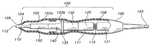
example, however, Fig. 1 shows one embodiment of the present invention with an
exemplary ultrasonic driver 100. The driver 100 is constructed from a
generally tubular
body 102 having a distal end 104 and a proximal end 106. A cavity 108 extends
through the body 102 between the distal end 102 and the proximal end 104. The
body
102 preferably includes a distal body end 102 a connected to a proximal body
end
102b. Such a two-piece body facilitates construction of the driver 100.
An ultrasonic driver unit 111 extends along the length of the cavity 108.
The distal end 104 includes an opening 109 therethrough to allow a tunneler
head 110
on the driver unit 111 to extend from inside the cavity 108 to an exterior of
the body
102. A tunneler tip 112 extends distally from the body 102 from a distal-most
portion
of the head 110. The proximal end 106 includes a connection for a power line
105 to
supply electrical power to the driver unit 111.
By means of power line 105, electrical energy, i.e., drive current, is sent
from a power supply proximate the driver 100 (shown for example, in Fig. 6, as
power
supply 600 and discussed in more detail below) to the driver 100 where the
power
supplied imparts ultrasonic longitudinal movement to head 110 at the distal
end of the
device. When power is applied to ultrasonic driver 100, the assembly
(discussed in
more detail below) will cause head 110 to vibrate longitudinally (for example
at
approximately 40 kHz). The amount of longitudinal movement will vary
proportionately
with the amount of driving power (current) applied, as adjustably selected by
the user.
CA 02575695 2007-01-31
WO 2006/015384 PCT/US2005/027880
-5-
Such ultrasonic vibration of the head 110 will generate heat as the tip
112 contacts tissue, i.e., the movement of the tip 112 through the tissue
converts the
mechanical energy of the moving head to thermal energy in a very localized
area at the
tip 112 of the head 110 (and therefore tunneler tip). This localized heat
creates a
narrow zone of coagulation, which will reduce or eliminate bleeding in small
vessels,
such as those less than one millimeter in diameter. The degree of hemostasis
will vary
with the level of driving power applied, the tunneling force applied by the
surgeon, the
nature of the tissue type, and the vascularity of the tissue, among other
factors.
Ultrasonic vibration at the tip 112 will also reduce friction which will
result in tunneling
io with less force exerted by the surgeon.
As illustrated in Fig. 1, this example of a suitable ultrasonic driver 100
houses a piezoelectric transducer 115 for converting electrical energy to
mechanical
energy that results in longitudinal vibrational motion of the ends of the
transducer.
Transducer 115 in this embodiment is in the form of a stack of ceramic
piezoelectric
elements with a motion null point located at some point along the stack, in
accordance
with the prior art. The transducer stack is mounted between two cylinders 120
and
121. Cylinder 130 is attached to cylinder 120, which in turn is mounted to the
housing
at another motion null point 135. Horn 140 is also attached to null point 135
on is
proximal side and to head 110 coupler 150 on its distal side. Head 110 is
affixed to
coupler 150. As a result, head 110 will vibrate in the longitudinal direction
at an
ultrasonic frequency rate with transducer 115.
The parts of the driver 100 are designed such that the combination will
oscillate at the same resonant frequency. In particular, the elements are
preferably
tuned such that the resulting length of each such element is one-half
wavelength.
Longitudinal back and forth motion is amplified as the diameter closer to head
110 of
the acoustical mounting horn 140 decreases. Thus, horn 140 as well as coupler
150
are shaped and dimensioned so as to amplify head 110 motion and provide
harmonic
vibration in resonance with the rest of the acoustic system, which produces
the
maximum back and forth motion of the end of the acoustical mounting horn 140
close
to head 110.
Fig. 2 shows an alternative embodiment of the present invention in which
head 110 is covered by tunneler tip 200. In this embodiment, tunneler tip 200
is
driven by head 110 and conveys the ultrasonic energy described above to a
larger tip to
aid in tunneling through tissue. In this embodiment, the tip 200 is removable
from the
rest of the driver 100. The tip 200 has a generally cone-shaped distal end 202
to
CA 02575695 2007-01-31
WO 2006/015384 PCT/US2005/027880
-6-
facilitate movement through the tissue, and an open proximal end 204 to
facilitate
insertion of the tip 200 over the distal end 104 of the unit 100.
In yet another embodiment, head 110 and tunneler tip 200 could be
formed of a single integral piece, such as is shown in Fig. 3 with integral
head 300. In
either event, head 110 and tunneler tip 200 (or simply head 300) are attached
to driver
100 through means known to those skilled in the art.
Fig. 4 shows the device of Fig. 2 attached to the end of tunneler 400.
The tunnelers used in accordance with the present invention are known to those
skilled
in the art. The tunnelers may be connected to the tips by known means,
including
threaded connections. Fig. 5 shows an alternative embodiment where tunneler
400
extends further along driver 100. In yet another embodiment (not shown), the
tunneler could extend even further along driver 100 and meet the tip 200 such
that the
entire driver 100 is contained within the tunneler except for that part
covered by tip
200.
Fig. 6 illustrates the driver of Fig. 5 connected to a power supply 600.
Power supply 600 is consistent with ultrasonic driver power sources known to
those
skilled in the art. Power supply 600 provides controllable current to power
line 105.
Included is handle 610 for the user to grasp and control the tunneler during
operation.
Fig. 7 shows an embodiment of the invention where tunneler 400 is
threadedly connected to driver 700. As described above, power line 105 feeds
current
to transducer 115 which oscillates and drives tip 315.
Fig. 8 illustrates still another embodiment where ultrasonic driver 100 is
disposed completely within tunneler 800. The tunneler 800 includes a body 802
having
a distal end 804 and a proximal end 806. A cavity 808 extends within the body
802
between the distal end 804 and the proximal end 806. In this embodiment, head
110
extends to an opening 809 in distal end 804 of the tunneler 800 sufficient to
provide
ultrasonic energy at the point of tissue contact as tunneler 800 is advanced
through
tissue during use.
In still another embodiment of the present invention, means for
dissecting tissue in the tunneler tip can be provided by direct electrical
current instead
of ultrasonic energy as described above. In this embodiment, as shown in for
example
in Fig. 9, a bipolar tip 900 is exposed at the distal end of tunneler 910. The
tunneler
910 includes a body 902 having a distal end 904 and a proximal end 906. A
cavity 908
extends within the body 902 between the distal end 904 and the proximal end
906.
Bipolar conductor leads 915, 920 extend through the cavity 908 and extend
slightliy
CA 02575695 2007-01-31
WO 2006/015384 PCT/US2005/027880
-7-
distally from the body 902 at a distal tip 912. The bipolar conductor leads
915 and 920
are coaxial with respect to each other at their distal end region, but at
their proximal
end are separate. The conductor leads 915 and 920 are separated by a coaxial
insulator (not shown) over that region where they are coaxial (toward the
distal end).
Fig. 10 shows an embodiment having bipolar tunneler tip 930 threadedly
connected to tunneler 940. The proximal end of each conductor lead 915 and 920
is in
electrical contact with power supply line 950. These bipolar tips are known to
those
skilled in the art for use in surgical pencils and cauterizing devices. The
present
invention, however, takes advantage of the delivery of this electrical energy
to separate
tissue layers during the advancement of the tunneler through the tissue. Also
possible
for use with the present invention would be a monopolar tip, which
configuration would
be known by those skilled in the art. With the tissue layer separation as
described
above, several advantages are realized, including cauterization and trauma
reduction.
Also as noted above, less force is needed by the operator to advance the
tunneler
is through the tissue.
Fig. 11 illustrates still yet another embodiment in which ultrasonic driver
100 is disposed in handle 610 of the device and head 110 extends throughout
tunneler
400. The distal tip of the device shown in Fig. 11 does not have a conical tip
as shown
in the embodiments of Figs. 2-6, but could have any of those tips disposed on
its distal
end. As described above, such conical tips could be attached to, or formed as
a part of,
head 110. Moreover, any combination of the embodiments disclosed above would
be
understood by one skilled in the art reading this disclosure.
The materials for the tunnelers and tips in accordance with the present
invention are typically stainless steel. Other possible materials would be
known,
however, to those skilled in the ultrasonic and tunneling arts.
Included in the present invention is a method of using the device
described above. Specifically, a method in accordance with the present
invention
includes the steps of advancing a tunneling device into living tissue and
separating
tissue layers at the tip of the tunneling device as the tunneling device is
advanced
through the tissue. This method is consistent with the use of the devices
described
above. The tissue is cauterized in accordance with the delivery of energy,
preferably
ultrasonic energy or direct electrical energy, as described above. The
tunneling
procedure generally, however, is that which is known to those skilled in the
art. The
advantages of the presently disclosed method, however, are described above,
and
3s include reduced trauma, reduced recovery time, higher patient comfort, less
pain, and
ease of use for the person performing the tunneling procedure. These
advantages,
CA 02575695 2007-01-31
WO 2006/015384 PCT/US2005/027880
-8-
achieved through this method and using the disclosed device, are a direct
result of the
delivery of energy (preferably ultrasonic or direct electrical energy) at the
distal tip of
the tunneler.
Although the invention is illustrated and described herein with reference
to specific embodiments, the invention is not intended to be limited to the
details
shown. Rather, various modifications may be made in the details within the
scope and
range of equivalents of the claims and without departing from the invention.