Note: Descriptions are shown in the official language in which they were submitted.
CA 02575783 2007-02-01
WO 2006/020327 PCT/US2005/025774
Cooling Tissue Inside the Body
TECHNICAL FIELD
This invention relates to cooling a target tissue region inside the body.
BACKGROUND
Myocardial ischemia, and in severe cases acute myocardial infarction (AMI),
can
occur when there is inadequate blood circulation to the myocardium due to
coronary artery
disease. Evidence suggests that early reperfusion of blood into the heart,
after removing a
blockage to blood flow, dramatically reduces damage to the myocardium.
However, the
reestablishment of blood flow into the heart may cause a reperfusion injury to
occur.
Reperfusion injury is believed to be due to the build up of waste products on
the myocardium
during the time blood flow was inadequate and the reaction of these waste
products with
oxygen in the blood when normal blood flow is reestablished. It is possible to
reduce
reperfusion injury to the myocardiuin by cooling the myocardial tissue prior
to reperfusion.
Mild cooling of the myocardial tissue to a temperature between 28 and 36
degrees Celsius
provides a protective effect, likely by the reduction in the rate of chemical
reactions and the
reduction of tissue activity and associated metabolic demands.
One method of cooling myocardial tissue is to place an ice pack over the
patient's
heart. Another method involves puncturing the pericardium and providing cooled
fluid to a
reservoir inserted into the pericardial space near the targeted myocardial
tissue. Cooling of
the myocardial tissue may also be accomplished by perfusing the target tissue
with cooled
solutions. A catheter having a heat transfer element located in the catheter's
distal tip may
also be inserted into a blood vessel to cool blood flowing into, and through,
the heart. It is
CA 02575783 2007-02-01
WO 2006/020327 PCT/US2005/025774
also possible to cool the myocardiai tissue by supplying cool blood to the
heart through a
catheter placed in the patient's coronary sinus.
SUMMARY
The invention features devices and methods to cool a target tissue region
inside the
body. In an aspect, the invention features a catheter that includes an
elongated meinber with
a lumen extending longitudinally through a portion of the member. The lumen
has an entry
port through which blood from a body vessel enters the lumen and an exit port
through which
the blood exits the lumen. An inflatable balloon is positioned between the
entry and exit
ports of the lumen, and when positioned within a body vessel and inflated, the
balloon
occludes the body vessel to prevent normal blood flow. A cooling element cools
blood as it
flows through the lumen.
In embodiments, the entry and exit ports of the lumen may be positioned so
that when
the catheter is in the body vessel, such as a coronary artery, the entry and
exit ports are both
within the body vessel. The inflated outer diameter of the inflatable balloon
may be
approximately five millimeters or less. The lumen may also be structured to
provide a blood
flow of twenty milliliters per ininute through the lumen with normal blood
pressure, and may
also have a diameter of less than about 45 thousandths of an inch.
In other embodiments, the cooling element may be located in a distal portion
of the
catheter. The cooling element may include a chamber that cools the blood by
using a Joule-
Thompson orifice to create a phase change of liquid to a gas. The inflatable
balloon can also
include an inflation chamber, and the balloon's inflation chamber may also
serve as the
chamber that cools the blood using the Joule-Thompson orifice. In other
embodiments, the
cooling element includes a thermoelectric cooler, which may include a
plurality of
thermoelectric semiconductors.
2
CA 02575783 2007-02-01
WO 2006/020327 PCT/US2005/025774
In another aspect, the invention features a catheter for providing cooled
blood to a
target tissue region inside a body. The catheter includes an elongated member
that has a
lumen extending longitudinally through a portion of the member. The lumen has
an entry
port through which blood from a body vessel enters the lumen and an exit port
through which
blood exits the lumen. A chamber is positioned in a distal portion of the
catheter between the
entry and exit ports of the lumen so that the chamber may cool the blood as it
flows through
the lumen by using a Joule-Thompson orifice to create a phase change of liquid
to a gas.
In embodiments, the entry and exit ports of the lumen may be positioned so
that when
the catheter is in the body vessel, such as a coronary artery, the entry and
exit ports are both
within the body vessel. In some embodiments, the chamber may also expand to
occlude a
body vessel to prevent nonnal blood flow to the target tissue region. The
chamber may
expand to an inflated outer diameter of approximately five millimeters or
less.
In another aspect, the invention features a metliod of providing cooled blood
to a
target tissue region inside a body. A catheter that has an inflatable balloon
near the catheter's
distal end is introduced into a body vessel. The balloon is inflated to
restrict normal blood
flow to the target tissue region through the body vessel. Blood is allowed to
flow through a
lumen in the balloon catlieter from an entry port proximal to the balloon to
an exit port distal
to the balloon, and the blood is cooled as it flows through the lumen.
In embodiments, the catheter may be positioned in the body vessel, for example
a
coronary artery, so that the entry and exit ports of the lumen are also within
the body vessel.
The method may also be performed during a percutaneous transluminal coronary
angioplasty.
The details of one or more embodiments of the invention are set forth in the
accompanying drawings and the description below. Other features, objects, and
advantages
of the invention will be apparent from the description and drawings, and from
the claims.
3
CA 02575783 2007-02-01
WO 2006/020327 PCT/US2005/025774
liEsc;x1Y'1'ION OF DRAWINGS
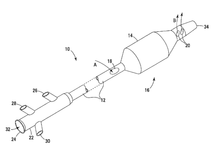
FIG 1 is a perspective view of a catheter in accordance with the invention.
FIG 2 is a side cross-sectional view, in a longitudinal plane, of a distal
portion of an
embodiment of a catheter of the type shown in FIG. 1.
FIG. 3 is a cross-sectional view of the catheter along the line 3-3 shown in
FIG. 2.
FIG. 4 is a perspective view of a thermoelectric cooler that may be used in a
catheter
in accordance with the invention.
FIG. 5 is a diagram of a side view of a distal portion of the FIG 1 catheter
positioned
in a coronary artery, shown in cross-section, and illustrates a method of
cooling a target tissue
region in the heart.
FIG. 6 is a diagram of a side view of a proximal end of a catheter used to
cool a target
tissue region and a control system connected to the proximal end of the
catheter, the control
system shown in block diagram.
Like reference symbols in the various drawings indicate like elements.
DETAILED DESCRIPTION
Referring to FIG 1, a catheter 10 includes an elongate tubular shaft 12 and an
inflatable balloon 14 at the catheter's distal portion 16. The catheter 10 may
be used to repair
a lesion in a body vessel, such as a coronary artery, that has reduced or
completely blocked
the flow of oxygenated blood to a tissue region. The catheter 10 may also be
used to provide
cooled blood to the oxygen-deprived, or ischemic, tissue region. A perfusion
lumen (not
shown in FIG 1) extends longitudinally through the shaft 12 at the catheter's
distal portion
16. When the balloon 14 is inflated in a body vessel so as to occlude blood
flow, blood will
be forced to enter the perfusion lumen through an entry port 18 in the
catheter shaft 12
proximal to the balloon 14, as indicated by arrow A. A cooling element located
in the
catheter's distal portion 16 (not shown in FIG 1) cools blood as it flows
through the perfiision
4
CA 02575783 2007-02-01
WO 2006/020327 PCT/US2005/025774
lumen, and the cooled blood exits the lumen distal to the balloon 14 through
exit ports 20, as
indicated by arrows B.
Delivery of cooled blood to the ischemic tissue region reduces the injury
associated
with the reperfusion of blood to the region without extending the time that
the tissue region is
deprived of oxygen. Because the blood provided to the tissue region during the
cooling
process is oxygenated, the cooling can be performed for as long as desired.
Further, the
oxygenated blood provided by the catheter 10 is cooled inside the body, and is
not removed
and cooled outside the body, which may damage blood cells. In addition,
providing blood to
the tissue region does not require the removal of the catheter's guide wire
(not shown in FIG
1) to infuse fluid into the vessel, which may compromise the position of the
catheter 10
during a procedure.
An adapter 22 is attached to the shaft 12 at the catheter's proximal end 24.
The
adapter includes a longitudinal opening 32 at the proximal end 24, which
provides access to a
lumen (not shown in FIG 1) inside the shaft 12. This internal lumen extends
through the
entire length of the shaft 12 to anotller longitudinal opening at the
catheter's distal end 34. A
guide wire (not shown) may be inserted through this internal lumen to allow a
physician to
maneuver the catheter through a body vessel and near a target tissue region.
Once the
catheter 10 is positioned, the guide wire may be removed and the lumen may
also be used to
provide fluid to the target tissue region.
The adapter 22 also includes ports 26, 28, and 30. The ports 26, 28, and 30
may
provide access to lumens or wires connecting internal devices, such as a
teinperature sensor,
that extend longitudinally through the catheter shaft 12 to the catheter's
distal portion 16.
The number of ports in the adapter, and the use of the ports, depends upon the
type of cooling
element used to cool the blood flowing through the perfusion lumen, as will be
described in
detail later.
5
CA 02575783 2007-02-01
WO 2006/020327 PCT/US2005/025774
In the FIG. 1 example, the catheter 10 may cool blood flowing through the
perfusion
lumen to a range of 25 to 36 degrees Celsius. The amount of cooling depends
upon a number
of factors, such as the volume flow rate of the blood through the perfusion
lumen, the length
and inside diameter of the perfusion lumen, and the cooling capability of the
cooling element.
For example, in an implementation where the length of the perfusion lumen is
approximately
20 millimeters and the perfusion lumen's inside diaineter is approximately 40
thousandths of
an inch, the volume flow rate of blood through perfusion lumen is
approximately 24 ml/min.
Also in this example, the temperature of the cooling element is approximately
minus 10
degrees Celsius, the blood flowing through the perfusion luinen can be cooled
from normal
body temperature of approximately 37 degrees Celsius to approximately 29
degrees Celsius.
The cooling of the blood may be varied by changing one or more of these
variables. For
example, by reducing the volume flow rate of the blood through the perfusion
lumen to 12
ml/min, with all other things remaining constant, the blood may be cooled to
25 degrees.
The volume flow rate of blood through the perfusion lumen is determined by the
size
of the perfusion lumen, the size and shape of the entry port 18 and the exit
ports 20, and, of
course, the blood pressure at the entry port 18. In the FIG 1 implementation,
the entry port
18 has a substantially oval shape and with axes of approximately 4.5 and 1.5
millimeters. In
other implementations, the entry port 18 may be configured in another shape
and the surface
area of the port 18 may be increased or decreased. Further, additional entry
ports may be
added to the catheter 10 to allow additional blood flow to enter the perfusion
lumen. The
FIG 1 catheter has two oval-shaped exit ports 20 with axes of approximately
five hundredths
and two hundredths of an inch. Like the entry port 18, the exit ports 20 may
also be
configured in another shape and the combined surface area of the exit ports 20
may be
increased or decreased as desired. In addition, additional exit ports may be
added to the
catheter shaft 12, or alternatively, the shaft 12 may have only one exit port.
In examples
6
CA 02575783 2007-02-01
WO 2006/020327 PCT/US2005/025774
where the blood flow rate through the perfusion lumen is reduced to increase
the cooling of
the blood, inflation/deflation cycling of the balloon 14 may be required to
oxygenate the
tissue distal to the balloon. To prevent reperfusion injury, however, the
balloon 14 should not
be deflated to allow oxygenated blood at body temperature to reach the tissue
region until the
tissue region has first been cooled.
The catheter 10 may cool blood flowing through the perfusion lumen with a
variety of
different cooling elements or mechanisms, depending upon factors such as the
length of the
perfusion lumen, the desired amount of cooling, the desired size of the
catheter's distal
portion 16, and the flexibility of the distal portion 16 of the catheter
required for the specific
application. The cooling element may be, for example, a chamber that is
positioned adjacent
to the perfusion lumen and is accessible via one or more lumens in the
catheter. In this
example, a cool fluid may be provided to the chamber, wliich in turn cools the
blood flowing
through the perfusion lumen.
In another embodiment, a chamber may be used to cool the blood that flows
through
the perfusion lumen using a physical process called the Joule-Thompson effect.
To use this
process, a highly-pressurized fluid is introduced into the chamber and is
allowed to change
phase from a liquid to a gas across an orifice located at a distal end of a
luinen. As the fluid
changes phase, energy in the form of heat is pulled form the surrounding area,
which cools
the chamber and the blood flowing through the perfusion lumen. An example of a
catheter
that uses the Joule-Thompson effect to cool blood is shown in FIGS. 2 and 3.
In other implementations, the cooling element may be thermoelectric cooler
(TEC)
(shown in FIG 4), which cools blood flowing through the perfusion lumen using
a process
called the Peltier effect. In this example, the TECs are positioned between
the entry port 18
and exit ports 20 and in thermal contact with the blood flowing through the
perfusion himen,
as will be discussed later. The TECs that are currently available do not have
the cooling
7
CA 02575783 2007-02-01
WO 2006/020327 PCT/US2005/025774
capability of a Joule=l'hompson cooling element of a similar size and cooling
surface area.
As a result, current TECs may not be capable of cooling blood to 29 degrees
Celsius as in the
previous example where the length of the perfusion lumen was 20 millimeters
with an inside
diameter of 40 thousandths of an inch and the volume flow rate of the blood
through
perfusion lumen is 24 ml/min. Thus, to achieve the same ainount of cooling,
TECs may
currently be used only in applications where the volume flow rate of blood is
reduced or the
length of the perfusion lumen is increased. As the cooling ability of TECs
continues to
increase, they may become suitable for more applications in the fiiture.
FIG. 2 is a side cross-sectional view, in a longitudinal plane, of a distal
portion 116 of
a catheter that uses the Joule-Thompson effect to cool blood as it flows
through the catheter's
perfusion lumen 136. The catheter's distal portion 116 includes an inflatable
balloon 114 that
is positioned over a shaft 112 between the entry port 118 and the exit ports
120 of the
perfusion lumen 136, and around the shaft's entire circumference. Welds (not
shown) secure
and seal the longitudinal ends 142 of the balloon 114 to the shaft 112, thus
forming a sealed
chamber 140 between the shaft 112 and the balloon 114. An infusion lumen 144
extends
through the shaft 112, from a port in an adapter (e.g., the port 26 of FIG. 1)
to, and into, the
sealed chamber 140. A highly pressurized fluid, such as COZ, N20, N2, or He,
is introduced
into the sealed chamber 140 and expands into a gas across a Joule-Thompson
orifice 146.
The phase change performs two functions in the FIG 2 catheter. In addition to
reducing the temperature of the chamber 140, the phase change to gas also
inflates the
balloon 114, which may repair a lesion in a body vessel, if necessary, and
also block normal
blood flow tlirough the body vessel and force the blood into the perfusion
himen 136. An
exhaust lumen (shown in FIG 3), which extends longitudinally from the sealed
chamber 140
to an adapter port (e.g., the port 28 shown in FIG 1), removes excess gas from
the sealed
8
CA 02575783 2007-02-01
WO 2006/020327 PCT/US2005/025774
chamber 140 to maintain a desired pressure in the chamber 140 and inflate the
balloon 114 to
a desired level.
In the FIG. 2 example, a temperature sensor 150 is located inside the chamber
140 and
monitors the temperature of the chamber 140. In this example, the temperature
sensor 150 is
a thermocouple. The thermocouple consists of two conductive wires 154 of
dissimilar
material that are insulated from each other. The wires 154 extend
longitudinally through the
catheter shaft 112 from a port in an adapter, for example the port 30 in the
adapter 22 shown
in FIG 1, and into the chamber 140. The conductive wires 154 are joined
together to form a
junction 152, which is in thermal contact with the gas inside the chamber 140.
When two
dissimilar conductors are joined in this maimer, an electro-motive force (emf)
is induced
across the junction 152, the magnitude of which varies as a function of the
junction's
temperature. The induced emf may be measured at the proximal ends of the
conductive wires
154, and thus allow the temperature of the chainber 140 to be measured. In
other
implementations, the temperature sensor 150 may be a tliermistor or other
suitable
temperature-sensing mechanism. The temperature sensor 150 may also be placed
in different
locations in the shaft 112 to measure the temperature of the chamber. In other
implementations, additional temperature sensors may be added to the catheter
to measure, for
example, the temperature of the blood exiting the exit ports 120.
A lumen 148 extends longitudinally through the catheter from an opening at the
catheter's proximal end (e.g., the longitudinal opening 32 shown in FIG 1) to
an opening in
the catheter's distal end 134. A guide wire (not shown) may be extended
longitudinally
through this lumen 148 to allow a physician to guide the catheter's distal
portion 116 through
a body vessel to a target tissue region. Once the catheter is positioned in
the body, the lumen
148 may also be used to provide fluid to the target tissue region if desired.
For example, cool
blood or a blood substitute could be provided to the target tissue region.
Cool saline or a
9
CA 02575783 2007-02-01
WO 2006/020327 PCT/US2005/025774
saiine soiunon containing annoxiaanis or other vascular agents such as nitric
oxide, lidocaine,
nitroglycerine, insulin, etc., may also be provided via lumen 148.
In the FIG 2 example, the walls of the balloon 114 have a greater thickness,
for
example 0.00 15 inch, than typical inflation balloons for balloon catheters,
which are
approximately 0.0007 inch. The increased thickness of the balloon walls
insulates bodily
fluids and tissues that contact the outer surface of the balloon 114. The
insulation may limit
the systematic cooling effects of the catheter and improve the efficiency of
the targeted
cooling of the blood flowing through the perfusion lumen 136. In other
implementations, the
balloon thickness may be increased or decreased as required. Alternatively, an
additional
outer layer may be added to the balloon 114. The additional outer layer may be
constructed
of a polymer, for example, polyester. In some implementations, a fluid or a
polymer material
may be placed between the balloon 114 and the additional outer layer to
provide an additional
insulation.
FIG. 3 shows a cross-sectional view of the catheter's distal portion 116 at
line 3-3 of
FIG 2 looking proximally from the balloon 114. The FIG 3 cross-section
illustrates the
relative size and location of the perfusion lumen 136, the lumen 148 for the
guide wire and
infusion of fluid to the target tissue region, the infusion lumen 144 and
exhaust lumen 156,
and the conductive wires 154. The balloon's longitudinal end portion 142 is
shown attached
to the shaft's outer surface 158.
The perfusion lumen 136 may have a diameter of approximately 39 to 42
thousandths
of an inch, and may vary depending upon the application. The diameter of the
perfusion
lumen 136 may be increased to increase the flow rate of blood through the
lumen, or
alternatively, the diameter may be decreased to reduce the flow rate of blood.
The lumen 148
may have a diameter of approximately 15 to 20 thousandths of an inch, and may
be increased
CA 02575783 2007-02-01
WO 2006/020327 PCT/US2005/025774
or decreased depending upon the application and the type of guide wire a
physician may want
to use to perform the procedure.
The infusion lumen 144 and exhaust lumen 156 in the FIG 3 exainple
collectively
form a half-circle in cross-section, with the infusion lumen 144 and exhaust
lumen 156 each
making up approximately half of the area. In other implementations, the
infusion lumen 144
and exhaust lumen 156 may have circular cross-sections, or be constructed in
another suitable
configuration.
FIG 4 is a perspective view of a TEC 200 that may be used to cool blood as it
flows
through a perfusion lumen for delivery to a target tissue region using a
thermal energy
process known as the Peltier effect. The TEC 200 includes a first and second
module 202 and
204, which when placed together, form a cylinder with a lumen 206 through
which blood
may flow. The TEC 200 may be placed in the outer wall of the perfusion lumen
so that the
blood flows through the lumen 206 of the TEC 200 for cooling as it flows
through the
perfusion lumen.
To form this cylinder-shaped structure, both the first and second modules 202
and 204
are in the shape of a half-cylinder, where the cylinder is split
longitudinally in two equally-
sized sections. The longitudinal edges of the first and second modules 202 and
204 are
separated by small gaps 208a and 208b.
The first module 202 of the TEC 200 is connected to wires 210 and 212 at the
first
module's proximal end 214, and connected to wires 216 and 218 at the first
module's distal
end 220. In this implementation, the wires extend 210 and 212 extend
longitudinally through
the shaft of the catheter toward the catheter's proximal end so that the
temperature of the
TECs may be controlled, as explained later. If the catheter includes
additional TECs 200,
then the wires 210 and 212 may be connected to the first module of another
TEC. If the TEC
200 is the most proximal TEC in the catheter shaft, the wires 210 and 212
extend
11
CA 02575783 2007-02-01
WO 2006/020327 PCT/US2005/025774
longitudinally through the shaft to the catheter's proximal end for access
outside of the
patient through a port in an adapter, for example the port 30 shown in FIG 1.
The wires 216
and 218 extend longitudinally through the catheter shaft toward the catheter's
distal end and
may be connected to the first module of another TEC located distal to the TEC
200.
The second module 204 is similarly connected to wires 222 and 224 at the
second
modules proximal end 214, and connected to wires 226 and 228 at the second
module's distal
end 220. The wires 222, 224, 226, and 228 extend longitudinally through the
shaft and
connect to the second modules of the various TECs in the catheter in the same
manner as
described for the first module 202.
The first and second modules 202 and 204 may, for example, contain a series of
thermoelectric cooling elements. The elements may be, for example, packaged
within an
electrical insulator and include an n-type semiconductor and a p-type
semiconductor
connected in series. In other implementations, the semiconductors may be
replaced with
other suitable materials. The semiconductors would typically be arranged
between a ceramic
substrate that electrically insulates the conductors from heat sinks attached
to the ceramic
substrate on two sides of the thermoelectric cooling element. The thermo
electric cooling
elements are arranged so that one heat sink is adjacent to contact the
internal surface of the
modules 202 and 204 (i.e., the surface that forms the lumen 206). The other
heat sink is
arranged to be adjacent to the external surface 230 of the modules 202 and
204.
To utilize the cooling effect of the TEC 200, a DC voltage may be applied to
the
elements via the wires 210, 212, 222, and 224, which causes a current to pass
through the
semiconductor pairs. The current causes heat to be drawn from the heat sink on
the surface
that forms the lumen 206 to the heat sink near the external surface of the
modules 230.
Through this process, the internal surface that forms the lumen 206 is cooled,
and at the same
12
CA 02575783 2007-02-01
WO 2006/020327 PCT/US2005/025774
time, the external surtace 230 is heated. By cooling the internal surface that
forms the lumen
206, the blood flowing through the perfusion lumen of the catheter may also be
cooled.
In an implementation where a TEC 200 is used for cooling, using both the
infusion
and exhaust lumens shown in FIGS. 2 and 3 may be unnecessary. A single lumen
may be
sufficient to inflate and deflate the balloon at the catheter's distal end.
Like the FIG. 2
infusion lumen, the balloon inflation lumen may extend longitudinally from the
sealed
chamber formed by the balloon to a port in the catheter's adapter.
FIG 5 is a diagram of a side view of a distal portion 16 of the FIG 1
perfusion
catheter positioned in a coronary artery, shown in cross-section, and
illustrates a method of
cooling a target tissue region 302 in the heart. In the FIG 5 example, the
distal portion 16 of
the perfusion catheter 10 is positioned in a coronary artery 300 of the heart,
via the aorta 304,
that contains a lesion or blockage and is being treated with percutaneous
transluminal
coronary angioplasty. Once the distal portion 16 of the catheter is positioned
in the artery
300, the balloon 14 is inflated to prevent normal blood flow to the target
tissue region 302,
and in some implementations, to open an occlusion of the artery 300. Blood
that enters the
perfusion lumen through entry port 18, as indicated by arrow A, is cooled by
the cooling
element in the catheter's distal portion 16. The blood then exits the
perfusion lumen through
exit ports 20, as indicated by arrows B, and is provided to the tissue region
302 to reduce
reperfusion injury.
The FIG 1 catheter may also be used to cool tissue regions in other areas of
the body.
For example, the catheter may be used in the brain, kidneys, and legs.
FIG. 6 shows a system including the previously described catheter (only a
portion of
which is shown in FIG 6) and various external equipment attached to the
catheter. In this
example, the catheter is attached to a control system 402, which includes a
controller 404, a
fluid pump 406, an exhaust valve 408, and a temperature monitor 410. The
controller 404
13
CA 02575783 2007-02-01
WO 2006/020327 PCT/US2005/025774
receives intormation trom ttie temperature monitor 410 and uses that
information to control
the operation of the fluid pump 406 and the temperature of the blood delivered
to a target
tissue region. The controller 404 also monitors the pressure in the catheter's
balloon (not
shown in FIG 6), which dictates the balloon's inflation and deflation, and
also permits the
continual expansion of gas into the balloon's chamber for cooling.
The catheter's proximal end 400 has an adapter 414 with ports 416, 418, and
420.
The port 416 provides access to an infusion lumen that extends longitudinally
through the
catheter to the balloon's chamber in the catheter's distal portion. The fluid
pump 406 is
connected to the infusion lumen via port 416. The controller 404 controls the
operation of the
fluid pump 406, and thus the amount and rate of super-cooled fluid provided to
the balloon's
chamber. The super-cooled fluid 412 provided to the sealed chamber may be C02,
N20, N2,
He, or another suitable fluid.
The port 418 provides access to an exhaust lumen that extends longitudinally
through
the catheter from the balloon's chamber. The exhaust valve 408 is connected to
the exhaust
lumen via port 418. The controller 404 controls and monitors the removal of
gas from the
balloon's chamber by exhaust valve 408. The port 420 provides access to a
temperature
sensor that senses the temperature of the sealed chainber. For example, in an
implementation
where the temperature sensor is a thermocouple (as shown in FIG 2), the port
420 provides
access to the conductive wires that extend from the thermocouple's junction in
the distal
portion of the catheter.
In other implementations, additional external devices may be added to the
control
system 402, or alternatively, some of the devices may be omitted. Further, the
control system
402 may be modified to control the cooling of catheters that use a TEC to cool
the blood
flowing through the perfusion lumen. In such an implementation, the fluid pump
406 may be
used to introduce and remove an inflation medium, and thus inflate and deflate
the catheter's
14
CA 02575783 2007-02-01
WO 2006/020327 PCT/US2005/025774
balloon. The exhaust valve may be replaced with a DC voltage source that
controls the
amount of cooling of the TECs. The temp monitor may be used to monitor a
temperature
sensor that measures the temperature of the fluid exiting the catheter's
perfusion lumen.
A number of embodiments of the invention have been described. Nevertheless, it
will
be understood that various modifications may be made without departing from
the spirit and
scope of the invention. Accordingly, other embodiments are within the scope of
the
following claims.