Note: Descriptions are shown in the official language in which they were submitted.
CA 02576324 2007-01-26
=
LUMEN REDUCTION METHODS AND DEVICES
FIELD OF THE INVENTION
[0001] The present invention relates to devices and methods for bariatric
surgery, and in
particular, to devices and methods for reducing the size of a lumen.
BACKGROUND OF THE INVENTION
[0002] One treatment for morbid obesity is bariatric surgery which involves
alteration of a
patient's digestive tract to encourage weight loss and to help maintain a
normal weight. A
common type of bariatric surgery is gastric bypass surgery, which aims to
decrease the size of a
patient's stomach by dividing it into upper and lower pouches using staples
and/or stitches. The
jejunum (the middle section of the small intestine) is also divided into two
parts. One part of the
jejunum, called the "Roux limb," is brought up behind the colon and lower
stomach pouch, and
joined or "anastamosed" to the upper stomach pouch. The remaining end of the
jejunum is
attached to the side of the Roux limb. As a result, a new digestive pathway is
created through
which food travels down the esophagus, into the upper stomach pouch, and
through the
anastomosis (or stoma) into the Roux limb. Digestive juices from the stomach,
the liver, and the
pancreas travel through the lower stomach pouch, down the duodenum and
jejunum, and into the
Roux limb where the two parts of the jejunum are attached and further
digestion takes place.
[0003] While effective, gastric bypass surgery is not without complications.
For example, the
stoma may dilate over time, allowing a patient to eat more and causing them to
gain weight.
Accordingly, there remains a need for improved devices and methods for
bariatric surgery, and
in particular, for devices and methods for reducing the size of a stoma.
SUMMARY OF THE INVENTION
[0004] The present invention provides various devices and methods for reducing
a size of a
lumen. In one embodiment, a lumen reduction device is provided having an end
effector with a
trough formed therein for receiving tissue around a lumen, and several
fasteners releasably
disposed therein. The fasteners can be positioned in a substantially
circumferential pattern, and
1
CA 02576324 2007-01-26
can engage tissue disposed within the trough. The device can also include at
least one suture
coupled to at least two of the fasteners and configured to be cinched to pull
the fasteners together
to reduce a size of a lumen. By way of non-limiting example, the device can
include a first
suture coupled to at least two fasteners, and a second suture coupled to at
least two fasteners
different from the fasteners coupled to the first suture.
[0005] The end effector can have a variety of configurations, but it is
preferably adapted to be
positioned within a lumen. In one embodiment, the end effector can have
proximal and distal
housing portions that define the trough therebetween. In an exemplary
embodiment, the trough
is formed around the circumference of the end effector. The trough can have a
fixed size or
alternatively it can have an adjustable size. For example, the proximal and
distal housing
portions can be movable to allow adjustment of the size of the trough. The
trough can include
features for gathering tissue, and in one embodiment the trough can include at
least one suction
port for suctioning tissue into the trough. In an exemplary embodiment, a
plurality of suction
ports are formed within the trough around the entire circumference thereof.
[0006] The trough can also optionally include features for injuring the tissue
such that it bleeds.
The tissue-injuring elements can be located at a variety of locations in the
trough, but in an
exemplary embodiment the tissue-injuring elements are disposed on opposed
walls of the trough
such that tissue within the trough can be positioned between the tissue-
injuring elements. A
variety of electrical and mechanical elements can be used as tissue-injuring
elements, such as RF
electrodes, monopolar electrodes, bipolar electrodes, mechanical scrapers, and
combinations
thereof.
[0007] The end effector can also be adapted to hold fasteners that can be
applied to tissue
disposed within the trough. While a variety of fastener holding techniques can
be used, in one
embodiment the trough can include channels formed therein. The shape and the
size of the
channels can vary depending upon the type of fasteners used. In one
embodiment, the fasteners
can have an elongate shape in an open position, and a ring-shape in a closed
position. The
fasteners can be biased to the closed position in which they are effective to
engage tissue. The
channels in the trough can be longitudinal cut-outs that extend between the
proximal and distal
housings to allow the fastener to extend across the trough. The fasteners can
be held within the
2
CA 02576324 2007-01-26
channel using a variety techniques, and in one embodiment the device can
include proximal and
distal fastener-retaining members located within the proximal and distal
housings. The fastener-
retaining members can have a variety of configurations depending upon the type
of fastener
used, however in one embodiment, the proximal fastener-retaining member is
adapted to retain a
first end of the fasteners, and the distal fastener-retaining member is
adapted to retain a second,
opposed end of the fasteners. In an exemplary embodiment, the proximal and
distal fastener-
retaining members are adapted to rotate to release the first and second ends
of the fasteners into
tissue. Rotation of the proximal and distal fastener-retaining members can be
affected, for
example, by first and second rotatable actuators that are coupled to the
proximal and distal
fastener-retaining members, respectively. The first and second actuators can
be configured to be
independently rotated, or alternatively, the first and second actuators can be
rotated
simultaneously. In other embodiments, the proximal and distal fastener-
retaining members can
be configured to release each fastener individually. For example, the legs of
the fastener-
retaining members can vary in length, such that the fastener-retaining members
will release the
fasteners sequentially as the fastener-retaining members are rotated.
[0008] Methods for reducing a size of a lumen are also provided. In one
embodiment, a method
can include positioning an end effector having a trough for receiving tissue
within a lumen,
actuating the end effector to deliver fasteners positioned within a
substantially circumferential
pattern around the end effector into the tissue, and cinching at least one
suture coupled to at least
two of the fasteners to pull the fasteners together and thereby reduce the
size of the lumen. In
one exemplary embodiment, actuation of the end effector can cause at least a
portion of one or
more of the fasteners to be simultaneously applied to tissue. For example, a
first end of the
fasteners can be released from a proximal fastener-retaining member formed
within a proximal
housing of the end effector, and a second end of the fasteners can be released
from a distal
fastener-retaining member formed within a distal housing of the end effector.
The first end of
the fasteners can be released independently from the second end of the
fasteners. The method
can also include suctioning tissue surrounding the end effector into the
trough, as well as
activating the end effector to injure tissue. While a variety of activation
techniques can be used,
in one embodiment, energy can be delivered to one or more tissue-injuring
elements disposed
within the trough to cause the tissue to bleed. In general, one or more
portions of the device can
be reconditioned after at least one use of the device. Such reconditioning can
include replacing
3
CA 02576324 2007-01-26
or cleaning at least a portion of any one of the pieces of the device, as well
as optionally
disassembling or reassembling the device.
BRIEF DESCRIPTION OF THE DRAWINGS
[0009] The invention will be more fully understood from the following detailed
description
taken in conjunction with the accompanying drawings, in which:
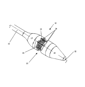
[0010] FIG. 1 is perspective view of one embodiment of a lumen reduction
device;
[0011] FIG. 2A is a perspective view of an end effector of the device of FIG.
1;
[0012] FIG. 2B is a cross-sectional view of the end effector of FIG. 2A taken
across line A-A,
showing the proximal and distal housing portions and the trough of the end
effector;
[0013] FIG. 2C is a perspective view of one embodiment of a fastener for use
with the lumen
reduction device of FIGS. 1-2B;
[0014] FIG. 3A is a perspective view of the actuators and fastener-retaining
members of the end
effector of FIG. 2B having a plurality of fasteners retained therein;
[0015] FIG. 3B is a front view of one of the fastener-retaining members of
FIG. 3A;
[0016] FIG. 3C is a transparent perspective view of the actuators and fastener-
retaining members
of the end effector of FIG. 2B;
[0017] FIG. 4 is a partially cross-sectional view of the end effector of FIG.
1 having sutures
coupled to fasteners contained therein;
[0018] FIG. 5A is a partially cut-away side view of the end effector of FIG. 1
positioned within a
stoma and suctioning tissue into a trough formed in the end effector;
[0019] FIG. 5B is a partially cut-away side view of the end effector and stoma
of FIG. 5A
showing release of the fasteners from the fastener-retaining members; and
[0020] FIG. 5C is a partially cut-away side view of the stoma of FIG. 5B
following removal of
the end effector of FIG. 2A, and showing sutures extending through the
fasteners for cinching
4
CA 02576324 2007-01-26
tissue around the stoma.
DETAILED DESCRIPTION OF THE INVENTION
[0021] Certain exemplary embodiments will now be described to provide an
overall
understanding of the principles of the structure, function, manufacture, and
use of the devices
and methods disclosed herein. One or more examples of these embodiments are
illustrated in the
accompanying drawings. Those of ordinary skill in the art will understand that
the devices and
methods specifically described herein and illustrated in the accompanying
drawings are non-
limiting exemplary embodiments and that the scope of the present invention is
defined solely by
the claims. The features illustrated or described in connection with one
exemplary embodiment
may be combined with the features of other embodiments. Such modifications and
variations are
intended to be included within the scope of the present invention.
[0022] The present invention provides devices and methods for reducing a size
of a lumen. In
general, the device can include an end effector having a trough formed therein
for receiving
tissue, and for delivering a plurality of fasteners to the tissue. The
fasteners can be coupled by
one or more sutures which can be used to cinch the tissue and thereby reduce
the size of the
lumen. The device can also include features to facilitate engagement of tissue
within the trough,
injury of tissue to promote healing, and various other features to facilitate
use of the device. A
person skilled in the art will appreciate that the present device can be used
in any procedure
where it is necessary to apply fasteners and/or reduce the size of a lumen,
such as stoma,
jejunum, duodenum, or colon reduction procedures.
[0023] FIG. 1 illustrates one exemplary embodiment of a lumen reduction device
10 for reducing
the size of a lumen. In general, the device 10 includes an outer shaft 12
having proximal and
distal ends 12a, 12b. The outer shaft 12 can have virtually any configuration,
and it can be
flexible or rigid. In an exemplary embodiment, the outer shaft 12 has a
configuration that allows
it to be endoscopically inserted through the esophagus. The proximal end 12a
can include a
handle 14 and the distal end 12b can include an end effector 16 for receiving
and treating tissue.
[0024] The end effector 16 is shown in more detail in FIGS. 2A-2B. While the
shape of the end
effector can vary, it is preferably shaped to be positioned within a lumen,
and includes a trough
CA 02576324 2007-01-26
=.
for holding tissue. The end effector 16 is also preferably adapted to
releasably retain one or
more fasteners for delivering the fasteners to tissue disposed within the
trough. In the illustrated
embodiment, the end effector 16 includes proximal and distal housing portions
17, 19 that are
connected by a connector portion 18, and that define the trough 20
therebetween. The housing
portions 17, 19 can be integrally formed with one another and/or the outer
shaft 12, or they can
be separate from one another and/or the outer shaft 12. While the housing
portions 17, 19 can
have a variety of configurations, in the embodiment shown in FIG. 2B each
housing portion 17,
19 has a substantially cylindrical, hollow configuration for retaining one or
more fasteners
therein, as will be discussed in more detail below. The connector portion 18
can have a diameter
smaller than a diameter of the proximal and distal housing portions 17, 19 to
define the trough 20
therebetween. The housing portions 17, 19 can also include features to
facilitate insertion into
the esophagus. By way of non-limiting example, the distal housing portion 19
can include a
tapered end with a blunt tip. The proximal and distal housing portions 17, 19
can also optionally
include a lumen 22 formed therethrough for receiving a guidewire to facilitate
positioning of the
device within a lumen.
[0025] The trough 20 formed between the proximal and distal housing portions
17, 19 can be
located at a variety of locations on the end effector 16, and it can extend
partially or entirely
around a circumference thereof. In an exemplary embodiment, the trough 20 is
formed around
the entire circumference of the end effector 16 to allow tissue surrounding a
lumen to be received
therein. The trough 20 can have any shape and size depending upon the amount
of tissue to be
received. In the illustrated embodiment, the trough 20 has a substantially
rectangular cross-
sectional shape with a backwall 21 that is defined by the connector 18, and
opposed endwalls 23,
25 that are defined by the proximal and distal housing portions 17, 19. The
size of the trough 20
should be sufficient to receive the amount of tissue to be fastened. In an
exemplary embodiment,
the trough 20 has a depth d of at least about 3 mm and a width w of at least
about 5 mm. The
trough 20 can also optionally have an adjustable size. For example, one or
both of the proximal
and distal housing portions 17, 19 can be movably coupled to the connector 18
to allow the
housing portions 17, 19 to slide relative to one another and thereby increase
or decrease the
width w of the trough 20. A lever located on the connector 18 can optionally
be provided for
controlling and adjusting the size of the trough.
6
CA 02576324 2007-01-26
[0026] As explained above, the trough 20 is configured to receive tissue.
While a variety of
techniques can be used to position tissue within the trough 20, in one
embodiment the trough 20
can include a plurality of suction elements 24 for suctioning tissue therein.
The trough 20 can
include any number of suction elements 24, and each suction element 24 can
have any shape,
such as ports or slots, and can have any size. The suction elements 24 can
also be formed
anywhere on the trough 20. As shown in FIG. 2B, the trough 20 includes suction
ports 24 that
are located around the entire circumference of the trough 20, that is, on the
basewall 21 and the
endwalls 23, 25. The suction ports 24 can also be positioned in any pattern
that is effective for
engaging tissue, such as in equally spaced rows within the trough 20. In use,
a suction force can
be generated using a pump or other element coupled to the proximal end of the
shaft or the
handle to pull air into the ports and suction the tissue therein.
[0027] The trough 20 can also optionally be adapted to injure or cause
intentional injury to
tissue, thereby promoting healing when the tissue is cinched together. Any
tissue-injuring
technique can be used, and one or more tissue-injuring elements can be
positioned anywhere
within the trough 20. In an exemplary embodiment, one or more tissue-injuring
elements are
positioned on the opposed endwalls 23, 25 of the trough 20. The tissue-
injuring elements can
also be located around the entire circumference of the trough 20 and spaced a
distance apart from
one another or located only in zones that are being cinched. The tissue-
injuring elements can be
in the form of electrical elements, such as electrodes for delivering RF,
monopolar, bipolar, or
other energy to the tissue, or mechanical elements, such as scrapers or little
blades located on the
endwalls of the trough that move to cut the tissue. In an exemplary
embodiment, the tissue-
injuring elements are in the form of two bipolar or monopolar strips that are
disposed on the
opposed endwalls 23, 25 and around the circumference of the trough 20.
Alternatively, a portion
of each endwall 23, 25 can be formed from a conductive material for receiving
energy. The
location of the tissue-injuring elements on the endwalls 23, 25 allows the
applied energy to travel
across or between the walls of the proximal and distal housing portions 17, 19
and through tissue
64 contained within the trough 20. Energy can be delivered to the strips
through one or more
leads extending through the housing and coupled to an internal or external
energy source.
[0028] As also explained above, the end effector can be adapted to hold one or
more fasteners
for delivering the fasteners to tissue disposed within the trough. While a
variety of techniques
7
CA 02576324 2007-01-26
can be used to hold the fasteners in the end effector, in one embodiment, the
trough 20 can
include one or more channels 26 formed therein for seating the fasteners. The
number and
location of the channels 26 can vary depending upon the desired amount of
tissue to be cinched.
In the exemplary embodiment shown in FIG. 2A the channels 26 are disposed
around the entire
circumference of the trough 20 such that the fasteners are located in a
circumferential pattern
therearound. The shape and size of the channels can also vary depending upon
the type of
fasteners used, and various fasteners known in the art can be used. In an
exemplary
embodiment, the fasteners 28d can have an elongate configuration with opposed
ends 28d1, 28d2
that are adapted to penetrate tissue as shown in FIG. 2C, and the channels 26
have an elongate
longitudinal configuration that extends through the sidewalls of the proximal
and distal housing
portions 17, 19, as well as the connector 18. The fasteners can be disposed
within the channels
26 such that the fasteners extend across the channel 26, as will be discussed
in more detail
below. In an exemplary embodiment, the fasteners are biased to a closed, ring-
shaped
configuration, and the ends can be expanded to have an elongate configuration
in an open
position. The opposed ends of the fasteners can be held within the channels 26
in an open
configuration using one or more fastener-retaining members, as will be
discussed below. Upon
release from the channels 26, the fasteners can close to form a ring-shaped
member that engages
the tissue. The fasteners can also include features to facilitate penetration
of tissue, such as
pointed ends and/or lubrication. FIG. 2C illustrates fastener 28d having
pointed end 28d2. The
size of each fastener can also vary depending upon the type and amount of
tissue to be cinched.
In an exemplary embodiment, the fasteners have a diameter that is about 3.5 mm
in a closed
position. A person skilled in the art will appreciate that the fasteners can
be formed from a
variety of biocompatible and superelastic materials, including, by way of non-
limiting example,
shape memory metals such as Nitinol.
[0029] As indicated above, the fasteners can be releasably retained within the
channels using
various techniques, but in an exemplary embodiment they are retained within
the channels with
proximal and distal fastener-retaining members that are disposed within the
proximal and distal
housings. FIG. 3B illustrates fastener-retaining member 30, which includes a
central disc with
several hook-shaped legs 34a, 34b, 34c, 34d, 34e, 34f, 34g, 34h (hereinafter
34a-h) extending
outwardly therefrom for holding the ends of the fasteners. As shown in FIG.
3A, the hook-
shaped legs on the proximal fastener-retaining member 30 are adapted to hold
the first end of the
8
CA 02576324 2007-01-26
fastener (first end 28d1 of fastener 28d is shown) within the channels in the
proximal housing
portion 17, and the hook-shaped legs on the distal fastener-retaining member
32 are adapted to
hold the second, opposed end of the fasteners (second end 28d2 of fastener 28d
is shown) within
the channels in the distal housing portion 19. The hook-shaped legs 34a-h on
each fastener-
retaining member are preferably bent in the same direction and have
substantially the same
length to effect the simultaneous release of the legs of the fasteners, as
will be discussed below.
In other embodiments, as discussed above, each of the hook-shaped legs on each
fastener-
retaining member can have a different length to release the legs of the
fasteners sequentially.
[0030] In use, the fastener-retaining members 30, 32 can be rotated to move
the hook-shaped
legs 34a-h out of the channels, and thereby release the fasteners from the
channels and into the
tissue disposed in the trough. While a variety of techniques can be used to
rotate the fastener-
retaining members, in an exemplary embodiment, a first actuator 38 extends
through the outer
shaft 12 and is coupled to a midportion of the proximal fastener-retaining
member 30, and a
second actuator 40 extends through the first actuator 38 and the connector 18
and is coupled to a
mid-portion of the distal fastener-retaining member 32. A proximal end of each
actuator 38, 40
can include a lever 42, 44 formed thereon and slidably disposed within a slot
formed in the
handle 14. In use, the levers 42, 44 can be rotated within the slots in the
handle 14 to rotate the
first and second actuators 38, 40 simultaneously or independently of one
another, thereby
releasing the ends of the fasteners from the hook-shaped members, and allowing
the fasteners to
penetrate through and close around tissue disposed within the trough. In
embodiments where the
hook-shaped members have legs of varying lengths, rotation of the first and
second actuators can
cause the shortest leg of the fastener-retaining members to release the ends
of the fastener held
therein. Further rotation of the levers to effect rotation of the first and
second actuators will
release additional fasteners sequentially. The levers 42, 44 can optionally be
biased, e.g. using a
spring, to a first position to retain the ends of the fastener-retaining
members within the channels,
thereby retaining the ends of the fasteners in the channels and preventing
accidental release of
the fasteners. Alternatively the handle can include a locking mechanism for
locking the levers in
a first position. A person skilled in the art will appreciate that a dial,
knob or any other
mechanism can be used to trigger rotation of the first and second actuators.
While rotatable
actuation is shown, a person skilled in the art will also appreciate that a
variety of other
techniques can be used to effect movement of the fastener-retaining members
30, 32, and thereby
9
CA 02576324 2007-01-26
release the ends of the fasteners.
[0031] The device can also be configured to hold one or more sutures to cinch
the tissue engaged
by the fasteners. FIG. 4 illustrates suture 45a, coupled to fasteners 28a,
28b, and suture 45b
coupled to fasteners 28c, 28d. The sutures 45a, 45b extend across the
fasteners 28a, 28b, 28c,
28d along the outside of the first actuator 38 and up through the outer shaft
(not shown).
Alternatively, the sutures can extend through the actuators or they can be
positioned external to
the device. The number of sutures can vary depending upon the amount of tissue
to be cinched,
and the sutures can be coupled to any number of fasteners. The sutures can
also be located in
predetermined zones, such that only a certain portion of the tissue
surrounding a lumen is
cinched. In use, when the fasteners are engaged with the tissue, the suture
will extend through
the ring-shaped fasteners, and the sutures can be pulled and tied to cinch the
tissue.
[0032] FIGS. 5A-5C illustrate one embodiment of an exemplary method for
reducing a size of a
lumen, such as a stoma, using, by way of non-limiting example, the device of
FIGS. 1-4. While
a variety of techniques can be used to access the stoma, in an exemplary
embodiment, the device
can be inserted down the esophagus. A scope can optionally be used to
facilitate positioning of
the end effector. As the end effector 16 enters the stomach, the stomach can
be insufflated to
prevent collapse thereof and to allow for visibility of the stomach and stoma
64. The trough 20
can then be directed towards and positioned at the stoma 64. Once at the site
of the stoma,
suction can be applied to the tissue 64 using the suction ports to cause the
tissue 64 to be
suctioned into the trough 20, as shown in FIG. 5A, and the tissue-injuring
elements can be
activated to cause injury to the tissue 64 within the trough 20.
Alternatively, in embodiments
where the trough 20 does not include tissue-injuring elements, a device
adapted to injure the
tissue can be positioned at the tissue prior to the application of fasteners
thereto from the end
effector. In one embodiment, argon plasma coagulation can be used to injure
the tissue, and a
catheter having a controlled argon source and a high frequency electrical
generator can be
positioned at the tissue. The generator can then be activated, using an
external energy source for
example, such that current is delivered to the tissue and the tissue is
injured.
[0033] Once the tissue 64 is injured, the fasteners can be applied thereto. In
an exemplary
embodiment as shown in FIG. 5B, the first actuator located within the proximal
housing portion
CA 02576324 2007-01-26
17 is actuated by rotating the lever on the handle (shown in FIG. 1) to cause
the proximal
fastener-retaining member to rotate. As the proximal fastener-retaining member
rotates, the first
end of the fasteners (first end 28d1 of fastener 28d is shown) are
simultaneously released from
the channels. The second actuator located within the distal housing portion 19
can then be
actuated independently of the first actuator to rotate the distal fastener-
retaining member. This
can be achieved using the lever on the handle (shown in FIG. 1). As a result,
the second end of
the fasteners (second end 28d2 of fastener 28d is shown) are simultaneously
released from the
channels. The ends will curve towards the first ends to form a ring-shaped
fastener in the closed
position. As noted above, the first and second actuators can optionally be
actuated at the same
time, causing both ends of the fasteners to be simultaneously released into
tissue, and/or the
fastener-retaining members can be adapted such that actuation of the actuators
causes the release
of a single fastener into tissue.
[0034] After the fasteners are released into the tissue, the stomach can
optionally be insufflated
again if necessary to separate the fasteners from the device to effect removal
thereof. The device
can be removed, leaving the fasteners (28d, 28e are shown) with the sutures
(suture 45a is
shown) extending therefrom, as shown in FIG. SC. The trailing ends of each
suture can be
tensioned to pull the fasteners together, thereby causing the tissue to cinch
to reduce the diameter
of the stoma. The sutures can be tied or a fastening device, such as a
knotting member, can be
used to secure the ends of the sutures to one another. The free ends can then
be cut off, or the
knotting member can include a cutting element to cut the suture ends off.
[0035] Lumen reduction devices, including portions thereof, can be designed to
be disposed after
a single use, or can be designed to be used multiple times. In either case,
however, the device
can be reconditioned for reuse after at least one use. Reconditioning can
include any
combination of the steps of disassembly of the device, followed by cleaning or
replacement of
particular pieces, and subsequent reassembly. By way of example, the lumen
reduction device of
FIGS. 1-4 can be reconditioned after the device has been used in a medical
procedure. The
device can be disassembled, and any number of the particular pieces (e.g., the
fasteners,
actuators, end effector, tissue-injury elements, and sutures) can be
selectively replaced or
removed in any combination. For example, the fasteners and sutures can be
replaced by adding a
new fastener cartridge to the end effector or by replacing the proximal and
distal fastener-
11
CA 02576324 2014-04-08
retaining members with fully loaded fastener-retaining members and/or
actuators. Upon
cleaning and/or replacement of particular parts, the device can be reassembled
for subsequent use
either at a reconditioning facility, or by a surgical team immediately prior
to a surgical
procedure. Those skilled in the art will appreciate that reconditioning of a
lumen reduction
device can utilize a variety of techniques for disassembly,
cleaning/replacement, and reassembly.
Use of such techniques, and the resulting reconditioned lumen reduction
device, are all within
the scope of the present application.
[0036] One skilled in the art will appreciate further features and advantages
of the invention
based on the above-described embodiments. Accordingly, the invention is not to
be limited by
what has been particularly shown and described, except as indicated by the
appended claims.
12