Note: Descriptions are shown in the official language in which they were submitted.
CA 02576394 2007-02-09
WO 2006/015600 PCT/DK2005/000524
1
Cannula device
The present invention relates to a cannula device, especially for use in
infusion sets. Such device may be used in various versions of infusion sets.
Normally an infusion set for intermittent or continuous administration of a
therapeutical substance, such as insulin, is in form of a two-part device.
A traditional infusion set comprises a base part having a cannula for
insertion
into a patient where said base part has means for receiving a connector
cannula extending from a connector and for bringing the connector cannula
into fluid communication with the cannula of the base part. Often, the
connector needle is in fluid communication with a drug delivery device, such
as an insulin pump.
Prior art
Different kinds of infusion sets are described in WO 02/068014 A2, EP 0 956
879 Al, US 5 522 803, US 2003/0225373 Al and WO 03/026728 Al.
US 2003/0176852A1 discloses an infusion set in which a base part
comprises a pivoting member, said base part comprising a cannula for
insertion into a patient and pivoting member has an inner cavity with one
receiving end adapted to receive an inserter needle or a connector cannula
and two connecting ends (3161 and 320) for further connection with the
cannula of the base part. During insertion the pivoting member is positioned
orthogonal to the base part and an inserter needle penetrates a membrane in
the receiving end and the needle passes through a canal and through the
first connecting end into the cannula which then can be inserted. After
insertion the needle is removed and the pivoting member is connected with a
connector. The connector and the pivoting member is connected from the
CA 02576394 2007-02-09
WO 2006/015600 PCT/DK2005/000524
2
same direction as the connection between the pivoting member and the
inserter. The pivoting member is then turn in order for the second connecting
end to align with the cannula. This device has the drawback that it is very
sensitive to movement of the pivoting member since a small tuning will close
of the delivery of drugs.
WO 02/094352 A2 discloses an infusion set having in the base part such a
construction that it can receive an insertion needle from one direction and a
connector needle from a second direction. This design does not allow the
patient to chose from which direction he/she wants to connect the connector
with the base part.
In these prior art infusion sets the construction of the cannula and the means
for providing fluid communication between the cannula and the cannula from
the connector is unique for each set. Normally each infusion set also utilises
a specific set of guiding and/or locking means thus allowing only for a
specific
connector to engage with the base part.
It would be highly desirable from both a production point of view and
practical
use if a universal part having a cannula and means adapted to receive the
cannula from the connector and fitting to most/all common infusion sets were
available. The invention is intended cover both infusions set as described
above and variants thereof when such a universal part is used therein.
Normally the connector and the base part are connected in the plane
essentially parallel to the surface of the skin of the carrier or in the
direction
essentially perpendicular to the skin of the carrier.
Furthermore in prior art infusion sets both the connector and the base part
have to be substituted if the person carrying the base part for some reason
wishes to shift to a different base part. It would be advantageous if
different
CA 02576394 2007-02-09
WO 2006/015600 PCT/DK2005/000524
3
types of connectors could be used with the same base part and visa versa,
and also if connection from different angles would be possible.
The object of the present invention is therefore to provide a cannula device
which can be used as a component in most/all common infusion sets and
which allows for connection from more than one direction.
According to the invention there is provided a cannula device for mounting in
a base part for infusion sets comprising a housing and at least one
membrane together defining at least one cavity adapted for receiving a
piercing member of a connector, the cannula device further comprising a
cannula mounted in said housing and being in fluid communication with said
at least one cavity, the device can receive the piercing member of a
connector from a first receiving direction and additionally can receive said
piercing member of a connector from a second receiving direction being
different from said first direction providing fluid communication between the
piercing member of a connector and the at least one cavity said device being
characterized in that the cannula device has means for attaching the cannula
device to corresponding means of the base part and that the housing has
such a geometry that the cannula can extend from the base part in more than
one direction.
The advantage of such a part is that it can be used as a key component in
infusion sets with both parallel and orthogonal connection between the
connector and the base part seen relative to the skin of the carrier. Thus
this
key component can be mass produced and be used as a component in
series of desired designs of the infusion sets. This results in lower
manufacturing costs, a more flexible production line and a more flexible
product.
CA 02576394 2007-02-09
WO 2006/015600 PCT/DK2005/000524
4
Suitable geometries that allow the cannula to extend from the base part in
more than one direction is e.g. an essential cubic housing or a housing with
at least two sides of a cube. Such a cubic housing can be orientated with the
cannula pointing in several directions in the same hole. Another suitable
geometry is a segment of a sphere defined by two cuts in a sphere or a
segment of an ellipsoid defined by two cuts in an ellipsoid. Preferably the
angle between the cuts in both the sphere and the ellipsoid is between 60
and 120 degree especially preferred is and angle of essentially 90 degrees.
Further a suitable housing could be of cylindrical shape or in form of a box
which could be attached to the base in a manner which allows it to rotate.
It should be emphasised that it is not mandatory that the several pointing
directions can be obtained when attached to the base part it can also results
from the possibility of multiple attachment directions.
In a preferred embodiment, the cannula device is attached to the base part in
a releasable manner, thus allowing exchange thereof. Said exchange can be
done by taking of the cannula device e.g. by sliding it out of the base part
guided by a set of guiding mean. Then another cannula device can be
provided in the same base part by sliding it into place via the same set of
guiding means or using a different set of guiding means in case a different
orientation of the soft cannula is desired. Hereby the advantage is obtained
that the base part can be reused several times by the patient thus saving
medical expenses. Further it provides the option of using different cannula
devices in the same base part for example having cannula devices with
different cannula sizes viz. different length and/or diameter, thereby making
infusion sets tailored for the patient using standard component.
The invention also relates to a base part having multiple set of guiding means
so as to allow it to engage with a cannula device of the above mentioned kind
in such manner, that the cannula device has more than one possible
CA 02576394 2007-02-09
WO 2006/015600 PCT/DK2005/000524
orientation of its main axis. In a preferred embodiment the base part further
has stoppers which allows the cannula device to be stopped in multiple
heights and/or distances from a bottom position, thus allowing an infusion set
with multiple length of exposed cannula (the part of the cannula which
5 penetrates into the patient) using the same cannula device.
In a preferred embodiment, the cannula device can be mounted in the base
part at different levels thus offering the option of varying the insertion
depth of
the cannula. This gives the advantage the one cannula device and one base
part together can be used for more than one insertion depth, thus further
adapting the infusion set to the individual patient, and furthermore as
infusion
set can e.g. be used both to children as well as adults.
Furthermore, different insertion angles can be used with the same base part.
By changing from a cannula device having a first angle between the cannula
and the housing to a cannula device having a second and different angle
between the cannula and the housing, more than one insertion angle are
possible using the same base part.
In another preferred embodiment the cannula device is provided with guiding
means for guiding the connecting with the connector and/or for guiding the
assembling with a base part. In an even more preferred embodiment the
cannula device has more than one type of guiding and/or locking means.
This allows the patient to use his or her preferred coupling directions, for
example some patients prefer a coupling in which the connection is parallel
with the skin and other patients prefer couplings being orthogonal to the
skin.
The guiding means described above are for guiding the coupling, thus
securing the needle and/or the cannula end in the right position and place, or
for guiding the attachment of the cannula device to the base part.
CA 02576394 2007-02-09
WO 2006/015600 PCT/DK2005/000524
6
In a preferred embodiment, the guiding means both guide the assembling of
the cannula device with a base part, and the coupling with a connector and/or
an inserter.
Preferably the cannula device is adapted to removably attach to a base part
of an infusion set.
Preferably the piercing member of the connector is in form of a cannula.
In a preferred embodiment, the locking means are for locking the connector
and the base part together in a releasable manner.
Infusion sets with a cannula device of the above mentioned kind provide a
unique option for the patients. It is now possible to insert the cannula using
a
traditional inserter, perpendicularly relatively to the skin giving the
advance of
a prefixed penetration depth, and to connect with the connector parallel to
the
surface of the skin, giving the lower profile of the infusion set.
In a preferred embodiment the cannula of the cannula device is a soft
cannula, preferably a soft cannula made of a plastic material. Preferred
plastic materials for the soft cannula are materials which are sufficiently
flexible to bend, when the patient moves and sufficiently rigid to avoid
kinking
closing off the drug supply. Further the material must be compatible with
medical use i.e. irritation of the skin must be kept at a minimum and being
non-toxic it must not decompose in the body, etc. Thermoplastic elastomers
(TPE) are a type of materials which satisfy these requirements. Examples of
such useful elastomers are: polyester ethers, ECDEL, styrene based TPE,
olefin based TPE, urethane based TPE, ester based TPE, amid based TPE,
polyolefines and silicone rubbers. In a preferred embodiment the material is
selected from the group consisting of polypropylene, C-FLEXTM, mixtures of
C-FLEXT"', and polypropylene, LUPOLENTM 1840H, LUPOLENTM 3020D,
CA 02576394 2007-02-09
WO 2006/015600 PCT/DK2005/000524
7
PELLETHANE TM 2363-75D, PELLETHANETM 2363-55D, TECOTHANE TM
and CARBOTHANETM
In a preferred embodiment, the housing is made of a plastic material,
preferably polypropylene.
In a preferred embodiment, there is an angle of at least 45 between the first
and the second directions of receiving said needle and/or piercing member,
more preferably there is at least 600 between said directions, even more
preferably there is at least 75 between said directions, and most preferably
there is at least 85 between said directions.
In an even more preferred embodiment, the cannula device can in addition to
the above stated two directions receive the piercing member from further
directions, said further directions preferably having angles to the first
direction extending from 5 to 1750, more preferably angles from 30 to 150 .
In a preferred embodiment of the cannula device, there is a membrane in the
cannula device which membrane may be penetrated by and seal around a
piercing member of a connector to enable fluid communication with at least
one cavity for each of the receiving directions.
In another preferred embodiment, there is one membrane through which the
cannula of the connector and/or the insertion needle penetrate/pehetrates, no
matter which of the possible receiving angles is used.
In a preferred embodiment, the cannula device comprises a plurality of wells
for receiving the piercing member of the connector. These wells are in fluid
communication with the cannula of the cannula device.
CA 02576394 2007-02-09
WO 2006/015600 PCT/DK2005/000524
8
In another preferred embodiment, the cannula device is revolving relatively to
the base part thus, allowing insertion subsequent turning of the base part
without movement of the inserted cannula. Alternatively the base part is
turned into a desired angle relatively to the cannula, and then the cannula is
inserted preferably using an inserter.
In a preferred embodiment, the cannula device is attached to the base part
via hinges.
In a preferred embodiment, the cannula device comprises a chamber which,
when a connector is connected to the base part, is in fluid communication
with the piercing member of a connector and which is also in fluid
communication with the cannula of the cannula device. Having a chamber
makes it easier to adjust the connection between the connector and the base
part.
In another preferred embodiment the piercing member of the connector is in
direct fluid communication with the cannula of the cannula device, i.e.
without
a chamber. This gives the advantages of a smaller dead volume.
In a preferred embodiment, the membrane is made of silicone, and even
more preferred self-sealing silicone.
In a preferred embodiment, the membrane crosses the axis of the cannula.
Another embodiment of the invention relates to an infusion set comprising a
cannula device of the above mentioned kind. The cannula device can be
attached to the base part by e.g. mechanical means, such as rims, grooves
or taps; by adhesives such as glues or by friction for example the cannula
device fits into a hole in the base part, and the friction between the sides
of
the hole and the cannula device keeps the cannula device in place. In a
CA 02576394 2007-02-09
WO 2006/015600 PCT/DK2005/000524
9
preferred embodiment the base part has a first set of locking means, and the
cannula device has a second set of locking means for securing the cannula
device in the base part. Preferably, the locking means also have a dis-
engagement member for releasing the cannula device from the base part.
In a preferred embodiment of the infusion set, the connector comprises a
cannula which can break at a predetermined place. This gives the possibility
of using the connector as an inserter for inserting the cannula of the cannula
device. After the insertion, the cannula of the connector is broken at the
desired spot, and the connector is used in traditional manner.
A second aspect of the invention relates to an infusion set comprising a base
part which comprises a cannula device of the above mentioned kind for
insertion into a patient and a connector for connecting the base part with a
medical device through a conduit, said connector comprises a piercing
member of a connector being in fluid communication with said tube; the base
part optionally comprises a first set of guiding mean and first set of locking
means for locking the connector to the base part; said connector optionally
comprises a second set of guiding means adapted to fit with the first set of
guiding means and a second set of locking means adapted engage with the
first set of locking means in a releasable manner.
According to another aspect of the present invention, an infusion set is
provided. The infusion set includes a base part and a cannula device
removably connected to the base part. The cannula device includes a
housing having at least one membrane secured thereto, and a cannula
mounted to the housing. The cannula device is adapted to receive a piercing
member of a connector from a first receiving direction and from a second
receiving direction different from the first direction, providing fluid
communication between the piercing member and the cannula of the cannula
device.
CA 02576394 2007-02-09
WO 2006/015600 PCT/DK2005/000524
The cannula device will be described in further detail with reference to the
figures.
5 FIG. I is a perspective view of an embodiment of the cannula device of the
present invention;
FIG. 2 is an enlarged view of the housing of the cannula device shown in
FIG. 1;
FIG. 3 is a sectional view of the cannula device shown in FIG. 2;
FIG. 4 is a perspective view of the cannula device coupled with an inserter;
FIG. 5 is a perspective view of the cannula device mounted in a base part of
an infusion set;
FIG. 6 is an exploded perspective view of the cannula device mounted in a
base part;
Fig. 7 is a top view of the cannula device mounted in a base part and
encapsulated by protecting members;
FIG. 8 is an exploded top perspective view showing the cannula device
mounted in a base part in which a cannula is essentially orthogonal to the
main plane of the base part;
FIG. 9 is a side view of an inserter coupled with the embodiment shown in
FIG. 8;
CA 02576394 2007-02-09
WO 2006/015600 PCT/DK2005/000524
11
FIG. 10 is a perspective view showing the mounting of the cannula device in
the base part;
FIG. 11 is an exploded side view showing another position for the cannula
device mounted in the base part;
FIG. 12 is an exploded view showing the embodiment of FIG. 11 from a
different angle;
FIG. 13 is an exploded view of the cannula device mounted in a second type
of base part including an inserter and a protective member;
FIG. 14 is an exploded view of the cannula device mounted in the base part
shown in FIG. 13 wherein the cannula device is mounted in an orthogonal
direction;
FIG. 15 is a top perspective view of the cannula device in the base part with
a protective member;
FIG. 16 is a sectional view of the embodiment shown in FIG. 15;
FIG. 17 is an exploded view of the embodiment shown in FIG. 14 having a
different protective member;
FIG. 18 is a perspective view of an embodiment of the cannula device, the
base part and an inserter;
FIG. 19 is a perspective view of the embodiment shown in FIG. 18 with the
inserter inserted in an orthogonal direction;
CA 02576394 2007-02-09
WO 2006/015600 PCT/DK2005/000524
12
FIG. 20 is a perspective view of the embodiment shown in FIG. 18 with the
inserter removed;
FIG. 21 is a top perspective view of the embodiment shown in FIG. 18 with
the protective member engaged with the base part;
FIG. 22A shows the cannula device of FIG. 1;
FIGS. 22B-D show the cannula device of FIG. 22A having various protective
members;
FIG. 23A-D show side views of the embodiments shown in FIGS. 22A-D;
FIG. 24A-D show top views of the embodiments shown in FIGS. 22A-D;
FIG. 25 is a perspective view of an embodiment of the present invention
having a protective member;
FIG. 26 is a sectional view of the embodiment shown in FIG. 25;
FIG. 27 is an exploded view of an embodiment of the present invention
including adhesive layer;
FIG. 28 is an exploded bottom view of the embodiment shown in FIG. 27;
FIG. 29A is an another embodiment of the cannula device of the present
invention;
FIG. 29B is a partial sectional view of the embodiment shown in FIG. 29A;
FIG. 30A is a front perspective view of the cannula device and a connector;
CA 02576394 2007-02-09
WO 2006/015600 PCT/DK2005/000524
13
FIG. 30B is top perspective view of the embodiment shown in FIG. 30A;
FIG. 31A is an exploded top view of the cannula device, the base and the
connector;
FIG. 31 B is a perspective view of the cannula device and base.part of the
embodiment shown in FIG. 31A;
FIG. 32A is a sectional view of the embodiment shown in FIG. 31A;
FIG. 32B is a sectional view of the embodiment shown in FIG. 31 B;
FIG. 32C is a side view of the embodiment shown in FIG. 31 B;
FIG. 32D is a top view of the embodiment shown in FIG. 31 B;
FIG. 33A is a perspective view of the cannula device shown in FIG. 31 B with
the cannula device in an orthogonal direction;
FIG. 33B is a perspective view of the cannula device shown in FIG. 31A with
the cannula device in an orthogonal direction;
FIGS. 33C and D show top views of the embodiments shown in FIGS. 33A
and B respectively;
FIG. 34A is a front perspective view of an embodiment of the cannula device;
FIG. 34B is a perspective view of an embodiment of the cannula device, the
base part and the adhesive layer;
FIG. 35 is a sectional view of the embodiment shown in FIG. 34B;
CA 02576394 2007-02-09
WO 2006/015600 PCT/DK2005/000524
14
FIGS. 36A-E are top and perspective views of an embodiment of the present
invention;
FIG. 37A is a top perspective view of the cannula device and the connector
showing the cannula in an orthogonal direction;
FIG. 37B is a side perspective view of the embodiment shown in FIG. 37A;
FIG. 37C is a top view of the embodiment shown in FIG. 37A;
FIG. 38A is a side perspective view of the cannula device and the connector
showing the cannula in a parallel direction;
FIG. 38B is a top view of the embodiment shown in FIG. 38A;
FIG. 39 is a perspective view showing an inserter with the cannula device;
FIGS. 40A and B are perspective views showing the cannula device mounted
in the connector at different positions;
FIGS. 41A and B are sectional views of the embodiment shown in FIGS. 41A
and B respectively;
FIGS. 42A and B are side views of the embodiment shown in FIGS. 41A and
B respectively;
FIG. 43 is a sectional view of an embodiment of the cannula device of the
present invention showing the connector in communication with the cannula;
FIG. 44 is a top perspective view of the cannula device, base part and
connector showing the locking members;
CA 02576394 2007-02-09
WO 2006/015600 PCT/DK2005/000524
FIG. 45 is a top view of an embodiment of the present invention;
FIG. 46 is a top perspective view of the cannula device, base part and
inserter;
5
FIG. 47 is a perspective view of the cannula device and the inserter mounted
on the base part in an orthogonal direction.
FIG. 48 shows the inserter that can be mounted on the cannula device;
FIG. 49 is a perspective view of an embodiment of the present invention
showing an angled base part;
FIG. 50 is a top view of the cannula device mounted in an angled base part;
FIG. 51 is a sectional view of the embodiment shown in FIG. 50;
FIG. 52 is a front perspective view of the embodiment shown in FIG. 50;
FIG. 53 is a top perspective view of the embodiment shown in FIG. 50;
FIG. 54 is a perspective view of the embodiment shown in FIG. 50;
FIG. 55 is a perspective view of the embodiment shown in FIG. 50;
FIG. 56 is a perspective view of the embodiment shown in FIG. 50;
FIG. 57 is a front perspective view of the embodiment shown in FIG. 50;
FIG. 58 is an exploded perspective view of another embodiment of the
present invention;
CA 02576394 2007-02-09
WO 2006/015600 PCT/DK2005/000524
16
FIG. 59 is an exploded perspective view of the cannula device and the base
part shown in FIG. 58;
FIG. 60 is a perspective view of the embodiment shown in FIG. 59 with the
cannula device in an orthogonal direction;
FIG. 61 is a perspective view of the embodiment shown in FIG. 60 with the
cannula device in a parallel direction;
FIG. 62 is an exploded perspective view of the embodiment shown in FIG.
61;
FIG. 63 is an exploded perspective view of the embodiment shown in FIG.
61; and
FIG. 64 is a sectional view of the cannula device shown in FIG. 58.
Fig. 65 shows a segment of a sphere defined by two cuts
DETAILED DESCRIPTION OF THE INVENTION
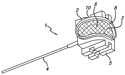
FIG. 1 shows a first embodiment of the present invention. In this
embodiment, the cannula device 1 includes a housing 2 and a membrane 3
which together define a cavity 6 being adapted to receive a piercing member
extending from a connector. A cannula 4 is mounted in the housing 2 and is
in fluid communication with the cavity 6. The membrane 3 may have an oval
or elongated shape covering at least a portion of two sides 8, 10 of the
cannula device 1. Alternatively, the membrane 3 may cover at least a portion
of one or more sides other than the two sides 8, 10. The membrane 3 may
also be configured in an alternative shape, for example, but not limited to,
circular, rectangular, triangular and others. This allows for connection with
a
CA 02576394 2007-02-09
WO 2006/015600 PCT/DK2005/000524
17
connector from more than one angle. The membrane 3 is shown as a single
membrane that curves around the cavity 6. Alternatively, more than one
membrane may be provided for entry of a piercing member in different
receiving directions as described below.
In the embodiment shown in FIG. 1, guiding members 5 are provided on the
housing 2, thus guiding the connection with a device such as the connector,
the inserter, or both. This helps ensure that the cannula from the connector,
the connector needles, or both, properly align as described below. As shown
in FIGS. 1 and 2, and described below, the guiding members 5 may guide
connection from at least two angles being essentially orthogonal to each
other.
FIG. 2 shows the cavity 6 defined by the housing 2 and the membrane 3.
The cavity 6 may be in fluid communication with the cannula 4. As shown, a
pair of guiding members 5 extends from the housing 2, opposite each other
on sides 9, 11 of the housing 2. Each guiding member 5 may include a pair
of elongate rail members 12 adapted to slidably engage with a device such
as a connector or an inserter as described below. Each rail member 12 may
further include a notch 14 defined in the rail member 12 for removably
engaging another or the same device. Additional guiding members 5 are
possible for removable engagement with the device such as the connector,
for example from an infusion set, or an inserter, as will be apparent to one
of
skill in the art.
A sectional view of the cannula device 1 shown in FIG. 3 shows the cavity 6
in further detail. The cavity 6 defines a chamber 16 which is in fluid
communication with the cannula 4. An piercing member (such the piercing
member 580, 680 described below) can penetrate the membrane 3 at any
position on the membrane 3 and communicate with the chamber 16. Thus,
the piercing member such as a piercing member may be in fluid
CA 02576394 2007-02-09
WO 2006/015600 PCT/DK2005/000524
18
communication with the cannula 4 of the device 1, and making it possible to
connect the connector to the cannula device 1 from different angles. As
shown in FIG. 3, the cannula device I may further include entry channels 17
and 19 of the cannula 4. Entry channels 17 and 19 may be in fluid
communication with the chamber 16 for reception of a therapeutic substance
through the piercing member. Alternatively, the entry channels 17 and 19
may directly receive the piercing member for delivery of the therapeutic
substance as described below.
As shown in FIG. 4, the cannula device 1 is connected to an inserter 50. A
needle 51 extends from the inserter 50, penetrating the membrane 3 at a first
position and extending out through the cannula 4. The inserter 50 is used to
place the cannula 4 of the device 1 subcutaneously in the patient. After
insertion, the inserter 50 is removed and the cannula 4 is left in the patient
for
delivery of a therapeutic substance and later withdrawal, for example, when a
base part (shown in FIG. 5), the cannula device 1, or both, are removed.
FIG. 4 shows that the inserter 50 can be connected to the cannula device I
from a first insertion direction 18 that may be generally parallel to or
aligned
with the axis 103 of the cannula 4. A portion of the membrane 3 remains
accessible and thus illustrates that it is possible to connect to a connector
from a second direction 20 which is different from the first direction 18. As
shown, there is approximately a 90 angle between the two directions 18, 20.
The first direction 18 and the second direction 20 may be predetermined by
the location of the entry chambers 17 and 19 for direct reception of the
piercing member. Alternatively, any reception direction may be used with the
chamber 16 that allows fluid communication between the piercing member
and the cannula 4 of the cannula device for delivery of the therapeutic
substance. By way of example, additional angles between the first direction
18 and the second direction 20 are possible. Preferably, the angle between
the first direction 18 and the second direction 20 is in the ranges from about
5o to about 175 , more preferably from about 30 to about 150 . Preferably,
CA 02576394 2007-02-09
WO 2006/015600 PCT/DK2005/000524
19
the angle between the first direction 18 and the second direction 20 is at
least
about 45 , more preferably about 60 , 75 , 85 most preferably about 90 .
One of skill in the art will understand that additional connection directions
are
possible and from many different angles.
As shown in FIG. 5, the cannula device I assembles into a base part 100 of
an infusion set 90. In this embodiment, the base part 100 includes base
guiding members 101 which fit together with the guiding members 5 mounted
on the housing 2 of the cannula device 1. The guiding members 5 on the
cannula device 1 may be used to guide and align the cannula device 1 into
the base guiding members 101 of the base part 100 and to guide the
connection to a connector. An opening 102 in a portion of the base guiding
members 101 of the base part 100 allows for connection of the guiding
members 5 of the cannula device 1 or a device, such as a connector or an
inserter from a second and different direction 20 than the first direction 18
which is parallel to the axis 103 of the cannula 4. The guiding members 5
may be elongated rectangular rails, as shown in this embodiment, or pins, or
other types of known alignment mechanisms.
Protecting members 104, 105 are also shown in FIG. 5. The protecting
members 104, 105 secure and at least partially enclose, and cover the
cannula device 1 on a plurality of sides. The protecting member 105 includes
guiding arms 106 and locking arms 107 similar to guiding and locking arms
that may be provided on a connector. Exemplary engagement of the locking
arms of a connector with an infusion set is described in detail in U.S.
5,522,803 which is incorporated by reference herein in its entirety. The
guiding arms 106 are adapted to slidably fit with mating openings 110 formed
in the inserter 50 as shown in FIGS. 5 and 6.
FIG. 6 illustrates an alternative, perspective view of the embodiment shown
in FIG. 5. Alternatively, the guiding arms 106 may slidably fit with the
guiding
CA 02576394 2007-02-09
WO 2006/015600 PCT/DK2005/000524
members 101 of the base 100 as shown in FIG. 8 showing the inserter 50
removed. The locking arms 107 may releasably engage openings 114
formed in the base 100. The releasable engagement of the cannula device 1
with the base part 100 allows for exchange of the base part 100 and the
5 cannula device 1. For example, but not limited to the following, the cannula
device 1 may be removed from the base part 100 by sliding the cannula
device 1 out of the base part 100. Another cannula device 1 may be slid into
the same base part 100 using the same guiding arms 106 of the base part
100. An advantage of the present invention is that the same base part may
10 be reused several times by the patient, thus saving medical expenses.
Additionally, the exchangeable cannula device and the base part allow for the
use of different cannula devices in the same base part, for example, but not
limited to, having a different size cannula 4 with the cannula device 1, i.e.,
the
length or the diameter of the cannula 4. This exchange allows the infusion
15 sets to be configured in sets that are tailored for the patient using these
modular components.
FIG. 7 illustrates the cannula device 1 releasably mounted on the base part
100 and the cannula device 1 and the base 100 are capped by protecting
20 members 104 and 105.
FIG. 8 shows the cannula device 1 mounted in the base part 100 in an
alternative position to that shown in FIGS. 5-7. In particular, the cannula
device 1 is mounted in the second direction 20 wherein the cannula 4 is
mounted on the base part 100 essentially orthogonal to a main plane 118 of
the base part 100. The base part 100 includes multiple guiding members
105 and openings 102 to allow removable attachment of the cannula device
I in at least these multiple orientations. As shown in FIG. 8, the guiding
members 5 of the housing 2 releasably fit with the opening 102 on the base
part 100. Changing the orientation of the guiding members 5 of the housing
2 with respect to the guiding members 101 and the openings 102 on the base
CA 02576394 2007-02-09
WO 2006/015600 PCT/DK2005/000524
21
part 100 allows the angle between the cannula 4 and the base part 100 to be
changed, which may be desired in certain circumstances. Protecting
members 104, 105 may also be engaged with the base member 100 when
the guiding members 5 of the housing 2 are slidably received in the openings
102 of the base part 100.
As shown in FIG. 9, in combination with FIG. 8, the inserter 50 including the
needle 51 extending through the cannula 4 is connected to the cannula
device 1 when the cannula device 1 is slidably received in the opening 102 in
the base part 100. The protecting members 104, 105 are also engaged with
the base part 100. Alternatively, a connector (for example, a connector 450,
550, 650, described below) may be engaged with the base part 100 in the
position shown for the inserter 50 or in the position shown for the protecting
member 105 and be connected to the cannula device 1. As shown in FIG. 9,
the cannula device 1 and the inserter 50 are engaged with the base part 100
in an orthogonal direction as compared to the parallel direction shown in FIG.
7.
In FIG. 10, the cannula device 1 is mounted in the base part 100 in the first
direction 18 where the cannula 4 is substantially parallel with the main plane
118 of the base part 100. The cannula device 1 may be slidably retained in
the base part 100 by the friction between the guiding members 5 of the
cannula device 1 and the guiding members 101 of the base part 100. The
guiding arms 106 of the protecting member 105 may be slidably engaged in
an opening 122 formed in the base 100 when the cannula device 1 is
engaged with the base 100. The cannula device 1 may also be engaged
with the base part 100 using several methods known to one of skill in the art.
For example, but not limited to, mechanical means, such as rims, grooves or
taps; by adhesives such as glue or by friction such as the cannula device 1
fitting with the opening 102 in the base part 100 and being retained therein
by
the friction between the sides of the guiding means 5 of the cannula device 1
CA 02576394 2007-02-09
WO 2006/015600 PCT/DK2005/000524
22
and the sides of the opening 102. The base part 100 may further include
locking members that removably secure the cannula device 1 in the base
part 100. In addition, the cannula device I may have locking members, such
as the engaging notch 14, shown iri FIG. 2, for securing the cannula device I
in the base part 100. The locking members may further include
disengagement members for releasing the locking members.
FIGS. 11 and 12 show how the locking arms of the protecting member 105,
or alternatively a connector having similar locking arms (as shown in FIGS.
52-57), can engage in openings 107 in the base part 100. The cannula 4 is
shown extending in the parallel direction, similar to FIG. 7.
Preferably, the cannula device 1 of the present invention is made from the
following materials, but is not limited to the materials described herein. One
of skill in the art will recognize that other materials are possible and are
within
the scope of the present invention.
The housing 2 of the cannula device 1 of the present invention is preferably
made from a plastic material, more preferably polypropylene. The membrane
of the present invention is preferably made of silicone, more preferably self-
sealing silicone. The membrane is preferably adapted to be penetrated by
and seal around a piercing member to enable fluid communication with the
cannula of the cannula device or with at least one cavity for each of the
receiving directions.
The cannula 4 of the cannula device 1, as shown, preferably is a soft
cannula. Preferably, the cannula 4 is made of a plastic material. Preferred
plastic materials for the soft cannula 4 are also materials which are
sufficiently flexible to bend, when the patient moves and sufficiently rigid
to
avoid kinking and closing off the drug supply. Further, the material should be
compatible with medical use i.e. minimal skin irritation, non-toxic, non-
CA 02576394 2007-02-09
WO 2006/015600 PCT/DK2005/000524
23
decomposable in the body, etc. Thermoplastic elastomers (TPE) are a type
of materials which satisfy these requirements. Examples of such elastomers
include, but are not limited to: polyester ethers, ECDEL, styrene based TPE,
olefin based TPE, urethane based TPE, ester based TPE, amid based TPE,
polyolefines and silicone rubbers. In a preferred embodiment, the material is
selected from the group consisting of polypropylene, C-FLEXTM, mixtures of
C-FLEXTM, and polypropylene, LUPOLENTM 1840H, LUPOLENTM 3020D,
PELLETHANE TM 2363-75D, PELLETHANETM 2363-55D, TECOTHANE
TM and CARBOTHANETM.
An alternative embodiment of the present invention is shown in FIGS. 13-25
illustrating the same cannula device 1 mounted in a different type of base
part 200. FIGS. 13-25 show how the cannula device of FIGS. 1-12 can be
mounted in different types of infusion sets, illustrating an advantage of the
cannula device of the present invention.
As shown in FIG. 13, the cannula device 1 is engaged with the base part 200
in the first direction 18 where the cannula 4 is generally parallel to a main
plane 218 of the base part 200. FIG. 13 also shows the inserter 50 with the
needle 51 connected to the cannula device 1 and extending through the
cannula 4. The projections 22 extend from the inserter 50 and slidably
engage with the guiding members 5 of the cannula device 1.
In this embodiment, the base part 200 includes guiding members 201 which
fit together with the guiding members 5 on the cannula device 1 as described
above. The cannula 4 extends through an opening 203 in the base part 200,
the opening 203 being sized to receive a annular ring 210 of the cannula
device 1. The base part 200 further includes upstanding guiding members
206, 207 adapted for sliding reception of corresponding guiding members
208, 209 of a protecting member 204 once the inserter 50 is removed, from
the cannula device 1. The protecting member 204 may cover the cannula
CA 02576394 2007-02-09
WO 2006/015600 PCT/DK2005/000524
24
device I while the cannula 4 is secured in the skin of the patient and capable
of delivering therapeutic substances to the patient through the cannula 4.
The guiding members 208, 209 can best be seen in FIG. 16 where the
guiding members 206, 207 of the base 200 and the guiding members 208,
209 of the protecting member 204 are adapted to rotatably fit together to
removably secure the protecting member 204 to the base part 200.
Preferably, the guiding members 207, 208 may further include barbed
projections 211 to facilitate the engagement. The guiding members 206, 209
may also include barbed projections 111. As shown in FIG. 13, the base part
200 preferably includes two upstanding guiding members 206, 207 on
opposite sides of the base part 200. Alternatively, any number of guiding
members 206, 207 or only guiding member 207 may be used to engage
corresponding guiding members 208, 209 or guiding member 208
respectively, and the guiding members 206, 207 and 208, 209 may be of any
size sufficient to removably secure the protecting member 204 to the base
200. other alignment, locking and/or guiding mechanism may be
implemented.
The protecting member 204 may include an opening 212 as best can be
seen in FIG. 15. Tubing (not shown) for delivering a therapeutic substance to
the cannula device 201 may be inserted through the opening 212 so that the
therapeutic substance may be delivered to the patient through the cannula 4
while the cover is in position on the base part 200. The protecting member
204 may further include elongate openings 214 on opposite sides of the
protecting member 204. Preferably, the openings 214 extend from the
periphery 216 of the member 204 inward and generally follow the contour of
the periphery 216. Sides 218 of the protecting member adjacent the
openings 214 provide partially flexible surfaces for gripping and turning the
protecting member 204 to engage or release the base member 200.
However, one of skill in the art will recognize that alternative engagement
CA 02576394 2007-02-09
WO 2006/015600 PCT/DK2005/000524
and release may be achieved by any mechanism commonly known to one of
skill in the art.
FIG. 14 illustrates the cannula device I engaged with the base part 200 in
the second direction 20 where the cannula 4 is generally perpendicular to the
5 main plane 218 of the base part 200. FIG. 14 also shows the inserter 50
with the needle 51 connected to the cannula device 1 and extending through
the cannula 4. The engagement of the inserter 50 and the protecting
member 204 are as described above in FIG. 13. Once the inserter 50 is
removed from the cannula device 1 and base part 200, the protecting
10 member 204 may be engaged with the guiding members 206, 207 of the
base part 200 as shown in FIG. 15. In some embodiments, protecting
members may be engaged with the base part while an inserter is also
engaged with the base part as described below. In the embodiment shown in
FIG. 15, the cannula 4 extends from the boitom of the base 200. As
15 discussed above, tubing may extend from the opening 212.
FIG. 16 illustrates a sectional view of the device shown in FIG. 15 where the
protecting member 204 is engaged with the base part 200 and the cannula
device 1 is positioned in the base part 200 with the cannula 4 projecting from
20 the base part perpendicular to the main plane 218 of the base part 200. The
protecting member 204 is sized and shaped to fit with the base part 200.
The guiding member 207 of the base part 200 and the guiding member 208
of the protecting member 204 having barbed projections 211 on the inner pair
25 of guiding members 207, 208 are shown rotatably secured together. As
discussed above and shown in FIG. 16, the outer pair of guiding members
207, 208 do not include barbed projections 211, however one of skill in the
art will recognize that each of the guiding members 207, 208 may include
guiding projections 211. The entry channels 17 and 19 of the cannula
device 1 are below the membrane 3 that is protected by the protecting
member 204.
CA 02576394 2007-02-09
WO 2006/015600 PCT/DK2005/000524
26
FIG. 17 illustrates the same cannula device I engaged with the same base
part 200 via the guiding members 5 of the cannula device 1 inserted into the
base guiding members 201 where the cannula 4 extends in the first direction
18 generally parallel to the main plane 218 of the base part 200. FIG. 17
illustrates a pair of protecting members 250, 252 that may be engaged with
the base part 200 to cover the cannula device I and the base part 200. The
protecting members 250, 252 are sized and shaped to fit together with the
base part 200 to cover the base part 200. As discussed above, alternative
shapes for the base part 200 and the protecting members 250, 252 are
possible. Other structures for mating these multiple members together may
also be utilized.
The protecting member 250 includes a pair of guiding members 254 that may
be adapted to engage the guiding members 207 of the base part 200. As
described above, the base part 200 may include guiding members 206. The
guiding members 206 may slide together with protrusions 256 on the
periphery 258 of the protecting member 250. The protecting member 250
may further include a notch 260 for engaging an edge 270 of the base part
200. The protecting member 250 covers a portion of the membrane 3 of the
cannula device 1 and may also engage the protecting member 252 to cover
the base part 200 when the base part 200 is adhered to the skin of the
patient and the cannula 4 is positioned transcutaneously for delivery of the
therapeutic substance. As shown in FIG. 17, the protecting member may
include a portion 264 for covering the membrane 3 of the cannula device 1
and the portion 264 may be made from a material that is different from the
remainder of the protecting member and penetrable by a needle, such as the
needle 51 of the inserter 50. For example, the portion 264 may be integrally
moulded to the member 250 and be formed from an elastomer. The
protecting member 252 may be slidably engaged with the protecting member
250.
CA 02576394 2007-02-09
WO 2006/015600 PCT/DK2005/000524
27
FIG. 18 shows the protecting member 250 engaged with the base part 200
as described above for FIG. 17. While the protecting member 250 is
engaged with the base part 200, the protecting member 252 may be removed
from the protecting member 250 and the base part 200. The needle 51 of the
inserter 50 may be inserted into the membrane 3 of the cannula device 1 and
extend through the cannula 4 in the first direction 18. The inserter 50 may be
removed and replaced with the protecting member 252 or alternatively with a
connector, such as a connector 450, 550, 650 described below, for delivery
of a therapeutic substance.
FIG. 19 illustrates the cannula device 1(beneath the protecting members
250, 252) engaged with the base part 200 in the second direction 20 and the
inserter 50 having needle 51 inserted into the cannula device 1 through the
cannula 4 in the second direction. FIG. 19 shows the needle 51 penetrating
the portion 264 of the protecting member 250. When the cannula device 1 is
engaged with the base part 200 in the second direction 20, the protective
members 250, 252 may remain engaged with the base part 200, protecting
the base part 200 and the cannula device 1 while the needle 51 of the
inserter 50 extends through the cannula 4.
FIG. 20 illustrates the embodiment described above for FIG. 17 showing the
protecting member 250 engaged with the base part 200 and the protecting
member 252 not yet engaged. As shown, the protecting member 252 may
be engaged parallel to the direction of the cannula 4 extending from the base
part 200. FIG. 21 illustrates the compact size of the device when the
protecting member 252 is engaged with the protecting member 250 and the
base part 200. In this configuration, the more sensitive parts of the device
are concealed and protected, and the assembly itself is easily transported or
packaged. As described above, the protecting member 252 may be removed
and a connector may be inserted in place of the protecting member 252,
having a similar compact size.
CA 02576394 2007-02-09
WO 2006/015600 PCT/DK2005/000524
28
FIG. 22 illustrates perspective views of the cannula device 1 and includes
several assembly configuration including the base parts 100, 200 and the
protecting members 104, 105, 204, 250, 252. FIG. 22A shows the cannula
device 1 itself. FIG. 22B shows the cannula device 1 inserted into the base
part 100. The protecting members 104, 105 cover the base part 100 and the
cannula device 1 with at least part of the membrane 3 accessible for insertion
of a needle of an inserter or a connector for delivery of a therapeutic
substance.
FIG. 22C shows the cannula device 1 engaged with the base part 200. The
protecting member 204 is shown covering the cannula device 1 and the base
part 200 and has been described above.
FIG. 22D shows the cannula device 1 engaged with the base part 200 and
protecting members 250, 252 covering the device 1 and base part 200.
FIGS. 23A-D illustrate side views of the cannula device I engaged with the
corresponding different embodiments shown in FIGS. 22B-D.
FIGS. 24A-D illustrate top views of the embodiments shown in FIGS. 22B-D
respectively.
FIG. 25 illustrates another embodiment of the present invention. As shown, a
base part 300 is engaged with protecting members 350, 352 to form a
compact device. An adhesive layer 310 for adhering the base part 300 to the
patient's skin is shown connected to the base part 300.
FIG. 26 illustrates a sectional view of the embodiment shown in FIG. 25. The
base part 300 includes a plurality of projections 320 having bottom surfaces
322 that together form the bottom surface 324 of the base part 300 as shown
in FIGS. 26 and 28. An entry port 328 may be formed in the base part 300
CA 02576394 2007-02-09
WO 2006/015600 PCT/DK2005/000524
29
wherein the entry port 328 is connected to a canal 330 that fluidly connects
with a cannula 304 that extends from the base part 300. The protecting
member 352 may be removed and a connector, such as the connector 450,
550, 650, described below, may be inserted at the same position for delivery
of a therapeutic substance. The adhesive layer 310 adheres to the bottom
surfaces 322 of the base part 300 and may be formed from a material that
may be penetrated by a needle of an inserter (not shown) extending through
the cannula 304. Alternatively, the adhesive layer 310 may include an
opening through which the cannula 304 may extend.
FIG. 27 shows an exploded view of the embodiment shown in FIG. 25. As
shown, the protecting member 352 includes guiding arms 306 and locking
arms 307 for engagement of the protecting member 352 with the base part
300. The locking arms 307 may engage the base part 300 through
corresponding openings 314 in the base part 200 as described above in
FIGS. 5 and 6.
FIG. 28 shows an exploded bottom view of the embodiment shown in FIG.
25. The projections 320 having bottom surfaces 322 on the base part 300
are shown. The base part 300 may further include a peripheral surface 332
that extends around the periphery of the base part 300 to which the adhesive
layer 310 may adhere in addition to the surfaces 322. As shown in the FIGS.
25-28, the general shape of the embodiment is cylindrical having the
adhesive layer 310, the base part 300, and the protecting members 350, 352
shaped to fit together to form a compact device. One of skill in the art will
recognize that alternative shapes are possible, including but not limited to
oval, rectangular, and square shapes, preferably where the assembly of
embodiment may form a compact device.
FIGS. 29A and 29B show an alternative embodiment of a cannula device
401. The cannula device 401 includes a housing 402, a membrane 403 and
CA 02576394 2007-02-09
WO 2006/015600 PCT/DK2005/000524
openings 405, 407. Preferably, the housing 402 may be essentially
cylindrically shaped, although other shapes are possible. In the present
embodiment, the cylindrical shape includes radially extending projections 413
(or flanges) that define openings 405 and 407 and may be used as guides
5 when positioned in a base part, such as base part 400, shown in FIG. 30 A.
As shown in the sectional view in FIG. 29B, the membrane 403, together with
the housing 402 define a cavity 406 within the housing 402 for reception of a
piercer from a connector (described below). A cannula 404 extends from the
housing 402 below the cavity 406 as shown in FIG. 29B. In this embodiment,
10 the housing 402 may be rotatably attached to the base part 400, shown in
FIG. 30, so that the rotatable attachment allows the housing 402 to rotate in
the base part 400 after insertion of the cannula 404 into the skin of the
patient, without movement of the cannula 404 and thereby minimizing pain to
the patient. Alternatively, the base part 400 may be turned to a desired angle
15 relative to the cannula 404 and then inserting the cannula 404 using an
inserter such as inserted 50 shown in FIG. 4. As shown in FIG. 29A, end
portions 412 protrude from the housing 402. Preferably, the end portions 412
may be generally circularly shaped to provide for rotation within the base
part
400. However, the end portions 412 may be any size and shape that is
20 movable within the base part 400 known to one of skill in the art.
In FIGS. 30A and B, the cannula 404 of the cannula device 401 can be seen
connected to the base part 400 and a connector 450. In this embodiment,
the base part 400 includes connecting members 446 adapted to receive the
25 end portions 412 of the housing 402. The connecting members 446 and the
end portions 412 may be shaped and sized to facilitate rotation of the
housing 402 with respect the base part 400. The device 401 further includes
an adhesive layer 410 for adhering the base part 400 to the patient's skin.
The adhesive layer 410 is shown connected to the base part 400 and the
30 adhesive layer 410 may include a cutout 448 for the cannula 404 when the
end portion 412 of the housing 402 is rotated in the connecting member 446
CA 02576394 2007-02-09
WO 2006/015600 PCT/DK2005/000524
31
of the base part 400 to change the angle of the cannula 404 with respect to
the skin.
The connector 450 connects to the cannula device housing 402 via openings
405, 407 in the housing 402. (Also shown and described below with
reference to FIG 31A.) The connector 450 may further include tubing 452 for
delivering a therapeutic substance to the cannula 404 for delivery to the
patient. The connector 450 may include a button 454 for release of the
connector 450 from the housing 402. Alternatively, any method for releasing
the connector 450 from the housing 402 may be used, including, but not
limited to pressing on an exterior portion 456 on each side of the connector
450 to release gripping arms 458 (shown in FIG. 31A).
FIG. 31A illustrates the connector 450 prior to connection with the housing
402. The connector 450 may include locking arms 458 and guiding members
470 similar to the locking arms and guiding members described above for the
protecting member with the cannula device 1. The locking arms 458 allow
the connector 450 to be inserted into the openings 405 of the housing 402
and be removably locked in place for delivery of the therapeutic substance.
The connector 450 may also include a piercing member 480 that connects
the connector 450 to the cannula 404. The piercing member 480 may be
inserted through the membrane 403 and into the cannula 404 or into the
chamber 406 to fluidly connect with the cannula 404. The piercing member
480 may be adapted to be broken at a predetermined place. This allows the
connector 450 to be used as an inserter for inserting the cannula 404 of the
cannula device 401. Then the piercing member 480 may be broken at the
desired spot and the connector 450 used in the traditional manner. The
piercing member 480 extends through the membrane and may piece the
membrane, but does not necessarily have to puncture the membrane. For
example, the piercing member may be inserted through a pre-formed hole in
the membrane. The term piercing member as used herein may include a
CA 02576394 2007-02-09
WO 2006/015600 PCT/DK2005/000524
32
cannula, including a rigid cannula or a needle, a semi-rigid cannula, or a
soft
cannula or any member suitable piercing the membrane.
As shown in FIGS. 31A and B, the base part 400 of this embodiment may be
flexible or hinged to facilitate the rotation of the housing 402 in the base
part
401. FIG. 31 B illustrates the cannula 404 rotated toward the skin and the
base part 400 flexing for rotation. The base part 401 may further include
hinges 472 joined to the connecting members 446. The hinges 472 may be
used for rotating the cannula 404 from parallel to the skin to angled toward
the skin. Rotation of the housing 402 also allows the cannula 404 to be
placed into the skin with an injector needle, such as the needle 51 shown
with the cannula device 1. The connector 450 may be connected at any
angle and the insertion device 480 can be inserted into the membrane 403
from a plurality of directions.
FIG. 32A shows the cavity 406 formed in the housing 402 and the cannula
404 extending from the cavity 406. FIG. 32B shows a portion of the housing
402 cut away from the base part 400. The connecting member 446 is shown
in a circular configuration having a raised circumference 474 and a central
depression 476 adapted to receive the end portion 412 of the housing 402.
A portion of the base part 400 is shown extending from the connecting
member 446. FIG. 32C shows the cannula 404 rotated from the direction
parallel to the skin. FIG. 32D shows a top view of the cannula device 401.
FIGS. 33A-D illustrate the cannula device 401 having the cannula 404
extending in a different direction as previously shown in FIGS. 30-32. The
cannula 404 is shown extending substantially orthogonally with respect to the
cannula 404 shown in FIGS. 31-32. FIG. 33B shows the cannula device 401
with the cannula 404 extending vertically from the base part 400. The
connector 450 is shown removably connected to the housing 402 in the
openings 405, 407 of the housing 402. An opening 482 in the adhesive layer
CA 02576394 2007-02-09
WO 2006/015600 PCT/DK2005/000524
33
410 is shown in FIG. 33B for the cannula 404 to extend through and into the
skin of the patient. FIGS. 33C and D show top views of the cannula device
401 shown in FIGS. 33A and B, respectively.
FIG. 34A illustrates the housing 402 having openings 405, 407. The
membrane 403 is shown in the center of the housing 402 for reception of an
insertion device for fluidly delivering a therapeutic substance through the
cannula 404. The end portion 412 is shown having a generally rectangular
shape for mating with a similarly shaped connecting member 446 of the base
part 400 shown in FIG. 34B. The housing 402 is rotatable in the base part
400 as described above.
FIG. 35 shows the cavity 406 formed inside the housing 402. The
membrane 403 may cover a portion of the cavity 406. A top portion 484 of
the cannula 404 connects with the cavity 406 in the housing 402. The cavity
406 allows for an piercing member of a connector to be inserted into the
membrane 403 in any reception direction and have the cannula 404 be in
fluid communication with the connector for delivery of the therapeutic
substance. In this embodiment, the cannula 404 is shown extending
vertically from the housing 402 in relation to the plane 418 of the base part
400. A pair of openings 486 is shown on the housing 402 for reception of a
connector or a protective member in two different directions. The base part
400 also includes the connecting member 446 in a generally square shape
for reception of the end portion 412 of the housing 402. The adhesive layer
410 may be adhered to the skin of the patient.
FIGS. 36A-E show the cannula device 401 from different angles. FIG. 36A
shows a top view of the cannula device 401 with the connector 450
connected to the housing 402. The cannula 404 is shown extending in a
direction generally parallel to the base part 400. Tubing 452 extends from
the connector 450 for connection to a medical device (not shown) for delivery
CA 02576394 2007-02-09
WO 2006/015600 PCT/DK2005/000524
34
of a therapeutic substance. FIG. 36B shows the cannula device 401 from
FIG. 36A with the connector 450 removed. FIG. 36C shows a perspective
view of the cannula device 401 of FIG. 36A. FIG. 36D shows a rear
perspective view of the cannula device 401 and FIG. 36E shows a rear
perspective view of the cannula device 401 with the connector 450
connected to the housing 402.
FIGS. 37A-C illustrate the cannula device 401 with the cannula 404
extending in a direction generally vertically to the plane 418 of the base
part
400. FIG. 37A shows the connector 450 connected in one set of the
openings 484 in the housing 402 wherein the connection is parallel to the
plane of the base part 400. The second set of the pair of openings 488 are
shown opening in a second direction that the connector 450 may be inserted
into wherein the connection is generally orthogonal to the plane 418 of the
base part 400. FIG. 37B shows a side perspective view of the cannula
device 401 and the cannula 404 extending below the base part 400. FIG
37C shows a top view of the device shown in FIG. 37A.
FIG. 38A shows the cannula device of FIG. 37A with the housing 402
repositioned in the base part 400 so that the cannula 404 extends generally
parallel main plane 418 of the base part 400. The connector 450 is shown
connected to the housing 402 in a parallel direction to the plane 418 of the
base part 400. FIG. 38B is a top view of the cannula device 401 shown in
FIG. 38A where the guiding arms 470 and the locking arms 458 from the
connector 450 can be seen connected to the housing 402 for removable
connection.
FIG. 39 illustrates a cannula device 501 connected to an inserter 550. The
cannula device 501 is shown removed from a base part to show the
connection of the inserter 560 with the guiding members 505 of the cannula
device 501.
CA 02576394 2007-02-09
WO 2006/015600 PCT/DK2005/000524
As shown in FIGS. 40A and B, the cannula device 501 may be connected to
a connector 550 at different heights and distances from skin of the patient so
that the cannula 504 may extend into the skin of the patient in varying
5 depths. A positioning member 507 is shown in FIGS. 40A and 41A
positioned beneath the cannula device 501. The positioning member 507
may be positioned between a base member 500 (shown in FIG. 43) and the
cannula device 501 for changing the height and distance that the cannula
501 may be inserted into the skin. The positioning member 507 allows the
10 cannula device 501 to be positioned higher in the connector 550 in FIG. 40A
when compared to the position of the cannula device in the connector 550 in
FIG. 40B without a positioning member (see also FIGS. 41A and 41 B). The
cannula device may also be positioned at multiple heights with respect to the
base part using multiple guiding members. Multiple positions of the cannula
15 device with respect to the base part allow one cannula device and one base
part together to be used for more than one insertion depth, adapting the
infusion set to the individual patient and also so that the infusion set may
also
be used for both children and adults. The cannula device 501 also includes a
membrane 503 that is adapted to receive a piercing member 580, such as a
20 cannula, from the connector 550 to fluidly connect the connector 550 to the
cannula 504 for delivery of a therapeutic substance. As will be understood
by one of skill in the art, the cannula may be rigid, for example, but not
limited
to, a needle, semi-rigid, or soft.
25 FIGS. 41A and B show a partial view of the embodiments shown in FIGS.
40A and B, respectively. FIG. 41A illustrates an embodiment that allows the
connection of the cannula device 501 with the connector 550 from two
different directions. As shown, the membrane 503 is a single membrane
mounted to the housing 502. As described blow, multiple membranes 503
30 are possible. The piercing member 580, shown as a cannula, may extend
from the connector 550 and penetrate the membrane 503 and depending on
CA 02576394 2007-02-09
WO 2006/015600 PCT/DK2005/000524
36
the direction from which the piercing member 580 is received in the cannula
device 501, the piercing member 580 may end in one of two cavities 512
formed in the cannula device 501. The two cavities 512 may be in fluid
communication with each other via a canal 514 formed in the cannula device
501. Preferably the canal 514 is provided in the interior of the housing 502.
This embodiment allows the piercing member 580 to be in fluid
communication with the cannula 504 regardless of the direction from which
the insertion device is inserted through the membrane 503. As shown in FIG.
41A, the piercing member 580 may be essentially orthogonal with the
cannula 504 of the cannula device 501 and the cavity 512, in to which the
piercing member 580 inserts and ends, fluidly connects the canal 514 and a
cavity 516 connected to the cannula 504. One of skill in the art will
recognize
that the piercing member 580 may also be inserted into the membrane 503
from a direction essentially parallel to the cannula 504 from the top of the
cannula device 501. The piercing member 580 may also be inserted through
the membrane 503 and the piercing member 580 may end in the cannula 504
and thus the piercing member 580 may fluidly connect directly with the
cannula 504 without connecting via a cavity 512 or a canal 514.
FIG. 41 B illustrates the cannula device 501 that may be connected to the
piercing member 580 from two directions and in which the piercing member
580 connects to the single cavity 522 from either direction through the
membrane 503. When the piercing member 580 is inserted through the
membrane 503 in the orthogonal direction to the direction of the cannula 504,
the insertion device may enter the cavity 522 from a hole 524 in a wall 523 of
the cavity 522. FIGS. 41A and B also illustrate the cannula device 501
connected at different heights in the connector 550.
FIGS. 42A and B illustrate side views of the cannula device 501 connected to
the cannula device 501 at different heights in the connector 550.
FIG. 43 illustrates the connector 550 connected to the cannula device 501
and the cannula device 501 may be mounted in a base part 500. As shown,
CA 02576394 2007-02-09
WO 2006/015600 PCT/DK2005/000524
37
the connector 550 is connected in a direction parallel to the main plane of
the
base part 500. The connector 550 includes the piercing member 580 that is
shown connecting through the membrane 503 directly with the cannula 504.
FIG. 44 shows the cannula device 501 connected to the connector 550 and
mounted in the base part 500. The cannula 504 extends in the direction
parallel to the plane 518 of the base part 500, orthogonal to the direction
shown in FIG. 42A and B. The connector 550 includes guiding arms 506 and
locking arms 507. As described above, the guiding arms help to position the
cannula device 501 in the base part 500 and the locking arms 507 removably
lock the connector 550 with the base part 500. The locking arms 507 are
shown extending into and locking with a portion of the base part 500 and into
an opening 511 on the base part 500. Side portions 513 of the locking arms
507 may include ridges 517 to help the patient grip the locking arms 507 and
slidably push together the connector 550 and the base part 500 to removably
lock the connector 550 with the base part 500. The gripping arms 507 may
also be flexible so that the patient may press inwardly on the locking arms
507 to release the locking arms from the base part 500. A portion of the
membrane 503 remains exposed at the top of the cannula device 501.
FIG. 45 shows the cannula device 501 together with the connector 550 and
tubing 552 and the base part 500. An adhesive layer 510 is also shown
having a cutout portion 515 for the cannula 504.
FIG. 46 illustrates the cannula device 501 mounted on the base part 500 with
the cannula 504 extending in the direction parallel to the main plane of the
base part 500. An inserter 560 may be connected to the base part 500 and
the cannula device 501. A needle 561 extends from the inserter 560 though
the membrane 503 of the cannula device and through the cannula 504. FIG.
47 illustrates the cannula device 501 shown in FIG. 46 may be rotated
orthogonally to the direction shown in FIG. 46 and mounted to the same base
CA 02576394 2007-02-09
WO 2006/015600 PCT/DK2005/000524
38
part 500 shown in FIG. 46. The cannula 504 extends downward from the
base part 500. The inserter 560 is also shown connected to the base part
500 and the cannula 501 in the direction orthogonal to that shown in FIG. 46.
FIG. 48 shows the inserter 560. The needle 561 extends from the inserter
560 and guiding arms 563 may extend on both sides of the needle 561 for
slidable connection with the guiding members of the cannula device
(described and shown above) to guide the inserter 560 into position in the
cannula device 501.
FIG. 49 shows an embodiment similar to the embodiment shown in FIG. 45
having an angled base part 600. The angled base part 600 can best be seen
in the side view shown in FIG. 51, wherein the connector 650 is shown
inserted in the base part 600 at an angle to the main plan of the base part
618. A cannula device 601 may be mounted in the base part 600 in several
directions including as shown in FIG. 49 having a cannula 604 extending
from the base part 600 in a direction generally parallel to the plane 618 of
the
base part 600. A connector 650 may be connected to the base part 600 and
the cannula device 601. The connector 650 may include an piercing member
680 that inserts through the membrane 603 of the cannula device 601 for
fluid connection between tubing 652 to the cannula 604 for delivery of a
therapeutic substance to the skin of a patient. The connector 650 may also
include locking arms 607 extending though an opening 611 in the connector
650 for removably locking the connector 650 to the base part 600. An
adhesive layer 610 is also shown connected to the base part 600 and the
adhesive layer 610 may include a cut out portion 615 for the cannula 604.
FIG. 50 shows a top view of the embodiment shown in FIG. 49.
FIG. 52 shows the cannula device 601 having an angled base part 600. As
described above, the connector 650 may be connected with the base part
600. The connector 650 further includes guiding arms to help position the
CA 02576394 2007-02-09
WO 2006/015600 PCT/DK2005/000524
39
cannula device 601 in the base part 600 (not shown, described above for
example in FIG. 6). Locking arms 607 removably engage the base part 600
though the opening 611 in the base part 600. The locking arms 607 are
shown extending into and removably locking with a portion of the base part
600 into the opening 611 on the base part 600. Side portions 613 of the
locking arms 607 may include ridges 617 to help the patient grip the locking
arms and removably lock the connector 650 with the base part 600. The
gripping arms 607 may also be flexible so that the patient may press inwardly
on the locking arms 607 to release the locking arms 607 from the base part
600. FIGS. 53-57 illustrate perspective views of the cannula device 601
shown in FIG. 52.
FIGS. 58=65 illustrate another preferred embodiment of the present invention.
In FIGS. 58 and 59, a cannula device 701 is cubically shaped and is
positioned in a base part 700 so that a cannula 704 of the cannula device
701 is essentially orthogonal to a main surface 705 of the base part 700. As
described above, other shapes for the cannula device 701 are possible. The
cannula device 701 can receive both an inserter needle of an inserter (as
described above and shown, for example, in FIG. 4) and a connector 750
having a cannula 751 from a direction being essentially parallel with the
cannula 704. The cannula device 701 includes at least one membrane.703
through which the inserter needle or the piercing member 751 inserts. FIGS.
58 and 59 illustrate the cannula device 701 having two membranes 703,
although one of skill in the art will understand that more membranes 703 are
possible. A protective member or inserter may also be used with the cannula
device 701 and base part 700 in place of the connector 750 as described
above.
Further FIGS. 58 and 59 show that it is possible for the connector 750 to
connect with the base part 700 from a direction being essentially orthogonal
to the direction of the cannula 704 and the piercing member 751 can pierce
the membrane 703 from a direction orthogonal to the direction of the
CA 02576394 2007-02-09
WO 2006/015600 PCT/DK2005/000524
connector 750 shown in FIG. 58. The cannula device 701 is constructed in
such a manner that the cannula 704 is in fluid communication with the
piercing member 751 when received regardless of the direction from which
the piercing member 751 is received. In the embodiment shown in FIGS. 58
5 and 59, the cannula device includes a cavity 706 similar to the cavity 6
described above for the cannula device 1. Alternatively, the cannula device
701 may not include a cavity 706 wherein the piercing member 751 fluidly
connects with the cannula 704, similar to the reception of the piercing
member without having a cavity 706, similar to the device described above in
10 FIG. 41 B.
FIGS. 58 and 59 also illustrate an alternative mechanism for removable
locking the connector 750 with the base part 700. The connector 750
includes locking arms 707 for removably locking the base part 700 to the
15 connector 750. The locking arms 707 further include notched projections 720
for engaging angled projections 722 on the base part 700. The notched
projections 720 of the connector 750 slidably engage the angled projections
722 of the base part 700. The projections 720 snap together with the
projections 722 and the projections 720 fit into a gap in the base part 700 to
20 releasably lock the base part 700 to the connector 750. The base part 700
may be released from the connector 750 by pressing on the projections 720.
The connector 750 may also include guiding arms 706 to slidably engage
guiding members 705 of the cannula device 701. Of course, alternative
locking means may be used to releasably engage the base part 700 with the
25 connector 751.
FIGS. 58-60 illustrate the cannula 704 extending essentially perpendicular to
the main surface 705 of the base part 700 so that the cannula 704 penetrates
the patient's skin essentially perpendicular to the main surface 705 of the
30 base part 700. The base part 700 includes an opening 709 through which
the cannula 704 of the cannula device 701 may extend.
CA 02576394 2007-02-09
WO 2006/015600 PCT/DK2005/000524
41
FIG. 61 illustrates the cannula 704 extending essentially parallel to the main
surface 705 of the base part 700 through the opening 709 in the base part
700. FIGS. 62 and 63 illustrate the cannula device 701 prior to connection
with the base part 700 where the cannula 704 will extend through the
opening 709 essentially parallel to the main surface 705 of the base part 700.
FIG. 64 shows an example of an interior portion 713 of the cannula device
701. In this embodiment, the piercing member 751 will insert directly into a
channel 715 without having a cavity for fluidly connecting the piercing
member 751 with the cannula 704 as described above. The cannula
connector 751 can pierce the membranes 703 from either direction shown
and directly enter the channels 715 to fluidly connect with the cannula 704.
Although the invention herein has been described in connection with a
preferred embodiment thereof, it will be appreciated by those skilled in the
art
that additions, modifications, substitutions, and deletions not specifically
described may be made without departing from the spirit and scope of the
invention as defined in the appended claims. It is therefore intended that the
foregoing detailed description be regarded as illustrative rather than
limiting,
and that it be understood that it is the following claims, including all
equivalents, that are intended to define the spirit and scope of this
invention.
Fig 65 shows a segment of a sphere defined by two planes cutting the
sphere in different segments and wherein the two planes have a line of
intersection within the sphere. The shape of this segment is useful as a
shape of the housing of the cannula device. In stead of a segment of a
sphere a segment of an ellipsoid defined in the same manner could be used
i.e. a segment of an ellipsoid defined by two planes cutting the ellipsoid in
different segments and wherein the two planes have a line of intersection
within the ellipsoid.