Note: Descriptions are shown in the official language in which they were submitted.
DEMANDES OU BREVETS VOLUMINEUX
LA PRESENTE PARTIE I)E CETTE DEMANDE OU CE BREVETS
COMPREND PLUS D'UN TOME.
CECI EST LE TOME DE _2
NOTE: Pour les tomes additionels, veillez contacter le Bureau Canadien des
Brevets.
JUMBO APPLICATIONS / PATENTS
THIS SECTION OF THE APPLICATION / PATENT CONTAINS MORE
THAN ONE VOLUME.
THIS IS VOLUME 1 OF 2
NOTE: For additional volumes please contact the Canadian Patent Office.
CA 02576904 2007-02-09
WO 2006/020579 PCT/US2005/028164
METHOD FOR RAPID 1T1DIEiT'lI"fFICATi IIGN OF I~VIii ~~~~ORGAI'1ISI~~S
This application claims priority to U.S. Provisional Application Serial No.
60/599,858, filed August 10, 2004, which is incorporated herein by reference
in its entirety.
Technical Field
The present invention relates, in general, to probes, methods, and kits used
to
determine the presence or absence of a microorganism in a sainple. The probes,
methods,
and kits comprise at least one capture probe and/or at least one detector
probe.
Background Art
In the following discussion certain articles and methods will be described for
background and introductory purposes. Nothing contained herein is to be
construed as an
"admission" of prior art. Applicant expressly reserves the right to
demonstrate, where
appropriate, that the articles and methods referenced herein do not constitute
prior art under
the applicable statutory provisions.
Bacteremia and fungemia are life-threatening infections that require timely
administration of appropriate antimicrobial therapy to prevent significant
mortality. The term
"septicemia" is used to describe the presence of organisms within the blood in
association
with laboratory and/or clinical findings that are indicative of infection such
as fever, chills,
malaise, tachycardia, hyperventilation, shock and leucocytosis. Weinstein et
al. (Rev. Ibzfect.
Dis. 5: 54-70 (1983)) determined that the overall rate of mortality was 42%
among 500
episodes of bacteremia and fungemia, with approximately half of the deaths
attributable
directly to septicemia. It has long been recognized, however, that the
majority of bacteremias
and fungemias are associated with the recovery of very low numbers of
organisms from the
blood. Indeed, it is not uncommon for less than 1 organism/mL of blood to be
present,
particularly after the initiation of antimicrobial therapy. The severity of
such infections and
the diverse spectrum of potential pathogens, therefore, necessitate highly
sensitive methods
of diagnosis that are capable of identifying a broad spectrum of bacteria and
fungi.
Classically, diagnosis is achieved through the use of broad-based cial_ture
methods that are
aiuenable to the growth of a wide variety of pathogens from low-level inocula.
Following
growth and isolation in pure culture, the organisms are identified through the
application of a
battery of biochemical tests. Antimicrobial susceptibility testing is then
conducted to permit
1
CA 02576904 2007-02-09
WO 2006/020579 PCT/US2005/028164
modification of empirical therapy to an efficacious pathogen-specific regimen
that minimizes
cost and toxicity. There remains, however, a need to reduce the time between
collection of
specimens from a patient and administration of targeted antimicrobial tlierapy
to provide an
opportunity to reduce morbidity and mortality, defray the cost of therapy and
hospitalization,
and miniinize the spread of antimicrobial drug resistance caused by
ineffective or
inappropriate therapy.
Disclosure of the Invention
The present invention relates to a method for identifying the presence of at
least
one microorganism in a sample, the method comprising: (a) releasing RNA or DNA
from the
at least one microorganism in the sample; (b) contacting the RNA or DNA with
at least one
capture probe capable of hybridizing to a first target sequence of the RNA or
DNA, wherein
the contacting is performed under conditions that pennit hybridization between
the first target
sequence and the at least one capture probe to fonn a microorganism-capture
probe hybrid
complex, and wherein the at least one capture probe comprises at least one
sequence selected
from the group consisting of SEQ ID NOs:1-53, 55, 56, 61, 62, 67, 68, and 72-
78; and (c)
detecting the presence of the microorganism-capture probe hybrid complex by
(i) contacting
the RNA or DNA with at least one detector probe capable of hybridizing to a
second target
sequence of the RNA or DNA, wherein the detector probe comprises at least one
reporter
group and wherein the contacting is perforined under conditions that permit
hybridization
between the second target sequence and the at least one detector probe to form
a
microorganism-capture probe-detector probe hybrid complex, and wherein the at
least one
detector probe also comprises at least one sequence selected from the group
consisting of
SEQ ID NOs:1-53, 55, 56, 61, 62, 67, 68, and 72-78; (ii) detecting the
microorganism-
capture probe-detector probe hybrid complex by detecting the at least one
reporter group,
wherein the presence of the microorganism-capture probe-detector probe hybrid
complex
indicates the presence of the at least one microorganism. In another
einbodiment, the reporter
group is selected from the group consisting of a radioactive isotope, an
enzyme, a fluorescent
molecule and an amplification sequence. In a further embodiment, the
amplification
sequence initiates an amplification reaction selected from the group
consisting of strand
displacement amplification (SDA), polymerase chain reaction (PCR), reverse
transcriptase-
strand displacement amplification (RT-SDA), reverse transcriptase-polymerase
chain reaction
(RT-PCR), nucleic acid sequence based amplification (NASBA), transcription-
mediated
2
CA 02576904 2007-02-09
WO 2006/020579 PCT/US2005/028164
aniplification (TMA), rolling circle ainplification and Q13 replicase
amplification. In an
additional embodiment, detection of the microorganism-capture probe-detector
probe hybrid
complex is accomplished via non-specifically labeling the hybrid complex.
In an additional aspect, the first target sequence and the second target
sequence
comprise the saine sequence. In another aspect, the capture probe is
iininobilized on a solid
support before hybridizing to the first target sequence. In yet another
aspect, the
microorganism-capture probe hybrid coinplex is innnobilized on a solid
support. In a further
aspect, the microorganism-capture probe-detector probe hybrid complex is
iininobilized on a
solid support. In anotller aspect, the solid support is selected from the
group consisting of
latex beads, agarose beads, paramagnetic beads, ferric oxide, microarray
chips, filter paper,
nitrocellulose filters, nylon membranes, glass slides and cellular
ineinbranes. In a further
aspect, the solid support is a inicroarray chip. In an additional aspect, two
or more capture
probes are iininobilized on a single spot of the solid support. In a further
aspect, the method
described above further coinprises an immobilization probe that is capable of
hybridizing to
the capture probe to be iinmobilized onto the solid support.
The methods of the present invention additionally provide a method for
identifying the species of one or more microorganisms in a sample, the method
comprising:
(a) releasing RNA or DNA fiom the at least one microorganism in the sample;
(b) contacting
the RNA or DNA with at least one species-specific capture probe capable of
hybridizing to a
first target sequence of the RNA or DNA, wherein the contacting is performed
under
conditions that permit hybridization between the first target sequence and the
at least one
species-specific capture probe to form a species-specific inicroorganism-
capture probe hybrid
coinplex, and wherein the at least one species-specific capture probe
comprises at least one
sequence selected from the group consisting of SEQ ID NOs:1-53, 55, 56, 61,
62, 67, 68, and
72-78; and (c) detecting the presence of the species-specific inicroorganism-
capture probe
hybrid complex by (i) contacting the RNA or DNA with at least one detector
probe capable
of hybridizing to a second target sequence of the RNA or DNA, wherein the
detector probe
comprises at least one reporter group and wherein the cop.tacting is performed
under
conditions that permit hybridization between the second target sequence and
the at least one
detector probe to form a species-specific microorganisin-captiarP probe-
detector probe hybrid
complex, and wherein the at least one detector probe also comprises at least
one sequence
selected from the group consisting of SEQ ID NOs:1-53, 55, 56, 61, 62, 67, 68,
and 72-78;
(ii) detecting the species-specific inicroorganisin-capture probe-detector
probe hybrid
3
CA 02576904 2007-02-09
WO 2006/020579 PCT/US2005/028164
complex by detecting the at least one reporter group, wherein the presence of
the species-
specific microorganism-capture probe-detector probe hybrid complex indicates
the presence
of the at least one inicroorganism belonging to the species. In a further
embodiment, the
amplification sequence initiates an amplification reaction selected from the
group consisting
of strand displaceinent anzplification (SDA), polyinerase chain reaction
(PCR), reverse
transcriptase-strand displaceinent ainplification (RT-SDA), reverse
transcriptase-polyinerase
chain reaction (RT-PCR), nucleic acid sequence based amplification (NASBA),
transcription-
mediated amplification (TMA), rolling circle amplification and QB replicase
amplification.In
another embodiment, the reporter group is selected from the group consisting
of a radioactive
isotope, an enzyme, a fluorescent molecule and an ainplification sequence. In
an additional
embodiment, detection of the microorganism-capture probe-detector probe hybrid
coinplex is
accomplished via non-specifically labeling the hybrid complex.
In an additional aspect, the first target sequence and the second target
sequence
comprise the same sequence. In anotller aspect, the species-specific capture
probe is
immobilized on a solid support before hybridizing to the first target
sequence. In yet another
aspect, the species-specific microorganism-capture probe hybrid complex is
immobilized on
a solid support. In a fu.rtller aspect, the species-specific microorganism-
capture probe-
detector probe hybrid coinplex is immobilized on a solid support. In another
aspect, the solid
support is selected from the group consisting of latex beads, agarose beads,
paramagnetic
beads, ferric oxide, microalTay chips, filter paper, nitrocellulose filters,
nylon membranes,
glass slides and cellular membranes. In a further aspect, the solid support is
a inicroarray
chip. In an additional aspect, two or more species-specific capture probes are
immobilized on
a single spot of the solid support. In a further aspect, the method described
above further
comprises an immobilization probe that is capable of hybridizing to the
capture probe to be
immobilized onto the solid support.
The present invention further provides a method of deterinining the efficacy
of an
antimicrobial patient therapy, coinprising: (a) identifying the presence or
absence of a
microorganism in a first patient sample according to the method claim 1; (b)
identifying the
presence or absence of the microorganism in a second patient sample according
to the method
of claim 1; wherein the first patient sample and the second patient sample are
taken
sequentially over time, and wherein detection of the microbial nucleic acid in
the first sample
and subsequent failure to detect nucleic acid in the second sainple indicates
a successful
response to therapy; and detection of the microbial nucleic acid in the second
sample
4
CA 02576904 2007-02-09
WO 2006/020579 PCT/US2005/028164
indicates the continued presence of viable organisms in the sample. In an
additional
einbodiment, the reporter group is selected from the group consisting of a
radioactive isotope,
an enzyme, a fluorescent molecule and an ainplification sequence. In another
einbodiment,
the ainplification sequence initiates an amplification reaction selected from
the group
consisting of strand displacement amplification (SDA), polyinerase chain
reaction (PCR),
reverse transcriptase-strand displaceinent amplification (RT-SDA), reverse
transcriptase-
polynlerase chain reaction (RT-PCR), nucleic acid sequence based amplification
(NASBA),
transcription-mediated amplification (TMA), rolling circle amplification, and
Q13 replicase
amplification. In a further einbodiment, the solid support is selected from
tlae group
consisting of latex beads, agarose beads, paramagnetic beads, ferric oxide,
microarray chips,
filter paper, nitrocellulose filters, nylon membranes, glass slides and
cellular membranes. In
an additional embodiment, the solid support is a microarray chip. In another
embodiment,
two or more capture probes are iminobilized on a single spot of the solid
support. In an
additional embodiment, the method further comprises an iininobilization probe
that is capable
of hybridizing to the capture probe to be immobilized onto the solid support.
In still another
embodiment, detection of the microorganism-capture probe-detector probe hybrid
complex is
accomplished via non-specifically labeling the hybrid complex.
The present invention provides a kit for detecting the presence or absence of
at
least one microorganism in a sample, comprising: (a) a solid support; (b) at
least one capture
probe comprising at least one capture sequence capable of hybridizing to at
least one target
sequence of RNA and/or DNA from the microorganism to form a microorganism-
capture
probe hybrid complex; wherein the at least the detector probe also comprises
at least one
sequeiice selected from the group consisting of SEQ ID NOs:l-53, 55, 56, 61,
62, 67, 68, and
72-78; (c) at least one detector probe capable of hybridizing to a second
sequence of the RNA
or DNA, wherein the detector probe comprises at least one reporter group, and
wherein the
detector probe coinprises at least one sequence selected from the group
consisting of SEQ ID
NOs:1-53, 55, 56, 61, 62, 67, 68, and 72-78; and (d) a vessel to collect,
concentrate, ainplify
or isolate the RNA or DNA. In one aspect, the vessel is selected from the
group consisting of
evacuated blood collection tubes, eppendorf tubes and test tubes. In another
aspect, the solid
support is selected from the group consisting of latex beads, agarose beads,
parainagnetic
beads, ferric oxide, microarray chips, filter paper, nitrocellulose filters,
nylon membranes,
glass slides and cellular membranes.
5
CA 02576904 2007-02-09
WO 2006/020579 PCT/US2005/028164
The present invention further provides an oligonucleotide for use in detecting
a
microorganism selected from the group consisting of Staphylococcus aw-eus,
Escherichia
coli, Staphylococcus epideriaaidis, Klebsiella pneutnoniae, Entel=ococcus
faecalis,
Pseudomonas aeruginosa, Str eptococcus pneumoniae, Streptococcus mutans,
Streptococcus
gordonii, Clostt idium peafi-ingens, Clostridiufn botulinum, Haenaopbilus
in7'luenzae,
EnteWcoccus durans, Streptococcus pyogeaaes, Str eptococcus agalacticae,
Clostr=idium
difficile and Ente7 ococcus faecium. In one einbodiment, Stapbylococcus
aui=eus is selected
from the group consisting of SEQ ID NOs:l, 2, 44, 47, 50, 61, 62, 73 and 76.
In another
embodiinent, Escherichia coli is selected from the group consisting of SEQ ID
NOs:3-7, 43,
469 49, 52, 53, 55, 56, 72, 75 and 78. In a further embodiment, Staphylococcus
epiderrnidis is
selected from the group consisting of SEQ ID NOs:B-10, 45, 48, 51, 67, 68, 74
and 77. In an
additional einbodiinent, Klebsiella pneun7oniae is selected from the group
consisting of SEQ
ID NOs:11-13. In yet another embodiment, Enterococcus faecalis is selected
from the group
consisting of SEQ ID NOs:14-16. In one aspect, Pseudoinonas aeruginosa is
selected from
the group consisting of SEQ ID NOs:17 and 18. In anotlier aspect,
Streptococcus
pneumoniae is selected from the group consisting of SEQ ID NOs:19 and 20. In a
further
aspect, Streptococcus rnutans is selected from the group consisting of SEQ ID
NOs:21 and
22. In an additional aspect, Streptococcus gordonii is selected from the group
consisting of
SEQ ID NOs:23 and 24. In yet another aspect Clostridium perfi ingens is
selected from the
group consisting of SEQ ID NOs:27 and 28. In another embodiment, Clostridiurn
botulinum
is selected from the group consisting of SEQ ID NOs:29 and 30. In a further
embodiment,
Haemophilus influenzae is selected from the group consisting of SEQ ID NOs:31
and 32. In
an additional embodirnent, Enterococcus durans is selected from the group
consisting of SEQ
ID NOs:35-37. In yet another embodiment, Streptococcus pyogenes is selected
from the
group consisting of SEQ ID NOs:38-40. In a further aspect, Streptococcus
agalacticae is
selected from the group consisting of SEQ ID NOs:41 and 42. In another aspect,
Clostridium
difficile is selected from the group consisting of SEQ ID NOs:25 and 26. In an
additional
aspect, Enterococcus faecium is selected from the group consisting of SEQ ID
NOs:33 and
34.
6
CA 02576904 2007-02-09
WO 2006/020579 PCT/US2005/028164
Brief Description Of The Drawings
FIGa I depicts one embodiment of the use of the probes and methods of the
present invention.
FIG. 2A depicts detection of a target oligonucleotide using species-specific
capture probes directly iininobilized to a solid support.
FIG. 2B depicts detection of a target oligonucleotide using species-specific
capture probes iminobilized to a solid support via immobilization probes.
FIG. 3 depicts an exainple of a solid support that may be used to irrnnobilize
probes of the present invention and may be used in the methods and kits of the
present
invention.
FIG. 4 depicts a vessel capable of concentrating the microorganisms in a
sainple,
such as blood, that can be used in the methods and kits of the present
invention.
FIGS. 5A-C depict exemplary capture probes according to the present invention
immobilized to a different spots of an array using inunobilization probes.
Target
oligonucleotides are bound to the capture probes, and detector probes are
bound to the target
oligonucleotides.
FIG. 6 depicts synthetic target sequences derived from discontiguous regions
within the ssrA (small stable RNA A) genes of E. coli, S. aureus, and S.
epideynzidis. Capture
probes and detector probes that may be used to capture and detect these
sequences are also
shown.
FIGS. 7A-E depict results from exemplary assays using methods described herein
using probes according to the invention.
FIG. 7F depicts the arrangement of immobilization probes and controls on chips
according to the invention.
FIG. SA depicts the arrangement on chips of capture probes according to the
invention and controls used in exeinplary assays using methods described.
FIG S. SB-E depict results from exeinplary assays using methods described
herein
using probes according to the invention.
Modes for Carrying Out the Invention
The present invention relates to probes, methods, and kits for identifying the
presence or absence of at least one microorganism in a sample. The probes of
the present
7
CA 02576904 2007-02-09
WO 2006/020579 PCT/US2005/028164
invention comprise single-stranded nucleic acid or nucleic acid derivatives
such as a peptide
nucleic acid. Probes of the preseiit invention comprise (a) nucleic acid
sequences capable of
hybridizing to nucleic acid sequences specific to microorganisms and/or (b)
nucleic acid
sequences capable of hybridizing to another probe according to the present
invention.
Probes capable of binding microorganism RNA and/or DNA are referred to herein
as "capture probes" and/or "detector probes." Capture probes are often, but
need not be,
irrnnobilized to a solid support. Detector probes often, but need not,
comprise a means for
facilitating detection of the microorganism RNA and/or DNA. For example,
detector probes
often, but need not, comprise a reporter group. Methods of the present
invention coinprise
releasing RNA and/or DNA from at least one microorganism in a sarnple and
contacting the
RNA and/or DNA with at least one capture probe under conditions that permit
specific
hybridization between the microorganism RNA and/or DNA and at least a portion
of the
probe to form a hybrid complex. A hybrid complex between a capture probe and
microorganisin RNA and/or DNA may be referred to herein as a "microorganism-
capture
probe hybrid complex." The microorganism-capture probe hybrid complex may, but
need
not, be detected with a detector probe that likewise forms a specific hybrid
with the
microorganism RNA and/or DNA. A hybrid complex between a detector probe and
microorganism RNA and/or DNA that is also hybridized to a capture probe may be
referred
to lierein as a "microorganism-capture probe-detector probe hybrid complex."
The presence
or absence of a specific hybrid complex correlates with the presence or
absence of the
microorganism.
The probes and/or identification methods of the present invention may be used
to
identify the genus and/or species of one or more microorganisms. The probes
and/or
identification methods of the present invention may be used to determine
whether one or
more microorganisms of a particular genus and/or species is present in a
sample.
Alternatively, the probes and/or identification metliods of the present
invention may be used
to identify whetlier a sample contains one or more microorganisms belonging to
a general
classification category such as a taxonoinic family, or to even a broader
category. As a non-
limiting example, the probes and/or methods of the present invention can be
used to
determine whetller a sampl_e contains a fungus, bacterium, virus, or parasitic
microorganism.
The probes and/or methods of the present invention may also be used to
determine
susceptibility to antimicrobial agents by determining the presence, absence
and/or expression
of specific marlcers, such as the antimicrobial drug resistance genes rnecA,
vanA or vanB.
8
CA 02576904 2007-02-09
WO 2006/020579 PCT/US2005/028164
For the purposes of the present invention, the term "microorganism" is used to
mean a prokaryotic organism, a bacterium, a fungus, a parasite, a protozoan,
or a virus.
These terms are not mutually exclusive; as tAvo non-limiting examples, inany
protozoa are
parasites, and all bacteria are prokaryotic organisms. A".-,ample," as the
term is used herein,
can be derived frorn an animal and can include, for example, blood, urine or
other body
fluids, organs, tissues and any portions thereof, or can be obtained from the
environment,
such as air, water or soil, or from material intended for huinan or animal use
or consuinption,
such as meat, fish or dairy produce, and even cosmetics. Furtherinore, the
methods of the
present invention may be perforined on the entire sainple or only a portion or
fraction thereof.
As a non-limiting example, a sample may be whole blood from a subject, or the
sample may
be a collection of platelets isolated or concentrated from a subject's blood.
As used herein,
"subject" means an animal. The term "animal" includes, but is not limited to,
birds, fish, and
mainmals, such as but not limited to, human and non-human primates, fann
animals, and
companion animals. As used herein, the terms "subject" and "patient" are used
intercliangeably. The sainple can also be derived from in vityo cultures of
cells. The cells of
the cell culture can be eukaryotic or prokaryotic including, but not limited
to, aniinal cells,
plant cells, and bacterial cells. The cell cultures can be, for example,
derived cells isolated
from tissues, organs, or body fluid of an animal or plant. In some
embodiments, the
biological sample comprises animal cells that are derived from a subject. The
term "sainple"
also encompasses a culture medium that has been inoculated with a sample taken
from a
mammal, food, the environment, cosmetics, or the like to permit any
microorganisms present
in the sample to replicate to detectable levels.
In certain embodiments, a sample is treated to concentrate or isolate
microorganisms before releasing nucleic acid from them. Microorganisms may be
concentrated in a sample prior to, or simultaneously with, the release of the
nucleic acids.
Alternatively, the DNA and/or RNA may be released prior to the concentration
process.
Many methods for concentrating and/or isolating microorganisms are lcnown in
the art. Exainples of ways to concentrate the microorganisms in the biological
sample
include, but are not limited to, using a Wampole IsolatorTM tube (Wampole
Laboratories,
New Jersey, USA), a BD CPTTM tube (Becton, Dickinson and Company, New Jersey,
USA),
di-electrophoresis, traveling wave field migration, and electrophoresis. For
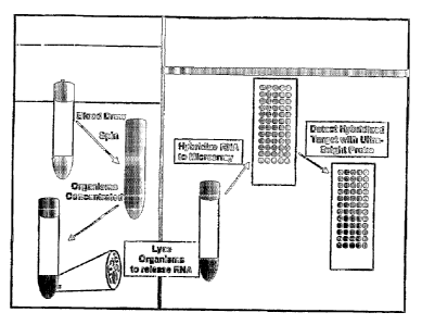
example, FIG. 1
illustrates one embodiment of the methods of the present invention in which
bacteria in blood
are concentrated into a volume of 300 L using a Wanlpole isolator tube. As
another
9
CA 02576904 2007-02-09
WO 2006/020579 PCT/US2005/028164
exarnple9 FIG. 4 depicts a vessel capable of concentrating the microorganisms
in a sarnple9
such as blood, that can be used in the n7ethods and kits of the present
invention. In sorne
embodiments, a sainple is treated to differentially separate microorganisms.
In such
embodiments9 separate samples containing different microorganisms may be
obtained.
Exainples 1-3 hereinbelow demonstrate differential separation of
microorganisms using
density gradients and matrices.
As the present invention contemplates, concentration of microorganisms and/or
their nucleic acids can be accoinplished in any number of steps including, but
not limited to,
one, two or three steps, where, in each step, the sample is progressively more
concentrated.
As a non-limiting example, microorganisms or their nucleic acids may be first
concentrated
using, for instance, a Wampole IsolatorTM tube. To continue this example, the
concentrated
sainple then may be concentrated further, separating intact microorganisms or
their nucleic
acids using, for instance, electrophoresis.
As used herein, the terms "nucleic acids" and "oligonucleotides" are used to
mean
DNA or RNA, as is recognized in the art. Nucleic acids may be single-stranded
or double-
stranded. Nucleic acids may be "released" from a microorganism using any means
that will
allow a capture probe access to hybridize the DNA or RNA. Hence, a "released"
nucleic acid
is a nucleic acid that is in a physical and chemical enviromnent that allows
nucleic acid
probes to bind to it. Additionally, both DNA and RNA may be released by the
same
processes. Examples of ways that DNA or RNA may be released include, but are
not limited
to, lysing the microorganisms using, for example, heat, enzymes, detergents,
buffers, acids,
bases, chaotropes, physical shearing in the presence of beads or particles and
the application
of pressure. As is contemplated by the present invention, the act of
collecting, isolating, or
concentrating the sample, or the portion thereof to be tested, may
sufficiently release the
nucleic acids that are subject to capturing. For the purposes of the present
invention, the
DNA or RNA to be used may be, but need not be, purified, isolated or
concentrated further
after release. Methods for purification, isolation, and concentration of
nucleic acids are well-
known in the art. It is preferable that nucleic acids released from
microorganisms be in
single-stranded form and laclc internal secondary structure before they' are
contacted with
probe(s) according to the present 'invention. Accordingly, the metllods of the
present
invention may also include one or more denaturing step, to denature any double-
stranded
nucleic acids or nucleic acids that possess internal secondary structure that
are released from
the microorganisms, prior to contacting the DNA or RNA with the capture probe.
However,
CA 02576904 2007-02-09
WO 2006/020579 PCT/US2005/028164
such a denaturing step is not required, paz-ticularly where the acts of
releasing, purifying,
isolating, and/or concentrating the nucleic acids also result in their
denaturation. Alethods for
denaturation of nucleic acids are well-known in the art.
Microorganisms that are to be detected may be referred to as "target
microorganisms." Nucleic acids released by microorganisms that are to be
detected may be
referred to as "target nucleic acids" or "target oligonucleotides." Target
oligonucleotides will
usually comprise at least one sequence that is capable of binding to a capture
probe and/or a
detector probe being used to detect microorganisms in a sainple. Such a
sequence may be
referred to herein as a "target sequence."
Once nucleic acids are released from the niicroorganislns, they may be
contacted
with one or more capture probes that are immobilized on a solid support. As an
alternative,
the released nucleic acids may be contacted with a fiee (non-immobilized)
capture probe to
form a hybrid complex, which is then contacted with a solid support that
irmnobilizes the
hybrid complex. As yet another alternative, capture probes may remain free
(i.e., not
immobilized). In such cases, hybrid conlplexes may be isolated by art-known
means such as
electrophoresis.
As used herein, a "capture probe" is a nucleic acid, or nucleic acid
derivative such
as a peptide nucleic acid, that is capable of binding to a released nucleic
acid. A capture
probe contains at least one single-stranded portion, or sequence, that is
capable of contacting
and hybridizing with released microorganism nucleic acids. A sequence that is
capable of
contacting and hybridizing with released microorganism nucleic acids may be
referred to
herein as a "capture sequence." As used herein, capture probes may be
classified by, for
example, the microorganism(s) to which their capture sequence(s) is capable of
binding.
Thus, capture probes of the same "type" comprise capture sequence(s) capable
of binding to
the saine microorganism(s). A capture probe comprises at least one capture
sequence. A
capture probe often also comprises, but is not required to comprise, sequences
in addition to
at least one capture sequence. Such additional sequences may, for exainple,
facilitate
iminobilization of the capture probe.
Detector probes may be used to facilitate the detection of nucleic acids that
have
been released from microorganisms. Nucleic acids that have been released from
microorganisms may be contacted with one or more detector probes. As used
herein, a
"detector probe" is a nucleic acid, or nucleic acid derivative such as a
peptide nucleic acid,
that is capable of binding to a released nucleic acid and that is capable of
being detected,
11
CA 02576904 2007-02-09
WO 2006/020579 PCT/US2005/028164
thereby facilitating detection of the released nucleic aeid. A detector probe
contains at least
one single-stranded portion, or sequence, that is capable of contacting and
hybridizing with a
released anicroorganisin nucleic acid. As with capture probes, a sequence of a
detector probe
that is capable of coritacting and hybridizing with released microorganisin
nucleic acids may
be referred to herein as a "capture sequence." A detector probe is preferably
bound to a
reporter group to facilitate detection. A detector probe to can be bound to a
reporter group
before or after the detector probe is hybridized to a target oligonucleotide.
Reporter groups
are known in the art and are discussed in more detail hereinbelow.
The capture sequence of a detector probe will usually hybridize to a different
target sequence of a target oligonucleotide than the target sequence to which
the capture
sequence of the capture probe hybridizes. Accordingly, a target
oligonucleotide can be
bound and detected by a detector probe while is bound to a capture probe. In
such
einbodiments, either the capture probe or the detector probe may be hybridized
to the target
oligonucleotide first. In certain embodiments it may be desirable to utilize a
capture probe
and detector probe each having a capture sequence that binds to the same
target sequence. In
such einbodiinents, a target oligonueleotide may be captured by a capture
probe, the capture
probe-target coinplex may be isolated, the capture probe-target complex may be
denatured,
and the detector probe may them be hybridized to the target oligonucleotide.
, As with capture probes, detector probes may be classified by, for example,
the
microorganism(s) to which their capture sequence(s) is capable of binding.
Thus, detector
probes of the same "type" comprise capture sequence(s) capable of binding to
the same
microorganism(s). A detector probe comprises at least one capture sequence. A
detector
probe often also comprises, but is not required to comprise, sequences in
addition to at least
one capture sequence. Such additional sequences may, for example, facilitate
the binding of
the detector probe to a reporter group.
In some einbodiinents, capture probes and/or detector probes coinprise linker
molecules such as, but not limited to, carbon chains or nucleic acid sequences
that are not
complementary to the target oligonucleotide. A linker molecule may serve, for
example, to
attach a probe to a solid surface, to bind a probe to another type of molecule
(sucll as, for
example, a protein), to attach a probe to a reporter group, or to bind a probe
to another probe.
A sequence that serves to immobilize a capture probe to a solid surface may be
referred to
herein as an "immobilization sequence." A probe comprising an immobilization
sequence
may be referred to herein as an "immobilization probe." Such non-complementary
lirikages
12
CA 02576904 2007-02-09
WO 2006/020579 PCT/US2005/028164
nlay reduce steric hindrance and may also improve the kinetics of
hybridization by increasing
the accessibility of the probes, particularly the capture sequence(s), to the
bulk solution.
Exainples of nucleic acid sequences that lnay be used as liiiker rnolecules
include the human
genes K-alphcr. (tifbailin alpha-1), PI'IA (Peptidldprolvl isofnercrse A), and
UBC (ubiquitin-
conjugating enzyfne E2A), and portions thereof. FIGS. 5A-C provide non-
limiting
illustrations of capture probes according to the present invention
iimiiobilized to an array
using linkers comprising portions of 1{ alpha, PPIA, and UBC.
Capture probes of the current invention may be "imnzobilized" onto a solid
support. As used herein, "immobilized" means affixed to a solid support such
that movement
of the capture probe in a solution is liinited, i.e., a capture probe that is
inunobilized on a
solid support will not dissociate from the solid support unless it is
subjected to a condition or
procedure that would cause it to dissociate.
As used herein, a "solid support" is a structure or a scaffold that will not
dissolve
in a liquid or gas solution. Examples of solid supports include, but are not
limited to, latex
beads, agarose beads, sepharose beads, paramagnetic beads, ferric oxide,
microarray cllips,
filter paper, nitrocellulose filters, nylon membranes, vessels, glass slides,
and even cellular
membranes. In some embodiments, the method of the present invention utilizes a
three-
dimensional microarray, such as, for example, the MetriGenix "' Flow-Thru
Chip'~
(MetriGenix, Inc., Maryland, USA), which facilitates increased llybridization
kinetics. An
example of a MetriGenix Flow-Thru Chip" is illustrated in FIG. 3. Such porous
arrays offer
increased surface area for attachment of probes over conventional two-
dimensional chips and
permit the flow of liquid back and fortll over the chip surface of the array,
thereby increasing
the opportunity for contact between the capture probe and target sequence. In
some
embodiments, each spot on an array may correspond to, as non-limiting
examples, a different
species of microorganisms, group of microorganisms or epideiniological marker.
In other
embodiments, different types of capture probes may be immobilized on the same
spot of an
array. Multiple spots for each analyte or group of analytes may also be
present.
Capture probes may be iminobilized onto solid supports using any of the many
art-known methods. Preferably, the immobilization does not adversely affect
the capture
probe's ability to bind to microorganism DNA and/or RNA or to other probes. A
capture
probe may be immobilized directly to the solid support, or it may be
immobilized indirectly
via attachment to another molecule that is immobilized on the solid support.
For example, a
capture probe may be immobilized using cheinical or linker moieties such as
carbon chains or
13
CA 02576904 2007-02-09
WO 2006/020579 PCT/US2005/028164
polyethylene glycol (PEG). In such cases, the binding of the capture probe may
be non-
specific. Alternatively, methods of using chenzical moieties to bind specific
nucleic acid
sequences are known and may be used with the present invention. As a non-
limiting
exainple, capture probes may be biotinylated, with biotin possessing the
ability to bind to
a~,,idin or streptavidin. Continuing the example, the solid support may have
avidin or
streptavidin bound to it. Such a scheme is a non-limiting example of a method
for
immobilizing capture probes without adversely affecting their ability to bind
microorganism
DNA and/or RNA because the biotin can be located at the opposite end of the
molecule fioin
the sequence capable of binding microorganism DNA and/or RNA (which may be
called a
"capture sequence"), or the biotin may be located on an internal branch of the
capture probe
that will result in its being located at a sufficient distance froin the
capture sequence that the
binding of the capture sequence to microorganism DNA and/or RNA is not
hindered.
As another exainple, capture probes may also be immobilized using another
single-stranded oligonucleotide probe that is itself iminobilized and that is
capable of
hybridizing with the capture probe to be immobilized. Such oligonucleotide
probes may be
called "immobilization probes." The use of immobilization probes is another
non-limiting
example of a method for iminobilizing capture probes without adversely
affecting their
ability to bind microorganism DNA and/or RNA because the sequence on the
capture probe
that is capable of binding to the immobilization probe (which may be called an
"immobilization sequence") can be located at the opposite end of the molecule
from the
sequence capable of binding microorganism DNA and/or RNA (which may be called
a
"capture sequence"), or the immobilization sequence may be located on an
internal branch of
the capture probe that will result in its being located at a sufficient
distance from the capture
sequence that the binding of the capture sequence to microorganism DNA and/or
RNA is not
hindered. An example of immobilization of a capture probe via an
immobilization probe is
illustrated in FIG. 2B. Examples of nucleic acid sequences that may be used as
immobilization probes include the human genes K-alpha (tubulin alpha-1), PPIA
(peptidylprolyl isomerase A), and UBC (ubiquitin-conjugating enzyme E2A).
(FIGS. 5A-C).
In an alternative embodiment, iinnZobilization probes may comprise non-
specific sequences
such as poly-A or poly-T oligomers. In a further embodiment they may also
comprise random
sequences of nucleotides or nucleotide homologues with no homology or
compleinentarity to
naturally occurring nucleic acid sequences.
14
CA 02576904 2007-02-09
WO 2006/020579 PCT/US2005/028164
In some embodiments, capture probes may be immobilized directly or indirectly
on a solid suppor-t in a pattern of discrete areas, or "spots." Such a
pattern, or a solid support
capable of supporting such a pattern, may be referred to herein as an "array,"
a"microarray,'9
or a "chip." The immobilization of probes of different types to a single
microarray or chip
facilitates the simultaneous deterinination of whether different
microorganisms are present in
a single sainple.
In certain embodlments, only a single type of capture probe is iinmobilized to
any
one spot, and different types of capture probes may be iminobilized to
different spots. In
such embodiments, the identity of the capture probe immobilized in any given
spot is laiown,
so microorganisms hybridized to capture probes in different spots can be
identified and
differentiated from one another by means of their locations. Examples of
innnobilization of
different types of capture probes in different spots of arrays are illustrated
in FIGS. 2A and
2B and in FIGS. 5A-C.
FIGS. 5A-C provide non-limiting exemplary illustrations of different types of
capture probes according to the present invention immobilized to different
spots of an array
using iinmobilization probes and immobilization sequences. Immobilization
probes
coinprising approximately 60 nucleotides in length to the human genes K-alpha
(tubulin
alpha-1) (FIG. 5A), PPIA (peptidylprolyl isomerase A) (FIG. 5B), or UBC
(ubiquitin-
conjugating enzyrne E2A) (FIG. 5C) are immobilized to an array. Each of the
immobilization
probes is hybridized to a capture probe comprising approximately 30 bases of
sequence
compleinentary to K-alpha (FIG. 5A), PPIA (FIG. 5B), or UBC (ubiquitin-
conjugating
enzyn7e E2A) (FIG. 5C) 3' to a capture sequence specific for E. coli (FIG.
5A), S. aureus
(FIG. 5B), or 0' epidef nfis (FIG. 5C).
In other embodiments, more t11an one type of capture probe is iminobilized in
a
single spot. In such embodiments, it will often be useful to use employ
detector probes such
that each detector probe of the saine type is bound to a reporter group that
is differentiable
from reporter groups bound to any other type of detector probe. As a non-
limiting example,
one could perforin an assay in which detector probes that bind to a target
sequence from
E. coli are labeled with fluorescein, and detector probes that bind to a
target sequence from
S. aureus are labeled with rhodamine. In such an assay, the presence of E.
coli could be
differentiated from the presence of S. aureus by the difference in the colors
of the fluorescent
labels. Of course, detector probes with differentiable labels may also be used
in conjunction
CA 02576904 2007-02-09
WO 2006/020579 PCT/US2005/028164
with the imn-iobilization of different types of capture probes in different
spots, thereby
facilitating the perforrnance of conlplex assays.
Detector probes may be attached directly or indirectly to a reporter group. As
an
example of an indirect attachment using a linker molecule, a detector probe
may coinprise a
"reporter adapter sequence" linker. A reporter adapter sequence is a portion
of a detector
probe that is capable of binding via hybridization to a single-stranded
oligonucleotide that
bears a reporter group, which may be referred to herein as a "reporter probe."
FIGS. 2A-B
provide exemplary illustrations of a capture probe hybridized to a target
oligonucleotide,
whicll is in turn hybridized to a detector probe. The detector probe is
hybridized to a reporter
probe. In certain embodiments, detector probes of different types inay
comprise the same
reporter adapter sequence, thereby facilitating the detection of different
microorganisms
using a single reporter probe, which may be referred to herein as a "universal
reporter probe."
Such an embodiment is illustrated in FIGS. 2A and B. In other embodiments,
detector probes
of different types may comprise different reporter adapter sequences, thereby
facilitating the
use of differentiable reporter probes to detect different microorganisms.
Capture probes and detector probes may be "protected" from prematurely
hybridizing to random nucleic acids by having a protecting group situated on
or near the
capture or detector probe. As non-limiting examples, protecting groups include
single-
stranded nucleic acid that is partially complementary to the capture probe to
be protected, or
an antibody or a binding fragment thereof that binds to the single-stranded
portion of the
capture or detector probe to be protected.
Hybridization between a microorganism nucleic acid and a capture probe or
detector probe may be referred to herein as a "hybridization event." A
hybridization event
will form a "hybrid complex." As used herein, a "hybrid complex" is a double-
stranded
nucleic acid comprising at least a portion of a capture probe or detector
probe (usually a
capture sequence) and at least a portion of a target oligonucleotide (usually
a target
sequence). A hybrid coinplex need not be double-stranded along its entire
length.
Furthennore, for the purposes of the present invention, a capture probe or
detector probe and
a target oligonucleotide need not have a complementary base pairing at every
base for a
hybridi_zation event to occur. Further still, for the purposes of the present
invention, a capture
sequence and a target sequence need not have a complementary base pairing at
every base for
a hybridization event to occur. In other words, the present invention
contemplates that a
hybrid complex will be formed even if a target oligonucleotide hybridizes to a
capture or
16
CA 02576904 2007-02-09
WO 2006/020579 PCT/US2005/028164
detector probe such that a portion of the capture or detector probe or target
nucleic acid is
single-stranded after hybridization because the target oligonucleotide did not
hybridize to the
entire length of the capture or detector probe. In some einbodiments of the
present invention,
a portion of a capture or detector probe remains single-stranded after
hybridization to a target
oligonucleotide. In some other eanbodiments, a portion of a target
oligonucleotide reinains
single-stranded after hybridization to a capture or detector probe. In still
other einbodiments,
portion(s) of each of a target oligonucleotide and a capture or detector probe
remain(s) single-
stranded after hybridization to one another. A "portion" can be one or more
nucleic acids in
length. Such single-stranded portions may occur within and/or outside of a
capture sequence
and/or a target sequence. Single-stranded portions within a capture sequence
and/or target
sequence may occur, as a non-liiniting example, because the capture sequence
and the target
sequence are not 100% complementary. Single-stranded portions outside of a
capture
sequence and/or target sequence may occur, as a non-limiting example, because
the capture
and/or detector probe contains portions that are not intended to bind to the
target
oligonucleotide. Single-stranded portions outside of a capture sequence and/or
target
sequence may occur, as another non-limiting exainple, because the target
oligonucleotide
comprises sequences in addition to the target sequence. For example, a capture
probe will
often (but need not) coinprise a sequence used to immobilize it to a solid
support. As another
example, a detector probe will often (but need not) coinprise a sequence used
to bind it to a
reporter group. As yet another example, a target oligonucleotide will often
(but need not)
comprise sequences 3' and/or 5' to the target sequence(s).
A solid support may have iminobilized to or on it one or various combinations
of
probes that are microorganism-specific, probes that are for epidemiological
markers (e.g.,
IS6110-based probes used for Mycobacterium tuberculosis), and/or probes that
are for drug
resistance markers (e.g., mecA-based probes for methicillin resistance in S.
aureus or rpoB-
based probes for detection of rifampin resistance in M. tuberculosis). As used
herein, the
term "microorganism-specific probe" includes probes that are capable of
hybridizing with a
target sequence derived or released from a single microorganism species. Such
probes may
also be referred to herein as "species-specific probes." The term
"microorganism-specific
probe" also includes probes that are capable of hybridizing with target
sequences from more
than one species of microorganism from a single genus of microorganism (e.g.,
IS6110 for
the detection of the M. tuberculosis complex (M. tuberculosis, M. bovis, M.
microti, and
M. af~icanuna); probes based on conserved regions of the 16S rRNA, 18S rRNA,
RNase P or
17
CA 02576904 2007-02-09
WO 2006/020579 PCT/US2005/028164
ssrA gene sequences). For exarnple, a microorganism-specific probe may be
designed to
form a hybrid with nucleic acid sequences from both S'. aureus and S.
epidea=nr.idis, but not
with E. coli, Such probes may also be referred to herein as "genus-specific
probes.'" In some
embodiments, a genus-specific probe will hybridize to sequences derived from
all or many of
the anicroorganisms belonging to the saine genus of classification. As used
herein, a' multi-
genus probe" will hybridize to nucleic acid from microorganisms belonging to
two or more
different genera. A probe may hybridize to an antimicrobial resistance marker
that may be
present in one or more species, for exalnple. Such a probe may be species-
specific, genus-
specific, or multi-genus, depending on how widely the antimicrobial resistance
marker is
distributed through phylogeny. Whether a microorganism-specific probe, as
contemplated by
the present invention, hybridizes to a target sequence derived or released
from a single
microorganism species, to target sequences derived or released from more than
one
microorganism species within the same genus of microorganisms, or to target
sequences
derived or released from microorganisms from different genuses may also depend
on the
hybridization and wash conditions used in the assay.
As described above for capture probes, detector probes may be microorganism-
specific probes, probes that are for epidemiological markers, and/or probes
that are for drug
resistance markers. Various combinations of capture probes and detector probes
may be used
to discriminate between organisms present in a sample. For instance, a genus-
specific
capture probe may be used to iminobilize microorganisms of a selected genus,
which then
may be detected as a genus with one or more genus-specific detector probes, or
which may be
discriminated by species with one or more species-specific detector probes.
More than one
type of capture probe may be used concurrently in the methods of the present
invention.
Likewise, more than one type of detector probe may be used concurrently in the
methods of
the present invention.
As non-limiting examples, oligonucleotide probes comprising one or more of the
sequences set forth in the following "table (Table 1) are particularly useful
for detecting and
identifying bacteria of the indicated species. Oligonucleotide probes
comprising one or more
of the sequences set forth in Table 1 can be used as capture and/or detector
probes to detect
nucleic acids from the indicated bacterial species. Oligonucleotide probes
comprising
regions that are homologous to the oligonucleotide probes set forth in Table 1
are also useful
for capturing and/or detecting the indicated species. In general,
oligonucleotides containing
18
CA 02576904 2007-02-09
WO 2006/020579 PCT/US2005/028164
sequences that are at least about 85%, at least about 90%, at least about 95%,
or about 100%
homologous to the oligonucleotides of Table 1 are useful.
The sequences in Table I can be used as species-specific capture and/or
detector
sequences to detect and/or differentiate between particular species of
inicroorganisms.
Sequences from Table 1 may, but need not, comprise portion(s) of longer
oligonucleotides.
For example, probes according to the invention may coinprise one or more
sequences from
Table 1 and/or additional sequences.
Table 1
SEQ Species Reference Target Oligo 5'-3' 'ICm Raffik::
II- Strain Gene Name Sequence ( G)''' ~
NO:
1 Staphylococcus NCTC ssrA S_aur-1 TTG ATT 58.9 1
aureus 8325 AAG TTT
CTT CTA
AAC AGA
2 Staphylococcus NCTC ssrA S_aur-2 TCA TGA 59.7 1
aureus 8325 AAA GTG
ATA AAC
AAC C
3 Escherichia coli 0157:H7 ssrA E_coli-1 AAT TCC 59.2 2
EDL933 TAC GTC
CTC GGT
A
4 Escherichia coli 0157:H7 ssrA E_coli-2 TAC ATT 60.0 2
EDL933 CGC TTG
CCA GC
5 Eschei=ichia coli 0157:H7 ssrA E_coli-3 CTA GCC 59.6 2
EDL933 TGA TTA
AGT TTT
AAC G
6 Escherichia coli ATCC ssrA E_coli-4 TCC TCG 59.8 2
133 GTA CTA
CA 1 GCT
TAG
7 Escherichia coli ATCC ssrA E_coli-5 TCC TAA 60.3 2
133 GAGCGG
AGG CTA
8 Staphylococcus SR1 ssrA S_epi-1 CAT CAT 61.4 3
epidermidis GCT AAG
CAA TAA
ACAA
19
CA 02576904 2007-02-09
WO 2006/020579 PCT/US2005/028164
SEQ Slaecies Refereance Target lig 5 -3' Tm Rank>
ID Straan Gene D'1am e Sequence ( G)"
PT :
9 Staplrylococcus SR1 ssrA S_epi-2 TTG ATT 60.0 3
epider iraidis ATA TTT
CAT CTA
AAC
AGA CT
Staphylococcus SR1 ssrA S_epi-3 CAG TTA 61.2 3
epiderrnidis TAT TTA
ACC GAA
ATG TGT
11 Klebsiella MGH ssrA K._pneu-1 ATT CCT 60.1 4
pneumoniae ACA TCC
TCG GCA
12 E'lebsiella MGH ssrA K_pneu-2 GTC TTA 59.0 4
pneumoniae AGA GCG
GAA GCT
AG
13 Klebsiella MGH ssrA K_pneu-3 AGC CTG 59.3 4
pneumoniae ATT AGA
TTT AAC
GC
14 Enterococcus 775 ssrA E_faeca-1 CAT ATT 59.0 5
faecalis GCC ACT
TAA ATC
TCT AC
Enterococcus 775 ssrA E_faeca-2 CTG TAT 60.9 5
faecalis TGC TAG
TCT GGT
AAG CT
16 Enterococcus 775 ssrA E_faeca-3 ACA CTC 59.5 5
faecalis ATT TAA
AGG TTC
GC
17 Pseudomonas ATCC ssrA P_aeru-1 GCT TAG 59.4 6
aeruginosa 25330 CCA GCT
CTA CTG
AG
18 Pseudomonas ATCC ssrA P_aeru-2 TTA AGC 59.9 6
aeruginosa 25330 AGC TAG
AGC GTA
GTT
19 Streptococcus Type 4 ssrA S_pneu-2 CTC AAG 59.4 8
pneuinoniae TCT AGA
AAC TGC
GAG
CA 02576904 2007-02-09
WO 2006/020579 PCT/US2005/028164
SEQ Species PLeference Target lgg 5Tm RaanV
fL) Straiaa CGene T'~iame Sequence ( G)
I'~T0:
20 Streptococcus Type 4 ssrA S_pneu-1 TTA TTT 60.6 8
pneumoniae TAA CAG
CCC CTC
G
21 Styeptococcus UA159 ssrA S_inut-1 TGT TTA 59.9 10
nmutans TTT AAC
ACC GTT
ACA AT
22 Streptococcus UA159 ssrA S_inut-2 TCA AAC 61.0 10
inutans TCT AAC
GAT GCG
AG
23 Streptococcus Not ssrA S_gord-1 TGT TTT 60.7 10
gordonii Known AAC TTG
ATT TTG
ACA CA
24 Streptococcus Not ssrA S_gord-2 CAA ATC 60.6 10
gordonii I~nown AAG CGA
GTC TAT
CAA
25 Clostridiuna 630 ssrA C_diff-1 CCA ACT 60.1 19
difficile TCA CTA
ATA TCT
CAC CT
26 Clostridiuin 630 ssrA C_diff-2 GTC CAG 59.6 19
difficile TCT TAG
TCG GCA
G
27 Clostridium (Shimizu) ssrA C_perf-1 AGC AGA 59.7 0
perftingens CCA GTA
AGA CTT
TCT AC
28 Clostridium (Shimizu) ssrA C_perf-2 AGA ACG 61.0 19
peNfi=ingens TCC ACA
GAC AAA
CTT
29 Clostridium Hall A ssrA C_bot-1 AAC AGG 60.1 19
botulinurn CTC CTA
GAT TCA
GTA G
30 Clostridium Hall A ssrA C_bot-2 CCG AGT 59.7 19
botulinuin GCA GTT
TAT CCT T
21
CA 02576904 2007-02-09
WO 2006/020579 PCT/US2005/028164
slEQ S Pecks Reference Target (DIig 5 -3 Tin Ranhlip "i'Ie aan Gene I\iame
Sequea~ce
Ii o
31 Haeinophilus Not ssrA H_infl-1 GAC ACG 60.8 23
influemae Kiiown CTA AAC
TTA AGC
TAG TT
32 Ilaeniophilus Not ssrA FI_infl-2 CCT CAA 60.7 23
influeMae I~nown ACG GTG
GCT TC
33 Enterococcus ATCC ssrA E_faeci-1 GTC AAC 60.0 25
faeciu.fza 35667 TCA TTT
AAG GAT
TCA CT
34 Entei=ococous ATCC ssrA E faeci-2 GAT GTT 60.5 25
faeciuni 35667 ~ CTC TTT
TTC AAC
TTA CAG
35 Enterococcus CNRZ129 ssrA E_dur-1 TCA ACT 60.5 NR
durans CAT TTG
AGG TTT
CG
36 Enterococcus CNRZ129 ssrA E_dur-2 TGA TGA 60.8 NR
durans TCT CTT
TTA AAC
TTT
ACA G
37 Enterococcus CNRZ129 ssrA E dur-3 AGG CAT 60.6 NR
durans ~ TCT GTA
TTG CTA
GTC T
38 Streptococcus M1 GAS ssrA S_pyo-1 TTA TGT 61.0 NR
pyogenes SF370 CTT CAT
TTA ACA
AAC
TAA AG
39 Streptococcus M1 GAS ssrA S_pyo-2 TCA AGC 59.8 NR
pyogenes SF370 CAT TAG
TTT GCG
40 Streptococcus Ml GAS ssrA S_pyo-3 GAC AAT 60.0 NR
pyogenes SF370 TTC GTA
ACC GTA
GC
41 Streptococcus NCTC ssrA S_agal-1 GTA TTG 60.8 NR
agalacticae 8181 ATT TAA
CTA GGT
GAT
GAC A
22
CA 02576904 2007-02-09
WO 2006/020579 PCT/US2005/028164
SlEQ) specl~~ ~~~erence Target Oligo S -3' Trn Rannh...
IID SErain Geaie Name Sequence ( C()'
HO:
42 Sti=eptococcus NCTC ssrA S_agal-2 TTA ACT 60.5 NR
agalacticae 8181 AAC TAG
ACA GTA
GCC
AAA C
43 Escherichia coli 0157:H7 ssrA Eco_ssrA TCA GTC 75.0 2
EDL933 _DP50 TTT ACA
TTC GCT
TGC CAG
CTG CGG
ACG GAC
ACG CCA
CTA
ACA AA
44 Staphylococcus NCTC ssrA Sau_ssrA_ CTT CAA 70.0 1
aureus 8325 DP50 ACG GCA
GTG TTT
AGC ATA
TCC TAT
TAA GGT
TGA ATC
GCG
TTA AC
45 Staphylococcus SR1 ssrA Sep_ssrA_ CCA ACA 69.0 3
epidermidis DP50 TGA TAC
TAG CTT
GAT TAT
ATT TCA
TCT AAA
CAG ACT
TCA
AGC GG
46 Escherichia coli 0157:H7 rnp EcoCP4 GCA CTG 63.2 2
GTC GTG
GGT TTC
47 Staphylococcus WCUH29 rnp SauCP5 TTA CTC 59.9 1
aureus TAT CCA
TAT CGA
AAG ACT
48 Staphylococcus SRl rnp SepCP6 CTA TTC 60.0 3
epiderriaidis TAA CCA
TAT CCA
ATG ACT
23
CA 02576904 2007-02-09
WO 2006/020579 PCT/US2005/028164
SEQ Species Reference Target Oligo 51-31 Tm RanL*
llF Strain Gene 11ame Sequence ( C);: 1'16:
49 Escherichia coli 0157:H7 rnp Eco_rnp_ CCC CCC 77.0 2
I)P50 AGGCGT
TAC CTG
GCA CCC
TGC CCT
ATG GAG
CCC GGA
CTT TCC
TC
50 Staphylococcus WCUH29 rnp Sau_rnp_ TAG GAT 67.0 1
aua eus DP50 ATT TCA
TTG CCG
TCA AAT
TAA TGC
CTT GAT
TTA TTG
TTT
CAT CA
51 Staphylococcus SR1 rnp Sep rnp TAG GTT 67.0 3
epidef iazidis DP50 ATT TCA
TTG CCG
TCA AAT
TAA TGC
CTT GAT
TTA TTG
TTT
CAT CA
52 Escherichia coli K12 16S EcoCP7 AGT GTG 59.0 2
rRNA GCT GGT
CAT CCT
53 Escherichia coli RREC I 16S Eco_16_D CTC AGA 75.0 2
rRNA P50 CCA GCT
AGG GAT
CGT CGC
CTT GGT
GAGCCG
TTA CCC
CAC
CAA CA
* Nearest neighbor analysis
** Ranking of species in top 25 (US) blood pathogens; NR = not ranked within
top 25 US
blood pathogens
For the purposes of present invention, a capture probe captures (by binding
to) an
oligonucleotide from a sample by hybridizing with it at a sequence (e.g., a
capture sequence)
24
CA 02576904 2007-02-09
WO 2006/020579 PCT/US2005/028164
that is at least partially complementary to a sequence (e.g., a target
sequence) of the
oligonucleotide being captured. Likewise, a detector probe detects (by binding
to) an
oligonucleotide from a sainple by hybridizing with it at a sequence (e.g., a
capture sequence)
that is at least partially complementary to a sequence of the oligonucleotide
being captured
(e.g., a target sequence). As used herein, the phrase "partially
complementary" means less
than 100% complementary, but at least about 85% compleinentary. Accordingly,
the phrase
"at least partially complementary" indicates that the capture sequence of a
capture and/or
detector probe may between about 85% complementary to about 100%
compleinentary to a
target sequence to be useful according to the present invention. A capture
sequence and a
target sequence of an oligonucleotide to be captured may be, as non-limiting
exainples, at
least about 85%, at least about 90%, at least about 95%, at least about 96%,
at least about
97%, at least about 98%, at least about 99%, or about 100% complementary to
one another.
For exainple, if a capture sequence is 100 bases long, and the target sequence
is 95%
complementary to the capture sequence, the base pairs of the capture sequence
and the target
sequence will matcll in 95 of 100 bases of the capture sequence.
As a practical matter, whether any particular nucleic acid molecule is at
least 85%,
at least 90%, at least 95%, at least 96%, at least 97%, at least 98%, at least
99%, or 100%
complementary to a target nucleic acid can be determined conventionally using
known
computer programs such as, for example, the Bestfit program (Wisconsin
Sequence Analysis
Paclcage, Version 8 for Unix, Genetics Computer Group, Wisconsin, USA).
Bestfit uses the
local homology algorithm of Smith and Waterinan, Advances in Applied
Mathematics 2:482-
489 (1981), to find the best segment of homology between two sequences. When
using
Bestfit or any other sequence alignment program to determine whether a
particular sequence
is, for example, 95% complementary to a reference sequence according to the
present
invention, the parameters are set such that the percentage of identity is
calculated over the full
length of the reference nucleotide sequence, whether that be the capture probe
or the target
nucleic acid, and that gaps in similarity of up to 5% of the total number of
nucleotides in the
reference sequence are allowed.
Whether the capture sequence of a capture probe and/or detector probe will
hybridize to the target sequence of a target oligonucleotide depends on the
degree of
complementarity between the target sequence and the capture sequence, as well
as both the
hybridization conditions and the stringency of the wash after hybridization.
As used herein,
the phrase "conditions that pei7nit hybridization" refers to hybridization
parameters, as well
CA 02576904 2007-02-09
WO 2006/020579 PCT/US2005/028164
as wash parameters9 that peri-nit hybridization between two oligonucleotides,
as are
understood in the art, For exarnpleq conditions that perinit hybridization
include, but are not
limited to, more stringent hybridization and wash conditions, such as
incubation at 42 C in a
solution comprising 50% formainide, 5X SSC (750 mM NaCI, 75 mM tri,-,odiuin
citrate), 50
mM sodium phosphate (pH 7.6), 5X Denhardt's solution, 10% dextran sulfate, and
20 g/ml
denatured, sheared salmon sperm DNA, followed by washing with 0.1X SSC. at
about 65 C,
68 C or 70 C. Of course, hybridization and wash conditions can be set to a
lower stringency.
Lower stringency hybridization and wash conditions include, but are not
limited to,
incubation at 42 C in a solution comprising 30% formamide, 5X SSC (750 mM
NaCI, 75
mM trisodium citrate), 50 mM sodium phosphate (pH 7.6), 5X Denhardt's
solution, 10%
dextran sulfate, and 20 g/ml denatured, sheared salmon sperm DNA, followed by
washing
the filters in a solution of 2X SSC or 1X SSC or 0.5X SSC at about 55 C or 60
C or 65 C.
As is within the capacity of one of ordinary skill in the art, the conditions
to permit
hybridization can be easily and routinely optimized to require a lower or
higher degree of
complementarity between a capture probe and/or detector probe and a target
nucleic acid
before hybridization will occur. For example, anionic detergents such as
sodium dodecyl
sulfate (SDS) may be used to enhance the stringency of hybridization or
washing, and
exclusion molecules such as PEG may be used to increase the effective
concentration of
reaction components.
A capture probe or detector probe may be an oligonucleotide or a
polynucleotide,
as these terms are understood in the art. A capture probe or detector probe
may be, for
example, at least 10, 15, 20, 25, 30, 35, 40, 45, 50, 75, 100, 150, 200, 300,
.400, 500 or 750
nucleotides in length. For convenience, the term "oligonucleotide" as used
herein
encompasses all of these lengths. In some einbodiments, capture probes and/or
detector
probes may be up to and including about 2000 nucleotides in length. In some
embodiments,
capture probes and/or detector probes are about 15 to about 60 nucleotides in
length.
The length of the capture probe and/or detector probe and the capture
sequence(s)
thereof and the conditions of hybridization may be tailored to form a specific
coiuplex witli
the nucleic acid of the intended target. The stability of a hybrid complex,
commonly
measured by its melting temperature, is related to the concentration of the
probe, the
hybridization conditions, the length of the hybrid complex and the degree of
sequence
identity between the capture sequence and the target sequence. The stability
of a hybrid
coinplex is decreased by mismatches and is increased by the number of base
pairs in the
26
CA 02576904 2007-02-09
WO 2006/020579 PCT/US2005/028164
hybrid complex. Such relati nships are detailed, for example, in Sarnbrook et
al.,
MOLECULAR C'LONING. A LAB kATORY mP,NUAL9 3rd ed., Cold Spring Harbor
Laboratory
Press, New York (2001), which is herein incorporated by reference. A probe can
be used to
distinguish betxveen closely related sequences that differ in the hybrid
complex by as little as
a single base. That is, a probe may be perfectly matched with a target nucleic
acid from one
species of microorganism but may differ in sequence by one base with the
nucleic acid target
of a second species, for example. Under the appropriate conditions, the
presence of the
mismatch causes the probe to form a hybrid coinplex with one target but not
with the other.
Because the relative difference in melting temperature between the matched and
mismatched
complexes decreases with increasing size of the probe, the probe preferably
should be 25
bases in length or less to detect a single mismatch. See Sambrook et al. While
longer probes
can be used to discriminate between closely related targets, the targets
preferably should
diverge to a progressively greater extent as the size of the probe increases.
After a hybrid complex is formed, the methods of the present invention then
detect
the hybrid complex. If a llybrid complex is detected, the presence of the
hybrid complex
indicates that the target sequence was released from a microorganism in the
sample tested.
Accordingly, the presence of a hybrid complex indicates the presence in the
sample of the
microorganism and/or epidemiological marker containing the target sequence. In
contrast,
when a hybrid complex is not detected, the absence of the hybrid complex
indicates that the
target sequence was not released from a microorganism in the sample tested.
Accordingly,
the absence of a hybrid complex indicates that the sample most likely does not
contain the
microorganism and/or epidemiological marker containing the target sequence.
Any method of detecting the hybrid complex can be used in the present
invention,
provided that one of skill in the art can rely on the detection methods to
identify the presence
of a hybrid complex. In some embodiments, at least one detector probe, in
addition to the
capture probe, is used to detect the hybrid complex. The detector probe
hybridizes to a
single-stranded portion (or "target sequence") of the target nucleic acids
that are captured.
A detector probe may comprise a reporter group to facilitate detection. As
used
herein, a "reporter group" means an entity that can generate a detectable
signal. A reporter
group may be incorporated into a detector probe, a reporter group may be
directly linlced or
bound to a detector probe, or a reporter group may be indirectly linked or
bound to a detector
probe. As explained in more detail hereinabove, as an example of an indirect
attachment
using a linker molecule, a detector probe may comprise a reporter adapter
sequence linker,
27
CA 02576904 2007-02-09
WO 2006/020579 PCT/US2005/028164
which is capable of binding via hybridization to a reporter probe, which is
bound to a reporter
group. Detector probes that "comprise4' reporter groups include detector
probes that have a
reporter group, reporter adapter sequence linkers, and the like incorporated
therein, as well as
detector probes that are directly or indirectly linked or bound to reporter
groups, reporter
adapter sequence linkers, and the like.
Many different reporter groups are lcnown in the art. For exainple,
radioactive
isotopes may be used as reporter groups. Radioactive isotopes may be, for
example,
incorporated into or attached to a detector probe, thereby generating a
radioactive probe.
Examples of a radioactive isotopes that can be used as reporter groups
include, but are not
limited to, 32P) 33P 131I9 90y9 188Re, 186Re! 67Cu9 198AU> 103pd and 212Pb/212
Bl.
Enzyines and/or enzyme systems that can be used to generate a detectable
signal
may also be used as reporter groups. Enzymes and/or other components of
enzyine systems
can be attached directly to a detector probe or can attach indirectly to a
detector probe. For
exainple, a detector probe may be biotinylated. As a specific, non-limiting
example, a
detector probe may be bound to BioTEG, which is biotin with a 15 atom tetra-
ethyleneglycol
spacer. Biotin possesses the ability to bind to avidin or streptavidin.
Continuing the
example, an enzyme such as horseradish peroxidase would be conjugated to
avidin or
streptavidin, thereby allowing the horseradish peroxidase to localize to the
hybrid complex
via binding of the biotin of the biotinylated- detector probe and the avidin
of the avidin-
enzyine conjugate. Upon addition of a substrate, horseradish peroxidase would
then generate
a detectable colorimetric signal as is readily understood in the art.
Additional examples of
enzymes that may be used as reporter groups include, but are not limited to
allcaline
phosphatase, glucose oxidase, P-galactosidase, soybean peroxidase and
luciferase.
Fluorescent or other detectable molecules may also be used as reporter groups
and
may be attached directly or indirectly to the detector probe. Non-limiting
examples of
detectable molecules that may be used as reporter groups include, but are not
limited to,
fluorescein, fluorescein isothiocyanate (FITC), rhodainine red, ROXTM
(Invitrogen,
California, USA), CyTM dyes (Amersliam, New Jersey, USA), BodipyTM dyes
(Molecular
Probes, Oregon, USA), TAMRAT"" dyes (Molecular Probes, Oregon, USA), TETTM
(Molecular Probes, Oregon, USA), Texas Red (Molecular Probes, Oregon, USA),
europium
dyes, chromogenic moieties and green fluorescent protein (GFP).
Reporter groups may also comprise an "amplification sequence," i. e., a
nucleotide
sequence that can initiate nucleic acid ainplification. For example, an
ainplification sequence
28
CA 02576904 2007-02-09
WO 2006/020579 PCT/US2005/028164
may be appended to a detector probe, or an ainplification sequence may be an
integral part of
a detector probe. An amplification sequence can initiate nucleic acid
replication, or
amplification, using any form of ainplification including, but not lirnited
to, strand
displacement amplification (SDA), polyinerase chain reaction (PCR), reverse
transcriptase-
strand displacement amplification (RT-SDA), reverse transcriptase-polymerase
chain reaction
(RT-PCR), nucleic acid sequence based amplification (NASBA), 1!/[essageAinpTM
ainplification (Ambion, Inc., Texas, USA), transcription-mediated
amplification (TMA),
rolling circle amplification, and QB replicase amplification. The nucleic
acids may be
ainplified prior to contacting them with a capture probe or after they are
contacted with the
capture probe.
Instead of or in addition to the use of a detector probe, a hybrid complex may
also
be detected using such standard methods as non-specifically labeling the
hybrid coinplex.
For exainple, intercalating dyes, such as ethidium bromide, may be used to
label the hybrid
complex for detection. Other exainples of non-specific labeling of the hybrid
complex
include, but are not limited to, acridine orange, SYBRTm Green UII (Molecular
Probes,
Oregon, USA), SYBR Gold, propidium iodide and cyanine monomers or dimers.
In some embodiments, the methods and/or probes of the present invention are
used to monitor the efficacy of antimicrobial patient therapy by sequential
sampling of
specimens over time. The detection of mRNA has been correlated with microbial
viability.
Hellyer et al., .I. Clin. lllicrobi l. 37: 290-295 (1999). Accordingly, the
detection of RNA in
a first sample, followed by administration of antimicrobial therapy and
subsequent failure to
detect RNA in a second sample, most likely indicates a successful response to
therapy. In
contrast, detection of RNA in the second sample would most likely indicate the
continued
presence of viable organisms in the specimen, and the need for continued
therapy or a change
in therapeutic regimen.
In some other embodiments, the methods and/or probes of the present invention
are used to quantify the number of organisms in a sample through the use of an
internal
standard and by comparison of signal intensities with controls. The internal
standard may be
RNA or DNA that is free in solution or encapsulated, such as in an Armored
RrLQ T ~ (Ainbion
Diagnostics, Texas, USA) particle or recombinant bacterium, to protect against
degradation.
The internal standard may be seeded into the sample at any point prior to
detection. In some
embodiments, the internal standard is seeded in the sample prior to
concentration and lysis of
the microorganism(s). The internal standard is detected using specific capture
probes that
29
CA 02576904 2007-02-09
WO 2006/020579 PCT/US2005/028164
pernlit distinctioii of the sta..ndard from target nucleic acid and from other
nucleic acids that
may be present. The signal generated by the internal standard in the test
sainple is compared
to that from controls that comprise different levels of the standard nucleic
acid conjugated
directly to the solid phase. By plotting a curve of signal intensities for the
controls, the
proportion of the internal standard recovered from the specimen may be
calculated. In similar
fashion, controls comprising different levels of the target sequence
conjugated directly to tlae
solid phase may be used to quantify the ainount of target present in the
processed sainple. By
correcting for recovery of nucleic acid using the internal standard, the
quantity of target
nucleic acid in the original saniple may then be calculated.
It is iinportant to note that the methods of the present invention also have
application outside the fields of huinan and animal infectious disease and are
particularly
suited to applications requiring rapid, sensitive and specific detection
and/or identification of
inultiple analytes. Accordingly, the inetllods of the present invention can be
used to detect
microorganisms in samples in many fields including, as non-limiting examples,
therapeutic
monitoring, food and environmental testing and monitoring deployment of
weapons of
bioterrorism.
The present invention also relates to kits for detecting the presence or
absence of
at least one microorganism in a sainple. The kits comprise a solid support, as
defined
hereinabove, comprising at least one capture probe, also defined hereinabove.
The at least
one capture probe may be a microorganism-specific probe, a probe that is for
an
epidemiological marker, and/or a probe that is for a drug resistance marker.
The kits of the
present invention also comprise at least one reporter group, as previously
described
hereinabove. The kits may also comprise a vessel. The vessel can be used to
collect or
concentrate the sample and/or to isolate the nucleic acids released from the
microorganisms.
The vessels may also be used when amplifying the nucleic acids released from
the
microorganism, if desired or necessary. Examples of vessels include, but are
not limited to,
evacuated blood collection tubes, eppendorf tubes, test tubes, etc. The kits
may further
comprise enzymes or other chemicals, such as media, detergents, buffers,
acids, bases, and
chaotropes used to lyse the microorganisms present in the sample. The reporter
group(s) of
the kits further comprises at least one detector probe. Ln such embodiments,
the reporter
group may be incorporated into or onto the detector probe, as previously
described herein.
As with the capture probe, the detector probe may be a universal probe or a
species-specific
probe. Kits of the present invention may also comprise positive, negative,
and/or internal
CA 02576904 2007-02-09
WO 2006/020579 PCT/US2005/028164
controls. In certain embodiments, kits of the present invention may comprise a
sufficient
number of probes and/or ainounts of other components to permit the performance
of only a
single assay or group of related assays using probes according to the present
invention. In
other einbodiinents, kits of the present invention may comprise a sufficient
number of probes
and/or ainounts of other components to perinit the performance of inultiple
assays using
probes according to the present invention.
The Figures provide non-limiting exemplary illustrations of embodiments of the
present invention and devices and non-limiting exemplary sequences useful in
practicing the
present invention. One exemplary embodiinent of the probes and methods of the
present
invention is illustrated in FIG. 1. In FIG. 1, bacteria in blood are
concentrated into a volume
of 300 p,L using a Wampole isolator tube. Next, the bacteria are lysed to
release RNA or
DNA, and this RNA- and/or DNA-containing solution in either a crude or
purified forin is
applied to a microarray solid support that has capture probes iinmobilized
tllereto. The
capture probes may be, for example, microorganism-specific probes, probes that
are for
epidenziological markers, and/or probes that are for drug resistance markers.
In this
particular embodiment, after the microbial RNA and/or DNA is allowed to
hybridize with the
capture probes to fonn hybrid complexes, the complexes are detected using a
detector probe
that bears a reporter group.
Two exemplary embodiments of the use of the probes and methods of the present
invention are illustrated in FIGS. 2A and 2B. The two embodiments of FIG. 2
differ in the
manner in which capture probes are immobilized on a solid support. The capture
probes of
the einbodiment illustrated in FIG. 2A are immobilized directly on the solid
support. The
capture probes are species-specific, and species A-specific capture probes are
immobilized to
one spot (A), while species B-specific capture probes are iinmobilized to a
different spot (B).
In a further embodiment, two or more capture probes may be immobilized on a
single spot to
permit the capture and detection of two or more target sequences derived from
different
organisms in a single location.
In contrast, the capture probes of the embodiment illustrated in FIG. 2B are
immobilized indirectly on the solid support through the use of immobilization
probes. Each
species-specific capture probe coinprises an immobilization seq ence that is
hybridized to an
immobilization probe. The immobilization probe is iinmobilized to a spot on
the solid
support. Each of the immobilization probes of the embodiinent illustrated in
FIG. 2B is
specific for a specific type of capture probe. Immobilization probes A
hybridize specifically
31
CA 02576904 2007-02-09
WO 2006/020579 PCT/US2005/028164
to capture probes A(ivhich are specific for species A), and immobilization
probes B
hybridize specifically to capture probes E(which are not shown, but wl-iieh
are specific for
species B). Capture probe A-specific iininobilization probes are iinnnobilized
to one spot
(A), while capture probe B-specific inunobilization probes are immobilized to
a different spot
(B). In a further embodnnent, two or more irrunobilization probes may be
iinmobilized on a
single spot to perinit the capture and detection of two or more target
sequences derived from
different organisms in a single location.
In each of the einbodiment of FIG. 2A and the einbodiinent of FIG. 2B, a first
target sequence of a target oligonucleotide released from inicroorganism
species A hybridizes
to a species A-specific capture sequence on a species A-capture probe, forming
a hybrid
coinplex. The hybrid complex is detected by allowing a second target sequence
on the target
oligonucleotide to hybridize to a species A-specific capture sequence on a
species A-specific
detector probe. A reporter adapter sequence on the detector probe is allowed
to hybridize
witli a universal reporter probe having a reporter group attached thereto.
Immobilization probe-capture probe combinations may be varied and customized,
however. As just one non-limiting example, using FIG. 2B for illustrative
purposes, the
immobilization probes specific for species A-specific capture probes could
also bind to
capture probes specific for a second species ("species C"). Species C could
be, for example,
of the saine genus as species A. In such a case, species A-specific capture
probes and species
C-specific capture probes would comprise the same iinmobilization sequences,
while having
capture sequences specific for species A or C, respectively.
FIG. 3 depicts an exemplary solid support that may be used to iinmobilize
probes of the
present invention and may be used in the methods and kits of the present
invention. The solid
support illustrated in FIG. 3 is a flow-through microarray chip, wliicli has
the advantage of
increasing the rate of hybridization between the capture probe and the RNA
and/or DNA of
the microorganism. The flow-through chip illustrated in FIG. 3 was produced by
MetriGeiiix, Inc. (Maryland, USA).
FIG. 4 depicts a vessel capable of concentrating the inicroorganisms in a
sample,
such as blood, that can be used in the methods arid kits of the present
invention.
FIGS. 5A-C illustrate embodiments of the invention in which immobilization
probes comprising sequences from the human genes K-alpha (tubulin alpha-1),
PPIA
(peptidylprolyl isonaer=ase A), and UBC (ubiquitin-conjugating enzyrne E2A)
are immobilized
onto different regions (or "spots") of a solid support. Each of the
immobilization probes is
32
CA 02576904 2007-02-09
WO 2006/020579 PCT/US2005/028164
hybridized to an oligonu.cleotide capture probe comprising (1) an
immobilization sequence
complementary to the inunobilization probe and (2) and an organism-specific
capture
sequence. The capture sequences of the capture probes are designed to
hybridize to
sequences that are specific for E. coli, S. aureus or S. epide7'777idis. An E.
c li, S. aureus or
S. epidern2idis target oligonucleotide is bound to each capture probe via
hybridization
between a capture sequence of the capture probe and a first target sequence of
the target
oligonucleotide.
A biotinylated detector probe is bound to each target oligonucleotide via
hybridization between a capture sequence of the detector probe and a second
target sequence
of the target oligonucleotide. Streptavidin conjugated to horseradish
peroxidase eiizyine is
bound to the biotin molecules of the biotinylated detector probes. A
horseradish peroxidase
enzyme substrate is added, and a detectable signal is generated.
FIG. 6 depicts synthetic target oligonucleotides derived from discontiguous
regions within the ssfA genes (small stable RNA A) of E. c li, S. aureus, and
S. epiderfnidis.
Target sequences in the target oligonucleotides are underlined or boxed.
Capture probes and
detector probes that may be used to capture and detect these sequences are
also shown, and
their capture sequences are underlined or boxed. Specifically, regions of
coinplementarity
between capture probes and target nucleic acids are underlined, and regions of
compleinentarity between detector probes and target nucleic acids are boxed.
The capture
probes also comprise iinmobilization sequences from the human genes K-alpha
(tubulin
alpha-1), PPIA (peptidylprolyl isomer ase A), and UBC (ubiquitin-conjugating
enzyyne E2A).
These human gene sequences (SEQ ID NOs:59, 65, and 71) are italicized in FIG.
6 and set
forth in Table 4a, rereinbelow. FIG. 6 is described in more detail in the
examples
hereinbelow.
FIG. 7F depicts the arrangement of imniobilization probes and controls on
chips
according to the invention. FIGS. 7A-E depict results from exemplary assays
using probes
and methods according to the invention. FIGS. 7A-F are described in more
detail in the
exainples hereinbelow.
FIG. 8A depicts the arrangement of capture probes and controls on chips
according to the invention. FIGS. 8B-E depict results from exemplary assays
using probes
and methods according to the invention. FIGS. 8A-E are described in more
detail in the
examples hereinbelow.
33
CA 02576904 2007-02-09
WO 2006/020579 PCT/US2005/028164
The following experirnental examples are provided to illustrate certaln
embodiments of the invention, but are not intended to limit the invention. The
exarnples and
embodiments described herein are illustrative, but not limiting, of the
probes, methods and
kits of the present invention. Other suitable modifications and adaptations of
the variety of
conditions and parameters nonilally encountered in typical laboratory and
which are obvious
to those skilled in the art are within the spirit and scope of the invention
described herein.
Example 1-- Detei minatian of tlae Specifi.c Gy-avities of E. coli, S. aureus
and C. albicans in
Comparison to Wh le Human Blood
To prepare a density gradient, 60% iodixanol (1.32 g/ml) was diluted with
Dulbecco's phosphate buffered saline to densities of 1.080, 1.101, 1.121 and
1.143 g/ml. A
2-ml volume of each of the density solutions was sequentially added to a
conical centrifuge
tube in order of density, starting with the highest.
A 1.25-ml volume of anticoagulated whole human blood was diluted 4-fold with
Dulbecco's phosphate-buffered saline. The diluted blood was inoculated with E.
coli,
S. aureus or C. albicans at a final concentration of 2500 organisms/mi. The
organism-spiked
blood was overlayered on the density gradient. The gradient was centrifuged at
3000X g for
minutes at ambient temperature. Following centrifugation, 2 ml fractions were
removed
from the density gradient. A 0.1-ml volume of each fraction was pipetted onto
a blood agar
20 plate and streaked for organism isolation. Each plate was incubated for
approximately 18
hours at 3 5 C under ambient air.
E. coli and S. aureus were isolated from the fractions with densities of
between
1.101 and 1.121 and 1.121 and 1.143 g/m, respectively. C. albicans was
isolated from the
fraction with a density of 1.143 g/ml. Blood cells were observed in the
fraction with a
density between 1.080 and 1.101 g/ml. The latter data are consistent with
reports in the
literature, in which the density of blood is estimated to be 1.090-1.101 g/ml.
These data
demonstrate that organisms can be differentially separated from blood in a
density solution
provided that their specific gravity is higher than that of blood.
Example 2- Differ=ential Separation of S. aureus and C. albicans From Blood in
a Density
Solution
A 5-ml volume of anticoagulated whole huinan blood was inoculated with
S. aureus or C. albicans at a final concentration of 2500 organismshnl. After
inoculation, a
34
CA 02576904 2007-02-09
WO 2006/020579 PCT/US2005/028164
2-ml volurne was Mrerlayered on a 3-ml volume of density solution at 1.090
g/ml, prepared
by dilution of 60% (1.32 g/nil) iodi~anol with Dulbecco's pliosphate-buffered
saline. The
density solution was centrifuged at 5445X g for 40 minutes at ambient
teinperature. After
ce.ntrifugation, 1-ml fractions were collected from the density solution. The
organisins were
isolated from the density fractions as described in Example 1. In contrast,
the blood cells
were obseived to remain at the top of the density solution.
These data demonstrate that both S. auf=eus and C. albicans can be separated
from
whole blood by ceiltrifugation tlirough a density matrix. These organisms can
be further
processed to purify protein, DNA or RNA and be used in downstreain molecular
or
microbiological applications.
Exaniple 3 - DiffeNential Separation f E. coli, S. aureus and C. albicans
fr~om Lysed Hufnan
Blood
A 5-ml voluine of whole human blood was lysed with Triton X-100 at a final
concentration of 1%. The detergent-treated blood was inoculated with E. c li,
S. aureus or
C. albicans at a final concentration 2500 organisms/ml. Sixty percent
iodixanol (1.32 g/ml)
was diluted to 1.090 g/ml with Dulbecco's phosphate-buffered saline. A 3-inl
voluine of
density solution at 1.090 g/ml was overlayered with a 2-ml volume of lysed
blood containing
the three organisms. The density solution was centrifuged and fractions were
collected as
described in Examples 1 and 2. The organisms were isolated from the fractions
as described
in Example 1. All three organisms were recovered from the bottom of the
density solution.
Meanwhile, the debris from the lysed blood cells was observed at the top of
the density
solution.
These data show that E. coli, S. aureus and C. albicans can be separated
efficiently from blood that has been lysed in Triton X-100. As described in
Example 2,
recovered organisms can be used in subsequent downstreain molecular or
microbiological
applications.
Example 4- Detection of E. coli-, S. aureus-, and S. ppideynzidis -specific
oligonucleotides
using MG.k universal chips
The following example demonstrates the detection of E. coli-, S. aut=eus- and
S. epidermidis- specific synthetic oligonucleotides using the MetriGenix
4DT"~ DNA chip.
CA 02576904 2007-02-09
WO 2006/020579 PCT/US2005/028164
VtetriGeni.x"" chips (IVI-etriGenixy Inc., Maryland, USA) were spotted with
inuiiobilization probes of approximately 60 nucleotides in length comprising
sequences from
the hLUnan genes h-allalaa (tub-arlita alpl7a-I) (SEQ ID NO:84), PPIA
(peptidylpralyl is ynerase
A~) (SEQ ID NO:85), and UBC (ulaiquitiyi-c ijugating d'?77.;y77ae E2A) (SEQ ID
NO:86). The
sequences of the immobilization probes (SEQ ID NOs:84-86) are set forth in
Table 4b. The
locations of the iininobilization probes irninobilized on the chips are shown
in FIG. 7F. Also
included on each chip were negative ("buffer") and positive ("staining")
control spots that
were prepared by spotting of buffer or iinmobilization of a biotinylated beta-
actin-derived
oligonucleotide, respectively. The "buffer" spots served as negative controls
for non-specific
hybridization. The "staining" spots served as positive staining controls for
the
chemiluminescent detection reaction. The sequence of the positive staining
control
biotinylated beta-actin-derived oligonucleotide (SEQ ID NO:83) was as follows:
5'-CCC AGG GAG ACC AAA AGC-Biotin. Each of the chips used in this example
(Chips
A-E) bore the indicated capture probes in the arrangement indicated in FIG.
7F.
Each of the immobilization probes was hybridized to an oligonucleotide capture
probe comprising (1) an iminobilization sequence of approximately 30 bases
complementary
to the immobilization probe and (2) and an organism-specific ssrA gene capture
sequence of
about 20-nucleotides. The sequences of the capture probes (SEQ ID NOs:55, 61,
67) are set
forth in Table 2 and in FIG. 6.
The capture sequences of the capture probes were designed to hybridize to
sequences that are specific for E. coli, S. aureus, or S. epidef midis. The
capture sequences
(SEQ ID NOs:l, 6, 10) of the capture probes are underlined in FIG. 6 and set
forth in Table 3.
The capture probe specific for E. coli also comprises an immobilization'
sequence (SEQ ID
NO:59; italicized in FIG. 6 and set forth in Table 4a) complementary to the K-
alpha
iminobilization probe. The capture probe specific for S. aureus also comprises
an
immobilization sequence (SEQ ID NO:65; italicized in FIG. 6 and set forth in
Table 4a)
compleinentary to the PPIA immobilization probe. The capture probe specific
for
S. epidermidis also comprises an immobilization sequence (SEQ ID NO:71;
italicized in FIG.
6 and set forth in Table 4a) complementary to the UBC immobilization probe.
(See FIGS.
5A-C for a schematic illustration.)
36
CA 02576904 2007-02-09
WO 2006/020579 PCT/US2005/028164
Table 2: Sequences of Capture Probes Used in Exarnple 4
Target Target 5'-3' Probe Sequence SEQ ID
Or ani,qaxa Gene NO:
E. c li ssrA TCC TCG GTA CTA CAT GCT TAG TAC 55
ATT CAA CAG AAT CCA CAC CAA
CCT CCT CAT A
S. crztr=ezts ssrA TTG ATT AAG TTT CTT CTA AAC AGA 61
TAC ATC ATA ATC. ATA AAC TTA ACT
CTG CAA TCC A
S. epider-anidis ssrA CAG TTA TAT TTA ACC GAA ATG TGT 67
ACA GAA AGT GCA ATG AAA TTT
GTT GAA ACC TTA
Table 3: Capture Sequences of Capture Probes Used in Example 4
Target Target 5'-3' Probe Sequence SEQ IID)
Or anism Gene 1' :
E. coli ssrA TCC TCG GTA CTA CAT GCT TAG 6
S. aur eus ssrA TTG ATT AAG TTT CTT CTA AAC AGA 1
S. epidernzidis ssrA CAG TTA TAT TTA ACC GAA ATG TGT 10
Table 4a: Immobilization Sequences of Capture Probes Used in Example 4
Capture Probes' Immobilization Sequence 5'-3' Probe Sequence SEQ
Target Organism Corresponds to Portion of ID
and Gene Human Gene Sequence NO:
E. coli ssrA K-alpha TTC AAC AGA ATC CAC 59
ACC AAC CTC CTC ATA
S. aureus ssrA PPIA TCA TAA TCA TAA ACT 65
TAA CTC TGC AAT CCA
S. epiderrnidis ssrA UBC GAA AGT GCA ATG AAA 71
TTT GTT GAA ACC TTA
37
CA 02576904 2007-02-09
WO 2006/020579 PCT/US2005/028164
Table 4b: Immobilization Probes Used in Example 4
Useci to fanm balize Immobilization Probe 5'-3' Probe Sequence SEQ
Capture Probe C rregp ncls to Portion ID
Specific f r Target of Human Gene i'~T :
Or anigan and Gene ~equence
E. coli ssrA K-alpha CGT GAA GAT ATG GCT 84
GCC CTT GAG AAG GAT
TAT GAG GAG GTT GGT
GTG GAT TCT GTT GAA
S. aureus ssrA PPIA ATG TTT TCC TTG TTC 85
CCT CCC ATG CCT AGC
TGG ATT GCA GAG TTA
AGT TTA TGA TTA TGA
Sepidey raaidis ssrA UBC TGG TCC TGC GCT TGA 86
GGG GGG GTG TCT AAG
TTT CCC CTT TTA AGG
TTT CAA CAA ATT TCA
TTG CAC TTT C
To mimic the presence of microorganism-derived target oligonucleotides in a
sample, synthetic target oligonucleotides comprising first target sequences
complementary to
the capture sequerices of the capture probes specific for each organism were
used to flood the
MetriGenix chip. The sequences of the synthetic target oligonucleotides (SEQ
ID NOs:54,
60, 66) are set forth in Table 5 and in FIG. 6. The synthetic target
oligonucleotides were
bound to (or "captured by") the organism-specific immobilization probes via
hybridization
between the capture sequence of the capture probe and the first target
sequence of the
synthetic target oligonucleotide. (See FIGS. 5A-C for a schematic
illustration.) The
sequences of the first target sequences (SEQ ID NOs:57, 63, 69) of the
synthetic target
oligonucleotides are underlined in FIG. 6 and set forth in Table 6.
38
CA 02576904 2007-02-09
WO 2006/020579 PCT/US2005/028164
Table 5: Sequences of Syntlletic Target Oligonucleotides Used in Example 4
rganioan Gene 5'-3' Target glag, nueflc tide Sequence SEQ ID
NO:
E. coli ssrA TCC CTA GCC TCC GCT CTT AGG ATA 54
AAG ACT GAC TAA GCA TGT AGT ACC
GAG GAT
S. ecureus ssrA AAG TCT GTT TAG AAG AAA CTT AAT 60
CAA ACT AGC ATC ATG TTG GTT GTT
TAT CAC TTT TCA TGA TGC
S. epideNfnidis ssrA AAC ACA TTT CGG TTA AAT ATA ACT 66
GAC AGT ATC ATG TTG GTT GTT TAT
TGC TTA GCA TGA TGC GA
Table 6: First Target Sequences of Synthetic Target Oligonucleotides Used in
Example 4
Organism Gene 5'-3' Target Olig raucle tide Sequence SEQ ID
HO:
E. eoli ssrA CTA AGC ATG TAG TAC CGA GGA 57
S. aureus ssrA TCT GTT TAG AAG AAA CTT AAT CAA 63
S. epiderinidis ssrA ACA CAT TTC GGT TAA ATA TAA CTG 69
The synthetic target oligonucleotides also comprised second target sequences
(SEQ ID NOs:58, 64, 70; boxed in FIG. 6 and set forth in Table 7) that were
complementary
to the organism-specific capture sequences of 5'-biotinylated detector probes.
The sequences
of the detector probes (SEQ ID NOs:56, 62, 68) are set forth in Table 8 and in
FIG. 6. The
5' biotin labels attached to the detector probes are also shown in Table 8 and
in FIG. 6.
Detection of a captured synthetic target oligonucleotide occurred through
1lybridization
between the second target sequence of the synthetic target oligonucleotide
(SEQ ID NOs:58,
64 or 70) and the capture sequence of the detector probe specific for that
target
oligonucleotide (SEQ ID NOs:1, 6 or 10). Therefore detector probes specific
for captured
target oligonucleotides were iminobilized. Unbound detector probes were
removed by
washing. Streptavidin-linked horseradish peroxidase and an appropriate
chemiluininescent
substrate (Luminol) were then added. The streptavidin-linked horseradish
peroxidase was
bound to immobilized biotin-bearing detector probes via bonding between
streptavidin and
biotin. In the presence of hydrogen peroxide, horseradish peroxidase catalyzes
the oxidation
of the substrate Luminol, resulting in the emission of light that is captured
by a Cliarge-
Coupled Device (CCD) camera. (See FIGS. 5A-C for a schematic illustration.)
The capture
39
CA 02576904 2007-02-09
WO 2006/020579 PCT/US2005/028164
sequences (SEQ ID NOs:2, 7, 8) of the detector probes are boxed in FIG. 6 and
set forth in
Table 9.
Table 7: Second Target Sequences of Synthetic Target ligonucleotides tlsed in
Exainple 4
Organism Gene 5'-3' Target (Dligonucleotide Sequence SEQ ID
I'i :
E. coli ssrA TAG CCT CCG CTC TTA GGA 58
S. aureus ssrA GGT TGT TTA TCA CTT TTC ATG A 64
S. epidef=midis ssrA TTG TTT ATT GCT TAG CAT GAT GC 70
Table 8: Sequences of Detector Probes Used in Example 4
Tl ax get Target 51-3' Probe Sequence SEQ ID
Organism Gene iT :
labeled with
biotin
E. coli ssrA Biotin-CAC TAC GAC TCT CGG 56
TCT GAT TCT ATT TGC TCC TAA
GAG CGG AGG CTA
S. aureus ssrA Biotin-CAC TAC GAC TCT CGG 62
TCT GAT TCT ATT TGC TCA TGA
AAA GTG ATA AAC AAC C
S. epiderinidis ssrA Biotin- CAC TAC GAC TCT CGG 68
TCT GAT TCT ATT TGC CAT CAT
GCT AAG CAA TAA ACA A
Table 9: Capture Sequences of Detector Probes Used in Example 4
Target Target 5'-3' Probe Sequence SEQ ID
Organism Gene NO:
E. coli ssrA TCC TAA GAG CGG AGG CTA 7
S. aureus ssrA TCA TGA AAA GTG ATA AAC AAC C 2
S. epidernaidis ssrA CAT CAT GCT AAG CAA TAA ACA A 8
The sample mix loaded onto to each of the chips used in this example (Chips A-
E)
contained 10 nM organism-specific capture probe, 100 nM biotinylated organism-
specific
detector probe, and 50 nM target oligonucleotide. The sample mix containing
the target
oligonucleotides and probes was heated on a 95 C heat block for five minutes
and
immediately cooled on ice for two minutes. Reagents were flowed sequentially
through
MetriGenix chips as follows using an MGX 2000 hybridization station
(MetriGenix, Inc.,
Maryland, USA):
CA 02576904 2007-02-09
WO 2006/020579 PCT/US2005/028164
a. 2500jiL Buffer 1(saline sodium phosphate-EDTA (SSPE) containing
Triton X-100) at a flow rate of 5001LL/min;
b. Blocking Reagent (buffered goat serum); 6 min at a flow rate of
L/min;
5 c. 1250gL Buffer 2(mrpholinoethane sulfonic acid buffer (MES)
containing forinamide, EDTA, sarcosine and NaCI) at a flow rate of
500 L/min;
d. Hybridization Mixture (Buffer 1 containing l OnM capture probe;
l OOnM detector probe; 50nM target oligonucleotide); 2 hours at a flow rate of
10 1 O L/min;
e. 2000 L Buffer 1 at a flow rate of 500 L/min;
f. Bloclcing Reagent; 6 min at a flow rate of 20 L/min;
g. 1000 L Buffer 1 at a flow rate of 500 L/min;
h. Staining Reagent (streptaifidin-conjugated horseradish peroxidase in a
solution containing NaH2PO4, EDTA, NP40 and Tween-20)
i. 2000gL Buffer 1 at a flow rate of 500 L/min.
Substrate (Luininol).
All steps were performed at ambient temperature with the exception of the 2
hour
hybridization incubation that was conducted at 37 C. Images of the arrays were
captured on
an MGX't 1200CL Detection Station using a CCD camera.
Table 10: Combinations of Targets and Detector Probes Tested
Chip Figure Target Capture Probes Detector Probes
Showing Oligonucleotides
Results
A FIG. 7A SEQ ID NOs:54, 60 SEQ ID NOs:55, 61 SEQ ID NOs:56, 62
and 66 and 67 and 68
B FIG. 7B None SEQ ID NOs:55, 61 SEQ ID NOs:56, 62
and 67 and 68
C FIG. 7C SEQ ID NO:54 SEQ ID NOs:55, 61 SEQ ID NOs:56, 62
and 67 and 68
D FIG. 7D SEQ ID NO:60 SEQ ID NOs:55, 61 SEQ ID NOs:56, 62
and 67 and 68
E FIG. 7E SEQ ID NO:66 SEQ ID NOs:55, 61 SEQ ID NOs:56, 62
and 67 and 68
41
CA 02576904 2007-02-09
WO 2006/020579 PCT/US2005/028164
Results are depicted in FIGS. 7A-E. On C1iip A (FIG. 7A), E. coli-, S. aureus-
and S. cpidernzidis- based DNA oligonucleotides were detected simultaneously.
Chip B
(FIG. 7A) coinprised a negative control without target oligonucleotides and
yielded only low
background signals. Chips C, D and E(FIGS. 7C-E, respectively) deinonstrated
specific
detection of each of the three patliogenic organisms independently. There was
no cross-
reaction between capture and detection systems for any of the three organisms,
tliereby
deinonstrating the ability to discriminate these species using the probes and
methods of the
invention.
Exafyzple 5-A Prophetic Exaffaple of Speci.fic detection of E. coli, S.
aureus, and
S. epiderrnidis fi=oin Whole Blood Zlsitzg ssrA of- RNase P RNAs as Targets
Sainple processing: A 10-nzl volume of anticoagulated whole human blood is
seeded with 10,000 organisms each of S. aureus, S. epidef yfaidis and E. coli
and is treated
with 1% (v/v) Triton X-100 for 10 minutes at ainbient temperature. A density
matrix is
forined by diluting iodixanol (1.32 g/ml) to a density of 1.090 g/ml witll
Dulbecco's
phosphate-buffered saline. The entire volume of lysed blood containing the
aforementioned
organisms is overlayered onto 15 ml of the density matrix. The organisms are
separated from
blood debris by centrifugation at 5440X g for 40 minutes at ambient
temperature. At the end
of the centrifugation step, the density matrix is decanted, and the resulting
organism pellet is
re-suspended in 100 L of RNase-free water prior to the isolation of total
RNA. The
recovery of three organisms at this stage may be verified by growth on
differential media,
followed by biochemical identification.
Target Preparation: Total RNA from 100 L of the bacterial suspension is
prepared using a QIAGEN RNeasy kit (QIAGEN, GmbH). Total bacterial RNA is
recovered and re-suspended in a final volume of 60 L of RNase-free water.
Optionally, the
RNA is fragmented in a fragmentation buffer (40 mM Tris, 100 mM potassium
acetate, 30
mM magnesium acetate, pH 8.0) at 95 C for 30 minutes to reduce secondary
structure of the
RNA.
Analysis On MetriGenix 'o' Chips: Custom DNA chips comprising specific DNA
capture probes for ssrA or RNaseP RNAs from each of the target organisms, as
well as other
species of potential interest, are manufactured by MetriGenix (MGX",
MetriGenix, Inc.,
Maryland, USA). The hybridization and detection process may take place on an
MGX 2000
hybridization station. Each capture probe comprises a target-specific region
(capture
42
CA 02576904 2007-02-09
WO 2006/020579 PCT/US2005/028164
sequence) of about 20 bases in length, which is immobilized on the surface of
the chip via a
5' 9-base lii-iker that has the sequence TTT TAA AAT (SEQ ID NO:87). Capture
probes for
each target organisrn are focused in discrete areas of the chip ("spots")
permitting specific
detection and identification of each species present within a sample. Negative
coiltrols for
non-specific hybridization are included in which only a phosphate-buffered
saline solution is
spotted on the chip. Biotin-labeled DNA oligonucleotides are also spotted
directly on the
surface to act as positive staining controls for the chemiluininescent
detection reaction. As
depicted in FIG. 5, biotin-labeled detection probes are about 50 nucleotides
in length and are
positioned downstream, i.e., 3' of the capture probes. Detection probes are
mixed with
varying amounts of the fraginented total RNA at a final concentration f 140
nM in
hybridization buffer (4X SSPE, 2.5X Denhardt's solution, with or without 30%
formamide,
pH 7.7). The probe-target RNA mixture is incubated at 95 C for five minutes
and then
placed in a 45 C water-bath for 10 minutes before applying a total volume of
66 .L to the
chip surface, which is pre-blocked with goat serum to reduce non-specific
binding. The
hybridization process occurs at 4 C, or room temperature, for 10 hours at a
flow rate of 10
L/minute. Streptavidin-horseradish peroxidase solution (1.25 pg/ L) is then
used to flood
the chip. The streptavidin molecules bind to the biotin labels on the
detection probes that are,
in turn, bound to the captured RNA target sequences. Unbound materials are
reinoved by
washing with 4X SSPE, pH 7.7.
Detection: Specific capture of the target RNA is visualized by
chemiluminescence using an MGX" 1200CL Detection Station. In the presence of
hydrogen
peroxide, horseradish peroxidase catalyzes the oxidation of the substrate
Luminol, resulting
in the emission of light that is captured by a CCD camera. The signal is
collected and
analyzed by MetriSoftTM software that corrects the signal intensity against
the local
background.
Predicted Results: No signals are detected in spots corresponding to negative
controls, while staining controls yield strong positive signals. Positive
signals are also
detected at positions corresponding to the specific organism(s) present in the
original sample.
There is no signal above background from spots corresponding to organisms not
seeded into
the original blood sample, i.e., spots corresponding to organisms other than
S. aureus,
S. epidermidis or E. coli.
43
CA 02576904 2007-02-09
WO 2006/020579 PCT/US2005/028164
Exaty7pl~.~ 6 - Si,ec~ic= Detecti 1? fBac tea=ial RNA usi77gprohesjraa 16,~
7=1Z1V;4, taaaRNA, (sst A
Transcripts), and Mase-P Transcripts
The following exainple demonstrates the specific detection of E. ccali and
S. cdur=eus RNA using the VietriGenix MGXTM 4DTM DNA chip (MetriGenix, Inc.,
Maryland,
USA).
Preparation Of Chips: MetriGenix"' chips were spotted with capture probes of
27-
33 nucleotides in length that were directed towards (1) transcript sequences
of the E. coli,
S. auyeus, or S. epideyyazidis ssrA gene (which encodes transfer-messenger RNA
("tmRNA"));
(2) transcript sequences of the E. coli, S. aureus, or S. epideynaidis
Ribonuclease P
("RNase P" or "rnp") gene; (3) E. c li 16S rRNA, (4) 7'f=ich n7onas vaginalis
18S rRNA, or
(5) Candida albicafzs 18S rRNA. In addition to an organism-specific capture
sequence, each
capture probe comprised a 9-mer immobilization sequence of 5'-TTT TAA AAT (SEQ
ID
NO:87), through which the capture probe was attached to the chip surface. The
sequences of
the capture probes, including the 5' immobilization sequences, are set forth
in Table 11. The
capture sequences of the capture probes are set forth in Table 12.
The locations of the capture probes immobilized on the chips are shown in FIG.
8A. As can be seen in FIG. 8A, in certain locations (labeled "buffer"), only
buffer was
spotted on the chip. These locations seived as negative controls for non-
specific
hybridization. In other locations, (labeled "staining"), biotin-labeled DNA
oligonucleotides
were also spotted directly on the surface to act as positive staining controls
for the
chemiluminescent detection reaction. The sequence of the staining control
oligonucleotide
(SEQ ID NQ:83) was as follows: 5'-CCC AGG GAG ACC AAA AGC-Biotin. Each of the
chips used in this exainple (Chip Nos. 1-4) bore the indicated capture probes
in the
arrangement indicated in FIG. 8A.
44
CA 02576904 2007-02-09
WO 2006/020579 PCT/US2005/028164
Table 11: Sequences of Capture Probes Used in Example 6
Probe Target 1 Target 5'-3' Probe Sequence SEQ ID
1'~Tanae rgani~in Gene NO:
CP1 E. coli ssrA TTT TAA AAT TCC TCG GTA 72
CTA CAT GCT TAG
CP2 S. auyeus ssrA TTT TAA AAT TTG ATT AAG 73
TTT CTT CTA AAC AGA
CP3 S. epiderinidis ssrA TTT TAA AAT CAT CAT GCT 74
AAG CAA TAA ACA A
CP4 E. coli rnp TTT TAA AAT GCA CTG GTC 75
GTG GGT TTC
CP5 S. aureus rnp TTT TAA AAT TTA CTC TAT 76
CCA TAT CGA AAG ACT
CP6 S. epidermidis rnp TTT TAA AAT CTA TTC TAA 77
CCA TAT CCA ATG ACT
CP7 E. coli 16S rRNA TTT TAA AAT AGT GTG GCT 78
GGT CAT CCT
CP8 Trichoinonas 18S rRNA TTT TAA AAT ATC CTG AAA 79
vaginalis GAC CCG AAG CCT GTC
CP9 Candida albicans 18S rRNA TTT TAA AAT TTG TTC CTC 80
GTT AAG GTA TTT ACA TTG
TAC TC
Table 12: Capture Sequences of Capture Probes Used in Example 6
Part of Target Target 5'-3' Probe Sequence SEQ ID
Probe Organism Gene NO:
CP1 E. coli ssrA TCC TCG GTA CTA CAT GCT TAG 6
CP2 S. aureus ssrA TTG ATT AAG TTT CTT CTA AAC 1
AGA
CP3 S. epidermidis ssrA CAT CAT GCT AAG CAA TAA ACA 8
A
CP4 E. coli rnp GCA CTG GTC GTG GGT TTC 46
CP5 S. aureus rnp TTA CTC TAT CCA TAT CGA AAG 47
ACT
CP6 S. epidermidis rnp CTA TTC TAA CCA TAT CCA ATG 48
ACT
CP7 E. coli 16S rRNA AGT GTG GCT GGT CAT CCT 52
CP8 Trichomonas 18S rRNA ATC CTG AAA GAC CCG AAG CCT 81
va inalis GTC
CP9 Candida 18S rRNA TTG TTC CTC GTT AAG GTA TTT 82
albicans ACA TTG TAC TC
CA 02576904 2007-02-09
WO 2006/020579 PCT/US2005/028164
Application Of Target RNA: Total RNA was isolated from E. c li, S aureus, and
S. epia'ea=raaidi.g= using aQIAGETI" , '-based extraction protocol. RNA (1.5
gg) from each
organism was added either alone or in combination to buffer containing 1X
SSPE, 0.15M
NaCl, 0.01M NaH2PO4, 0.001M EDTA), 2.5% Triton X-100 and used to flood
replicate
IvletriGenix'chips essentially as described in Exainple 4. Hybridization took
place over 10
hours at room temperature at a flow rate of 10 L/min. The combinations in
which the RNA
was used are set forth below and in Table 13. Chips were washed twice with MES
buffer
containing 0.88M IlTaCl, 0.02M EDTA, 0.5% sarcosine, 33% formainide prior to
staining.
Detection Of Captured RNA: Organism-specific 50-mer detector probes that were
labeled at the 3' end with BioTEG (Biotin with a 15 atom tetra-ethyleneglycol
spacer). The
detector probes were washed over the chips in combinations set forth below and
in Table 13.
The sequences of the detector probes are set forth in Table 14. The 3' BioTEG
labels
attached to the detector probes are also shown in Table 14. Streptavidin-
horseradish
peroxidase solution (1.25 pg/gL) was then used to flood the chip. The
streptavidin molecules
bound to the biotin labels on the detection probes that were, in turn, bound
to the captured
RNA target sequences. Unbound materials were removed by washing with 1X MES.
Bound
detector probes were visualized by chemiluminescence. Specifically, a
chemiluminescent
substrate (Luminol) was used to flood the chips. In the presence of hydrogen
peroxide,
horseradish peroxidase catalyzed the oxidation of the substrate Luminol,
resulting in the
emission of light that was captured by a CCD caniera. The assays were
performed on an
MGX't 2000 hybridization station (MetriGenix, Inc., Maryland, USA). Images of
the array
were captured over a 10 second period using an MGX't 1200CL Detection Station
(MetriGenix, Inc., Maryland, USA) equipped with a CCD camera. Results are
depicted in
FIGS. 8B-E.
46
CA 02576904 2007-02-09
WO 2006/020579 PCT/US2005/028164
Table 13: Combinations of Targets and Detector Probes Tested in Exarnple 6
Chip Figure Target Detector Pr be5 ECOiita.cted with RNA from
No. Sh wiiig Organisms Target Organism lmm balized on Chip
Res"lts
1 FIG. SB E. coli Eco_ssrA_DP50 (specifc for E. coli ssrA)
Eco_rnp_DP50 (specific for E. coli rnp)
Eco 16 DP50 (specific for E. coli 16S rRNA)
2 FIG. SC S. aureus Eco_ssrA_DP50 (specific for E. coli ssrA)
S. epia'ermidis Eco_rnp DP50 (specific for E. coli rnp)
Eco 16 DP50 (specific for E. coli 16S rRNA)
3 FIG. 8D S. aureus Sau_ssrA_DP50 (specific for S. aureus ssrA)
Sau nn DP50 (specific for S. aureus rn )
4 FIG. 8E E. coli Sau_ssrA_DP50 (specific for S. aureus ssrA)
S. epidernaidis Sau rnp_DP50 (specific for S. aui eus rnp)
Table 14: Sequences of Detector Probes Used in Example 6
Probe Name Target Target 5'-3' Probe Sequence SEQ ID
Organism Gene 110:
labeled with
Bi0'1["EG+
Eco_ssrA_DP50 E. coli ssrA TCA GTC TTT ACA TTC GCT 43
TGC CAG CTG CGG ACG GAC
ACG CCA CTA ACA AA-
BioTEG
Sau_ssrA_DP50 S. auf eus ssrA CTT CAA ACG GCA GTG TTT 44
AGC ATA TCC TAT TAA GGT
TGA ATC GCG TTA AC-
BioTEG
Eco_rnp_DP50 E. coli rnp CCC CCC AGG CGT TAC CTG 49
GCA CCC TGC CCT ATG GAG
CCC GGA CTT TCC TC-
BioTEG
Sau_rnp_DP50 S. aureus rnp TAG GAT ATT TCA TTG CCG 50
TCA AAT TAA TGC CTT GAT
TTA TTG TTT CAT CA-
BioTEG
Eco_16_DP50 E. coli 16S CTC AGA CCA GCT AGG GAT 53
rRNA CGT CGC CTT GGT GAG CCG
TTA CCC CAC CAA CA-
BioTEG
$BioTEG = 3' Biotin with a 15 atom tetra-ethyleneglycol spacer
FIG. 8B is a CCD image depicting Chip No. 1 after performance of the assay
described in this example. Chip No. 1 was flooded with total RNA from E. coli.
After
47
CA 02576904 2007-02-09
WO 2006/020579 PCT/US2005/028164
washing to remove unbound RNA, the immobilized RNA on the chip was contacted
with
detector probes specific for E. c, oli ssrA, rnp and 16S rRNA transcript
sequences
(Eco_ssrA_DP50 (SEQ ID NG:43)9 Eco Rnp_DP50 (SEQ ID NO:49), and Eco 16 DP50
(SEQ ID NO:53)). As can be seen in FIG. 8E, only the spots on the chip
corresponding to
the E. coli-specific target sequences and the positive staining controls
yielded signals above
background.
FIG. 8C is a CCD image depicting Chip No. 2 after performance of the assay
described in this example. Chip No. 2 was flooded with total RNA fiom S.
aureus and
S. epiderra2idis. After washing to remove unbound RNA, the RNA inunobilized on
the chip
was contacted with detector probes specific for E. eoli ssrA, mp and 16S rRNA
transcript
sequences (Eco_ssrA .DP50 (SEQ ID NO:43), Eco Rnp_DP50 (SEQ ID NO:49), and
Eco 16 DP50 (SEQ ID NO:53)). As can be seen in FIG. 8C, only the spots on the
chip
corresponding to the positive staining controls yielded signals above
background.
FIG. 8D is a CCD image depicting Chip No. 3 after performance of the assay
described in this example. Chip No. 3 was flooded with total RNA from S. auy-
eus. After
washing to remove unbound RNA, the immobilized RNA on the chip was contacted
with
detector probes specific for S. aureus ssrA and mp transcript sequences (Sau
ssrA DP50
(SEQ ID NO:44) and Sau Rnp_DP50 (SEQ ID NO:50)). As can be seen in FIG. 8D,
only
the spots on the chip corresponding to the S. aureus-specific target sequences
and the positive
staining controls yielded signals above background.
FIG. 8E is a CCD image depicting Chip No. 4 after performance of the assay
described in this example. Chip No. 4 was flooded with total RNA from E. coli
and
S. epidern2idis. After washing to remove unbound RNA, the immobilized RNA on
the chip
was contacted with detector probes specific for S. aureus ssrA and rnp
transcript sequences
(Sau ssrA DP50 (SEQ ID NO:44) and Sau Rnp_DP50 (SEQ ID NO:50)). As can be seen
in
FIG. 8E, only the spots on the chip corresponding to the positive staining
controls yielded
signals above background.
For all combinations studied, no signals were detected in spots corresponding
to
negative controls, while staining controls yielded strong positive signals.
Strong positive
signals were also detected at spots bearing immobilized RNA derived from a
given specific
organism when detector probes specific for that orgaiiism were contacted to
the iiumobilized
RNA on the chip. No signals were detected at spots bearing RNA derived from a
given
specific organism when no detector probes specific for that organism were
contacted to the
48
CA 02576904 2007-02-09
WO 2006/020579 PCT/US2005/028164
inimobilized RNA on the chip. Contacting R_tIA derived from a given specific
organism witll
detector probe(s) specific for different organism(s) produced no signals.
These data
demonstrate that both the capture probes and the detector probes used were
able to selectively
bind to specific target RNA. There was no cross-reaction between capture and
detection
systems for any of the three organisms, thereby demonstrating the ability to
discriminate
these species using the probes and methods of the invention.
There is no signal above baclcground from spots corresponding to organisins
not
seeded into the original sainple, i.e., spots corresponding to organisms other
than S. czuyeus,
S. epideNfzidis or E. coli.
VVliile this invention is satisfied by embodiments in many different fonns, as
described in detail in connection with preferred embodiments of the invention,
it is
understood that the present disclosure is to be considered as exemplary of the
principles of
the invention and is not intended to limit the invention to the specific
embodiments illustrated
and described herein. Numerous variations may be made by persons skilled in
the art without
departure from the spirit of the invention. The scope of the invention will be
measured by the
appended claims and their equivalents.
49
DEMANDES OU BREVETS VOLUMINEUX
LA PRESENTE PARTIE DE CETTE DEMANDE OU CE BREVETS
COMPREND PLUS D'UN TOME.
CECI EST LE TOME 1 DE 2
NOTE: Pour les tomes additionels, veillez contacter le Bureau Canadien des
Brevets.
JUMBO APPLICATIONS / PATENTS
THIS SECTION OF THE APPLICATION / PATENT CONTAINS MORE
THAN ONE VOLUME.
THIS IS VOLUME 1 OF 2
NOTE: For additional volumes please contact the Canadian Patent Office.