Note: Descriptions are shown in the official language in which they were submitted.
DEMANDE OU BREVET VOLUMINEUX
LA PRESENTE PARTIE DE CETTE DEMANDE OU CE BREVET COMPREND
PLUS D'UN TOME.
CECI EST LE TOME 1 DE 2
CONTENANT LES PAGES 1 A 74
NOTE : Pour les tomes additionels, veuillez contacter le Bureau canadien des
brevets
JUMBO APPLICATIONS/PATENTS
THIS SECTION OF THE APPLICATION/PATENT CONTAINS MORE THAN ONE
VOLUME
THIS IS VOLUME 1 OF 2
CONTAINING PAGES 1 TO 74
NOTE: For additional volumes, please contact the Canadian Patent Office
NOM DU FICHIER / FILE NAME:
NOTE POUR LE TOME / VOLUME NOTE:
CA 02578131 2007-02-27
WO 2006/031653 PCT/US2005/032196
HUMANIZED ANTI-5T4 ANTIBODIES AND
ANTI-5T4 ANTIBODY / CALICHEAMICIN CONJUGATES
RELATED APPLICATIONS
Priority is claimed to U.S. Provisional Patent Application No. 60/608,494,
filed on
September 10, 2004, and incorporated by reference in its entirety herein.
FIELD OF THE INVENTION
The present invention generally relates to humanized antibodies and
antibody/drug
conjugates (i.e., immunoconjugates) for the treatment of malignant disorders.
More
particularly, the present invention relates to humanized anti-5T4 antibodies,
isolated variable
region nucleic acids and polypeptides for preparing the antibodies, and anti-
5T4/cytotoxin
conjugates, particularly, anti-5T4/calicheamicin conjugates.
BACKGROUND OF THE INVENTION
Drug conjugates developed for systemic pharmacotherapy are target-specific
therapeutic agents. The concept involves coupling a therapeutic agent to a
carrier molecule
with binding specificity for a defined target cell population. The
availability of high affinity
monoclonal antibodies has fostered the development of immunotherapies, i.e.,
antibody-
targeted drugs. Therapeutic agents that have been conjugated to monoclonal
antibodies
include cytotoxins, biological response modifiers, enzymes (e.g.,
ribonucleases), apoptosis-
inducing proteins and peptides, and radioisotopes. Antibody/cytotoxin
conjugates are
frequently termed immunocytotoxins, whereas antibody/drug conjugates
consisting of
antibodies and low-molecular-weight drugs such as methothrexate and adriamycin
are called
chemoantibody/drug conjugates. lmmunomodulators contain biological response
modifiers
that are known to have regulatory functions such as lymphokines, growth
factors, and
complement-activating cobra venom factor (CVF). Radioantibody/drug conjugates
consist of
radioactive isotopes, which may be used as therapeutics to kill cells by their
radiation or
used for imaging. Antibody-mediated drug delivery to tumor cells augments
tumor-killing
efficacy of the drug by minimizing its uptake in normal tissues. See e.g.,
Reff et al. (2002)
Cancer Contro/9:152-66; Sievers (2000) Cancer Chemother. Pharmacol. 46
SuppI:S18-22;
Goldenberg (2001) Crit. Rev. Oncol. Hematol. 39:195-201. MYLOTARG (gemtuzumab
ozogamicin) is a commercially available antibody/drug conjugate that works
according to this
principle and which is approved for the treatment of acute myeloid leukemia in
elderly
patients. See Sievers et al. (1999) Blood 93: 3678-84. In this case, the
targeting molecule
is an anti-CD33 monoclonal antibody that is conjugated to calicheamicin.
CA 02578131 2007-02-27
WO 2006/031653 PCT/US2005/032196
tr' i;:,' "j~,.. t' .,.
I.,,,n;i, ll;::if 'Es ff il ;i
Immunotherapy in humans has been limited, in part due to adverse responses to
non-human monoclonal antibodies. Early clinical trials using rodent antibodies
revealed
human anti-mouse antibody (HAMA) and human anti-rat antibody (HARA) responses,
which
lead to rapid clearance of the antibody. Less immunogenic antibodies have
since been
developed, including chimeric antibodies, humanized antibodies, PRIMATIZEDO
antibodies,
and human antibodies prepared using transgenic mice or phage display
libraries. See
Morrison et W. (1984) Proc. Natl. Acad. Sci. USA 81:6851-5; Queen et al.
(1989) Proc. Natl.
Acad. Sci. USA 86:10029-33; Newman et al. (1992) Biotechnology (NY) 10:1455-
60; Green
et al. (1994) Nat. Genet. 7:13-21; Marks et al. (1991) J. Mol. Biol. 222:581-
97. Avoidance of
a HAMA response permits high dose and repeated dose administration to achieve
a
therapeutic response.
Chimeric antibodies are prepared using recombinant cloning techniques to
include
variable regions, which contain the antigen-binding sites, from a non-human
species
antibody (i.e., a species immunized with the antigen) and constant regions
from a human
immunoglobulin. Humanized antibodies are a type of chimeric antibody, wherein.
only those
residues of the variable regions that are responsible for antigen binding are
derived from a
non-human species, while the remaining variable region residues as well as the
constant
regions are human. Humanized antibodies are even less immunogenic than
traditional
chimeric antibodies and show improved stability following administration to
humans. See
Benincosa et al. (2000) J. Pharmacol. Exp. Ther. 292:810-6; Kalofonos et al.
(1994) Eur. J.
Cancer 30A: 1842-50; Subramanian et al. (1998) Pediatr. Infect. Dis. J. 17:110-
5.
Candidate antibodies for drug targeting include antibodies that recognize
oncofetal
antigens, i.e., antigens present on fetal cells and neoplastic cells, and
which are largely
absent from normal adult cells. See e.g., Magdelenat (1992) J. Immunol.
Methods 150: 133-
43. The 5T4 oncofetal antigen is a 72 kDa highly glycosylated transmembrane
glycoprotein
comprising a 42 kDa non-glycosylated core (Hole et al. (1988) Br. J. Cancer
57: 239-46,
Hole et al. (1990) lnt. J. Cancer 45:179-84; PCT International Publication No.
W089/07947;
U.S. Patent No. 5,869,053). 5T4 includes an extracellular domain characterized
by two
leucine-rich repeats (LRRs) and an intervening hydrophilic region, which is an
accessible
antigen for targeted therapy (Myers et al. (1994) J. Biol. Chem. 269: 9319-
24).
Human 5T4 is expressed in numerous cancer types, including carcinomas of the
bladder, breast, cervix, endometrium, lung, esophagus, ovary, pancreas,
stomach, and
testes, and is substantially absent from normal tissues, except for
syncytiotrophoblast in
placenta (see, e.g., Southall et al. (1990) Br. J. Cancer 61: 89-95
(immunohistological
distribution of 5T4 antigen in normal and malignant tissues); Mieke et al.
(1997) Clin. Cancer
Res. 3: 1923-1930 (low intercellular adhesion molecule 1 and high 5T4
expression on tumor
cells correlate with reduced disease-free survival in colorectal carcinoma
patients);
-2-
CA 02578131 2007-02-27
WO 2006/031653 PCT/US2005/032196
Starzynska et al. (1994) Br. J. Cancer 69: 899-902 (prognostic significance of
5T4 oncofetal
antigen expression in colorectal carcinoma); Starzynska et al. (1992) Br. J.
Cancer 66: 867-
869 (expression of 5T4 antigen in colorectal and gastric carcinoma); Jones et
al. (1990) Br.
J. Cancer6l: 96-100 (expression of 5T4 antigen in cervical cancer); Connor and
Stern (199)
int J. Cancer 46: 1029-1034 (loss of MHC class-I expression in cervical
carcinomas); Ali et
al. (2001) Oral Oncology 37: 57-64 (pattern of expression of the 5T4
oncofoetal antigen on
normal, dysplastic and malignant oral mucosa); PCT International Publication
No.
W089/07947; U.S. Patent No. 5,869,053). For example, tissues reported to have
no
expression of 5T4 include the liver, skin, spleen, thymus, central nervous
system (CNS),
adrenal gland, and ovary. Tissues reported to have focal or low expression of
5T4 include
the liver, skin, spleen, lymph node, tonsil, thyroid, prostate, and seminal
vesicles. Weak-
moderate diffuse expression of 5T4 has been reported in the kidney, lung,
pancreas,
pharynx, and gastro-intestinal tract. The only tissue reported to have high
expression of 5T4
is syncytiotrophoblast; 5T4 was also absent from normal serum or the serum of
pregnant
women (i.e., Ievels < 10 ng/ml). Overexpression of 5T4 in tumors has been
correlated with
disease progression, and assessment of 5T4 expression has been suggested as a
useful
approach for identifying patients with short-term prognosis (Mulder et al.
(1997) Clin. Cancer
Res. 3: 1923-30, Naganuma et al. (2002) Anticancer Res. 22: 1033-8, Starzynska
et al.
(1994) Br. J. Cancer 69: 899-902, Starzynska et al. (1998) Eur. J.
Gastroenterol. HepatoL
10: 479-84, Wrigley et al. (1995) Int. J. Gynecol. Cancer 5: 269-274).
Several non-human anti-5T4 antibodies have been described, including mAb5T4,
also called the H8 antibody, which recognizes a conformational epitope of the
5T4 antigen
(Shaw et al. (2002) Biochem. J. 363: 137-45, PCT International Publication No.
W098/55607), a rat monoclonal antibody (Woods et al. (2002) Biochem. J. 366:
353-65),
and the 5T4 mouse monoclonal antibody (U.S. Patent No. 5,869,053). Single
chain anti-5T4
antibodies have also been described, as well as fusion proteins that include
anti-5T4
antibody sequences fused to a therapeutic molecule. For example, anti-5T4
antibody
sequences fused to the human IgGi constant domain or the extracellular domain
of murine
B7.1 induces cytolysis of 5T4-expressing tumor cell lines (Myers et al. (2002)
Cancer Gene
Ther. 9: 884-96, Shaw et al. (2000) Biochim. Biophys. Acta. 1524: 238-46; U.S.
Patent
Application Publication No. 2003/0018004). Similarly, a single chain anti-5T4
antibody fused
to a superantigen may stimulate T cell-dependent cytolysis of non-small cell
lung carcinoma
cells in vitro (Forsberg et al. (2001) Br. J. Cancer 85: 129-36). A phase I
clinical trial using
PNU-214936, a murine Fab fragment of the monoclonal antibody 5T4 fused to a
mutated
superantigen staphylococcal enterocytotoxin A (SEA), showed limited
cytotoxicity and some
anti-tumor response (Cheng et al. (2004) J. Clin. Oncol. 22(4):602-609). As an
alternate
therapeutic approach, recombinant 5T4 vaccines are suggested for the treatment
of cancers
-3-
CA 02578131 2007-02-27
WO 2006/031653 PCT/US2005/032196
it,; 1:11
,.
(Mulryan et al. (2002) Mol. Cancer Ther. 1: 1129-37; UK Patent Application
Publication Nos.
2,370,571 and 2,378,704; EP Patent Application Publication Nos. EP 1,160,323
and
1,152,060).
Notwithstanding substantial interest in 5T4 as a potential target for
immunotherapy,
therapies that employ an anti-5T4 antibody conjugated to a therapeutic agent
have not been
described. The present invention provides humanized anti-5T4 antibodies and
antibody/drug
conjugates, as well as methods for producing the disclosed antibodies and
antibody/drug
conjugates and methods for their therapeutic use(s).
SUMMARY OF THE INVENTION
The present invention provides chimeric and humanized anti-5T4 antibodies and
antibody fragments, and methods for preparing and using the same. The anti-5T4
antibodies of the invention comprise at least one light chain or at least one
heavy chain, or
fragments thereof, wherein the chimeric or humanized anti-5T4 antibody or
antibody
fragment (a) specifically binds to human 5T4 antigen with a binding affinity
of at least about 1
x 10"' M to about 1 x 10"12 M; (b) specifically binds to human 5T4 antigen
with a binding
affinity greater than 1 x 10"i' M; (c) specifically binds to human 5T4 antigen
with a binding
affinity greater than 5 x 10" 1 M; (d) specifically binds to human 5T4 antigen
with a binding
affinity greater than a binding affinity of murine H8 anti-5T4 antibody
binding to human 5T4
antigen; (e) specifically targets 5T4-expressing cells in vivo; (f) competes
for binding to
human 5T4 antigen with an antibody of any one of (a)-(e); (g) specifically
binds to an epitope
bound by any one of (a)-(e); or (h) comprises an antigen binding domain of any
one of (a)-
(e). Chimeric and humanized anti-5T4 antibodies of the invention comprise
constant regions
that are derived from human constant regions, such as IgG1 or IgG4 constant
regions. For
example, the human IgGi heavy chain constant region can comprise an amino acid
sequence of any one of SEQ ID NOs:25 or 85-89. As another example, the human
IgG4
heavy chain constant region can comprise proline at position 241.
Representative chimeric anti-5T4 antibodies of the invention include
antibodies
comprising (a) a light chain variable region sequence comprising amino acids 1-
107 of SEQ
ID NO:1, (b) heavy chain variable region sequence comprises amino acids 1-120
of SEQ ID
NO:2, or (c) a light chain comprising a variable region comprising an amino
acid sequence of
residues 1-107 of SEQ ID NO:1, and a heavy chain comprises a variable region
comprising
an amino acid sequence of residues 1-120 of SEQ ID NO:2. Additional
representative
chimeric anti-5T4 antibodies include antibodies comprising (a) a light chain
comprising an
amino acid sequence of SEQ ID NO:1, and a heavy chain comprising an amino acid
sequence of SEQ ID NO:2; or (b) a light chain comprising an amino acid
sequence of SEQ
ID NO:3, and a heavy chain comprising an amino acid sequence of SEQ ID NO:4.
-4-
CA 02578131 2007-02-27
WO 2006/031653 PCT/US2005/032196
ij ;ft S{õo; ., ii ==. '' ii,..(i': õiE ~,,.ii i3,::it ,. '' ;;, ;{r iiõ!'. ;
;if., j iii~ ii;;,i~
Representative humanized anti-5T4 antibodies of the invention include
antibodies
comprising (a) framework regions comprising residues of a human antibody
framework
region; and (b) one or more CDRs of the light chain variable region of SEQ ID
NO:17 or one
or more CDRs of the heavy chain variable region of SEQ ID NO:18. For example,
residues
of a human antibody framework region can comprise (a) a human antibody light
chain
frdmework region of a DPK24 subgroup IV germ line clone, a Viclll subgroup, or
a Vxi
subgroup germ line clone; (b) a human antibody heavy chain framework region
selected from
the group consisting of DP-75, DP-8(VH1-2), DP-25, VI-2b and VI-3 (VH1-03), DP-
15 and
V1-8 (VH1-08), DP-14 and V1-18 (VH1-18), DP-5 and V1-24P (VH1-24), DP-4 (VH1-
45),
DP-7 (VH1-46), DP-10, DA-6 and YAC-7 (VH1-69), DP-88 (VH1-e), DP-3 and DA-8
(VH1-f);
(c) a consensus sequence of a heavy chain framework region of (b); or (d) a
framework
region that is at least 95% identical to a framework region of (a)-(c).
Representative humanized anti-5T4 antibodies of the invention can also include
two
or more CDRs of SEQ ID NOs:17 or 18, such as two or all three CDRs of the
light chain
variable region of SEQ ID NO:17, or two or all three CDRs of the heavy chain
variable region
of SEQ ID NO:18, or one or more CDRs or the light chain variable region of SEQ
ID NO:17
and one or more CDRs of the heavy chain variable region of SEQ ID NO:18, or
all of the
CDRs or SEQ ID NOs:17 and 18.
Representative humanized anti-5T4 antibodies of the invention can also
comprise a
light chain variable region comprising (a) an amino acid sequence of SEQ ID
NO:17 or 23;
(b) an amino acid sequence that is at least 78% identical to SEQ ID NO:17; or
(c) an amino
acid sequence that is at least 81% identical to SEQ ID NO:23. Similarly,
humanized anti-
5T4 antibodies of the invention can comprise a light chain variable region
sequence encoded
by a nucleic acid comprising: (a) a nucleotide sequence of SEQ ID NO:22 or 81;
(b) a
nucleotide sequence that is at least 90% identical to the nucleic acid of SEQ
ID NO:22; (c)a
nucleotide sequence that is at least 91% identical to the nucleic acid of SEQ
ID NO:81; or (d)
a nucleic acid that specifically hybridizes to the complement of SEQ ID NO:22
or SEQ ID
NO:81 under stringent hybridization conditions.
Representative humanized anti-5T4 antibodies of the invention can also
comprise a
heavy chain variable region comprising (a) an amino acid sequence set forth as
any one of
SEQ ID NOs:18, 19, and 21; (b) an amino acid sequence that is at least 83%
identical to
SEQ ID NO:18; (c) an amino acid sequence that is at least 81% identical to SEQ
ID NO:19;
or (d) an amino acid sequence that is at least 86% identical to SEQ ID NO:21.
Similarly,
humanized antibodies of the invention can comprise a heavy chain variable
region sequence
encoded by a nucleic acid comprising (a) a nucleotide sequence of SEQ ID
NO:20, 82, or
83; (b) a nucleotide sequence that is at least 91% identical to the nucleic
acid of SEQ ID
NO:20 or SEQ ID NO:83; (c) a nucleotide sequence that is at least 94%
identical to the
-5-
CA 02578131 2007-02-27
WO 2006/031653 PCT/US2005/032196
nucleic acid of SEQ ID NO:82; or (d) a nucleic acid that specifically
hybridizes to the
complement of any one of SEQ ID NOs:20, 82, and 83 under stringent
hybridization
conditions.
Additional representative humanized anti-5T4 antibodies of the invention
include
antibodies comprising (a) a light chain variable region comprising an amino
acid sequence of
residues 1-107 of SEQ ID NO:5, and a heavy chain variable region comprising an
amino
acid sequence of residues 1-120 of SEQ ID NO:6; (b) a light chain amino acid
sequence of
SEQ ID NO:5, and a heavy chain amino acid sequence of SEQ ID NO:6; (c) a light
chain
variable region comprising an amino acid sequence of residues 1-107 of SEQ ID
NO:7, and
a heavy chain variable region comprising an amino acid sequence of residues 1-
120 of SEQ
ID NO:8; (d) a light chain amino acid sequence of SEQ ID NO:7, and a heavy
chain amino
acid sequence of SEQ ID NO:8; (e) a light chain variable region comprising an
amino acid
sequence of residues 1-107 of SEQ ID NO:9, and a heavy chain variable region
comprising
an amino acid sequence of residues 1-120 of SEQ ID NO:10; (f) a light chain
amino acid
sequence of SEQ ID NO:9, and a heavy chain amino acid sequence of SEQ ID
NO:10; (g) a
light chain variable region comprising an amino acid sequence of residues 1-
107 of SEQ ID
NO:1 1, and a heavy chain variable region comprising an amino acid sequence of
residues 1-
120 of SEQ ID NO:12; (h) a light chain amino acid sequence of SEQ ID NO:11,
and a heavy
chain amino acid sequence of SEQ ID NO:12; (i) a light chain variable region
comprising an
amino acid sequence of residues 1-107 of SEQ ID NO:1 1, and a heavy chain
variable region
comprising an amino acid sequence of SEQ ID NO:19; Q) a light chain amino acid
sequence
of SEQ ID NO:11, and a heavy chain amino acid sequence of SEQ ID NO:84; (k) a
light
chain variable region comprising an amino acid sequence of residues 1-107 of
SEQ ID
NO:11, and a heavy chain variable region comprising an amino acid sequence of
residues 1-
120 of SEQ ID NO:8; and (I) a light chain amino acid sequence of SEQ ID NO:11,
and a
heavy chain amino acid sequence of SEQ ID NO:8.
Also provided are antibody/drug conjugates for drug delivery comprising (a) a
chimeric or humanized anti-5T4 antibody or antibody fragment of the invention;
and (b) a
drug, which is directly or indirectly bound to the antibody. Representative
drugs include
therapeutic agents, such as cytotoxins, radioisotopes, immunomodulatory
agents, anti-
angiogenic agents, anti-proliferative agents, pro-apoptotic agents,
chemotherapeutic agents,
and therapeutic nucleic acids. A cytotoxin may be, for example, an antibiotic,
an inhibitor of
tubulin polymerization, an alkylating agent, a protein synthesis inhibitor, a
protein kinase
inhibitor, a phosphatase inhibitor, a topoisomerase inhibitor, or an enzyme.
Antibiotic
cytotoxins, such as calicheamicin, calicheamicin, N-acetyl- y -calicheamicin,
or derivatives
-6-
CA 02578131 2007-02-27
WO 2006/031653 PCT/US2005/032196
the'reo'frsuch as1 h~facetyli'~ ~y -calicheamicin dimethyl hydrazide, are
particularly useful for anti-
cancer therapies.
The disclosed anti-5T4 antibody/drug conjugates may include a linker for
binding the
antibody to the drug. Representative linkers include 4-
(4'acetylphenoxy)butanoic acid
(AcBut), 3-acetylphenyl acidic acid (AcPac), and 4-mercapto-4-methyl-pentanoic
acid
(Amide). The antibody/drug conjugates may also include polyethylene glycol or
other agents
to enhance drug incorporation.
The present invention further provides a method for preparing antibody/drug
conjugates having the formula:
5T4Ab(-X-W)m
wherein 5T4Ab is a chimeric or humanized anti-5T4 antibody or antibody
fragment; X is a
linker that comprises a product of any reactive group that may react with an
anti-5T4
antibody; W is a drug; m is the average loading for a purified conjugation
product; and (-X-
W)m is a drug derivative. According to the method, the drug derivative is
added to the
chimeric or humanized anti-5T4 antibody or antibody fragment wherein the drug
is 3-10% by
weight of the chimeric or humanized anti-5T4 antibody or antibody fragment.
The drug
derivative and the chimeric or humanized anti-5T4 antibody or antibody
fragment are then
incubated in a non-nucleophilic, protein-compatible, buffered solution having
a pH in a range
from about 7 to 9 to produce an antibody/drug conjugate, wherein the solution
further
comprises (i) a suitable organic cosolvent, and (ii) one or more additives
comprising at least
one bile acid or its salt, and wherein the incubation is conducted at a
temperature ranging
from about 30 C to about 35 C for a period of time ranging from about 15
minutes to about
24 hours. The resultant conjugate is then subjected to a chromatographic
separation
process to separate antibody/drug conjugates with a loading in the range of 3-
10% by weight
drug and with low conjugated fraction (LCF) from unconjugated chimeric or
humanized anti-
5T4 antibody or antibody fragment, drug derivative, and aggregated conjugates.
Antibody/drug conjugates produced by the method are also provided.
For delivery of a drug to 5T4-expressing cells, the present invention provides
methods whereby cells are contacted with an antibody/drug conjugate comprising
(i) a
chimeric or humanized anti-5T4 antibody, and (ii) a drug which is bound to the
humanized
anti-5T4 antibody directly or indirectly. According to the disclosed methods,
the drug is
internalized within the target cell. Therapeutic methods are also disclosed
herein, which
comprise administering to the subject having a 5T4-positive cancer a
therapeutically
effective amount of an anti-5T4 antibody/drug conjugate comprising (i) a
chimeric or
humanized anti-5T4 antibody or antibody fragment, and (ii) a therapeutic agent
which is
bound to the humanized anti-5T4 antibody or antibody fragment directly or
indirectly. Anti-
5T4 therapies of the invention may be combined with any other known therapy
for improved
-7-
CA 02578131 2007-02-27
WO 2006/031653 PCT/US2005/032196
õ' õ ii II 'if
ellect. A seconct trlerapeutic agent may be administered in combination with
an anti-5T4
antibody/drug conjugate simultaneously or consecutively in any order.
BRIEF DESCRIPTION OF THE DRAWINGS
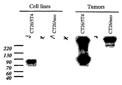
Figure 1 shows the results of Western blot analysis to assess 5T4 expression
in
tumorigenic cell lines. Western blots were generated from lysates of cultured
cells as well as
from allografted tumors in nude mice. CT26/neo, CT26 mouse colon carcinoma
cells
expressing the neomycin resistance gene; CT26/5T4, CT26 cells expressing 5T4
antigen.
Figure 2 shows the results of Western blot analysis of CT26/5T4 and CT26/neo
samples after exposure of the cell lines to biotin. Sample A is the fraction
of 5T4 that has
been biotinylated and capable of binding to avidin. Sample S is the residual
amount of 5T4
present in the supernatant after precipitation of the cell extract with
avidin. This represents a
fraction that has not been biotinylated and is therefore located in the cell
plasma. 5T4 is
detected in both membrane (A) and intracellular (S) fractions.
Figures 3A-3B show the results of experiments to quantify intracellular versus
membrane-associated 5T4 antigen. Figure 3A shows Western blots prepared using
CT26/5T4 cell extracts diluted as indicated. The biotinylated sample
represents the residual
amount of 5T4 present in the sample following depletion of the biotinylated
sample using
avidin, i.e., the amount of non-membrane-associated 5T4. The total sample
represents the
sum of the residual amount and the amount depleted by avidin, i.e., amount of
non-
membrane-associated and membrane associated 5T4 antigen. Figure 3B shows the
linear
regression curves determined by the dilution of the sample and the optical
density of the H8-
reactive band. The amount of membrane-associated 5T4 antigen is depicted as
the
difference between the optical density of the total sample and the optical
density of the
biotinylated sample after avidin depletion. As described in Example 1, the
amount of 5T4 on
the cell membrane (5T4M) was calculated to be 24% of total cellular 5T4 in
CT26/5T4 cells.
Figure 4 shows Western blot results that demonstrate 5T4 antigen on the cell
surface
of CT26/5T4 cells, DLD-1 cells (human colon carcinoma cells), N87 cells (human
gastric
carcinoma cells), PC3-MM2 cells (human prostate carcinoma cells), and PC3
cells (human
prostate carcinoma cells).
Figures 5A-5B show results of FACS analysis to detect membrane localization of
5T4
antigen. In MDAMB435/neo cells, the signal of H8 coincides with that of a
control IgG
(Figure 5A). In contrast, in MDAMB435/5T4 cells, the signal resulting from the
H8 antibody
is more than 100-fold greater than that of the control antibody, indicating
the presence of
5T4 on the cell membrane (Figure 5B). Black, detection of 5T4 antigen; gray,
detection by a
control IgG.
-8-
CA 02578131 2007-02-27
WO 2006/031653 PCT/US2005/032196
Figure
s
~how rfesults of FACS analysis to detect 5T4 antigen on the membranes of
N87 (human gastric carcinoma cells), PC14PE6 (human lung carcinoma cells), and
NCI-
H157 cells (human lung carcinoma cells). In each case, the signal resulting
from the H8
antibody is about 10-fold greater than that of the control antibody,
indicating the presence of
5T4 on the cell membrane. Gray, detection of 5T4 antigen; black, detection by
a control IgG.
Figure 7 is a line graph depicting measurements of fluorescently labeled H8
antibody
detected on the cell surface of CT26/5T4 cells and in the cell culture medium.
The mean
fluorescence of membrane-associated antibody decreased as a function of time.
The
antibody was not released in the medium. These results demonstrate that the H8
antibody/5T4 complex is internalized by CT26/5T4 cells.
Figure 8 is a line graph depicting selective cytolysis of MDAMB435/5T4 cells
exposed to an anti-5T4 conjugate comprising H8 antibody conjugated to
calicheamicin using
4-mercapto-4-methyl-pentanoic acid as a linker.
Figures 9A-9B are line graphs that depict selective cytolysis of 5T4-
expressing cells
exposed to an anti-5T4 conjugate (H8PEG2K-AcBut-CalichDMH) comprising
PEGylated H8
antibody conjugated to calicheamicin using 4-(4'-acetylphenoxy)butanoic acid
(AcBut) as a
linker. See Example 2. Figure 9A shows that MDAMB435/neo cells lacking 5T4
antigen are
approximately equally susceptible to cytolysis by H8PEG2K-AcBut-CalichDMH as
by free
calicheamicin. Figure 9B shows enhanced cytolysis of 5T4-expressing cells
exposed to
H8PEG2K-AcBut-CalichDMH as compared to free calicheamicin.
Figure 10 is a line graph that depicts growth inhibition of MDAMB435/5T4
tumors
exposed to H8-calicheamicin conjugates prepared using the indicated linkers.
PBS,
phosphate buffered saline; H8-AcBut-CalichDMH, H8 antibody conjugated to
calicheamicin
using 4-(4'-acetylphenoxy)butanoic acid (AcBut); H8-AcPac-CalichDMH, H8
antibody
conjugated to calicheamicin using 3-acetylphenyl acidic acid; H8-Amide-
CalichDMH, H8
antibody conjugated to calicheamicin using 4-mercapto-4-methyl-pentanoic acid;
H8PEG(mal2)-AcBut-CalichDMH, PEGylated H8 antibody conjugated to calicheamicin
using
4-(4'-acetylphenoxy)butanoic acid (AcBut).
Figures 11A-11B are line graphs that depict growth inhibition of MDAMB435/5T4
tumors in the presence of control substances (Figure 11A) or H8-calicheamicin
conjugates
(Figure 11 B). CMA, anti-CD33 antibody conjugated to calicheamicin (negative
control, i.e.,
used to assess cytotoxicity due to tumor uptake of a conjugate by cells
lacking the targeted
antigen); PBS, phosphate buffered saline; H8+CalichDMH, a mixture of H8
antibody and
calicheamicin (unconjugated); CalichDMH, free calicheamicin; H8-AcPac-
CalichDMH, H8
antibody conjugated to calicheamicin using 3-acetylphenyl acidic acid; H8-
amide-
CalichDMA, H8 antibody conjugated to calicheamicin using 4-mercapto-4-methyl-
pentanoic
acid.
-9-
CA 02578131 2007-02-27
WO 2006/031653 PCT/US2005/032196
7i::3r {L". ,..~.... It tfi ;i It Ifit
Figure 12 is a line graph that depicts growth inhibition of NCI-H157 tumors
exposed
to the indicated H8-calicheamicin conjugates or control substances. H8-AcPac-
CalichDMH,
H8 antibody conjugated to calicheamicin using 3-acetylphenyl acidic acid; H8-
amide-
CalichDMA, H8 antibody conjugated to calicheamicin using 4-mercapto-4-methyl-
pentanoic
acid; CMA, anti-CD33 antibody conjugated to calicheamicin (negative control);
PBS,
phosphate buffered saline; H8, unconjugated H8 antibody.
Figures 13A-13B are line graphs that depict growth inhibition of N87 tumors in
the
presence of control substances (Figure 13A) or H8-calicheamicin conjugates
(Figure 13B).
CMA, anti-CD33 antibody conjugated to calicheamicin (positive control); PBS,
phosphate
buffered saline; H8+CalichDMH, a mixture of H8 antibody and calicheamicin
(unconjugated);
CalichDMH, free calicheamicin; H8-AcPac-CalichDMH, H8 antibody conjugated to
calicheamicin using 3-acetylphenyl acidic acid; H8-amide-CalichDMA, H8
antibody
conjugated to calicheamicin using 4-mercapto-4-methyl-pentanoic acid.
Figure 14 is a line graph that depicts growth inhibition of PC14PE6 tumors
exposed
to H8/calicheamicin conjugates or control substances. H8-AcPac-CalichDMH, H8
antibody
conjugated to calicheamicin using 3-acetylphenyl acidic acid; H8-amide-
CalichDMA, H8
antibody conjugated to calicheamicin using 4-mercapto-4-methyl-pentanoic acid;
CMA, anti-
CD33 antibody conjugated to calicheamicin (negative control); PBS, phosphate
buffered
saline; H8, unconjugated H8 antibody.
Figures 15A-15G are images of normal and tumor-infested lungs of an orthotopic
model of lung cancer. Figure 15A is a picture of an excised normal mouse lung;
the heart
appears dark. Figure 15B is a picture of an excised mouse lung infested with
tumor nodules
following intravenous injection of PC14PE6 tumor cells (see Example 4); H,
heart. Figure
15C is a macroscopic image (4X magnification) showing the thorax after
collapse of the
lungs. Lung nodules (LN) are distinguishable from normal lung tissue (L). The
thoracic
cavity was filled with hemorrhagic fluid (pleural effusion, PE). Figures 15D-
15G are
photomicrographs of hematoxylin and eosin stained sections of paraffin-
embedded lung and
heart tissue, which demonstrate the extent of tumor infiltration and
destruction of normal
tissue. Figures 15D-15E show infiltrates of tumor cells in the pleural cavity
(15D) and the
pericardium (1 5E). Figures 15F-15G show the reduction of functional lung
tissue by
proliferating tumor tissue in the perialveolar space.
Figure 16 is a line graph showing the surviving fraction (%) of mice bearing
orthotopic
lung tumors that have received the indicated treatments. All treatments were
administered
intraperitoneally 6 days after injection of the PC14PE6 cells. See Example 4.
H8 (thick solid
black line), unconjugated murine H8 antibody; PBS (solid white line),
phosphate-buffered
saline; CMA 2 (thin solid black line), anti-CD33 antibody conjugated to
calicheamicin
administered at a dose of 2 g calicheamicin; CMA 4 (line with small dashes),
anti-CD33
-10-
CA 02578131 2007-02-27
WO 2006/031653 PCT/US2005/032196
il!',,;i .
antibody conjugated to calicheamicin administered at a dose of 4 g
calicheamicin; H8-
AcPac-CalichDMH 2 (line with large dashes), H8-calicheamicin conjugate
administered at a
dose of 2 g calicheamicin; H8-AcPac-CalichDMH 4 (line with large dashes), H8-
calicheamicin conjugate administered at a dose of 4 g calicheamicin. The
results for H8-
calicheacmicin conjugate administered at a dose of 2 g or at a dose of 4 g
were
indistinguishable over a period of 120 days. Ten animals were included in each
treatment
group. Each treatment regimen consisted of 3 doses administered
intraperitoneally with 4
days interval between each dose.
Figure 17 is a bar graph showing pleural volumes in mice that died from lung
tumors
following the indicated control treatments. PBS, phosphate buffered saline;
H8,
unconjugated H8 antibody; CMA 2, f anti-CD33 antibody conjugated to
calicheamicin
administered at 2 g per dose; CMA 4, anti-CD33 antibody conjugated to
calicheamicin
administered at 4 g per dose; n, number of animals. Pleural effusion volume
was not
reduced following administration of unconjugated H8 antibody or the control
conjugate CMS.
Figure 18 is an alignment of the murine H8 light chain.variable region (amino
acids
21-127 of SEQ ID NO:16) and the DPK24 germ line clone (SEQ ID NO:63). Boxed
sequences, CDRs; asterisks, positions at which amino acids of murine H8 are
maintained in
humanized H8 light chain variable region version 1, and at which amino acids
of human
DPK24 are maintained in humanized light chain variable region version 2;
underlined
residues, mutations that increase antibody expression.
Figure 19 is an alignment of human light chain variable region sequences of
subgroup Vxlll (SEQ ID NOs:65-70) and the murine H8 light chain variable
region (amino
acids 21-127 of SEQ ID NO:16). Residues that differ in human framework
sequences when
compared to H8 framework sequences are underlined. For humanization of H8, one
or more
residues at the corresponding positions in H8 is substituted with a residue of
a human
framework sequence. Boxed sequences, CDRs.
Figure 20 is an alignment of human light chain variable region sequences of
subgroup Vxl (SEQ ID NOs:71-80) and the murine H8 light chain variable region
(amino
acids 21-127 of SEQ ID NO:16). For humanization of H8, one or more residues at
the
corresponding positions in H8 is substituted with a residue of a human
framework sequence.
Boxed sequences, CDRs.
Figure 21 is an alignment of the murine H8 heavy chain variable region (amino
acids
20-139 of SEQ ID NO:14) and the DP75 germ line clone (SEQ ID NO:64). Boxed
sequences, CDRs; asterisks, positions at which amino acids of murine H8 are
maintained in
humanized H8 heavy chain variable region version 1(i.e., K38, S40, and 148),
and at which
-11-
CA 02578131 2007-02-27
WO 2006/031653 PCT/US2005/032196
amino acids of human DP75 are maintained in humanized heavy chain variable
region
version 2.
Figure 22 is an alignment of. human heavy chain variable region sequences of
subgroup I (SEQ ID NOs:52-60) and the consensus framework sequences derived
there
from (SEQ ID NO:49-51).
Figure 23 is an alignment of the murine H8 heavy chain variable region (amino
acids
20-139 of SEQ ID NO:14) and the humanized H8 heavy chain variable region
derived from
the consensus sequence of heavy chain variable region subgroup I, Le.,
humanized heavy
chain variable region version 3 (SEQ ID NO:1 9). Boxed sequences, CDRs.
Figures 24A-24C show sequences of representative light chain variable region
sequences (Figure 24A) and heavy chain variable region sequences (Figures 24B-
24C) of
humanized anti-5T4 antibodies.
Figures 25A-250 show the results of BLAST analysis performed using humanized
variable regions as query sequences. See also Tables 6 and 7.
Figures 26A-26B shows the sequences of representative human constant regions
used to prepare humanized anti-5T4 antibodies.
Figures 27A-27G show the light chain and heavy chain amino acid sequences of
representative anti-5T4 antibodies. Figure 27A shows a chimeric anti-5T4
antibody having
(a) a light chain comprising the murine H8 light chain variable region and a
human kappa
constant region (SEQ ID NO:1), and (b) a heavy chain comprising the murine H8
heavy
chain variable region and a human IgG1 constant region (SEQ ID NO:2). Figure
27B shows
a chimeric anti-5T4 antibody having (a) a light chain comprising the murine H8
light chain
variable region and a human kappa constant region (SEQ ID NO:3), and (b) a
heavy chain
comprising the murine H8 heavy chain variable region and a mutated human IgG4
constant
region (SEQ ID NO:4). Figure 27C shows a semi-human anti-5T4 antibody having
(a) a light
chain comprising the humanized H8 light chain variable region version 1 and a
human kappa
constant region (SEQ ID NO:5), and (b) a heavy chain comprising the murine H8
heavy
chain variable region and a mutated human IgG4 constant region (SEQ ID NO:6).
Figure
27D shows a humanized anti-5T4 antibody having (a) a light chain comprising
the
humanized H8 light chain variable region version 1 and a human kappa constant
region
(SEQ ID NO:7), and (b) a heavy chain comprising the humanized H8 heavy chain
variable
region version 1 and a mutated human IgG4 constant region (SEQ ID NO:8).
Figure 27E
shows a humanized anti-5T4 antibody having (a) a light chain comprising the
humanized H8
light chain variable region version 1 and a human kappa constant region (SEQ
ID NO:9),
and (b) a heavy chain comprising the humanized H8 heavy chain variable region
version 2
and a human IgG1 constant region (SEQ ID NO:10). Figure 27F shows a humanized
anti-
5T4 antibody having (a) a light chain comprising the humanized H8 light chain
variable
-12-
CA 02578131 2007-02-27
WO 2006/031653 PCT/US2005/032196
:ii
region version 2 and a human kappa constant region (SEQ ID NO:11), and (b) a
heavy chain
comprising the humanized H8 heavy chain variable region version 2 and a
mutated human
IgG4 constant region (SEQ ID NO:12). Figure 27G shows a humanized anti-5T4
antibody
having (a) a light chain comprising the humanized H8 light chain variable
region version 2
and a human kappa constant region (SEQ ID NO:11), and (b) a heavy chain
comprising the
humanized H8 heavy chain variable region version 3 and a mutated human IgG4
constant
region (SEQ ID NO:84). Single underlining, variable regions; boxed sequences,
CDRs;
asterisk, proline mutation.
Figures 28A-28B show results of FACS analysis to detect 5T4 antigen on
MDAMB435/neo cells (Figure 28A) or on MDAMB435/5T4 cells (Figure 28B) using
murine
H8, chimeric versions of H8, and humanized versions of H8 at the indicated
concentrations.
All antibodies show selective binding to MDAMB435/5T4 cells.
Figure 29 is a line graph that shows the binding properties of chimeric H8
antibody
and humanized H8 versions 1-3, which were determined using a competitive
binding assay.
The IC50 for the chimeric H8 antibody and humanized H8 versions 1-3 were 1.0 X
10"9M, 1.0
X 10'9M, 1.4 X 10"9M, and 1.5 X 10"9M, respectively. See Example 5.
Figure 30 is a line graph that shows detection of chimeric H8 antibody and
humanized H8 antibody on the cell surface of MDAMB435/5T4 cells over a period
of 25
hours. The reduced level of detection over the observation period demonstrates
internalization of both antibodies. No detectable antibody was present in the
conditioned
medium during the course, of the experiment.
Figure 31 is a bar graph depicting levels of transient expression of chimeric
H8
antibody and humanized H8 versions 1-3 in COS-1 cells. The three humanized H8
antibodies were expressed at a similar level (version 1, 4.4 mg/U48hours;
version 2, 2.7
mg/U48hours; version 3, 3.9 mg/U48hours) which was greater than that observed
for
chimeric H8 antibody (0.6 mg/U48hours). See Example 6.
Figures 32A-32B are line graphs that show inhibition of spheroid growth of
MDAMB435/neo and MDAMB435/5T4 cells in vitro following 144 hours of exposure
to H8-
AcBut-CalichDMH, (humanized H8 antibody conjugated to calicheamicin using 4-
(4'-
acetylphenoxy)butanoic acid (AcBut)) at the indicated concentrations.
Figures 33A-33C are line graphs that depict growth inhibition of N87 tumors in
the
presence of control substances (Figure 33A) or humanized H8-calicheamicin
conjugates
(Figure 33B) and response calculations (Figure 33C). PBS, phosphate buffered
saline;
huH8+CalichDMH, a mixture of H8 antibody and calicheamicin (unconjugated);
CMA, anti-
CD33 antibody conjugated to calicheamicin; CMC, anti-CD22 antibody conjugated
to
calicheamicin; huH8-AcBut-CalichDMH, humanized H8 antibody conjugated to
calicheamicin
using 4-(4'-acetylphenoxy)butanoic acid (AcBut); (4), antibody-calicheamicin
conjugate
-13-
CA 02578131 2007-02-27
WO 2006/031653 PCT/US2005/032196
~...
..., r, !f,.,fi f.,
administered at a dose of 4 g calicheamicin; (2), antibody-calicheamicin
conjugate
administered at a dose of 2 g calicheamicin; (1), antibody-calicheamicin
conjugate
administered at a dose of 1 g calicheamicin; arrows, dosing schedule on days
1, 5, and 9;
CR, complete response; PR, partial response; TR, no response; NR, no response.
See
Example 9.
Figures 34A-34C are line graphs that depict growth inhibition of MDAMB435/5T4
tumors in the presence of control substances (Figure 34A) or humanized H8-
calicheamicin
conjugates (Figure 34B) and response calculations (Figure 34C). PBS, phosphate
buffered
saline; huH8+CalichDMH, a mixture of H8 antibody and calicheamicin
(unconjugated); CMA,
anti-CD33 antibody conjugated to calicheamicin; CMC, anti-CD22 antibody
conjugated to
calicheamicin; huH8-AcBut-CalichDMH, humanized H8 antibody conjugated to
calicheamicin
using 4-(4'-acetylphenoxy)butanoic acid (AcBut); (4), antibody-calicheamicin
conjugate
administered at a dose of 4 g calicheamicin; (2), antibody-calicheamicin
conjugate
administered at a dose of 2 g calicheamicin; (1), antibody-calicheamicin
conjugate
administered at a dose of 1 g calicheamicin arrows, dosing schedule on days
1, 5, and 9;
CR, complete response; PR, partial response; TR, no response; NR, no response.
See
Example 9.
Figures 35A-35E are line graphs that depict growth inhibition of PC14PE6
tumors in
the presence of control substances (Figure 35A) or humanized H8-calicheamicin
conjugates
(Figures 35B, 35D, 35E) and calculated responses (Figure 35C). Figures 35A-35C
present
data pertaining to new growth tumors, and Figure 35D presents data pertaining
to treatment
of relapsed tumors. PBS, phosphate buffered saline; huH8+CalichDMH, a mixture
of H8
antibody and calicheamicin (unconjugated); CMA, anti-CD33 antibody conjugated
to
calicheamicin; huH8-AcBut-CalichDMH, humanized H8 antibody conjugated to
calicheamicin
using 4-(4'-acetylphenoxy)butanoic acid (AcBut); (4), antibody-calicheamicin
conjugate
administered at a dose of 4 g calicheamicin; (2), antibody-calicheamicin
conjugate
administered at a dose of 2 g calicheamicin; (1), antibody-calicheamicin
conjugate
administered at a dose of 1 g calicheamicin; (4*), antibody-calicheamicin
conjugate
administered at a dose of 4 g calicheamicin after tumors allowed to grow to
approximately
1.08 cm3 prior to treatment with the conjugate; arrows, dosing schedule on
days 1, 5, and 9
(Figures 35A, 35B, and 35D) or days 19, 23, and 27 (Figure 35C); CR, complete
response;
PR, partial response; TR, no response; NR, no response. See Example 9.
Figures 36A-36B show photographs of mice harboring PC14PE6 tumors 21 days
following treatment with vehicle (phosphate buffered saline) (Figure 367A) or
with huH8-
AcBut-CalichDMH (humanized H8 antibody conjugated to calicheamicin using 4-(4'-
acetylphenoxy)butanoic acid (AcBut)) (Figure 36B). PC14PE6 tumors were
approximately
-14-
CA 02578131 2007-02-27
WO 2006/031653 PCT/US2005/032196
8u mm at the time vehicle or humanized H8-calicheamicin conjugate were
administered.
Agents were administered by intraperitoneal injection in a total of three
doses of 4 g
calicheamicin per dose, each dose separated by three days. Arrow in Figure 36A
identifies
visible tumor. Area circumscribed by dotted line in Figure 36B identifies area
where
PC14PE6 tumor has regressed. See Example 9.
DETAILED DESCRIPTION OF THE INVENTION
1. Chimeric and Humanized Anti-5T4 Antibodies
H8 is a hybridoma-generated monoclonal mouse IgGi antibody which is described
in
PCT International Publication No. WO 98/55607 and in Forsberg et al. (1997) J.
Biol. Chem.
272(19):124430-12436. Chimeric anti-5T4 antibodies of the invention include
variable
region sequences of the murine anti-5T4 antibody and additional residues
derived from
human antibody sequences. Humanized anti-5T4 antibodies of the invention
include antigen
binding residues from mouse anti-5T4 antibody H8 and additional residues
derived from
human antibody sequences. The disclosed chimeric and humanized anti-5T4
antibodies are
therefore also called chimeric H8 antibodies and humanized H8 antibodies.
Representative
chimeric and humanized H8 antibodies are set forth in Figures 27A-27F.
The term antibody refers to an immunoglobulin protein, or antibody fragments
that
comprise an antigen binding site (e.g., Fab, modified Fab, Fab', F(ab')2 or Fv
fragments, or a
protein having at least one immunoglobulin light chain variable region or at
least one
immunoglobulin heavy chain region). Humanized antibodies of the invention
include
diabodies, tetrameric antibodies, single chain antibodies, tretravalent
antibodies,
multispecific antibodies (e.g., bispecific antibodies), domain-specific
antibodies that
recognize a particular epitope (e.g., antibodies that recognize an epitope
bound by the H8
antibody).
The term anti-5T4 antibody refers to an antibody that specifically binds to
5T4
antigen, particularly human 5T4 antigen. The 5T4 antigen is a 72 kDa non-
glycosylated
phosphoprotein found on the surface of trophoblast cells and numerous cancer
cell types
See Hole et al. (1988) Br. J. Cancer 57: 239-46, Hole et al. (1990) Int J.
Cancer 45: 179-
184; PCT International Publication No. WO89/07947; U.S. Patent No. 5,869,053.
The term binding refers to an affinity between two molecules, for example, an
antigen
and an antibody. As used herein, specific binding means a preferential binding
of an
antibody to an antigen in a heterogeneous sample comprising multiple different
antigens.
The binding of an antibody to an antigen is specific if the binding affinity
is at least about 10"7
M or higher, such as at least about 10"8 M or higher, including at least about
10"9 M or higher,
at least about 10'11 M or higher, or at least about 10'12 M or higher. For
example, specific
-15-
CA 02578131 2007-02-27
WO 2006/031653 PCT/US2005/032196
~.fl {{. iV it jt jti f';li ir3 1f,::1k
binding of an antibody of the invention to a human 5T4 antigen includes
binding in the range
of at least about 1 x 10"'to about 1 x 10"12. Specific binding of an antibody
of the invention
to a human 5T4 antigen also includes binding in the range of at least about 3
x 10'10 M to
about 12 x 10'10 M, such as within the range of about 4 x 10'10 M to about 9 x
10"10 M, or
such as within the range of about 7 x 10'10 M to about 12 x 10'10 M, or such
as within the
range of about 7 x 10"10 M to about 9 x 10"10 M, or such as within the range
of about 9 x 10'10
M to about 12 x 10"10 M, or such as within the range of about 11 x 10"10 M to
about 12 x 10"10
M, or greater binding affinities such as about 1.0 x 10"11 M to about 10 x
10"" M, or about 1.0
x 10'" M to about 5 x 10"" M, or about 5.0 x 10-" M to about 10 x 10'" M. The
phrase
specifically binds also refers to selective targeting to 5T4-expressing cells
when
administered to a subject.
The term chimeric antibody is used herein to describe an antibody comprising
sequences from at least two different species. Humanized antibodies are one
type of
chimeric antibody. A chimeric anti-5T4 antibody may comprise (a) a light chain
variable
region having an amino acid sequence of residues 1-107 of SEQ ID NO:1 and a
heavy chain
variable region having an amino acid sequence of residues 1-120 of SEQ ID
NO:2; (b) a light
chain amino acid sequence of SEQ ID NO:1 and a heavy chain amino acid sequence
of
SEQ ID NO:2; or (c) a light chain amino acid sequence of SEQ ID NO:3 and a
heavy chain
amino acid sequence of SEQ ID NO:4.
The term humanized is used herein to describe an antibody, wherein variable
region
residues responsible for antigen binding (i.e., residues of a complementarity
determining
region and any other residues that participate in antigen binding) are derived
from a non-
human species, while the remaining variable region residues (i.e., residues of
the framework
regions) and constant regions are derived, at least in part, from human
antibody sequences.
Residues of the variable regions and variable regions and constant regions of
a humanized
antibody may also be derived from non-human sources. Variable regions of a
humanized
antibody are also described as humanized (i.e., a humanized light or heavy
chain variable
region). The non-human species is typically that used for immunization with
antigen, such
as mouse, rat, rabbit, non-human primate, or other non-human mammalian
species.
Representative chimeric and humanized anti-5T4 antibodies of the invention
comprise at least one light chain or at least one heavy chain, or fragments
thereof, wherein
the chimeric or humanized anti-5T4 antibody or antibody fragment (a)
specifically binds to
human 5T4 antigen with a binding affinity of at least about 1 x 10'' M to
about 1 x 10"12 M; (b)
specifically binds to human 5T4 antigen with a binding affinity greater than 1
x 10'" M; (c)
specifically binds to human 5T4 antigen with a binding affinity greater than 5
x 10'11 M; (d)
specifically binds to human 5T4 antigen with a binding affinity greater than a
binding affinity
of murine H8 anti-5T4 antibody binding to human 5T4 antigen; (e) specifically
targets 5T4-
-16.
CA 02578131 2007-02-27
WO 2006/031653 PCT/US2005/032196
expressing cells in vivo; (f) competes for binding to human 5T4 antigen with
an antibody of
any one of (a)-(e); (g) specifically binds to an epitope bound by any one of
(a)-(e); or (h)
comprises an antigen binding domain of any one of (a)-(e).
The murine H8 anti-5T4 antibody has been shown to recognize a conformational
epitope proximal to transmembrane domain of 5T4. Glycosylation, which is
important for
structure and immunogenicity, and intramolecular disulphide bonds are required
for binding
of the antibody. It has also been shown that the H8 anti-5T4 antibody does not
bind mouse
5T4, although there is 84% identity between mouse and human 5T4 and 6 of 7 N-
linked
glycosylation sites are conserved between the two. The N-terminal and C-
terminal cysteines
are also completely conserved between mouse and human 5T4. The murine H8
antibody
has also been shown to bind human 5T4 when the N-linked glycosylation site at
amino acid
192 is removed (Shaw et al. (2002) Biochem J. 365: 137-145). There is some
evidence
suggesting that the H8 anti-5T4 antibody does not bind a human/mouse 5T4
chimera having
mouse LRR2 (residues 173-361 replacing human residues 173-355) and yet the
antibody
does bind to the reciprocal chimera. There is also evidence suggesting that
both a chimeric
H8 antibody and a humanized H8 antibody bind to a 5T4 chimera containing mouse
residues
282-361. This evidence leads to the conclusion that the H8 epitope is located
between
amino acids 173 and 252. Additional evidence suggests that chimeric H8 may not
bind to a
human/mouse anti-5T4 chimera containing mouse residues 173-258, while a
humanized H8
antibody has slight binding at higher concentrations.
Naturally occurring antibodies are tetrameric (H2L2) glycoproteins of about
150,000
daltons, composed of two identical light (L) chains and two identical heavy
(H) chains. The
two heavy chains are linked to each other by disulfide bonds and each heavy
chain is linked
to a light chain by a disulfide bond. Each of the light and heavy chains is
further
characterized by an amino-terminal variable region and a constant region. The
term variable
refers to the fact that certain portions of the variable domains differ
extensively in sequence
among antibodies and substantially determine the binding affinity and
specificity of each
particular antibody for its particular antigen. The variable regions of each
of light and heavy
chain align to form the antigen-binding domain. Representative humanized H8
variable
regions are set forth in Figures 24A-22C (SEQ ID NOs:17, 18, 19, 21, and 23).
Antibodies having a tetrameric structure, similar to naturally occurring
antibodies,
may be recombinantly prepared using standard techniques. Recombinantly
produced
antibodies also include single chain antibodies, wherein the variable regions
of a single light
chain and heavy chain pair include an antigen binding region, and fusion
proteins, wherein a
variable region of a humanized anti-5T4 antibody is fused to an effector
sequence, such as
an Fc domain, a cytokine, an immunostimulant, a cytotoxin, or any other
therapeutic protein.
See e.g., Harlow & Lane (1988) Antibodies: A Laboratory Manual, Cold Spring
Harbor
-17-
CA 02578131 2007-02-27
WO 2006/031653 PCT/US2005/032196
Laboratory Press, Cold Spring Harbor, New York and U.S. Patent Nos. 4,196,265;
4,946,778; 5,091,513; 5,132,405; 5,260,203; 5,677,427; 5,892,019; 5,985,279;
6,054,561.
Tetravalent antibodies (H4L4) comprising two intact tetrameric antibodies,
including
homodimers and heterodimers, may be prepared for example as described in PCT
International Publication No. WO 02/096948. Antibody dimers may also be
prepared via
introduction of cysteine residue(s) in the antibody constant region, which
promote interchain
disulfide bond formation, using heterobifunctional cross-linkers (Wolff et al.
(1993) Cancer
Res. 53: 2560-5), or by recombinant production to include a dual constant
region (Stevenson
et al. (1989) Anticancer Drug Des. 3: 219-30).
The term complementarity determining region or CDR refers to residues of the
antibody variable regions that participate in antigen binding. A number of
definitions of the
CDRs are in common use. The Kabat definition is based on sequence variability,
and the
Chothia definition is based on the location of the structural loop regions.
The AbM definition
is a compromise between the Kabat and Chothia approaches. The CDRs of the
light chain
variable region are bounded by the residues at positions 24 and 34 (CDR1-L),
50 and 56
(CDR2-L), and 89 and 97 (CDR3-L) according to the Kabat, Chothia, or AbM
algorithm.
According to the Kabat definition, the CDRs of the heavy chain variable region
are bounded
by the residues at positions 31 and 35B (CDR1-H), 50 and 65 (CDR2-H), and 95
and 102
(CDR3-H) (numbering according to Kabat). According to the Chothia definition,
the CDRs of
the heavy chain variable region are bounded by the residues at positions 26
and 32 (CDR1-
H), 52 and 56 (CDR2-H), and 95 and 102 (CDR3-H) (numbering according to
Chothia).
According to the AbM definition, the CDRs of the heavy chain variable region
are bounded
by the residues at positions 26 and 35B (CDR1-H), 50 and 58 (CDR2-H), and 95
and 102
(CDR3-H) (numbering according to Kabat). See Martin et al. (1989) Proc. Natl.
Acad. Sci.
USA 86: 9268-9272; Martin et al. (1991) Methods Enzymol. 203: 121-153;
Pedersen et al.
(1992) Immunomethods 1: 126; and Rees et al. (1996) In Sternberg M.J.E. (ed.),
Protein
Structure Prediction, Oxford University Press, Oxford, pp. 141-172.
The term specificity determining region or SDR refers to those residues within
CDRs
that directly interact with antigen, which correspond to hypervariable
residues. See (Padlan
et al. (1995) FASEB J. 9: 133-9).
Framework residues are those residues of the variable region other than
hypervariable residues. Representative human frameworks of a heavy chain
variable region
that may be used to prepare humanized anti-5T4 antibodies include the
framework regions
of DP-75 and DP-8(VH1-2), DP-25, VI-2b and VI-3 (VH1-03), DP-15 and V1-8 (VH1-
08), DP-
14 and V1-18 (VH1-18), DP-5 and V1-24P (VH1-24), DP-4 (VH1-45), DP-7 (VH1-46),
DP-
10, DA-6 and YAC-7 (VH1-69), DP-88 (VH1-e), DP-3, and DA-8 (VH1-f). Consensus
framework sequences based on the foregoing individual sequences may also be
used. See
-18-
CA 02578131 2007-02-27
WO 2006/031653 PCT/US2005/032196
ti"ifi
Figures 21-23. Representative human frameworks of a light chain variable
region include
that of human germ line clone DPK24 and germ line clone subgroups Vtclll and
Vxl, each of
which shows greater than 60% amino acid identity when compared to the H8 light
chain
variable region. See Figures 18-20.
The constant regions.of the disclosed humanized anti-5T4 antibodies are
derived
from constant regions from any one of IgA, IgD, IgE, IgG, IgM, and any
isotypes thereof
(e.g., IgG1, IgG2, IgG3, or IgG4 isotypes of IgG). The choice of the human
isotype (IgGi,
IgG2, IgG3, IgG4) and modification of particular amino acids in the human
isotype may
enhance or eliminate activation of host defense mechanisms and alter
biodistribution of a
humanized antibody of the invention. See (Reff et al. (2002) Cancer Control9:
152-66).
Humanized antibodies may be prepared using any one of a variety of methods
including veneering, grafting of complementarity determining regions (CDRs),
grafting of
abbreviated CDRs, grafting of specificity determining regions (SDRs), and
Frankenstein
assembly, as described below. These general approaches may be combined with
standard
mutagenesis and synthesis techniques to produce an anti-5T4 antibody of any
desired
sequence.
Veneering is based on the concept of reducing potentially immunogenic amino
acid
sequences in a rodent or other non-human antibody by resurfacing the solvent
accessible
exterior of the antibody with human amino acid sequences. Thus, veneered
antibodies
appear less foreign to human cells. See Padian (1991) Mol. Immunol. 28:489-98.
A non-
human antibody is veneered by (1) identifying exposed exterior framework
region residues in
the non-human antibody, which are different from those at the same positions
in framework
regions of a human antibody, and (2) replacing the identified residues with
amino acids that
typically occupy these same positions in human antibodies.
Grafting of CDRs is performed by replacing one or more CDRs of an acceptor
antibody (e.g., a human antibody) with CDRs of a donor antibody (e.g., a non-
human
antibody). Acceptor antibodies may be selected based on similarity of
framework residues
between a candidate acceptor antibody and a donor antibody and may be further
modified to
introduce similar residues. For example, a human acceptor framework may
comprise a
heavy chain variable region of a human sub-group I consensus sequence,
optionally with
non-human donor residues at one or more of positions 1, 28, 48, 67, 69, 71,
and 93. As
another example, a human acceptor framework may comprise a light chain
variable region of
a human sub-group I consensus sequence, optionally with non-human donor
residues at one
or more of positions 2, 3, 4, 37, 38, 45 and 60. Following CDR grafting,
additional changes
may be made in the donor and/or acceptor sequences to optimize antibody
binding and
functionality. See e.g., PCT International Publication No. WO 91/09967.
-19-
CA 02578131 2007-02-27
WO 2006/031653 PCT/US2005/032196
P' :;õ I..... jf1t i! ;;t it ii T;,i; ii: 'f "If..
~"rafting of abbreviated CDRs is a related approach. Abbreviated CDRs include
the
specificity-determining residues and adjacent amino acids, including those at
positions 27d-
34, 50-55 and 89-96 in the light chain, and at positions 31-35b, 50-58, and 95-
101 in the
heavy chain (numbering convention of (Kabat et al. (1987)). See (Padian et al.
(1995)
FASEB J. 9: 133-9). Grafting of specificity-determining residues (SDRs) is
premised on the
understanding that the binding specificity and affinity of an antibody
combining site is
determined by the most highly variable residues within each of the
complementarity
determining regions (CDRs). Analysis of the three-dimensional structures of
antibody-
antigen complexes, combined with analysis of the available amino acid sequence
data was
used to model sequence variability based on structural dissimilarity of amino
acid residues
that occur at each position within the CDR. See Padlan et al. (1995) FASEB J.
9: 133-139.
Minimally immunogenic polypeptide sequences consisting of contact residues,
which are
referred to as specificity-determining residues (SDRs), are identified and
grafted onto human
framework regions.
According to the Frankenstein approach, human framework regions are identified
as
having substantial sequence homology to each framework region of the relevant
non-human
antibody, and CDRs of the non-human antibody are grafted onto the composite of
the
different human framework regions. A related method also useful for
preparation of
antibodies of the invention is described in U.S. Patent Application
Publication No.
2003/0040606.
Humanized anti-5T4 antibodies disclosed herein typically comprise at least one
humanized light chain variable region or heavy chain variable region. Thus, a
humanized
anti-5T4 antibody of the invention may comprise a light chain variable region
prepared by
veneering, grafting of abbreviated CDRs or SDRs, or Frankenstein assembly, as
above, and
a heavy chain variable region of a non-human antibody (e.g., -the H8 antibody
or other non-
human anti-5T4 antibody). Alternatively, a light chain variable region of a
non-human
antibody may be combined with a humanized heavy chain variable region.
Representative humanized anti-5T4 antibodies of the invention include (a)
antibodies
having one or more CDRs of a non-human anti-5T4 antibody selected from CDRs of
the light
chain variable region of SEQ ID NO:17 or the heavy chain variable region of
SEQ ID NO:18,
such as two or more CDRs selected from CDRs of the light chain variable region
of SEQ ID
NO:17 or the heavy chain variable region of SEQ ID NO:18; (b) antibodies
having a light
chain comprising a variable region having two or three CDRs of SEQ ID NO:17;
and (c)
antibodies having a heavy chain comprising a variable region having two or
three CDRs of
SEQ ID NO:18. Representative humanized anti-5T4 antibodies of the invention
also include
those antibodies having (a) a light chain variable region amino acid sequence
set forth as
SEQ ID NO:17 or 23; (b) a light chain variable region amino acid sequence that
is at least
-20-
CA 02578131 2007-02-27
WO 2006/031653 PCT/US2005/032196
If ;:it
78% identical to SEQ ID NO:17; or (c) a light chain variable region amino acid
sequence that
is at least 81% identical to SEQ ID NO:23. A light chain variable region of a
functional
humanized anti-5T4 antibody (i.e., an anti-5T4 antibody that specifically
binds to 5T4
antigen) may be encoded by (a) a nucleic acid of SEQ ID NO:22 or SEQ ID NO:81;
(b) a
nucleic acid that is at least 90% identical to the nucleic acid of SEQ ID
NO:22; (c) a nucleic
acid that is at least 91% identical to the nucleic acid of SEQ ID NO:81; or
(d) a nucleic acid
that specifically hybridizes to the complement of SEQ ID NO:22 or SEQ ID NO:81
under
stringent hybridization conditions, for example final wash conditions of 0.1 X
SSC at 65 C.
Representative humanized anti-5T4 antibodies of the invention further include
those
antibodies having (a) a heavy chain variable region amino acid sequence set
forth as any
one of SEQ ID NOs:18, 19, and 21; (b) a heavy chain variable region amino acid
sequence
that is at least 83% identical to SEQ ID NO:18; (c) a heavy chain variable
region amino acid
sequence that is at least 81% identical to SEQ ID NO:19; or (d) a heavy chain
variable
region amino acid sequence that is at least 86% identical to SEQ ID NO:21. A
heavy chain
variable region of a functional humanized anti-5T4 antibody (1.e., an anti-5T4
antibody that
specifically binds to 5T4 antigen) may be encoded by (a) a nucleic acid of any
one of SEQ
ID NOs:20, 82, and 83; (b) a nucleic acid that is at least 91 % identical to
the nucleic acid of
SEQ ID NO:20; (c) a nucleic acid that is at least 94% identical to the nucleic
acid of SEQ ID
NO:82; (d) a nucleic acid that is at least 91% identical to the nucleic acid
of SEQ ID NO:83;
or (e) a nucleic acid that specifically hybridizes to the complement of any
one of SEQ ID
NOs:20, 82, and 83 under stringent hybridization conditions, for example final
wash
conditions of 0.1'X SSC at 65 C.
A humanized anti-5T4 antibody may comprise (a) a light chain variable region
having
an amino acid sequence of residues 1-107 of SEQ ID NO:5 and a heavy chain
variable
region having an amino acid sequence of residues 1-120 of SEQ ID NO:6; (b) a
light chain
amino acid sequence of SEQ ID NO:5 and a heavy chain amino acid sequence of
SEQ ID
NO:6; (c) a light chain variable region having an amino acid sequence of
residues 1-107 of
SEQ ID NO:7 and a heavy chain variable region having an amino acid sequence of
residues
1-120 of SEQ ID NO:8; (d) a light chain amino acid sequence of SEQ ID NO:7 and
a heavy
chain amino acid sequence of SEQ ID NO:8; (e) a light chain variable region
having an
amino acid sequence of residues 1-107 of SEQ ID NO:9 and a heavy chain
variable region
having an amino acid sequence of residues 1-120 of SEQ ID NO:10; (f) a light
chain amino
acid sequence of SEQ ID NO:9 and a heavy chain amino acid sequence of SEQ ID
NO:10;
(g) a light chain variable region having an amino acid sequence of residues 1-
107 of SEQ ID
NO:11 and a heavy chain variable region having an amino acid sequence of
residues 1-120
of SEQ ID NO:12; or (h) a light chain amino acid sequence of SEQ ID NO:11 and
a heavy
chain amino acid sequence of SEQ ID NO:12.
-21-
CA 02578131 2007-02-27
WO 2006/031653 PCT/US2005/032196
i
il ; ~ ,.,.,~f .,= ,,,..r
umanized anti-5T4 antibodies of the invention may be constructed wherein the
variable region of a first chain (i.e., the light chain variable region or the
heavy chain variable
region) is humanized, and wherein the variable region of the second chain is
not humanized
(i.e., a variable region of an antibody produced in a non-human species).
These antibodies
are referred to herein as semi-humanized antibodies. For example, an anti-5T4
antibody
may comprise a humanized light chain= variable region of SEQ ID NO:17 or 23,
and a heavy
chain variable region of a non-human anti-5T4 antibody, such as the murine H8
heavy chain
variable region of SEQ ID NO:14. Alternatively, an anti-5T4 antibody may
comprise a
humanized light chain variable region of a non-human anti-5T4 antibody, such
as the murine
H8 light chain variable region of SEQ ID NO:16, and a humanized heavy chain
variable
region of any one of SEQ ID NOs:18, 19, or 21. Anti-5T4 non-human antibodies
other than
murine H8 may be used to prepare semi-humanized antibodies, for example the
rat
monoclonal antibody described by Woods et al. (2002) Biochem. J. 366: 353-65).
Variants of the disclosed humanized anti-5T4 antibodies may be readily
prepared to
include various changes, substitutions, insertions, and deletions, where such
changes
provide for advantages in use. For example, to increase the serum half life of
the antibody,
a salvage receptor binding epitope may be incorporated, if not present
already, into the
antibody heavy chain sequence. See U.S. Patent No. 5,739,277. Additional
modifications to
enhance antibody stability include modification of IgG4 to replace the serine
at residue 241
with proline. See Angal et al. (1993) Mol. lmmunol. 30: 105-108. Other useful
changes
include substitutions as required to optimize efficiency in conjugating the
antibody with a
drug. For example, an antibody may be modified at its carboxyl terminus to
include amino
acids for drug attachment, for example one or more cysteine residues may be
added. The
constant regions may be modified to introduce sites for binding of
carbohydrates or other
moieties.
Variants of humanized anti-5T4 antibodies of the invention may be produced
using
standard recombinant techniques, including site-directed mutagenesis, or
recombination
methods. A diversified repertoire of humanized anti-5T4 antibodies may be
prepared via
gene arrangement and gene conversion methods in transgenic non-human animals
(U.S.
Patent Publication No. 2003/0017534), which are then tested for relevant
activities using
functional assays. In particular embodiments of the invention, anti-5T4
variants are obtained
using an affinity maturation protocol such as mutating the CDRs (Yang et al.
(1995) J. Mol.
Biol. 254: 392-403), chain shuffling (Marks et al. (1992) Biotechnology (NY)
10: 779-783),
use of mutator strains of E. coli (Low et al. (1996) J. Mol. Biol. 260: 359-
368), DNA shuffling
(Patten et al. (1997) Curr. Opin. Biotechnol. 8: 724-733), phage display
(Thompson et al.
(1996) J. Mol. Biol. 256: 77-88), and sexual PCR (Crameri et al. (1998) Nature
391: 288-
291). For immunotherapy applications, relevant functional assays include
specific binding to
-22-
CA 02578131 2007-02-27
WO 2006/031653 PCT/US2005/032196
i~f4'~ antigeni, internalization of the antibody when conjugated to a
cytotoxin, and
targeting to a tumor site(s) when administered to a tumor-bearing animal, as
described in the
Examples. See Examples 1-11.
The present invention further provides cells and cell lines expressing
humanized anti-
5T4 antibodies of the invention. Representative host cells include mammalian
and human
cells, such as CHO cells, HEK-293 cells, HeLa cells, CV-1 cells, and COS
cells. Methods
for generating a stable cell line following transformation of a heterologous
construct into a
host cell are known in the art. Representative non-mammalian host cells
include insect cells
(Potter et al. (1993) Int Rev. Immunol. 10(2-3):103-112). Antibodies may also
be produced
in transgenic animals (Houdebine (2002) Curr. Opin. Biotechnol. 13(6):625-629)
and
transgenic plants (Schillberg et al. (2003) Cell Mol. Life Sci. 60(3):433-45).
I.A. Chimeric and Humanized Anti-5T4 Nucleic Acids
The present invention further provides isolated nucleic acids encoding
humanized
anti-5T4 light and heavy chain variable regions. The isolated nucleic acids
may be used to
prepare a humanized anti-5T4 antibody, as disclosed herein.
The terms nucleic acid molecule and nucleic acid each refer to
deoxyribonucleotides
or ribonucleotides and polymers thereof in single-stranded, double-stranded,
or triplexed
form. Unless specifically limited, the term encompasses nucleic acids
containing known
analogues of natural nucleotides that have similar properties as the reference
natural nucleic
acid. The terms nucleic acid molecule or nucleic acid may also be used in
place of gene,
cDNA, mRNA, or cRNA. Nucleic acids may be synthesized, or may be derived from
any
biological source, including any organism. Representative methods for cloning
nucleic acids
that encode humanized anti-5T4 antibodies are described in Example 5.
Representative nucleic acids of the invention comprise the nucleotide sequence
of
any one of SEQ ID NOs:20, 22, 81, 82, or 83. Nucleic acids of the invention
may also
comprise a nucleotide sequence that is substantially identical to any one of
SEQ ID NOs:20,
22, 81, 82, or 83, for example, at least 91% identical to any one of SEQ ID
NOs:20, 81, or
83, or at least 90% identical to SEQ ID NO:22, or at least 94% identical to
SEQ ID NO:82.
Sequences are compared for maximum correspondence using a sequence comparison
algorithm using the full-length sequence of any one of SEQ ID NOs:20, 22, 81,
82, or 83 as
the query sequence, as described herein below, or by visual inspection. See
also Example
and Table 6.
With respect to substantially identical nucleic acids having a specified
minimal
percentage identity to the disclosed humanized H8 variable region nucleic
acids, the
substantially identical sequences may also be at least about 92% identical to
SEQ ID NO:20.
81, or 83, such as at least 93% identical, or at least 94% identical, or at
least 95% identical,
-23-
CA 02578131 2007-02-27
WO 2006/031653 PCT/US2005/032196
ii:;;i1
or at least 96% identical, or at least 97% identical, or at least 98%
identical, or at least 99%
identical. Similarly, substantially identical sequences also include sequences
that are at
least about 91% identical to SEQ ID NO:22, for example, at least about 92%
identical, or at
least 93% identical, or at least 94% identical, or at least 95% identical, or
at least 96%
identical, or at least 97% identical, or at least 98% identical, or at least
99% identical; and
sequences that are at least 95% identical to SEQ ID NO:82, such as at least
96% identical,
or at least 97% identical, or at least 98% identical, or at least 99%
identical.
Substantially identical sequences may be polymorphic sequences. The term
polymorphic refers to the occurrence of two or more genetically determined
alternative
sequences or alleles in a population. An allelic difference may be as small as
one base pair.
Substantially identical sequences may also comprise mutagenized sequences,
including sequences comprising silent mutations. A mutation may comprise one
or more
residue changes, a deletion of one or more residues, or an insertion of one or
more
additional residues.
Substantially identical nucleic acids are also identified as nucleic acids
that hybridize
specifically to or hybridize substantially to the full length of any one of
SEQ ID NOs:20, 22,
81, 82, and 83 under stringent conditions. In the context of nucleic acid
hybridization, two
nucleic acid sequences being compared may be designated a probe and a target.
A probe
is a reference nucleic acid molecule, and a target is a test nucleic acid
molecule, often found
within a heterogeneous population of nucleic acid molecules. A target sequence
is
synonymous with a test sequence.
A preferred nucleotide sequence employed for hybridization studies or assays
includes probe sequences that are complementary to or mimic at least an about
14 to 40
nucleotide sequence of a nucleic acid molecule of the present invention.
Preferably, probes
comprise 14 to 20 nucleotides, or even longer where desired, such as 30, 40,
50, 60, 100,
200, 300, or 500 nucleotides or up to the full length of any one of SEQ ID
NOs:20, 22, 81,
82, and 83. Such fragments may be readily prepared, for example by chemical
synthesis of
the fragment, by application of nucleic acid amplification technology, or by
introducing
selected sequences into recombinant vectors for recombinant production.
The phrase hybridizing specifically to refers to the binding, duplexing, or
hybridizing
of a molecule only to a particular nucleotide sequence under stringent
conditions when that
sequence is present in a complex nucleic acid mixture (e.g., total cellular
DNA or RNA).
The phrase hybridizing substantially to refers to complementary hybridization
between a probe nucleic acid molecule and a target nucleic acid molecule and
embraces
minor mismatches that may be accommodated by reducing the stringency of the
hybridization media to achieve the desired hybridization.
-24-
CA 02578131 2007-02-27
WO 2006/031653 PCT/US2005/032196
Stringent hybridization conditions and stringent hybridization wash conditions
in the
context of nucleic acid hybridization experiments such as Southern and
Northern blot
analysis are both sequence- and environment-dependent. Longer sequences
hybridize
specifically at higher temperatures. An extensive guide to the hybridization
of nucleic acids
is found in Tijssen (1993) Laboratory Techniaues in Biochemistry and Molecular
Biolocil[-
Hybridization with Nucleic Acid Probes, part I chapter 2, Elsevier, New York,
New York.
Generally, highly stringent hybridization and wash conditions are selected to
be about 5 C
lower than the thermal melting point (Tm) for the specific sequence at a
defined ionic strength
and pH. Typically, under stringent conditions a probe will hybridize
specifically to its target
subsequence, but to no other sequences.
The Tm is the temperature (under defined ionic strength and pH) at which 50%
of the
target sequence hybridizes to a perfectly matched probe. Very stringent
conditions are
selected to be equal to the Tm for a particular probe. An example of stringent
hybridization
conditions for Southern or Northern Blot analysis of complementary nucleic
acids having
more than about 100 complementary residues is overnight hybridization in 50%
formamide
with 1 mg of heparin at 42 C. An example of highly stringent wash conditions
is 15 minutes
in 0.1X SSC at 65 C. An example of stringent wash conditions is 15 minutes in
0.2X SSC
buffer at 65 C. See Sambrook et al., eds (1989) Molecular Cloninca: A
Laboratory Manual,
Cold Spring Harbor Laboratory Press, Cold Spring Harbor, New York, for a
description of
SSC buffer. Often, a high stringency wash is preceded by a low stringency wash
to remove
background probe signal. An example of medium stringency wash conditions for a
duplex of
more than about 100 nucleotides, is 15 minutes in 1X SSC at 45 C. An example
of low
stringency wash for a duplex of more than about 100 nucleotides, is 15 minutes
in 4X to 6X
SSC at 40 C. For short probes (e.g., about 10 to 50 nucleotides), stringent
conditions
typically involve salt concentrations of less than about 1 M Na+ ion,
typically about 0.01 to 1 M
Na+ ion concentration (or other salts) at pH 7.0-8.3, and the temperature is
typically at least
about 30 C. Stringent conditions may also be achieved with the addition of
destabilizing
agents such as formamide. In general, a signal to noise ratio of 2-fold (or
higher) than that
observed for an unrelated probe in the particular hybridization assay
indicates detection of a
specific hybridization.
The following are examples of hybridization and wash conditions that may be
used to
identify nucleotide sequences that are substantially identical to reference
nucleotide
sequences of the present invention: a probe nucleotide sequence preferably
hybridizes to a
target nucleotide sequence in 7% sodium dodecyl sulphate (SDS), 0.5M NaPO4,
1mM EDTA
at 50 C followed by washing in 2X SSC, 0.1% SDS at 50 C; more preferably, a
probe and
target sequence hybridize in 7% sodium dodecyl sulphate (SDS), 0.5M NaPO4, 1mM
EDTA
-25-
CA 02578131 2007-02-27
WO 2006/031653 PCT/US2005/032196
pt ;,,,. i f .. ,:; f.,'~::il(
at 5C followed by washing in 1X SSC, 0.1% SDS at 50 C; more preferably, a
probe and
target sequence hybridize in 7% sodium dodecyl sulphate (SDS), 0.5M NaPO4, 1mM
EDTA
at 50 C followed by washing in 0.5X SSC, 0.1% SDS at 50 C; more preferably, a
probe and
target sequence hybridize in 7% sodium dodecyl sulphate (SDS), 0.5M NaPO4, 1mM
EDTA
at 50 C followed by washing in 0.1 X SSC, 0.1 % SDS at 50 C; more preferably,
a probe and
target sequence hybridize in 7% sodium dodecyl sulphate (SDS), 0.5M NaPO4, 1mM
EDTA
at 50 C followed by washing in 0.1 X SSC, 0.1% SDS at 65 C.
A further indication that two nucleic acid sequences are substantially
identical is that
proteins encoded by the nucleic acids are substantially identical, share an
overall three-
dimensional structure, or are biologically functional equivalents. These terms
are defined
further herein below. Nucleic acid molecules that do not hybridize to each
other under
stringent conditions are still substantially identical if the corresponding
proteins are
substantially identical. This may occur, for example, when two nucleotide
sequences
comprise conservatively substituted variants as permitted by the genetic code.
The term conservatively substituted variants refers to nucleic acid sequences
having
degenerate codon substitutions wherein the third position of one or more
selected (or all)
codons is substituted with mixed-base and/or deoxyinosine residues. See Batzer
et al.
(1991) Nucleic Acids Res. 19:5081; Ohtsuka et al. (1985) J. Biol. Chem.
260:2605-2608;
and Rossolini et al. (1994) Mol. Ce/l Probes 8:91-98.
Nucleic acids of the invention also comprise nucleic acids complementary to
any one
of SEQ ID NOs:20, 22, 81, 82, and 83, and subsequences and elongated sequences
of the
nucleic acids and complementary nucleic acids of any one of SEQ ID NOs:20, 22,
81, 82,
and 83.
The term complementary sequences, as used herein, indicates two nucleotide
sequences that comprise antiparallel nucleotide sequences capable of pairing
with one
another upon formation of hydrogen bonds between base pairs. As used herein,
the term
complementary sequences means nucleotide sequences which are substantially
complementary, as may be assessed by the same nucleotide comparison methods
set forth
below, or is defined as being capable of hybridizing to the nucleic acid
segment in question
under relatively stringent conditions such as those described herein. A
particular example of
a complementary nucleic acid segment is an antisense oligonucleotide.
The term subsequence refers to a sequence of nucleic acids that comprises a
part of
a longer nucleic acid sequence. An exemplary subsequence is a probe, described
herein
above, or a primer. The term primer as used herein refers to a contiguous
sequence
comprising about 8 or more deoxyribonucleotides or ribonucleotides, preferably
10-20
nucleotides, and more preferably 20-30 nucleotides of a selected nucleic acid
molecule. The
-26-
CA 02578131 2007-02-27
WO 2006/031653 PCT/US2005/032196
primers of the invention encompass oligonucleotides of sufficient length and
appropriate
sequence so as to provide initiation of polymerization on a nucleic acid
molecule of the
present invention.
The term elongated sequence refers to an addition of nucleotides (or other
analogous molecules) incorporated into the nucleic acid. For example, a
polymerase (e.g., a
DNA polymerase) may add sequences at the 3' terminus of the nucleic acid
molecule. In
addition, the nucleotide sequence may be combined with other DNA sequences,
such as
promoters, promoter regions, enhancers, polyadenylation signals, intronic
sequences,
additional restriction enzyme sites, multiple cloning sites, and other coding
segments. Thus,
the invention also provides vectors comprising the disclosed nucleic acids,
including vectors
for recombinant expression, wherein a nucleic acid of the invention is
operatively linked to a
functional promoter.
The term operatively linked, as used herein, refers to a functional
combination
between a promoter region and a nucleotide sequence such that the
transcription of the
nucleotide sequence is controlled and regulated by the promoter region.
Techniques for
operatively linking a promoter region to a nucleotide sequence are known in
the art.
The term vector is used herein to refer to a nucleic acid molecule having
nucleotide
sequences that enable its replication in a host cell. A vector may also
include nucleotide
sequences to permit ligation of nucleotide sequences within the vector,
wherein such
nucleotide sequences are also replicated in a host cell. Representative
vectors include
plasmids, cosmids, and viral vectors.
Nucleic acids of the present invention may be cloned, synthesized, altered,
mutagenized, or combinations thereof. Standard recombinant DNA and molecular
cloning
techniques used to isolate nucleic acids are known in the art. Site-specific
mutagenesis to
create base pair changes, deletions, or small insertions are also known in the
art. See e.g.,
Sambrook et al. (eds.) (1989) Molecular Cloning: A Laboratory Manual. Cold
Spring Harbor
Laboratory Press, Cold Spring Harbor, New York; Silhavy et al. (1984)
Experiments with
Gene Fusions. Cold Spring Harbor Laboratory Press, Cold Spring Harbor, New
York; Glover
& Hames (1995) DNA Clonina: A Practical Approach, 2nd ed. IRL Press at Oxford
University
Press, Oxford/New York; Ausubel (ed.) (1995) Short Protocols in Molecular
Biology, 3rd ed.
Wiley, New York.
I.B. Chimeric and Humanized Anti-5T4 Polypeptides
The present invention also provides isolated humanized anti-5T4 polypeptides.
Representative light chain and heavy chain polypeptides of the invention are
set forth as
SEQ ID NOs:1-12. Representative light chain variable region polypeptide and
heavy chain
-27-
CA 02578131 2007-02-27
WO 2006/031653 PCT/US2005/032196
variable region polypeptides are set forth as SEQ ID NOs:17 and 23 and SEQ ID
NOs:18,
19, and 21, respectively.
The terms polypeptide and protein each refer to a compound made up of a single
chain of amino acids joined by peptide bonds. The antibodies of the invention
are alternately
referred to as polypeptides or proteins. Polypeptides of the invention may
comprise naturally
occurring amino acids, synthetic amino acids, genetically encoded amino acids,
non-
genetically encoded amino acids, and combinations thereof. Polypeptides may
include both
L-form and D-form amino acids.
Representative non-genetically encoded amino acids include but are not limited
to 2-
aminoadipic acid; 3-aminoadipic acid; (i-aminopropionic acid; 2-aminobutyric
acid; 4-
aminobutyric acid (piperidinic acid); 6-aminocaproic acid; 2-aminoheptanoic
acid; 2-
aminoisobutyric acid; 3-aminoisobutyric acid; 2-aminopimelic acid; 2,4-
diaminobutyric acid;
desmosine; 2,2'-diaminopimelic acid; 2,3-diaminopropionic acid; N-
ethylglycine; N-
ethylasparagine; hydroxylysine; allo-hydroxylysine; 3-hydroxyproline; 4-
hydroxyproline;
isodesmosine; allo-isoleucine; N-methylglycine (sarcosine); N-
methylisoleucine; N-
methylvaline; norvaline; norieucine; and ornithine.
Representative derivatized amino acids include, for example, those molecules
in
which free amino groups have been derivatized to form amine hydrochlorides, p-
toluene
sulfonyl groups, carbobenzoxy groups, t-butyloxycarbonyl groups, chloroacetyl
groups or
formyl groups. Free carboxyl groups may be derivatized to form salts, methyl
and ethyl
esters or other types of esters or hydrazides. Free hydroxyl groups may be
derivatized to
form 0-acyl or 0-alkyl derivatives. The imidazole nitrogen of histidine may be
derivatized to
form N-im-benzylhistidine.
The present invention also provides functional fragments of a humanized anti-
5T4
polypeptide, for example, a variable region polypeptide. Functional
polypeptide sequences
that are longer than the disclosed sequences are also provided. For example,
one or more
amino acids may be added to the N-terminus or C-terminus of an antibody
polypeptide.
Such additional amino acids may be employed in a variety of applications,
including but not
limited to purification applications. Methods of preparing elongated proteins
are known in
the art.
Polypeptides of the invention include (a) a light chain variable-region
polypeptide
having the amino acid sequence of SEQ ID NO:17 or 23; (b) a light chain
variable region
polypeptide having an amino acid sequence that is at least 78% identical to
SEQ ID NO:17;
and (c) a light chain variable region polypeptide having an amino acid
sequence at least
81% identical to SEQ ID NO:23. Additional polypeptides of the invention
include (a) a heavy
chain variable region polypeptide having the amino acid sequence set forth as
any one of
SEQ ID NOs:18, 19, and 21; (b) a heavy chain variable region polypeptide
having an amino
-28-
CA 02578131 2007-02-27
WO 2006/031653 PCT/US2005/032196
acid sequence that is at least 83% identical to SEQ ID NO:18; (c) a heavy
chain variable
region polypeptide having an amino acid sequence that is at least 81%
identical to SEQ ID
NO:19; and (d) a heavy chain variable region polypeptide having an amino acid
sequence
that is at least 86% identical to SEQ ID NO:21. Sequences are compared for
maximum
correspondence using a sequence comparison algorithm using the full-length
sequence of
any one of SEQ ID NO:17, 18, 19, 21, or 23 as the query sequence, as described
herein
below, or by visual inspection. See also Example 5.
With respect to substantially identical polypeptides having a specified
minimal
percentage identity to the disclosed humanized H8 variable region
polypeptides,
substantially identical polypeptides may also be at least about 87% identical
to the amino
acid sequence of any one of SEQ ID NO:17, 18, 19, 21, or 23, such as at least
88%
identical, or at least 89% identical, or at least 90% identical, or at least
91% identical, or at
least 92% identical, or at least 93% identical, or at least 94% identical, or
at least 95%
identical, or at least 96% identical, or at least 97% identical, or at least
98% identical, or at
least 99% identical. The invention further encompasses polypeptides encoded by
any one
of the nucleic acids disclosed herein.
Substantially identical proteins also include proteins comprising amino acids
that are
conservatively substituted variants of any one of the disclosed humanized
variable region
polypeptides and variable region antibodies. The term conservatively
substituted variant
refers to a polypeptide comprising an amino acid in which one or more residues
have been
conservatively substituted with a functionally similar residue and which
specifically binds to
human anti-5T4 with similar affinity as any of the disclosed chimeric and
humanized H8
antibodies. The phrase conservatively substituted variant also includes
peptides wherein a
residue is replaced with a chemically derivatized residue.
Examples of conservative substitutions include the substitution of one non-
polar
(hydrophobic) residue such as isoleucine, valine, leucine or methionine for
another; the
substitution of one polar (hydrophilic) residue for another such as between
arginine and
lysine, between glutamine and asparagine, between glycine and serine; the
substitution of
one basic residue such as lysine, arginine or histidine for another; or the
substitution of one
acidic residue, such as aspartic acid or glutamic acid for another.
Isolated polypeptides of the invention may be purified and characterized using
a
variety of standard techniques that are known to the skilled artisan. See
e.g., Schroder &
Lubke (1965) The Peptides. Academic Press, New York; Bodanszky (1993)
Principies of
Peptide Synthesis, 2nd rev. ed. Springer-Verlag, Berlin/ New York; Ausubel
(ed.) (1995)
Short Protocols in Molecular Biology, 3rd ed. Wiley, New York.
-29-
CA 02578131 2007-02-27
WO 2006/031653 PCT/US2005/032196
iY.:L, ,.,II,. 1111 1I::31
I.C. Nucleotide and Amino Acid Sequence Comparisons
The terms identical or percent identity in the context of two or more
nucleotide or
polypeptide sequences, refer to two or more sequences or subsequences that are
the same
or have a specified percentage of amino acid residues or nucleotides that are
the same,
when compared and aligned for maximum correspondence, as measured using one of
the
sequence comparison algorithms disclosed herein or by visual inspection.
The term substantially identical in regards to a nucleotide or polypeptide
sequence
means that a particular sequence varies from the sequence of a naturally
occurring
sequence by one or more deletions, substitutions, or additions, the net effect
of which is to
retain biological function of a humanized anti-5T4 nucleic acid or
polypeptide.
For comparison of two or more sequences, typically one sequence acts as a
reference sequence to which one or more test sequences are compared. When
using a
sequence comparison algorithm, test and reference sequences are entered into a
computer
program, subsequence coordinates are designated if necessary, and sequence
algorithm
program parameters are selected. The sequence comparison algorithm then
calculates the
percent sequence identity for the designated test sequence(s) relative to the
reference
sequence, based on the selected program parameters.
Optimal alignment of sequences for comparison may be conducted, for example,
by
the local homology algorithm of Smith & Waterman (1981) Adv. Appl. Math 2:482-
489, by
the homology alignment algorithm of Needleman & Wunsch (1970) J. Mol. BioL
48:443-453,
by the search for similarity method of Pearson & Lipman (1988) Proc. Natl.
Acad. Sci. USA
85:2444-2448, by computerized implementations of these algorithms (GAP,
BESTFIT,
FASTA, and TFASTA in the Wisconsin Genetics Software Package, Genetics
Computer
Group, Madison, Wisconsin), or by visual inspection. See generally, Ausubel
(ed.) (1995)
Short Protocols in Molecular Bioloav, 3rd ed. Wiley, New York.
A preferred algorithm for determining percent sequence identity and sequence
similarity is the BLAST algorithm, which is described in Altschul et al.
(1990) J. Mol. Biol.
215:403-410. Software for performing BLAST analyses is publicly available
through the
National Center for Biotechnology Information (http://www.ncbi.nlm.nih.gov/).
The BLAST
algorithm parameters determine the sensitivity and speed of the alignmeht. For
comparison
of two nucleotide sequences, the BLASTn default parameters are set at W=1 1
(wordlength)
and E=1 0 (expectation), and also include use of a low-complexity filter to
mask residues of
the query sequence having low compositional complexity. For comparison of two
amino acid
sequences, the BLASTp program default parameters are set at W=3 (wordiength),
E=10
(expectation), use of the BLOSUM62 scoring matrix, gap costs of existence=1 1
and
extension=l, and use of a low-complexity filter to mask residues of the query
sequence
having low compositional complexity. See Example 5.
-30-
CA 02578131 2007-02-27
WO 2006/031653 PCT/US2005/032196
õ
I.D. Functional Assays
The present invention further discloses in vitro and in vivo assays to
characterize
activities of a humanized anti-5T4 antibody, including 5T4 binding activity,
cellular
internalization following binding to 5T4 antigen presented on a cell surface,
and targeting to
5T4-expressing cells in a subject. When conjugated to a cytotoxin, the
disclosed antibodies
of the invention may elicit anti-cancer activity, including inhibition of
growth of 5T4-
expressing cancer cells and/or induction of cell death in 5T4-expressing
cells. Humanized
anti-5T4 antibodies of the invention may comprise one or more of the foregoing
activities.
Techniques for detecting binding of humanized anti-5T4 antibodies to 5T4
antigen
are known in the art, including for example, BIACORE assays as described in
Example 5.
Additional representative techniques include centrifugation, affinity
chromatography and
other immunochemical methods. See e.g., Manson (1992) Immunochemical
Protocols,
Humana Press, Totowa, New Jersey, United States of America; Ishikawa (1999)
Ultrasensitive and Rapid Enzyme Immunoassay, Elsevier, Amsterdam/New York.
Antigen
binding assays may be performed using isolated 5T4 antigen or 5T4-expressing
cells. See
Examples 1 and 5.
The term anti-cancer activity is used to generally describe an ability to
destroy
existing cancer cells, or to delay or prevent growth of cancer cells. The term
cancer refers to
both primary and metastasized tumors and carcinomas of any tissue in a
subject, including
carcinomas and hematopoietic malignancies such as leukemias and lymphomas. In
vitro
assays for determining anti-cancer activity are described in Examples 2 and 8,
and
representative animal models are described in Examples 3, 4, and 9.
The term growth inhibitory is used herein to describe an ability of anti-5T4
antibodies
to eliminate 5T4-expressing cells or to prevent or reduce proliferation of 5T4-
expressing
cells. As described in Examples 2-4 and 8-9, humanized anti-5T4 antibodies of
the invention
may inhibit cancer cell growth. Additional representative methods for rapid in
vitro
assessment of cell growth inhibition are described in Jones et al. (2001) J.
lmmunol.
Methods 254:85-98.
An ability to induce cell death includes induction of programmed cell death,
which is
characterized by nuclear DNA degradation, nuclear degeneration and
condensation, loss of
membrane integrity, and phagocytosis. Representative assays to assess cell are
described
in Hoves et al. (2003) Methods 31:127-34; Peng et al. (2002) Chin. Med. Sci.
J. 17:17-21;
Yasuhara et al. (2003) J. Histochem. Cytochem. 51:873-85.
II. Anti-5T4 Antibody/Drug Coniugates
The present invention further provides antibody/drug conjugates comprising a
chimeric or humanized anti-5T4 antibody of the invention. Also provided are
methods for
-31-
CA 02578131 2007-02-27
WO 2006/031653 PCT/US2005/032196
If,, th, i} }}1I1;::U It If ;:;::U [::iil 11:õI1
preparing the antibody/drug conjugates, such that the drug is bound to the
antibody either
directly or indirectly. Antibody/drug conjugates of the invention have the
general formula
5T4Ab(-X-W)R,
wherein:
5T4Ab is a chimeric or humanized anti-5T4 antibody or antibody fragment
described
herein;
X is a linker that comprises a product of any reactive group that may react
with an
anti-5T4 antibody of antibody fragment;
W is a drug;
m is the average loading for a purified conjugation product (e.g., m such that
the drug
constitutes about 3 - 10% of the conjugate by weight); and
(-X-W)m is a drug derivative.
Also provided are methods for preparing antibody/drug conjugates of the
invention.
As one example, an antibody/drug conjugate of the formula 5T4Ab(-X-W)m may be
prepared
by (a) adding the drug derivative to the chimeric or humanized anti-5T4
antibody wherein the
drug is 3-10% by weight of the chimeric or humanized anti-5T4 antibody; (b)
incubating the
drug derivative and the chimeric or humanized anti-5T4 antibody in a non-
nucleophilic,
protein-compatible, buffered solution having a pH in a range from about 7 to 9
to produce an
antibody/drug conjugate, wherein the solution further compromises (i) a
suitable organic
cosolvent, and (ii) and one or more additives comprising at least one bile
acid or its salt, and
wherein the incubation is conducted at a temperature ranging from about 30 C
to about
35 C for a period of time ranging from about 15 minutes to about 24 hours; and
(c)
subjecting the conjugate produced in step (b) to a chromatographic separation
process to
separate antibody/drug conjugates with a loading in the range of 3-10% by
weight drug and
with low conjugated fraction (LCF) from unconjugated chimeric or humanized
anti-5T4
antibody, drug derivative, and aggregated conjugates.
IIA. Drugs
The term drug as used herein refers to any substance having biological or
detectable
activity, for example therapeutic agents, detectable labels, binding agents,
etc., and
prodrugs, which are metabolized to an active agent in vivo. The term drug also
includes
drug derivates, wherein a drug has been functionalized to enable conjugation
with an
antibody of the invention. Generally, these types of conjugates are referred
to as
immunoconjugates.
The term therapeutic agent refers to any composition that may be used to treat
or
prevent a condition in a subject in need thereof. In particular, drugs useful
in the invention
-32-
CA 02578131 2007-02-27
WO 2006/031653 PCT/US2005/032196
will include anti-cancer drugs. 5T4-expressing cells include cancer cells from
squamous/adenomatous lung carcinoma (non-small-cell lung carcinoma), invasive
breast
carcinoma, colorectal carcinoma, gastric carcinoma, squamous cervical
carcinoma, invasive
endometrial adenocarcinoma, invasive pancreas carcinoma, ovarian carcinoma,
squamous
vesical carcinoma, and choriocarcinoma.
Representative therapeutic drugs include cytotoxins, radioisotopes,
chemotherapeutic agents, immunomodulatory agents, anti-angiogenic agents, anti-
proliferative agents, pro-apoptotic agents, and cytostatic and cytolytic
enzymes (e.g.,
RNAses). A drug may also include a therapeutic nucleic acid, such as a gene
encoding an
immunomodulatory agent, an anti-angiogenic agent, an anti-proliferative agent,
or a pro-
apoptotic agent. These drug descriptors are not mutually exclusive, and thus a
therapeutic
agent may be described using one or more of the above-noted terms. For
example,
selected radioisotopes are also cytotoxins. Therapeutic agents may be prepared
as
pharmaceutically acceptable salts, acids or derivatives of any of the above.
Generally,
conjugates having a radioisotope as the drug are referred to as
radioimmunoconjugates and
those having a chemotherapeutic agent as the drug are referred to as
chemoimmunoconjugates.
Examples of suitable drugs for use in immunoconjugates include the taxanes,
maytansines, CC-1065 and the duocarmycins, the calicheamicins and other
enediynes, and
the auristatins. Other examples include the anti-folates, vinca alkaloids, and
the
anthracyclines. Plant toxins, other bioactive proteins, enzymes (i.e., ADEPT),
radioisotopes,
photosensitizers (i.e., for photodynamic therapy) can also be used in
immunoconjugates. In
addition, conjugates can be made using secondary carriers as the cytotoxic
agent, such as
liposomes or polymers, for example.
The term cytotoxin generally refers to an agent that inhibits or prevents the
function
of cells and/or results in destruction of cells. Representative cytotoxins
include antibiotics,
inhibitors of tubulin polymerization, alkylating agents that bind to and
disrupt DNA, and
agents that disrupt protein synthesis or the function of essential cellular
proteins such as
protein kinases, phosphatases, topoisomerases, enzymes, and cyclins.
Representative
cytotoxins include, but are not limited to, doxorubicin, daunorubicin,
idarubicin, aclarubicin,
zorubicin, mitoxantrone, epirubicin, carubicin, nogalamycin, menogaril,
pitarubicin, valrubicin,
cytarabine, gemcitabine, trifluridine, ancitabine, enocitabine, azacitidine,
doxifluridine,
pentostatin, broxuridine, capecitabine, cladribine, decitabine, floxuridine,
fludarabine,
gougerotin, puromycin, tegafur, tiazofurin, adriamycin, cisplatin,
carboplatin,
cyclophosphamide, dacarbazine, vinbiastine, vincristine, mitoxantrone,
bleomycin,
mechlorethamine, prednisone, procarbazine, methotrexate, flurouracils,
etoposide, taxol,
taxol analogs, platins such as cis-platin and carbo-platin, mitomycin,
thiotepa, taxanes,
-33-
CA 02578131 2007-02-27
WO 2006/031653 PCT/US2005/032196
vincristine, daunorubicin, epirubicin, actinomycin, authramycin, azaserines,
bleomycins,
tamoxifen, idarubicin, dolastatins/auristatins, hemiasterlins, esperamicins
and
maytansinoids.
In particular embodiments of the invention, the cytotoxin is an antibiotic
such as a
calicheamicin, also called the LL-E33288 complex, for example, gamma-
calicheamicin ('yi).
See U.S. Patent No. 4,970,198. Early studies with antibody conjugates of gamma
calicheamicin hydrazide derivatives showed antigen-based cytotoxicity in vitro
and activity in
xenograft experiments. The therapeutic index of these conjugates was improved
initially by
using a less potent derivative, N-acetyl gamma. Stabilizing the disulfide bond
that is present
in all calicheamicin conjugates by adding dimethyl substituents made
additional
improvements. Additional examples of calicheamicins suitable for use in
preparing
antibody/drug conjugates of the invention are disclosed in U.S. Patent Nos.
4,671,958;
5,053,394; 5,037,651; 5,079,233; and 5,108,912; which are incorporated herein
in their
entirety. These compounds contain a methyltrisulfide that may be reacted with
appropriate
thiols to form disulfides, at the same time introducing a functional group
such as a hydrazide
or other functional group that is useful for conjugating calicheamicin to an
anti-5T4 antibody.
Stabilizing the disulfide bond that is present in all calicheamicin conjugates
by adding
dimethyl substituents made additional improvements. This led to the choice of
N-acetyl
gamma calicheamicin dimethyl hydrazide, or NAc-gamma DMH (CL-184,538), as one
of the
optimized derivatives for conjugation. Disulfide analogs of calicheamicin can
also be used,
for example, analogs described in U.S. Patent Nos. 5,606,040 and 5,770,710,
which are
incorporated herein in their entirety.
For radiotherapy applications, a chimeric or humanized anti-5T4 antibody of
the
invention may comprise a high energy radioisotope. The isotope may be directly
bound to
the antibody, for example, at a cysteine residue present in the antibody, or a
chelator may be
used to mediate the binding of the antibody and the radioisotope.
Radioisotopes suitable for
radiotherapy include but are not limited to a-emitters, (3-emitters, and auger
electrons. For
diagnostic applications, useful radioisotopes include positron emitters and y-
emitters. A
humanized anti-5T4 antibody of the invention may further be iodinated, for
example on a
tyrosine residue of the antibody, to facilitate detection or therapeutic
effect of the antibody.
Representative radioisotopes that may be conjugated to an anti-5T4 antibody
include
1efluorine, 64copper, 65copper, 67gallium, 68gallium, "bromine, 80n1bromine,
95ruthenium,
97ruthenium, 103ruthenium, 105ruthenium, 99rntechnetium, 107mercury,
203mercury, 123iodine,
124iodine,125iodine,126iodine,131iodine,'33iodine, "'indium, i'3indium,
99nirhenium, io5rhenium,
io'rhenium, 186rhenium, 188rhenium, i21mtellurium, 99technetium,
122nttellurium, 125rntellurium,
165thulium, 167thulium, '68thulium, 90yttrium, and nitride or oxide forms
derived there from.
-34-
CA 02578131 2007-02-27
WO 2006/031653 PCT/US2005/032196
Other suitable radioisotopes include alpha emitters, such as 213bismuth,
2131ead, and
225actinium.
Angiogenesis and suppressed immune response play a central role in the
pathogenesis of malignant disease and tumor growth, invasion, and metastasis.
Thus,
drugs useful in the methods of the present invention also include those able
to induce an
immune response and/or an anti-angiogenic response in vivo.
The term immune response is meant to refer to any response to an antigen or
antigenic determinant by the immune system of a vertebrate subject, including
humoral
immune responses (e.g. production of antigen-specific antibodies) and cell-
mediated
immune responses (e.g. lymphocyte proliferation). Representative
immunomodulatory
agents include cytokines, xanthines, interieukins, interferons, and growth
factors (e.g., TNF,
CSF, GM-CSF and G-CSF), and hormones such as estrogens (diethylstilbestrol,
estradiol),
androgens (testosterone, HALOTESTIN (fluoxymesterone)), progestins (MEGACEO
(megestrol acetate), PROVERAO (medroxyprogesterone acetate)), and
corticosteroids
(prednisone, dexamethasone, hydrocortisone).
Immunomodulatory agents useful in the invention also include anti-hormones
that
block hormone action on tumors and immunosuppressive agents that suppress
cytokine
production, downregulate self-antigen expression, or mask MHC antigens.
Representative
anti-hormones include anti-estrogens including for example tamoxifen,
raloxifene, aromatase
inhibiting 4(5)-imidazoles, 4-hydroxytamoxifen, trioxifene, keoxifene, LY
117018,
onapnstone, and toremifene; and anti-androgens such as flutamide, nilutamide,
bicalutamide, leuprolide, and goserelin; and anti-adrenal agents.
Representative
immunosuppressive agents include 2-amino-6-aryl-5-substituted pyrimidines,
azathioprine,
cyclophosphamide, bromocryptine, danazol, dapsone, glutaraldehyde, anti-
idiotypic
antibodies for MHC antigens and MHC fragments, cyclosporin A, steroids such as
glucocorticosteroids, cytokine or cytokine receptor antagonists (e.g., anti-
interferon
antibodies, anti-IL10 antibodies, anti-TNFa antibodies, anti-IL2 antibodies),
streptokinase,
TGF(3, rapamycin, T-cell receptor, T-cell receptor fragments, and T cell
receptor antibodies.
Representative anti-angiogenic agents include inhibitors of blood vessel
formation,
for example, farnesyltransferase inhibitors, COX-2 inhibitors, VEGF
inhibitors, bFGF
inhibitors, steroid sulphatase inhibitors (e.g., 2-methoxyoestradiol bis-
sulphamate (2-
MeOE2bisMATE)), interieukin-24, thrombospondin, metallospondin proteins, class
I
interferons, interieukin 12, protamine, angiostatin, laminin, endostatin, and
prolactin
fragments.
Anti-proliferative agents and pro-apoptotic agents include activators of PPAR-
gamma
(e.g., cyclopentenone prostaglandins (cyPGs)), retinoids, triterpinoids (e.g.,
cycloartane,
-35-
CA 02578131 2007-02-27
WO 2006/031653 PCT/US2005/032196
Il ;~o II . ~I ~i,.11 tfi.;tt
lupane, ursane, oleanane, friedelane, dammarane, cucurbitacin, and limonoid
triterpenoids),
inhibitors of EGF receptor (e.g., HER4), rampamycin, CALCITRIOL (1,25-
dihydroxycholecalciferol (vitamin D)), aromatase inhibitors (FEMARAO
(letrozone)),
telomerase inhibitors, iron chelators (e.g., 3-aminopyridine-2-carboxaldehyde
thiosemicarbazone (Triapine)), apoptin (viral protein 3 - VP3 from chicken
aneamia virus),
inhibitors of Bcl-2 and Bcl-X(L), TNF-alpha, FAS ligand, TNF-related apoptosis-
inducing
ligand (TRAIUApo2L), activators of TNF-alpha/FAS Iigand/TNF-related apoptosis-
inducing
ligand (TRAIUApo2L) signaling, and inhibitors of P13K-Akt survival pathway
signaling (e.g.,
UCN-01 and geldanamycin).
Representative chemotherapeutic agents include alkylating agents such as
thiotepa
and cyclosphosphamide; alkyl sulfonates such as busulfan, improsulfan and
piposulfan;
aziidines such as benzodopa, carboquone, meturedopa, and uredopa;
ethylenimines and
methylamelamines including altretamine, triethylenemelamine,
triethylenephosphoramide,
triethylenethiophosphoramide and trimethylolomelamine; nitrogen mustards such
as
chlorambucil, chlornaphazine, cholophosphamide, estramustine, ifosfamide,
mechiorethamine, mechiorethamine oxide hydrochloride, melphalan, novembichin,
phenesterine, prednimustine, trofosfarnide, uracil mustard; nitrosureas such
as carmustine,
chlorozotocin, fotemustine, lomustine, nimustine, ranimustine; antibiotics
such as
aclacinomysins, actinomycin, authramycin, azaserine, bleomycins, cactinomycin,
calicheamicin, carabicin, carminomycin, carzinophilin, chromomycins,
dactinomycin,
daunorubicin, detorubicin, 6-diazo-5-oxo-L-norieucine, doxorubicin,
epirubicin, esorubicin,
idarubicin, marcellomycin, mitomycins, mycophenolic acid, nogalamycin,
olivomycins,
peplomycin, potfiromycin, puromycin, quelamycin, rodorubicin, streptonigrin,
streptozocin,
tubercidin, ubenimex, zinostatin, zorubicin; anti-metabolites such as
methotrexate and 5-
fluorouracil (5-FU); folic acid analogues such as denopterin, methotrexate,
pteropterin,
trimetrexate; purine analogs such as fludarabine, 6-mercaptopurine,
thiamiprine,
thioguanine; pyrimidine analogs such as ancitabine, azacitidine, 6-azauridine,
carmofur,
cytarabine, dideoxyuridine, doxifluridine, enocitabine, floxuridine, 5-EU;
androgens such as
calusterone, dromostanolone propionate, epitiostanol, mepitiostane,
testolactone; anti-
adrenal such as arninoglutethimide, mitotane, trilostane; folic acid
replenisher such as
frolinic acid; acegiatone; aidophospharnide glycoside; arninolevulinic acid;
amsacrine;
bestrabucil; bisantrene; edatraxate; defofamine; demecolcine; diaziquone;
elfornithine;
elliptinium acetate; etoglucid; gallium nitrate; hydroxyurea; lentinan;
lonidamine;
mitoguazone; mitoxantrone; mopidamol; nitracrine; pentostatin; phenamet;
pirarubicin;
podophyllinic acid; 2-ethylhydrazide; procarbazine; razoxane; sizofiran;
spirogermanium;
tenuazonic acid; triaziquone; 2, 2', 2' -trichlorotriethylamine; urethan;
vindesine; dacarbazine;
mannomustine; mitobronitol; mitolactol; pipobroman; gacytosine; arabinoside
(Ara-C);
-36-
CA 02578131 2007-02-27
WO 2006/031653 PCT/US2005/032196
,{I,,,I~ 11.,.11, ;:;It if:;:IF
cyclophosphamide; thiotepa; taxoids, e.g. paclitaxel (TAXOL , Bristol-Myers
Squibb
Oncology of Princeton, New Jersey) and doxetaxel (TAXOTEREO, Rhone-Poulenc
Rorer of
Antony, France); chiorambucil; gemcitabine; 6-thioguanine; mercaptopurine;
methotrexate;
platinum analogs such as cisplatin and carboplatin; vinblastine; platinum;
etoposide (VP-16);
ifosfamide; mitomycin C; mitoxantrone; vincristine; vinorelbine; navelbine;
novantrone;
teniposide; daunomycin; aininopterin; xeloda; ibandronate; CPT-1 1;
topoisomerase inhibitor
RFS 2000; difluoromethylornithine (DMFO); retinoic acid; esperamicins; and
capecitabine.
Additional therapeutic agents that may be conjugated to the chimeric and
humanized
anti-5T4 antibodies disclosed herein and used in accordance with the
therapeutic methods
of the present invention include but are not limited to photosensitizing
agents (U.S. Patent
Publication No. 2002/0197262 and U.S. Patent No. 5,952,329) for photodynamic
therapy;
magnetic particles for thermotherapy (U.S. Patent Publication No.
2003/0032995); binding
agents, such as peptides, ligands, cell adhesion ligands, etc., and prodrugs
such as
phosphate-containing prodrugs, thiophosphate-containing prodrugs, sulfate
containing
prodrugs, peptide containing prodrugs, (3-lactam-containing prodrugs,
substituted
phenoxyacetamide-containing prodrugs or substituted phenylacetamide-containing
prodrugs,
5-fluorocytosine and other 5-fluorouridine prodrugs that may be converted to
the more active
cytotoxic free drug.
For diagnostic methods using chimeric or humanized anti-5T4 antibodies, a drug
may
comprise a detectable label that may be used to detect the presence of 5T4-
expressing cells
in vitro or in vivo. Radioisotopes useful for clinical diagnostic applications
include labels that
are detectable in vivo, such as those labels that are detectable using
scintigraphy, magnetic
resonance imaging, or ultrasound. Useful scintigraphic labels include positron
emitters and
y-emitters. Representative contrast agents for magnetic source imaging are
paramagnetic
or superparamagnetic ions (e.g., iron, copper, manganese, chromium, erbium,
europium,
dysprosium, holmium and gadolinium), iron oxide particles, and water soluble
contrast
agents. For ultrasonic detection, gases or liquids may be entrapped in porous
inorganic
particles that are released as microbubble contrast agents. For in vitro
detection, useful
detectable labels include a fluorophore, an epitope, or a radioactive label.
II.B. Linker Molecules
Drugs are conjugated to chimeric and humanized anti-5T4 antibodies of the
invention
either directly or indirectly via a linker molecule. The linker molecule may
be stable or
hydrolyzable, whereby it is released following cellular entry. The major
mechanisms by
which the drug is cleaved from the antibody include hydrolysis in the acidic
pH of the
lysosomes (hydrazones, acetals, and cis-aconitate-like amides), peptide
cleavage by
lysosomal enzymes (the cathepsins and other lysosomal enzymes), and reduction
of
-37-
CA 02578131 2007-02-27
WO 2006/031653 PCT/US2005/032196
disuifides. As a result of these varying mechanisms for cleavage, mechanisms
of linking the
drug to the antibody also vary widely and any suitable linker can be used.
Preferably, the
conjugation method produces a sample with minimal low conjugate fraction (LCF,
the
fraction of mostly unconjugated antibody), i.e. less than about 10%.
One example of a suitable conjugation procedure relies on the conjugation of
hydrazides and other nucleophiles to the aldehydes generated by oxidation of
the
carbohydrates that naturally occur on antibodies. Hydrazone-containing
conjugates can be
made with introduced carbonyl groups that provide the desired drug-release
properties.
Conjugates can also be made with a linker that has a disulfide at one end, an
alkyl chain in
the middle, and a hydrazine derivative at the other end. The anthracyclines
are one
example of cytotoxins that can be conjugated to antibodies using this
technology.
Linkers containing functional groups other than hydrazones have the potential
to be
cleaved in the acidic milieu of the lysosomes. For example, conjugates can be
made from
thiol-reactive linkers that contain a site other than a hydrazone that is
cleavable
intracellularly, such as esters, amides, and acetals/ketals. Camptothecin is
one cytotoxic
agent that can be conjugated using these linkers. Ketals made from a 5 to 7-
member ring
ketone and that has one of the oxygens attached to the cytotoxic agent and the
other to a
linker for antibody attachment also can be used. The anthracyclines are also
an example of
a suitable cytotoxin for use with these linkers.
Another example of a class of pH sensitive linkers are the cis-aconitates,
which have
a carboxylic acid juxtaposed to an amide bond. The carboxylic acid accelerates
amide
hydrolysis in the acidic lysosomes. Linkers that achieve a similar type of
hydrolysis rate
acceleration with several other types of structures can also be used. The
maytansinoids are
an example of a cytotoxin that can be conjugated with linkers attached at C-9.
Another potential release method for drug conjugates is the enzymatic
hydrolysis of
peptides by the lysosomal enzymes. In on example, a peptide is attached via an
amide
bond to para-aminobenzyl alcohol and then a carbamate or carbonate is made
between the
benzyl alcohol and the cytotoxic agent. Cleavage of the peptide leads to the
collapse, or
self-immolation, of the aminobenzyl carbamate or carbonate. The cytotoxic
agents
exemplified with this strategy include anthracyclines, taxanes, mitomycin C,
and the
auristatins. In one example, a phenol can also be released by collapse of the
linker instead
of the carbamate. In another variation, disulfide reduction is used to
initiate the collapse of a
para-mercaptobenzyl carbamate or carbonate.
Many of the cytotoxic agents conjugated to antibodies have little, if any; -
solubility in
water and that can limit drug loading on the conjugate due to aggregation of
the conjugate.
One approach to overcoming this is to add solublizing groups to the linker.
Conjugates
made with a linker consisting of PEG and a dipeptide can been used, including
those having
-38-
CA 02578131 2007-02-27
WO 2006/031653 PCT/US2005/032196
li,,:n iE;:;~; ..fi". ='' ~fõii ={ 6 il:;fi ;j ~:it õ~' ;;;;1~ ii: iE
:;ffõ'~~il~ if"a~
a PEG di-acid, thiol-acid, or maleimide-acid attached to the antibody, a
dipeptide spacer,
and an amide bond to the amine of an anthracycline or a duocarmycin analogue.
Another
example is conjugates that are made with a PEG-containing linker disulfide
bonded to a
cytotoxic agent and amide bonded to an antibody. Approaches that incorporate
PEG groups
may be beneficial in overcoming aggregation and limits in drug loading.
Representative -linkers preferred for preparation of antibody/drug conjugates
of the
invention include linkers of the formula:
(CO - Alk' - Sp' -Ar-Sp2-AIk2-C(Z')=Q-Sp)
wherein
AlW and AIk2 are independently a bond or branched or unbranched (C1-Clo)
alkylene
chain;
Sp' is a bond, -S-, -0-, -CONH-, -NHCO-, -NR'-, -N(CH2CH2)2N-, or -X-Ar'-Y-
(CH2)n-Z
wherein X, Y, and Z are independently a bond, -NR'-, -S-, or -0-, with the
proviso that when
n = 0, then at least one of Y and Z must be a bond and Ar' is 1,2-, 1,3-, or
1,4-phenylene
optionally substituted with one, two, or three groups of (C1-C5) alkyl, (Ci-
C4) alkoxy, (Ci-C4)
thioalkoxy, halogen, nitro, -COOR', -CONHR', -(CH2)nCOOR', -S(CH2)õCOOR',
-O(CH2)õCONHR', or -S(CH2)nCONHR', with the proviso that when Alk' is a bond,
Sp' is a
bond;
n is an integer from 0 to 5;
R' is a branched or unbranched (Ci-C5) chain optionally substituted by one or
two
groups of -OH, (C1-C4) alkoxy, (C1-C4) thioalkoxy, halogen, nitro, (C1-C3)
dialkylamino, or
(Ci-C3) trialkylammonium -A' where A' is a pharmaceutically acceptable anion
completing a
salt;
Ar is 1,2-, 1,3-, or 1,4-phenylene optionally substituted with one, two, or
three groups
of (Ci-Cs) alkyl, (Ci-C5) alkoxy, (Ci-C4) thioalkoxy, halogen, nitro, -COOR', -
CONHR',
-O(CH2),,COOR', -S(CH2)nCOOR', -O(CH2)õCONHR', or -S(CH2)õCONHR' wherein n and
R'
are as hereinbefore defined or a 1,2-, 1,3-, 1,4-, 1,5-, 1,6-, 1,7-, 1,8-, 2,3-
, 2,6-, or 2,7-
naphthylidene or
aC S:O
I
with each naphthylidene or phenothiazine optionally substituted with one, two,
three,
or four groups of (C1-Cs) alkyl, (CI-C5) alkoxy, (Cl-C4) thioalkoxy, halogen,
nitro, -COOR',
-CONHR', -O(CH2),,COOR', -S(CH2),,COOR', or -S(CH2)nCONHR' wherein n and R'
are as
defined above, with the proviso that when Ar is phenothiazine, Spi is a bond
only connected
to nitrogen;
-39-
CA 02578131 2007-02-27
WO 2006/031653 PCT/US2005/032196
fi;:li j#';i; ;;ri ii;!' (1õiil~ ;;;~
Sp2' is-a bond, -S-, or -0-, with the proviso that when AIk2 is a bond, Sp2 is
a bond;
Zi is H, (Ci-C5) alkyl, or phenyl optionally substituted with one, two, or
three groups
of (Ci-C5) alkyl, (Cl-C5) alkoxy, (CI-C4) thioalkoxy, halogen, nitro, -COOR', -
ONHR',
-O(CH2),~COOR', -S(CH2),,COOR', -O(CH2)nCONHR', or -S(CH2),~CONHR' wherein n
and R'
are as defined above;
Sp is a straight or branched-chain divalent or trivalent (C1-C18) radical,
divalent or
trivalent aryl or heteroaryl radical, divalent or trivalent (C3-Ci8)
cycloalkyl or heterocycloalkyl
radical, divalent or trivalent aryl- or heteroaryl-aryl (C1-C1e) radical,
divalent or trivalent
cycloalkyl- or heterocycloalkyl-alkyl (Ci-C18) radical or divalent or
trivalent (C2-C1e)
unsaturated alkyl radical, wherein heteroaryl is preferably furyl, thienyl, N-
methylpyrrolyl,
pyridinyl, N-methylimidazolyl, oxazolyl, pyrimidinyl, quinolyl, isoquinolyl, N-
methylcarbazoyl,
aminocourmarinyl, or phenazinyl and wherein if Sp is a trivalent radical, Sp
may be
additionally substituted by lower (C1-C5) dialkylamino, lower (Ci-C5) alkoxy,
hydroxy, or lower
(Ci-C5) aikylthio groups; and
Q is =NHNCO-, =NHNCS-, =NHNCONH-, =NHNCSNH-, or =NHO-.
Preferably, Alki is a branched or unbranched (Ci-Cio) alkylene chain; Sp' is a
bond,
-S-, -0-, -CONH-, -NHCO-, or -NR' wherein R' is as hereinbefore defined, with
the proviso
that when Alk' is a bond, Sp' is a bond;
Ar is 1,2-, 1,3-, or 1,4-phenylene optionally substituted with one, two, or
three groups
of (Ci-Cs) alkyl, (C1-C5) alkoxy, (Ci-C4) thioalkoxy, halogen, nitro, -COOR', -
CONHR',
-O(CH2)nCOOR', -S(CH2)nCOOR', -O(CH2)nCONHR', or -S(CH2)õCONHR' wherein n and
R'
are as hereinbefore defined, or Ar is a 1,2-, 1,3-, 1,4-, 1,5-, 1,6-, 1,7-,
1,8-, 2,3-, 2,6-, or 2,7-
naphthylidene each optionally substituted with one, two, three, or four groups
of (Ci-C6)
alkyl, (Ci-C5) alkoxy, (Ci-C4) thioalkoxy, halogen, nitro, -COOR', -CONHR', -
O(CH2)nCOOR',
-S(CH2)nCOOR', -O(CH2)nCONHR', or -S(CH2)nCONHR'.
Z' is (C1-C5) alkyl, or phenyl optionally substituted with one, two, or three
groups of
(Ci-C5) alkyl, (C1-C4) alkoxy, (Ci-C4) thioalkoxy, halogen, nitro, -COOR', -
CONHR',
-O(CH2)nCOOR', -S(CH2)nCOOR', -O(CH2)nCONHR', or -S(CH2)nCONHR'; Alk2 and Sp2
are
together a bond; and Sp and Q are as immediately defined above.
U.S. Patent No. 5,773,001, incorporated herein in its entirety, discloses
linkers that
may be used with nucleophilic drugs, particularly hydrazides and related
nucleophiles,
prepared from the calicheamicins. These linkers are especially useful in those
cases where
better activity is obtained when the linkage formed between the drug and the
linker is
hydrolyzable. These linkers contain two functional groups, including (1) a
group for reaction
with an antibody (e.g., carboxylic acid), and (2) a carbonyl group (e.g., an
aidehyde or a
ketone) for reaction with a drug. The carbonyl groups may react with a
hydrazide group on
-40-
CA 02578131 2007-02-27
WO 2006/031653 PCT/US2005/032196
the drug to form a hydrazone linkage. This linkage is hydrolyzable, allowing
for release of
the therapeutic agent from the conjugate after binding to the target cells.
As one example, a chimeric or humanized H8 antibody may be conjugated to a
cytotoxic drug by (1) adding the cytotoxic drug derivative to the anti-5T4
antibody wherein
the cytotoxic drug is 4.5 - 11% by weight of the proteinaceous carrier; (2)
incubating the
cytotoxic drug derivative and anti-5T4 antibody in a non-nucleophilic, protein-
compatible,
buffered solution having a pH in the range from about 7 to 9 to produce a
monomeric
cytotoxic drug/antibody conjugate, wherein the solution further comprises (a)
a suitable
organic cosolvent, and (b) an additive comprising at least one C6-C18
carboxylic acid or its
salt, and wherein the incubation is conducted at a temperature ranging from
about 30 C to
about 35 C for a period of time ranging from about 15 minutes to 24 hours; and
(3)
subjecting the conjugate produced in step (2) to a chromatographic separation
process to
separate monomeric conjugates with a loading in the range of 3% to 10 % by
weight
cytotoxic drug and with low conjugated fraction (LCF) below 10 percent from
unconjugated
antibody, cytotoxic drug derivative, and aggregated conjugates.
The chromatographic separation of step (3) can include processes such as size
exclusion chromatography (SEC), ultrafiltration/diafiltration, HPLC, FPLC, or
Sephacryl S-
200 chromatography. The chromatographic separation may also be accomplished by
hydrophobic interaction chromatography (HIC) using Phenyl Sepharose 6 Fast
Flow
chromatographic medium, Butyl Sepharose 4 Fast Flow chromatographic medium,
Octyl
Sepharose 4 Fast Flow chromatographic medium, Toyopearl Ether-650M
chromatographic
medium, Macro-Prep methyl HIC medium or Macro-Prep t-Butyl HIC medium.
Representative methods for preparing anti-H8 antibody/drug conjugates include
those described for preparation of CMC-544 in co-pending published U.S. Patent
Application
Publication No. 2004-082764A1 and U.S. Patent Application No. 10/699,874,
which are
incorporated herein in their entirety. Conjugation may be performed using the
following
conditions: 10 mg/mI antibody, 8.5% (w/w) calicheamicin derivative, 37.5 mM
sodium
decanoate, 9% (v/v) ethanol, 50 mM HEPBS (N-(2-Hydroxyethyl)piperazine-N'-(4-
butanesulfonic acid)), pH 8.5, 32 C, 1 hour. Hydrophobic interaction
chromatography (HIC)
may be performed using a butyl sepharose FF resin, 0.65 M potassium phosphate
loading
buffer, 0.49 M potassium phosphate wash buffer, and 4 mM potassium phosphate
elution
buffer. Buffer exchange may be accomplished by size exclusion chromatography,
ultrafiltration/diafiltration, or other suitable means. The antibody/drug
conjugate may be
formulated in 1.5% Dextran-40, 0.9% sucrose, 0.01% TWEENO-80, 20 mM Tris/50 mM
NaCI, pH 8Ø An alternative formulation solution containing 5% sucrose, 0.01%
TWEENO-
80, 20 mM Tris/10 mM NaCI, pH 8.0 may also be used. Lyophilization cycles are
adjusted
based on the formulation. The concentration of the formulated bulk may be 0.5
mg
-41-
CA 02578131 2007-02-27
WO 2006/031653 PCT/US2005/032196
conjugate/ml. Each may vial contain 1 mg of conjugate, i.e., 2 ml fill. Other
fill volumes may
be prepared as desired, e.g., 5 mi fill.
Other representative methods include those described for CMD-193, also
described
in co-pending U.S. Patent Application No. 11/080,587. Conjugation may be
performed using
the following conditions: 10 mg/mI antibody, 7% (w/w) calicheamicin
derivative, 10 mM
deoxycholate, 50 mM HEPBS (N-(2-Hydroxyethyl)piperazine-N'-(4-butanesuifonic
acid)), 9%
(v/v) ethanol, pH 8.2, 32 C, 1 hour. The reaction may be diluted 10-fold with
0.66 M
potassium phosphate pH 8.56, and HIC may be performed using a butyl sepharose
FF resin,
0.60 M potassium phosphate loading buffer and wash buffer, and 20 mM Tris/25
mM NaCi
elution buffer. Buffer exchange may be accomplished using
ultrafiltration/diafiltration with a
regenerated cellulose membrane. The conjugate may be diafiltered against 20 mM
Tris/10
mM NaCi pH 8.0 (10 diavolumes). The antibody/drug conjugate may be formulated
in 5%
sucrose, 0.01% TWEENO-80, 20 mM Tris/10 mM NaCI, pH 8Ø The concentration of
the
bulk conjugate after formulation may be 1 mg/ml, and the vial fill may be 5
mg/vial, i.e., 5 ml
fill, or other fill volumes may be prepared as desired.
In a particular embodiment of the invention, the linker employed is 4-(4-
acetylphenoxy) butanoic acid (AcBut). Antibody/drug conjugates are prepared by
reacting R-
calicheamicin, y-calicheamicin or N-acetyl y-calicheamicin, or derivatives
thereof, with 3-
mercapto-3-methyl butanoyl hydrazide, the AcBut linker, and an anti-5T4
antibody of the
invention. See e.g., U.S. Patent No. 5,773,001. This linker produces
conjugates that are
substantially stable in circulation, releasing an estimated 2% of the NAc-
gamma DMH per
day, and which release the NAc-gamma DMH readily in the acidic lysosomes. In
other
embodiments of the invention, antibody/drug conjugates are prepared using 3-
acetylphenyl
acidic acid (AcPac) or 4-mercapto-4-methyl-pentanoic acid (Amide) as the
linker molecule.
See Example 2.
Representative linkers useful for conjugation of radioisotopes include
diethylenetriamine pentaacetate (DTPA)-isothiocyanate, succinimidyl 6-
hydrazinium
nicotinate hydrochloride (SHNH), and hexamethylpropylene amine oxime (HMPAO)
(Bakker
et al. (1990) J. Nucl. Med. 31: 1501-9, Chattopadhyay et al. (2001) Nucl. Med.
Biol. 28: 741-
4, Dewanjee et al. (1994) J. Nucl. Med. 35: 1054-63, Krenning et al. (1989)
Lancet 1: 242-4,
Sagiuchi et al. (2001) Ann. Nucl. Med. 15: 267-70); U.S. Patent No.
6,024,938).
Alternatively, a targeting molecule may be derivatized so that a radioisotope
may be bound
directly to it (Yoo et al. (1997) J. Nucl. Med. 38: 294-300). lodination
methods are also
known in the art, and representative protocols may be found, for example, in
Krenning et al.
(1989) Lancet 1:242-4 and in Bakker et al. (1990) J. Nucl. Med. 31:1501-9.
To further increase the number of drug molecules per antibody/drug conjugate,
the
drug may be conjugated to polyethylene glycol (PEG), including straight or
branched
-42-
CA 02578131 2007-02-27
WO 2006/031653 PCT/US2005/032196
1~3t i1.õ ...jk .1fIG :' at u,:'f ;;;iiõ't:.,~ 11: e
polyethylene glycol polymers and monomers. A PEG monomer is of the formula:
-(CH2CH2O)-. Drugs and/or peptide analogs may be bound to PEG directly or
indirectly, i.e.
through appropriate spacer groups such as sugars. A PEG/antibody/drug
composition may
also include additional lipophilic and/or hydrophilic moieties to facilitate
drug stability and
delivery to a target site in vivo. Representative methods for preparing PEG-
containing
compositions may be found in U.S. Patent Nos. 6,461,603; 6,309,633; and
5,648,095,
among other places.
The hydrophobic nature of many drugs, including calicheamicins, may results in
aggregation of antibody/drug conjugates. To produce monomeric antibody/drug
conjugates
with higher drug loading/yield and decreased aggregation, the conjugation
reaction may be
performed in a non-nucleophilic, protein-compatible, buffered solution
containing (i)
propylene glycol as a cosolvent and (ii) an additive comprising at least one
C6-C18 carboxylic
acid. Useful acids include C7 to C12 acids, such as octanoic acid or caprylic
acid, or its salts.
Other protein-compatible organic cosolvents other than propylene glycol, such
as ethylene
glycol, ethanol, DMF, DMSO, etc., may also be used. Some or all of the organic
cosolvent is
used to transfer the drug into the conjugation mixture. Useful buffers for the
preparation of
antibody/drug conjugates using N-hydroxysuccinimide (OSu) esters or other
comparably
activated esters include phosphate-buffered saline '(PBS) and N-2-hydroxyethyl
piperazine-
N'-2-ethanesulfonic acid (HEPES buffer). The buffered solution used in
conjugation
reactions should substantially lack free amines and nucleophiles. As another
approach, the
conjugation reactions may be performed in a non-nucleophilic, protein-
compatible, buffered
solution containing t-butanol without the additional additives. See e.g., U.S.
Patent Nos.
5,712,374 and 5,714,586. Additional methods for conjugation and calicheamicin-
containing
conjugates are described in U.S. Patent Nos. 5,739,116 and 5,877,296.
Optimal reaction conditions for formation of a monomeric conjugate may be
empirically determined by variation of reaction variables such as temperature,
pH,
calicheamicin derivative input, and additive concentration. Representative
amounts of
propylene glycol range from 10% to 60%, for example 10% to 40%, or about 30%
by volume
of the total solution. Representative amounts of an additive comprising at
least one C6-C18
carboxylic acid or its salt range from 20 mM to 100 mM, such as from 40 mM to
90 mM, or
about 60 mM to 90 mM. The concentration of the C6-C18 carboxylic acid or its
salt may be
increased to 150-300 mM and the cosolvent dropped to 1% to 10%. In
representative
embodiments of the invention, the carboxylic acid is octanoic acid, decanoic
acid, or the
corresponding salts. For example, 200 mM caprylic acid may be used with 5%
propylene
glycol or ethanol. The conjugation reaction may be performed at slightly
elevated
temperature (30-35 C) and pH (8.2-8.7). The concentration of antibody may
range from 1 to
15 mg/mI and the concentration of a calicheamicin derivative, e.g., N-Acetyl
gamma-
-43-
CA 02578131 2007-02-27
WO 2006/031653 PCT/US2005/032196
lI, f+ (L.., ._ii _ . {LIf it; .lt (L,,II ;;:I If;;fli I4;:;i[
calicheamicin DMH AcBut OSu ester may range from about 4.5% to 11% by weight
of the
antibody. Conditions suitable for conjugation of other drugs may be determined
by those
skilled in the art without undue experimentation.
II.C. Purification of Antibody/Drug Coniugates
Following conjugation, the monomeric conjugates may be separated from
unconjugated reactants and/or aggregated forms of the conjugates by
conventional
methods, for example size exclusion chromatography (SEC), hydrophobic
interaction
chromatography (HIC), ion exchange chromatography (IEC), or chromatofocusing
(CF). The
purified conjugates are monomeric, and usually contain from 3% to 10% drug by
weight.
Antibody/drug conjugates may also be purified using hydrophobic interaction
chromatography (HIC), which offers some advantages over SEC including (1) a
capability to
efficiently reduce the LCF content as well as aggregate; (2) accommodation of
large reaction
volumes; and (3) minimal dilution of the product. High-capacity HIC media
suitable for
production scale use include Phenyl Sepharose 6 Fast Flow chromatographic
medium, Butyl
Sepharose 4 Fast Flow chromatographic medium, Octyl Sepharose 4 Fast Flow
chromatographic medium, Toyopearl Ether-650M chromatographic medium, Macro-
Prep
methyl HIC medium or Macro-Prep t-Butyl HIC medium.
Ultrafiltration/diafiltration may also
be used for buffer exchange.
In a representative purification process, multiple steps are performed,
including a
centrifuge cell removal step, a Protein A affinity capture step followed by
one or two
orthogonal chromatographic polishing steps, a virus filtration step, and a
tangential-flow
filtration step for concentration and formulation. The purification process
preferably yields
product with less than 5% aggregate, less than 20pprn Protein A, less than
50ppm host cell
protein, and overall recovery of greater than 50%.
A typical humanized anti-5T4/calicheamicin preparation contains predominantly
(-95%) conjugated antibody containing 5-7'moles calicheamicin per mole
antibody. The
conjugate has been reproducibly prepared at the laboratory scale (10-200 mg).
Drug
loading, which is expressed as pg calicheamicin/mg mAb, is determined by
dividing the
calicheamicin concentration ( g/mL) by the antibody concentration (mg/mL).
These values
are determined by measuring the UV absorbance of the conjugate solution at
280nm and
310nm. It is important to note that this is an average loading and that the
actual loading is a
quasi-gaussian distribution centered on the average loading value, i.e., some
of the antibody
is loaded higher than average and some of the antibody is loaded lower than
the average.
Unconjugated antibody (low conjugated fraction), which can be measured using
analytical
HIC-HPLC (hydrophobic interaction high-performance liquid chromatography), is
the
population of antibody that has little or no conjugated calicheamicin. This
value is a
-44-
CA 02578131 2007-02-27
WO 2006/031653 PCT/US2005/032196
õ ..,n u . =~õr ic .cõd. .,JV õ ,111.11 It{;. =li=. :tiP 116U
measure of calicheamicin distribution on the antibody and does not generally
affect the
amount of calicheamicin dosed. Unconjugated calicheamicin, which can be
measured using
ELISA, refers to the amount of calicheamicin that is not conjugated to the
antibody and is
expressed in terms of percent of total calicheamicin. Drug-loading assays do
not differentiate
between unconjugated and conjugated calicheamicin. The amount of unconjugated
calicheamicin is undetectable or negligible when using drug-loading assays,
and therefore
these assays effectively measure the amount of conjugated calicheamicin.
Analytical methods can be used to assay for release and stability testing of
humanized anti-5T4 calicheamicin conjugates. The conjugates can be evaluated
for identity
(IEF), strength (total protein and total calicheamicin loading), purity
(unconjugated
calicheamicin, low conjugated antibody, aggregate content and SDS-PAGE
Reduced), and
immunoaffinity (antigen binding ELISA). Additional assays known to those of
skill in the art
can be used. Using these assays, batch-to-batch consistency can be maintained
in
commercial manufacture.
IID. Pharmacokinetics of Antibody/Drug Coniuaates
The pharmacokinetics of 5T4-targeted immunoconjugates can be evaluated and
compared to the pharmacokinetics of unconjugated calicheamicin in various
animals. For
example, this can be done following a single intravenous bolus administration
in female nude
mice, male Sprague-Dawley rats, and female cynomoigus monkeys.
Pharmacokinetics of
an anti-5T4 antibody are generally characterized by low clearance, low volume
of
distribution, and long apparent terminal half-life in various species. The
serum
concentrations of unconjugated calicheamicin derivatives are expected to be
below the
quantification limit. The toxicity profile for these conjugates in single-dose
toxicity ranging
studies is expected to be similar to that obtained for other
antibody/calicheamicin conjugates
at comparable doses.
111. Uses of Chimeric and Humanized Anti-5T4 Antibodies and Antibody/Druq
Coniugates
The humanized anti-5T4 antibodies and antibody/drug conjugates of the
invention
are useful both in vitro and in vivo for applications related to 5T4-
expressing cells. As
described in Example 1, 5T4-expressing cancer cells include
squamous/adenomatous lung
carcinoma (non-small-cell lung carcinoma), invasive breast carcinoma,
colorectal carcinoma,
gastric carcinoma, squamous cervical carcinoma, invasive endometrial
adenocarcinoma,
invasive pancreas carcinoma, ovarian carcinoma, squamous vesical carcinoma,
and
choriocarcinoma. 5T4 was detected at high levels on carcinomas of bronchi,
breast, colon,
rectum, stomach, cervix, endometrium, pancreas, ovaria, chorium, and seminal
vesicles.
-45-
CA 02578131 2007-02-27
WO 2006/031653 PCT/US2005/032196
it: I4 iii
For in vivo applications, the utility of the disclosed humanized antibodies as
drug
carriers relies on their ability to behave as targeting molecules. The term
targeting refers to
the preferential movement and/or accumulation of a peptide or peptide analog
in a target
tissue as compared with a control tissue. The term target tissue as used
herein refers to
tissues comprising 5T4-expressing cells, i.e., an intended site for
accumulation of an
antibody/drug conjugate of the invention following administration to a
subject. The term
control tissue as used herein refers to a site suspected to substantially lack
binding and/or
accumulation of an administered antibody/drug conjugate, i.e., tissues that
substantially lack
5T4-expressing cells. The term selective targeting is used herein to refer to
a preferential
localization of an antibody/drug conjugate such that an amount of
antibody/drug conjugate in
a target tissue is about 2-fold greater than an amount of peptide analog in a
control tissue,
such as an amount that is about 5-fold or greater, or about 10-fold or
greater.
III.A. In Vitro Applications
The present invention provides in vitro methods using humanized anti-5T4
antibodies. For example, the disclosed antibodies may be used, either alone or
in
combination with cytotoxic agents or other drugs to specifically bind 5T4-
positive cancer
cells to deplete such cells from a cell sample.
Methods are also provided for targeted cytolysis via contacting 5T4-expressing
cells
with an antibody/drug conjugate comprising an anti-5T4 antibody conjugated to
a cytotoxin.
See Examples 3 and 8. Also provided are methods for inhibiting proliferation
of 5T4-
expressing cells and for inducing apoptosis of 5T4-expressing cells via
contacting the cells
with an antibody/drug conjugate comprising a cytotoxic drug. The contacting of
the 5T4-
expressing cells with the antibody/drug conjugate may be accomplished in vitro
or in vivo.
III.B. Diagnostic and Detection Methods
Humanized anti-5T4 antibodies of the invention also have utility in the
detection of
5T4+ cells in vitro and in vivo based on their ability to specifically bind
the 5T4 antigen. A
method for detecting 5T4-expressing cells may comprise: (a) preparing a
biological sample
comprising cells; (b) contacting a humanized anti-5T4 antibody with the
biological sample in
vitro, wherein the antibody comprises a detectable label; and (c) detecting
the detectable
label, whereby 5T4-expressing cells are detected.
The disclosed detection methods may also be performed in vivo, for example as
useful for diagnosis, to provide intraoperative assistance, or for dose
determination.
Following administration of a labeled humanized anti-5T4 antibody to a
subject, and after a
time sufficient for binding, the biodistribution of 5T4-expressing cells bound
by the antibody
may be visualized. The disclosed diagnostic methods may be used in combination
with
-46-
CA 02578131 2007-02-27
WO 2006/031653 PCT/US2005/032196
treatment methods. In addition, humanized anti-5T4 antibodies of the invention
may be
administered for the dual purpose of detection and therapy.
Representative non-invasive detection methods include scintigraphy (e.g.,
SPECT
(Single Photon Emission Computed Tomography), PET (Positron Emission
Tomography),
gamma camera imaging, and rectilinear scanning), magnetic resonance imaging
(e.g.,
convention magnetic resonance imaging, magnetization transfer imaging (MTI),
proton
magnetic resonance spectroscopy (MRS), diffusion-weighted imaging (DWI) and
functional
MR imaging (fMRI)), and ultrasound.
III.C. Therapeutic Applications
The present invention further relates to methods and compositions useful for
inducing
cytolysis of 5T4-expressing cancer cells in a subject. Thus, the disclosed
methods are
useful for inhibiting cancer growth, including delayed tumor growth and
inhibition of
metastasis. While not intending to be bound by any single mode of operation,
both antigen-
guided targeting (see e.g., Examples 3, 4, and 9) as well as passive targeting
(see e.g.,
Example 10) of humanized H8-calicheamicin conjugates may contribute to anti-
tumor
efficacy.
Representative cancers treatable using the disclosed anti-5T4 antibodies and
antibody/drug conjugates include 5T4-expressing primary and metastatic tumors
in breast,
colon, rectum, lung, oropharynx, hypopharynx, esophagus, stomach, pancreas,
liver,
gallbladder, bile ducts, small intestine, urinary tract including kidney,
bladder and urothelium,
female genital tract, cervix, uterus, ovaries, male genital tract, prostate,
seminal vesicles,
testes, an endocrine gland, thyroid gland, adrenal gland, pituitary gland,
skin, bone, soft
tissues, blood vessels, brain, nerves, eyes, meninges. In particular, the
disclosed anti-5T4
aritibody/drug conjugates of the invention may be used for the treatment of
non-small cell
lung cancer, metastatic breast cancer, and pancreatic cancer as both second-
line
monotherapy and as part of first-line combination therapy. Target cancers may
also express
the Lewis Y carbohydrate antigen, including breast, colon, gastric,
esophageal, pancreatic,
duodenal, lung, bladder and renal carcinomas and gastric and islet cell
neuroendocrine
tumors. See U.S. Patent No. 6,310,185.
The disclosed methods also pertain to 5T4-expressing leukemia and lymphoma
cells,
including Hodgkin's lymphoma cells and non-Hodgkin's lymphoma cells. The
lymphoma
cells may be lymphoma cells indolent, aggressive, low-grade, intermediate-
grade, or high-
grade.
Thus, patients to be treated with the humanized anti-5T4
antibody/calicheamicin
conjugates of the invention may be selected based on biomarker expression,
including but
not limited to elevated -expression of 5T4 antigen-, resulting in a patient
population selected- -
-47-
CA 02578131 2007-02-27
WO 2006/031653 PCT/US2005/032196
fP" 11:;; w,fr,". i ji ;;;iF II::Jf t~. iE ,~==;,;~i ;"
~or 'enricFied target expression rather than tumor origin or histology. Target
expression can
be measured as a function of the number of cells staining combined with the
intensity of the
cells staining. For example, classification of high expression of 5T4 includes
those patients
with greater than 30% (i.e., 40%, 50% or 60%) of the cells tested by
immunohistochemical
staining positive for 5T4 at a level of 3+ (on a scale of 1 to 4), while
moderate expression of
the 5T4 can include those patients with greater than 20% of the cell cells
staining at 1+ to
2+.
Biomarkers other than expression of 5T4 antigen can be also used for patient
selection, including characterization of the tumor based on multi-drug
resistence (MDR), for
example. Nearly 50 per cent of human cancers are either completely resistant
to
chemotherapy or respond only transiently, after which they are no longer
affected by
commonly used anticancer drugs. This phenomenon is referred to as MDR and is
inherently
expressed by some tumor types, while others acquire MDR after exposure to
chemotherapy
treatment. The drug efflux pump P-glycoprotein mediates a majority of the MDR
associated
with cytotoxic chemotherapeutics. Phenotypic and functional analysis of MDR
mechanisms
present in cancer patient tumor specimens can be conducted in order to relate
specific MDR
mechanism(s) with resistance to chemotherapy in specific tumor types.
The term cancer, as used herein, also encompasses non-neoplastic proliferative
disorders. Thus, the methods of the present invention are contemplated for the
treatment or
prevention of hyperplasia, metaplasia, or most particularly, dysplasia (for
review of such
abnormal growth conditions, see DeVita, Jr. et a. (2001), Cancer: Principles
and Practice, 6th
edition, Lippincott Williams & Wilkins.
The term cancer growth generally refers to any one of a number of indices that
suggest change within the cancer to a more developed form. Thus, indices for
measuring an
inhibition of cancer growth include but are not limited to a decrease in
cancer cell survival, a
decrease in tumor volume or morphology (for example, as determined using
computed
tomographic (CT), sonography, or other imaging method), a delayed tumor
growth, a
destruction of tumor vasculature, improved performance in delayed
hypersensitivity skin test,
an increase in the activity of cytolytic T-lymphocytes, and a decrease in
levels of tumor-
specific antigens. The term delayed tumor growth refers to a decrease in
duration of time
required for a tumor to grow a specified amount. For example, treatment may
delay the time
required for a tumor to increase in volume 3-fold relative to an initial day
of measurement
(day 0) or the time required to grow to 1 cm3.
III.D. Formulation
Chimeric and humanized anti-5T4 antibodies of the invention are readily
prepared
and formulated for s-afeandefficacious clinical use. Suitable formulati'ons
for administration
-48-
CA 02578131 2007-02-27
WO 2006/031653 PCT/US2005/032196
If.,f .~~,, ff:;ll~
to a subjecf ude aqueous and non-aqueous sterile injection solutions which may
contain
anti-oxidants, buffers, bacteriostats, antibacterial and antifungal agents
(e.g., parabens,
chlorobutanol, phenol, ascorbic acid, and thimerosal), solutes that render the
formulation
isotonic with the bodily fluids of the intended recipient (e.g., sugars,
salts, and polyalcohols),
suspending agents and thickening agents. Suitable solvents include water,
ethanol, polyol
(e.g., glycerol, propylene glycol, and liquid polyethylene glycol), and
mixtures thereof. The
formulations may be presented in unit-dose or multi-dose containers, for
example sealed
ampoules and vials, and may be stored in a frozen or freeze-dried
(lyophilized) condition
requiring only the addition of sterile liquid carrier immediately prior to use
for administration
to a subject or for subsequent radiolabeling with an isotope appropriate for
the intended
application. Anti-5T4 antibodies and antibody/drug conjugates of the invention
are
preferably formulated as an effective dose, described below.
As one example, a representative anti-5T4 antibody formulation comprises a
multi-
dose formulation of 40 mg/mI antibody or antibody/drug conjugate, 25 mM
acetate, 150 mM
trehalose, 0.9% benzyl alcohol, 0.02% polysorbate 20 at pH 5.0, and which has
a minimum
shelf life of two years storage at 2-8 C. As another example, an anti-5T4
antibody
formulation may comprise 10 mg/ml antibody or antibody/drug conjugate in 9.0
mg/mI
sodium chloride, 7.35 mg/mI sodium citrate dihydrate, 0.7 mg/mi polysorbate
80, and sterile
water, pH 6.5. Representative formulations of an anti-5T4/calicheamicin
conjugate for
administration to experimental mouse models include 2 g or 4 g calicheamicin
(see
Examples 3, 4, and 7), which may be scaled accordingly for administration to
humans.
A stable lyophilized formulation of an anti-5T4 antibody or antibody/drug
conjugate
may be prepared by (a) dissolving an antibody/drug conjugate to a final
concentration of 0.5
to 2 mg/mI in a solution comprising a cryoprotectant at a concentration of
1.5%-5% by
weight, a polymeric bulking agent at a concentration of 0.5-1.5% by weight,
electrolytes at a
concentration 0.01 M to 0.1 M, a solubility facilitating agent at a
concentration of 0.005% to
0.05% by weight, buffering agent at a concentration of 5-50 mM such that the
final pH of the
solution is 7.8-8.2, and water; (b) dispensing the above solution into vials
at a temperature of
+5 C to +10 C; (c) freezing the solution at a freezing temperature of -35 C to
-50 C; (d)
subjecting the frozen solution to an initial freeze drying step at a primary
drying pressure of
20 to 80 microns at a shelf temperature at -10 C to -40 C for 24 to 78 hours;
and (e)
subjecting the freeze-dried product of step (d) to a secondary drying step at
a drying
pressure of 20 to 80 microns at a shelf temperature of +10 C to + 35 C for 15
to 30 hours.
Representative cryoprotectants useful for lyophilization of the cryoprotectant
include
aiditol, mannitol, sorbitol, inositol, polyethylene glycol, aidonic acid,
uronic acid, aidaric acid,
aidoses, ketoses, amino sugars, alditols, inositols, glyceraidehydes,
arabinose, lyxose,-._
-49-
CA 02578131 2007-02-27
WO 2006/031653 PCT/US2005/032196
i~:i !;; ..... pentose, ribose, xylose, galactose, glucose, hexose, idose,
mannose, talose, heptose,
glucose, fructose, gluconic acid, sorbitol, lactose, mannitol, methyl a-
glucopyranoside,
maltose, isoascorbic acid, ascorbic acid, lactone, sorbose, glucaric acid,
erythrose, threose,
arabinose, allose, altrose, gulose, idose, talose, erythrulose, ribulose,
xylulose, psicose,
tagatose, glucuronic acid, gluconic acid, glucaric acid, galacturonic acid,
mannuronic acid,
glucosamine, galactosamine, sucrose, trehalose, neuraminic acid, arabinans,
fructans,
fucans, galactans, galacturonans, glucans, mannans, xylans, levan, fucoidan,
carrageenan,
galactocarolose, pectins, pectic acids; amylose, pullulan, glycogen,
amylopectin, cellulose,
dextran, pustulan, chitin, agarose, keratin, chondroitin, dermatan, hyaluronic
acid, alginic
acid, xanthan gum, starch, sucrose, glucose, lactose, trehalose, ethylene
glycol,
polyethylene glycol, polypropylene glycol, glycerol and pentaerythritol.
For example, the cryoprotectant sucrose may be used at a concentration of 1.5%
by
weight, the polymeric bulking agent Dextran 40 or hydroxyethyl starch 40 may
be used at a
concentration of 0.9% by weight, the electrolyte used in the lyophilization
solution is sodium
chloride, which is present at a concentration of 0.05 M, and the buffering
agent
tromethamine may be used at a concentration of 0.02 M. A solubility
facilitating agent (e.g.,
a surfactant such as Polysorbate 80) may also be used during the
lyophilization process.
Usually this solubility facilitating agent is a surfactant. Representative
steps for preparation
of a lyophilized formulation include freezing the vials at a temperature of -
45 C; the frozen
solution is subjected to an initial freeze drying step at a primary drying
pressure of 60
microns and at a shelf temperature of -30 C for 60 hours; and subjecting the
freeze-dried
product to a secondary drying step at a drying pressure of 60 microns at a
shelf temperature
of +25 C for 24 hours.
Anti-5T4 antibodies and antibody/drug conjugates are formulated in a
pharmaceutically acceptable carrier, for example, large slowly metabolized
macromolecules
such as proteins, polypeptides, liposomes, polysaccharides, polylactic acids,
polyglycolic
acids, polymeric amino acids, amino acid copolymers and inactive virus
particles.
Pharmaceutically acceptable salts may also be used, for example, mineral acid
salts, such
as hydrochlorides, hydrobromides, phosphates and sulfates, or salts of organic
acids, such
as acetates, propionates, malonates and benzoates. Formulations may
additionally contain
liquids such as water, saline, glycerol, and ethanol, and/or auxiliary
substances, such as
wetting or emulsifying agents or pH buffering substances, may be present in
such
compositions. Such carriers enable the compositions to be formulated as
tablets, pills,
dragees, capsules, liquids, gels, syrups, slurries and suspensions, for
ingestion by the
patient.
-50-
CA 02578131 2007-02-27
WO 2006/031653 PCT/US2005/032196
ir 1f,..fi~
III.E. Dose and Administration
A humanized anti-5T4 antibody may be administered parenterally, for example,
via
intravascular, subcutaneous, intraperitoneal, or intramuscular administration.
For delivery of
compositions to pulmonary pathways, compositions may be administered as an
aerosol or
coarse spray, Le. transnasal administration. Intrathecal or intramedullary
administration may
be used for treatment of central nervous- system (CNS) and CNS-related
cancers. An anti-
5T4 antibody of the invention may also be administered transdermally,
transcutaneously,
topically, enterally, intravaginally, sublingually or rectally. A delivery
method is selected
based on considerations such as the condition and site to be treated, the type
of antibody
formulation, and the therapeutic efficacy of the composition. Intravenous
administration may
be routinely used in the clinic.
The present invention provides that an effective amount of a humanized anti-
5T4
antibody is administered to a subject. The term effective amount is used
herein to describe
an amount of a humanized anti-5T4 antibody sufficient to elicit a desired
biological response.
For example, when administered to a cancer-bearing subject, an effective
amount comprises
an amount sufficient to elicit an anti-cancer activity, including cancer cell
cytolysis, inhibition
of cancer cell proliferation, induction of cancer cell apoptosis, reduction of
cancer cell
antigens, delayed tumor growth, and inhibition of metastasis. Tumor shrinkage
is well
accepted as a clinical surrogate marker for efficacy. Another well accepted
marker for
efficacy is progression-free survival. Anti-5T4/calicheamicin conjugates
generally
demonstrate at least a 25% improvement in key efficacy parameters, such as
improvement
in median survival, time to tumor progression, and overall response rate.
Generally, an effective dose is in the range from about 0.01 mg/m2 to about 50
mg/m2, such as from about 0.1 mg/m2 to about 20 mg/m2, or about 15 mg/m2,
which dose is
calculated based on the amount of anti-5T4 antibody or based upon the amount
of
calicheamicin in the antibody/calicheamicin preparation. Representative doses
of an anti-
5T4/calicheamicin conjugate for administration to experimental mouse models
include 2 g
or 4 g calicheamicin (see Examples 3-4 and 9), which may be scaled
accordingly for
administration to humans. For example, anti-5T4/calicheamicin conjugates of
the invention
may be administered to human patients once every 3 weeks for up to 6 cycles.
For a
radiolabeled anti-5T4 antibody, an effective dose is typically in the range
from about 1 mCi to
about 300 mCi, normally about 5 mCi to 100 mCi, depending on the radioisotope
and the
binding affinity of the antibody.
For detection of 5T4-positive cells using the disclosed chimeric and humanized
anti-
5T4 antibodies, a detectable amount of a composition of the invention is
administered to a
subject. A detectable amount, as used herein to refer to a diagnostic
composition, refers to
-51-
CA 02578131 2007-02-27
WO 2006/031653 PCT/US2005/032196
a dose of a chimeric or humanized H8 antibody such that the presence of the
antibody may
be determined in vitro or in vivo. For scintigraphic imaging using
radioisotopes, typical
doses of a radioisotope may include an activity of about 10 Ci to 50 mCi, or
about 100 Ci
to 25 mCi, or about 500 Ci to 20 mCi, or about 1 mCi to 10 mCi, or about 10
mCi.
Actual dosage levels of active ingredients in a composition of the invention
may be
varied so as to administer an amount of the composition that is effective to-
achieve the
desired diagnostic or therapeutic outcome. Administration regimens may also be
varied. A
single injection or multiple injections may be used. The selected dosage level
and regimen
will depend upon a variety of factors including the activity and stability
(i.e., half life) of the
therapeutic composition, formulation, the route of administration, combination
with other
drugs or treatments, the disease or disorder to be detected and/or treated,
and the physical
condition and prior medical history of the subject being treated.
For any anti-5T4 or antibody/drug conjugate of the invention, the
therapeutically
effective dose may be estimated initially either in cell culture assays or in
animal models,
usually in rodents, rabbits, dogs, pigs, and/or or primates. The animal model
may also be
used to determine the appropriate concentration range and route of
administration. Such
information may then be used to determine useful doses and routes for
administration in
humans. Typically a minimal dose is administered, and the dose is escalated in
the absence
of dose-limiting cytotoxicity. Determination and adjustment of an effective
amount or dose,
as well as evaluation of when and how to make such adjustments, are known to
those of
ordinary skill in the art of medicine.
For additional guidance regarding formulation, dose, administration regimen,
and
measurable therapeutic outcomes, see Berkow et al. (2000) The Merck Manual of
Medical
Information, Merck & Co., Inc., Whitehouse Station, New Jersey; Ebadi (1998)
CRC Desk
Reference of Clinical Pharmacolopy, CRC Press, Boca Raton, Florida; Gennaro
(2000)
Remington: The Science and Practice of Pharmacy, Lippincott, Williams &
Wilkins,
Philadelphia, Pennsylvania; Katzung (2001) Basic & Clinical Pharmacology,
Lange Medical
Books / McGraw-Hill Medical Pub. Div., New York; Hardman et al. (2001) Goodman
&
Gilman's the Pharmacological Basis of Therapeutics, The McGraw-Hill Companies,
Columbus, Ohio; 'Speight & Holford (1997) Avery's Drug Treatment: A Guide to
the
Properties, Choices, Therapeutic Use and Economic Value of Drugs in Disease
Management, Lippincott, Williams, & Wilkins, Philadelphia, Pennsylvania.
III.F. Combination Therapies
The disclosed anti-5T4 antibodies may be administered as an initial treatment,
or for
treatment of conditions that are unresponsive to conventional therapies. In
addition, the
disclosed anti-5T4 antibodies may be used in combination with other therapies
(e.g., surgical
-52-
CA 02578131 2007-02-27
WO 2006/031653 PCT/US2005/032196
{~~ {~;;: I~ .; '" ii= ~i ti::;it (~;;if is~i; , ,,:; ie;'' ;'{{,= i:;;l(
If;;;i
excision, radiation, additional targeted anti-cancer drugs or systemic anti-
cancer drugs, etc.)
to thereby elicit additive or potentiated therapeutic effects and/or reduce
hepatocytotoxicity
of some anti-cancer agents. Chimeric and humanized anti-5T4 antibodies of the
invention
may be co-administered or co-formulated with additional agents, or formulated
for
consecutive administration in either order.
Representative agents useful. for combination therapy include any of the drugs
described herein above as useful for preparation of anti-5T4/drug conjugates.
Chimeric and
humanized anti-5T4 antibodies of the invention may also be used in combination
with other
therapeutic antibodies and antibody/drug conjugates, including anti-5T4
antibodies other
than the disclosed humanized anti-5T4 antibodies, as well as anti-CD19, anti-
CD20 (e.g.,
RITUXANO, ZEVALIN , BEXXARO), anti-CD22 antibodies, anti-CD33 antibodies
(e.g.,
MYLOTARGO), anti-CD33 antibody/drug conjugates, anti-Lewis Y antibodies (e.g.,
Hu3S193, Mthu3S193, AGmthu3S193), anti-HER-2 antibodies (e.g., HERCEPTINO
(trastuzumab), MDX-210, OMNITARGO (pertuzumab, rhuMAb 2C4)), anti-CD52
antibodies
(e.g., CAMPATH ), anti-EGFR antibodies (e.g., ERBITUXO (cetuximab), ABX-EGF
(panitumumab)), anti-VEGF antibodies (e.g., AVASTINO (bevacizumab)), anti-
DNA/histone
complex antibodies (e.g., ch-TNT-1/b), anti-CEA antibodies (e.g., CEA-Cide,
YMB-1003)
hLM609, anti-CD47 antibodies (e.g., 6H9), anti-VEGFR2 (or kinase insert domain-
containing
receptor, KDR) antibodies (e.g., iMC-1C11), anti-Ep-CAM antibodies (e.g., ING-
1), anti-FAP
antibodies (e.g., sibrotuzumab), anti-DR4 antibodies (e.g., TRAIL-R), anti-
progesterone
receptor antibodies (e.g., 2C5), anti-CA19.9 antibodies (e.g., GIVAREXO) and
anti-fibrin
antibodies (e.g., MH-1).
Anti-5T4 antibody/drug conjugates may also be administered together with one
or
more combinations of cytotoxic agents as part of a treatment regimen. Useful
cytotoxic
preparations for this purpose include CHOPP (cyclophosphamide, doxorubicin,
vincristine,
prednisone and procarbazine); CHOP (cyclophosphamide, doxorubicin,
vincristine, and
prednisone); COP (cyclophosphamide, vincristine, prednisone); CAP-BOP
(cyclophosphamide, doxorubicin, procarbazine, bleomycin, vincristine and
prednisone); m-
BACOD (methotrexate, bleomycin, doxorubicin, cyclophosphamide, vincristine,
dexamethasone, and leucovorin; ProMACE-MOPP (prednisone, methotrexate,
doxorubicin,
cyclophosphamide, etoposide, leukovorin, mechloethamine, vincristine,
prednisone and
procarbazine); ProMACE-CytaBOM (prednisone, methotrexate, doxorubicin,
cyclophosphamide, etoposide, leukovorin, cytarabine, bleomycin and
vincristine); MACOP-B
(methotrexate, doxorubicin, cyclophosphamide, vincristine, prednisone,
bleomycin and
leukovorin); MOPP (mechloethamine, vincristine, prednisone and procarbazine);
ABVD
(adriamycin/doxorubicin, bleomycin, vinblastine and dacarbazine); MOPP
(mechloethamine,
vincristine, prednisone and procarbazine) alternating with ABV
(adriamycin/doxorubicin,
-53-
CA 02578131 2007-02-27
WO 2006/031653 PCT/US2005/032196
f;(e i. .~ ,. .;.l~ ~,;lt
bleomycin, vinblastine); MOPP (mechloethamine, vincristine, prednisone and
procarbazin)
alternating with ABVD(adriamycin/doxorubicin, bleomycin, vinblastine and
dacarbazine);
ChIVPP (chlorambucil, vinblastine, procarbazine, prednisone); IMVP-16
(ifosfamide,
methotrexate, etoposide); MIME (methyl-gag, ifosfamide, methotrexate,
etoposide); DHAP
(dexamethasone, high-dose cytaribine and cisplatin); ESHAP (etoposide,
methyipredisolone,
I~LDI cytarabine, and cisplatin); CEPP(B) (cyclophosphamide, etoposide,
procarbazine,
prednisone and bleomycin); CAMP (lomustine, mitoxantrone, cytarabine and
prednisone);
and CVP-1 (cyclophosphamide, vincristine and prednisone); DHAP (cisplatin,
high-dose
cytarabine and dexamethasone); CAP (cyclophosphamide, doxorubicin, cisplatin);
PV
(cisplatin, vinblastine or vindesine); CE (carboplatin, etoposide); EP
(etoposide, cisplatin);
MVP (mitomycin, vinblastine or vindesine, cisplatin); PFL (cisplatin, 5-
flurouracil, leucovorin);
IM (ifosfamide, mitomycin); IE (ifosfamide, etoposide); IP (ifosfamide,
cisplatin); MIP
(mitomycin, ifosfamide, cisplatin); ICE (ifosfamide, carboplatin, etoposide);
PIE (cisplatin,
ifosfamide, etoposide); Viorelbine and cisplatin; Carboplatin and paclitaxel;
CAV
(cyclophosphamide, doxorubicin, vincristine); CAE (cyclophosphamide,
doxorubicin,
etoposide); CAVE (cyclophosphamide, doxorubicin, vincristine, etoposide); EP
(etoposide,
cisplatin); and CMCcV (cyclophosphamide, methotrexate, lomustine,
vincristine).
Anti-5T4/calicheamicin conjugates may be used in combination with systemic
anti-
cancer drugs, such as epithilones (BMS-247550, Epo-906), reformulations of
taxanes
(Abraxane, Xyotax), microtubulin inhibitors (MST-997, TTI-237), or with
targeted cytotoxins
such as CMD-193 and SGN-15. Additional useful anti-cancer agents include
TAXOTERE ,
TARCEVAO, GEMZAR (gemcitabine), 5-FU, AVASTINO, ERBITUX , TROVAXO,
anatumomab mafenatox, letrazole, docetaxel, and anthracyclines.
For combination therapies, a humanized anti-5T4 antibody and additional
therapeutic
or diagnostic agents are administered within any time frame suitable for
performance of the
intended therapy or diagnosis. Thus, the single agents may be administered
substantially
simultaneously (i.e., as a single formulation or within minutes or hours) or
consecutively in
any order. For example, single agent treatments may be administered within
about 1 year of
each other, such as within about 10, 8, 6, 4, or 2 months, or within 4, 3, 2
or 1 week(s), or
within about 5, 4, 3, 2 or 1 day(s). The administration of the anti-
5T4/calicheamicin
conjugate and the second therapeutic agent preferably elicits a greater effect
than
administration of either alone.
EXAMPLES
The following examples have been included to illustrate modes of the
invention.
Certain aspects of the following examples are described in terms of techniques
and
procedures found or contemplated by the present co-inventors to work well in
the practice of
-54-
CA 02578131 2007-02-27
WO 2006/031653 PCT/US2005/032196
Il;;;i~
the invention. These examples illustrate standard laboratory practices of the
co-inventors.
In light of the present disclosure and the general level of skill in the art,
those of skill will
appreciate that the following examples are intended to be exemplary only and
that numerous
changes, modifications, and alterations may be employed without departing from
the scope
of the invention.
EXAMPLE 1
5T4 Expression in Normal and Malignant Tissues
To consider 5T4 antigen as a target for cancer therapy, the distribution of
5T4 on
normal and malignant tissues was determined. 5T4 was observed at high levels
on the
surface of various tumor cells, in some cases correlating with progression of
the disease,
and was substantially absent from most normal cells. This expression profile
suggested 5T4
as a plausible target for cancer immunotherapy.
The expression of 5T4 in normal and cancerous tissues was assayed using the
murine H8 anti-5T4 antibody according to standard techniques such as Western
blot.
Affinity of various antibodies and conjugates for 5T4 was verified by plasmon
resonance or
FACS analysis. H8 is a hybridoma generated monoclonal mouse IgG1 antibody
which is
described in PCT International Publication No. WO 98/55607 and in Forsberg et
al. (1997) J.
Biol. Chem. 272(19):124430-12436. For use as a positive control in in vitro
and in vivo
assays, tumor cells that express reproducibly high levels of 5T4 were
established by
transfecting a vector expressing 5T4 into CT26 mouse colon carcinoma and
MDAMB435
human breast carcinoma cells. See Figure 1 and Table 1.
Tumor samples tested included squamous/adenomatous lung carcinoma (non-small-
cell lung carcinoma), invasive breast carcinoma, colorectal carcinoma, gastric
carcinoma,
squamous cervical carcinoma, invasive endometrial adenocarcinoma, invasive
pancreas
carcinoma, ovarian carcinoma, squamous vesical carcinoma, and choriocarcinoma.
5T4
was detected at high levels on carcinomas of bronchi, breast, colon, rectum,
stomach,
cervix, endometrium, pancreas, ovaria, chorium and seminal vesicles. The cell
surface
distribution of the 5T4 antigen (e.g., homogeneous or heterogeneous) varied
according to
tumor type.
To assay cellular localization of the 5T4 antigen, 5T4-expressing cells were
cultured
as monolayers and then exposed to biotinylated murine H8 antibody. Following
cell lysis,
membrane-bound 5T4 antigen was separated by avidin binding, and intracellular
5T4 was
detected in the non-avidin binding fraction. See Figure 2.
To quantify the percentage of 5T4 antigen present on the cell surface,
extracts of
biotinylated and control cultures of CT26/5T4 were mixed with avidin-coated
beads.
-55-
CA 02578131 2007-02-27
WO 2006/031653 PCT/US2005/032196
Ii õV(;;lit {i;;Jl
Western blot analysis was performed on proteins in the supernatant, and the
amount of 5T4
was estimated by densitometry. See Figures 3A-3B. Based on the equations of
the linear
regression lines determined by the dilution of the sample and the optical
density of the H8-
reactive band, the amount of 5T4 on the cell membrane (5T4M) was calculated
using the
formula: 100*(1-internal optical density / total optical density). 5T4M was
calculated for three
ccil, types: CT26/5T4, 24%; PC3-MM2, 15%, N87, 41 %.
Membrane localization of 5T4 on DLD-1 cells (human colon carcinoma cells), N87
cells (human gastric carcinoma cells), PC3-MM2 cells (human prostrate
carcinoma cells),
and PC3 cells (human prostrate carcinoma cells) was determined by Western blot
analysis
following avidin depletion of biotinylated cell cultures. See Figure 4.
Membrane localization
of 5T4 on MDAMB435/5T4 cells (human breast cancer cells) was determined by
FACS
analysis. See Figures 5A-5B. Membrane localization of 5T4 on N87, PC14PE6, and
NCI-
H157 cells was determined by FACS analysis. See Figure 6. Membrane
localization of 5T4
antigen on PC3-MM2 cells was also determined by histochemical detection in
tissue
samples.
To assess whether the H8 antibody is internalized following binding to 5T4
antigen,
the amount of antibody detected at the cell surface was determined over a
period of several
hours. The disappearance of H8 from the surface of CT26/5T4 cells demonstrates
internalization of the 5T4/H8 complex. See Figure 7.
Table 1 summarizes 5T4 expression in tumor cells. In colorectal carcinoma,
gastric
carcinoma, and ovarian carcinoma, expression of 5T4 is directly related to
progression of the
disease. In breast carcinoma, increased intensity of 5T4 staining on
metastatic nodules was
observed. However, no correlation was found between 5T4 expression in primary
tumors
and the stage of the disease. See Table 2.
-56-
ATTY. DKT. 040000-0317753
Table 1 mm o
5T4 Expression in Tumor Cells Y W
Cell line Carcinoma 5T4 Expression Presence of 5T4 on cell membrane =
Westem blot Westem blot FACS
+ biotin ation
DLD-1 colon 2/2(a) 1/1 10(b)
GEO colon 0/2 n.d. 4.0
HCT116 colon 1/1 n.d. 2.2
HT29 colon 2/2 0/1 2.7 0
LOVO colon 0/2 0/1 3.3 Ln
HCA7 colon 1/1 n.d. 8.0
MDAMB361 breast 3/3 2/2 38.0, 38.0, 34.0 w
MDAMB435 breast (melanoma?) 0/2 n.d. 2.4 [ o
0
PC3 prostate 4/4 1/1 3.0
PC3MM2 prostate 6/6 2/2 11.0, 14.0 N
H157 lung (NSCLC) n.d. 1/1 36.3,41.8 PC14 lung (NSCLC) 1/1 n.d. 3.5
PC14/PE6 lung (NSCLC) 1/1 n.d. 3.6
N87 gastric 4/4 3/3 7.6
JAR chorion 2/2 1/1 9.0
JEG chorion 2/2 1/1 2.3
LOX melanoma 212 0/1 0.2, 0.9 y
A431 cervix 3/3 1/2 2.3,2.3 BXPC3 pancreas 1/1 n.d. 3.9
(a) = fraction of 5T4-positive cuitures
(') = relative mean channel fluorescence = MCF following staining with H8 /
MCF following staining with mlgG
ATTY. DKT. 040000-0317753
Table 2
~. o
---
5T4 Expression in Breast Carcinoma
. ~,
'_?1
neoplasia primary anaplasia metastatic =
oA%
benign fibrocystic benign fibroadenoma grade 1 grade 2 grade 3 metastatic CA
Expression 67% (6/9) 80% (4/5) 75% (3/4) 100% (4/4) 89% (8/9) 100% (19/19) -
Homogeneity
100% 33% (3/9) 60% (3/5) 75% (3/4) 75% (3/4) 78% (7/9) 68% (13/19)
0
90% 0% 0% 0% 0 ~
75% 0% 0% 0% 11 %(1/9) ~
50% 33% (3/9) 20% (1/5) 0% 25% (1/4) 0% }32% (6/19) ~
N
25% 0% 0% 0% 0% 0
0% 33% (3/9) 20% (1 /5) 25% (1 /4) 0% 11 %(1 /9) 0% I
N
Intensity
4+ 0% 0% 0% 0% 0% 0%
3+ 11% (1/9) 0% 0% 25% (1/4) 22% (2/9) 47%(9/19)
2+ 56% (5/9) 0% 25% (1/4) 75% (3/4) 37%(7/19*)
1+ 80% (4/5) 50% (2/4) 67% (6/9) 16% (3/19)
4/191-2+
CA 02578131 2007-02-27
WO 2006/031653 PCT/US2005/032196
Example 2
Preparation and Characterization of
Anti-5T4 - Calicheamicin (CM) Coniuaates
The murine H8 antibody was used for preparation of antibody/drug conjugates.
The
conjugates were then tested in vitro for ability to bind human 5T4 antigen and
to induce
cytolysis of cancer cells. Three linkers were used to ligate calicheamicin to
H8: 4-(4'-
acetylphenoxy)butanoic acid (AcBut), 3-acetylphenyl acidic acid (AcPac) and 4-
mercapto-4-
methyl-pentanoic acid (Amide). To increase the amount of calicheamicin in H8-
calicheamicin conjugates, the antibody was conjugated to PEG prior to
conjugation with
calicheamicin, for example, using PEG-SPA, PEG-SBA, and PEG-bis-maleimide. In
Table
4, the efficiency of each of the H8-calicheamicin conjugates is reported as
ED50, which is
the amount of calicheamicin given as conjugate or as free drug that caused 50%
reduction of
a cell culture relative to an untreated control. The number of cells was
determined using a
vital dye (MTS).
Direct linkage of calicheamicin to H8 via the AcBut (4-(4'-
acetylphenoxy)butanoic
acid) acid-hydrolyzable linker proved difficult and generated mostly
aggregated conjugate
and conjugate with low amounts of CM per antibody (approximately 1 mole/mole).
The low
level of conjugation and aggregation properties of the resultant complexes
rendered these
complexes unsuitable for use.
Linkage of calicheamicin to H8 via a stable amide linker (4-mercapto-4-methyl-
pentanoic acid) resulted in a conjugate that was 200-fold more cytotoxic
against a 5T4
expressing carcinoma when compared to its 5T4-negative counterpart. See Figure
8 and
Table 4. Despite this selectivity, the conjugate was less cytotoxic than free
calicheamicin
and was unable to completely destroy the cell culture at concentrations of 500
ng
calicheamicin per ml of medium.
Conjugates prepared using polyethylene glycol (PEG), H8, calicheamicin, and
the
AcBut linker did not form aggregates and had a loading of approximately 6 mole
calicheamicin per mole H8. Multiple types of PEG tested reduced the binding of
H8 to 5T4
by 50% to 90%. See Table 3.
-59-
CA 02578131 2007-02-27
WO 2006/031653 PCT/US2005/032196
Table 3
Binding Activity of H8 Antibody/PEG
Binding of H8PEG*
Name MW (kDa) BIACORE FACS
PEG-SPA (PEG2K) 2 69% 64%
PEG-SBA (PEG5K) 5.3 79% 79%
PEG-bis-maleimide 3.4 43% not
(PEGmai2) determined
* fraction of binding to 5T4 lost following antibody modification
Despite reduced binding, two H8PEG-AcBut-CalichDMH conjugates, H8PEG2K-
AcBut-CalichDMH and H8PEGmal2-AcBut-CalichDMH, proved selectively efficacious
for
inducing cytolysis of 5T4-expressing cells in vitro. Selective cytotoxicity
was observed for
H8-calicheamicin conjugates that retained only about 30% of binding activity.
H8PEG2K-
AcBut-CalichDMH was also more cytotoxic than free calicheamicin and completely
destroyed the cell culture at concentrations of 500 ng calicheamicin per ml of
medium. See
Figures 9A-9B and Table 4.
-60-
ATTY. DKT. 040000-0317753
Table 4
Selective Cytolysis of 5T4-Expressing Cells by H8-Calicheamicin ConiuQates
Treatment Cell lines
MDAMB435 1VIDAMB435/neo MDAMB435/5T4 =
name antibody PEG linker EDso (ng/ml) range (ng/ml) ID50 (ng/ml) range (ng-ml)
EDSO (ng/ml) range (ng-ml)
CM none none none 3.80 (4)a 0.70-7.00 11.90 (7) 2.00-30.00 10.50 (6) 5.00-
20.00
CMA p67.6 none AcBut 41.00 (3) 23.00-70.00 50.00 (5) 20.00-90.00 55.00 (4)
20.00-80.00
H8-CM H8 none AcBut 3.00 (1) 3.00-3.00 2.30 (1) 2.30-2.30 01a (1) 0.04-0.04 N
H$~C1VS H8 none AcPac n.d. n.d. 1.00 (1) 1.00-1.00 0.02 (1) 0.02-0.02 ~
-: H$''CM H8 none Amide n.d. nd. 40.00 (1) 40.00-40.00 0:18 (1) 0.18-0.18 w
H8PEG2K-CI14 H8 PEG2K AcBut 31.00(2) 12.00-50.00 41.50(2) 23.00-60.00 2.60
(2), 2.00-3.20 0
H8PEG5K-CM H8 PEG5K AcBut 36.00(2) 22.00-50.00 45.00 (2) 40.00-50.00 31.50(2)
30.00-33.00 0
H8PEGinal2-C1VI H8 PEGmal2 AcBut n.d. n.d. 7.00 (1) 7.00-7.00 0:90 (1) 0.90-
0.90 N
N
Numbers in parentheses indicate the number of experiments.
CA 02578131 2007-02-27
WO 2006/031653 PCT/US2005/032196
T jii q:::li
Example 3
Anti-Tumor Efficacy of H8-Calicheamicin Coniuctates
Using Subcutaneous Xenoprafts
To assess the cytotoxicity of H8-calicheamicin conjugates -n vivo, tumors were
prepared in nude mice by subcutaneous injection of MDAMB435/5T4 cells (human
breast
carcinoma cells overexpressing human 5T4 antigen), NCI-H157 cells (human non-
small cell
lung cancer cells), PC14PE6 cells (human non-small cell lung cancer cells), or
N87 cells
(human gastric carcinoma cells). H8-calicheacmicin conjugates and control
compounds
were administered by intraperitoneal injection to tumor-bearing mice in a
total of 3 doses
given at 4-day intervals, i.e., on days 1, 5, and 9. H8-calicheamicin
conjugates inhibited
growth of all tumor types. See Figures 10, 11 A-11 B, 12, 13A-13B, and 14.
Example 4
Anti-Tumor Efficacy of H8-Calicheamicin Coniuaates
Using an Orthotopic Model of Human Lung Cancer
To further assess the targeting ability of antibodies having an H8-
calicheamicin
conjugates, an orthotopic model for non-small cell and small cell cancer was
used,
essentially as described by Onn et al. (2003) Clin. Cancer Res. 9(15):5532-
5539. In brief,
human lung adenocarcinoma (PC14PE6) cells were injected into tail veins of
nude mice,
which then migrated to form tumors in lung. Tumors appeared as solid nodules
in the lung
parenchyma and caused hemorrhagic pleural effusions containing suspended tumor
cells.
See Figures 15A-15G. Injection of control compounds and H8-calicheacmicin
conjugates
were administered by intraperitoneal injection to tumor-bearing mice beginning
at 6 days
after injection of tumor cells for a total of 3 doses given at 4-day
intervals, i.e., on days 6, 10,
and 14. Administration of H8-calicheacmicin conjugates resulted in increased
survival of
tumor-bearing animals. See Figure 16. Administration of unconjugated H8
antibody or the
control conjugate CMA did not reduce the pleural effusions. However, CMS
slightly
increased the average survival of the tumor-bearing mice. See Figure 17.
-62-
CA 02578131 2007-02-27
WO 2006/031653 PCT/US2005/032196
Example 5
Preparation and Characterization of Humanized Anti-5T4 Antibodies
Chimeric and humanized anti-5T4 antibodies were prepared using sequences
derived from the murine H8 antibody and human antibody sequences. The
sequences of
representative antibodies of the invention are shown in Figures 27A-27F.
Chimeric H8 antibodies were constructed having murine H8 heavy chain and light
chain variable regions sequences and human constant regions sequences (Figures
27A-
27B). Representative human constant regions that were used to prepare chimeric
and
humanized H8 antibodies include those of human IgG1, human kappa, and human
IgG4.
Mutations were optionally* introduced to alter constant region effector
functions, such as
cellular dependent cytotoxicity (CDC), complement lysis, and antibody
dependent cellular
cytotoxicity (ADCC). See Figures 26A-26B. For cloning of sequences encoding
IgG
constant regions, intronic sequences may optionally be deleted. See Example 6.
Antibodies
were also prepared wherein one antibody chain comprises the murine H8 variable
region (as
in a chimeric antibody) and the other antibody chain comprises a humanized H8
variable
region, i.e., a semi-humanized antibody (Figure 27C).
Humanized H8 variable regions were constructed to include the CDRs of murine
H8
grafted onto human or substantially human framework regions. The CDRs of the
murine H8
antibody were identified using the AbM definition, which is based on sequence
variability as
well as the location of the structural loop regions. Human acceptor frameworks
were
selected on the basis that they were substantially similar to the framework
regions of the
murine H8 antibody, or which were most similar to the consensus sequence of
the variable
region subfamily. See Figures 18-23. Consideration was also given to
representation of the
framework loci in humans, such that widely represented sequences were
preferred over less
populous sequences. Additional mutations of the human framework acceptor
sequences
were made, for example to restore murine residues believed to be involved in
antigen
contacts and/or residues involved in the structural integrity of the antigen-
binding site. The
amino acid sequence was also optimized for codon preference of CHO cells and
to remove
restriction enzyme sites. A peptide structure prediction program was used to
analyze the
humanized variable heavy and light region sequences to identify and avoid post-
translational
protein modification sites introduced by the humanization design. Using this
strategy, three
versions of humanized H8 variable regions were constructed. Version 1 retains
murine H8
residues at positions within the framework sequence believed to be critical
for antibody
integrity and antigen binding. Version 2 retains murine residues only in the
CDRs. Version
3 is similar to version 2, with the exception that a consensus variable region
sequence was
-63-
CA 02578131 2007-02-27
WO 2006/031653 PCT/US2005/032196
used as the heavy chain acceptor framework. The light chain variable region of
the version
3 antibody is the same as that of the Version 2 antibody. See Figures 24A-24C.
The H8 anti-5T4 antibody variable heavy and light regions were cloned using
the
SMARTO cDNA synthesis (Clontech) followed by PCR amplification. The cDNA was
synthesized from 1 g total RNA isolated from the H8 hybridoma cells, using
oligo(dT) and
ine SMART IIA oligo with POWERSCRIPTTM reverse transcriptase (Clontech). The
cDNA
was then amplified by PCR using a primer which anneals to the SMARTO IIA oligo
sequence
and a human constant region specific primer (mouse Kappa for the light chain,
mouse IgG1
for the heavy chain) with VENTO polymerase. Heavy and light chain PCR products
were
subcloned into the pED6 expression vector and the nucleic acid sequence was
determined.
This method is advantageous in that no prior knowledge of the DNA sequence is
required.
In addition, the resultant DNA sequence is not altered by use of degenerate
PCR primers.
Several discrepancies were noted between the nucleotide sequence of murine H8
when compared to the published nucleotide sequence (PCT International
Publication No.
WO 98/55607 and in Forsberg et al. (1997) J. Biol. Chem. 272(19):124430-
12436). The
noted differences do not alter the protein sequence. The T(published) to C
difference
present in the codon for amino acid 133 of the heavy chain variable region
correlates with
the codons ACT and ACC, respectively. Both ACT and ACC code for the amino acid
threonine (T). The T (published) to C difference present in the codon for
amino acid 138 of
the heavy chain variable region correlates with the codons TCT and TCC,
respectively. Both
TCT and TCC code for the amino acid serine (S). The A (published) to C
difference present
in the codon for amino acid 126 of the light chain variable region correlates
with the codons
ATA and ATC, respectively. Both ATA and ATC code for the amino acid isoleucine
(I).
For construction of humanized H8 light chain variable regions, the DPK24 germ
line
sequence VL-IV/Iocus B3 was used as the acceptor framework. The DPK24 sequence
is
68% identical to the murine H8 light chain variable region and contains 18
amino acid
substitutions when compared to the murine H8 light chain framework sequences.
Humanized H8 light chain variable region version 1 maintained murine H8
residues S43,
S49, and F87. Mutations which were determined to increase expression include
F10S,
T45K, 163S, Y67S, F73L, and T77S. See Figure 18.
Humanized antibodies were also constructed using framework regions of the
light
chain variable region of germline clone subgroups Vxlll and Vxi. See Figures
19-20. In
particular, antibodies that include light chain Viclll subgroups framework
regions and the
disclosed humanized H8 antibody version 1 are both highly expressed and
stable. Nine
mutations (T7S, D17E, V19A, S50Y, 163S, Y67S, F73L, T77S, and F87Y) for
humanization
of the H8 light chain variable region based on the VxIIi germline frameworks
L16, L2, A27,
-64-
CA 02578131 2007-02-27
WO 2006/031653 PCT/US2005/032196
= ,r; iE i~;~Ã tT:;it ,; ';i :;11:;t( II;'i
L6, L1 U, and L25 have been introduced in humanized H8 antibody version 2 and
do not
compromise binding affinity. Similarly, ten mutations (T7S, F10S, V19A, T46K,
S50Y, 163S,
T67S, F73L, T77S, and F87Y) for humanization of the H8 light chain variable
region based
on the germline frameworks from subgroup Vicl have been introduced in
humanized H8
antibody without affecting binding affinity. In addition, substitutions of
amino acids at
positions -1, 9, 10, 12, 15, 22, 43, 45, and 83 of the H8 light chain variable
region do not
affect antigen binding.
For construction of humanized H8 heavy chain variable regions, the DP75 germ
line
sequence VH-1/locus 1-02 was used as the acceptor sequence. The DP75 sequence
is 65%
identical to the murine H8 heavy chain variable region and contains 28 amino
acid
substitutions when compared to the murine H8 heavy chain framework sequence.
Humanized H8 heavy chain variable region version 1 maintained murine H8
residues K38
and S40, which are important for antigen contact with the heavy chain and
light chain
variable regions, as well as 148, which is important for antigen contact with
the variable
regions and with CDR2. See Figure 21. Alternatively, humanized H8 heavy chain
version 3
was prepared using a heavy chain variable region subgroup consensus sequence.
The
consensus sequence contains 25 amino acid substitutions when compared to the
murine H8
heavy chain framework sequence. See Figures 22-23.
The humanized H8 heavy chain and light chain variable regions were constructed
by
annealing together overlapping oligonucleotides and ligating them into the
pED6 expression
vector containing a human antibody constant region. Humanized heavy chain and
light
chain variable regions may also be constructed using PCR mutagenesis or site-
directed
mutagenesis. Design of the oligonucleotides included optimization of codon
usage for CHO
cell expression and removal of restriction enzyme sites. A Bg/ II restriction
site was removed
from the H8 variable heavy region. Oligonucleotides used to synthesize
humanized H8 light
chain variable region version 1 are set forth as SEQ ID NOs:27-32.
Oligonucleotides used
to synthesize humanized H8 light chain variable region version 2 are set forth
as SEQ ID
NOs:33-36. Oligonucleotides used to synthesize humanized H8 heavy chain
variable region
version 1 are set forth as SEQ ID NOs:37-44. Oligonucleotides used to
synthesize
humanized H8 heavy chain variable region version 2 are set forth as SEQ ID
NOs:37, 39-42,
and 44-46. Oligonucleotides used to synthesize humanized H8 heavy chain
variable region
version 3 are set forth as SEQ ID NOs:37, 40, 41, and 44-48.
To assess the novelty of the humanized H8 variable region sequences, BLASTp
searches (for protein query sequences) were conducted using default parameters
of
Expect=l0, Word Size=3, a low complexity filter, and the BLOSUM62 matrix,
permitting gap
costs of existence=1 1, and extension=1. BLASTn searches (for nucleotide query
sequences)
were conducted using default parameters of Expect=10, Word Size=11, and a low
-65-
CA 02578131 2007-02-27
WO 2006/031653 PCT/US2005/032196
. . ,..
=n
corrpleX'ity filter. search results are reported as a list of sequences
related to the
query sequence, ranked in order of E value, which is an indicator of the
statistical
significance of matches identified in the database. Sequences most closely
related to the
humanized variable region sequences used for BLAST analysis are identified in
Table 5
(BLASTp) and Table 6 (BLASTn). The BLAST results and an alignment of each
query
sequence with the most closely related subject sequence are shown i.n Figures
25A-250.
Table 5
BLASTP Analysis
Identity (%) of
Description of Closest Subject
Query Sequence Closest Subject Figure
Sequence
Sequence
VL version 1 gi12295281prf1751423A
77% 25A
(SEQ ID NO:17) protein Len, Bence-Jones
VL version 2 gil2295281prf1751423A
80% 25C
(SEO ID NO:23) protein Len, Bence-Jones
VH version 1 giJ161791 igblAAB16857.1
(SEQ ID NO:18) 82% Homo sapiens immunoglobulin IgM 25B
heavy chain VH1 region
VH version 2 gil161791 IgblAAB16857.1
85 Homo sapiens immunoglobulin IgM 25D
(SEQ ID NO:21)
heavy chain VH1 region
VH version 3 gil179396581gblAAH19337.1 1
80% 25E
(SEQ ID NO:19) Homo sapiens IGHG1 protein
-66-
CA 02578131 2007-02-27
WO 2006/031653 PCT/US2005/032196
It:;3+ if. ,' -t.,,ft" il'f {(:::ft
Table 6
BLASTn Analysis
Identity (%) of
Description of Closest Subject
Query Sequence Closest Subject Figure
Sequence
Sequence
g i 17769338 1g b lA F20 6032.1
VL version 1
90% Mus musculus hybridoma 16B2-A1 anti- 25J-25K
(SEQ ID NO:81) myosin immunoglobulin light chain
variable region mRNA, partial cds
g i l7769338 ig b lA F206032.1
VL version 2
89% Mus musculus hybridoma 16B2-A1 25F-25G
(SEQ ID NO:22) anti-myosin immunoglobulin light chain
variable region mRNA, partial cds
g i l34539549 1g b lAY36 9876.1
VH version 1
93% Mus musculus clone BaPFO-17 25L-25M
(SEQ ID NO:82) immunoglobulin mu heavy chain
variable region mRNA, partial cds
VH version 2 g i l47109385 lem b lAJ 7045366.1
(SEQ ID NO:20) 90% synthetic construct for anti-PLAP ScFv 25H-251
antibody, clone GLC4
VH version 3 gil47109385lemblAJ704536.1 1
90% Synthetic construct for anti-PLAP ScFv 25N-250
(SEQ ID NO:83)
antibody, clone GLC4
To confirm that the chimeric and humanized antibodies could be expressed, COS-
1
cells were transiently transfected with plasmids encoding representative anti-
5T4 antibodies
of the invention. After a period of 48 hours, the cell culture medium was
assayed to
-67-
CA 02578131 2007-02-27
WO 2006/031653 PCT/US2005/032196
~t ft ~:::,: ~E . -~.~~:t, ~ iti ~ ~, .;; .: ;.:.ir ~~:,:{ 'lt
determine IeveYs of huinan IgG antibodies using an ELISA. As shown in Table 7,
all of the
anti-5T4 antibodies were expressed.
Table 7
Expression of Chimeric and Humanized Anti-5T4 Antibodies
Antibody human IgG in cell culture fold increase in expression
medium ( g/ml) relative to chimeric H8
Chimeric H8
0.5 --
(mVH / mVL)
Semi-Humanized H8
(mVH / hVL1) 8.4 17
Humanized H8 version 1
(hVH1 / hVL1) 3.6 7
Humanized H8 version 2/1
(hVH2 / hVL1) 2.0 4
Humanized H8 version 1/2
(hVH1 / hVL2) 4.3 9
Hu,manized H8 version 2
(hVH2 / hVL2) 2.1 4
mVH, murine H8 heavy chain variable region
mVL, murine H8 light chain variable region
hVH1, humanized H8 heavy chain variable region version 1
hVL1, humanized H8 light chain variable region version 1
hVH2, humanized H8 heavy chain variable region version 2
hVL2, humanized H8 light chain variable region version 2
To assess the binding specificity and affinity of chimeric and humanized H8
antibodies, BIACORE analysis was performed using human 5T4 antigen
immobilized on a
CM5 chip. BIACOREO technology utilizes changes in the refractive index at the
surface
layer upon binding of the antibody to the 5T4 antigen immobilized on the
layer. Binding is
detected by surface plasmon resonance (SPR) of laser light refracting from the
surface.
Analysis of the signal kinetics on rate and off rate allows the discrimination
between non-
specific and specific interaction. The concentration of antibody used was in
the range of
12.5 nM to 200 nM. See Table 8. Chimeric, semi-humanized, and humanized H8
antibodies
performed similarly to the murine H8 antibody in the BIACORE assay.
-68-
CA 02578131 2007-02-27
WO 2006/031653 PCT/US2005/032196
fi .~ii...fit :,1t I~,..{~ ~õ";U .'~ . li li; tl" ji ~i ; fi
Table 8
Results of BIACORE Assay
Antibody KD (M) kd (1/s) ka (1/Ms)
MurineH8 10-10 5.9X10" 1.0X10
Chimeric H8
3.0 X 10"10 4.2 X 10"5 1.4 X 10⁵
(mVH / mVL)
Humanized H8 version 1
8.4 X 10"10 7.8 X 10"5 0.9 X 10⁵
(hVH1 / hVL1)
Humanized H8 version 2
1.2 X 10"10 1.3 X 10"5 1.1 X 10⁵
(hVH2 / hVL2)
Humanized H8 version 3
1.2X10"10 1.9X10'5 1.5X10⁵
(hVH3 / hVL2)
mVH, murine H8 heavy chain variable region
mVL, murine H8 light chain variable region
hVH1, humanized H8 heavy chain variable region version 1
hVL1, humanized H8 light chain variable region version 1
hVH2, humanized H8 heavy chain variable region version 2
hVL2, humanized H8 light chain variable region version 2
hVH3, humanized H8 heavy chain variable region version 3
To assess selectivity of binding, FACS analysis was performed to detect 5T4
antigen
on MDAMB435/neo cells or on MDAMB435/5T4 cells using murine H8, chimeric
versions of
H8, and humanized versions of H8 at the indicated concentrations. All
antibodies show
selective binding to 5T4-expressing cells. See Figures 28A-28B.
The binding properties of chimeric H8 antibody and humanized H8 versions 1-3
were
determined using a competitive binding assay as follows. ELISA plates were
coated with
human 5T4 antigen. To each well was added 100 1 of 1 g/ml 5T4 antigen in PBS-
CMF pH
7.2. The plates were incubated overnight at 4 C. After coating with antigen,
plates were
washed in a blocking solution of 0.02% casein in PBS-CMF, pH 7.2 for 2-4 hours
at room
temperature. Serial dilutions of 250ng/ml chimeric H8 antibody in assay buffer
(0.5% BSA +
0.02% TWEENO-20 in PBS) were prepared, transferred to the coated and blocked
ELISA
plate, and incubated for 1-2 hours at room temperature. Plates were washed 4
times with
200 l of 0.03% TWEENO-20 in PBS. The signal was developed for 10-15 minutes
at room
temperature following addition of 100 1 BIOFX TMB (Biofx Laboratories, Inc.
of
Randalistown, Maryland) per well. The reaction was stopped by addition of 100
I per well of
0.18 N H2SO4. All incubation and wash steps were performed with gentle
agitation. ELISA
-69-
CA 02578131 2007-02-27
WO 2006/031653 PCT/US2005/032196
.. ; . _... , .-, .._.
plates were read at 450nm. The ED50 of biotinylated antibody binding to
antigen was
determined by plotting OD45o (duplicate data points averaged) as a function of
biotinylated
antibody concentration. Competition ELISAs were performed by preparing coated
and
blocked ELISA plates as above, transferring to such plates serial dilutions of
test antibody
along with biotinylated chimeric H8 antibody at the calculated ED50
concentration. The biotin
label was amplified using streptavidin-HRP diluted 1:10,000 in assay buffer,
followed by
signal development and quantification as above. See Figure 29.
To assess whether humanized anti-5T4 antibodies are internalized following
binding
to 5T4 antigen, the amount of antibody detected at the cell surface of
MDAMB435/5T4 cells
was determined using FACS analysis as described in Example 1. As observed for
chimeric
H8 antibody and H8 antibody/calicheamicin conjugates, humanized H8 antibodies
were
internalized by 5T4-expressing cells. See Figure 30.
Example 6
Transient and Stable Expression of Humanized H8 Antibodies
For large scale production of humanized H8 antibodies, stable CHO cell lines
that
express humanized H8 versions 1-3 were prepared. As an initial step, the level
of antibody
production was assessed following transient expression of the encoding vectors
in COS-1
cells. DNA encoding humanized H8 heavy chains and light chains were subcloned
into the
bi-cistronic expression vectors pSMED2 (methotrexate resistance) and pSMEN2
(neomycin
resistance), respectively. The three humanized H8 antibodies were expressed at
similar
levels, which were greater than that observed for chimeric H8 antibody. See
Figure 31.
For expression in CHO cells, the human IgG4 mutated constant region was
further
optimized by removing three introns, which resulted in higher expression and
stability of
humanized H8 antibodies. CHO cell lines expressing humanized H8 antibodies
were
prepared by co-transfecting pSMED2_huH8 heavy chain and pSMEN2_huH8 light
chain into
the pre-adapted CHO Dukx cell line 153.8. The expression level of the lead
clone, selected
in 50 nM methotrexate, had an average titer of 17 mg/liter/24 hours and an
average cellular
productivity of 10 g/106 cells/24 hours. The lead pool, selected in 50 nM
methotrexate and
G418 (1 mg/mI), had an average titer of 8 mg/liter/24 hours and its average
cellular
productivity was 6 g/106 cells/24 hours.
-70-
CA 02578131 2007-02-27
WO 2006/031653 PCT/US2005/032196
Example 7
Preparation and Characterization of
Humanized H8-Calicheamicin Coniu_gates
Humanized H8 antibodies were conjugated to calicheamicin essentially as
described
in Example 2. Additives deoxycholate and sodium decanoate each produced a
conjugate
with low levels of unconjugated protein and aggregate. See Table 9.
Table 9
Coniuaation of Humanized H8 and Calicheamicin at 5 ma Production Scale
End of reaction data
Protein Loading
Additive/conc (mg/mL) (mcg/mg) Aggregate LCF
Deoxycholate/lOmM 5.27 78.9 7.1 1.88
Decanoate/ 37.5mM 4.95 84.8 7.07 0
The binding kinetics of murine H8, humanized H8, and humanized H8
calicheamicin
conjugate were compared by plasmon resonance, essentially as described in
Example 2.
The conjugate sample contained 61 g CalichDMH per mg protein, 1% free
antibody, and
1.4 % aggregate. The binding properties of humanized H8 version 2-
calicheamicin
conjugates were comparable to that of murine H8-calicheamicin conjugates
(Table 10),
indicating that neither humanization of the antibody nor conjugation to
calicheamicin affected
the binding to 5T4. These results were independently confirmed by determining
the binding
of the antibodies and conjugates on 5T4 expressing tumor cells using flow
cytometry.
Table 10
Results of BIACOREO Assay Using Antibody/Calicheamicin (CM) Coniuqates
Conjugate KD (M) kd (1/s) ka (1/Ms)
Murine H8 / CM 5.8 X 10⁻¹⁰ 6.9 X 10" 1.O X 10
Humanized H8 version 2 / CM 1.7 X 10⁻¹⁰ 2.0 X 10' 1.2 X 10
Humanized H8 version 2/ AcBut / CalichDMH 2.6 X 10' 3.4 X 10" 1.3 X 10
-71-
CA 02578131 2007-02-27
WO 2006/031653 PCT/US2005/032196
p . ...li ft,~,l}
Example 8
Anti-Tumor Efficacy of Humanized H8-Calicheamicin Coniuaates in Vitro
To assess cytotoxicity of humanized H8-calicheamicin conjugates in vitro,
MDAMB435/5T4 cells (human breast carcinoma cells overexpressing human 5T4
antigen)
and MDAMB435/neo cells (control cells) were cultured in the presence of
antibody-
calicheamicin conjugates or free calicheamicin, essentially as described by
Boghaert et al.
(2004), Clin. Cancer Res., 10: 4538-4549. In Table 11, the cytotoxicity of
each agent is
reported as ED50 (ng/ml), which is the amount of calicheamicin given as
conjugate or as
free drug that caused 50% reduction of a cell culture relative to an untreated
control. The
number of cells in culture was determined using a vital dye (MTS) following 96
hours of drug
exposure. The ED50 or the conjugate was consistently lower (3-fold t 6-fold)
when added to
MDAMB435/5T4 cells than when added to MDAMB435/neo cells.
The cytotoxicity of humanized H8-calicheamicin conjugates was also assessed
using
MDAMB435/5T4 and MDAMB435/neo cells, cultured in a manner suitable for
spheroid
growth. This model approximates the conditions of a developing tumor and has
an inherent
greater resistance to cytotoxic drugs. Another advantage of the model is that
it allows longer
culturing periods. Following 144 hours of culture in the presence of antibody-
calicheamicin
conjugates or free calicheamicin, the dimensions of each spheroid was
determined. As
shown in Table 12, the ED50 of huH8-AcBut-CalichDMH was 6-fold lower when
added to
MDAMB435/5T4 cells than when added to MDAMB435/neo cells.
Using either assay, humanized H8-calicheamicin conjugates were substantially
more
potent at inducing cytotoxicity and inhibiting spheroid growth when compared
to free
calicheamicin or to CMA-676, an anti-CD33-calicheamicin conjugate. Selective
toxicity of
PC14PE6 cells could be demonstrated in a colony forming assay and spheroid
assay but not
in a vital dye assay. The results demonstrate that the cytotoxicity of the
conjugate relates
directly to the amount of 5T4 expressed by the cells. In addition the
conjugate is more
efficacious than free drug (CalichDMH) or control conjugate (CMA-676).
-72-
CA 02578131 2007-02-27
WO 2006/031653 PCT/US2005/032196
Table 11
Results for MTS Assay
Cells MDAMB435/5T4 MDAMB435/neo
Experiment A B A B
~
ED50 (ng/ml) CalichDMH 10 4 2.3 2.2
ED50 (ng/ml) huH8-AcBut-CalichDMH 0.15 0.13 0.43 0.8
ED50 (ng/mi) CMA-676 40 >400 30 70
X-fold potency of huH8-AcBut- 100 31 5.3 2.8
CalichDMH compared to CalichDMH
X-fold potency of huH8-AcBut- 267 >3,077 70 80
CalichDMH compared to CMA-676
CalichDMH, free calicheamicin
huH8-AcBut-CalichDMH, humanized H8 antibody conjugated to calicheamicin using
4-(4'-acetylphenoxy)butanoic acid (AcBut)
CMA-676, anti-CD33 antibody conjugates to calicheamicin
Experiments A & B, experiments performed on separate days
Table 12
Results for Sgheroid Growth Assay
Cells MDAMB435/5T4 MDAMB435/neo
ED50 (ng/ml) CalichDMH 1.6 2
ED50 (ng/mi) huH8-AcBut-CalichDMH 0.5 0.08
ED50 (ng/ml) CMA-676 11 3.2
X-fold potency of huHB-AcBut- 3.2 25
CalichDMH compared to CalichDMH
X-fold potency of huH8-AcBut- 22 40
CalichDMH compared to CMA-676
CalichDMH, free calicheamicin
huH8-AcBut-CalichDMH, humanized H8 antibody conjugated to calicheamicin using
4-(4'-acetylphenoxy)butanoic acid (AcBut)
CMA-676, anti-CD33 antibody conjugates to calicheamicin
-73-
CA 02578131 2007-02-27
WO 2006/031653 PCT/US2005/032196
IP" li : ' ...i~... , ii,,,l{ e;
Example 9
Anti-Tumor Efficacy of Humanized H8-Calicheamicin Coniugates
Usina Subcutaneous Xenografts
To assess the cytotoxicity of humanized H8-calicheamicin conjugates in vivo,
tumors
were prepared in nude mice by subcutaneous injection of N87 cells (human
gastric
carcinoma cells), MDAMB435/5T4 cells (human breast carcinoma cells
overexpressing
human 5T4 antigen), or PC14PE6 cells (human non-small cell lung cancer cells).
Humanized H8-calicheacmicin conjugates and control agents were administered by
intraperitoneal injection to tumor-bearing mice in a total of 3 doses given at
4-day intervals,
i.e., on days 1, 5, and 9 following selection of tumors having achieved a size
of
approximately 0.08 cm3 (Figures 33A-33C, 34A-34C, 35A-35C, and 35E) or on days
19, 23,
and 27 following selection of tumors having achieved a size of 1.08 cm3
(Figure 35D). In
one study, animals bearing relapsed tumors were treated (Figure 35E). A total
of 11 animals
were treated with humanized H8-calicheamicin conjugates, and 13 animals were
treated with
the indicated control substances.
Response rates to therapy were determined 99 days after the first dose.
Complete
response rate (CR) is the percentage of surviving mice with a tumor size
smaller or equal to
the average initial tumor volume of the group. Partial response rate (PR) is
the percentage
of surviving mice with a tumor size smaller or equal to twice the average
initial tumor volume
of the group. Total response (TR) is the sum of CR and PR. No response (NR) is
calculated
as (100-TR). See Figures 33C, 34C, and 35C. Humanized H8-calicheamicin
conjugates
inhibited growth of all tumor types. See Figures 33A-33B, 34A-34B, 35A-35D,
and 36A-36B.
The amount of huH8-AcBut-CalichDMH needed to inhibit PC14PE6 cells, i.e, the
minimal
effective dose, is at least 16-fold lower than the maximal non-lethal dose.
-74-
DEMANDE OU BREVET VOLUMINEUX
LA PRESENTE PARTIE DE CETTE DEMANDE OU CE BREVET COMPREND
PLUS D'UN TOME.
CECI EST LE TOME 1 DE 2
CONTENANT LES PAGES 1 A 74
NOTE : Pour les tomes additionels, veuillez contacter le Bureau canadien des
brevets
JUMBO APPLICATIONS/PATENTS
THIS SECTION OF THE APPLICATION/PATENT CONTAINS MORE THAN ONE
VOLUME
THIS IS VOLUME 1 OF 2
CONTAINING PAGES 1 TO 74
NOTE: For additional volumes, please contact the Canadian Patent Office
NOM DU FICHIER / FILE NAME:
NOTE POUR LE TOME / VOLUME NOTE: