Note: Descriptions are shown in the official language in which they were submitted.
CA 02578322 2007-02-13
ANNULZAR DISK FOR REDUCTION OF ANASTOMOTIC TENSION AND
METHODS OF USING THE SAME
BACKGROUND
Technical Field
[0001] The present disclosure relates to annular surgical structures and, more
particularly, to adhesive structures, gaskets, disks and the like for use in
conjunction with
circular stapling devices, for reducing occurrences of anastomotic tension and
the like.
Background of Related Art
10002] Staples have traditionally been used to replace suturing when joining
or
anastomosing various body structures, such as, for example, the bowel or
bronchus. The
surgical stapling devices employed to apply these staples are generally
designed to
simultaneously cut and seal an extended segment of tissue in a patient, thus
vastly
reducing the time and risks of such procedures.
[0003] Linear or annular surgical stapling devices are employed by surgeons to
sequentially or simultaneously apply one or more linear rows of surgical
fasteners, e.g.,
staples or two-part fasteners, to body tissue for the purpose of joining
segments of body
tissue together and/or for the creation of anastomoses. Linear surgical
stapling devices
generally include a pair of jaws or finger-like structures between which body
tissue to be
joined is placed. When the surgical stapling device is actuated and/or
"fired", firing bars
1
CA 02578322 2007-02-13
move longitudinally and contact staple drive members in one of the jaws, and
surgical
staples are pushed through the body tissue and into/against an anvil in the
opposite jaw
thereby crimping the staples closed. A knife blade may be provided to cut
between the
rows/lines of staples. Examples of such surgical stapling devices are
described in U.S.
Patent Nos. 4,354,628, 5,014,899 and 5,040,715, the entirety of each of which
is
incorporated herein by reference.
[0004] Annular surgical stapling devices generally include an annular staple
cartridge assembly including a plurality of annular rows of staples, typically
two, an anvil
assembly operatively associated with the annular cartridge assembly, and an
annular
blade disposed internal of the rows of staples. Examples of such annular
surgical stapling
devices are described in U.S. Patent Nos. 5,799,857 and 5,915,616 to Robertson
et al., the
entirety of each of which is incorporated herein by reference.
[0005] For most procedures, the use of bare staples, with the staples in
direct
contact with the patient's tissue, is generally acceptable. The integrity of
the tissue will
normally serve to prevent the staples from tearing out of the tissue and
compromising the
sealing before healing has occurred. However, in some surgical operations,
surgical
supports, e.g., meshes, are employed by surgeons to bridge, repair and/or
reinforce tissue
defects with a patient, especially those occurring in the abdominal wall,
chest wall,
diaphragm and other musculo-aponeurotic areas of the body. Examples of
surgical
supports are disclosed in U.S. Patent Nos. 3,054,406, 3,124,136, 4,347,847,
4,655,221,
4,838,884 and 5,002,551, the entirety of each of which is incorporated herein
by
reference.
2
CA 02578322 2007-02-13
[0006] When the staples are applied in surgical procedures utilizing surgical
supports (i.e., reinforcing material), the legs of the staple typically pass
from the cartridge
jaw through a layer of the surgical support, and through the patient's tissue
before
encountering the anvil jaw.. In an alternative procedure, the legs of the
staple typically
pass from the cartridge jaw through a first layer of the surgical support,
then through the
patient's tissue, and finally through a second layer of the surgical support
before
encountering the anvil jaw. With the staples in place, the stapled tissue is
clamped
between the layers of the surgical support.
[0007] The surgical supports described above are used in conjunction with
linear
surgical stapling devices. An end-to-end anastomosis stapler such as a Model
"EEATM"
instrument is available from United States Surgical, a Division of Tyco Health-
Care
Group, LP, Norwalk, CT and disclosed in U.S. Patent No. 5,392,979 to Green et
al.. In
general, an end-to-end anastomosis stapler typically places an array of
staples into the
approximated sections of a patient's bowels or other tubular organs. The
resulting
anastomosis contains an inverted section of bowel which contains numerous "B"
shaped
staples to maintain a secure connection between the approximated sections of
bowel.
100081 In addition to the use of surgical staples, biological tissue adhesives
have
been developed for tissue repair and the creation of anastomoses. Generally,
biological
adhesives bond separated tissues together to aid in the healing process and to
enhance the
tissue strength. Such adhesives may be used instead of suturing and stapling,
for
example, in surgical procedures, for the repair of tissue or the creation of
anastomoses.
3
CA 02578322 2007-02-13
[0009] In addition to the use of biological adhesives, following the formation
of
the anastomosis, a separate instrument or device is used to apply biological
sealants to the
anastomosis. Typically, in a separate step, the biological sealants are
applied to the outer
surface of the anastomosis by spraying on, brushing on, swabbing on, any
combinations
thereof, or any other method contemplated by those skilled in the art. The
biological
sealants act to reduce and/or stop the incidents of leakage from the
anastomosis.
[0010] One possible side effect of any end-to-end bowel anastomosis is its
tendency to stenos over time, which stenosis can decrease the diameter of the
lumen over
time. Accordingly, the need exists for a structure which assists in
maintaining the lumen
of the anastomosed bowel or other tubular organ open over time.
[0011] The application of a suitable biocompatible adhesive offers many
advantages to the patient and the surgeon alike, such as, for example, the
possible
reduction in the number of staples used, immediate sealing of the tissue being
treated, a
strengthening of the anastomosis, and a reduction in the occurrence of
bleeding from the
blood vessels, leakage through the tissue joint, and stricture. Moreover, use
of
biocompatible adhesives tends to minimize foreign body reaction and scarring.
[0012] An anastomosis is subjected to some degree of tension in the body. The
tension places strain on the staples joining the sections of intestinal
tissue, especially the
radially outward row of staples. As a result, this degree of tension may
result in
anastomotic leakage occurring between the adjoining sections of intestinal
tissue, and/or
it may result in a decreased flow of blood to the surgical site thus
compromising the rate
of healing.
4
CA 02578322 2007-02-13
[0013] Accordingly, the need exists for annular supports which operate in
conjunction with any end-to-end, annular or circular stapling device to reduce
the
occurrence of anastomotic tension acting on the surgical staple at the
interface between a
radially outer surgical staple and the anastomotic tissue.
SUMMARY
[0014] The present disclosure relates to adhesive disks for use in conjunction
with
circular stapling devices, for reducing occurrences of anastomotic tension and
the like.
[0015] According to an aspect of the present disclosure an apparatus for
forming
an anastomosis between adjacent tissue sections is provided. The apparatus
includes an
anastomosis device including an anvil assembly having a shaft which is
selectively
attachable to a tubular body portion, wherein the tubular body portion
includes at least
one annular row of staples operatively disposed therein. The apparatus further
includes a
disk having an outer terminal portion and a substantially centrally located
aperture. The
disk including an adhesive material at the outer terminal portion. The outer
terminal
portion of the disk extends radially outward beyond the outer-most row of the
at least one
annular row of staples to adhesively attach the tissue sections together
radially outward of
the at least one annular row of staples.
[0016] According to another aspect of the present disclosure, a method of
performing an anastomotic procedure on adjacent tissue sections is provided.
The
method includes the steps of a) providing a surgical stapling device including
an anvil
assembly and a body portion, the anvil assembly including an anvil member
supported on
an anvil shaft and the body portion carrying a plurality of surgical staples
arranged in an
CA 02578322 2007-02-13
annular row and a knife; and b) providing an adhesive disk having an outer
terminal
portion and a substantially centrally located aperture, wherein the outer
terminal portion
of adhesive disk extends radially outward beyond an outer-most row of the at
least one
annular row of staples.
[0017] The method further includes the steps of: c) inserting the anvil
assembly
into a first tissue section; d) inserting the body portion into a second
tissue section; e)
disposing the adhesive disk between the first tissue section and the second
tissue section;
f) approximating the anvil assembly and body -portion with one another so that
an end
portion of the first tissue section, an end portion of the second tissue
section and the
adhesive disk are disposed between the anvil member and the body portion,
wherein the
adhesive disk is disposed between the first tissue section and the second
tissue section
and the outer terminal edge thereof extends radially outward beyond the outer-
most row
of the at least one annular row of staples, wherein the adhesive disk adheres
the end
portions of the first and second tissue sections to one another; g) deploying
the staples
from the body portion; and h) cutting the first tissue section, the second
tissue section,
and the adhesive disk with the knife.
[0018] According to yet another aspect of the present disclosure, a method of
joining adjacent tissue sections is provided. The method includes the steps
of: a)
providing a surgical stapling device including an anvil assembly having an
anvil shaft,
and a body portion carrying a plurality of surgical staples arranged in an
annular row and
a knife; and b) providing an adhesive disk having an outer terminal portion
which extends
radially outward beyond an outer-most row of the at least one annular row of
staples.
6
CA 02578322 2007-02-13
[00191 The method further includes the steps of: c) inserting the anvil
assembly
into a first tissue section; d) inserting the body portion into a second
tissue section; e)
disposing the adhesive disk between the first tissue section and the second
tissue section;
f) approximating the anvil assembly and body portion with one another so that
the first
tissue section, the second tissue section and the adhesive disk are disposed
between the
anvil member and the body portion, wherein the adhesive disk is interposed
between the
first tissue section and the second tissue section, and wherein the outer
terminal portion
of the adhesive disk extends radially outward beyond the outer-most row of the
at least
one annular row of staples; g) deploying the staples from the body portion;
and h) cutting
the first tissue section, the second tissue section, and the adhesive disk
with the knife.
[0020] It is envisioned that the adhesive disk may be fabricated from at least
one
of a bioabsorbable and a non-bioabsorbable material.
[0021] The adhesive disk may include a material selected from the group
consisting of an adhesive, a sealant, a hemostat, and a medicament.
[0022] It is envisioned that at least a portion of the adhesive disk which
extends
radially outward beyond the outer-most row of the at least one annular row of
staples
adheres the adjacent tissue sections to one another. The adhesive disk may
adhere the
adjacent tissue sections to one another at least at a location radially
outward of the outer-
most row of the at least one annular row of staples.
[0023] The adhesive disk reduces the tension exhibited on the outer-most row
of
the at least one annular row of staples when the adjacent tissue sections are
pulled away
from one another.
7
CA 02578322 2007-02-13
BRIEF DESCRIPTION OF DRAWINGS
[0024] The accompanying drawings, which are incorporated in and constitute a
part of this specification, illustrate embodiments of the disclosure and,
together with a
general description of the disclosure given above and the detailed description
of the
embodiments given below, serve to explain the principles of the disclosure,
wherein:
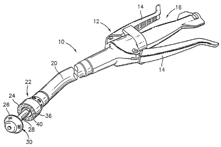
[0025] FIG. I is a perspective view of an exemplary annular surgical stapling
device;
[0026] FIG. 2 is a cross-sectional schematic illustration of a pair of
adjacent
tissue sections joined to one another pursuant to a prior art anastomotic
procedure, and
exhibiting tension therebetween and on the resulting staple line;
[0027] FIG. 3 is a perspective view of an adhesive disk according to an
embodiment of the present disclosure;
[0028] FIG. 4 is a perspective view of the intestinal area of a patient,
illustrating a
method of positioning the adhesive disk of FIG. 3 on the anvil rod of the
surgical stapling
device of FIG. 1;
[0029] FIG. 5 is a schematic perspective view of the intestinal area of FIG.
4,
illustrating the anvil rod mounted to the annular stapling device and having
the adhesive
disk of FIG. 3 disposed therebetween; and
[0030] FIG. 6 is a cross-sectional schematic illustration of the pair of
adjacent
tissue sections of the intestinal area of FIGS. 4 and 5, joined to one another
pursuant to a
8
CA 02578322 2007-02-13
method of the present disclosure, and exhibiting a reduction of tension
therebetween and
on the resulting staple line.
DETAILED DESCRIPTION OF EMBODIMENTS
[00311 Embodiments of the presently disclosed center hub will now be described
in detail with reference to the drawing figures wherein like reference
numerals identify
similar or identical elements. As used herein and as is traditional, the term
"distal" refers
to that portion which is furthest from the user while the term "proximal"
refers to that
portion which is closest to the user.
[0032] Referring initially to FIG. 1, an annular surgical stapling device, for
use
with the annular adhesive structures disclosed herein, is generally designated
as 10.
Surgical stapling device 10 includes a handle assembly 12 having at least one
pivotable
actuating handle member 14, and an advancing member 16. Extending from handle
member 12, there is provided a tubular body portion 20 which may be
constructed so as
to have a curved shape along its length. Body portion 20 terminates in a
staple cartridge
assembly 22 which includes a pair of annular arrays of staple receiving slots
36 having a
staple (not shown) disposed in each one of staple receiving slots 36.
Positioned distally
of staple cartridge assembly 22 there is provided an anvil assembly 30
including an anvil
member 26 and an anvil rod 28 operatively associated therewith for removably
connecting anvil assembly 30 to a distal end portion or connection member 40
of stapling
device 10.
[0033] Staple cartridge assembly 22 may be fixedly connected to the distal end
of
tubular body portion 20 or may be configured to concentrically fit within the
distal end of
9
CA 02578322 2007-02-13
tubular body portion 20. Typically, staple cartridge assembly 22 includes a
staple pusher
(not shown) including a proximal portion having a generally frusto-conical
shape and a
distal portion defining two concentric rings of peripherally spaced fingers
(not shown),
each one of which is received within a respective staple receiving slot 36.
[0034] Typically, a knife (not shown), substantially in the form of an open
cup
with the rim thereof defining a knife edge, is disposed within staple
cartridge assembly
22 and mounted to a distal surface of a staple pusher (not shown). The knife
edge is
disposed radially inward of the pair of annular arrays of staples.
Accordingly, in use, as
the staple pusher is advanced, the knife is also advanced axially outward.
[0035] Reference may be made to U.S. Patent 5,915,616 to Viola et al., the
entire
content of which is incorporated herein by reference, for a detailed
discussion of annular
stapling device 10.
[0036] Turning now to FIG. 2, a cross-sectional schematic illustration of a
pair of
adjacent tissue sections 66, 68 (i.e., intestinal sections), joined to one
another with
annular stapling device 10 according to the method described above, is shown.
[0037] As seen in FIG. 2, when tissue sections 66 and 68 undergo a degree of
tension (i.e., being pulled in opposite directions from one another), as
evidenced by
arrows "Al, A2", a degree of mechanical strain is placed upon staples "S". A
greater
degree of strain is exhibited on the radially outwardly disposed staples "S1"
as compared
to the radially inward disposed staples "S2". In other words, as tissues
sections 66 and 68
are pulled apart, in the direction of arrows "Al, A2", a relatively high
degree of strain is
placed on outer staples "S 1" and then on inner staples "S2". Additionally,
stress
CA 02578322 2007-02-13
concentrations are formed and/or exhibited at each outer staple "S 1" of the
outer row of
staples.
[0038] Turning now to FIG. 3, an adhesive disk, in accordance with an
embodiment of the present disclosure, is generally designated as 100. Adhesive
disk 100
desirably has a shape corresponding to the arrays of staple receiving slots
36. Preferably,
adhesive disk 100 includes a washer-like or gasket-like body portion 102
including a
substantially centrally located aperture 104 formed therethrough. Adhesive
disk 100 is
defined by an outer terminal edge or portion 106, an inner terminal edge 108
defining the
size of aperture 104, an upper surface 110, and a bottom surface 112.
[0039] In one embodiment, adhesive disk 100 is sized such that when adhesive
disk 100 is operatively associated with stapling device 10, as will be
described in greater
detail below, outer terminal edge or portion 106 extends radially beyond
staple retaining
pockets 36 of staple cartridge assembly 22. Additionally, aperture 104 of
structure 100 is
sized to at least receive shaft 28 of anvil assembly 30 therethrough. In
another
embodiment, the distance between outer terminal edge 106 and inner terminal
edge 108
may be substantially equal to a width of a tissue contact surface 24 (see FIG.
1) of staple
cartridge assembly 22.
[0040] It is contemplated that adhesive disk 100 may be fabricated from or
include a surgical grade, biocompatible, non-absorbable (i.e., permanent)
material;
desirably a mesh impregnated with an adhesive, sealant and/or wound treatment
material.
For example, adhesive disk 100 may be fabricated from "TEFLON", which is a
registered trademark owned by DuPont de Nemours & Co. It is further
contemplated that
11
CA 02578322 2007-02-13
adhesive disk 100 may be fabricated from a biocompatible polymeric foam, felt,
polytetrafluoroethylene (ePTFE), gelatin, fabric or the like, or any other
biocompatible
material.
[0041] Non-absorbable materials used for adhesive disk 100 include, and are
not
limited to, those that are fabricated from such polymers as polyethylene,
polypropylene,
nylon, polyethylene terephthalate, polytetrafluoroethylene, polyvinylidene
fluoride, and
the like. Further non-absorbable materials include and are not limited to
stainless steel,
titanium and the like.
[0042] In one embodiment, adhesive disk 100 may be fabricated from a bio-
absorbable material which is desirably impregnated with an adhesive, sealant,
and/or
other wound treatment material (e.g., a medicament). Accordingly, a sealant
component
of adhesive disk 100 can be used to retard any bleeding which may occur from
the tissue,
an adhesive component of adhesive disk 100 can be used to secure the
approximated
tissue together, and the bio-absorbability of adhesive disk 100 allows for
adhesive disk
100 to be absorbed into the body after a predetermined amount of time. For
example,
adhesive disk 100 may remain in place in the body for approximately 2-3 weeks
in order
for the anastomosis to sufficiently heal prior to adhesive disk 100 being
absorbed into the
body. In other embodiments, adhesive disk 100 has at least one portion that is
absorbable
and at least one portion that is not absorbable.
[0043] Bio-absorbable materials used for adhesive disk 100 include, and are
not
limited to, those fabricated from homopolymers, copolymers or blends obtained
from one
or more monomers selected from the group consisting of glycolide, glycolic
acid, lactide,
12
CA 02578322 2007-02-13
lactic acid, p-dioxanone, a-caprolactone and trimethylene carbonate. Other bio-
absorbable materials include and are not limited to, for example, Polyglycolic
Acid
(PGA) and Polylactic Acid (PLA). In one embodiment, adhesive disk 100 may be
fabricated from bio-absorbable felt, ePTFE, gelatin or any other bio-
absorbable materials.
[0044] It is contemplated that the adhesive is a biocompatible adhesive
including,
but not limited to, adhesives which cure upon tissue contact, which cure upon
exposure to
ultraviolet (UV) light, which are two-part systems which are kept isolated
from one
another and cure upon coming into contact with one another, which are pressure
sensitive, which are any combinations thereof, or any other known suitable
adhesive. In
one embodiment, it is contemplated that an adhesive having a cure time of from
about 10
to 15 seconds may be used. In another embodiment, it is contemplated that an
adhesive
having a cure time of about 30 seconds may be used.
[0045] It is envisioned that adhesive disk 100 may be impregnated with a pre-
cured adhesive or sealant. The pre-cured sealant or adhesive will react with
the moisture
and/or heat of the body tissue to thereby activate the sealing and/or adhesive
properties of
the sealant or adhesive. It is envisioned that the pre-cured sealant or
adhesive may be a
hydro-gel or the like.
[0046] It is envisioned that the wound treatment material "W ' includes and is
not
limited to one or a combination of adhesives, hemostats, sealants, coagulants,
astringents,
and medicaments. Other surgically biocompatible wound treatment materials "W"
which
may be employed in or applied by surgical instruments, including surgical
staplers,
include adhesives whose function is to attach or hold organs, tissues or
structures;
13
CA 02578322 2007-02-13
sealants to prevent fluid leakage; hemostats to halt or prevent bleeding;
coagulants,
astringents (e.g., sulfates of aluminum) and medicaments. Examples of
adhesives which
can be employed include protein derived, aldehyde-based adhesive materials,
for
example, the commercially available albumin/glutaraldehyde materials sold
under the
trade designation BioGlueTm by Cryolife, Inc., and cyanoacrylate-based
materials sold
under the trade designations IndermilTm and Derma BondTm by Tyco Healthcare
Group,
LP and Ethicon Endosurgery, Inc., respectively. Examples of sealants, which
can be
employed, include fibrin sealants and collagen-based and synthetic polymer-
based tissue
sealants. Examples of commercially available sealants are synthetic
polyethylene glycol-
based, hydrogel materials sold under the trade designation CoSealTm by
Cohesion
Technologies and Baxter International, Inc. Examples of hemostat materials,
which can
be employed, include fibrin-based, collagen-based, oxidized regenerated
cellulose-based
and gelatin-based topical hemostats. Examples of commercially available
hemostat
materials are fibrinogen-thrombin combination materials sold under the trade
designations CoStasisTm by Tyco Healthcare Group, LP, and TisseelTm sold by
Baxter
Interna.tional, Inc.
[0047] The wound treatment material may include a cross-linking material
and/or
reactive agent that reacts with the annular structure, tissue or both. The
resulting material
acts as a seal or tissue-joining material that is non-absorbable. For example,
the wound
treatment material may be based on biocompatible cross-linked polymers formed
from
water soluble precursors having electrophilic and nucleophilic groups capable
of reacting
and cross-linking in situ, including those disclosed in U.S. Patent No.
6,566,406, the
entire contents of which are incorporated herein by reference.
14
CA 02578322 2007-02-13
[0048] The wound treatment material may be disposed on adhesive disk 100
and/or impregnated into adhesive disk 100. Medicaments may include one or more
medically and/or surgically useful substances such as drugs, enzymes, growth
factors,
peptides, proteins, dyes, diagnostic agents or hemostasis agents, monoclonal
antibodies,
or any other pharmaceutical used in the prevention of stenosis.
[0049] Wound treatment material "W ' may include visco-elastic film forming
materials, cross-linking reactive agents, and energy curable adhesives. It is
envisioned
that wound treatment material "W", and in particular, adhesive may be cured
with the
application of water and/or glycerin (e.g., 1,2,3-pranatetriol, also known as
glycerol and
glycerine) thereto. In this manner, the water and/or glycerin cure the
adhesive and
hydrate the wound.
[0050] In one embodiment, it is contemplated that adhesive disk 100 may be
impregnated with a first component of a two-part adhesive and that the device
deploys
the second component of the two-part adhesive. For example, in a surgical
stapler 10, the
staples "S", which are retained in staple receiving slots 36 of staple
cartridge assembly
22, may be coated with a second component (e.g., a reactant) of the two-part
adhesive. In
this manner, the first component of the adhesive is activated when the staples
"S"
penetrate and capture adhesive disk 100 during the firing sequence of surgical
stapling
device 10, and the two components of the adhesive contact one another.
[0051] It is further envisioned that adhesive disk 100 may be single layered
including a homogeneous array of bio-absorbable or non-absorbable materials or
a
heterogeneous array of bio-absorbable and/or non-absorbable materials. In
certain
CA 02578322 2007-02-13
embodiments, adhesive disk 100 may be impregnated with a pressure sensitive
adhesive
which is activated when adjacent layers of tissue are approximated, with
adhesive disk.
100 disposed therebetween.
[0052] In an alternate embodiinent, it is contemplated that adhesive disk 100
may
be layered, i.e., having at least two layers. In this embodiment, each layer
may include a
homogeneous or heterogeneous array of bio-absorbable and/or non-absorbable
materials.
It is envisioned that each layer may be separated from one another prior to
the surgical
procedure.
[0053] Turning now to FIGS. 4-6, there is illustrated the use of surgical
stapling
device 10 and adhesive disk 100 in an anastomosis procedure to effect joining
of
intestinal sections 66 and 68. The anastomosis procedure is typically
performed using
minimally invasive surgical techniques including laparoscopic means and
instrumentation. At the point in the procedure shown in FIG. 4, a diseased
intestinal
section has been previously removed, anvil assembly 30 has been introduced to
the
operative site either through a surgical incision or trans-anally and
positioned within
intestinal section 68, and tubular body portion 20 of surgical stapling device
10 has been
inserted trans-anally into intestinal section 66. Intestinal sections 66 and
68 are also
shown temporarily secured about their respective components (e.g., shaft 28 of
anvil
assembly 30, and the distal end of tubular body portion 20) by conventional
means such
as a purse string suture "P".
[0054] As seen in FIG. 5, adhesive disk 100 is then placed onto shaft 28 of
anvil
assembly 30 prior to the coupling of anvil assembly 30 to the distal end of
tubular body
16
CA 02578322 2007-02-13
portion 20 in order for adhesive disk 100 to be located between intestinal
sections 66 and
68. In particular, shaft 28 of anvil assembly 30 is inserted through aperture
104 of
adhesive disk 100. In this position, adhesive disk 100 is located adjacent
intestinal
section 68. Following positioning of adhesive disk 100 onto shaft 28 of anvil
assembly
30, the surgeon maneuvers anvil assembly 30 until the proximal end of shaft 28
is
inserted into the distal end of tubular body portion 20 of surgical stapling
device 10,
wherein the mounting structure (not shown) within the distal end of tubular
body portion
20 engages shaft 28 to effect the mounting.
[0055] Thereafter, anvil assembly 30 and tubular body portion 20 are
approximated to approximate intestinal sections 66, 68 and capture adhesive
disk 100
therebetween. As seen in FIG. 6, outer terminal edge or portion 106 of
adhesive disk 100
extends radially outward, a distance "D", beyond the outer-most row of staples
"S 1".
With adhesive disk 100 captured between intestinal sections 66, 68, surgical
stapling
device 10 may be fired thereby stapling intestinal sections 66, 68 to one
another, securing
adhesive disk 100 between intestinal section 66, 68, and cutting the portion
of tissue and
adhesive disk 100 disposed radially inward of the knife, to complete the
anastomosis. As
seen in FIG. 6, following cutting of the knife through adhesive disk 100, a
new inner
terminal edge 108a of adhesive disk 100 is defined.
[0056] It is envisioned that anvil assembly 30 and tubular body portion 20 are
maintained in the approximated condition for a time sufficient for that
portion of
adhesive disk 100 located radially outward of the outer-most row of staples "S
1" to
adhere and/or bond with each intestinal section 66 and 68.
17
CA 02578322 2007-02-13
[0057] As seen in FIG. 6, any tension which may be experienced by intestinal
sections 66 and 68, as illustrated by arrows "Al, A2" directed in opposite
directions from
one another, is initially absorbed by adhesive disk 100 in the location
radially outward of
the outer-most row of staples "S 1". In this manner, the degree of strain
exhibited on the
outer-most row of staples "S 1" is reduced as compared to when no adhesive
disk 100 is
present between intestinal sections 66 and 68. In other words, as intestinal
sections 66
and 68 are pulled apart, in the direction of arrows "Al, A2", a relatively low
degree of
strain is placed on the outer-most row of staples "S 1" and an even lower
degree of strain
is placed on the inner-most row of staples "S2". Additionally, stress
concentrations at
each outer staple "S 1" of the outer row of staples is reduced by the
inclusion of adhesive
disk 100 between intestinal sections 66 and 68.
[0058) It is envisioned and understood that the greater the distance "D" that
adhesive disk 100 extends beyond the outer-most row of staples "S 1", the less
the degree
of strain which is placed on the outer-most row of staples "S 1".
[0059] As seen in FIG. 3, it is contemplated that adhesive disk 100 may
include a
slit 120 extending between inner terminal edge 108 and outer terminal edge or
portion
106 thereby enabling adhesive disk 100 to be positioned between intestinal
sections 66
and 68 following connection of anvil assembly 30 and tubular body portion 20
of surgical
stapling device 10
[0060] It will be understood that various modifications may be made to the
embodiments of the presently disclosed surgical stapling apparatus and the
various
dispensing systems and methods described above. Therefore, the above
description
18
CA 02578322 2007-02-13
should not be construed as limiting, but merely - as exemplifications of
preferred
embodiments. Those skilled in the art will envision other modifications within
the scope
and spirit of the present disclosure.
19