Note: Descriptions are shown in the official language in which they were submitted.
CA 02578419 2007-02-26
WO 2006/023589 PCT/US2005/029305
1
1 TITLE OF THE INVENTION
2
3 DIALYSIS IMPLANT AND METHODS OF USE
4
Daniel R. Burnett
6 Gregory Hall
7
8 BACKGROUND OF THE INVENTION
9 1. Field of the Invention
[00011 The present invention relates to an implantable device for drug
delivery and
11 dialysis, particularly for peritoneal dialysis, and a method of using the
system.
12
13 2. Description of the Related Art
14 [0002] Kidney failure is typically treated by dialysis until a kidney
transplant or
other treatment can replace the kidney function. Dialysis can be performed by
16 hemodialysis or peritoneal dialysis (PD).
17 100031 Hemodialysis treatment removes the blood from the body, often about
0.25 L
18 (8.5 fl. oz.) at a time, and often from a blood vessel in the arm. The
extra-corporeal
19 blood is then passed through a semi-permeable membrane that removes the
waste -
including excess water - otherwise filtered by healthy kidneys, from the blood
without
21 the loss of desirable molecules. Hemodialysis patients typically receive
three
22 treatment sessions per week, with each session lasting 3 to 5 hours.
Because proper
23 maintenance of hemodialysis equipment (e.g., membranes, pumps) is critical,
24 hemodialysis sessions are often performed at a treatment center.
CA 02578419 2007-02-26
WO 2006/023589 PCT/US2005/029305
2
1 100041 PD treatment introduces a dialysis solution to the peritoneal cavity.
The
2 blood is naturally filtered through the organ membranes in the peritoneum.
Blood
3 waste naturally passes through the organ membranes in the peritoneal cavity.
The
4 waste is drawn into the peritoneal cavity by the osmotic pressure gradient
created by
the properly-formulated dialysis solution. After a few hours, the dialysis
solution,
6 loaded with waste, can be removed. A patient can perform the "exchanges" of
7 dialysis solution at home, but must drain an extra-corporeal bag of dialysis
solution
8 into the peritoneal cavity, and then drain their own peritoneal cavity into
an extra-
9 corporeal bag - all through a trans-peritoneum catheter. Patients also
usually undergo
four to six exchanges a day.
11 [0005] PD is widely considered to be a more effective treatment for
removing waste
12 from the blood, but patients often prefer the relative infrequency and
convenience of
13 hemodialysis. Most patients also prefer not to receive the large quantity
and depth of
14 the punctures associated with PD.
[0006] U.S. Patent No. 5,037,385 to O'Byrne discloses an implantable
peritoneal
16 dialysis system. The system includes an implanted trans-peritoneum
catheter. The
17 trans-peritoneal catheter terminates outside the peritoneal cavity at a
subcutaneous
18 self-sealing terminal structure and terminates inside the peritoneal cavity
at an open
19 end. Dialysis solution can be introduced directly into the subcutaneous
self-sealing
terminal structure. The solution then flows into the peritonea] cavity. The
system
21 also includes an implanted catheter that drains the peritoneal cavity into
the bladder
22 via a pump.
23 [0007] The system disclosed by O'Byrne may reduce the number of times the
24 patient must drain their peritonea] cavity and may reduce the depth of the
punctures
needed to introduce dialysis solution to the peritoneal cavity. The system
disclosed
CA 02578419 2007-02-26
WO 2006/023589 PCT/US2005/029305
3
1 by O'Byrne, however, fails to increases the number of painfiil punctures
needed to
2 introduce the dialysis solution, fails to incorporate safeguards against
pathologically
3 high pressures in the urinary bladder or pathologically low levels of
peritoneal fluid,
4 fails to incorporate control mechanisms required for effective dialysis
without
dehydration, and fails to prevent loss of peritoneal proteins with extended
use.
6 [0008] A need therefore exists for methods and devices for performing more
7 convenient and painless PD. There exists a need to reduce the frequency of
punctures
8 patients receive during PD treatment. There also exists a need to reduce the
depth of
9 punctures during PD therapy. Furthermore, there exists a need to fulfill the
above
needs without negatively affecting the quality of blood waste removal.
11
12 BRIEF SUMMARY OF THE INVENTION
13 [0009] An implantable dialysis device is disclosed. In one embodiment of
the
14 implantable dialysis, the device has two components: an implantable
peritoneourinary
pump system and an implantable dialysate infusion system.
16 100101 The implantable peritoneourinary pump system can have a first
discharge
17 conduit for the withdrawal of peritoneal fluid from the peritoneal cavity.
The
18 implantable peritoneourinary pump system can have a peritoneourinary pump.
The
19 implantable peritoneourinary pump system can have a second discharge
conduit. The
second discharge (i.e., exit) conduit can shunt the fluid into the bladder.
The
21 implantable peritoneourinary pump system can have peritoneal and urinary
pressure
22 sensors. The implantable peritoneourinary pump system can have a
magnetically
23 coupled pump powering or recharging mechanism.
24 [0011] The first discharge conduit can be in fluid communication with the
peritoneal
cavity and the peritoneourinary pump. The first discharge conduit can have one
or
CA 02578419 2007-02-26
WO 2006/023589 PCT/US2005/029305
4
1 more perforations. The perforations can allow for the influx of the
peritoneal fluid.
2 The first discharge conduit can have a semi-permeable membrane or reservoir.
The
3 membrane or reservoir can restrict the flow of certain components of the
peritoneal
4 fluid based on size and/or charge.
[0012] The peritoneourinary pump can be attached to the first and/or second
6 conduits. The peritoneourinary pump can be programmable and/or controllable
via an
7 external signal generator. The peritoneourinary pump can be controlled as a
function
8 of time. The peritoneourinary pump can be controlled through negative and/or
9 positive feedback loops, for example, using input from the pressure sensors.
100131 The second discharge conduit can be in fluid communication with the
11 peritoneourinary pump and the urinary bladder. The second discharge conduit
can be
12 fixedly attached to the bladder wall. The second discharge conduit can be
coated, for
13 example, to prevent encrustation.
14 [0014] The peritoneal and urinary pressure sensors can be loose in the
peritoneal
cavity and bladder, respectively, for example by being tethered but free-
floating. The
16 peritoneal and urinary pressure sensors can be incorporated into the first
and second
17 discharge conduits, respectively. The pressure sensors can be incorporated
into the
18 peritoneourinary pump housing. The peritoneal and urinary pressure sensors
control
19 the peritoneourinary pump in order to prevent excessive bladder pressure or
abnormally low or high peritoneal pressure. The implantable dialysis device
can also
21 have moisture, protein, strain (e.g., in the bladder wall), nerve sensors
(e.g., to detect
22 nerve signals in the bladder, for example, to detect fullness), or
combinations thereof.
23 [0015] The magnetically coupled pump powering mechanism can be used to
directly
24 drive the peritoneourinary pump by the transdermally application of
magnetic forces
and/or to inductively recharge the internal battery. In one embodiment, for
example
CA 02578419 2007-02-26
WO 2006/023589 PCT/US2005/029305
1 when the peritoneourinary pump is directly driven by magnetic forces, the
first
2 discharge conduit can pass from the subcutaneous space into the peritoneal
cavity.
3 The peritoneourinary pump can reside in the subcutaneous space. The second
4 discharge conduit can pass from the subcutaneous space into the bladder. The
5 subcutaneous location of the peritoneourinary pump can increase the applied
strength
6 of magnetic forces used to drive the peritoneourinary pump.
7 [0016] In a second embodiment, for example when the internal battery is
inductively
8 recharged, the implantable peritoneourinary pump system can be located
anywhere in
9 the peritoneal, urinary or subcutaneous space. The inductive recharging coil
can be
located in close proximity to the skin, for example, to increase the
effectiveness of
11 battery recharging.
12 [0017] When activated, the implantable peritoneourinary pump system can
transfer
13 peritoneal fluid into the bladder via the first discharge conduit, the
peritoneourinary
14 pump and the second discharge conduit. Peritoneal fluid transfer, for
example
through control of the peritoneourinary pump and/or valves, can be internally
16 controlled via negative or positive feedback from pressure sensors and/or
externally
17 activated, for example, by a transdermal signal.
18 [0018] The implantable dialysate infusion system can elute concentrated
dialysate,
19 other osmotic agents, or other therapeutic and/or diagnostic agents, or
combinations
thereof, into the peritoneal cavity. The eluting can be performed chronically.
The
21 implantable dialysate infusion system can have a reservoir. The implantable
dialysate
22 infusion system can have a first transfer conduit. The implantable
dialysate infusion
23 system can have an infusion pump. The infusion pump and the
peritoneourinary
24 pump can be the same pump. The infusion pump and the peritoneourinary pump
can
CA 02578419 2007-02-26
WO 2006/023589 PCT/US2005/029305
6
1 be separate pumps. The implantable dialysate infusion system can have a
second
2 transfer conduit. The implantable dialysate infusion system can have a
filling port.
3 [0019] The reservoir can be in fluid communication with the first transfer
conduit
4 and the filling port. The reservoir can be made, in part or whole, from an
impermeable material. The impermeable material can prevent or minimize
undesired
6 leakage of dialysate into the peritoneal cavity. The implanted location of
the reservoir
7 can allow for the accommodation of large volumes of concentrated solute
inside the
8 reservoir. The reservoir can be located within the peritoneal cavity.
9 100201 The first transfer conduit can be in fluid communication with the
reservoir
and the infusion pump. The first transfer conduit can be absent from the
implantable
11 dialysate infusion system, for example if the infusion pump is incorporated
into the
12 reservoir.
13 100211 The infusion pump can be attached to the first and/or second
transfer
14 conduits. The infusion pump can be incorporated into the implantable
peritoneourinary pump system. The infusion pump can be programmable and/or
16 controllable via an external signal generator. The infusion pump can be
controlled
17 through either negative or positive feedback loops using the pressure
sensors of the
18 implantable peritoneourinary pump system. The infusion pump can be driven
by
19 methods similar to methods'described supra for powering the
peritoneourinary pump,
for example, the infusion pump can be externally powered or rechargeable. The
21 infusion pump can be activated and deactivated in conjunction with the
implantable
22 peritoneourinary pump system.
23 [0022] The second conduit can be in fluid communication with the infusion
pump
24 and the peritoneal cavity. The second conduit, with one or more
perforations, can
function as the first conduit of the implantable peritoneourinary pump system
CA 02578419 2007-02-26
WO 2006/023589 PCT/US2005/029305
7
1 component of the device. The second conduit can terminate in a mixing
chamber.
2 The mixing chamber can dilute the concentrated or solid dialysate with the
peritoneal
3 fluid, for example, prior to discharge into the peritoneal cavity. Diluting
and/or
4 mixing the concentrated or solid dialysate with the peritoneal fluid can
prevent local
reaction, for example a hyperosmotic reaction, to the mixed fluid.
6 [0023] The filling port can be in fluid communication with the reservoir.
The filling
7 port can be implanted in a position providing minimally invasive or
percutaneous
8 access to the filling port. The filling port can have a self-sealing
puncture membrane.
9 The filling port can have a locating mechanism, for example, a magnetic
field or
another signal generating mechanism. The filling port can be locatable via
palpation.
11 [0024] When activated, the implantable dialysate infusion system can
transfer
12 concentrated or solid dialysate from the reservoir into the peritoneal
cavity, or mixing
13 chamber, via the first conduit, the infusion pump and the second conduit.
The
14 implantable dialysate infusion system can have slow-release formulation of
concentrated dialysate in the form of a dialysate solid or concentrated
solute.
16 [0025] A method of using the implantable dialysis device in an animal
having a
17 peritoneal cavity and a bladder is disclosed. The method can include
pumping
18 dialysate, or other osmotic or other agent, from the reservoir into the
peritoneal cavity.
19 The method can include pumping some or all of the contents of the
peritoneal cavity
into the urinary bladder for evacuation, for example, after a time-delay from
the
21 introduction of additional agents into the peritoneal cavity. The method
can include
22 the percutaneous refilling of the reservoir. The method can include the use
of timers
23 and pressure sensors to automatically administer peritoneal dialysis. The
method can
24 minimize conscious patient interaction, for example, only requiring
conscious patient
CA 02578419 2007-02-26
WO 2006/023589 PCT/US2005/029305
8
1 interaction for the refilling of the reservoir and the recharging or
activating of the
2 pumps.
3 [0026] The implantable dialysate infusion system can be used to administer
any
4 agent such as a drug, diagnostic, or therapeutic, for example, when large
volumes of
the agent are to be administered. Due to the implantable dialysate infusion
system's
6 rechargeable nature, the implantable dialysate infusion system's ability to
be refilled
7 and its large volume peritoneal reservoir, large amounts of drug or
therapeutic could
8 be administered intravenously, subcutaneously or intraperitoneally over
extended
9 periods of time with only infrequent puncture for refilling of the
reservoir.
11 BRIEF DESCRIPTION OF THE DRAWINGS
12 [0027] Figure 1 illustrates an embodiment of the implantable dialysis
device.
13 100281 Figure 2 illustrates cross-section A-A of an embodiment of the
distributor.
14 [0029] Figure 3 illustrates an embodiment of the implantable dialysis
device.
100301 Figure 4 illustrates cross-section B-B of an embodiment of the
distributor.
16 [0031] Figure 5 illustrates cross-section C-C of an embodiment of the
distributor.
17 [00321 Figure 6 illustrates an embodiment of the implantable dialysis
device.
18 [0033] Figure 7 illustrates cross-section D-D of an embodiment of the
distributor.
19 [0034] Figure 8 illustrates an embodiment of the implantable dialysis
device.
[0035] Figure 9 illustrates an embodiment of the exit conduit and the exit.
21 [0036] Figure 10 illustrates cross-section C-C of an embodiment of the
distributor.
22 [0037] Figures 11-13 illustrate various embodiments of the implantable
dialysis
23 device.
24 100381 Figures 14 and 15 illustrate various embodiments of the transfer
element.
CA 02578419 2007-02-26
WO 2006/023589 PCT/US2005/029305
9
1 100391 Figure 16 illustrates a method and placement for implanting the
implantable
2 dialysis device.
3 100401 Figures 17-22 illustrate an embodiment of a method for peritoneal
dialysis
4 using the implantable dialysis device.
[0041] Figures 23-27 illustrate an embodiment of a method for peritoneal
dialysis
6 using the implantable dialysis device.
7 [0042] Figures 28-32 illustrate various embodiments of a method for
peritoneal
8 dialysis using the implantable dialysis device.
9 [0043] Figures 33 illustrates an embodiment of a method for using the
implantable
dialysis device having a mixing chamber.
11 [0044] Figures 34 illustrates an embodiment of a method for using the
implantable
12 dialysis device having a inductive dipole transducer.
13 100451 Figure 35 illustrates an embodiment of a method for using the
implantable
14 dialysis device implanted wholly in the peritoneal cavity and the bladder.
100461 Figure 36 illustrates an embodiment of a method for using the
implantable
16 dialysis device with a first component and a second component.
17
18 DETAILED DESCRIPTION
19 100471 Figure 1 illustrates an implantable dialysis device 2. The
implantable
dialysis device 2 can have a distributor 4. The distributor 4 can be
configured to
21 receive and distribute a dialysate and/or any other fluid or fluids, for
example a
22 solution of therapeutic and or diagnostic agents. The dialysate can be
received by the
23 distributor 4 and initially distributed through a reservoir conduit 6 to a
reservoir 8. At
24 a later time, the distributor 4 can withdraw the dialysate from the
reservoir 8 and
distribute the dialysate through a discharge conduit 10 to a peritoneal cavity
(shown
CA 02578419 2007-02-26
WO 2006/023589 PCT/US2005/029305
1 infra). At a later time, the distributor 4 can withdraw the dialysate and
other waste
2. fluids and solids from the peritoneal cavity through the discharge conduit
10. The
3 distributor 4 can then distribute the withdrawn dialysate and waste fluids
and solids
4 through the exit conduit 12 and out an exit 14 to a bladder (shown infra).
5 [0048] The distributor 4 can be attached to a reservoir conduit 6. The
reservoir
6 conduit 6 can be attached to the reservoir 8. A reservoir connector 18 can
attach the
7 reservoir conduit 6 to the reservoir 8. The reservoir 8 can be in fluid
communication
8 with a reservoir conduit first end 20a. The reservoir connector 18 can
reinforce the
9 attachment between the reservoir 8 and the reservoir conduit first end 20a.
10 [0049] The reservoir 8 can be a substantially or completely impermeable,
leak-proof
11 container for indefinite storage of therapeutic and/or diagnostic fluids
and/or solids.
12 The reservoir 8 can be hollow. A reservoir sensor 22, such as a reservoir
pressure
13 sensor, reservoir pH sensor, reservoir temperature sensor, reservoir
electrolyte sensor,
14 reservoir analyte sensor, or combinations thereof, can be attached to the
inside of the
reservoir 8.
16 [0050] The reservoir 8 can be substantially spherical, circular,
cylindrical, tubular,
17 or have a shape similar to a partially flattened sphere. The reservoir 8
can be shaped
18 to fit in the negative space around organs, for example in the cul-de-sac
of the
19 peritoneal cavity. The reservoir 8 can be made from at least two pieces of
material.
The pieces of material can be joined at the perimeters of the pieces of
material. The
21 pieces of material can be substantially circular.
22 [0051] The reservoir 8 can have a reservoir diameter 24. The reservoir
diameter 24
23 can be from about 2 cm (0.8 in.) to about 20 cm (8 in.), more narrowly from
about 4
24 cm (2 in.) to about 10 cm (4 in.), for example about 2 cm (0.8 in.), about
4 cm (2 in.),
about 10 cm (4 in.), or about 20 cm (8 in.). The reservoir 8 can have a
reservoir
CA 02578419 2007-02-26
WO 2006/023589 PCT/US2005/029305
11
1 volume. The reservoir volume can be from about 10 mL (0.6 in.3) to about
3000 mL
2 (200 in.), more narrowly from about 200 mL (10 in.3) to about 2000 mL (100
in.),
3 for example about 1500 mL (92 in.3). The reservoir volume can depend on the
4 potency (e.g., solute concentration) of the reservoir contents used with the
reservoir 8.
100521 The reservoir 8 can be substantially impermeable, for example the outer
6 surface of the reservoir 8 can be made from a nonporous membrane or a
membrane
7 with sufficiently small pores to minimize or prevent flow across the surface
of the
8 reservoir 8.
9 [0053] The pore size can be dependent on the particle size of an agent
(e.g., osmotic
agent, dialysate) dispensed into the surrounding body cavity and/or tissue.
The pore
11 size can prevent leakage, for example, of particles with a molecular weight
(MW)
12 from about 50 to about 5000, more narrowly a MW less than about 800, yet
more
13 narrowly a MW from about 50 to about 100. The pores can be configured to
exclude,
14 for example, sugars and dialysates (e.g., with a MW of about 800),
synthetic osmotic
agents (e.g., a MW of less than or equal to about 5000), glucose (e.g., about
2.27%
16 solution, MW of about 180.16), maltose, such as maltose disaccharide (e.g.,
about
17 4.32% solution, MW of about 342.30), maltotriose, such as maltotriose
trisaccharide
18 (e.g., about 6.36% solution, MW of about 504.44), and maltopentaose, such
as
19 maltopentaose pentasaccharide (e.g., about 10.4% solution, MW of about
828.72),
any other osmotically active material, and combinations thereof.
21 [0054] The reservoir 8 can have pores having diameters substantially
smaller than
22 about 500 m (19.7 mil), yet more narrowly from about 5 m (0.2 mil) to
about 200
23 m (7.87 mil). ("Substantially smaller" can be having about 95% or more of
the
24 pores being smaller.) The reservoir 8 can have an average pore diameter
from about 5
m (0.2 mil) to about 500 m (1.97 mil), for example about 10 m (0.39 mil).
The
CA 02578419 2007-02-26
WO 2006/023589 PCT/US2005/029305
12
1 reservoir 8 can be made from any of the materials disclosed infra for all
elements of
2 the implantable dialysis device 2. The reservoir 8 can be made from a
biocompatible
3 impermeable membrane. The reservoir 8 can be made from, for example
polymers,
4 such as polyacrylonitrile (PAN), polysulfone (PS), polyethersulfone,
poluethylene,
polymethylmethacrylate (PMMA), polytetrafluoroethylene (PTFE) (e.g., TEFLONO,
6 E. I. Du Pont de Nemours and Company, Wilmington, DE), expanded PTFE (ePTFE)
7 (e.g., GORE-TEXO from W.L. Gore & Associates, Inc., Newark, DE), polyester
8 (e.g., DACRONO from E. I. Du Pont de Nemours and Company, Wilmington, DE),
9 polypropylene, polyether ether ketone (PEEK), Nylon, polyether-block co-
polyamide
polymers (e.g., PEBAX from ATOFINA, Paris, France), polyurethanes such as
11 aliphatic polyether polyurethanes (e.g., TECOFLEXO from Thermedics Polymer
12 Products, Wilmington, MA), polyvinyl chloride (PVC), thermoplastic,
fluorinated
13 ethylene propylene (FEP), cellulose (e.g., VISKINGO> SERVAPORO> MEMBRA-
14 CEL , or SPECTRA/PORO 1, 3 and 6 Dialysis Tubing from SERVA
Electrophoresis GmbH of Heidelberg, Germany; Cuprophane PT-150 from Enka-
16 Glanstoff of Germany) such as a seamless regenerated cellulose and/or
cellulose
17 acetate (CA), extruded collagen, silicone, a metal, such as single or
multiple stainless
18 steel alloys, nickel titanium alloys (e.g., Nitinol), cobalt-chrome alloys
(e.g.,
19 ELGILOYO; CONICHROME ), molybdenum alloys (e.g., molybdenum TZM
alloy), tungsten-rhenium alloys, or combinations of any of the above.
21 [0055] The reservoir 8, as well as other elements in contact with the
stored fluids,
22 for example the elements from a filling port to the reservoir 8 and from
the reservoir 8
23 to the distributor 4, can be made from strong and/or redundant materials
having a
24 thickness and construction such that the material can remain intact without
leaking or
becoming substantially permeable during conditions of extreme acceleration,
for
CA 02578419 2007-02-26
WO 2006/023589 PCT/US2005/029305
13
1 example in a halting car accident at about 89 km/h (55 miles per hour)
producing, for
2 example, an acceleration of about 991.5 m/sz (3,253 f/s2).
3 100561 The reservoir 8 can be made from a multi-layer and/or fiber-
reinforced
4 material. The reservoir 8 can be made from strong and redundant materials.
The
reservoir 8 can be made from a flexible or rigid material.
6 [0057] The reservoir conduit 6 can be configured to enable the fluid
communication
7 of dialysate or other fluid between the distributor 4 and the reservoir 8.
The reservoir
8 8 can be fixedly, removably and/or rotatably attached, directly or
indirectly, to the
9 reservoir conduit first end 20a. The reservoir 8 can be in fluid
communication with
the reservoir conduit first end 20a. The distributor 4 can be attached to a
reservoir
11 conduit second end 20b. The distributor 4 can be in fluid communication
with the
12 reservoir conduit second end 20b.
13 100581 The reservoir conduit 6 can be flexible or rigid. The reservoir
conduit 6 can
14 be deformable or resilient. The reservoir conduit 6 can be substantially
impermeable.
[0059] The reservoir conduit 6 can have a reservoir conduit diameter 26 and a
16 reservoir conduit length 28. The reservoir conduit diameter 26 can be from
about 1
17 mm (0.04 in.) to about 10 mm (0.4 in.), more narrowly from about 2 mm (0.08
in.) to
18 about 5 mm (0.2 in.), for example about 1 mm (0.04 in.), about 2 mm (0.08
in.), about
19 5 mm (0.2 in.), or about 10 mm (0.4 in.). The reservoir conduit length 28
can be from
about 0 cm (0 in.) to about 50 cm (20 in.), more narrowly from about 5 cm (2
in.) to
21 about 20 cm (8 in.), for example about 5 cm (2 in.), about 10 cm (4 in.),
about 20 cm
22 (8 in), or about 50 cm (20 in.).
23 [0060] The discharge conduit 10 can be configured to enable fluid
communication
24 of dialysate, waste liquids and solids, and/or other fluid between the
distributor 4 and
the peritoneal cavity. The peritoneal cavity can be in fluid communication
with a
CA 02578419 2007-02-26
WO 2006/023589 PCT/US2005/029305
14
1 discharge conduit first port 30 at a discharge conduit first end 32a. The
distributor 4
2 can be attached to a discharge conduit second end 32b. The distributor 4 can
be in
3 fluid communication with the discharge conduit second end 32b.
4 [0061] The discharge conduit 10 can be substantially impermeable, permeable,
semi-permeable or combinations thereof. The discharge conduit first port 30
can have
6 an opening, and/or a permeable, and/or a semi-permeable surface. The
discharge
7 conduit 10 can have multiple (not shown) discharge conduit first ports 30
that can be
8 at the discharge conduit first end 32a and/or along a discharge conduit
length 34. The
9 discharge conduit first port 30 can be configured to minimize and/or prevent
fluid
communication of proteins, for example by size or charge exclusion (e.g., as
11 described in detail supra for the reservoir and infra for the transfer
element and
12 barriers). A peritoneal cavity sensor 36, such as a peritoneal cavity
pressure sensor,
13 peritoneal cavity pH sensor, peritoneal cavity temperature sensor,
peritoneal cavity
14 electrolyte sensor, peritoneal cavity analyte sensor, or combinations
thereof, can be
attached to the discharge conduit 10, for example on or adjacent to the
discharge
16 conduit first port 30.
17 100621 The discharge conduit 10 can have one or more perforations 38 along
part or
18 all of the discharge conduit length 34. The perforations 38 can be along
the discharge
19 conduit first end 32a and/or along the discharge conduit second end 32b.
The
perforations 38 can be configured to allow the fluid communication of the
dialysate or
21 other fluids. The perforations 38 can be configured to minimize and/or
prevent fluid
22 communication of proteins for example by size or charge exclusion (e.g., as
described
23 herein). The perforations 38 can be configured to minimize and/or prevent
fluid
24 communication of dialysate solute.
CA 02578419 2007-02-26
WO 2006/023589 PCT/US2005/029305
1 [0063] The discharge conduit 10 can be flexible or rigid. The discharge
conduit 10
2 can be deformable or resilient. The discharge conduit 10 can have a
discharge
3 conduit diameter 40 and the discharge conduit length 34. The discharge
conduit
4 diameter 40 can be from about 1 mm (0.04 in.) to about 10 mm (0.4 in.), more
5 narrowly from about 2 mm (0.08 in.) to about 5 mm (0.2 in.), for example
about 1 mm
6 (0.04 in.), about 2 mm (0.08 in.), about 5 mm (0.2 in.), or about 10 mm (0.4
in.). The
7 discharge conduit length 34 can be from about 0 cm (0 in.) to about 50 cm
(20 in.),
8 more narrowly from about 5 cm (2 in.) to about 20 cm (8 in.), for example
about 5 cm
9 (2 in.), about 10 cm (4 in.), about 20 cm (8 in), or about 50 cm (20 in.).
The discharge
10 conduit 10 can be shaped to fit in the negative space around one or more
organs
11 within the peritoneal cavity. The discharge conduit 10 can permit the
inflow of bodily
12 fluids required to mix with dialysate fluid (e.g., in concentrated form) or
solid
13 dialysate material prior to transfer into the peritoneal cavity.
14 [0064] The outer surface of the reservoir conduit 6 can be attached to the
outer
15 surface of the discharge conduit 10 along the entire, part, or none of the
reservoir
16 conduit length 28 and the discharge conduit length 34. The reservoir
conduit 6 and
17 the discharge conduit 10 can share a common outer conduit (not shown) along
the
18 entire or part of the reservoir conduit length 28 and the discharge conduit
length 34.
19 The common outer conduit can be distinct or integral with the reservoir
conduit 6
and/or the discharge conduit 10.
21 [0065] The exit conduit 12 can be configured to enable the fluid
communication of
22 dialysate or other fluid between the distributor 4 and the bladder. The
distributor 4
23 can be fixedly, removably and/or rotatably attached, directly or
indirectly, to an exit
24 conduit first end 42a. The distributor 4 can be in fluid communication with
the exit
conduit first end 42a. The bladder (shown infra) can be attached to an exit
conduit
CA 02578419 2007-02-26
WO 2006/023589 PCT/US2005/029305
16
1 second end 42b, for example by fixedly attaching an anchor 44 at the exit
conduit
2 second end 42b against a wall of the bladder. For example, the anchor 44 can
have a
3 flange that can form a one-way interference fit with the wall of the
bladder. The
4 bladder, for example via an exit port 46, can be in fluid communication with
the exit
conduit second end 42b. A bladder sensor 48, such as a bladder pressure
sensor,
6 bladder pH sensor, bladder temperature sensor, bladder electrolyte sensor,
bladder
7 analyte sensor, or combinations thereof, can be attached to the exit conduit
12, for
8 example on or adjacent to the exit port 46.
9 [0066] The exit conduit 12 can be substantially impermeable (e.g., outside
the
bladder) and/or semi-permeable (e.g., inside the bladder) and/or permeable
(e.g.,
11 inside the bladder). The exit conduit 12 can be flexible or rigid. The exit
conduit 12
12 can be deformable or resilient.
13 100671 The exit conduit 12 can have an exit conduit diameter 50 and an exit
conduit
14 length 52. The exit conduit diameter 50 can be from about l mm (0.04 in.)
to about
10 mm (0.4 in.), more narrowly from about 2 mm (0.08 in.) to about 5 mm (0.2
in.),
16 for example about 1 mm (0.04 in.), about 2 mm (0.08 in.), about 5 mm (0.2
in.), or
17 about 10 mm (0.4 in.). The exit conduit length 52 can be from about 0 cm (0
in.) to
18. about 50 cm (20 in.), more narrowly from about 5 cm (2 in.) to about 20 cm
(8 in.),
19 for example about 5 cm (2 in.), about 10 cm (4 in.), about 20 cm (8 in), or
about 50
cm (20 in.).
21 [0068] The exit conduit 12 can be distinct from the reservoir conduit 6
and/or the
22 discharge conduit 10. The exit conduit 12 can be integral with the
reservoir conduit 6
23 and/or the discharge conduit 10. The exit conduit 12 can be in fluid
communication
24 with the reservoir conduit 6 and/or the discharge conduit 10.
CA 02578419 2007-02-26
WO 2006/023589 PCT/US2005/029305
17
1 [0069] Any or all elements of the implantable dialysis device 2 can be made
from,
2 for example, a single or multiple stainless steel alloys, nickel titanium
alloys (e.g.,
3 Nitinol), cobalt-chrome alloys (e.g., ELGILOY(V from Elgin Specialty Metals,
Elgin,
4 IL; CONICHROMEO from Carpenter Metals Corp., Wyomissing, PA), molybdenum
alloys (e.g., molybdenum TZM alloy, for example as disclosed in International
Pub.
6 No. WO 03/082363 A2, published 9 October 2003), tungsten-rhenium alloys, for
7 example, as disclosed in International Pub. No. WO 03/082363, polymers such
as
8 polyester (e.g., DACRONO from E. I. Du Pont de Nemours and Company,
9 Wilmington, DE), polypropylene, PTFE, ePTFE, PEEK, Nylon, polyether-block co-
polyamide polymers (e.g., PEBAXO from ATOFINA, Paris, France), polyurethanes
11 such as aliphatic polyether polyurethanes (e.g., TECOFLEXO from Thermedics
12 Polymer Products, Wilmington, MA), PVC, PAN, PS, polyethersulfone,
polyethylene,
13 polymethylmethacrylate (PMMA), thermoplastic, FEP, cellulose (e.g.,
VISKINGO,
14 SERVAPORO, MEMBRA-CELO, or SPECTRA/PORO 1, 3 and 6 Dialysis Tubing
from SERVA Electrophoresis GmbH of Heidelberg, Germany; Cuprophane PT-150
16 from Enka-Glanstoff of Germany), such as a seamless regenerated cellulose
and CA,
17 extruded collagen, silicone, echogenic, radioactive, radiopaque materials
or
18 combinations thereof. Examples of radiopaque materials are barium sulfate,
titanium,
19 stainless steel, nickel-titanium alloys, tantalum and gold.
[0070] Any or all elements of the implantable dialysis device 2 can be a
matrix for
21 cell ingrowth or used with a fabric, for example a covering (not shown)
that acts as a
22 matrix for cell ingrowth. The matrix and/or fabric can be, for example,
polyester
23 (e.g., DACRON from E. I. du Pont de Nemours and Company, Wilmington, DE),
24 polypropylene, PTFE, ePTFE, nylon, extruded collagen, silicone or
combinations
thereof.
CA 02578419 2007-02-26
WO 2006/023589 PCT/US2005/029305
18
1 [00711 The elements of the implantable dialysis device 2 and/or the fabric
can be
2 filled and/or coated with an agent delivery matrix known to one having
ordinary skill
3 in the art and/or a therapeutic and/or diagnostic agent. The agents within
these
4 matrices can include radioactive materials; radiopaque materials; cytogenic
agents;
cytotoxic agents; cytostatic agents; thrombogenic agents, for example
polyurethane,
6 cellulose acetate polymer mixed with bismuth trioxide, and ethylene vinyl
alcohol;
7 lubricious, hydrophilic materials; phosphor cholene; anti-inflammatory
agents, for
8 example non-steroidal anti-inflammatories (NSAIDs) such as cyclooxygenase-1
9 (COX-1) inhibitors (e.g., acetylsalicylic acid, for example ASPIRINO from
Bayer
AG, Leverkusen, Germany; ibuprofen, for example ADVILO from Wyeth,
11 Collegeville, PA; indomethacin; mefenamic acid), COX-2 inhibitors (e.g.,
VIOXXO
12 from Merck & Co., Inc., Whitehouse Station, NJ; CELEBREXO from Pharmacia
13 Corp., Peapack, NJ; COX-1 inhibitors); immunosuppressive agents, for
example
14 Sirolimus (RAPAMUNE , from Wyeth,, Collegeville, PA), or matrix
metalloproteinase (MMP) inhibitors (e.g., tetracycline and tetracycline
derivatives)
16 that act early within the pathways of an inflammatory response. Examples of
other
17 agents are provided in Walton et al, Inhibition of Prostoglandin E2
Synthesis in
18 Abdominal Aortic Aneurysms, Circulation, July 6, 1999, 48-54; Tambiah et
al,
19 Provocation of Experimental Aortic Inflammation Mediators and Chlamydia
Pneumoniae, Brit. J. Surgery 88 (7), 935-940; Franklin et al, Uptake of
Tetracycline
21 by Aortic Aneurysm Wall and Its Effect on Inflammation and Proteolysis,
Brit. J.
22 Surgery 86 (6), 771-775; Xu et al, Spl Increases Expression of
Cyclooxygenase-2 in
23 Hypoxic Vascular Endothelium, J. Biological Chemistry 275 (32) 24583-24589;
and
24 Pyo et al, Targeted Gene Disruption of Matrix Metalloproteinase-9
(Gelatinase B)
Suppresses Development of Experimental Abdominal Aortic Aneurysms,.I. Clinical
CA 02578419 2007-02-26
WO 2006/023589 PCT/US2005/029305
19
1 Investigation 105 (11), 1641-1649 which are all incorporated by reference in
their
2 entireties. The reservoir 8 can be made from any of the materials disclosed
herein for
3 all elements of the implantable dialysis device 2. The reservoir 8 can be
made from a
4 biocompatible impermeable membrane. The reservoir 8 can be made from, for
example, silicone, cellulose (e.g., VISKING , SERVAPORO, MEMBRA-CELO, or
6 SPECTRA/PORO 1, 3 and 6 Dialysis Tubing from SERVA Electrophoresis GmbH of
7 Heidelberg, Germany; Cuprophane PT-150 from Enka-Glanstoff of Germany), such
8 as a seamless regenerated cellulose and CA, extruded collagen, silicone,
polymers,
9 such as PAN, PS, polyethersulfone, polyether ether ketone (PEEK), Nylon,
polyether-
block co-polyamide polymers (e.g., PEBAXO from ATOFINA, Paris, France),
11 polyurethanes such as aliphatic polyether polyurethanes (e.g., TECOFLEX(&
from
12 Thermedics Polymer Products, Wilmington, MA), polyvinyl chloride (PVC),
13 poluethylene, polyester (e.g., DACRONO from E. I. Du Pont de Nemours and
14 Company, Wilmington, DE), polypropylene, PMMA, thermoplastic, fluorinated
ethylene propylene (FEP), PTFE, and ePTFE, a metal, such as single or multiple
16 stainless steel alloys, nickel titanium alloys (e.g., Nitinol), cobalt-
chrome alloys (e.g.,
17 ELGILOYO; CONICHROMEO), molybdenum alloys (e.g., molybdenum TZM
18 alloy), tungsten-rhenium alloys, or combinations of any of the above.
19 [00721 The reservoir 8 can be made from an non-permeable material. The
reservoir
8 can be made from a material having a thickness and construction such that
the
21 material can remain intact without leaking or becoming substantially
permeable
22 during conditions of extreme acceleration, for example in a halting car
accident at
23 about 89 km/h (55 miles per hour) producing, for example, an acceleration
of about
24 991.5 m/s2 (3,253 f/SZ). The reservoir 8 can be made from a multi-layer
and/or fiber-
reinforced material. The reservoir 8 can be made from a rigid material. The
reservoir
CA 02578419 2007-02-26
WO 2006/023589 PCT/US2005/029305
1 8 can be made from any material listed herein, for example, polymers such as
2 polyester (e.g., DACRONO from E. I. Du Pont de Nemours and Company,
3 Wilmington, DE), polypropylene, polytetrafluoroethylene (PTFE) (e.g.,
TEFLONO,
4 E. I. Du Pont de Nemours and Company, Wilmington, DE), expanded PTFE (ePTFE)
5 (e.g., GORE-TEXO from W.L. Gore & Associates, Inc., Newark, DE), polyether
6 ether ketone (PEEK), Nylon, polyether-block co-polyamide polymers (e.g.,
PEBAX
7 from ATOFINA, Paris, France), polyurethanes such as aliphatic polyether
8 polyurethanes (e.g., TECOFLEXO from Thermedics Polymer Products, Wilmington,
9 MA), PVC, PAN, PS, polyethersulfone, polyethylene, PMMA, thermoplastic, FEP,
10 cellulose (e.g., VISKINGO, SERVAPORO, MEMBRA-CELO, or SPECTRA/PORO
l 1 1, 3 and 6 Dialysis Tubing from SERVA Electrophoresis GmbH of Heidelberg,
12 Germany; Cuprophane PT-150 from Enka-Glanstoff of Germany), such as a
seamless
13 regenerated cellulose and/or CA, extruded collagen, silicone or
combinations thereof.
14 [00731 The implantable dialysis device 2 can have one or more reservoir
sensors 22.
15 The reservoir sensors 22 can be in the reservoir 8, and/or in the reservoir
connector
16 18, and/or in the reservoir conduit 6. The reservoir sensors 22 can be
configured to
17 measure pressure, pH, temperature, electrolyte concentration, analyte
concentration,
18 or combinations thereof in the reservoir 8.
19 [0074] The implantable dialysis device 2 can have one or more peritonea]
cavity
20 sensors 36. The peritoneal cavity sensors 36 can be on the discharge
conduit 10, for
21 examples, at the discharge conduit first end 32a and/or along the discharge
conduit
22 length 34. The peritonea] cavity sensors 36 can be configured to measure
pressure,
23 pH, temperature, electrolyte concentration, analyte concentration, or
combinations
24 thereof in the peritoneal cavity.
CA 02578419 2007-02-26
WO 2006/023589 PCT/US2005/029305
21
1 [0075] The implantable dialysis device 2 can have one or more bladder
sensors 48.
2 The bladder sensors 48 can be on the exit 14. The bladder sensors 48 can be
3 configured to measure pressure, pH, temperature, electrolyte concentration,
analyte
4 concentration, or combinations thereof in the bladder. The sensors 22, 36,
and 48 can
measure concentration of dialysate solutes in the fluids. The sensors 22, 36,
and 48
6 can send signals indicating respective measured metrics to the distributor
4.
7 [0076] Figure 2 illustrates that the distributor 4 can have a pump 54. The
pump 54
8 can be a mechanical, electromechanical, osmotic or diffusion pump, or
combinations
9 thereof. The pump 54 can be a hand-powered pump, for example the pump can be
a
resilient, compressible bulb pump. The pump 54 can be a miniature gear-pump.
The
11 pump 54 can be strong enough to clear clogs from the discharge conduit 10
and/or the
12 exit conduit 12. The pump 54 can produce a flow rate in the discharge
conduit 10
13 from about 50 mL/min. (3.0 in.3/min.) to about 5000 mL/min. (300
in.3/min.), more
14 narrowly from about 250 mL/min. (15 in.3/min.) to about 500 mL/min. (30
in.3/min.).
The flow rate can be set to prevent bladder spasm with the rapid influx of the
fluid.
16 [0077] The pump 54 can have and/or be in fluid communication with a
distributor
17 valve 56 (shown infra). The distributor valve 56 can be a mechanical valve,
a semi-
18 permeable membrane or combinations thereof. The distributor valve 56 can be
a
19 single, three-way valve.
100781 The distributor 4 can have a distributor first conduit 58a. The
distributor first
21 conduit 58a can be in fluid communication with the reservoir conduit second
end 20b.
22 The distributor first conduit 58a can be in fluid communication with the
distributor
23 valve 56. The distributor first conduit 58a can be integral with the
reservoir conduit
24 second end 20b.
CA 02578419 2007-02-26
WO 2006/023589 PCT/US2005/029305
22
1 100791 The distributor 4 can have a distributor second conduit 58b. The
distributor
2 second conduit 58b can be in fluid communication with the discharge conduit
10.
3 The distributor second conduit 58b can be in fluid communication with the
distributor
4 valve 56. The distributor second conduit 58b can be integral with the
discharge
conduit 10.
6 100801 The distributor 4 can have a distributor third conduit 58c. The
distributor
7 third conduit 58c can be in fluid communication with the exit conduit first
end 42a.
8 The distributor third conduit 58c can be in fluid communication with the
distributor
9 valve 56. The distributor third conduit 58c can be integral with the exit
conduit first
end 42a.
11 [0081] The distributor valve 56 can be configured to route flow between a
12 distributor first conduit 58a, the distributor second conduit 58b, and the
distributor
13 third conduit 58c. The distributor valve 56 can be configured as a one-way
flow or
14 check valve, for example, preventing backflow in any direction. The
distributor valve
56 can be a one-way valve preventing flow in the direction from the
distributor third
16 conduit 58c to either the distributor first conduit 58a or the distributor
second conduit
17 58b.
18 [0082] The distributor valve 56 can be a pressure sensing valve. The
distributor
19 valve 56 can be configured to shut off flow if backpressure exceeds a pre-
determined
threshold. If pressure in the peritoneal cavity is less than about 1.5 kPa
(0.15 psi),
21 more narrowly less than about I kPa (0.1 psi), yet more narrowly less than
about 0.5
22 kPa (0.07 psi), then the pump 54 can be inhibited (e.g., stopped or
slowed), for
23 example be the distributor valve 56 and/or a controller. If the absolute
pressure in the
24 bladder is greater than or equal to about 3 kPa (0.4 psi), more narrowly,
greater than
or equal to about 4 kPa (0.6 psi), then the pump 54 can be inhibited. If the
differential
CA 02578419 2007-02-26
WO 2006/023589 PCT/US2005/029305
23
1 between the pressure in the peritoneal cavity and the pressure in the
bladder pressure
2 is greater than or equal to about 2 kPa (0.3 psi), more narrowly greater
than or equal
3 to about 3 kPa (0.4 psi), then the pump 54 can be inhibited.
4 [0083] The distributor 4 can have a power storage and/or regulation device,
for
example a battery 60. The battery 60 can be configured to supply power to the
pump
6 54 and/or the distributor valve 56. The battery 60 can be one or more power
storage
7 devices (not shown), for example capacitors, dry or wet cells, flywheels,
springs, or
8 combinations thereof. The battery 60 can hold a charge of more than about
500 mAh,
9 for example about 1000 mAh. For example 3 AA Nickel Cadmium about 1000 mAh
batteries can be used. The battery 60 can be configured to provide a current
of greater
11 than about 0.2 DCA and/or less than about 2.0 DCA, for example about 0.42
DCA.
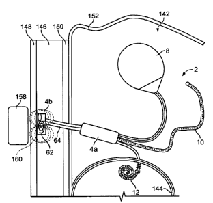
12 [0084] The distributor 4 can have an internal transducer 62. The internal
transducer
13 62 can receive energy in a first form (e.g. moving magnetic fields),
convert the energy
14 into a second form (e.g., direct current electricity), and deliver the
second form of
energy to appropriate elements (e.g., pump 54, distributor valve 56,
controller) in the
16 implantable dialysis device 2. The internal transducer 62 can be wholly or
partially
17 inside a distributor case. An internal transducer connector 64 (shown
infra) can be
18 configured to deliver the energy to the appropriate elements. The internal
transducer
19 connector 64 can be wholly within the distributor case.
[0085] The distributor 4 can have an internal filling port 66. The internal
filling port
21 66 can have a self-sealing membrane forming at least part of the external
wall of the
22 distributor 4. The internal filling port 66 can be configured to receive
injections (e.g.,
23 of dialysate solution and/or other agent), for example from a
transcutaneous needle.
24 The internal filling port 66 can have a locating mechanism, for example, a
magnetic
field or another signal generating mechanism. The locating mechanism can aid
CA 02578419 2007-02-26
WO 2006/023589 PCT/US2005/029305
24
1 targeting the internal filling port 66, for example, when injecting
dialysate solution
2 and/or other agent. The internal filling port 66 can have a storage volume.
The
3 internal filling port 66 can have a non-corrosive internal surface. The
internal filling
4 port 66 can be a receptacle for a cartridge or ampoule. A filling conduit 68
can be
configured to create fluid communication between the internal filling port 66
and the
6 reservoir conduit 6.
7 [0086] Figure 3 illustrates the implantable dialysis device 2 that can have
a first
8 component 72a and a second component 72b. The first component 72a can be
9 physically unattached to the second component 72b.
[0087] The first component 72a can be configured to pump fluid from a drainage
11 conduit 74 to, and out, the exit conduit 12. The drainage conduit 74 can
have a
12 drainage conduit first port 75. The first component 72a can have a first
distributor 4a.
13 The first distributor 4a can be attached to the drainage conduit 74. The
first
14 distributor 4a can be attached to the exit conduit 12.
100881 The second component 72b can be configured to receive a solution, for
16 example, dialysate by injection into a second distributor 4b. The second
component
17 72b can be configured to deliver and store the solution in the reservoir 8.
The second
18 component 72b can be configured to deliver the stored solution from the
reservoir 8
19 to, and out, the discharge conduit 10.
[0089] The second distributor 4b can be attached to the reservoir conduit 6
and the
21 reservoir 8. The second distributor 4b can be attached to the discharge
conduit 10.
22 [0090] The first component 72a can be in data and/or power communication
with
23 the second component 72b. One or more wires (not shown) can attach the
first
24 component 72a to the second component 72b. The first component 72a can
communicate with the second component 72b over a data network, for example, a
CA 02578419 2007-02-26
WO 2006/023589 PCT/US2005/029305
1 wired and/or wireless network, such as Ethernet (IEEE 802.3), Firewire (IEEE
1394),
2 802.11 (wireless LAN), Bluetooth, cellular communication, serial port (RS-
232, RS-
3 485), parallel port (IEEE 1284), Fiber Channel, IRDA infrared data port,
radio such as
4 900 MHz RF or FM signal, or combinations thereof.
5 [0091] Any implantable dialysis device 2 can also use the communication
networks
6 supra to communicate data with an extracorporeal component, for example, a
7 monitoring device such as a handheld diagnostic computer or peripheral
device (e.g.,
8 a personal data assistant). The extracorporeal component can transmit and
receive
9 data and/or energy from the implantable dialysis device 2 (e.g., from the
internal
10 transducer 62 and/or controller and/or battery 60). The extra corporeal
component
11 can be used to control operation of, or provide an energy charge to, the
implantable
12 dialysis device 2.
13 100921 Figure 4 illustrates the first distributor 4a that can have no
internal filling
14 port 66. The first distributor 4a can have no distributor third conduit
58c. The
15 exterior of the distributor 4 can be the distributor case 76. The
distributor case 76 can
16 be made from, coated, or otherwise surrounded with a biocompatible
material.
17 [0093] The distributor 4 can have a distributor first port 78a and a
distributor second
18 port 78b. The distributor ports 78a and 78b can be voids in the distributor
case 76,
19 semi-permeable membranes, permeable membranes, or combinations thereof. The
20 distributor first port 78a can be fixedly or releasably attached to a
conduit, for
21 example, the drainage conduit 74. The distributor second port 78b can be
fixedly or
22 releasably attached to a conduit, for example the exit conduit 12.
23 [0094] A distributor first port 78a can be fixedly or releasably attached
to and/or
24 integral with, and in fluid communication with, the drainage conduit 74. A
distributor
25 second port 78b can be fixedly or releasably attached to and/or integral
with, and in
CA 02578419 2007-02-26
WO 2006/023589 PCT/US2005/029305
26
1 fluid communication with, the exit conduit 12. The distributor valve 56 can
be a one-
2 way check valve permitting flow from the distributor first port 78a to the
distributor
3 second port 78b, but preventing or minimizing flow from the distributor
second port
4 78b to the distributor first port 78a.
100951 The internal transducer 62 can be outside the distributor case 76. The
6 internal transducer 62 can be an induction coil. The internal transducer
connector 64
7 can connect the internal transducer 62 to the pump 54 and/or to one or more
power
8 storage devices (not shown), for example capacitors, dry or wet cells,
flywheels,
9 springs, or combinations thereof. The internal transducer connector 64 can
pass
through the distributor case 76.
11 [00961 For implantable dialysis devices 2 that have more than one
distributor 4, any
12 or each distributor 4 can have a separate pump 54.
13 100971 Figure 5 illustrates that the second distributor 4b can have the
storage
14 volume of the internal filling port 66 surrounding the pump 54. The
distributor case
76 can be a self-sealing material configured to allow a needle puncture in one
or more
16 locations.
17 [0098] The reservoir conduit second end 20b (not shown) can be fixedly or
18 releasably attached to and/or integral with, and in fluid communication
with, the
19 distributor first port 78a. The discharge conduit second end 32b (not
shown) can be
fixedly or releasably attached to and/or integral with, and in fluid
communication
21 with, the distributor second port 78b.
22 [00991 Figure 6 illustrates the implantable dialysis device 2 that can have
a first
23 discharge conduit l0a and a second discharge conduit I Ob. The first and
second
24 discharge conduits I Oa and I Ob can have first and second discharge
conduit lengths
34a and 34b and first and second discharge conduit diameters 40a and 40b that
can be
CA 02578419 2007-02-26
WO 2006/023589 PCT/US2005/029305
27
1 equivalent to those supra for the discharge conduit 10. The first and/or
second
2 discharge conduits l0a and/or l Ob can have first and/or second peritoneal
cavity
3 sensors 36a and/or 36b, respectively.
4 101001 The first and/or second discharge conduits 10a and/or I Ob can have a
first
and/or second discharge conduit first port guards 80a and/or 80b. The guards
80a and
6 80b can be rigid, semi-rigid or flexible. The port guards 80a and 80b can be
wire
7 screens, permeable membranes, or combinations thereof The port guards 80a
and
8 80b can be configured to filter particles based on size and/or charge.
9 [0101] Figure 7 illustrates that the distributor 4 can have the distributor
first conduit
58a, the distributor second conduit 58b, and the distributor third conduit 58c
that can
11 be segmented from a single channel, and/or be adjacent to each other. The
distributor
12 first, second, and third conduits 58a, 58b and 58c can all open on the same
side of the
13 distributor 4. The distributor 4 can have a distributor fourth conduit 58d.
The
14 distributor fourth conduit 58d can open on a different side of the
distributor 4 than the
first, second and third conduits 70a, 70b and 70c.
16 [0102] The reservoir conduit second end 20b can be fixedly or releasably
attached to
17 and/or integral with, and in fluid communication with, the distributor
first conduit
18 58a. The first discharge conduit second end 32b' can be fixedly or
releasably
19 attached to and/or integral with, and in fluid communication with, the
distributor
second conduit 58b. The second discharge conduit second end 32b can be fixedly
or
21 releasably attached to and/or integral with, and in fluid communication
with, the
22 distributor third conduit 58c. The fourth conduit 58d can be fixedly or
releasably
23 attached to and/or integrated with, and in fluid communication with, the
exit conduit
24 12.
CA 02578419 2007-02-26
WO 2006/023589 PCT/US2005/029305
28
1 [0103] Figure 8 illustrates that the implantable dialysis device 2 can have
the first
2 and second distributors 4a and 4b. The reservoir conduit 6 can have an
inflow
3 channel 86 and an outflow channel 88.
4 [01041 The inflow and outflow channels 86 and 88 can be separated by a
septum, be
otherwise attached or integral, or be contained within two distinct, and
separate tubes.
6 The inflow channel 86 can be attached to the outflow channel 88 along part
or all of
7 the lengths of the inflow channel 86 and the outflow channel 88.
8 [0105] The inflow channel 86 can provide fluid communication between the
9 internal filling port 66 and the reservoir 8. The internal filling port 66
and/or filling
conduit (not shown in Figure 8) can be attached to the inflow channel 86. The
11 reservoir 8 and/or the reservoir connector 18 can be attached to the inflow
channel 86.
12 The inflow channel 86 can be attached to and/or integral with the reservoir
8 and the
13 second distributor 4b, for example with the internal filling port 66. The
inflow
14 channel 86 can be in direct fluid communication with, and/or attached to,
the first
distributor 4a. The first distributor 4a can be configured to provide a
positive and/or
16 negative pressure to the inflow channel 86.
17 101061 The outflow channel 88 can be in direct fluid communication with,
and
18 attached to and/or integral with the first distributor 4a and the reservoir
8 and/or the
19 reservoir connector 18.
101071 The discharge conduit 10 can have one or more perforations 38 along
part or
21 all of the discharge conduit length 34. The perforations 38 can be along
the discharge
22 conduit first end 32a and/or along the discharge conduit second end 32b.
The
23 perforations 38 can be configured to allow the fluid communication of
dialysate
24 solute. The perforations 38 can be configured to disallow fluid
communication of
CA 02578419 2007-02-26
WO 2006/023589 PCT/US2005/029305
29
1 proteins. The perforations 38 can be configured to disallow fluid
communication of
2 dialysate solute.
3 [01081 The first distributor 4a can have the pump 54 (not shown). The second
4 distributor 4b can have the internal filling port 66. The second distributor
4b can have
the internal transducer 62. The internal transducer connector 64 can be
attached to the
6 first distributor 4a and/or the second distributor 4b. The internal
transducer connector
7 64 can transfer power from the second distributor 4b to the first
distributor 4a. The
8 first and/or second distributors 4a and/or 4b can have the batteries 60 (not
shown in
9 Figure 8).
101091 Figures 8 and 9 illustrate that the exit conduit 12 can have an exit
extension
11 90. The exit extension 90 can be semi-permeable, permeable, impermeable, or
12 combinations thereof. The exit extension 90 can have a length of conduit,
for
13 example a coiled or "pigtail" catheter. The exit extension 90 can have one
or more
14 exit ports 46. The exit extension 90 can have an exit tip 94. The exit tip
94 can have
the exit port 46 (not shown in Figures 8 or 9). The exit tip 94 can be semi-
permeable,
16 impermeable, permeable, or combinations thereof.
17 [0110] The exit conduit 12 can have an exit conduit longitudinal axis 96.
The exit
18 conduit 12 can have one or more sub-anchors 98. The sub-anchors 98 can be
19 substantially perpendicular to the exit conduit longitudinal axis 96. The
anchor 44
can be substantially perpendicular to the exit conduit longitudinal axis 96.
The sub-
21 anchors 98 can be flanges. The sub-anchors 98 can be rigid or flexible.
22 101111 Figure 10 illustrates that the pump 54 can have or be mechanically
attached
23 to a rotational electromechanical motor 99. The motor 99 can be configured
to be
24 inductively driven. The motor 99 can have a first pole 100a and a second
pole 100b.
A pole axle 102 can attach the first pole 100a to the second pole 1 OOb. The
pole axle
CA 02578419 2007-02-26
WO 2006/023589 PCT/US2005/029305
1 102 can rotate about a motor rotation axis 104, for example when the first
and second
2 poles 100a and 100b are urged by a dynamic external magnetic field. The pole
axle
3 102 can be mechanically coupled to a flow driving mechanism (not shown). The
4 pump 54 and/or motor 99 can be the taught by PCT Patent Application titled
5 Magnetic Circumferentially Coupled Implantable Pump, filed 18 August 2004
6 (attorney docket number TN 1004-PCT), and hereby incorporated by reference
in its
7 entirety.
8 [01121 Figures 11 through 13 (not showing elements of the implantable
dialysis
9 device 2 for clarity) illustrate various configurations of the peritoneal
cavity sensor 36
10 and bladder sensor 48. The peritonea] cavity sensor 36 and bladder sensor
48 can be
11 in fluid communication with the discharge conduit 10 and/or exit conduit
12,
12 respectively (i.e., and the peritoneal cavity and the bladder,
respectively, during use).
13 As shown in Figure 11, the peritoneal cavity sensor 36 and bladder sensor
48 can be
14 attached to the discharge conduit 10 and exit conduit 12. The peritoneal
cavity sensor
15 36 and the bladder sensor 48 can be on the inside (as shown) and/or outside
of the
16 discharge and exit conduits 10 and 12. As shown in Figure 12, the
peritoneal cavity
17 sensor 36 and bladder sensor 48 can be located in the distributor 4. As
shown in
18 Figure 13, the peritoneal cavity sensor 36 can be attached to a peritoneal
tether 106.
19 The bladder sensor 48 can be attached to a bladder tether 108. Multiple
sensors 36
20 and 48 can be attached to each tether 106 and 108. The tethers 106 and 108
can be
21 attached to the respective conduits 10 and 12, and/or the distributor 4,
and/or to other
22 elements of the implantable dialysis device 2. The tethers 106 and 108 can
be flexible
23 or rigid.
CA 02578419 2007-02-26
WO 2006/023589 PCT/US2005/029305
31
1 [01131 The implantable dialysis device 2 can have more than_one of each
peritoneal
2 cavity sensor 36 and bladder sensor 48. The peritoneal cavity sensor 36 and
bladder
3 sensor 48 can be in any combination of configurations.
4 101141 Figures 14 and 15 illustrate that the implantable dialysis device 2
can have a
transfer element 110 at the first end of the drainage (e.g., shown without the
transfer
6 element 110 in Figure 3) and/or discharge (e.g., shown without the transfer
element
7 110 in Figure 6) conduits 74 and/or 10. The transfer element 110 can be
integral with,
8 and/or attached to, the conduits 74 and/or 10 via a transfer element
connector 111.
9 The transfer element l 10 can have a permeable surface. The transfer element
110 can
be configured to filter peritoneal fluids across a transfer element face 112.
The
11 transfer element 110 can be configured to filter fluid across the transfer
element face
12 112 through size and/or charge exclusion. The transfer element 110 can be
13 configured to allow water and waste in the peritoneal fluid to osmotically
transfer into
14 the transfer element 110.
10115J Figure 14 illustrates that the transfer element 110 can be configured
to
16 resiliently expand and compress, as shown by arrows. The transfer element
110 can
17 be configured to transfer liquids out of the transfer element 110 and into
the drainage
18 and/or discharge conduits 74 and/or 10. The transfer element 110 can be
biased to
19 stay in an expanded configuration at rest. The transfer element 110 can be
hollow.
The hollow inside the transfer element 110 can be in fluid communication with
the
21 drainage and/or discharge conduits 74 and/or 10. A one-way valve (not
shown) in the
22 drainage and/or discharge conduits 74 and/or 10, the transfer element
connector 111,
23 or the transfer element 110, can be configured to prevent or minimize fluid
24 communication from the drainage and/or discharge conduits 74 and/or 10 to
the
CA 02578419 2007-02-26
WO 2006/023589 PCT/US2005/029305
32
1 reservoir 8. The transfer element 110 can have a substantially cylindrical
2 configuration.
3 [01161 The transfer element 110 can have a transfer element face 112. The
transfer
4 element 110 can have two or more transfer element faces 112. The transfer
element
faces 112 can be made from a substantially impermeable, semi-permeable,
permeable
6 material, or combinations thereof. The transfer element face 112 can be
configured to
7 be substantially or wholly permeable to dialysate solutes. The transfer
element face
8 112 can be substantially or wholly impermeable to proteins. The transfer
element
9 face 112 can be made from the materials listed herein, for example,
polyester (e.g.,
DACRON from E. I. Du Pont de Nemours and Company, Wilmington, DE),
11 polypropylene, PTFE (e.g., TEFLON , E. I. Du Pont de Nemours and Company,
12 Wilmington, DE), ePTFE (e.g., GORE-TEX from W.L. Gore & Associates, Inc.,
13 Newark, DE), PEEK, Nylon, polyether-block co-polyamide polymers (e.g.,
PEBAX(M
14 from ATOFINA, Paris, France), polyurethanes such as aliphatic polyether
polyurethanes (e.g., TECOFLEX from Thermedics Polymer Products, Wilmington,
16 MA), polyvinyl chloride (PVC), PAN, PS, polyethersulfone, polyethylene,
PMMA,
17 thermoplastic, FEP, cellulose (e.g., VISKING , SERVAPOR , MEMBRA-CEL ,
18 or SPECTRA/POR 1, 3 and 6 Dialysis Tubing from SERVA Electrophoresis GmbH
19 of Heidelberg, Germany; Cuprophane PT-150 from Enka-Glanstoff of Germany),
such as a seamless regenerated cellulose and CA, extruded collagen, silicone,
21 echogenic, radioactive, radiopaque materials or combinations thereof. Any
of the
22 polymers can be permeable if woven loosely enough, as known to those having
23 ordinary skill in the art.
24 [01171 The transfer element faces 112 can be made from a porous membrane.
The
transfer element faces 112 can have pores having diameters substantially
smaller than
CA 02578419 2007-02-26
WO 2006/023589 PCT/US2005/029305
33
1 about 500 m (19.7 mil), yet more narrowly from about 5 m (0.2 mil) to
about 200
2 m (7.87 mil). ("Substantially smaller" is having about 95% or more of the
pores
3 being smaller.) The transfer element faces 112 can have an average pore
diameter
4 from about 5 m (0.2 mil) to about 500 m (1.97 mil), for example about 10
m (0.39
mil). The transfer element faces 112 can contain pores having diameters less
than
6 about 10 mm (0.4 in.), more narrowly less than about 5 mm (0.2 in.). For
example
7 the pores can have diameters less than about 2 mm (0.08 in.), more narrowly
less than
8 about 1 mm (0.04 in.), yet still more narrowly less than about 0.5 mm (0.02
in.). For
9 example the pores can have diameters of about 2 mm (0.08 in.).
101181 The transfer element 110 can have a transfer element side 114. The
transfer
11 element side 114 can be made from a substantially impermeable, semi-
permeable,
12 permeable material, or combinations thereof. The transfer element side 114
can be
13 configured to be substantially or wholly permeable to dialysate solutes.
The transfer
14 element side 114 can be substantially or wholly impermeable to proteins.
The transfer
element sides 114 can be made from a material that has a permeability that is
not
16 substantially effected by expansion and contraction. The transfer element
side 114
17 can be made from materials listed herein, for example the materials listed
for the
18 transfer element faces 112.
19 [0119] The transfer element side 114 can be made from one or more material
listed
infra for making the transfer element faces 112.
21 101201 The transfer element 110 can have one or more transfer element
frames 116.
22 The frames 116 can be wires or filaments. The frames 116 can be rigid,
flexible,
23 resilient, deformable, or combinations thereof. The frames 116 can be made
from, for
24 example, Nitinol or stainless steel. The frames 116 can be circular, oval,
triangular,
square, pentagonal, hexagonal, or combinations thereof. The frames 116 can be
on
CA 02578419 2007-02-26
WO 2006/023589 PCT/US2005/029305
34
1 the outside of, the inside of, embedded into, or any combination thereof
with, the
2 material on the surface of the transfer element 110.
3 [01211 The transfer element side 114 can have one or more bellows 1] 8. The
4 transfer element side 114 can have about three bellows 118. The bellows 118
can be
covered by a flexible material. Each bellow 118 can have one frame 116 on each
side
6 of the bellow 118.
7 [01221 The transfer element 110 can have one or more struts 120. The struts
120
8 can provide resiliency to the transfer element 110. When the transfer
element 110 is
9 in the expanded configuration, the struts 120 can be fully extended and/or
straight or
slightly curved. The struts 120 can attach a first frame 116a to a second
frame 116b.
11 One strut 120 can attach to all of the frames 116. One strut 120 can attach
to the
12 frame 116 on a first transfer element face 112 and the frame 116 on a
second transfer
13 element face 112.
14 [01231 The transfer element l 10 can be resilient. During use, the
resiliency of the
transfer element 110 can produce a slow and steady negative pressure in the
16 peritoneal cavity. The negative pressure can be from about -500 mmHg (-10
psi) to
17 about -5 mmHg (-0.1 psi), more narrowly from about -300 mm Hg (-6 psi) to
about -
18 50 mmHg (-1 psi), for example -500 mmHg (-10 psi), about -300 mm Hg (-6
psi),
19 about -50 mmHg (-1 psi), or about -5 mmHg (-0.1 psi).
[01241 The transfer element 110 can have a transfer element height 124. The
21 transfer element height 124 can be from about 0 cm (0 in.) to about 8 cm (3
in.), more
22 narrowly from about 1 cm (0.4 in.) to about 4 cm (2 in.), for example about
0 cm (0
23 in.), about 1 cm (0.4 in.), about 2 cm (0.8 in.), about 4 cm (2 in.), or
about 8 cm (3
24 in.).
CA 02578419 2007-02-26
WO 2006/023589 PCT/US2005/029305
1 [01251 The transfer element 110 can have a transfer element radius 126. The
2 transfer element radius 126 can vary over the transfer element height 124.
The
3 transfer element radius 126 can be from about 1 cm (0.4 in.) to about 10 cm
(4 in.),
4 more narrowly from about 2 cm (0.8 in.) to about 4 cm (2 in.), for example
about 1
5 cm (0.4 in.), about 2 cm (0.8 in.), about 4 cm (2 in.), or about 10 cm (4
in.).
6 [0126] Figure 15 illustrates that the reservoir can have a first barrier
128a and/or a
7 second barrier 128b. The transfer element 110 can have more than two
barriers 128.
8 The barriers 128 can have barrier sides 130. The barrier sides 130 can be
rigid or
9 flexible. The barriers 128 can have barrier faces 132. The barrier faces 132
can be
10 supported away from the transfer element faces 112, for example, by the
barrier sides
11 130. The barrier faces 132 can be in contact with the transfer element
faces 112.
12 [0127] The barriers 128 can be made from a substantially impermeable, semi-
13 permeable, permeable material, or combinations thereof. The barriers 128
can be
14 configured to be substantially or wholly permeable to dialysate solutes.
The barriers
15 128 can be substantially or wholly impermeable to proteins. The barriers
128 can be
16 made from, for example, polymers such as polyester (e.g., DACRONO from E.
I. Du
17 Pont de Nemours and Company, Wilmington, DE), polypropylene, PTFE (e.g.,
18 TEFLONO, E. I. Du Pont de Nemours and Company, Wilmington, DE), ePTFE
(e.g.,
19 GORE-TEX from W.L. Gore & Associates, Inc., Newark, DE), PEEK, Nylon,
20 polyether-block co-polyamide polymers (e.g., PEBAXO from ATOFINA, Paris,
21 France), polyurethanes such as aliphatic polyether polyurethanes (e.g.,
TECOFLEXO
22 from Thermedics Polymer Products, Wilmington, MA), PVC, PAN, PS,
23 polyethersulfone, polyethylene, PMMA, thermoplastic, FEP, cellulose (e.g.,
24 VISKINGO, SERVAPORO, MEMBRA-CELO, or SPECTRA/PORO 1, 3 and 6
25 Dialysis Tubing from SERVA Electrophoresis GmbH of Heidelberg, Germany;
CA 02578419 2007-02-26
WO 2006/023589 PCT/US2005/029305
36
1 Cuprophane PT- 150 from Enka-Glanstoff of Germany), such as a seamless
2 regenerated cellulose and CA, extruded collagen, silicone, echogenic,
radioactive,
3 radiopaque materials or combinations thereof.
4 [01281 The barriers 128 and/or the transfer element faces 112 and/or the
transfer
element side 114 can be electrically charged, for example negatively charged.
6 Conductive filaments (not shown) can be sewn, fused, embedded, or otherwise
7 attached into, onto, or under the barriers 128, and/or the transfer element
faces 112,
8 and/or the transfer element sides 114. The materials used to make the
barriers 128,
9 and/or the transfer element faces 112, and/or the transfer element sides 114
can be
embedded and/or partially or substantially coated with a conductive material.
The
11 conductive material and/or conductive filament can be statically charged
before
12 deployment, and/or receive a charge from the distributor 4 and/or another
energy
13 source during use. The charge on the barriers 128 and/or the transfer
element faces
14 112 and/or the transfer element side 114 can repel proteins. The barriers
128 can be
made from a conductive material, for example a metal. The conductive material
can
16 be in electrical current communication, for example directly or
inductively, with the
17 power storage device, for example the battery 60. The conductive material
can
18 generate a low-level charge on the barriers 128. The low-level charge on
the barriers
19 128 can repel charged particles, for example proteins.
[01291 The barriers 128 can have a barrier height 138. The barrier height 138
can
21 be from about 0 mm (0 in.) to about 10 mm (0.4 in.), more narrowly from
about 1 mm
22 (0.04 in.) to about 5 mm (0.2 in.), yet more narrowly from about 2 mm (0.08
in.) to
23 about 5 mm (0.2 in.), for example about 0 mm (0 in.), about 1 mm (0.04
in.), about 2
24 mm (0.08 in.), about 5 mm (0.2 in.) or about 10 mm (0.4 in.).
CA 02578419 2007-02-26
WO 2006/023589 PCT/US2005/029305
37
1 [0130] In some embodiments of the implantable dialysis device 2, the
distributor
2 valve 56 can be a one-way valve, and the implantable dialysis device 2 can
have no
3 pump 54. The distributor valve 56 can have a semi-permeable membrane between
the
4 internal filling port 66 and the distributor first conduit 58a.
6 METHOD OF USE
7 [0131] Figure 16 illustrates a method for implanting the implantable
dialysis device
8 2 in a recipient 140. The recipient 140 can have a peritoneal cavity 142 and
a bladder
9 144. The reservoir 8 can be placed in the peritoneal cavity 142, for example
in the
cul-de-sac of the peritoneal cavity 142. The discharge conduit 10 (e.g., the
11 perforations 38, not shown in Figure 16) and/or the discharge conduit first
port 30 can
12 be placed in the peritoneal cavity 142. The discharge conduit 10 can be
placed such
13 that the discharge conduit first port 30 can be in fluid communication with
the
14 peritoneal cavity 142. The exit conduit 12 can be placed across the wall of
the
bladder 144. The anchor 44 can be placed adjacent to and/or against the
outside of
16 the bladder 144. The anchor 44 can interference fit against the outside of,
or
17 otherwise be attached to, the bladder 144. The exit port 46 can be in fluid
18 communication with the inside of the bladder 144.
19 [0132] Figures 17 through 22 illustrate a method for performing dialysis
using the
implantable dialysis device 2. Figure 17 illustrates that the second
distributor 4b can
21 be placed in a subcutaneous layer 146 between skin 148 and a muscle layer
150. The
22 second distributor 4b can be placed directly in contact with the skin 148.
The
23 internal filling port 66 can be implanted for optimized access, for
example, for access
24 by a percutaneous injection. The first distributor 4a can be placed in the
peritoneal
cavity 142. The implantable dialysis device 2 can be tethered to the skin 148
and/or
CA 02578419 2007-02-26
WO 2006/023589 PCT/US2005/029305
38
1 subcutaneous layer 146 and/or muscle layer 150 and/or peritoneal layer 152,
for
2 example, by the internal transducer connector 64 and/or part or all of the
reservoir
3 conduit 6.
4 101331 The sub-anchors 98 can interference fit with the bladder 144. The sub-
anchors 98 can fix the exit conduit 12 and/or the exit 14 to the bladder 144.
The
6 anchor 44 can prevent the exit 14 from moving outside of the bladder 144.
The exit
7 extension 90 can prevent the exit 14 from moving outside of the bladder 144.
8 [0134] A liquid, such as a solution of dialysate solute, another therapeutic
or
9 diagnostic agent, or combinations thereof, can be inserted, as shown by
arrow 154,
into the internal filling port 66. The liquid in the internal filling port 66
can be
11 pumped, shown by the arrows 156, through the reservoir conduit 6 and into
the
12 reservoir 8. The liquid can be pumped, for example, through the inflow
channel 86.
13 The reservoir conduit 6 can pass through the first distributor 4a. The pump
or pumps
14 54 (not shown) pumping the liquid to the reservoir 8 can be in the first
distributor 4a
and/or the second distributor 4b. The distributor valve (not shown), for
example in
16 the first distributor, can be adjusted to permit flow from the internal
filling port 66 to
17 the reservoir 8.
18 [0135] The reservoir 8 can be non-permeable. The reservoir conduit 6 can be
non-
19 permeable.
[0136] Figure 18 illustrates that an external transducer 158 can be placed
adjacent to
21 and/or against the skin 148. The external transducer 158 can transfer
energy to the
22 internal transducer 62. The external transducer 158 can transmit energy
waves 160.
23 The energy waves 160 can be periodic magnetic fields. The energy waves can
pass
24 through the skin 148 and subcutaneous layer 146. The internal transducer 62
can
receive the energy waves 160. The internal transducer 62 can convert the
energy
CA 02578419 2007-02-26
WO 2006/023589 PCT/US2005/029305
39
1 waves 160 into a form of energy more readily usable by the distributor. The
internal
2 transducer 62 can convert the energy waves 160 from magnetic energy into
electrical
3 energy. The internal transducer 62 can transmit energy via the internal
transducer
4 connector 64 to the pump 54 (not shown) and/or the energy storage device
(not
shown).
6 [01371 Figure 19 illustrates that the first distributor 4a can pump, as
shown by
7 arrows, some or all of the liquid from the reservoir 8 to the peritoneal
cavity 142
8 through the reservoir conduit 6 and the discharge conduit 10. The liquid can
be
9 pumped through the outflow channel 88. The distributor valve 56 can be
adjusted to
permit flow from the reservoir 8 to the peritoneal cavity 142. The liquid can
contain
11 dissolved and/or undissolved dialysate solids 162 (i.e., dialysate solute).
The liquid
12 can decrease the osmotic pressure in the peritoneal cavity 142.
13 101381 Figure 20 illustrates that the dialysate solids 162 left in the
peritoneal cavity
14 142 can draw, as shown by arrows, additional fluids and waste (e.g.,
toxins) across
organ walls and the peritoneum (i.e., the peritoneal layer 152) and into the
peritoneal
16 cavity 142.
17 101391 Figure 21 illustrates that, as shown by arrows, the pump (not shown)
can
18 create pressure pulling fluids from the peritoneal cavity 142 into the
discharge conduit
19 10, and through the exit conduit 12 into the bladder 144. The distributor
valve 56 can
be adjusted to permit flow from the peritoneal cavity 142 to the bladder 144.
When a
21 suitable amount of liquid and waste has been removed from the peritoneal
cavity 142,
22 the method shown in Figure 19 can singularly or repeatedly release
additional fluid
23 from the reservoir, if more fluid is desired.
24 101401 Figure 22 illustrates that the fluid in the peritoneal cavity, for
example
including the waste, can be drained, as shown by arrow. The bladder can be
drained
CA 02578419 2007-02-26
WO 2006/023589 PCT/US2005/029305
1 with natural bladder evacuation (i.e., urination) and/or with a urethral
(e.g., Foley)
2 catheter.
3 101411 The controller (not shown), for example in the first distributor 4a,
can control
4 the energy storage device. The controller can be a processor, such as a
central
5 processing unit (CPU).
6 [0142]
7 The controller can communicate data with an external controller. The first
component
8 72a can have a first controller. The second component 72b can have a second
9 controller. The first controller can be in data communication with the
second
10 controller. The controller can receive signals from the reservoir sensor
22, peritoneal
11 cavity sensor 36, and bladder sensor 48 by a wire or over a data network,
as described
12 infra between controllers.
13 [0143] If the pressures in the peritoneal cavity 142 or the bladder 144
exceed
14 pressure thresholds levels, the controller can stop or slow the pump 54.
For example,
15 the controller can stop or slow the pump 54 if the peritoneal pressure
drops below
16 about 11 mm Hg (0.21 psi), more narrowly below about 7 mm Hg (0.1 psi), yet
more
17 narrowly below about 4 mm Hg (0.08 psi). The controller can stop or slow
the pump
18 54 if the absolute bladder pressure rises above about 22 mm Hg (0.43 psi),
yet more
19 narrowly above about 29 mm Hg (0.56 psi). The controller can stop or slow
the
20 pump 54 if the differential between the peritoneal and bladder pressure
rises above
21 about 15 mm Hg (0.29 psi), more narrowly above about 22 mm Hg (0.43 psi).
22 101441 The controller can stop the pump 54 and/or adjust the distributor
valve 56 to
23 release the excess pressure (e.g., from the peritoneal cavity into the
bladder).
24 [01451 The controller can control the distributor 4, for example including
the pump
25 54 and/or the distributor valve 56. The controller can monitor the quantity
and/or
CA 02578419 2007-02-26
WO 2006/023589 PCT/US2005/029305
41
1 quality (e.g., ratio of dialysate solute volume to solvent or solution
volume, solution
2 temperature) of stored liquid in the implantable dialysis device 2. The
controller can
3 regulate valve adjustments. The controller can regulate the distribution of
fluids and
4 solutes by the implantable dialysis device 2. The controller can have a
clock. The
controller can control the implantable dialysis device based on the clock. For
6 example, the controller can be programmed to deliver about 100 mL (6 in.) of
7 dialysate solution from the reservoir 8 into the peritoneal cavity 142 for
one-hour of
8 every six hours.
9 101461 When the implantable dialysis device 2 is low or out of stored liquid
or
dialysate solute, the controller can create, for example, through the
distributor, a
11 vibration or other signal to indicate that the implantable liquid or
dialysis device 2 is
12 low or out of stored dialysate solute.
13 [01.471 Figures 23 through 27 illustrate a method for performing dialysis
using the
14 implantable dialysis device 2. The distributor 4 can be placed in the
subcutaneous
layer 146 adjacent to and/or against the muscle layer 150. The internal
transducer 62
16 can be placed in the subcutaneous layer 146 adjacent to and/or against the
skin 148.
17 The discharge conduit first port 30 and the drainage conduit first port 75
can be in the
18 peritoneal cavity 142.
19 [0148] Figure 23 illustrates that a needle and syringe 164 can be injected
into the
internal filling port 66. The syringe can hold liquid. Pressure can be
applied, as
21 shown by arrow 166, to a plunger 167 on the syringe 164. The liquid can
then enter,
22 as shown by arrow 168, the internal filling port. The distributor valve 56
can be
23 configured so the liquid can controllably flow out of the internal filling
port 66.
24 101491 Figure 24 illustrates that the liquid in the internal filling port
66 can be
pumped, shown by the arrows, by the distributor 4 to the reservoir 8. The
distributor
CA 02578419 2007-02-26
WO 2006/023589 PCT/US2005/029305
42
1 valve 56 can be adjusted to permit flow from the internal filling port 66 to
the
2 reservoir 8. The reservoir 8 can then hold the liquid. The pump (not shown)
can be
3 powered using one or more methods described supra. When the distributor 4
has
4 completed pumping liquid to the reservoir conduit 6 and/or the reservoir 8,
the
distributor valve 56 can be adjusted to prevent flow out of the reservoir
conduit 6 to or
6 through the distributor 4.
7 101501 Figure 25 illustrates that, when appropriate, the distributor 4 can
pump, as
8 shown by arrows, some or all of the liquid from the reservoir 8 to the
peritoneal cavity
9 142, for example, via the discharge conduit 10. The distributor valve 56 can
be
adjusted to permit flow from the reservoir 8 to the discharge conduit 10. The
liquid
11 can be enter the peritoneal cavity 142. The liquid can have dialysate
solids 162. The
12 liquid can decrease the osmotic pressure in the peritoneal cavity 142.
13 [01511 Figure 26 illustrates that dialysate solids 162 left in the
peritoneal cavity 142
14 can draw, as shown by arrows, additional fluids and waste across organ
walls and the
peritoneum and into the peritoneal cavity 142. The additional fluids can
increase the
16 fluid pressure in the peritoneal cavity 142.
17 [01521 Figure 27 illustrates that fluids in the peritoneal cavity 142 can
be evacuated
18 by the peritoneal dialysis device 2. The distributor valve 56 can permit
flow from the
19 drainage conduit 74 to the exit conduit 12. As shown by arrows, the pump
(not
shown) can create pressure pulling fluids from the peritoneal cavity 142 into
the
21 drainage conduit 74, and through the exit conduit 12 into the bladder 144.
The patient
22 can dispose of fluids in the bladder 144 through urination or a catheter.
Fluids can
23 enter the drainage conduit 74 through the perforations 38, and/or the
drainage conduit
24 first port 75, and/or the second discharge conduit first port guard 80a.
CA 02578419 2007-02-26
WO 2006/023589 PCT/US2005/029305
43
1 [0153] Figures 28 through 32 illustrate various methods for performing
dialysis
2 using the implantable dialysis device 2 having the transfer element l 10.
Figures 28
3 and 29 illustrate various methods for introducing dialysate solids into the
peritoneal
4 cavity 142. The transfer element 110 can be resiliently biased in an
expanded
configuration.
6 [0154] Figure 28 illustrates that a dialysate implant 170 can be placed in
the
7 peritoneal cavity 142. The dialysate implant 170 can elute dialysate solids
162 in the
8 peritoneal cavity 142.
9 [0155] The dialysate implant 170 can be a solution, a gel matrix with
dialysate
solids, a polymer matrix with dialysate solids, made wholly of dialysate solid
or
11 combinations thereof. The gel matrix with dialysate solids, polymer matrix
with
12 dialysate solids, wholly dialysate solid, or combinations thereof can be
formulated to
13 time-release dialysate solids. The dialysate implant 170 can be made from
alginate
14 cross-linked with calcium.
[0156] The dialysate solids can be any dialysate solutes out of solution. The
16 dialysate solids can be, for example bicarbonate, dextrose, glucose,
sodium, sodium
17 chloride, sodium lactate, calcium chloride, magnesium chloride, citric
acid, one or
18 combinations of glucose (e.g., about 2.27% solution, MW of about 180.16),
maltose,
19 such as maltose disaccharide (e.g., about 4.32% solution, MW of about
342.30),
maltotriose, such as maltotriose trisaccharide (e.g., about 6.36% solution, MW
of
21 about 504.44), maltopentaose, such as maltopentaose pentasaccharide (e.g.,
about
22 10.4% solution, MW of about 828.72), Icodextran and/or any other
osmotically active
23 material or combinations thereof.
24 [0157] Figure 29 illustrates that the distributor 4 can pump, as shown by
arrows, the
contents of the internal filling port 66 to the peritoneal cavity 142. The
external
CA 02578419 2007-02-26
WO 2006/023589 PCT/US2005/029305
44
l transducer 158 and/or an energy storage device in the implantable dialysis
device 2
2 can provide the energy to pump. Placing the dialysate implant 170 in the
peritoneal
3 cavity 142, as shown in Figure 28, can be performed alone or in combination
with
4 pumping the contents of the internal filling port 66 to the peritoneal
cavity 142 (as
shown in Figure 29) and/or to the reservoir 8.
6 [0158] If the dialysate solids or solutes are introduced into the peritonea]
cavity 142,
7 the osmotic pressure in the peritoneal cavity can decrease, thereby drawing
fluid, and
8 the associate waste, from the vascular system and the adjacent organs into
the
9 peritoneal cavity 142. The fluid pressure in the peritoneal cavity 142 can
increase. A
pressure gradient across the surface of the reservoir 8 can force fluid from
the
11 peritoneal cavity 142 into the reservoir 8. The resiliency of the reservoir
8 can keep
12 the reservoir in an expanded configuration when the pressure in the
peritoneal cavity
13 increases, thereby potentially creating a larger pressure gradient across
the surface of
14 the reservoir 8 and potentially increasing the fluid flow rate across the
surface of the
reservoir 8.
16 [0159] Figure 30 illustrates that fluid in the peritoneal cavity can
permeate, as
17 shown by arrows, into the transfer element 110. When fluid permeates into
the
18 transfer element 110, the transfer element 110 can expand. Figure 31
illustrates that
19 particles, for example small solutes, such as urea and creatinine, can
permeate, as
shown by arrows, into the transfer element 110. Particles, such as proteins,
can be
21 filtered from entering the transfer element 110 based on particle size
and/or particle
22 charge.
23 [01601 The fluid, as shown in Figure 30, and the particles, as shown in
Figure 31,
24 can concurrently permeate the transfer element 110, for example across the
transfer
CA 02578419 2007-02-26
WO 2006/023589 PCT/US2005/029305
1 element face 112. The transfer element 110 can fill with a waste fluid, and,
if used,
2 dialysate solids and/or solution.
3 [0161] Figure 32 illustrates that the distributor 4 can pump, as shown by
arrow 172,
4 the waste fluid out of the reservoir 8. When the waste fluid is pumped out
of the
5 transfer element 110, the transfer element 110 can remain resiliently in an
expanded
6 configuration. When the waste fluid is pumped out of the transfer element
110, the
7 transfer element l 10 can resiliently contract. The distributor 4 can pump
the waste
8 fluid through the discharge conduit 10. The distributor 4 can pump the waste
fluid
9 through the distributor 4. The distributor 4 can pump the waste fluid
through the exit
10 conduit 12. The distributor 4 can pump the waste fluid through the exit 14.
The
11 waste fluid can be pumped, as shown by arrow 174, or otherwise flow, into
the
12 bladder 144.
13 101621 The transfer element 110 can be continuous emptied of waste fluids
and
14 solids by the distributor 4. The transfer element 110 can be emptied of
fluid by the
15 distributor 4, then the distributor 4 can wait until the transfer element
110 accumulates
16 a minimum quantity or pressure of fluid before the distributor 4 again
empties the
17 transfer element 110 of fluids and solids.
18 [0163] Figure 33 illustrates a method for using the implantable dialysis
device with
19 a mixing chamber 176. The discharge conduit 10 can have a pre-mix channel
178 and
20 a drainage channel 180. The mixing chamber 176 can be attached to the
discharge
21 conduit first end 32a. The mixing chamber 176 can be configured to mix
peritoneal
22 fluid with the dialysate or other liquid before the liquid flows from the
discharge
23 conduit 10 to the peritoneal cavity 142. The mixing chamber 176 can be a
perforated
24 or non-perforated chamber. The mixing chamber 176 can draw peritoneal fluid
into
25 the mixing chamber. The mixing chamber 176 can then mix the peritoneal
fluid with
CA 02578419 2007-02-26
WO 2006/023589 PCT/US2005/029305
46
1 the liquid (e.g., concentrated dialysate) prior to release into the
peritoneal cavity 142.
2 The mixing chamber 176 can be separate from the discharge conduit 10 and/or
3 drainage conduit 74. The mixing chamber 176 can also prevent trapping the
bowel or
4 other peritoneal contents in the discharge conduit first port 30
[01641 If the dialysate solution is mixed with peritoneal fluid to reduce the
solute-to-
6 solvent ratio of the fluid before the fluid enters the peritoneal cavity
142, the dialysate
7 solution can be held in the reservoir 8 to allow for dilution of the solute
prior to
8 release into the peritonea] cavity. The discharge conduit 10 can have the
pre-mix
9 channel 178 and the drainage channel 180.
[0165] Figure 34 illustrates that the internal transducer can have first and
second
11 first and second magnetic poles 100a and 100b. The pole axle 102 can attach
the first
12 pole 100a to the second pole 100b. The pole axle 102 can be configured to
rotate
13 about the motor rotation axis 104 or be otherwise attached (e.g., via a
geared
14 transmission, driveshaft, or combinations thereof) to mechanically transmit
rotational
force to the motor rotation axis 104. The external transducer 158 can have
magnetic
16 poles offset from the first and second poles 100a and 100b of the internal
transducer
17 62 (e.g., the negative pole in the external transducer 158 can align with
the positive
18 pole of the internal transducer 62).
19 101661 If the poles in the external transducer 158 are rotated about the
motor
rotation axis 104, the first and second poles 100a and l 00b of the internal
transducer
21 62 can exert a rotational force about the motor rotation axis 104 on the
pole axle 102.
22 The pole axle 102 can rotate about the motor rotation axis 104. The pole
axle 102 can
23 drive the flow driving mechanism (e.g., a crankshaft on the pump 54).
24 [01671 The pump 54 can drive, as shown by arrows 182, fluid flow in the
reservoir
conduit 6 to or from the reservoir 8. The pump 54 can drive, as shown by
arrows 184,
CA 02578419 2007-02-26
WO 2006/023589 PCT/US2005/029305
47
1 fluid flow in the discharge conduit 10 from the reservoir 8 or to the
distributor 4. The
2 pump 54 can drive, as shown by arrows 186, fluid flow in the exit conduit 12
to the
3 exit 14 from the distributor 4. The pump 54 can drive, as shown by arrows
188, fluid
4 flow in the internal filling port 66 to the reservoir conduit 6 or the
discharge conduit
10. Fluid flow can be driven by dynamic mechanical pressure or by osmotic
pressure
6 gradients.
7 [01681 Figure 35 illustrates that the distributor 4 can be placed wholly in
the
8 peritoneal cavity 142. The external transducer 158 can transmit energy waves
160
9 into the peritoneal cavity 142. The internal transducer 62 can receive the
energy
waves 160. The drainage conduit 74 can be wholly within the peritoneal cavity
142.
11 [01691 Figure 36 illustrates the implantable dialysis device 2 that can
have the first
12 component 72a and the second component 72b. The first component 72a can be
13 placed at a distance away from the second component 72b. The first and/or
second
14 components 72a and/or 72b can have a distinct and separate internal
transducer 62.
[01701 For any embodiment of the implantable dialysis device 2, the port
guards 80
16 can prevent the discharge conduit ports 30, or other ports (e.g., drainage
conduit ports
17 75) from being blocked by solid objects (e.g., organs and/or the dialysate
implant
18 170), for example, in the peritoneal cavity 142.
19 101711 The distributor 4 can have or otherwise be in contact with the
battery 60,
capacitor or other energy storage device (not shown). The external transducer
158
21 can charge the energy storage device, for example, via the internal
transducer 62. The
22 energy storage device can be used to power the distributor 4 and/or other
components
23 of the implantable dialysis device 2. When the energy storage device is low
on stored
24 power, a vibration or other signal, for example from the distributor, can
be created to
indicate that the energy storage device is low on power.
CA 02578419 2007-02-26
WO 2006/023589 PCT/US2005/029305
48
1 [0172) The patient can manually control the distributor 4, for example with
the
2 external transducer 158, and/or the controller can control the internal
transducer 62.
3 When the pressures, or other characteristics, sensed by the reservoir sensor
22,
4 peritoneal cavity sensor 36 and/or bladder sensor 48, are out of a
predetermined
range, the controller can create, for example, through the distributor, a
vibration or
6 other signal to indicate that the pressures, or other characteristics, are
out of a
7 predetermined range; and/or control, for example by stopping, the pump 54
and
8 distributor valve 56. The controller can shut-off the pump 54, and/or
override manual
9 control, when the bladder sensor 48 reports a bladder pressure, or other
characteristic,
above or below a predetermined safe level. The controller can activate the
pump 54,
11 and/or override manual control, when the reservoir sensor 22 reports a
reservoir
12 pressure, or other characteristic, above or below a predetermined safe
level.
13 [0173) A cleaning fluid, for example saline solution, can be injected, for
example
14 under high pressure, into the reservoir 8 and/or transfer element 110, for
example
directly into the transfer element 110 and/or via the distributor 4 and/or the
discharge
16 conduit 10. The cleaning fluid can exit the transfer element 110 into the
peritoneal
17 cavity 142. The cleaning fluid can backwash the transfer element 110. The
cleaning
18 fluid can dislodge particles, for example proteins, in the pores of the
transfer element
19 110.
101741 The implantable dialysis device 2 can be used to treat and prevent
congestive
21 heart failure (CHF) and high blood pressure. By draining fluid from the
peritoneal
22 cavity 142, and thereby reducing the fluid pressure in the peritoneal
cavity 142, the
23 implantable dialysis device 2 can induce venous fluid loss into the
peritoneal cavity
24 142. This induction of venous fluid loss into the peritoneal cavity 142 can
reduce
venous pressure, and prevent or minimize venous fluid release in the lungs.
CA 02578419 2007-02-26
WO 2006/023589 PCT/US2005/029305
49
1 Regardless of disease state being treated, the patient can maintain
hydration after
2 implantation of the implantable dialysis device 2 by drinking fluids or
otherwise
3 receiving supplemental intravenous fluids.
4 [0175] It is apparent to one skilled in the art that various changes and
modifications
can be made to this disclosure, and equivalents employed, without departing
from the
6 spirit and scope of the invention. Elements shown with any embodiment are
7 exemplary for the specific embodiment and can be used on other embodiments
within
8 this disclosure. Some elements have been omitted from some figures for
clarity of
9 illustration, but the omission of these elements does not constitute lack of
written
disclosure of the use of these elements with the embodiments shown in the
figures in
11 which these elements are not shown.
12 [0176] Furthermore, use of delineating nomenclature (e.g., first, second)
is not
13 intended to be limiting. For example, designs and methods of use described
for the
14 first and second distributors 4a and 4b can be used for the distributor 4
and vice versa.