Note: Descriptions are shown in the official language in which they were submitted.
CA 02578581 2007-01-10
WO 2006/017204 PCT/US2005/024418
PATENT
COMPOSITE VASCULAR GRAFT INCLUDING BIOACTIVE AGENT COATING
AND BIODEGRADABLE SHEATH
CROSS-REFERENCE TO RELATED APPLICATIONS
This International Application claims the benefit of U.S. Application No.
10/889,432,
filed July 12, 2004.
FIELD OF THE INVENTION
The present invention relates to implantable medical devices which inhibit or
reduce
bacterial growth during their use in a living body. More particularly, the
present invention
relates to composite vascular grafts which incorporate bioactive agents to
deliver therapeutic
materials and/or to inhibit or reduce bacterial growth during and following
the introduction of
the graft to the implantation site in the body.
BACKGROUND OF THE INVENTION
In order to repair or replace diseased or damaged blood vessels it is well
known to use
implantable vascular grafts in the medical arts. These vascular grafts, which
are typically
polymeric tubular structures, may be implanted during a surgical procedure or
maybe
interluminally implanted in a percutaneous procedure.
Such medical procedures employing vascular grafts introduce a foreign object
into a
patient's vascular system. Therefore, the risk of infection must be addressed
in any such
procedure.
Vascular graft infection is reported to occur in from about 1% to 6% of the
procedures. More significantly, vascular graft infections are associated with
a high mortality
rate of between 25% to 75%. Moreover, morbidity rates for vascular graft
infections are in
the range of between 40% and 75%. Infections caused by vascular grafts are
also known to
prolong hospital stays, thereby greatly increasing the cost of medical care.
CA 02578581 2007-01-10
WO 2006/017204 PCT/US2005/024418
Numerous factors contribute to the risk of vascular graft infection. Such
factors
include the degree of experience of the surgeon and operating room staff. The
age of the
patent and the degree to which the patient is immunocompromised also are
strong risk factors
with respect to vascular graft insertion. Other common factors associated with
vascular graft
infection risks include sterility of the skin of the patient, as well as the
materials being
implanted.
It has been found that the mechanism of infection for many implanted devices
is
attributed to local bacterial contamination during surgery. Bacteria on the
device produce an
extracellular slime matrix/biofilm during colonization, which coats the
polymer surface. This
biofilm protects the bacteria against the patient's defense mechanisms. The
biofilm layer also
reduces the penetration of antibiotics.
The most common infectious agents are: staphylococcus aureus, pseudomonas
aeruginosa, and staphylococcus epidermis. These agents have been identified in
over 75% of
all reported vascular infections. Both staplaylococcus aureus and pseudomonas
aeruginosa,
show high virulence and can lead to clinical signs of infection early in the
post-operative
period (less than four months). It is this virulence that leads to septicemia
and is one main
factor in the high mortality rates. Staphylococcus epidermis is described as a
low virulence
type of bacterium. It is late occurring, which means it can present clinical
signs of infection
up to five years post-operative. This type of bacterium has been shown to be
responsible for
up to 60% of all vascular graft infections. Infections of this type often
require total graft
excision, debridement of surrounding tissue, and revascularization through an
uninfected
route.
Such high virulence organisms are usually introduced at the time of
implantation. For
example, some of the staplaylococcus strains (including staplaylococcus
au3=eus) have
receptors for tissue ligands such as fibrinogen molecules which are among the
first deposits
seen after implantation of a graft. This tissue ligand binding provides a way
for the bacteria
to be shielded from the host immune defenses as well as systemic antibiotics.
The bacteria
can then produce polymers in the form of a polysaccharide that can lead to the
2
CA 02578581 2007-01-10
WO 2006/017204 PCT/US2005/024418
aforementioned slime layer on the outer surface of the graft. In this
protective environment,
bacterial reproduction occurs and colonies form within the biofilm that can
shed cells to
surrounding tissues (Calligaro, K. and Veith, Frank, Surgery, 1991 V110-No. 5,
805-811).
Infection can also originate from transected lymphatics, from inter-arterial
thrombus, or be
present within the arterial wall.
There are severe complications as a result of vascular graft infections. For
example,
anastonomic disruption due to proteolytic enzymes that the more virulent
organisms produce
can lead to a degeneration of the arterial wall adjacent to the anastomosis.
This can lead to a
pseudoaneurism which can rupture and cause hemodynamic instability. A further
complication of a vascular graft infection can be distal styptic embolisms,
which can lead to
the loss of a limb, or aortoenteric fistulas, which are the result of a
leakage from a graft that is
infected and that leads to gastrointestinal bleeding (Greisler, H., Infected
Vascular Grafts.
Maywood, IL, 33-36).
Desirably, it would be beneficial to prevent any bacteria from adhering to the
graft, or
to the immediate area surrounding the graft at the time of implantation. It
would further be
desirable to prevent the initial bacterial biofilm formation described above
by encouraging
normal tissue ingrowth within the tunnel, and by protecting the implant itself
from the biofilm
formation.
It is known to incorporate antimicrobial agents into a medical device. For
example,
prior art discloses an ePTFE vascular graft, a substantial proportion of the
interstices of which
contain a coating composition that includes: a biomedical polyurethane;
poly(lactic acid),
which is a biodegradable polymer; and the antimicrobial agents, chlorhexidine
acetate and
pipracil. The prior art further describes an ePTFE hernia patch which is
impregnated with a
composition including silver sulfadiazine and chlorhexidine acetate and
poly(lactic acid).
Moreover, prior art is known, which discloses a stent or vascular prosthesis
having an
overlying biodegradable coating layer that contains a drug. The coating layer
is disclosed as
including an anticoagulant drug, and, optionally, other additives such as an
antibiotic
3
CA 02578581 2007-01-10
WO 2006/017204 PCT/US2005/024418
substance.
Further prior art describes a medical implant wherein an antimicrobial agent
penetrates the exposed surfaces of the implant and is impregnated throughout
the material of
the implant. The medical implant may be a vascular graft and the material of
the implant may
be polytetrafluoroethylene (PTFE). The antimicrobial agent is selected from
antibiotics,
antiseptics and disinfectants.
Moreover, there is prior art that discloses that silver, which is a known
antiseptic
agent, can be deposited onto the surface of a porous polymeric substrate via
silver ion assisted
beam deposition prior to filling the pores of a porous polymeric material with
an insoluble,
biocompatible, biodegradable material. This prior art further discloses that
antimicrobials can
be integrated into the pores of the polymeric substrate. The substrate may be
a porous
vascular graft of ePTFE.
It is also known to provide an anti-infective medical article including a
hydrophilic
polymer having silver chloride bulk distributed therein. The hydrophilic
polymer may be a
laminate over a base polymer. Preferred hydrophilic polymers are disclosed as
melt
processible polyurethanes. The medical article may be a vascular graft. A
disadvantage of
this graft is that it is not formed of ePTFE, which is known to have natural
antithrombogenic
properties. A fiuther disadvantage is that the hydrophilic polyurethane matrix
into which the
silver salt is distributed does not itself control the release of silver into
the surrounding body
fluid and tissue at the implantation site of the graft.
Furthermore, there is prior art describing an implantable medical device that
can
include a stent structure, a layer of bioactive material posited on one
surface of the stent
structure, and a porous polymeric layer for controlled release of a bioactive
material which is
posited over the bioactive material layer. The thickness of the porous
polymeric layer is
described as providing this controlled release. The medical device can further
include another
polymeric coating layer between the stent structure and the bioactive material
layer. This
polymeric coating layer is disclosed as preferably being formed of the same
polymer as the
4
CA 02578581 2007-01-10
WO 2006/017204 PCT/US2005/024418
porous polymeric layer. Silver can be included as the stent base metal or as a
coating on the
stent base metal. Alternatively, silver can be in the bioactive layer or can
be posited on or
impregnated in the surface matrix of the porous polymeric layer. Polymers of
polytetrafluoroethylene and bioabsorbable polymers can be used. A disadvantage
of this
device is that it is not designed to achieve fast tissue ingrowth within the
tunnel to discourage
initial bacterial biofilm formation.
Further prior art describes an antimicrobial vascular graft made with a porous
antimicrobial fabric formed by fibers which are laid transverse to each other,
and which
define pores between the fibers. The fibers may be of ePTFE. Ceramic particles
are bound to
the fabric material, the particles including antimicrobial metal cations
thereon, which may be
silver ions. The ceramic particles are exteriorly exposed and may be bound to
the graft by a
polymeric coating material, which may be a biodegradable polymer. A
disadvantage of this
device is that the biodegradable coating layer does not provide sufficient
rigidity during
implantation for an outer graft layer.
There is a need for additional antimicrobial vascular grafts. In particular,
there is a
need for multi-layered vascular grafts which incorporate antimicrobial agents
and, optionally,
other therapeutic or diagnostic agents that can be controllably released upon
implantation
from biodegradable materials in the graft to suppress infection and to prevent
biofilm
formation. It would also be desirable to provide such grafts with sufficient
rigidity in the
tissue-contacting outer layer and with good cellular communication between the
blood and
the perigraft tissue in the luminal layer.
SUMMARY OF THE INVENTION
The present invention provides a composite vascular graft having a bioactive
agent
incorporated therein. The graft includes a flexible, porous tubular graft
member that may be
an ePTFE tube and/or a textile. The porous tubular graft member may be covered
with one or
more biodegradable, bioactive agent coating layers. Desirably, the bioactive
agent coating
layer includes an antimicrobial agent. The graft further includes a
biodegradable sheath
disposed over the one or more bioactive agent coating layers. The sheath has a
rigidity greater
5
CA 02578581 2007-01-10
WO 2006/017204 PCT/US2005/024418
than the flexible tubular graft member; and is biodegradable to expose the
bioactive agent
coating layer so as to re-establish the flexibility of the tubular graft
member. The sheath
optionally includes a bioactive agent, such as an antimicrobial agent.
The present invention also provides a method for forming a composite vascular
graft
which incorporates bioactive agents therein. The method can include the steps
of providing a
porous, flexible tubular graft member; and applying a biodegradable coating
material having
at least one bioactive agent incorporated therein to the graft member so as to
form one or
more overlying biodegradable, bioactive agent coating layers. A biodegradable
sheath, which
optionally includes a bioactive agent, is then disposed over the one or more
bioactive agent
coating layers overlying the graft member.
BRIEF DESCRIPTION OF THE DRAWINGS
Figure lA is a schematic longitudinal cross-sectional representation of an
embodiment
of the vascular graft of the present invention, wherein the graft includes a
single bioactive
agent coating layer.
Figure lB is a schematic longitudinal cross-sectional representation of a fiu-
tlier
embodiment of the vascular graft of the present invention wherein the graft
includes multiple
bioactive agent coating layers.
Figure 2 is a schematic longitudinal cross-sectional representation of yet
another
embodiment of the vascular graft of the present invention, wherein the
biodegradable sheath
of the composite graft includes bioactive agents therewithin.
Figure 3 is a perspective view of a tubular vascular graft according to the
present
invention.
Figure 4 is a cross-sectional showing of an embodiment of a stent/graft
composite of
the present invention wherein the inner porous tubular graft member is an
ePTFE tube.
6
CA 02578581 2007-01-10
WO 2006/017204 PCT/US2005/024418
Figure 5 is a perspective view of a textile tubular graft member useful in the
composite graft of the present invention.
Figure 6 is a schematic showing of a conventional weave pattern useful for the
textile
tubular graft member in Figure 5.
Figure 7 is a perspective showing of a biodegradable sheath in tubular
configuration
useful in the composite graft of the present invention.
Figure 8 is a perspective showing of a biodegradable sheath in sheet-like
configuration
useful in the composite graft of the present invention.
DETAILED DESCRIPTION OF THE INVENTION
In preferred embodiments of the present invention, the implantable composite
device
is a multi-layered tubular structure, which is particularly suited for use as
a vascular graft.
The prosthesis preferably includes at least one porous, flexible tubular graft
member made of
a textile and/or ePTFE. Furthermore, the prosthesis preferably includes one or
more
biodegradable coating layers disposed over the graft member and designed to
regulate
delivery of an antimicrobial agent associated therewith to the site of
implantation. The
prosthesis also includes a biodegradable sheath disposed over the one or more
coating layers
overlying the graft member.
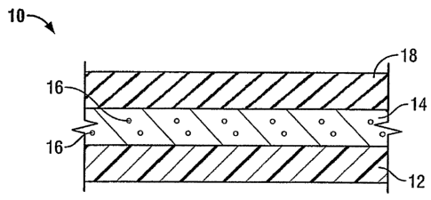
Figure lA shows vascular graft 10 of the present invention. As noted above,
the
present invention takes the preferred embodiment of a tubular graft having a
composite
structure. The layers shown in Figure 1 represent the tubular members forming
the composite
structure. However, it may be appreciated that the present invention also
contemplates other
implantable multi-layer prosthetic structures such as vascular patches, blood
filters, film
wraps for implantable devices such as stents, hernia repair fabrics and plugs
and other such
devices where such structures may be employed. As shown in Figure lA, the
composite
device 10 of the present invention includes a tubular flexible vascular graft
member 12, which
is porous and made of a textile and/or ePTFE. A biodegradable, bioactive agent
coating layer
7
CA 02578581 2007-01-10
WO 2006/017204 PCT/US2005/024418
14 covers the graft member 12. Biodegradable coating layer 14 permits
controlled delivery of
bioactive agents 16 associated with coating layer 14 therethrough. These
bioactive agents 16
are preferably distributed substantially evenly throughout the bulk of the
bioactive agent
coating layer 14, as will be described in greater detail below. Bioactive
agents 16 desirably
include antimicrobial agents. Device 10 of the present invention further
includes a
biodegradable sheath 18, which has a rigidity greater than that of flexible
graft member 12.
After implantation, sheath 18 biodegrades upon exposure to blood and/or other
physiological
fluids. This biodegradation of the sheath 18 decreases the rigidity of the
graft so as to re-
establish the flexibility of the tubular graft member 12. Once the sheath has
degraded, it
exposes bioactive agent coating layer 14. Desirably, antimicrobial agents are
posited on or
incorporated within coating layer 14 to reduce infection after implantation.
Sheath 18 may be
in a tubular configuration and placed over the graft member 12 or may be in a
sheet-like
configuration and wrapped about the tubular graft member 12, as fiirther
described below.
The biodegradable sheath 18 is desirably flexible and slightly elastic in
nature to allow it to be
placed on top of or wrapped about the vascular graft 12.
With reference now to Figure 1B, in one aspect of the present invention the
bioactive
agent coating is applied to graft member 12 in multiple coating layers, such
as 14a and 14b.
It is well within the contemplation of the present invention that coating
layers 14a and 14b
may contain the same or different bioactive agents 16. For example, as shown
in the
embodiment in Fig. 1B, bioactive agent 16a in coating layer 14a is an
antibiotic agent,
whereas bioactive agent 16b in coating layer 14b is an antiseptic agent. It
can be appreciated
that these multiple coating layers can be applied onto graft member 12 for a
longer term anti-
infective effect. Bioactive agent coating layer 14a is exposed after bioactive
agent coating
layer 14b has been biodegraded. Desirably, the bioactive agent coating layers
are both
biodegradable, as well as bioresorbable.
Referring now to Figure 2, in another aspect of the present invention,
biodegradable
sheath 18 also includes one or more bioactive agents. In desired embodiments,
the bioactive
agents in the biodegradable sheath include at least one antimicrobial agent
such that
antimicrobial agents are controllably released from the biodegradable sheath
immediately
8
CA 02578581 2007-01-10
WO 2006/017204 PCT/US2005/024418
upon implantation to reduce infection after implantation. Once the sheath
biodegrades and is
desirably resorbed, the one or more bioactive agent coating layers 14 are
exposed for a longer
term anti-infective effect.
Referring now to Figure 3, a preferred embodiment of a composite tubular graft
of the
present invention is shown, wherein the layers shown in Figure 1A represent
the tubular
members in Figure 3 forming the composite structure. Device 20 includes an
inner porous
tubular graft member 22, which is flexible; and a medial coating layer 24
disposed coaxially
thereover. Medial layer 24 includes bioactive agent 26 which is preferably
distributed
substantially evenly throughout the bulk of the biodegradable matrix of layer
24. An outer
tubular biodegradable sheath member 28 is disposed coaxially over
biodegradable bioactive
coating layer 24. As will be described in further detail below, the porous
flexible tubular
graft member 22 can be an ePTFE tube and/or a textile. A central lumen 29
extends
throughout the tubular composite graft 20 defined further by the inner wal122a
of luminal
tube 22, which permits the passage of blood through graft 20 once the graft is
properly
implanted in the vascular system.
It is well within the contemplation of the present invention that a stent can
be
interposed between the tubular members of the graft of the present invention.
With reference
to Figure 4, a stent/graft composite device 30 of the present invention is
shown. Device 30
includes inner porous tubular graft member 22, which in the present figure is
depicted as an
ePTFE tubular member. Device 30 also includes at least one medial,
biodegradable,
bioactive agent coating layer 24 disposed coaxially over graft member 22. As
described
above, coating layer 24 includes at least one bioactive agent which can be
controllably
released from the biodegradable matrix of coating layer 24. Composite device
30 further
includes a biodegradable tubular sheath member 28 which is disposed coaxially
over tubular
member 24. As described above and as shown in Figure 2, sheath member 28 can
also
include bioactive agents. In desired embodiments, the bioactive agents
associated with
coating layer 24 and optionally with biodegradable sheath 28, include an
antimicrobial agent
that can be controllably released from coating layer 24 and sheath 28
depending on the rate of
hydrolysis of the bonds within these biodegradable members. Central lumen 29
extends
9
CA 02578581 2007-01-10
WO 2006/017204 PCT/US2005/024418
throughout tubular composite graft 30. An expandable stent 32 may be
interposed between
inner ePTFE tubular member 22 and biodegradable coating layer 24. Stent 32,
which may be
associated with the graft of the present invention, is used for increased
support of the blood
vessel and increased blood flow through the area of implantation. It is noted
that increased
radial tensile strength at the outer sheath member 28 enables the graft to
support, for example,
radial expansion of stent 32, when present. In order to facilitate
hemodialysis treatment, a
significant number of patients suffering from hypertension or poor glycemic
control in
diabetes will have a synthetic vascular graft surgically implanted between the
venous and
arterial systems. Typically, these grafts become occluded over time. In these
instances, a
covered stent across the venous anastomotic site in patients with significant
stenosis may aid
in prolonging the patency of these grafts, which would avoid painful and
typically expensive
surgical revisions. For these reasons, it is well within the contemplation of
the present
invention that a stent covered with or incorporated within the vascular graft
of the present
invention may be useful for AV access.
The bioactive agents may include antimicrobial agents. In one embodiment, the
antimicrobial agents are antibiotic or antiseptic agents, or combinations
thereof. The
antibiotic agents can be of the type including, but not limited to,
ciprofloxacin, vancomycin,
minocycline, rifampin and other like agents, as well as combinations thereof.
Suitable antiseptic agents include, but are not limited to, the following:
silver agents,
chlorhexidine, triclosan, iodine, benzalkonium chloride and other like agents,
as well as
combinations thereof.
For example, silver is an antiseptic agent that has been shown in vitro to
inhibit
bacterial growth in several ways. For example, it is known that silver can
interrupt bacterial
growth by interfering with bacterial replication through a binding of the
microbial DNA, and
also through the process of causing a denaturing and inactivation of crucial
microbial
metabolic enzymes by binding to the sulfliydryl groups (Tweten, K., J. of
Heart Valve
Disease 1997, V6, No. 5, 554-561). It is also known that silver causes a
disruption of the cell
membranes of blood platelets. This increased blood platelet disruption leads
to increased
CA 02578581 2007-01-10
WO 2006/017204 PCT/US2005/024418
surface coverage of the implants with platelet cytoskeletal remains. This
process has been
shown to lead to an encouragement of the formation of a more structured
(mature state)
pannus around the implant. This would likely discourage the adhesion and
formation of the
biofilm produced by infectious bacteria due to a faster tissue ingrowth time
(Goodman, S. et
al, 24th Annual Meeting of the society for Biomaterials, April 1998, San
Diego, CA; pg. 207).
The silver agent can be a silver metal ion such as silver iodate, silver
iodide, silver
nitrate, and silver oxide. These silver ions are believed to exert their
effects by disrupting
respiration and electron transport systems upon absorption into bacterial or
fungal cells.
Antimicrobial silver ions are useful for in vivo use because they are not
substantially absorbed
into the body, and typically pose no hazard to the body.
Referring again to Figure lA, the aforementioned antiseptic or antibiotic
bioactive
agents 16 can be used alone or in combination of two or more of them. These
agents 16 can
be posited on coating layer 14 or can be dispersed throughout coating layer
14. The amount
of each antimicrobial or antibiotic bioactive agent 16 used to posit onto or
to impregnate the
coating layer 14 varies to some extent, but is at least of an effective
concentration to inhibit
the growth of bacterial and fungal organisms. 1
As noted above, in one aspect of the present invention, composite device 10
includes
an ePTFE graft member as the porous graft member 12 depicted in Figure 1A.
PTFE exhibits
superior biocompatibility and low thrombogenicity, which makes it particularly
useful as
vascular graft material. Desirably, the ePTFE graft member is a tubular
structure 22, as
depicted in Figure 4. The ePTFE material has a fibrous state, which is defined
by interspaced
nodes interconnected by elongated fibrils. The space between the node surfaces
that is
spanned by the fibrils is defined as the internodal distance. In the present
invention, the
intemodal distance in a luminal ePTFE graft member is desirably about 70 to
about 90
microns in order to achieve fast tissue ingrowth within the tunnel to
discourage initial
bacterial biofilm formation. When the term "expanded" is used to describe
PTFE, i.e.
ePTFE, it is intended to describe PTFE which has been stretched, in accordance
with
techniques which increase the intemodal distance and, concomitantly, porosity.
The
11
CA 02578581 2007-01-10
WO 2006/017204 PCT/US2005/024418
stretching may be done uni-axially, bi-axially, or multi-axially. The nodes
are stretched apart
by the stretched fibrils in the direction of the expansion. Methods of making
conventional
longitudinally expanded ePTFE are well known in the art.
It is fin-ther contemplated that the ePTFE may be a physically modified ePTFE
tubular
structure having enhanced axial elongation and radial expansion properties of
up to 600% by
linear dimension. The physically modified ePTFE tubular structure is able to
be elongated or
expanded and then returned to its original state without an elastic force
existing therewithin.
Additional details of physically-modified ePTFE and methods for making the
same can be
found in commonly assigned Application Title "ePTFE Graft With Axial
Elongation
Properties", assigned U.S. Application No. 09/898,418, filed on July 3, 2001,
published on
January 9, 2003 as U.S. Application Publication No. 2003-0009210A1, the
contents of which
are incorporated by reference herein in its entirety.
As noted above, in another aspect of the present invention, composite device
10
includes a textile graft member as the porous graft member 12 in Figure lA. As
will be
described in further detail below, virtually any textile construction can be
used for the graft
12, including weaves, knits, braids, filament windings, spun fibers and the
like. Any weave
pattern in the art, including, simple weaves, basket weaves, twill weaves,
velour weaves and
the like may be used. With reference to Figures 5 and 6, the weave pattern of
a textile tubular
graft member 40 shown in Figure 5 includes warp yams 40a running along the
longitudinal
length (L) of the graft and fill yams 40b running around the circumference (C)
of the graft,
the fill yams being at approximately 90 degrees to one another with fabrics
flowing from the
machine in the warp direction. A central lumen 29 extends throughout the
tubular graft
member 40, which permits the passage of blood through the composite vascular
graft of the
present invention once it is assembled and is properly implanted in the
vascular system.
Any type of textile products can be used as yarns for a textile graft member.
Of
particular usefulness in forming a textile graft member for the composite
device of the present
invention are synthetic materials such as synthetic polymers. Synthetic yams
suitable for use
in the textile graft member include, but are not limited to, polyesters,
including PET
12
CA 02578581 2007-01-10
WO 2006/017204 PCT/US2005/024418
polyesters, polypropylenes, polyethylenes, polyurethanes and
polytetrafluoroethylenes. The
yarns may be of the mono-filament, multi-filament, spun-type or combinations
thereof. The
yarns may also be flat, twisted or textured, and may have high, low or
moderate shrinkage
properties or combinations thereof. Additionally, the yam type and yam denier
can be
selected to meet specific properties desired for the prosthesis, such as
porosity and flexibility.
The yarn denier represents the linear density of the yarn (number of grams
mass divided by
9,000 meters of length). Thus, a yarn with a small denier would correspond to
a very fine
yam, whereas a yam with a large denier, e.g., 1,000, would correspond to a
heavy yarn. The
yams used for the textile graft member of the device of the present invention
may have a
denier from about 20 to about 200, preferably from about 30 to about 100.
Desirably, the
yarns are polyester, such as polyethylene terephthalate (PET). Polyester is
capable of
shrinking during a heat-set process, which allows it to be heat-set on a
mandrel to form a
generally circular shape.
After forming the textile layer of the present invention, it is optionally
cleaned or
scoured in a basic solution of warm water. The textile is then rinsed to
remove any remaining
detergent, and is then compacted or shrunk to reduce and control in part the
porosity of the
textile layer. Porosity of a textile material is measured on the Wesolowski
scale and by the
procedure of Wesolowski. In this test, a textile test piece is clamped
flatwise and subjected to
a pressure head of about 120 mm of mercury. Readings are obtained which
express the
number of mm of water permeating per ininute through each square centimeter of
fabric. A
zero reading represents absolute water impermeability and a value of about
20,000 represents
approximate free flow of fluid.
The porosity of the textile layer is often about 5,000 to about 17,000 on the
Wesolowski scale. The textile layer may be compacted or shrunk in the wale
direction to
obtain the desired porosity. A solution of organic component, such as
hexafluoroisopropanol
or trichloroacetic acid, and a halogenated aliphatic hydrocarbon, such as
methylene chloride,
can be used to compact the textile graft by immersing it into the solution for
up to 30 minutes
at temperatures from about 15 C to about 160 C.
13
CA 02578581 2007-01-10
WO 2006/017204 PCT/US2005/024418
Yarns of the textile layer may be one ply or multi-ply yams. Multi-ply yams
may be
desirable to impart certain properties onto the drawn yam, such as higher
tensile strengths for
the textile layer.
A further aspect of the composite device of the present invention relates to
the
biodegradable, bioactive agent coating layer shown as layer 14 in Figure 1A.
In one
embodiment, the bioactive agent coating is applied to the porous tubular graft
member as one
or more coating layers. For example, a coating material can be applied (prior
to
polymerization) as a liquid to the outside surface of an ePTFE and/or textile
graft member by
such means as dipping, spraying or painting.
The coating layer may be comprised of natural, modified natural or synthetic
polymers, copolymers, block polymers, as well as combinations thereof. It is
noted that a
polymer is generally named based on the monomer it is synthesized from.
Examples of
suitable biodegradable polymers or polymer classes include fibrin, collagen,
elastin,
celluloses, gelatin, vitronectin, fibronectin, laminin, reconstituted basement
membrane
matrices, starches, dextrans, alginates, hyaluronic acid, poly(lactic acid),
poly(glycolic acid),
polypeptides, glycosaminoglycans, their derivatives and mixtures thereof. For
both glycolic
acid and lactic acid, an intermediate cyclic dimer is typically prepared and
purified, prior to
polymerization. These intermediate dimers are called glycolide and lactide,
respectively.
Other useful biodegradable polymers or polymer classes for the bioactive agent
coating layer include the following: polydioxanones, polyoxalates, poly(a-
esters),
polyanhydrides, polyacetates, polycaprolactones, poly(orthoesters), polyamino
acids,
polyamides and mixtures and copolymers thereof.
Additional useful biodegradable polymers for the bioactive agent coating layer
include, stereopolymers of L- and D-lactic acid, copolymers of bis(p-
carboxyphenoxy)
propane acid and sebacic acid, sebacic acid copolymers; copolymers of
caprolactone,
poly(lactic acid)/poly(glycolic acid)/polyethyleneglycol copolymers,
copolymers of
polyurethane and (poly(lactic acid), copolymers of polyurethane and
poly(lactic acid),
14
CA 02578581 2007-01-10
WO 2006/017204 PCT/US2005/024418
copolymers of a-amino acids, copolymers of a-amino acids and caproic acid,
copolymers of
a-benzyl glutamate and polyethylene glycol, copolymers of succinate and
poly(glycols),
polyphosphazene, polyhydroxy-alkanoates and mixtures thereof. Binary and
ternary systems
are contemplated.
Factors affecting the mechanical performance of in vivo biodegradable polymers
are
well known to the polymer scientist, and include monomer selection, initial
process
conditions, and the presence of additives. Biodegradation has been
accomplished by
synthesizing polymers that have unstable linkages in the backbone, or linkages
that can be
safely oxidized or hydrolyzed in the body. The most common chemical functional
groups
having this characteristic are ethers, esters, anhydrides, orthoesters and
amides.
As described above, the biodegradable coating layer includes a bioactive
agent. In
one desired embodiment, the bioactive agent is an antimicrobial agent. For
example, the
antimicrobial agent can be an antibiotic or antiseptic agent. Examples of
suitable antibiotic
and antiseptic agents for use in the present invention are provided above.
The bioactive agent is desirably evenly distributed throughout the bulk of the
biodegradable coating layer and is controllably released from the
biodegradable coating layer
to the site of implantation of the graft by hydrolysis of chemical bonds in
the biodegradable
polymer. It is also contemplated that a bioactive agent can be posited on the
coating layer.
A solution of biodegradable material that includes a mon.omer (or an
intermediate
cyclic dimer) on which the biodegradable polymer is based can be applied as a
coating to the
external side of the ePTFE and/or textile graft member. This can be
accomplished by such
means as dipping, spraying, painting, etc. A bioactive agent can be blended
into the wet or
fluid biodegradable material to form a coating mixture which is then applied
to the porous
tubular graft member by a spraying process, for example. Alternatively, the
bioactive agent
may be applied in powdered form to wet or fluid biodegradable material after
the
biodegradable material has been applied as a coat to the porous tubular graft
member, but
prior to its polymerization.
CA 02578581 2007-01-10
WO 2006/017204 PCT/US2005/024418
In preparing the biodegradable, bioactive agent coating layer, a solution or
fluid of a
biocompatible, biodegradable material can be formed. For example,
extracellular matrix
proteins which are used in fluid/solution may be soluble. However, some
materials may be
difficult to dissolve in water. Collagen, for example, is considered insoluble
in water, as is
gelatin at ambient temperature. To overcome such difficulties, collagen or
gelatin may
preferably formed at an acidic pH, i.e. at a pH less than 7 and, preferably,
at a pH of about 2
to about 4. The temperature range at which such fluid/solutions are formed is
between about
4 C to about 40 C, and preferably about 30 C - 35 C.
In situations where the bioactive agent is insoluble in the wet or fluid
biodegradable
coating material, the agent may be finely subdivided as by grinding with a
mortar and pestle.
The fmely subdivided bioactive agent can then be distributed desirably
substantially evenly
throughout the bulk of the wet or fluid biodegradable coating material before
cross-linking or
cure solidifies the coating layer.
It is well within the contemplation of the present invention that the coating
layer can
be combined with various carrier, drug, prognostic, or therapeutic materials.
For example,
the coating layer can be combined with any of the following therapeutic
agents: antimicrobial
agents, such as the antibiotic agents and antiseptic agents listed above; anti-
thrombogenic
agents, such as heparin, heparin derivatives, urokinase, and PPack
(dextrophenylalanine
proline, arginine, chloromethylketone); anti-proliferative agents (such as
enoxaprin,
angiopeptin, or monoclonal antibodies capable of blocking smooth muscle cell
proliferation,
hirudin, and acetylsalicylic acid); anti-inflammatory agents, such as
dexamethasone,
prednisolone, corticosterone, budesonide, estrogen, sulfasalazine, and
mesalamine); anti-
neoplastics/anti-proliferative/anti-miotic agents (such as paclitaxel, 5-
flurouracil, cisplatin,
vinblastine, vincristine, epothilones, endostatin, angiostatin and thymidine
kinase inhibitors);
anesthetic agents (such as lidocaine, bupivacaine, and ropivacaine); anti-
coagulants (such as
D-Phe-Pro-Arg chloromethyl keton, an RGD peptide-containing compound, heparin,
antithrombin compounds, platelet receptor antagonists, anti-thrombin
antibodies, anti-platelet
receptor antibodies, aspirin, prostaglandin inhibitors, platelet inhibitors
and tick anti-platelet
peptides); vascular cell growth promoters (such as growth factor inhibitors,
growth factor
16
CA 02578581 2007-01-10
WO 2006/017204 PCT/US2005/024418
receptor antagonists, transcriptional activators, and translational
promoters); vascular cell
growth inhibitors (such as growth factor inhibitors, growth factor receptor
antagonists,
transcriptional repressors, translational repressors, replication inhibitors,
inhibitory
antibodies, antibodies directed against growth factors, bi-fiuictional
molecules consisting of a
growth factor and a cytotoxin, bi-functional molecules consisting of an
antibody and a
cytotoxin); cholesterol-lowering agents; vasodilating agents; and agents which
interfere with
andogenous or vascoactive mechanisms. In addition, cells which are able to
survive within
the body and are dispersed within the coating layer may be therapeutically
useful. These cells
themselves may be therapeutically useful or they may be selected or engineered
to produce
and release therapeutically useful compositions.
In other embodiments, bioactive agents associated with the composite device of
the
present invention may be genetic agents. Examples of genetic agents include
DNA, anti-
sense DNA, and anti-sense RNA. DNA encoding one of the following may be
particularly
useful in association with an implantable device according to the present
invention: (a) tRNA
or RRNA to replace defective or deficient endogenous molecules; (b) angiogenic
factors
including growth factors such as acidic and basic fibroblast growth factors,
vascular
endothelial growth factor, epidennal growth factor, transforming growth factor
a and (3,
platelet-derived endothelial growth factor, platelet-derived growth factor,
tumor necrosis
factor a, hepatocyte growth factor and insulin-like growth factor; (c) cell
cycle inhibitors; (d)
thymidine kinase and other agents useful for interfering with cell
proliferation; and (e) the
family of bone morphogenic proteins. Moreover, DNA encoding molecules capable
of
inducing an upstream or downstream effect of a bone morphogenic protein may be
useful.
A fiuther aspect of the present invention relates to the biodegradable sheath
shown as
layer 18 in Figure 1A. In one embodiment, the biodegradable sheath is
comprised of a
material selected from, but not limited to, the following: polylactides,
polyanhydrides,
polyvinyl alcohol, polyvinylpyrolidone, polyglycols, gelatin derivatives and
combinations
thereof. The biodegradable sheath can have a tubular or sheet-like
configuration for disposal
over the bioactive coating layer. For example, referring to Figure 7 of the
present invention,
there is shown a biodegradable sheath in a tube-like configuration 50 used in
combination
17
CA 02578581 2007-01-10
WO 2006/017204 PCT/US2005/024418
with a tubular composite vascular graft of the present invention.
Specifically, the tube 50 can
be placed over the bioactive coating layer overlying the porous, flexible
tubular graft member.
Alternatively, the biodegradable sheath can be in a sheet-like configuration
as shown
in Figure 8. Sheath 60 shown in Figure 8 is used in combination with a tubular
composite
vascular graft of the present invention. Specifically, the sheath 60 can be
wrapped about the
bioactive coating layer overlying the porous, flexible tubular graft member.
The sheet 60 is
seamed along the longitudinal axis.
The sheath provides a desired degree of initial rigidity to the flexible
tubular textile
and/or ePTFE graft member during implantation. After implantation, the sheath
biodegrades
upon exposure to blood and/or other physiological fluids. The biodegradation
of the sheath
decreases the rigidity of the graft and re-establishes the flexibility of the
graft member. After
the sheath has, degraded, it exposes the underlying bioactive agent coating
layer which is
desirably incorporated with antimicrobial agents to reduce infection after
implantation. In
embodiments where multiple bioactive agent coating layers are present, each
coating layer
controllably releases bioactive agents associated therewith after the coating
layer overlying it
is resorbed. This provides a longer term anti-infective effect.
The biodegradable sheath of the composite graft of the present invention can
include
bioactive agents. For example, the biodegradable sheath can be incorporated
with
antimicrobial agents so as to controllably release the antimicrobial agents
immediately upon
implantation.
In one of the embodiments of the present invention, it is contemplated that a
dry,
finely subdivided antimicrobial agent may be blended with wet or fluid
material to form a
mixture which is used to impregnate the pores of a porous biodegradable
sheath.
Altematively, it is contemplated that air pressure or other suitable means may
then be
employed to disperse the antimicrobial agent substantially evenly within the
filled pores.
18
CA 02578581 2007-01-10
WO 2006/017204 PCT/US2005/024418
In one example, a bioactive agent or drug can be incorporated into the sheath
in the
following manner: mixing into a fluid material used to make the sheath, a
crystalline,
particulate material like salt or sugar that is not soluble in a solvent used
to form the sheath;
casting the solution with particulate material into a film or sheet; and then
applying a second
solvent, such as water, to dissolve and remove the particulate material,
thereby leaving a
porous sheath. The sheath may then be placed into a solution containing a
bioactive agent in
order to fill the pores. Preferably, a vacuum would be pulled on the sheath to
insure that the
bioactive agent applied to it is received into the pores.
It is also contemplated that the bioactive agent or drug may be encapsulated
in
microparticles, such as microspheres, microfibers or microfibrils, which can
then be
incorporated into or on the sheath. Various methods are known for
encapsulating bioactive
agents or drugs within microparticles or microfibers (see Patrick B. Deasy,
Microencapsulation and Related Drug Processes, Marcel Dekker, Inc., New York,
1984). In
one example, a suitable microsphere for incorporation can have a diameter of
about 10
microns or less. The niicrosphere could be contained within the biodegradable
polymeric
matrix of the sheath. The microparticles containing the bioactive agent can be
incorporated
within the sheath by adhesively positioning them onto the sheath material or
by mixing the
microparticles with a fluid or gel and flowing them into the sheath layer. The
fluid or gel
mixed with the microparticles could, for example, be a carrier agent designed
to improve the
cellular uptake of the bioactive agent incorporated into the sheath. Moreover,
it is well within
the contemplation of the present invention that carrier agents, which can
include hyaluronic
acid, may be incorporated within each of the embodiments of the present
invention so as to
enhance cellular uptake of the bioactive agent(s) associated with the device.
The microparticles may have a polymeric wall surrounding the bioactive agent
or a
matrix containing the bioactive agent and optional carrier agents, which due
to the potential
for varying thicknesses of the polymeric wall and for varying porosities and
permeabilities
suitable for containing a bioactive agent, there is provided the potential for
an additional
mechanism for controlling the release of a therapeutic agent.
19
CA 02578581 2007-01-10
WO 2006/017204 PCT/US2005/024418
Moreover, microfibers or microfibrils, which may be loaded with the bioactive
agent
by extrusion, can be adhesively layered or woven into the sheath material for
drug delivery.
The bioactive agents, which can optionally be associated with the
biodegradable
sheath of the composite graft of the present invention, may be selected from
drugs, prognostic
agents, carrier agents, therapeutic agents, and genetic agents. Suitable
bioactive agents
include, but are not limited to, growth factors, anti-coagulant substances,
stenosis inhibitors,
thrombo-resistant agents, antibiotic agents, anti-tumor agents, anti-
proliferative agents,
growth hormones, antiviral agents, anti-angiogenic agents, angiogenic agents,
anti-mitotic
agents, anti-inflammatory agents, cell cycle regulating agents, genetic
agents, cholesterol-
lowering agents, vasodilating agents, agents that interfere with endogenous
vasoactive
mechanisms, hormones, their homologs, derivatives, fragments, pharmaceutical
salts and
combinations thereof. Specific examples of such agents are provided above.
As described above, a further aspect of the present invention relates to a
method of
making the inventive composite vascular graft. The method includes the steps
of providing a
flexible, porous tubular graft member, such as an ePTFE and/or textile graft
member; and
applying a biodegradable coating material to the porous tubular graft member
so as to form
one or more overlying coating layers, wherein the biodegradable coating
material has at least
one bioactive agent incorporated therein. The method further includes
disposing a
biodegradable sheath over the one or more coating layers overlying the ePTFE
and/or textile
graft member.
Generally, tubular textile layers are manufactured in a single long tube and
cut to a
pre-determined length. To cut the textile layer, a laser would be desirably
used, which cuts
and fuses the ends simultaneously. The textile layer is typically cleaned,
desirably with
sodium dodecyl sulfate and then rinsed with deionized water. The textile layer
can then be
placed over a cylindrical mandrel and heat set to precisely set the diameter
and to remove any
creases or wrinkles. Typically, heat setting is carried out at the temperature
range from about
125 C to about 225 C using a convection oven for a time of 20 minutes. Any
known means
for heating may be used.
CA 02578581 2007-01-10
WO 2006/017204 PCT/US2005/024418
Alternatively, the composite device of the present invention may be formed by
expanding a_thin wall PTFE inner luminal tube at a relatively high degree of
elongation, on
the order of approximately between 400% and 2,000% elongation and preferably
from about
between 700% and 900%. The inner luminal tube is desirably expanded over a
cylindrical
mandrel, such as a stainless steel mandrel at a temperature of between room
temperature and
640 F, preferably about 500 F. The luminal tube is preferably, but not
necessarily fully
sintered after expansion. Sintering is typically accomplished at a temperature
of between
640 F and 800 F, preferably at about 660 F and for a time of between about 5
minutes to 30
minutes, preferably about 15 minutes. The resulting luminal tube formed by
this method
desirably exhibits an IND of greater than 40 microns, and in particular
between 40 and 100
microns, most desirably between 70 to about 90 microns, spanned by a moderate
number of
fibrils. Such_a microporous structure is sufficiently large so as to promote
enhanced cell
endothelization once blood flow is established through the graft. Such cell
endothelization
enhances the long-term patency of the graft.
The combination of the luminal ePTFE and/or textile tube over the mandrel is
then
employed as a substrate over which the biodegradable, bioactive coating layer
can be
disposed. In particular, the biodegradable, bioactive coating layer can be
applied as a fluid
coating material on the external surface of the luminal tube by such means as
dipping,
spraying or painting. The bioactive agent coating can be applied in a single
layer or in
multiple layers. Within the bioactive agent coating material is preferably
substantially evenly
dispersed a bioactive agent, which may be in dry powdered form.
The biodegradable sheath, which can be in the form of a tube or sheet, is then
disposed over the bioactive agent coating layer(s). For example, the tube or
sheet may
correspond to a porous, biodegradable polymeric matrix, wherein the pores can
optionally be
filled with a bioactive agent. The interior diameter of a biodegradable
tubular sheatli member
is selected so that it may be easily, but tightly disposed over the outside
diameter of the
coated graft member. In one embodiment, the sheath is cross-linlced and bonds
to the
underlying bioactive agent coating layer. It is further contemplated that the
biodegradable
sheath can be~ secured to the coated graft member using techniques that would
avoid
21
CA 02578581 2007-01-10
WO 2006/017204 PCT/US2005/024418
degrading or damaging the bioactive agents in the coating layer(s). For
example, where silver
metal ions are the bioactive agents, it may be suitable to sinter the
composite structure formed
between the coated, tubular graft member and the tubular sheath using similar
parameters to
those described above.
Alternatively, the biodegradable sheath may be securably affixed to the coated
graft
member by means of a bonding agent. The bonding agent may include various
biocompatible, elastomeric bonding agents such as urethanes,
styrene/isobutylene/styrene
block copolymers (SIBS), silicones, and combinations thereof. Once the
composite
prosthesis is formed, one or more layers of elastic tubing, preferably
silicone, can then be
placed over this composite structure. This holds the composite structure
together and assures
that complete contact and adequate pressure is maintained for bonding
purposes.
While the invention has been described in relation to the preferred
embodiments with
several examples, it will be understood by those skilled in the art that
various changes may be
made without deviating from the spirit and scope of the invention as defmed in
the appended
claims.
22